IL-28, IL-29 and their class II cytokine receptor IL-28R
6
www.nature.com/natureimmunology • january 2003 • volume 4 no 1 • nature immunology Paul Sheppard*,Wayne Kindsvogel*,Wenfeng Xu*, Katherine Henderson, Stacy Schlutsmeyer,Theodore E.Whitmore, Rolf Kuestner, Ursula Garrigues, Carl Birks, Jenny Roraback, Craig Ostrander, Dennis Dong, Jinu Shin, Scott Presnell, Brian Fox, Betty Haldeman, Emily Cooper, David Taft,Teresa Gilbert, Francis J. Grant, Monica Tackett, William Krivan, Gary McKnight, Chris Clegg, Don Foster and Kevin M. Klucher Published online 2 December 2002; doi:10.1038/ni873 Cytokines play a critical role in modulating the innate and adaptive immune systems. Here, we have identified from the human genomic sequence a family of three cytokines, designated interleukin 28A (IL-28A), IL-28B and IL-29, that are distantly related to type I interferons (IFNs) and the IL-10 family.We found that like type I IFNs, IL-28 and IL-29 were induced by viral infection and showed antiviral activity. However, IL-28 and IL-29 interacted with a heterodimeric class II cytokine receptor that consisted of IL-10 receptor β ( IL-10Rβ) and an orphan class II receptor chain, designated IL-28Rα.This newly described cytokine family may serve as an alternative to type I IFNs in providing immunity to viral infection. ZymoGenetics, Inc., 1201 Eastlake Avenue E., Seattle,WA 98102, USA. *These authors contributed equally to this work. Correspondence should be addressed to K.K. ([email protected]). IL-28, IL-29 and their class II cytokine receptor IL-28R Class II cytokine receptor ligands, which include the interferons (IFNs) 1 , interleukin 10 (IL-10) 2 and IL-10–related proteins 3 , play a crit- ical role in response to microbial challenge by modulating the innate and adaptive immune systems. Viral infection elicits the production of type I IFNs that, upon interaction with their receptor, induce a set of IFN-stimulated genes that inhibit viral replication and increase the lytic potential of natural killer (NK) cells 4 . In addition to this role in innate immunity, type I IFNs modulate the adaptive immune response by increasing major histocompatibility complex (MHC) class I expression to promote antigen presentation 5 , also promoting T cell survival 6 and stimulating dendritic cell maturation 7 . In humans, the type I IFN family consists of at least 13 nonallelic IFN-α genes, a single IFN-β gene and a single IFN-ω gene 1 . Two addi- tional type I IFNs, limitin and IFN-κ 8,9 , have also been identified. All A RTICLES 63 Figure 1. Sequence alignment of IL-28 and IL-29. Cytokines IL-28A, IL-28B and IL-29 were aligned with a structure-based multiple alignment of IL-10, IL-22 and IFN-α2. Known helical regions of IL-10 and IFN-α2 are underlined. Hydrophobic residues are in bold. © 2003 Nature Publishing Group http://www.nature.com/natureimmunology
-
Upload
independent -
Category
Documents
-
view
0 -
download
0
Transcript of IL-28, IL-29 and their class II cytokine receptor IL-28R

www.nature.com/natureimmunology • january 2003 • volume 4 no 1 • nature immunology
Paul Sheppard*,Wayne Kindsvogel*,Wenfeng Xu*, Katherine Henderson,Stacy Schlutsmeyer,Theodore E.Whitmore, Rolf Kuestner, Ursula Garrigues, Carl Birks,Jenny Roraback, Craig Ostrander, Dennis Dong, Jinu Shin, Scott Presnell, Brian Fox,Betty Haldeman, Emily Cooper, David Taft,Teresa Gilbert, Francis J. Grant, Monica Tackett,William Krivan, Gary McKnight, Chris Clegg, Don Foster and Kevin M. KlucherPublished online 2 December 2002; doi:10.1038/ni873
Cytokines play a critical role in modulating the innate and adaptive immune systems. Here, we haveidentified from the human genomic sequence a family of three cytokines, designated interleukin28A (IL-28A), IL-28B and IL-29, that are distantly related to type I interferons (IFNs) and the IL-10family. We found that like type I IFNs, IL-28 and IL-29 were induced by viral infection and showedantiviral activity. However, IL-28 and IL-29 interacted with a heterodimeric class II cytokinereceptor that consisted of IL-10 receptor β (IL-10Rβ) and an orphan class II receptor chain,designated IL-28Rα .This newly described cytokine family may serve as an alternative to type I IFNsin providing immunity to viral infection.
ZymoGenetics, Inc., 1201 Eastlake Avenue E., Seattle,WA 98102, USA. *These authors contributed equally to this work.Correspondence should be addressed to K.K. ([email protected]).
IL-28, IL-29 and their class II cytokinereceptor IL-28R
Class II cytokine receptor ligands, which include the interferons(IFNs)1, interleukin 10 (IL-10)2 and IL-10–related proteins3, play a crit-ical role in response to microbial challenge by modulating the innateand adaptive immune systems. Viral infection elicits the production oftype I IFNs that, upon interaction with their receptor, induce a set ofIFN-stimulated genes that inhibit viral replication and increase the lyticpotential of natural killer (NK) cells4. In addition to this role in innate
immunity, type I IFNs modulate the adaptive immune response byincreasing major histocompatibility complex (MHC) class I expressionto promote antigen presentation5, also promoting T cell survival6 andstimulating dendritic cell maturation7.
In humans, the type I IFN family consists of at least 13 nonallelicIFN-α genes, a single IFN-β gene and a single IFN-ω gene1. Two addi-tional type I IFNs, limitin and IFN-κ8,9, have also been identified. All
ARTICLES
63
Figure 1. Sequence alignment ofIL-28 and IL-29. Cytokines IL-28A,IL-28B and IL-29 were aligned with astructure-based multiple alignment ofIL-10, IL-22 and IFN-α2. Known helicalregions of IL-10 and IFN-α2 areunderlined. Hydrophobic residues arein bold.
©20
03 N
atu
re P
ub
lish
ing
Gro
up
h
ttp
://w
ww
.nat
ure
.co
m/n
atu
reim
mu
no
log
y

nature immunology • volume 4 no 1 • january 2003 • www.nature.com/natureimmunology
ARTICLES
the human type I IFNs are colocalized to chromosome 9 and interactwith the same receptor, the IFN-αβ receptor (IFN-αβR)1. IFN-αβRconsists of two subunits, IFNAR-1 and IFNAR-2, which form a het-erodimer upon IFN stimulation10,11. This initiates a signaling cascadethat involves Jak1- and Tyk2-dependent tyrosine phosphorylation ofsignal transducers and activators of transcription 1 (STAT1) and STAT2proteins that interact with a third protein, the p48 DNA–binding sub-unit, to form the IFN-stimulated gene factor 3 complex (ISGF3). ISGF3ultimately interacts with the IFN-stimulated response element (ISRE)in the upstream regulatory regions of a number of genes involved in theresponse induced by type I IFNs12.
Other class II cytokine receptor ligands appear to play a direct rolein inflammation. IL-22, an IL-10–related protein, is rapidly induced inmouse liver after lipopolysaccharide injection13. In turn, IL-22 inducesacute-phase reactants in the liver, which suggests that IL-22 plays a rolein innate immunity via induction of an inflammatory process13. In con-trast, the primary function of IL-10 may be to dampen inflammatoryresponses14,15. IL-22 and IL-10 share a common receptor β subunit, IL-10Rβ16–18, but have the distinct receptor α subunits, IL-22Rα1 and IL-10Rα, respectively17,19. In addition to IL-22Rα1, IL-22 interacts with asoluble class II cytokine receptor, IL-22Rα2, which may function as anaturally occurring IL-22 antagonist20. Like the type I IFNs, both IL-22and IL-10—through their respective receptors—activate the Jak-STATsignaling system. However, unlike type I IFNs, IL-22 and IL-10 pri-marily activate STAT317,21.
Given the central role played by class II cytokine receptor ligands inimmunity, the identification of additional members of this family mayincrease our understanding of regulation of the immune system inresponse to microbial challenge. We describe here the identification ofa new family of cytokines, IL-28A, IL-28B and IL-29, and their class
II cytokine receptor, IL-28R, from human genomic sequence. IL-28and IL-29 were induced by viral infection and showed antiviral activi-ty. This cytokine family may serve as an alternative to type I IFNs inproviding immunity to viral infection.
ResultsNewly identified IFN-like cytokinesUsing a method that combines multiple computational techniques (seeMethods) to detect families of uncharacterized proteins from humangenomic sequence, we identified two protein sequences, designated IL-28A and IL-29, as a pair of paralogous sequences with 81% amino acididentity. The results of profile and multiple sequence alignment tech-niques applied to this pair of protein sequences indicated low sequencehomology to the IL-10 and type I IFN helical cytokine families(11–13% amino acid identity to IL-10, 15–19% amino acid identity toIFN-α and IL-22) (Fig. 1). Although the sequence homology is low, anumber of features in the multiple alignment strengthen the predictionthat these proteins are helical cytokine family members. In particular,this was shown by the conservation of the cysteine pattern and compat-ibility with the expected amphipathic profile of helical cytokines.Characterization of IL-28A and IL-29 indicated that both genes werelocated on the same genomic contig, AC011445.6, which is from thechromosomal region 19q13.1322. This chromosomal location differsfrom the type I IFN family clustered on chromosome 91. A third fami-ly member, designated IL-28B, was identified on the same genomiccontig with BLAST23 analysis. IL-28A and IL-28B are virtually identi-cal, with 96% amino acid identity (Fig. 1).
Polymerase chain reaction (PCR) was used to confirm mRNAexpression and predicted splicing patterns of IL-28A, IL-28B and IL-29 from a panel of ∼ 100 cDNA libraries. IL-28 and IL-29 expressionwas detected at low amounts in libraries from a wide range of tissuetypes, including blood, brain, lung, ovary, pancreas, pituitary, placenta,prostate and testis (data not shown). Comparison of the correspondingcDNA and genomic sequences revealed that, like the IL-10 gene fami-ly2, IL-28 and IL-29 contained multiple exons, with IL-28A and IL-28Bhaving six exons and IL-29 five exons. This is in contrast to the type IIFNs, which are encoded within a single exon1.
Induction of IL-28 and IL-29Analysis of the putative 5′ regulatory regions of IL-28 and IL-29 iden-tified a number of DNA binding elements involved in type I IFN generegulation (unpublished data). To determine whether expression of thegenes encoding IL-28 and IL-29 is regulated in a manner similar to typeI IFNs, human peripheral blood mononuclear cells were treated withthe double-stranded RNA polymer poly(I)•poly(C), an inducer of type
64
Figure 2. RNA expression of IL-28 and IL-29. RT-PCR analysis of IL-28A, IL-28B, IL-29, IFN-α and IFN-β RNA expression in untreated (–),poly(I)•poly(C)–treated and EMCV-infectedhuman peripheral blood mononuclear cells.ActinRNA was measured as a noninducible control.
Figure 3. IFN-like activities of IL-28and IL-29. (a) Antiviral activity of IL-28Aand IL-29. IL-28A, IL-29 and IFN-α2a wereadded at varying concentrations to HepG2cells before EMCV infection and dye-uptakeassays were done. Mean ± s.d. A570 values,which were directly proportional to antivi-ral activity, are shown (n = 3). (b) IL-28 andIL-29 signal through ISRE. HepG2 cellswere transfected with an ISRE reporterplasmid and luciferase activity was analyzedafter treatment with varying concentra-tions of IL-28A, IL-29 or IFN-α2a. Fold acti-vation was determined by dividing the rela-tive light units (RLU) of each experimentalsample by the RLU of media alone. Mean ±s.d. data are shown (n = 2).
a b
©20
03 N
atu
re P
ub
lish
ing
Gro
up
h
ttp
://w
ww
.nat
ure
.co
m/n
atu
reim
mu
no
log
y
ARTICLES
www.nature.com/natureimmunology • january 2003 • volume 4 no 1 • nature immunology
I IFNs24, or with infection by encephalomyocarditis virus (EMCV).Total RNA was isolated from treated and untreated cells and cytokine-specific RNA levels were measured by reverse transcription PCR (RT-PCR) with primers specific for the individual genes. Low to unde-tectable amounts of IL-28, IL-29, IFN-α and IFN-β RNA were seen inuntreated cells (Fig. 2). In contrast, the amount of IL-28 and IL-29RNA was increased by both poly(I)•poly(C) treatment and viral infec-tion, as was also seen for the type I IFNs. Similar induction of IL-28and IL-29 RNA by poly(I)•poly(C) treatment was seen in a variety ofcell types tested (data not shown). These experiments indicated that IL-28 and IL-29, like type I IFNs, can be induced by double-stranded RNAor viral infection and suggested they play a role in antiviral immunity.
Antiviral activityIL-28 and IL-29 were tested for antiviral activity by challenging thehuman hepatocellular carcinoma cell line HepG2 with infection by
EMCV after pretreatment of the cells with IL-28 or IL-29. Purifiedrecombinant IL-28A and IL-29, like IFN-α2a, were able to protectHepG2 cells from viral-induced cytopathogenic effect when applied 24h before adding EMCV (Fig. 3a). Half-maximal protection (EC50) wasachieved with ∼ 2 ng/ml of IL-29 and 30 ng/ml of IL-28A compared to0.5 ng/ml of IFN-α2a. These results showed that IL-28 and IL-29, liketype I IFNs, have intrinsic cellular antiviral activity and are able to fullyprotect HepG2 cells challenged with EMCV.
IL-28 and IL-29 signalingInteraction of type I IFNs with their receptor initiates a signaling cas-cade that results in the interaction of ISGF3 with the IFN-stimulatedresponse element (ISRE) found in the upstream regulatory regions ofgenes induced by type I IFNs12. To test whether IL-28 and IL-29 signalthrough ISRE, HepG2 cells were transiently transfected with a reporterconstruct containing ISRE sequences upstream of the luciferasereporter gene. Stimulation of the transfected cells with IL-28A and IL-29 resulted in a half-maximal response of twofold above media aloneat 1–4 ng/ml (Fig. 3b). In comparison, IFN-α2a gave a half-maximalresponse of threefold above media alone at 0.05 ng/ml. These resultsindicated that IL-28 and IL-29, like type I IFNs, can signal throughISRE regulatory sites. Therefore, it is likely they provide antiviral activ-ity by the induction of at least a subset of IFN-stimulated genes12.
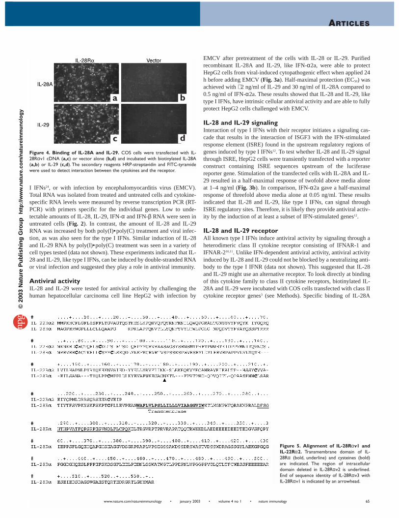
IL-28 and IL-29 receptorAll known type I IFNs induce antiviral activity by signaling through aheterodimeric class II cytokine receptor consisting of IFNAR-1 andIFNAR-210,11. Unlike IFN-dependent antiviral activity, antiviral activityinduced by IL-28 and IL-29 could not be blocked by a neutralizing anti-body to the type I IFNR (data not shown). This suggested that IL-28and IL-29 might use an alternative receptor. To look directly at bindingof this cytokine family to class II cytokine receptors, biotinylated IL-28A and IL-29 were incubated with COS cells transfected with class IIcytokine receptor genes3 (see Methods). Specific binding of IL-28A
65
Figure 4. Binding of IL-28A and IL-29. COS cells were transfected with IL-28Rαv1 cDNA (a,c) or vector alone (b,d) and incubated with biotinylated IL-28A(a,b) or IL-29 (c,d). The secondary reagents HRP-streptavidin and FITC-tyramidewere used to detect interaction between the cytokines and the receptor.
a b
c d
Figure 5. Alignment of IL-28Rαv1 andIL-22Rα2. Transmembrane domain of IL-28Rα (bold, underline) and cysteines (bold)are indicated. The region of intracellulardomain deleted in IL-28Rαv2 is underlined.End of sequence identity of IL-28Rαv3 withIL-28Rαv1 is indicated by an arrowhead.
©20
03 N
atu
re P
ub
lish
ing
Gro
up
h
ttp
://w
ww
.nat
ure
.co
m/n
atu
reim
mu
no
log
y

nature immunology • volume 4 no 1 • january 2003 • www.nature.com/natureimmunology
ARTICLES
and IL-29 to an orphan class II cytokine receptor, designated IL-28receptor α (IL-28Rα) was observed (Fig. 4).
IL-28Rα was initially identified through scanning translated humangenomic sequences for sequences related to class II cytokine receptors.IL-28Rα was found on genomic clone AL358412, which is from chro-mosomal region 1p36.1122. Using the predicted IL-28Rα open-readingframe as a template, we isolated full-length cDNAs that encoded aputative class II cytokine receptor. Three different forms of IL-28RαcDNA were isolated that may derive from alternative splicing (Fig. 5).The predicted proteins from two of the cDNA forms contain an extra-cellular region consisting of a cytokine-binding domain with twofibronectin III domains25, followed by transmembrane and intracellularregions (IL-28Rαv1 and IL-28Rαv2). These two predicted proteinsdiffer only in the intracellular domain where one of the predicted pro-teins contains a 29–amino acid deletion (IL-28Rαv2). The third formof IL-28Rα (IL-28Rαv3) appears to use an alternative splice site todelete the predicted transmembrane and intracellular regions and there-fore is likely to encode a soluble form of IL-28Rα. BLAST analysisindicated that IL-28Rα (v1, v2 and v3) has the highest sequence iden-tity with the soluble class II cytokine receptor IL-22Rα220, with 23%amino acid identity (Fig. 5).
We analyzed the expression pattern of IL-28Rα using northern blotanalysis with a probe detecting RNA encoding the cytoplasmic domainof IL-28Rα (v1 and v2). Transcripts of 4.5 and 2.0 kb were detected inmost tissues, with the highest amounts seen in pancreas, thyroid, skele-tal muscle, heart, prostate and testis (Fig. 6). The 4.5 kb IL-28Rα tran-script was also readily detected in three cell lines, Raji (Burkitt’s lym-phoma), HL-60 (promyelocytic leukemia) and SW480 cells (colorectaladenocarcinoma).
To determine whether IL-28Rα was functionally important for IL-28 activity, full-length IL-28Rα (v1) was overexpressed in thehuman embryonic kidney cell line 293 HEK, and the antiviral activityof IL-28 was measured. The IL-28Rα overexpressing 293 HEK cells
were ∼ five-times more fluorescent than wild-type 293 HEK cells uponthe addition of biotinylated IL-29 and a fluorescent secondary detec-tion reagent (data not shown). Half-maximal protection against EMCVinfection in 293 HEK cells overexpressing IL-28Rα was seen at 0.2ng/ml of IL-28A (Fig. 7a). In contrast, wild-type 293 HEK cellsrequired ∼ 1000 ng/ml of IL-28A to achieve a similar protection (Fig.7b). Similar results were seen with IL-29 (data not shown). Theseresults indicated that the class II cytokine receptor IL-28Rα mediatesthe antiviral activity of IL-28 and IL-29.
Overexpression of IL-28Rα in 293 HEK cells resulted in decreasedantiviral activity of IFN-α2a. In wild-type 293 HEK cells, half-maxi-mal protection against EMCV infection was seen at 0.3 ng/ml of IFN-α2a (Fig. 7b). In contrast, IFN-α2a was unable to provide this degreeof protection against EMCV in 293 HEK cells overexpressing IL-28Rαat any of the IFN-α2a concentrations tested (Fig. 7a).
Subunits of IL-28RClass II cytokine receptors generally function as heterodimeric com-plexes3. To identify a second potential IL-28R subunit, baby hamsterkidney (BHK) cells were transfected with the ISRE reporter plasmidand IL-28Rα together with other class II cytokine receptor subunitcDNAs. Hamster cells were chosen for these experiments to potential-ly avoid signaling through endogenous class II cytokine receptor sub-units18. Signaling assays suggested IL-10Rβ is a potential partner sub-unit for IL-28Rα. Thus, coexpression of human IL-28Rα and IL-10Rβin BHK cells increased the sensitivity to IL-28 and IL-29 more thantenfold compared to expression of IL-28Rα alone (data not shown). Toconfirm that IL-10Rβ was involved in IL-28 signaling, 293 HEK cellsoverexpressing IL-28Rα (Fig. 7c) or wild-type 293 HEK cells (Fig. 7d)were treated with a neutralizing human IL-10Rβ antibody or a non-neu-tralizing control antibody before the addition of IL-28A and subsequentviral infection. A marked reduction in IL-28A–dependent antiviralactivity was seen only in cells pretreated with the neutralizing antibody
66
Figure 7. Requirement of IL-28Rα and IL-10Rβ for functional signaling. IL-28Rα overexpressing (a) or wild-type (b) 293 HEK cells were pretreated with varyingconcentrations of IL-28A or IFN-α2a before EMCV infection and dye-uptake assays were done. IL-28Rα overexpressing (c) or wild-type (d) 293 HEK cells were treatedwith a human IL-10Rβ neutralizing antibody or a non-neutralizing control antibody before the addition of IL-28A—0.5 ng/ml (c), 200 ng/ml (d)—or IFN-α2a—100 ng/ml (c),1 ng/ml (d).After cytokine stimulation, EMCV was added and the antiviral activity of cytokines was monitored with a dye-uptake assay. Mean ± s.d.A570 values, which weredirectly proportional to antiviral activity, are shown (n = 2).
a b c d
Figure 6. IL-28Rα RNA expression analysis. (Upper panel) Northern blots ofhuman tissue and cell line mRNA hybridized to an IL-28Rα probe. Tissue and cellsources and molecular size markers (in kb) are indicated. (Lower panel) Expressionof β-actin, as a control.
©20
03 N
atu
re P
ub
lish
ing
Gro
up
h
ttp
://w
ww
.nat
ure
.co
m/n
atu
reim
mu
no
log
y

ARTICLES
www.nature.com/natureimmunology • january 2003 • volume 4 no 1 • nature immunology
to IL-10Rβ. In contrast, no effect on IFN-dependent antiviral activitywas seen. Similar results were seen with IL-29 (data not shown).Combined with the previous data, these results indicated that IL-28 andIL-29 signal through a heterodimeric class II cytokine receptor thatconsists of IL-28Rα and IL-10Rβ.
DiscussionWe have identified here a cytokine family consisting of IL-28A, IL-28Band IL-29 and a component of their receptor, IL-28Rα, from humangenomic sequence. The complexity and volume of genomic sequencemakes the identification of proteins from some structure classesstraightforward, but for others it is more problematic, particularly assequence similarity to related proteins decreases. We have succeeded incharacterizing a potentially important family of cytokines by taking anapproach that allowed us to focus our attention on a few candidategenes. We evaluated candidate proteins based not just on their individ-ual or local protein sequences, but on their relatedness to other unchar-acterized proteins, their potential biological context and propensity fora particular secondary structure. Although this approach was tailored tolook for a particular structural class of proteins, in this case helicalcytokines, it might also be applied to other protein families for whichdistant similarity detection is difficult.
IL-28 and IL-29 represent an evolutionary link between type I IFNsand the IL-10 family. At the amino acid level, IL-28 and IL-29 are moreclosely related to type I IFNs than IL-10, but their genomic structure ismore similar to members of the IL-10 gene family. Functionally, IL-28and IL-29 resemble type I IFNs in their ability to induce an antiviralstate in cells but, unlike type I IFNs, they do not display antiprolifera-tive activity when applied to the IL-28Rα–expressing human Burkittlymphoma cell line, Daudi (unpublished data). Finally, the IL-28R usesa signal transduction pathway that involves the IFN-stimulatedresponse element like the type I IFNR, but consists of a class IIcytokine receptor subunit, IL-28Rα, and IL-10Rβ, a receptor subunitalso used in the IL-10 and IL-22 receptors16–18. Although our currentunderstanding of the biology of IL-28 and IL-29 suggests a role simi-lar to type I IFNs in immunity to viral infection, additional biologicalactivities analogous to those of the IL-10 family members or other bio-logical properties are also possible.
Although IL-28 and IL-29 induce ISRE signaling and mediate antivi-ral activity in cells in response to viral infection, they have a lower spe-cific activity than IFN-α2a on the wild-type cells tested. This may bedue to a lower density of IL-28R molecules on the surface of the celltypes tested than the IFNR. Alternatively, the IL-28 and IL-29 used inthese experiments may have a lower affinity for the IL-28R comparedto IFN-α2a with its receptor. We have observed that overexpression ofIL-28Rα in cells resulted in increased antiviral activity of IL-28 and IL-29 with a corresponding decrease in the antiviral activity of IFN-α2a.A similar decrease in IFN-dependent antiviral activity was reportedpreviously in cells overexpressing the β1 chain of the IL-12R. It wasproposed that overexpression of the β1 chain was interfering with thetype I IFN response due to competition for a shared Jak kinase26. A sim-ilar mechanism may explain our findings. Whether there is a naturallyoccurring cell type that responds predominantly to IL-28 and IL-29rather than type I IFN remains to be determined.
Type I IFNs have been approved for use in multiple clinical indica-tions, including hairy cell leukemia, malignant melanoma, AIDS-relat-ed Kaposi’s sarcoma, multiple sclerosis and chronic hepatitis B and C27.However, patients undergoing IFN therapy often have only a limitedresponse and most have adverse side effects including fatigue, fever,anorexia, depression and myelosuppression28. Although further studies
will provide a better understanding of potential therapeutic utilities, IL-28 and IL-29 with their receptor may serve as an alternative thera-peutic choice to type I IFNs. In particular, differences in the range ofbiological activities of IL-28 and IL-29 and the expression level and tis-sue distribution of IL-28R relative to the type I IFNs and their receptormay reduce the adverse side effects and/or increase the efficacy typi-cally seen in type I IFN therapy.
MethodsComputational analysis. The genomic sequence was collected from publicly availablehuman genomic sequence data. GENSCAN was used for gene prediction from genomicsequence with standard parameters29. For cytokine identification, SIGNAL—a profilesearch tool based on amino acid preferences of signal peptidase30—was then used to predictsecretion signal sequences for GENSCAN predicted peptides. Initial predicted secreted pro-tein sequences were checked for homology to characterized protein and DNA sequencesusing the WU-BLAST23 suite of sequence comparison tools. The remaining unknown pro-teins were characterized with respect to each other by comparing each protein sequence toeach of the others in the set to identify potentially new protein families. After this analysis,predicted previously unidentified protein family members were then evaluated for their heli-cal content and potential to fold as helical bundle cytokines with 4HB31. Manual sequencealignment of the known helical regions of IL-1032 and IFN-α233 was used as a starting pointfor manual addition of IL-28A, IL-28B, IL-29 and IL-22 to the multiple alignment.Predicted helical regions and cysteine pairs of the newly identified genes were used as ini-tial anchors in the multiple alignment. Hydrophobic residues were used to place helices inregister with known structures.
Protein expression and purification. Full-length cDNAs of IL-28 and IL-29 were gener-ated with PCR and cloned into a modified pFastBac1 (Invitrogen, Carlsbad, CA) donor vec-tor containing the coding sequence for a glutamic acid tag and a stop signal at the 3′ end ofthe multiple cloning region. Donor vectors were transformed into DH10Bac cells(Invitrogen) to generate bacmid DNA that was used to transfect Spodoptera frugiperda(Sf9) cells. Amplified virus was collected and used to infect Sf9 cells for protein produc-tion. Protein was purified from baculovirus-conditioned media with a protein A anti–Glu-Glu antibody-affinity column (Applied Biosystems, Foster City, CA) followed by aSuperdex 75 gel filtration column (Amersham Pharmacia Biotech, Piscataway, NJ).
RNA expression analysis. Freshly isolated human peripheral blood mononuclear cellswere grown in the presence of poly(I)•poly(C) (100 µg/ml, Sigma, St. Louis, MO), EMCV(MOI of 0.1) or in medium alone. After a 15-h incubation, total RNA was isolated from cellsand treated with RNase–free DNase. Total RNA (100 ng) was used as template for one-stepRT-PCR with the Superscript One-Step RT-PCR with Platinum Taq kit and gene-specificprimers as suggested by the manufacturer (Invitrogen). Multiple tissue northern blots(Clontech, Palo Alto, CA) were hybridized with a radioactively labeled PCR fragment gen-erated from the intracellular region of IL-28Rα or β-actin. Hybridizations were done inULTRAhyb (Ambion, Austin, TX). After hybridization, nonspecific radioactive signal wasremoved by treating the blots with 0.1X SSC–0.5% SDS at 50 °C. The blots were exposedwith BioMax MR Film and intensifying screens (Eastman Kodak, Rochester, NY), as perthe manufacturer’s recommendations for 3 days.
Secretion trap assay. COS-7 cells were transfected with class II cytokine receptor cDNAsas pools of all possible heterodimeric receptor pairs (IFNAR1, IFNAR2, IFN-γR1, IFN-γR2, IL-10Rα, IL-10Rβ, IL-20Rα, IL-20Rβ, IL-22Rα1, IL-28Rα and tissue factor) or vec-tor alone with lipofectamine (Invitrogen) ∼ 24 h before ligand binding. Cells were blockedthen incubated with biotinylated IL-29 for 1 h. Cells were then washed, incubated withhorseradish peroxidase (HRP)-streptavidin (NEN kit) for 1 h, washed again and then fixedwith 1.8% formaldehyde in PBS. Positive binding was detected with fluorescein tyramide(NEN kit) and visualized with a fluorescein isothiocyanate (FITC) filter on a fluorescentmicroscope. Positive pools were broken down into individual receptor cDNAs and retestedby final analysis with biotinylated IL-29 and IL-28A.
Signaling and antiviral assays. 293 HEK cells were transfected with an IL-28Rαv1-pZP7expression vector and placed under 2 µg/ml puromycin selection. These cells were main-tained as a pool of cells after selection. For signaling assays, cells were transfected withpISRE-Luciferase DNA (Stratagene, San Diego, CA). Forty-eight hours after transfection,cells were stimulated with dilutions of ligand for 4 h, lysed and relative light units (RLU)in the protein extracts were measured on a luminometer after addition of a luciferase sub-strate (Promega, Madison, WI). An antiviral assay was adapted for EMCV (ATCC numberVR-129B) with human cells34. Cells were plated with cytokines and incubated 24 h beforechallenge by EMCV at a MOI of 0.1 to 1. The cells were analyzed for viability with a dye-uptake bioassay 24 h after infection35. Target cells were given MTT and incubated at 37 °Cfor 2 h. A solubilizer solution was added, incubated overnight at 37 °C and the absorbanceat 570 nm was determined. A570 was directly proportional to antiviral activity. In someexperiments cells were pretreated with a neutralizing polyclonal goat antibody to human IL-10Rβ (5 µg/ml, R&D Systems, Minneapolis, MN) or a non-neutralizing polyclonal goatcontrol antibody (5 µg/ml) for 1 h at 37 °C before cytokine stimulation.
67
©20
03 N
atu
re P
ub
lish
ing
Gro
up
h
ttp
://w
ww
.nat
ure
.co
m/n
atu
reim
mu
no
log
y

nature immunology • volume 4 no 1 • january 2003 • www.nature.com/natureimmunology
ARTICLES
Accession numbers. Sequences are available at GenBank under accession numbersAY129148 (IL-28A), AY129149 (IL-28B), AY129150 (IL-29), AY129151 (IL-28Rαv1),AY129152 (IL-28Rαv2) and AY129153 (IL-28Rαv3). Sequences for the helical regions ofcytokines were PDB entry 1INR32 for IL-10 and PDB entry 1RH233 for IFN-α2
AcknowledgmentsWe thank P. O’Hara, R.Adams and D. Sawislak for project advice.
Competing interests statementThe authors declare competing financial interests: see the Nature Immunology website(http://www.nature.com/natureimmunology) for details.
Received 3 September 2002; accepted 28 October 2002.
1. Pestka, S., Langer, J.A., Zoon, K.C. & Samuel, C.E. Interferons and their actions. Annu. Rev. Biochem. 56,727–777 (1987).
2. Moore, K.W., de Waal Malefyt, R., Coffman, R.L. & O’Garra,A. Interleukin-10 and the interleukin-10receptor. Annu. Rev. Immunol. 19, 683–765 (2001).
3. Kotenko, S.V.The family of IL-10-related cytokines and their receptors: related, but to what extent?Cytok. Growth Factor Rev. 13, 223–240 (2002).
4. Biron, C.A. Role of early cytokines, including alpha and beta interferons (IFN-α/β), in innate andadaptive immune responses to viral infections. Semin. Immunol. 10, 383–390 (1998).
5. Fellous, M. et al. Interferon-dependent induction of mRNA for the major histocompatibility antigensin human fibroblasts and lymphoblastoid cells. Proc. Natl. Acad. Sci. USA 79, 3082–3086 (1982).
6. Marrack, P., Kappler, J. & Mitchell,T.Type I interferons keep activated T cells alive. J. Exp. Med. 189,521–530 (1999).
7. Buelens, C. et al. Interleukin-3 and interferon β cooperate to induce differentiation of monocytesinto dendritic cells with potent helper T-cell stimulatory properties. Blood 99, 993–998 (2002).
8. Oritani, K. et al. Limitin:An interferon-like cytokine that preferentially influences B-lymphocyte pre-cursors. Nat. Med. 6, 659–666 (2000).
9. LaFleur, D.W. et al. Interferon-κ, a novel type I interferon expressed in human keratinocytes. J. Biol.Chem. 276, 39765–39771 (2001).
10. Novick, D., Cohen, B. & Rubinstein, M.The human interferon α/β receptor: characterization andmolecular cloning. Cell 77, 391–400 (1994).
11. Uze, G., Lutfalla, G. & Gresser, I. Genetic transfer of a functional human interferon α receptor intomouse cells: cloning and expression of its cDNA. Cell 60, 225–234. (1990).
12. Stark, G.R., Kerr, I.M.,Williams, B.R., Silverman, R.H. & Schreiber, R.D. How cells respond to interfer-ons. Annu. Rev. Biochem. 67, 227–264 (1998).
13. Dumoutier, L.,Van Roost, E., Colau, D. & Renauld, J.C. Human interleukin-10-related T cell-derivedinducible factor: molecular cloning and functional characterization as an hepatocyte-stimulating fac-tor. Proc. Natl. Acad. Sci. USA 97, 10144–10149 (2000).
14. de Waal Malefyt, R.,Abrams, J., Bennett, B., Figdor, C.G. & de Vries, J.E. Interleukin 10 (IL-10) inhibitscytokine synthesis by human monocytes: an autoregulatory role of IL-10 produced by monocytes.J. Exp. Med. 174, 1209–1220 (1991).
15. Fiorentino, D. F., Zlotnik,A., Mosmann,T. R., Howard, M. & O’Garra,A. IL-10 inhibits cytokine produc-tion by activated macrophages. J. Immunol. 147, 3815–3822 (1991).
16. Kotenko, S.V. et al. Identification and functional characterization of a second chain of the interleukin-10 receptor complex. EMBO J. 16, 5894–5903 (1997).
17. Xie, M.H. et al. Interleukin (IL)-22, a novel human cytokine that signals through the interferon recep-tor-related proteins CRF2-4 and IL-22R. J. Biol. Chem. 275, 31335–31339 (2000).
18. Kotenko, S.V. et al. Identification of the functional interleukin-22 (IL-22) receptor complex: the IL-10R2 chain (IL-10Rβ) is a common chain of both the IL-10 and IL-22 (IL-10-related T cell-derivedinducible factor, IL-TIF) receptor complexes. J. Biol. Chem. 276, 2725–2732 (2001).
19. Liu,Y.,Wei, S.H., Ho,A.S., de Waal Malefyt, R. & Moore, K.W. Expression cloning and characterizationof a human IL-10 receptor. J. Immunol. 152, 1821–1829 (1994).
20. Xu,W. et al. A soluble class II cytokine receptor, IL-22RA2, is a naturally occurring IL-22 antagonist.Proc. Natl. Acad. Sci. USA 98, 9511–9516 (2001).
21. Weber-Nordt, R.M. et al. Stat3 recruitment by two distinct ligand-induced, tyrosine-phosphorylateddocking sites in the interleukin-10 receptor intracellular domain. J. Biol. Chem. 271, 27954–27961 (1996).
22. Hubbard,T. et al. The Ensembl genome database project. Nucleic Acids Res. 30, 38–41 (2002).23. Altschul, S.F., Gish,W., Miller,W., Myers, E.W. & Lipman, D.J. Basic local alignment search tool. J. Mol.
Biol. 215, 403–410 (1990).24. Raj, N.B. & Pitha, P.M.Two levels of regulation of β-interferon gene expression in human cells. Proc.
Natl. Acad. Sci. USA 80, 3923–3927 (1983).25. Bazan, J.F. Structural design and molecular evolution of a cytokine receptor superfamily. Proc. Natl.
Acad. Sci. USA 87, 6934–6938 (1990).26. Dondi, E. et al. Down-modulation of type 1 interferon responses by receptor cross-competition for
a shared Jak kinase. J. Biol. Chem. 276, 47004–47012 (2001).27. Gutterman, J.U. Cytokine therapeutics: lessons from interferon alpha. Proc. Natl. Acad. Sci. USA 91,
1198–1205 (1994).28. Dusheiko, G. Side effects of α interferon in chronic hepatitis C. Hepatology 26 (Suppl.) 112–121 (1997).29. Burge, C. & Karlin, S. Prediction of complete gene structures in human genomic DNA. J. Mol. Biol.
268, 78–94 (1997).30. von Heijne, G., Marrack, P., Kappler, J. & Mitchell,T. Patterns of amino acids near signal-sequence
cleavage sites. Eur. J. Biochem. 133, 17–21 (1983).31. Blumberg, H. et al. Interleukin 20: discovery, receptor identification, and role in epidermal function.
Cell 104, 9–19 (2001).32. Zdanov,A. et al. Crystal structure of interleukin-10 reveals the functional dimer with an unexpected
topological similarity to interferon γ. Structure 3, 591–601 (1995).33. Radhakrishnan, R. et al. Zinc mediated dimer of human interferon-α2 revealed by X-ray crystallogra-
phy. Structure 4, 1453–1463 (1996).34. Familletti, P.C., Rubinstein, S. & Pestka, S.A convenient and rapid cytopathic effect inhibition assay for
interferon. Meth. Enzymol. 78, 387–394 (1981).35. Berg, K., Hansen, M.B. & Nielsen, S.E.A new sensitive bioassay for precise quantification of interferon
activity as measured via the mitochondrial dehydrogenase function in cells (MTT-method). Apmis 98,156–162 (1990).
68
©20
03 N
atu
re P
ub
lish
ing
Gro
up
h
ttp
://w
ww
.nat
ure
.co
m/n
atu
reim
mu
no
log
y