
Tissue distribution of lymphocytes in rheumatic heart valves as defined by monoclonal anti-T cell...
7
Tissue Distribution of Lymphocytes in Rheumatic Heart Valves as Defined by Monoclonal Anti-T Cell Antibodies VEENA RAIZADA RALPH C. WILLIAMS Jr., M.D. Albuquerque, New Mexico PREM CHOPRA N. GOPINATH KUNTI PRAKASH K. B. SHARMA New Delhi, India K. M. CHERIAN Madras, India SHARAD PANDAY Bombay, India RAMESH ARORA MADHURI NIGAM New Delhi, India JOHN B. ZABRISKIE, M.D. New York, New York GUNNAR HUSBY, M.D. Tromso, Norway From the Department of Internal Medicine, Uni- versity of New Mexico School of Medicine, Albu- querque, New Mexico; the Departments of Pa- thology and Surgery, All-India Institute for Medical Sciences (PC, NG), the Streptococcal Research Unit, Lady Hardinge Medical College (KP, KBS), and the Departments of Medicine and Thoracic Surgery, G. B. Pant Hospital (RA, MN), New Delhi, India; the Department of Surgery, King Edward Memorial Hospital, Bombay, India; the Department of Surgery, Cardiothoracic and Vascular Unit, South Railway Headquarters Hospital, Madras, India; Rockefeller University, New York, New York; and the Department of Rheumatology, Uni- versity of Tromso, Tromso. Norway. This work was supp%ted in part by grants AMAI 13690 and AMA1 13814 from the U.S. Public Health Service and in part by a grant from the American Heart Association. Requests for reprints should be ad- dressed to Dr. Ralph C. Williams Jr.. Department of Medicine, University of New Mexico School of Medicine, Albuquerque, New Mexico 87 13 1. Manuscript accepted April 21, 1982. Fresh cardiac valvular tissues and atrial appendages removed from 106 Indian patients with rheumatic heart disease at the tlme of corrective cardiac surgery were examined to determine the char- acteristics of valvular Interstitial lymphocytlc infiltrates uslng con- ventional histologic staining along with indirect immunofluorescent techniques. Precise identification of the phenotypic profiles of in- flammatory mononuclear cells was attempted using anti-LgG,anti-la, and monoclonal mouse hybridoma reagents Identifying 1 cells (OKf3) as well as 1 cell subsets (OKT4 helper/Inducer and 0KT6 suppressor/cytotoxic cells). A similar group of 21 patients under- going cardiac valvular resection in Albuquerque was studied. The mean age of Indian patients providing valve tissues was 27.7, whereas in those in Albuquerque, it was 52 years. Twenty-five percent of rheumatic heart valves in Indian patients showed sig- nificant lnterstltial lymphold Infiltrates, and one third of the rheumatic valves from patients in Albuquerque showed similar mononuclear cell collections. Lymphoid infiltrates contained a predominance of T cells (70 to 60 percent) and only occasional B cells. Most of the T cells were OKT4-posltive, with only a minor representation of suppressorkytotoxic OKTO-positive T cells. In many instances, OKT4-positive helper T cell collections were closely juxtaposed to flbroblasts and collagen fibrlls. These findings suggest that the chronic rheumatic scarring process may involve helper/inducer T cells as an ancillary factor in the indolent contracture and fibrosis of deformed cardiac valvular structures. Attempts to demonstrate residual streptococcal antigens by indirect immunofluorescence using a wide panel of heterologous rabbit F(ab’)* reagents with specificity for group A streptococcal membranes, cell wall muco- peptide, or group A carbohydrate gave negative results. Our recent studies of mitral valves removed during cardiac surgery in Indian patients revealed that a significant proportion of such pathologic material shows impressive valvular substance infiltration by plasma cells, lymphocytes, and tissue mononuclear cells [ 11. When we compared similar pathologic material available to us from several large hospitals with active cardiac surgical programs in the Albu- querque area, less in the way of similar valvular lymphoid infiltrates was seen. The average age of Indian patients undergoing mitral val- vular resection and placement of artificial valves was considerably lower (mean age 27.7 years) compared with that of patients in Albu- querque who were available for study (mean age 52 years). Therefore, it seemed that the Indian material might well reflect histopathologic events intrinsic to the rheumatic process two to three decades before 90 January 1993 The American Journal of Medlclne Volume 74
Transcript of Tissue distribution of lymphocytes in rheumatic heart valves as defined by monoclonal anti-T cell...
Tissue Distribution of Lymphocytes in
Rheumatic Heart Valves as Defined by
Monoclonal Anti-T Cell Antibodies
VEENA RAIZADA RALPH C. WILLIAMS Jr., M.D.
Albuquerque, New Mexico
PREM CHOPRA N. GOPINATH KUNTI PRAKASH K. B. SHARMA
New Delhi, India
K. M. CHERIAN
Madras, India
SHARAD PANDAY
Bombay, India
RAMESH ARORA MADHURI NIGAM
New Delhi, India
JOHN B. ZABRISKIE, M.D.
New York, New York
GUNNAR HUSBY, M.D.
Tromso, Norway
From the Department of Internal Medicine, Uni- versity of New Mexico School of Medicine, Albu- querque, New Mexico; the Departments of Pa- thology and Surgery, All-India Institute for Medical Sciences (PC, NG), the Streptococcal Research Unit, Lady Hardinge Medical College (KP, KBS), and the Departments of Medicine and Thoracic Surgery, G. B. Pant Hospital (RA, MN), New Delhi, India; the Department of Surgery, King Edward Memorial Hospital, Bombay, India; the Department of Surgery, Cardiothoracic and Vascular Unit, South Railway Headquarters Hospital, Madras, India; Rockefeller University, New York, New York; and the Department of Rheumatology, Uni- versity of Tromso, Tromso. Norway. This work was supp%ted in part by grants AMAI 13690 and AMA1 13814 from the U.S. Public Health Service and in part by a grant from the American Heart Association. Requests for reprints should be ad- dressed to Dr. Ralph C. Williams Jr.. Department of Medicine, University of New Mexico School of Medicine, Albuquerque, New Mexico 87 13 1. Manuscript accepted April 21, 1982.
Fresh cardiac valvular tissues and atrial appendages removed from 106 Indian patients with rheumatic heart disease at the tlme of corrective cardiac surgery were examined to determine the char- acteristics of valvular Interstitial lymphocytlc infiltrates uslng con- ventional histologic staining along with indirect immunofluorescent techniques. Precise identification of the phenotypic profiles of in- flammatory mononuclear cells was attempted using anti-LgG, anti-la, and monoclonal mouse hybridoma reagents Identifying 1 cells (OKf3) as well as 1 cell subsets (OKT4 helper/Inducer and 0KT6 suppressor/cytotoxic cells). A similar group of 21 patients under- going cardiac valvular resection in Albuquerque was studied. The mean age of Indian patients providing valve tissues was 27.7, whereas in those in Albuquerque, it was 52 years. Twenty-five percent of rheumatic heart valves in Indian patients showed sig- nificant lnterstltial lymphold Infiltrates, and one third of the rheumatic valves from patients in Albuquerque showed similar mononuclear cell collections. Lymphoid infiltrates contained a predominance of T cells (70 to 60 percent) and only occasional B cells. Most of the T cells were OKT4-posltive, with only a minor representation of suppressorkytotoxic OKTO-positive T cells. In many instances, OKT4-positive helper T cell collections were closely juxtaposed to flbroblasts and collagen fibrlls. These findings suggest that the chronic rheumatic scarring process may involve helper/inducer T cells as an ancillary factor in the indolent contracture and fibrosis of deformed cardiac valvular structures. Attempts to demonstrate residual streptococcal antigens by indirect immunofluorescence using a wide panel of heterologous rabbit F(ab’)* reagents with specificity for group A streptococcal membranes, cell wall muco- peptide, or group A carbohydrate gave negative results.
Our recent studies of mitral valves removed during cardiac surgery in Indian patients revealed that a significant proportion of such pathologic material shows impressive valvular substance infiltration by plasma cells, lymphocytes, and tissue mononuclear cells [ 11. When we compared similar pathologic material available to us from several large hospitals with active cardiac surgical programs in the Albu- querque area, less in the way of similar valvular lymphoid infiltrates was seen. The average age of Indian patients undergoing mitral val- vular resection and placement of artificial valves was considerably lower (mean age 27.7 years) compared with that of patients in Albu- querque who were available for study (mean age 52 years). Therefore, it seemed that the Indian material might well reflect histopathologic events intrinsic to the rheumatic process two to three decades before
90 January 1993 The American Journal of Medlclne Volume 74
what is now evident in the material generally seen after cardiac surgery in this country. The present report provides information on tissue distribution as well as relative proportions in rheumatic heart valves of T cells, T cell subsets, B cells, and tissue mononuclear cells using immunofluorescent techniques with some of the newly developed monoclonal mouse hybridoma re-
agents [2-!!I]. Furthermore, our results suggest that an active ongoing immunologic process may indeed be present within rheumatic heart valves 10 to 20 years after initial acute rheumatic fever activity.
MATERIALS AND METHODS
Patients. Patient material was collected from four different cardiac surgical programs in India. These included 42 sam- ples of cardiac tissues obtained at the K.E.M. Hospital in Bombay, 21 samples from the South Railway Headquarters Hospital in Madras, 22 samples from G.B. Pant Hospital in New Delhi, and 32 samples from All-India Institute for Medical Sciences in New Delhi. In some patients, multiple valves removed at surgery were available. In all, 106 different pa- tients from three widely differing geographic areas of India provided the tissues collected for study. The ratio of males to females among patients studied was approximately equal, there being 54 men to 52 women. The average age of Indian patients studied was 27.7 years (range 12 to 51).
A smaller group of fresh surgical heart valve specimens from patients in Albuquerque were studied; these included 21 heart valves (15 mitral and six aortic) from 18 patients. Fourteen patients had rheumatic heart disease; the remaining four had coronary artery disease (one), calcific aortic stenosis (two), and mitral valve prolapse (one). The average age of these subjects was 58 years (range 47 to 81). Equal numbers of men and women were included.
Fresh surgical specimens of heart valves or atrial ap- pendages removed during corrective cardiovascular surgery were snap-frozen in a dry ice-isopentane bath within two hours of surgical excision. In 70 patients, mitral valve tissues were collected; however, in 24 patients aortic valves were similarly processed. Two tricuspid valves were studied. In addition, 20 tissue samples were collected as atrial ap- pendages at surgery. Frozen valve tissues and atrial ap- pendages were examined grossly to establish the degree of obvious valvular deformity or gross pathologic change before processing for special immunologic studies.
All frozen tissues were sectioned shortly after collection, producing 4 p frozen sections suitable for both conventional hematoxylin and eosin staining as well as immunofluorescent staining [6,7]. Twenty to 30 parallel frozen sections were cut in groups of five or six from each valvular sample at various levels through the tissue. Special lmmunofluorescent Studies. Frozen sections of tissue from an average of three different levels through each valve were first studied using conventional hematoxylin and eosin stain. The areas of valvular substance clearly identified as containing lymphocytic or mononuclear-plasmacytic in- filtrates were then studied by immunofluorescence in at- tempts to establish the cellular phenotypic profile of lymphoid Cells actually present within the valvular lesions. The types
TISSUE T CELLS AND RHEUMATIC HEART DISEASE-RAIZADA ET AL
of lymphocytic or mononuclear cell infiltrates selected for study are shown in Figure 1. Our methods of tissue immu- nofluorescent study were similar to those used by others employing similar or identical reagents [8,9]. B cells were identified using monoclonal mouse (Ortho) anti-human la antibody followed by goat F(ab’)n anti-mouse IgG conjugated with either fluorescein or rhodamine. In addition, parallel sections were stained using rabbit or goat F(ab’)s anti-human IgG, IgA, and IgM followed by an appropriate fluorescein- conjugated F(ab’)* anti-rabbit or anti-goat F(ab$ second layer. Pepsin F(ab’)s reagents were used to avoid inadvertent reactivity with tissue Fc receptors. Large tissue monocytes, interdigitating cells, or macrophages staining positively for human la antigen could easily be differentiated from la-pos- itive B cells in parallel hematoxylin and eosin sections by their characteristic morphologic aspects.
Tissue T cells were identified using the Ortho mouse mo- noclonalOKT3 reagent followed, as previously noted herein, by goat F(ab’)* anti-mouse IgG labelled with fluorescein. Lymphocyte T cell subpopulations encompassing helper/ inducer (OKTCpositive) as well as suppressor/cytotoxic (OKTbpositive) cells were identified using a similar two-step immunofluorescent technique [ 7-91. Mouse ascites fluid alone without specific monoclonal antibody as well as nonimmunologically reactive mouse myeloma IgG protein (0.01 mglml) was used as the control for any background nonspecific staining. In addition, fluorescein goat F(ab’)* anti-mouse IgG absorbed with insolubilized human IgG was used as the control for possible nonspecific tissue binding of the second fluorescein antibody.
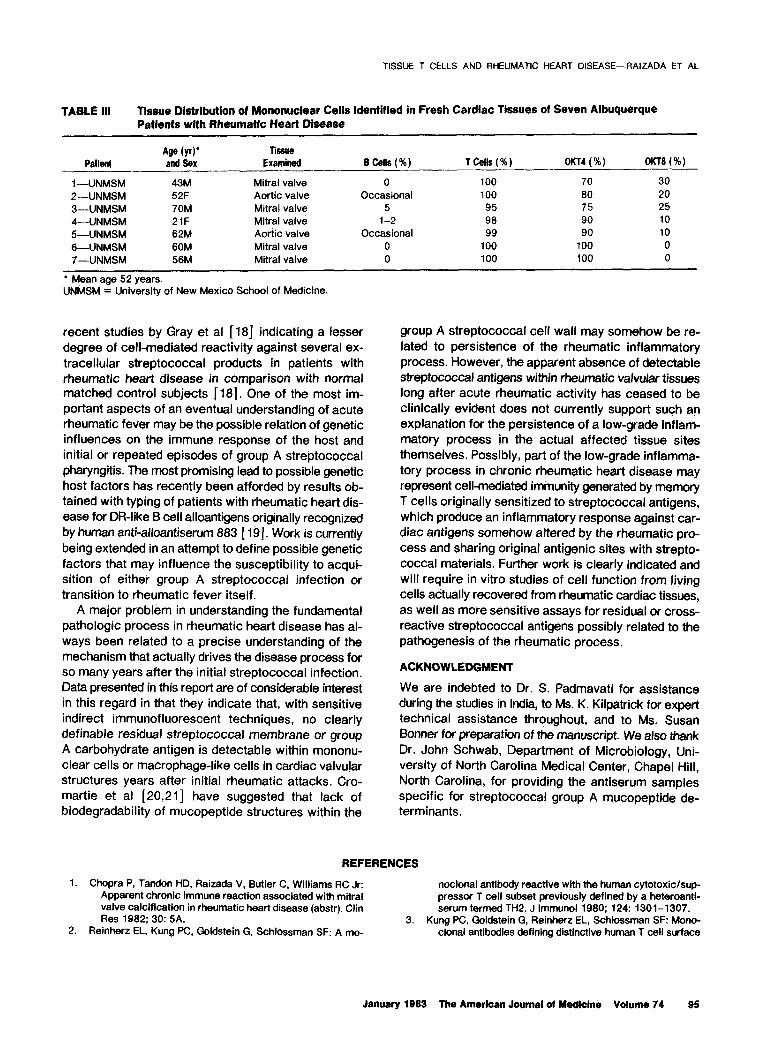
In 10 instances, rheumatic valvular tissues as well as atrial appendages were also examined by immunofluorescence using pepsindigested F(ab’)* rabbit antiserum to strepto- coccal membranes (group A, group G, and group D) [ IO] as well as antiserum to streptococcal group A carbohydrate. In addition, tissues were also examined using F(ab’)s rabbit antiserum samples reactive with group A streptococcal mucopeptide determinants (anti-peptidoglycan and rhamnose polysaccharide and a second reagent reacting with L-al- anyl-D-alanyl D-alanine). (The latter reagents were kindly furnished by Dr. John Schwab, Department of Microbiology, University of North Carolina, Chapel Hill, North Carolina.) Controls included F(ab’)l prepared .from rabbit antiserum samples specific for group B meningococci or gonococcal pili. Second layer in these immunofluorescent reactions employed fluorescein- or rhodamine-conjugated F(ab’)s goat anti-rabbit F(ab’)*. As previously noted, pepsin-digested F(ab’), reagents were used when appropriate throughout to avoid inadvertent binding of various reagents to tissue Fc receptors [ 10,l I].
RESULTS
Indian Patient Material. Conventional histologic study of fresh cardiac tissues using frozen sections stained with hematoxylin and eosin showed that 25 percent of valves or atrial tissues from Indian subjects with es- tablished rheumatic heart disease contained focal mononuclear or lymphocytic infiltrations. The immu- nofluorescent studies were concentrated on serial tis-
January 1993 The Amerlcan Journal of Medicine Volume 74 91
TISSUE T CELLS AND RHEUMATIC HEART DISEASE-RAIZADA ET AL
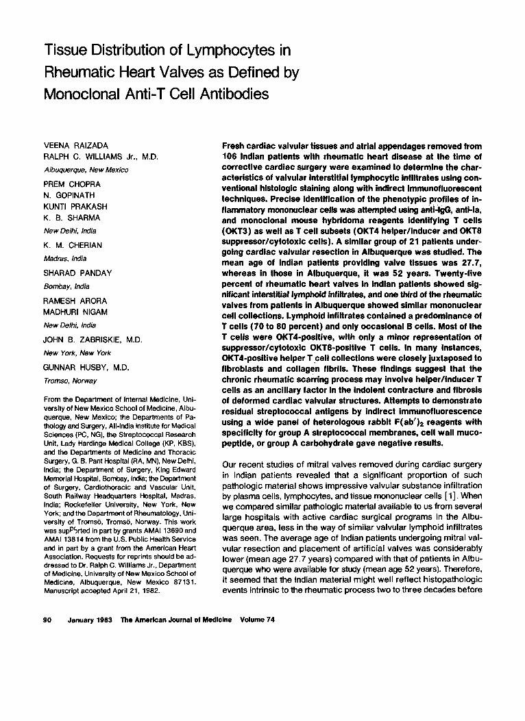
sue sections containing welldefined mononuclear cell infiltrates (Figure 1, top and bottom).
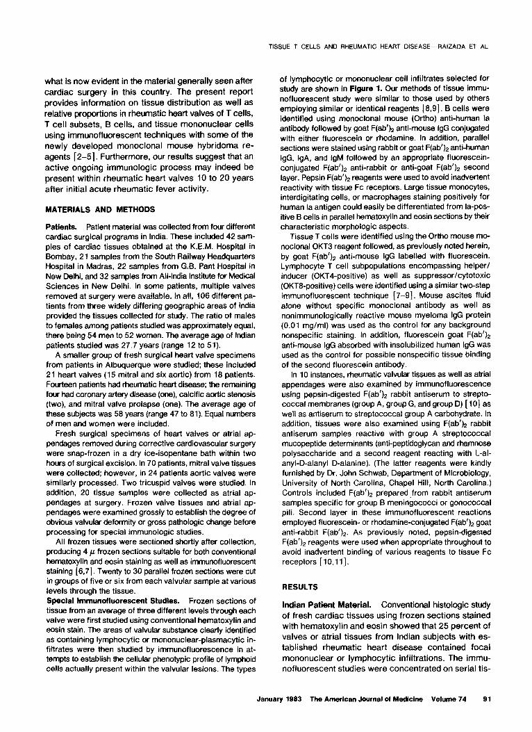
With reagents aimed at defining at least part of the mononuclear cell infiltrate profile, it was clear that a major preponderance of T lymphocytes was present in most instances. Relative proportions of T or 6 lymphoid cell subpopulations were estimated by counting 100 to 200 cells in multiple different (average of three) parallel microscopic fields. Data obtained in 22 patients’ tissues in which mononuclear cell infiltrates were extensive enough to make meaningful observations are shown in Table I. With occasional exceptions (Patients 8 and 12), the majority of lymphocytes within focal valvular or atrial mononuclear cell infiltrates were T cells reacting with the pan-T OKT3 reagent. Examples of T cell infiltrates
Figure 7. Top, focal intense mononu- cl&r cell infiltrates in rheumatic cardiac valvular structures (original magnification X 340, reduced by 35 percent). Bottom, higher-power views of mononuclear vai- vular infiltrates in close juxtaposition to fibroblasts and collagen fibrils (original magnification X 480, reduced by 35 percent).
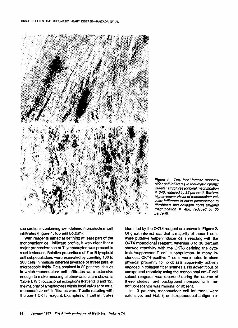
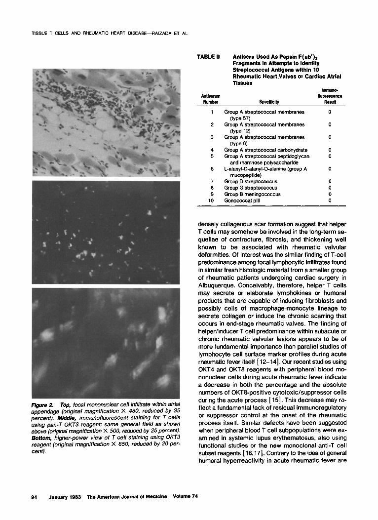
identified by the 0KT3 reagent are shown in Figure 2. Of great interest was that a majority of these T cells were putative helper/inducer cells reacting with the 0KT4 monoclonal reagent, whereas 0 to 30 percent showed reactivity with the OKT8 defining the cyto- toxic/suppressor T cell subpopulation. In many in- stances, OKTCpositive T cells were noted in close physical proximity to fibroblasts apparently actively engaged in collagen fiber synthesis. No adventitious or unexpected reactivity using the monoclonal anti-T cell subset reagents was recorded during the course of these studies, and background nonspecific immu- nofluorescence was minimal or absent.
In 10 patients, mononuclear cell infiltrates were extensive, and F(ab’)* antistreptococcal antigen re-
92 January 1983 The American Journal of Medicine Volume 74
TISSUE T CELLS AND RHEUMATIC HEART DISEASE-RAIZAUA ET AL
TABLE I Tissue Distribution of Mononuclear Ceils Identified In Fresh Cardiac Tissues of 22 Indian Patients with Rheumatic Heart Disease
Patient and Hospital
Age (yr) and Sex’ Tissue Examined B Cells (%)
1 cells (CfKT3) (%) W-f4 (%) gffT6 (%)
1 -AIMI 2-AIMI 3-AIMI 4-KEM
5-KEM 6-AIMI 7-AIMI
8--AIMI 9-KEM
lo-KEM
1 l-KEM 12-AIMI 13-KEM 14-KEM 15-G.B. Pant 16-G.B. Pant 17-KEM 18-G.B. Pant 1 O-Madras 20-G.B. Pant 21-G.B. Pant 22-Madras
37M 25M 13F 22M
21F Mitral valve 22F Mitral valve 25F Mitral valve
18F 21M 15F
20M 32M 23M 48F 16F 25F 30F 42F 17M 29F 25M 17M
Mitral valve Mitral valve Mitral valve Aortic valve
Aortic valve Mitral valve Mitral valve
Mitral valve Aortic valve Mitral valve Mitral valve Atrial appendage Atrial appendage Tricuspid valve Mitral valve Aortic valve Atrial appendage Atrial appendage Mitral valve
None Occasional (l-2) Occasional (l-2) None
5
Occasional (l-2) None
10 Occasional (l-2) Occasional (l-5)
(focal) 30-40 Occasional (l-2) None None 5 Occasional (l-2) l-5 2-5 (focal) 2-5 (very focal) None Occasional (l-2)
100 >95 >95 100
(focal) 95
>95 100
(focal) 90
>95 100-95
60-70 90 10
>95 70 30 100 ND ND 100 >95 (5 95 90-95 (5
>95 98 l-2 >95 >95 <5
95 90 10 95 >95 <5
100 100 0 >95 >95 <5 100 90 10
95 95 80 75
95 <5 95 <5
100 None
70 >95 >95
<5 <5 20 25
30 (5
0
l Mean age 24.7 years; female-to-male patient ratio 11:ll. AIMI = All-India Medical Institute, New Delhi, India; KEM = King Edward Memorial Hospital, Bombay, India.
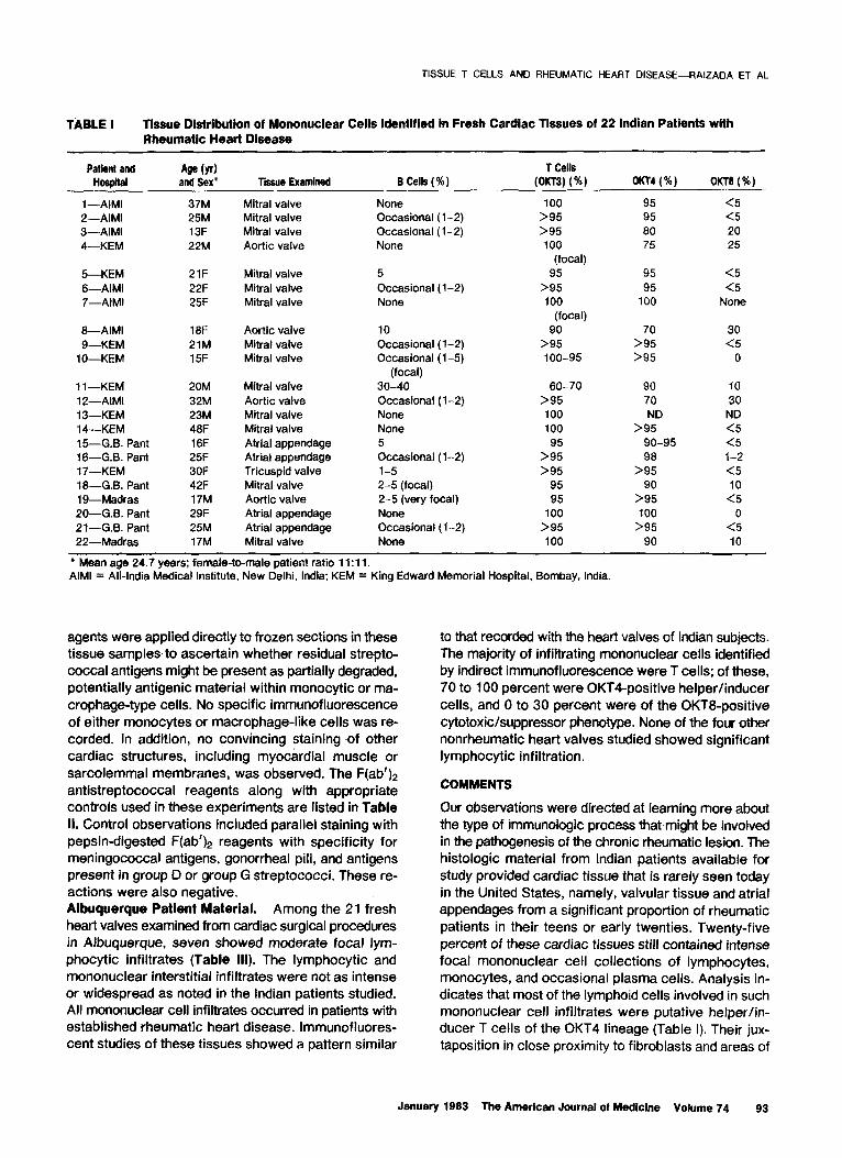
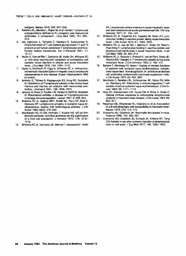
agents were applied directly to frozen sections in these tissue samples- to ascertain whether residual strepto- coccal antigens might be present as partially degraded, potentially antigenic material within monocytic or ma- crophage-type cells. No specific immunofluorescence of either monocytes or macrophage-like cells was re- corded. In addition, no convincing staining .of other cardiac structures, including myocardial muscle or sarcoiemmal membranes, was observed. The F(ab’)n antistreptococcal reagents along with appropriate controls used in these experiments are listed in Table II. Control observations included parallel staining with pepsin-digested F(ab$ reagents with specificity for meningococcal antigens, gonorrhea1 pili, and antigens present in group D or group G streptococci. These re- actions were also negative. Albuquerque Patient Material. Among the 21 fresh heart valves examined from cardiac surgical procedures in AfbUQUerQUe, seven showed moderate focal lym- phocytic infiltrates (Table Ill). The lymphocytic and mononuclear interstitial infiltrates were not as intense or widespread as noted in the Indian patients studied. All mononuclear cell infiltrates occurred in patients with established rheumatic heart disease. Immunofiuores- cent studies of these tissues showed a pattern similar
to that recorded with the heart valves of Indian subjects. The majority of infiltrating mononuclear cells identified by indirect immunofluorescence were T cells; of these, 70 to 100 percent were OKT4-positive helper/inducer cells, and 0 to 30 percent were of the OKTB-positive cytotoxic/suppressor phenotype. None of the four other nonrheumatic heart valves studied showed significant lymphocytic infiltration.
COMMENTS
Our observations were directed at learning more about the type of immunologic process that.might be involved in the pathogenesis of the chronic rheumatic lesion. The histologic material from Indian patients available for study provided cardiac tissue that is rarely seen today in the United States, namely, valvular tissue and atrial appendages from a significant proportion of rheumatic patients in their teens or early twenties. Twenty-five percent of these cardiac tissues still contained intense focal mononuclear cell collections of lymphocytes, monocytes, and occasional plasma cells. Analysis in- dicates that most of the lymphoid cells involved in such mononuclear cell infiltrates were putative helper/in- ducer T cells of the 0KT4 lineage (Table I). Their jux-
taposition in close proximity to fibroblasts and areas of
January 1983 The American Journal of Medicine Volume 74 93
TISSUE T CELLS AND RHEUMATIC HEART DISEASE-RAIZADA ET AL
F/gure 2. Top, focal mononuclear cell infiltrate within atria/ appendage (original magnification X 480, reduced by 35 percent). Middle, immunofluorescent staining for T cells using pan-T OKT3 reagent; same general field as shown above (original magnification X 500, reduced by 25 percent). Bottom, higher-power view of T cell staining using 0KT3 reagent {original magnification X 850, reduced by 20 per- cent).
TABLE II
Antiserum
Antisera Used As Pepsin F( ab’)* Fragments in Attempts to Identify Streptococcal Antigens within 10 Rheumatic Heart Valves or Cardiac Atrial Tissues
Immuno- fluorescence
Number speclflcity Resull
1
2
3
4 5
6
7 6 9
10
Group A streptococcal membranes
(type 67) Group A streptococcal membranes
(type 12) Group A streptococcal membranes
(type 6) Group A streptococcal carbohydrate Group A streptococcal peptidoglycan
and rhamnose polysaccharide L-alanyl-D-alanyl-Dalanine (group A
mucopeptide) Group D streptococcus Group G streptococcus Group B meningococcus Gonococcal pili
0
0
0
0 0
0
densely collagenous scar formation suggest that helper T cells may somehow be involved in the long-term se- quellae of contracture, fibrosis, and thickening well known to be associated with rheumatic valvular deformities. Of interest was the similar finding of T-cell predominance among focal lymphocytic infiltrates found in similar fresh histologic material from a smaller group of rheumatic patients undergoing cardiac surgery in Albuquerque. Conceivably, therefore, helper T cells may secrete or elaborate lymphokines or humoral products that are capable of inducing fibroblasts and possibly cells of macrophage-monocyte lineage to secrete collagen or induce the chronic scarring that occurs in end-stage rheumatic valves. The finding of helper/inducer T cell predominance within subacute or chronic rheumatic valvular lesions appears to be of more fundamental importance than parallel studies of lymphocyte cell surface marker profiles during acute rheumatic fever itself [ 12-141. Our recent studies using OKT4 and OKT8 reagents with peripheral blood mo- nonuclear cells during acute rheumatic fever indicate a decrease in both the percentage and the absolute numbers of OKT8-positive cytotoxic/suppressor cells during the acute process [IS]. This decrease may ro- fleet a fundamental lack of residual immunoregulatory or suppressor control at the onset of the rheumatic process itself. Similar defects have been suggested when peripheral blood T cell subpopulations were ex- amined in systemic lupus erythematosus, also using functional studies or the new monoclonal anti-T cell subset reagents [ 16,171. Contrary to the idea of general humoral hyperreactivity in acute rheumatic fever are
94 January 1983 The American Journal of Medicine Volume 74
TISSUE T CELLS AND RHEUMATIC HEART DISEASE-RAIZADA ET AL
TABLE III Tissue Distribution of Mononuclear Ceils Identified in Fresh Cardiac Tissues of Seven Albuquerque Patlents with Rheumatic Heart Disease
P&ll Age (YV Tiswe and sex Examinsd BCells(%) T Cells ( % ) 0KT4 (%) OKTB (%)
1 -UNMSM 43M Mitral valve 0 100 70 30 P-UNMSM 52F Aortic valve Occasional 100 80 20 3-UNMSM 70M Mitral valve 5 95 75 25 4-UNMSM 21F Mitral valve l-2 98 90 10 J-UNMSM 62M Aortic valve Occasional 99 90 10 6-UNMSM 60M Mitral valve 0 100 100 0 7-UNMSM 56M Mitral valve 0 100 100 0
l Mean age 52 years. UNMSM = University of New Mexico School of Medicine.
recent studies by Gray et al [18] indicating a lesser degree of cell-mediated reactivity against several ex- tracellular streptococcal products in patients with rheumatic heart disease in comparison with normal matched control subjects [ 181. One of the most im- portant aspects of an eventual understanding of acute rheumatic fever may be the possible relation of genetic influences on the immune response of the host and initial or repeated episodes of group A streptococcal pharyngitis. The most promising lead to possible genetic host factors has recently been afforded by results ob- tained with typing of patients with rheumatic heat-t dis- ease for DR-like B cell alloantigens originally recognized by human anti-alloantiserum 883 [ 191. Work is currently being extended in an attempt to define possible genetic factors that may influence the susceptibility to acqui- sition of either group A streptococcal infection or transition to rheumatic fever itself.
A major problem in understanding the fundamental pathologic process in rheumatic heart disease has al- ways been related to a precise understanding of the mechanism that actually drives the disease process for so many years after the initial streptococcal infection. Data presented in this report are of considerable interest in this regard in that they indicate that, with sensitive indirect immunofluorescent techniques, no clearly definable residual streptococcal membrane or group A carbohydrate antigen is detectable within mononu- clear cells or macrophage-like cells in cardiac valvular structures years after initial rheumatic attacks. Cro- martie et al [20,21] have suggested that lack of biodegradability of mucopeptide structures within the
group A streptococcal cell wall may somehow be re- lated to persistence of the rheumatic inflammatory process. However, the apparent absence of detectable streptococcal antigens within rheumatic valvular tissues long after acute rheumatic activity has ceased to be clinically evident does not currently support such an explanation for the persistence of a low-grade inflam- matory process in the actual affected tissue sites themselves. Possibly, part of the low-grade inflamma- tory process in chronic rheumatic heart disease may represent cell-mediated immunity generated by memory T cells originally sensitized to streptococcal antigens, which produce an inflammatory response against car- diac antigens somehow altered by the rheumatic pro- cess and sharing original antigenic sites with strepto- coccal materials. Further work is clearly indicated and will require in vitro studies of cell function from living cells actually recovered from rheumatic cardiac tissues, as well as more sensitive assays for residual or cross- reactive streptococcal antigens possibly related to the pathogenesis of the rheumatic process.
ACKNOWLEDGMENT
We are indebted to Dr. S. Padmavati for assistance during the studies in India, to Ms. K. Kilpatrick for expert technical assistance throughout, and to Ms. Susan Banner for preparation of the manuscript. We also thank Dr. John Schwab, Department of Microbiology, Uni- versity of North Carolina Medical Center, Chapel Hill, North Carolina, for providing the antiserum samples specific for streptococcal group A mucopeptide de- terminants.
REFERENCES
1. Chopra P, Tandon I-ID, Raizada V, Butler C, Williams RC Jr: noclonal antibody reactive with the human cytotoxickup- Apparent chronic immune reaction associated with mitral pressor T cell subset previously defined by a heteroanti- valve calcification in rheumatic heart disease (abstr). Clin serum termed TH2. J lmmunol 1980; 124: 1301-1307. Res 1982; 30: 5A. 3. Kung PC, Goldstein G, Reinherz EL, Schlossman SF: Mono-
2. Reinherz EL, Kung PC, Goldstein G, Schlossman SF: A mo- clonal antibodies defining distinctive human T cell surface
Janusry lBE3 The American Journal ol Medktne Volume 74 95
TISSUE T CELLS AND RHEUMATIC HEART DISEASE-RAIZADA ET AL
4.
5.
6.
7.
6.
9.
10.
11.
12.
antigens. Nature 1979; 206: 347-349. Reinherz EL, Moretta L. Roper M, et al: Human T lymphocyte
subpopulations defined by Fc receptors and monoclonal antibodies. A comparison. J Exp Med 1960; 151: 969- 974.
van Agthoven A, Terhorst C, Reinherz E, Schlossman S: Characterization of T cell surface glycoproteins Tl and T3 present on all human peripheral T lymphocytes and func- tionally mature thymocytes. Eur J lmmunol 1981; 11: 18-21.
Husby G, Van de Rijn I, Zabriskie JB, Abdin ZH, Williams RC Jr: Anti-body reacting with cytoplasm of subthalamic and caudate nuclei neurons in chorea and acute rheumatic fever. J Exp Med 1976; 144: 1094-1110.
Husby G, Blomhoff JP, Elgjo K, Williams RC Jr: Immunohis- tochemical characterization of hepatic tissue lymphocyte subpopulations in liver disease. Stand J Gastroenterol 1982 (in press).
Janossy G, Tidman N, Papageorgin ES, Kung PC, Goldstein G: Distribution of T lymphocyte subsets in the human bone marrow and thymus: an analysis with monoclonal anti- bodies. J lmmunol 1981; 126: 1608-1613.
Janossy G, Duke 0, Poulter LW, Panayi G, Bofill M, Goldstein G: Rheumatoid arthritis: a disease of T-lymphocyte/ma- crophage immunoregulation. Lancet 1981; II: 839-842.
Williams RC Jr, Hughes GRV, Snaith ML, Parry HF, Diao E, Greaves MF: Lymphocyte antigens in systemic lupus er- ythematosus: studies with heterologous antisera. J Clin Invest 1980; 65(2): 379-389.
Winchester RJ, Fu SM. Hoffman T, Kunkel HG: IgG on lym- phocyte surfaces: technical problems and the significance of a third cell population. J lmmunol 1975; 114: 1210- 1212.
Williams RC Jr, Zabriskie JB, Mahros F, Hassaballa F, Abdin
13.
14.
15.
16.
17.
18.
19.
20.
21.
ZH: Lymphocyte surface markers in acute rheumatic fever and post-streptococcal acute glomerulonephritis. Clin Exp lmmunol 1977; 27: 135-142.
Williams RC Jr, Kilpatrick KA, Kassaby M, Abdin ZH: Lym- phocytes binding C-reactive protein during acute rheumatic fever. J Clin Invest 1978; 61: 1384-1393.
Williams RC Jr, van de Rijn I, Mahros F, Abdin ZH, Reid H, Poon-King T: Lymphocytes binding C-reactive protein and streptococcal membranes in acute rheumatic fever. J Lab Clin Med 1980; 96: 803-814.
Williams RC Jr, Raizada V, Prakash K, van de Rijn I, Stobo JD, Sharma KB: Changes in T lymphocyte subsets during acute rheumatic fever. J Clin lmmunol 1982; 2: 166-172.
Sakane T, Steinberg AD, Green I: Studies of immune functions of patients with systemic lupus erythematosus: comple- ment-dependent immunoglobulin M anti-thymusderived cell antibodies preferentially inactivate suppressor cells. J Clin Invest 1979; 63: 954-965.
Morimoto C, Reinherz EL, Schlossman SF, Schur PH, Mills JA, Steinberg AD: Alterations in immunoregulatory T cell subsets in active systemic lupus erythematosus. J Clin In- vest 1980; 66: 1171-1174.
Gray ED, Wannamaker LW, Ayoub EM, El Kholy A. Abdin Z: Cellular immune responses to extracellular streptococcal products in rheumatic heart disease. J Clin Invest 1981; 68: 665-671.
Patarroyo ME, Winchester RJ, Vejerano A, et al: Association of a B-cell alloantigen with susceptibility to rheumatic fever. Nature 1979; 278: 172-174.
Cromartie WJ, Craddock JH: Rheumatic-like lesions in mice. Science 1966; 154: 285-287.
Cromartie WJ, Craddock JG, Schwab JH, Anderle SK, Yang CH: Arthritis in rats after systemic injection of streptococcal cells or cell walls. J Exp Med 1977; 146: 1585-1602.
96 January 1993 The American Journal of Medicine Volume 74