Thyroid disruption in the lizard Podarcis bocagei exposed to a mixture of herbicides: a field study
10
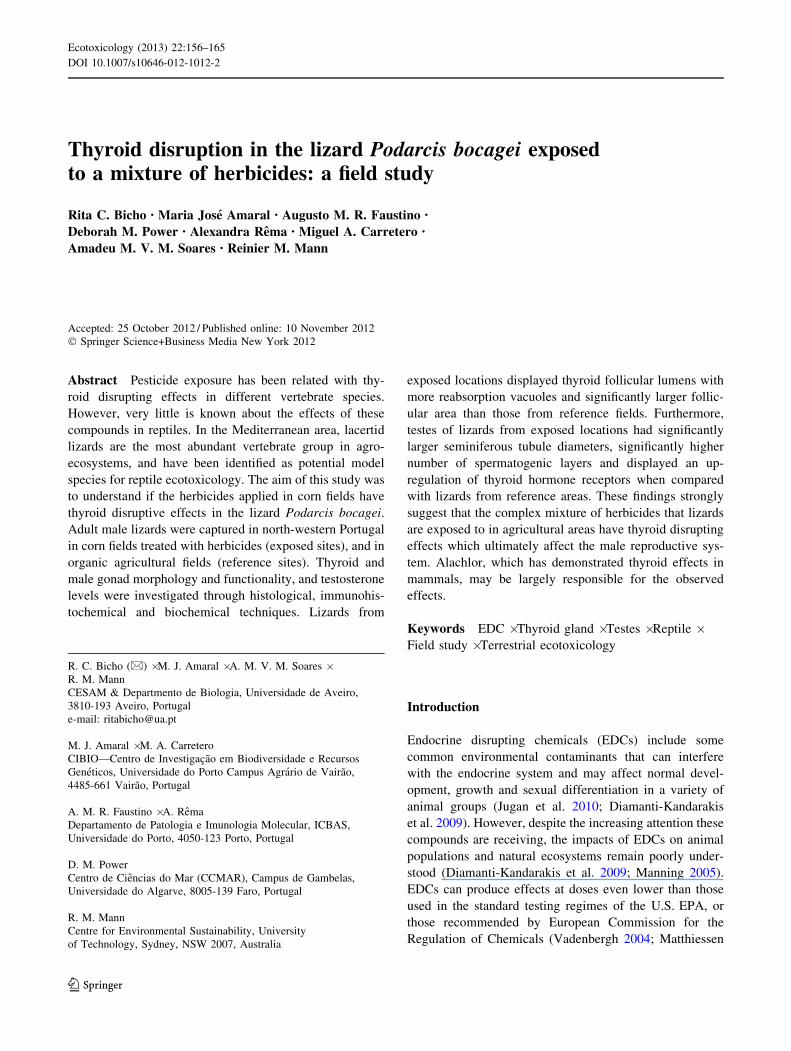
Thyroid disruption in the lizard Podarcis bocagei exposed to a mixture of herbicides: a field study Rita C. Bicho • Maria Jose ´ Amaral • Augusto M. R. Faustino • Deborah M. Power • Alexandra Re ˆma • Miguel A. Carretero • Amadeu M. V. M. Soares • Reinier M. Mann Accepted: 25 October 2012 / Published online: 10 November 2012 Ó Springer Science+Business Media New York 2012 Abstract Pesticide exposure has been related with thy- roid disrupting effects in different vertebrate species. However, very little is known about the effects of these compounds in reptiles. In the Mediterranean area, lacertid lizards are the most abundant vertebrate group in agro- ecosystems, and have been identified as potential model species for reptile ecotoxicology. The aim of this study was to understand if the herbicides applied in corn fields have thyroid disruptive effects in the lizard Podarcis bocagei. Adult male lizards were captured in north-western Portugal in corn fields treated with herbicides (exposed sites), and in organic agricultural fields (reference sites). Thyroid and male gonad morphology and functionality, and testosterone levels were investigated through histological, immunohis- tochemical and biochemical techniques. Lizards from exposed locations displayed thyroid follicular lumens with more reabsorption vacuoles and significantly larger follic- ular area than those from reference fields. Furthermore, testes of lizards from exposed locations had significantly larger seminiferous tubule diameters, significantly higher number of spermatogenic layers and displayed an up- regulation of thyroid hormone receptors when compared with lizards from reference areas. These findings strongly suggest that the complex mixture of herbicides that lizards are exposed to in agricultural areas have thyroid disrupting effects which ultimately affect the male reproductive sys- tem. Alachlor, which has demonstrated thyroid effects in mammals, may be largely responsible for the observed effects. Keywords EDC Á Thyroid gland Á Testes Á Reptile Á Field study Á Terrestrial ecotoxicology Introduction Endocrine disrupting chemicals (EDCs) include some common environmental contaminants that can interfere with the endocrine system and may affect normal devel- opment, growth and sexual differentiation in a variety of animal groups (Jugan et al. 2010; Diamanti-Kandarakis et al. 2009). However, despite the increasing attention these compounds are receiving, the impacts of EDCs on animal populations and natural ecosystems remain poorly under- stood (Diamanti-Kandarakis et al. 2009; Manning 2005). EDCs can produce effects at doses even lower than those used in the standard testing regimes of the U.S. EPA, or those recommended by European Commission for the Regulation of Chemicals (Vadenbergh 2004; Matthiessen R. C. Bicho (&) Á M. J. Amaral Á A. M. V. M. Soares Á R. M. Mann CESAM & Departmento de Biologia, Universidade de Aveiro, 3810-193 Aveiro, Portugal e-mail: [email protected] M. J. Amaral Á M. A. Carretero CIBIO—Centro de Investigac ¸a ˜o em Biodiversidade e Recursos Gene ´ticos, Universidade do Porto Campus Agra ´rio de Vaira ˜o, 4485-661 Vaira ˜o, Portugal A. M. R. Faustino Á A. Re ˆma Departamento de Patologia e Imunologia Molecular, ICBAS, Universidade do Porto, 4050-123 Porto, Portugal D. M. Power Centro de Cie ˆncias do Mar (CCMAR), Campus de Gambelas, Universidade do Algarve, 8005-139 Faro, Portugal R. M. Mann Centre for Environmental Sustainability, University of Technology, Sydney, NSW 2007, Australia 123 Ecotoxicology (2013) 22:156–165 DOI 10.1007/s10646-012-1012-2
-
Upload
independent -
Category
Documents
-
view
2 -
download
0
Transcript of Thyroid disruption in the lizard Podarcis bocagei exposed to a mixture of herbicides: a field study
Thyroid disruption in the lizard Podarcis bocagei exposedto a mixture of herbicides: a field study
Rita C. Bicho • Maria Jose Amaral • Augusto M. R. Faustino •
Deborah M. Power • Alexandra Rema • Miguel A. Carretero •
Amadeu M. V. M. Soares • Reinier M. Mann
Accepted: 25 October 2012 / Published online: 10 November 2012
� Springer Science+Business Media New York 2012
Abstract Pesticide exposure has been related with thy-
roid disrupting effects in different vertebrate species.
However, very little is known about the effects of these
compounds in reptiles. In the Mediterranean area, lacertid
lizards are the most abundant vertebrate group in agro-
ecosystems, and have been identified as potential model
species for reptile ecotoxicology. The aim of this study was
to understand if the herbicides applied in corn fields have
thyroid disruptive effects in the lizard Podarcis bocagei.
Adult male lizards were captured in north-western Portugal
in corn fields treated with herbicides (exposed sites), and in
organic agricultural fields (reference sites). Thyroid and
male gonad morphology and functionality, and testosterone
levels were investigated through histological, immunohis-
tochemical and biochemical techniques. Lizards from
exposed locations displayed thyroid follicular lumens with
more reabsorption vacuoles and significantly larger follic-
ular area than those from reference fields. Furthermore,
testes of lizards from exposed locations had significantly
larger seminiferous tubule diameters, significantly higher
number of spermatogenic layers and displayed an up-
regulation of thyroid hormone receptors when compared
with lizards from reference areas. These findings strongly
suggest that the complex mixture of herbicides that lizards
are exposed to in agricultural areas have thyroid disrupting
effects which ultimately affect the male reproductive sys-
tem. Alachlor, which has demonstrated thyroid effects in
mammals, may be largely responsible for the observed
effects.
Keywords EDC � Thyroid gland � Testes � Reptile �Field study � Terrestrial ecotoxicology
Introduction
Endocrine disrupting chemicals (EDCs) include some
common environmental contaminants that can interfere
with the endocrine system and may affect normal devel-
opment, growth and sexual differentiation in a variety of
animal groups (Jugan et al. 2010; Diamanti-Kandarakis
et al. 2009). However, despite the increasing attention these
compounds are receiving, the impacts of EDCs on animal
populations and natural ecosystems remain poorly under-
stood (Diamanti-Kandarakis et al. 2009; Manning 2005).
EDCs can produce effects at doses even lower than those
used in the standard testing regimes of the U.S. EPA, or
those recommended by European Commission for the
Regulation of Chemicals (Vadenbergh 2004; Matthiessen
R. C. Bicho (&) � M. J. Amaral � A. M. V. M. Soares �R. M. Mann
CESAM & Departmento de Biologia, Universidade de Aveiro,
3810-193 Aveiro, Portugal
e-mail: [email protected]
M. J. Amaral � M. A. Carretero
CIBIO—Centro de Investigacao em Biodiversidade e Recursos
Geneticos, Universidade do Porto Campus Agrario de Vairao,
4485-661 Vairao, Portugal
A. M. R. Faustino � A. Rema
Departamento de Patologia e Imunologia Molecular, ICBAS,
Universidade do Porto, 4050-123 Porto, Portugal
D. M. Power
Centro de Ciencias do Mar (CCMAR), Campus de Gambelas,
Universidade do Algarve, 8005-139 Faro, Portugal
R. M. Mann
Centre for Environmental Sustainability, University
of Technology, Sydney, NSW 2007, Australia
123
Ecotoxicology (2013) 22:156–165
DOI 10.1007/s10646-012-1012-2

and Johnson 2007). Also, there are a multitude of physio-
logical processes that can be disrupted, which may only be
sensitive for relatively short periods during organogenesis.
Several EDCs are recognized for their effects on the
reproductive system, having estrogenic, androgenic, anti-
estrogenic and anti-androgenic effects on wildlife (Schmut-
zler et al. 2007; Jugan et al. 2010). More recently, the thyroid
axis was also recognized as a target for the action of endocrine
disruptors (Sciarrillo et al. 2008; Jugan et al. 2010; Schmutzler
et al. 2007). However, the complexity of the hypothalamus-
pituitary-thyroid axis and the numerous target organs for this
system means that there is a multiplicity of targets for the
action of chemicals with thyroid disruptive activities. As a
consequence, our understanding of the interaction between
environmental contaminants and the thyroid axis is still poor.
Most attention has been concentrated on the thyroid gland
itself, and it is now well established that some chemicals can
interfere directly with the synthesis of thyroid hormones and
its regulation by the hypothalamus and pituitary gland. Fur-
thermore, several histological biomarkers for increased or
decreased thyroid activity have been developed. For example,
increased follicular cell height and follicle area within thyroid
tissue are evident in thyroid glands that were actively stimu-
lated by the thyroid stimulating hormone (TSH) (Leatherland
1994; Hewitt et al. 2002).
Many pesticides are suspected EDCs and different lab-
oratory studies have demonstrated their effects on the
endocrine system of different taxa, including wildlife
populations heavily exposed to these contaminants
(Manning 2005; Meeker and Boas 2011). However, there
are limited data available that describe relationships
between pesticides exposure and altered thyroid function
(Meeker and Boas 2011). Among herbicides, some classes
such as the chloroacetanilide herbicides (e.g., alachlor);
triazines (e.g., atrazine); dinitroanalines (e.g., trifluralin)
and phenoxy acids (e.g., 2,4-D) have recognized thyroid
status-disrupting properties (Meeker and Boas 2011).
The disruption of thyroid gland homeostasis can also
affect the normal function or development of target organs
such as the testes (Zhang et al. 2009). Thyroid hormones
have an important role in regulation of testis development
and function in all life stages in mammals (Wagner et al.
2008; Jannini et al. 1995) and in reptiles (Haldar-Misra and
Thapliyal 1981; Plowman and Lynn 1973; Thapliyal et al.
1974), and this role is confirmed by the expression of
thyroid hormone receptors (TR) in testes (Cardone et al.
2000; Jannini et al. 1994; Buzzard et al. 2000).
Reptiles offer several useful attributes for discerning the
endocrine disrupting effects of pesticides and other chemicals
(Crain and Guillette Jr 1998). For example, reptiles appear to
have a lower capacity for detoxifying organic pollutants than
other vertebrates (Walker and Ronis 1989). However, com-
pared with other vertebrate classes, very little is known about
the effects of contaminants in these animals (Sparling et al.
2010). They are not routinely included in toxicity tests and
there is little information about their sensitivity to pesticides,
despite being identified by some authors as more vulnerable
than other taxa (Bruce et al. 2010; De Lange et al. 2009).
Moreover, most studies in reptile ecotoxicology concern
chemical tissue residues in field collected individuals
(Hopkins 2006; Linder et al. 2010). A far smaller proportion of
studies examined the effects of exposure to environmental
contaminants, and these are generally laboratory based
manipulations (McFarland et al. 2009; Capriglione et al.
2011). Although these studies are important to understand
responses, they do not provide enough information on the
effects on populations in complex natural environments
(Mann et al. 2009). Therefore, studies that examine the effects
of contaminant exposure in field populations are essential. In
Europe, lacertid lizards have been proposed as potential model
species for reptile ecotoxicology because of their high site
fidelity (Galan 1999; Carretero et al. 2006) and abundance in
agricultural areas (compared to mammals; Valverde, 1967)
and because they are relatively easy to maintain in captivity
(Mann et al. 2007; Cardone et al. 2008).
The current work forms part of a larger study on the
effects of corn pesticides in lacertid lizards. This larger
study included an examination of population indices and
biomarkers of pesticide exposure and effect among several
sub-populations of Podarcis bocagei (Amaral et al. 2012a,
b). The present study, aims to understand if pesticides
applied in agricultural fields have any thyroid disruptive
effects in the lizard Podarcis bocagei. We evaluated effects
on the thyroid gland and male gonad morphology and
functionality, and on the thyroid–gonadal axis function.
Materials and methods
Lizard collection and maintenance
Adult male P. bocagei lizards (n = 37) were collected in the
field margins of four agricultural areas in north-western
Portugal. The collection sites included two fields used for
corn growing where a mixture of pesticides has been applied
routinely for over 30 years (exposed), and two organic
agricultural fields with no history of pesticide application. A
detailed description of the collection sites: Exp 1, Exp 4,
Ref 1 and Ref 2 is published elsewhere (Amaral et al.
2012b). Lizards were collected from four sites to avoid
interference with population structures in any one popula-
tion. The major chemicals detected in the exposed sites were
alachlor, bentazone, dicamba, dimethenamid-p, mesotrione
and terbuthylazine (Table 1). Lizards were caught with a
noose during Autumn (November, 2009) corresponding to
the pre-hibernation period described for the species (Galan
Thyroid disruption in the lizard Podarcis bocagei 157
123

1995). Each animal was housed individually in glass terraria
(40 9 20 9 25 cm) in a climate control room. Each terrar-
ium contained a terracotta vase ([ = 16 cm) that provided
refuge and a basking location, and a 25 W incandescent lamp
(8 h/day) that created a thermal gradient (25–35 �C). Room
temperature was maintained at 22 ± 1 �C. Lighting in the
climate room was provided by external natural sunlight,
fluorescent lighting (2 9 40 W) and a high pressure sodium
lamp (400 W, 7000–12 000 Lx) for 12 h/day. Water was
provided in shallow dishes and renewed every 2–3 days, and
animals were fed with mealworms (Tenebrio molitor L.).
Lizards were held in captivity for 15 days during which
various parameters were measured (i.e., morphological,
behavioral and physiological); these data are described
elsewhere (Amaral et al. 2012a). Animal handling complied
with Portuguese animal ethics guidelines as stipulated by
Direccao Geral de Veterinaria and Instituto da Conservacao
da Natureza e Biodiversidade.
Dissection and histology
Lizards were killed by cervical dislocation. From each
animal, blood samples were taken and plasma was obtained
by centrifugation (800 rpm for 10 min at 4 �C) and stored
at -20 �C until hormonal analysis. Thyroid glands and the
left testis were removed, testes were weighed and tissues
were fixed in Davidson’s solution for 24 h. Tissues were
then washed in 70 % ethanol and stored in 70 % ethanol
until further processing. An automated tissue processor
(Microm STP 120) was used for paraffin embedding.
Sections (2 lm) were cut on a rotary microtome (Leica RM
2035). Thyroid sections were stained with Cleveland-Wolf
(CW) and testes sections stained with Hematoxylin and
Eosin (H&E) for light microscopic examination. Obser-
vations and photographs were made using an Olympus
BX51 microscope with an Olympus camera attached. To
perform thyroid histomorphometry, 20 follicles from each
animal were chosen randomly, and follicle area and follicle
cell height were measured. For testes histomorphometry,
20 seminiferous tubules from each animal were randomly
chosen for analysis. Spermatogenesis development was
assessed by measuring tubular diameter, and along this
axis, the width of Sertoli cells and the width of each
spermatogenesis layer (spermatogonia, spermatocytes I and
II, spermatids, spermatozoa). The number of spermato-
genesis layers observed in each animal was also recorded.
All measurements were carried out using ImageJ (Rasband
1997–2008).
Immunohistochemistry
Expression of thyroid receptors (TR) in testes was detected
by immunohistochemistry using the primary antibody—
TRa/b sc-772 (Santa Cruz Biotechnology, Inc. Santa Cruz,
Table 1 Average pesticide
content of soils (lg/kg
soil) ± SD: seven herbicides
were detected (LOD = 0.5 lg/
kg)—pre and post pesticide
application in 2009
Exp exposed sites, Ref reference
sites
Pesticide residue concentration
(lg/kg soil)
Sampled sites
Ref 1 Ref 2 Exp 1 Exp 4
Alachlor
Pre 10.23 ± 7.18 \0.5 7.03 ± 8.18 30.66 ± 8.25
Post 3.75 ± 3.75 \0.5 2.36 ± 4.13 16.97 ± 9.48
Bentazone
Pre \0.5 \0.5 \0.5 \0.5
Post \0.5 \0.5 39.88 ± 88.70 \0.5
Dicamba
Pre \0.5 \0.5 \0.5 \0.5
Post \0.5 \0.5 9.75 ± 27.75 \0.5
Dimethenamid
Pre \0.5 \0.5 13.52 ± 17.68 14.47 ± 12.26
Post \0.5 \0.5 0.59 ± 0.27 30.08 ± 28.13
Glyphosate
Pre \0.5 \0.5 \0.5 \0.5
Post \0.5 \0.5 \0.5 1.23 ± 1.55
Mesotrione
Pre 2.93 ± 4.85 3.58 ± 6.15 8.35 ± 9.71 \0.5
Post \0.5 \0.5 23.59 ± 34.12 20.7 ± 40.28
Terbuthylazine
Pre \0.5 \0.5 \0.5 \0.5
Post \0.5 \0.5 \0.5 33.48 ± 40.22
158 R. C. Bicho et al.
123

CA) as described in Virgilio et al. (2004). This is a rabbit
polyclonal antibody raised against a recombinant protein
corresponding to amino acids 1-408 that represents the full
length of TRa1 and reacts both with TRa and TRb iso-
forms. Testes sections were immersed in 1 L of extran
solution and microwaved for 20 min at 700 watts for
antigen retrieval. Antigen visualization was done with the
Novocastra Novolink Polymer Detection System (Leica
Microsystems GmbH, Wetzlar, Germany) and involved the
following steps: Sections were incubated with H2O2 (3 %)
for 10 min to eliminate endogenous peroxidase activity
followed by a 5 min incubation with a protein blocking
agent. Sections were subsequently incubated overnight at
4 �C with the primary antibody diluted at 1:45 with BSA
(5 %) and Triton X-100 (0.3 %), and on the following day,
washed in TBS-TRIS-buffered saline solution before
incubation for 30 min with the secondary antibody system
using diaminobenzidine (DAB) as a chromogen. Observa-
tions and photographs were made using an Olympus BX51
microscope with an Olympus camera attached. TR-positive
cells were identified as dark reaction product in the cell
nuclei, and their number was counted in 30 randomly
chosen seminiferous tubules.
Testosterone assays
Plasma testosterone (T) was determined using a specific
radioimmunoassay (RIA) as described by Scott et al.
(1984). Duplicate aliquots (50 lL) of diluted denatured
plasma (prepared as indicated above) were used in the T
assay. Preliminary analysis revealed parallelism between
dilutions of the samples and the standard curve. Intra-assay
and inter-assay coefficients of variation were 8 and 12 %,
respectively.
Statistical analysis
Quantitative data obtained from histological sections and radio-
immunoassays were tested for normality and homogeneity of
variance (Levene’s test). Differences between lizards from
exposed and reference sites (Type) were examined using analysis
of covariance (ANCOVA, a B 0.05) with log SVL as the
covariate. Differences in the number of spermatogenesis layers
were tested with the non-parametric Mann–Whitney test.
Results
Thyroid histology and histomorphometry
The thyroid gland of P. bocagei is a single discrete ribbon-
like structure that transversely crosses the middle of the
trachea, as described for other lizards. Thyroids of all
individuals presented a normal histological appearance—
colloid filled follicles with cuboidal epithelial cells sur-
rounded by interfollicular connective tissue and blood
vessels. However, in lizards from exposed sites, there were
a large number of reabsorption vacuoles in follicular col-
loid, whereas in lizards from reference sites these vacuoles
were scarce (Fig. 1a–c). Lizards from exposed areas also
displayed a significantly larger follicular area (Fig. 2a,
ANCOVA Type: F1,19 = 6.9, p = 0.02). No differences
were found in the follicular epithelial cell height (Fig. 2b,
ANCOVA Type: F1,19 = 1.5, p = 0.24).
Testis histology, histomorphometry
and immunohistochemistry
Macroscopically, testes from both reference and exposed
lizards had a normal appearance. Histological examination
of testes sections revealed that lizards from exposed sites
possessed a significantly higher number of spermatogenesis
layers (Figs. 3a,b, 4; Mann–Whitney test: z = -2.6,
p = 0.009). Specifically, eight lizards (&44 %) of this
group presented all spermatogenesis phases, from sper-
matogonia to spermatozoa; whereas, only one animal
(&6 %) from the reference locations presented all phases.
Moreover, individuals from exposed sites had a
Fig. 1 Thyroid gland sections of Podarcis bocagei stained with
Cleveland-Wolf. a Representative section of lizard from reference
site. b Representative section of lizard from exposed site with
enlarged follicular area, c High magnification (9400) showing
reabsorption vacuoles (arrow) in thyroid section of lizard from
exposed site
Thyroid disruption in the lizard Podarcis bocagei 159
123

significantly smaller layer of spermatogonia (Fig. 5a,
ANCOVA Type: F1,31 = 9.1, p = 0.005) and significantly
larger seminiferous tubule diameter (Fig. 5b, ANCOVA
Type: F1,31 = 4.5, p = 0.04).
Immunohistochemistry revealed expression and locali-
zation of TR proteins in both groups of lizards. In lizards
from reference locations, TR-positive staining was only
seen in Leydig cells and all spermatogonia and spermato-
cyte were TR-negative (Fig. 6a). In lizards from exposed
sites, TR-positive staining was seen in some spermatogo-
nia, spermatocyte and spermatids, and there was consistent
TR-positive staining in all mature spermatozoa (Fig. 6b).
Testosterone radioimmunoassay
No differences were found in the concentrations of circu-
lating testosterone between animals from exposed and
reference sites (Fig. 7, ANCOVA Type: F1,22 = 0.1,
p = 0.73).
Discussion
Thyroid disrupting effects related to exposure to herbicides
have been described in different species related to exposure
to herbicides (Mann et al. 2009; Meeker and Boas 2011;
Wilson et al. 1996). However, very little is known about
the effects of these compounds on reptiles (Cardone et al.
2008). Reptiles are an important non-target group of ver-
tebrate animals that will be exposed in regions of intensive
agriculture where many different pesticides are applied
annually, and in varying combinations. Although many
fauna persist in agricultural systems, it is unclear to what
extent they cope with the constantly changing cocktails of
these compounds. Prominent within the agricultural fields
of Portugal (and other Mediterranean countries) are abun-
dant populations of lacertid lizards. Their ubiquity in
Mediterranean agricultural ecosystems provides impetus
for the use of these species as biological models for eco-
toxicological studies in these environments.
An annual cycle in thyroid activity, influenced by cli-
matic variations and temperature, has been described for
several reptile species, including Italian Podarcis sicula, an
ecologically similar species to P. bocagei (Sciarrillo et al.
2000). In P. sicula, a peak in thyroid activity is observed
between March and July during the reproduction period,
and a minimum is recorded in winter before and during
hibernation (Sciarrillo et al. 2000). Our individuals were
collected in November, just before winter, and we expected
to find low thyroid activity. Indeed, individuals not
exposed to herbicides presented low thyroid activity.
However, lizards from exposed sites showed a more active
thyroid, with significantly larger follicular area, and a far
greater abundance of reabsorption vacuoles. Furthermore,
these lizards also displayed an up-regulation of TR in
testes. Similar findings were observed in amphibians from
agricultural areas, where individuals exposed to pesticides
presented increased levels of circulating T3 and T4
(Mosconi et al. 2005). Analogous histological and hor-
monal alterations were also observed in other animal
classes in different contamination scenarios. For example,
in mummichog (Fundulus heteroclitus), individuals from a
polluted habitat exposed to non essential metals and
organic materials had larger thyroid follicles, greater epi-
thelial cell heights, and elevated concentrations of circu-
lating T4 (Zhou et al. 1999).
The physiological mechanisms involved in the thyroid
effects of herbicides or other contaminants are not well
understood. Several pesticides that have known thyroid
disruptive activities generally decrease thyroid function
(Meeker and Boas 2011; Boas et al. 2006). Therefore,
within the context of the increased thyroid activity
observed in the present study, the chloroacetanilide herbi-
cides, a group that includes acetochlor, metolachlor and
Fig. 2 Thyroid gland histomorphometry results of Podarcis bocagei.a Follicular area of lizards from reference sites compared with lizards
from exposed sites. b Follicle epithelial cell height of lizards from
reference sites compared with lizards from exposed sites. *p \ 0.05;
error bars represent standard deviations, (n = 10 lizards from
reference sites; n = 12 lizards from exposed sites)
160 R. C. Bicho et al.
123

alachlor (among others), are of particular interest. In the
present study, lizards from contaminated sites were
exposed to a mixture of herbicides and alachlor was one of
the most persistent of the chemicals detected (Amaral et al.
2012b). In a study with rats, alachlor increased thyroid
activity (Wilson et al. 1996). In amphibians, acetochlor,
which is structurally very similar to alachlor, increased
thyroid activity (Helbing et al. 2006; Crump et al. 2002;
Cheek et al. 1999), and appears to be associated with
increased expression of TR (Veldhoen and Helbing 2001;
Helbing et al. 2006). It is hypothesized that the observed
alterations in thyroid function and the up-regulation of TR
in testes observed in lizards exposed to herbicides, were
consequent to alachlor exposure.
Reproduction in P. bocagei is characterized by an
annual spermatogenic cycle, which is influenced by climate
(Carretero et al. 2006). The maritime Atlantic climate of
north-western Portugal provides suitable temperature and
humidity conditions for P. bocagei activity during most of
Fig. 3 Cross sections of testes of Podarcis bocagei stained with
Hematoxylin and Eosin. a Representative section of lizard from
reference site: seminiferous tubules showing only 2 spermatogenesis
layers (spermatogonia (black arrow) and spermatocyte I (circled).
b Representative section of lizard from exposed site: seminiferous
tubule presenting all spermatogenesis layers (spermatogonia (blackarrow), spermatocyte I (circled) and spermatocyte II/spermatids (in
square) and spermatozoa (white arrow)
Fig. 4 Number of spermatogenic layers observed in seminiferous
tubules of testes of Podarcis bocagei. 2 layers include spermatogonias
and spermatocytes I; 3 layers include spermatogonias, spermatocytes
I and spermatocytes II/spermatids; 4 layers include spermatogonias,
spermatocytes I, spermatocytes II/spermatids and spermatozoa.
**p \ 0.01 Mann–Whitney test. (n = 16 lizards from reference sites,
n = 18 lizards from exposed sites)
Fig. 5 Testes histomorphometry results of Podarcis bocagei.a Width of each spermatogenic layer observed in seminiferous
tubule: A spermatogonia, B spermatocytes I, C spermatocytes
II/spermatids, D spermatozoa. b diameter of seminiferous tubules.
*p \ 0.05, **p \ 0.01; error bars represent standard deviations,
(n = 16 lizards from reference sites, n = 18 lizards from exposed
sites)
Thyroid disruption in the lizard Podarcis bocagei 161
123

the year including sunny days in winter (Carretero et al.
2006). As a consequence, the reproductive season starts in
February and lasts until July with spermatozoa production
starting between late November and December, peaking in
the middle of the breeding season and disappearing
towards its end (Carretero et al. 2006). Lizards in our study
were collected over two days in early November and were
subsequently maintained under identical conditions for
15 days before dissection.
Most lizards from reference sites presented only two
spermatogenesis layers (spermatogonias and spermatocytes
I) with a few presenting also the spermatocytes II/sper-
matids layer, and only one presenting all spermatogenesis
layers (including spermatozoa). In contrast, many of the
lizards from exposed sites had larger seminiferous tubules
and presented all spermatogenesis layers. These results
indicate that these lizards were in a more advanced stage of
spermatogenic activity than those from reference sites.
Furthermore, the spermatogonia layer was generally
smaller in exposed lizards, suggesting a higher recruitment
rate of these cells for further differentiation into more
mature germ cells when under active spermatogenesis. No
environmental factors offer any explanation for the
observed differences; temperature, relative humidity,
rainfall, and levels of solar radiation were all similar
between reference and exposed sites (Amaral et al. 2012b).
Therefore, it is possible that field exposure to the described
mixture of herbicides affected the normal spermatogenic
cycle in these lizards by triggering the beginning of sper-
matozoa production. The physiological mechanisms by
which the mixture of herbicides caused this effect in testes
are still unknown, and to the best of our knowledge, no
other studies have described inappropriate gonadal devel-
opment following exposure to environmental contami-
nants. However, some herbicides are able to affect
hormone metabolism and displace testosterone bound to
androgen receptors, ultimately changing normal testis
function and the progress of spermatogenesis (Cristino
et al. 2002; Cook et al. 1993; Cardone et al. 2008).
An annual cycle of sex steroids, including testosterone is
described for Podarcis sicula (Ando et al. 1992). Plasma
levels of testosterone reach a peak in the beginning of the
breeding season (March), starts decreasing towards the end
(July) and are low during hibernation period (Ando et al.
1992). In the present study, plasma titres of testosterone
revealed no significant differences between lizards from
different sites, suggesting that the spermatogenic activity
observed in lizards from exposed localities was probably
not induced through a mechanism involving increased
testosterone activity. However, several studies have dem-
onstrated both for reptiles and mammals that spermato-
genesis is under direct control of the thyroid gland (Jannini
et al. 1995; Holsberger and Cooke 2005; Cardone et al.
2008; Haldar-Misra and Thapliyal 1981). As indicted
above, in the present study, histology of the thyroid indi-
cated that this gland was significantly more active in lizards
from exposed sites, and that there was an up-regulation in
expression of TR in testes of these lizards. In a study with
P. sicula, the authors demonstrated that the induction of
Fig. 6 Expression and localization of thyroid receptor (TR) proteins
in testes of Podarcis bocagei. a Cross section of testis from reference
animal: positive nuclear immunostaining of Leydig cell (arrow).
b Cross section of testis from exposed animal: positive nuclear
immunostaining of spermatogonia (arrow), spermatocytes/spermatids
(circled) and spermatozoa (white arrow)
Fig. 7 Testosterone concentrations in Podarcis bocagei. Plasma
titres of testosterone (n = 10 lizards from reference sites, n = 15
lizards from exposed sites). Error bars represent standard deviations
162 R. C. Bicho et al.
123

androgen receptor (AR) mRNA was regulated by testos-
terone and T3 independently (Cardone et al. 2000).
Therefore, increased levels of T3 would increase the
expression of AR mRNA (Cardone et al. 2000), thereby
making sertoli and germ cells more responsive to circu-
lating testosterone. We can speculate that the premature
commencement of spermatogenesis in lizards exposed to
herbicides, which was not evident in lizards from reference
sites, was through the induction of AR mRNA, as a pre-
dictable consequence of higher thyroid gland activity and
up-regulation of TRs.
Amaral et al. (2012b) studied the demographics and mor-
phological aspects of natural field populations of P. bocagei
from these same agricultural sites. In general, few statistically
significant differences between exposed and reference sub-
populations were detected. However, among the same lizards
as were used in the present study, Amaral et al. (2012a)
examined various other physiological, behavioural and bio-
chemical biomarkers, and demonstrated that lizards exposed
to pesticides were affected by the exposure in a manner con-
sistent with an effect on the thyroid axis. Significantly
decreased body condition index, and marginally insignificant
increases in standard metabolic rate (SMR) were demon-
strated among exposed lizards (Amaral et al. 2012a). Avail-
able data has demonstrated that in lizards, SMR is influenced
by thyroid status (John-Alder 1984, 1990; Gupta and
Thapliyal 1985); and by sex hormones, as indicated by plasma
titres of testosterone (Gupta and Thapliyal 1985). Since in our
study no differences were observed in testosterone plasma
levels, our findings suggest that the increased SMR in exposed
lizards can be related to the disruption of the thyroid status
observed in these lizards. Furthermore, the lower body con-
dition index of exposed lizards could result from increased
energetic demands; an increase in SMR for maintenance
requirements has to be compensated for, either by an increase
in energy assimilation (i.e. feeding) or by a decrease in energy
available for growth, storage or reproduction (Amaral et al.
2012a; Carretero et al. 2006).
Conclusions
This study has demonstrated that histology of thyroid gland
and testes are valuable biomarkers for the evaluation of
endocrine disruption in lacertid lizards. Our results strongly
suggest that exposure of lizards to a complex mixture of
herbicides had thyroid disrupting effects that ultimately
affected the male reproductive system. Taking in consid-
eration that alachlor is known to have thyroid disruptive
effects in mammals, this herbicide may be largely
responsible for the observed alterations. It remains unclear
if the observable effects will affect the long term viability
of P. bocagei populations, or for that matter, other verte-
brate taxa living within the vicinity of agricultural fields.
Acknowledgments We appreciate the assistance of Ricardo Valente
and CIBIO members. All lizards were collected under a permit issued by
the Instituto da Conservacao da Natureza e Biodiversidade. This research
and the technical position of R.C. Bicho was supported by FEDER
through COMPETE-Programa Operacional Factores de Competitivid-
ade and National funding through FCT-Fundacao para a Ciencia e
Tecnologia, within the research project LAB-PET—Lacertid Lizards as
Bioindicators of Pesticide Exposure and Toxicity in intensive market
garden agriculture (FCT PTDC/AMB/64497/2006). M. J. Amaral ben-
efited from a doctoral grant from FCT (SFRH/BD/31470/2006).
Conflict of interest The authors declare that they have no conflict
of interests.
References
Amaral MJ, Bicho RC, Carretero MA, Sanchez-Hernandez JC,
Faustino AMR, Soares AMVM, Mann RM (2012a) The use of a
lacertid lizard as a model for reptile ecotoxicology studies: part
2—biomarkers of exposure and toxicity among pesticide
exposed lizards. Chemosphere 87:765–774
Amaral MJ, Carretero MA, Bicho RC, Soares AMVM, Mann RM
(2012b) The use of a lacertid lizard as a model for reptile
ecotoxicology studies: part 1—field demographics and morphol-
ogy. Chemosphere 87:757–764
Ando S, Ciarcia G, Panno ML, Imbrogno E, Tarantino G, Buffone M,
Beraldi E, Angelini F, Botte V (1992) Sex steroids levels in the
plasma and testis during the reproductive cycle of lizard
Podarcis s. sicula raf. Gen Comp Endocrinol 85:1–7
Boas M, Feldt-Rasmussen U, Skakkebæk NE, Main KM (2006)
Environmental chemicals and thyroid function. Eur J Endocrinol
154(5):599–611
Bruce P, Stacey M, Donald S (2010) Ecotoxicology of pesticides in
reptiles. In: Ecotoxicology of amphibians and reptiles, 2nd edn.
CRC Press, Boca Raton, pp 203–224
Buzzard JJ, Morrison JR, O’Bryan MK, Song Q, Wreford NG (2000)
Developmental expression of thyroid hormone receptors in the
rat testis. Biol Reprod 62:664–669
Capriglione T, De Iorio S, Gay F, Capaldo A, Vaccaro M,
Morescalchi M, Laforgia V (2011) Genotoxic effects of the
fungicide thiophanate-methyl on Podarcis sicula assessed by
micronucleus test, comet assay and chromosome analysis.
Ecotoxicology 20:885–891
Cardone A, Angelini F, Esposito T, Comitato R, Varriale B (2000) The
expression of androgen receptor messenger RNA is regulated by tri-
iodothyronine in lizard testis. J Steroid Biochem Mol Biol 72:133–141
Cardone A, Comitato R, Angelini F (2008) Spermatogenesis, epididymis
morphology and plasma sex steroid secretion in the male lizard
Podarcis sicula exposed to diuron. Environ Res 108:214–223
Carretero MA, Ribeiro R, Barbosa D, Sa-Sousa P, Harris DJ (2006)
Spermatogenesis in two Iberian Podarcis lizards relationships
with male traits. Anim Biol 56:1–12
Cheek AO, Ide CF, Bollinger JE, Rider CV, McLachlan JA (1999)
Alteration of leopard frog (Rana pipiens) metamorphosis by the
herbicide acetochlor. Arch Environ Contam Toxicol 37:70–77
Cook JC, Mullin LS, Frame SR, Biegel LB (1993) Investigation of a
mechanism for leydig cell tumorigenesis by linuron in rats.
Toxicol Appl Pharmacol 119:195–204
Thyroid disruption in the lizard Podarcis bocagei 163
123
Crain DA, Guillette LJ Jr (1998) Reptiles as models of contaminant-
induced endocrine disruption. Anim Reprod 53:77–86
Cristino L, Pica A, Della Corte F (2002) Studio sull’omeostasi dei
vertebrati ectotermi del Parco Regional del Matese attraverso
indaginiematologiche. In: Odierna G, Guarino FM (eds) I
Vertebrati Ectotermi del Parco Regionale del Matese. Napoli,
pp 149–162
Crump D, Werry K, Veldhoen N, Van Anggelen G, Helbing C (2002)
Exposure to the herbicide acetochlor alters thryoid hormone-
dependent gene expression and metamorphosis in XenopusLaevis. Environ Health Perspect 110:1199–1205
De Lange HJ, Lahr J, Van der Pol JJC, Wessels Y, Faber JH (2009)
Ecological vulnerability in wildlife: an expert judgment and
multicriteria analysis tool using ecological traits to assess relative
impact of pollutants. Environ Toxicol Chem 28:2233–2240
Diamanti-Kandarakis E, Bourguignon J, Giudice LC, Hauser R, Prins
GS, Soto AM, Zoeller RT, Gore AC (2009) Endocrine-disrupting
chemicals: an endocrine society scientific statement. Endocr Rev
30:293–342
Galan P (1995) Ciclos de actividad de Podarcis bocagei en el
noroeste iberico. Rev Espanola Herpetol 9:37–47
Galan P (1999) Demography and population dynamics of the lacertid
lizard Podarcis bocagei in north-west Spain. J Zool (Lond)
249:203–218
Gupta BBP, Thapliyal JP (1985) Role of thyroid and testicular
hormones in the oxidative metabolism of the Indian garden
lizard, Calotes versicolor. Gen Comp Endocrinol 58:20–27
Haldar-Misra C, Thapliyal JP (1981) Thyroid in reproduction of
reptiles. Gen Comp Endocrinol 43:537–542
Helbing CC, Ovaska K, Ji L (2006) Evaluation of the effect of
acetochlor on thyroid hormone receptor gene expression in the
brain and behavior of Rana catesbeiana tadpoles. Aquat Toxicol
80:42–51
Hewitt EA, Crain DA, Gunderson MP, Guillette LJ (2002) Thyroid
status in juvenile alligators (Alligator mississippiensis) from
contaminated and reference sites on Lake Okeechobee, Florida,
USA. Chemosphere 47:1129–1135
Holsberger D, Cooke P (2005) Understanding the role of thyroid
hormone in sertoli cell development: a mechanistic hypothesis.
Cell Tissue Res 322:133–140
Hopkins WA (2006) Use of tissue residues in reptile ecotoxicology: a
call for integration and experimentalism. In: Gardner SC,
Oberdorster E (eds) Toxicology of reptiles. Taylor & Francis,
Boca Raton, pp 35–62
Jannini EA, Dolci S, Ulisse S, Nikodem VM (1994) Developmental
regulation of the thyroid hormone receptor alpha 1 mRNA
expression in the rat testis. Mol Endocrinol 8:89–96
Jannini EA, Ulisse S, D’Armiento M (1995) Thyroid hormone and
male gonadal function. Endocr Rev 16:443–459
John-Alder HB (1984) Reduced aerobic capacity and locomotory
endurance in thyroid-deficient lizards. J Exp Biol 109:175–189
John-Alder HB (1990) Thyroid regulation of resting metabolic rate
and intermediary metabolic enzymes in a lizard (Sceloporusoccidentalis). Gen Comp Endocrinol 77:52–62
Jugan M-L, Levi Y, Blondeau J-P (2010) Endocrine disruptors and
thyroid hormone physiology. Biochem Pharmacol 79:939–947
Leatherland JF (1994) Reflections on the thyroidology of fishes: from
molecules to humankind. Guelph Ichthyol 2:1–67
Linder G, Lehman CM, Bidwell JR (2010) Ecotoxicology of
amphibians in a nutshell. In: Sparling DW, Linder G, Bishop
CA, Krest SK (eds) Ecotoxicology of amphibians and reptiles,
2nd edn. Society of Environmental Toxicology and Chemistry
(SETAC), Pensacola, pp 69–103
Mann RM, Sanchez-Hernandez JC, Serra EA, Soares AMVM (2007)
Bioaccumulation of Cd by a European lacertid lizard after
chronic exposure to Cd-contaminated food. Chemosphere
68:1525–1534
Mann RM, Hyne RV, Choung CB, Wilson SP (2009) Amphibians and
agricultural chemicals: review of the risks in a complex
environment. Environ Pollut 157:2903–2927
Manning T (2005) Endocrine-disrupting chemicals: a review of the
state of the science. Australas J Ecotoxicol 11:1–52
Matthiessen P, Johnson I (2007) Implications of research on
endocrine disruption for the environmental risk assessment,
regulation and monitoring of chemicals in the European Union.
Environ Pollut 146:9–18
McFarland CA, Quinn MJ, Bazar MA, Talent LG, Johnson MS
(2009) Toxic effects of oral hexahydro-1,3,5-trinitro-1,3,
5-triazine in the western fence lizard (Sceloporus occidentalis).
Environ Toxicol Chem 28:1043–1050
Meeker JD, Boas M (2011) Pesticides and thyroid hormones. In:
Jerome ON (ed) Encyclopedia of environmental health. Elsevier,
Burlington, pp 428–437
Mosconi G, Di Rosa I, Bucci S, Morosi L, Franzoni MF, Polzonetti-
Magni AM, Pascolini R (2005) Plasma sex steroid and thyroid
hormones profile in male water frogs of the Rana esculentacomplex from agricultural and pristine areas. Gen Comp
Endocrinol 142:318–324
Plowman MM, Lynn WG (1973) The role of the thyroid in testicular
function in the gecko, Coleonyx variegatus. Gen Comp Endo-
crinol 20:342–346
Rasband WS (1997–2008) ImageJ. National Institutes of Health,
Bethesda
Schmutzler C, Gotthardt I, Hofmann PJ, Radovic B, Kovacs G,
Stemmler L, Nobis I, Bacinski A, Mentrup B, Ambrugger P,
Gruters A, Malendowicz LK, Christoffel J, Jarry H, Seidlova-
Wuttke D, Wuttke W, Kohrle J (2007) Endocrine disruptors and
the thyroid gland—a combined in vitro and in vivo analysis of
potential new biomarkers. Environ Health Perspect 115:77–83
Sciarrillo R, Laforgia V, Cavagnuolo A, Varano L, Virgilio F (2000)
Annual variations of thyroid activity in the lizard Podarcis sicula(Squamata, Lacertidae). Ital J Zool 67:263–267
Sciarrillo R, De Falco M, Virgilio F, Laforgia V, Capaldo A, Gay F,
Valiante S, Varano L (2008) Morphological and functional
changes in the thyroid gland of methyl thiophanate-injected
lizards, Podarcis sicula. Arch Environ Contam Toxicol
55:254–261
Scott AP, MacKenzie DS, Stacey NE (1984) Endocrine changes
during natural spawning in the white sucker, Catostomuscommersoni: II. Steroid hormones. Gen Comp Endocrinol
56:349–359
Sparling D, Linder G, Bishop C, Krest S (2010) Recent advancements
in amphibian and reptile ecotoxicology. In: Sparling D, Linder
G, Bishop C, Krest S (eds) Ecotoxicology of amphibians and
reptiles, 2nd edn. Taylor and Francis, New York, pp 1–14
Thapliyal JP, Gupta SC, Garg RK (1974) Effects of thyroidectomy on
checkered water-snake, Natrix piscator. J Endocrinol 60:517
Vadenbergh JG (2004) Animal models and studies of in utero
endocrine disruptor effects. ILAR J 45:438–442
Valverde JA (1967) Estructura de una comunidad de vertebrados
terrestres. Monografıas de Ciencia Moderna, 76. Consejo
Superior de Investigaciones Cientıficas. Madrid, 218 p
Veldhoen N, Helbing CC (2001) Detection of environmental endo-
crine-disruptor effects on gene expression in live Ranacatesbeiana tadpoles using a tail fin biopsy technique. Environ
Toxicol Chem 20:2704–2708
Virgilio FSR, De Falco M, Comitato R, Laforgia V, Varano L,
Cardone A (2004) Temporal expression of thyroid hormone
receptor alpha1 in the liver of the lizard Podarcis sicula. J Exp
Zool A 301A:212–217
164 R. C. Bicho et al.
123

Wagner MS, Wajner SM, Maia AL (2008) The role of thyroid
hormone in testicular development and function. J Endocrinol
199:351–365
Walker CH, Ronis MJJ (1989) The monooxygenases of birds, reptiles
and amphibians. Xenobiotica 19:1111–1121
Wilson AGE, Thake DC, Heydens WE, Brewster DW, Hotz KJ
(1996) Mode of action of thyroid tumor formation in the male
Long-Evans rat administered high doses of alachlor. Fundam
Appl Toxicol 33:16–23
Zhang J, Zuo Z, He C, Wu D, Che Y, Wang C (2009) Inhibition of
thyroidal status related to depression of testicular development in
Sebasticus marmoratus exposed to tributyltin. Aquat Toxicol
94:62–67
Zhou T, John-Alder HB, Weis P, Weis JS (1999) Thyroidal status of
mummichogs (Fundulus heteroclitus) from a polluted versus a
reference habitat. Environ Toxicol Chem 18:2817–2823
Thyroid disruption in the lizard Podarcis bocagei 165
123