Biomarkers of exposure and effect in a lacertid lizard (Podarcis bocagei Seoane) exposed to...
9
BIOMARKERS OF EXPOSURE AND EFFECT IN A LACERTID LIZARD (PODARCIS BOCAGEI SEOANE) EXPOSED TO CHLORPYRIFOS MARIA JOSE ´ AMARAL ,*yz JUAN C. SANCHEZ-HERNANDEZ ,§ RITA C. BICHO, y MIGUEL A. CARRETERO, z RICARDO VALENTE , y AUGUSTO M.R. FAUSTINO, k AMADEU M.V.M. SOARES, y and REINIER M. MANNy # yCESAM and Department of Biology, University of Aveiro, Aveiro, Portugal zCIBIO—Research Center in Biodiversity and Genetic Resources, University of Porto, Vaira ˜o, Portugal §Laboratory of Ecotoxicology, Faculty of Environmental Sciences, University of Castilla-La Mancha, Toledo, Spain kDepartment of Pathology and Molecular Immunology, ICBAS, University of Porto, Porto, Portugal #Centre for Environmental Sustainability, University of Technology, Sydney, Australia (Submitted 6 December 2011; Returned for Revision 24 January 2012; Accepted 13 June 2012) Abstract —In Europe, reptiles have been recently included in environmental risk-assessment processes for registration of plant- protection products. However, data on toxicity effects of most compounds are lacking. Chlorpyrifos is the most commonly used organophosphorus insecticide worldwide. In the present study, the authors exposed a lacertid lizard, Podarcis bocagei, to sublethal concentrations of chlorpyrifos. Individuals were exposed through spiked food for a period of 20 d (low dose 0.12 mg/kg/d, high dose 1.57 mg/kg/d). After exposure, various biomarkers of exposure and effect were evaluated, including the activities of glutathione S-transferase and enzymes involved in the glutathione redox cycle, glutathione concentrations, activities of esterases, liver and testes histopathologies, as well as locomotory and predatory behavior. The results indicate that sublethal, subchronic exposure to chlorpyrifos can affect P. bocagei in a dose-dependent manner. Adverse effects occurred at both the subindividual and individual levels, including inhibition of carboxylesterases and cholinesterases (ChEs), liver histopathological changes, and altered predatory behaviors. Animals exposed to chlorpyrifos took more time to capture and subdue prey items. The results suggest a link between effects at subindividual levels of organization with those observed at the whole individual level after exposure to environmentally realistic dosages of chlorpyrifos. Environ. Toxicol. Chem. 2012;31:2345–2353. # 2012 SETAC Keywords —Organophosphate Feeding study Reptile Ecological risk assessment Terrestrial ecotoxicology INTRODUCTION Globally, chlorpyrifos (O,O-diethyl-O-3,5,6-trichloro-2- pyridyl-phosphorothioate) is one of the most commonly used organophosphorus (OP) insecticides in agricultural areas. Chlorpyrifos tops the list of the 10 most used insecticides in the European Union, with an annual consumption of 1,226 tons of active substance [1]. As a member of the OP family, chlorpyrifos inhibits the hydrolytic activity of cholinesterases (ChEs) [2], and exposure to this pesticide is commonly eval- uated by determination of inhibition of acetylcholinesterase (AChE; Enzyme Commission [EC] 3.1.1.7) in nervous tissue. Nevertheless, adverse effects of chlorpyrifos are not exclusively limited to neurotoxicity; and it has been shown to induce oxidative stress [3] and genotoxicity [4] and to affect repro- ductive parameters [5] and locomotor performance [6] in differ- ent vertebrate groups. The effects of chlorpyrifos have been extensively studied in different nontarget organisms in the aquatic environment [7,8] and in laboratory bioassays using rats as an animal model as well as in mammalian organs and tissues [9]. However, the detrimental effects from chlorpyrifos exposure in terrestrial ecosystems are less understood. Reptiles, like other terrestrial vertebrates, respond to OP pesticides through a demonstrable inhibition of both brain and plasma ChE activities (S.R. Schmidt, 2003, Master’s thesis, Texas Tech University, Lubbock, TX, USA). Several studies have examined the toxic effects of OP insecticides in reptiles, measuring ChE activities [10,11] as well as other physiological and behavioral parameters [12,13]. Reptiles have seldom been considered in risk-assessment processes of pesticides, and toxicity data from surrogate species such as birds are commonly used to represent adverse effects in reptiles [14]. These extrap- olations, while not always valid, assume that birds are equally or more sensitive to contaminants and that contaminant exposure is higher in birds [14]. Under the new European Commission Regulation 1107/2009 concerning the placement of plant- protection products on the European Union market, reptiles would be considered as nontarget organisms to be included in the risk-assessment framework for plant-protection product author- ization. However, their inclusion depends on the availability of exposure and toxicity data, which are currently lacking. The mode of action of environmental contaminants is a focus of predictive ecotoxicology [15]. Conceptual frameworks that try to link effects at the level of molecular or biochemical systems with adverse effects observed at the whole individual level are increasingly common in predictive risk-assessment processes [16,17]. An understanding of the mechanisms of toxic action in nontarget organisms is helpful to establish a relation- ship between the interaction of the pesticide at the target site and the subsequent adverse effects at the whole individual level. Available data from the scientific literature have enabled this mechanistic approach with regard to several classes of environmental contaminants, such as narcotic chemicals, estro- gen receptor agonists (e.g., nonylphenol), aryl hydrocarbon receptor agonists (e.g., 2,3,7,8-tetrachlorodibenzo-p-dioxin, planar polychlorinated biphenyls), and vitellogenesis disrupters Environmental Toxicology and Chemistry, Vol. 31, No. 10, pp. 2345–2353, 2012 # 2012 SETAC Printed in the USA DOI: 10.1002/etc.1955 * To whom correspondence may be addressed ([email protected]). Published online 23 July 2012 in Wiley Online Library (wileyonlinelibrary.com). 2345
-
Upload
independent -
Category
Documents
-
view
0 -
download
0
Transcript of Biomarkers of exposure and effect in a lacertid lizard (Podarcis bocagei Seoane) exposed to...

BIOMARKERS OF EXPOSURE AND EFFECT IN A LACERTID LIZARD(PODARCIS BOCAGEI SEOANE) EXPOSED TO CHLORPYRIFOS
MARIA JOSE AMARAL,*yz JUAN C. SANCHEZ-HERNANDEZ,§ RITA C. BICHO,yMIGUEL A. CARRETERO,z RICARDO VALENTE,yAUGUSTO M.R. FAUSTINO,k AMADEU M.V.M. SOARES,y and REINIER M. MANNy#
yCESAM and Department of Biology, University of Aveiro, Aveiro, Portugal
zCIBIO—Research Center in Biodiversity and Genetic Resources, University of Porto, Vairao, Portugal
§Laboratory of Ecotoxicology, Faculty of Environmental Sciences, University of Castilla-La Mancha, Toledo, Spain
kDepartment of Pathology and Molecular Immunology, ICBAS, University of Porto, Porto, Portugal
#Centre for Environmental Sustainability, University of Technology, Sydney, Australia
(Submitted 6 December 2011; Returned for Revision 24 January 2012; Accepted 13 June 2012)
Abstract—In Europe, reptiles have been recently included in environmental risk-assessment processes for registration of plant-protection products. However, data on toxicity effects of most compounds are lacking. Chlorpyrifos is the most commonly usedorganophosphorus insecticide worldwide. In the present study, the authors exposed a lacertid lizard, Podarcis bocagei, to sublethalconcentrations of chlorpyrifos. Individuals were exposed through spiked food for a period of 20 d (low dose 0.12mg/kg/d, highdose 1.57mg/kg/d). After exposure, various biomarkers of exposure and effect were evaluated, including the activities of glutathioneS-transferase and enzymes involved in the glutathione redox cycle, glutathione concentrations, activities of esterases, liver and testeshistopathologies, as well as locomotory and predatory behavior. The results indicate that sublethal, subchronic exposure to chlorpyrifoscan affect P. bocagei in a dose-dependent manner. Adverse effects occurred at both the subindividual and individual levels, includinginhibition of carboxylesterases and cholinesterases (ChEs), liver histopathological changes, and altered predatory behaviors. Animalsexposed to chlorpyrifos took more time to capture and subdue prey items. The results suggest a link between effects at subindividuallevels of organization with those observed at the whole individual level after exposure to environmentally realistic dosages ofchlorpyrifos. Environ. Toxicol. Chem. 2012;31:2345–2353. # 2012 SETAC
Keywords—Organophosphate Feeding study Reptile Ecological risk assessment Terrestrial ecotoxicology
INTRODUCTION
Globally, chlorpyrifos (O,O-diethyl-O-3,5,6-trichloro-2-pyridyl-phosphorothioate) is one of the most commonly usedorganophosphorus (OP) insecticides in agricultural areas.Chlorpyrifos tops the list of the 10 most used insecticides inthe European Union, with an annual consumption of 1,226 tonsof active substance [1]. As a member of the OP family,chlorpyrifos inhibits the hydrolytic activity of cholinesterases(ChEs) [2], and exposure to this pesticide is commonly eval-uated by determination of inhibition of acetylcholinesterase(AChE; Enzyme Commission [EC] 3.1.1.7) in nervous tissue.Nevertheless, adverse effects of chlorpyrifos are not exclusivelylimited to neurotoxicity; and it has been shown to induceoxidative stress [3] and genotoxicity [4] and to affect repro-ductive parameters [5] and locomotor performance [6] in differ-ent vertebrate groups. The effects of chlorpyrifos have beenextensively studied in different nontarget organisms in theaquatic environment [7,8] and in laboratory bioassays usingrats as an animal model as well as in mammalian organs andtissues [9]. However, the detrimental effects from chlorpyrifosexposure in terrestrial ecosystems are less understood.
Reptiles, like other terrestrial vertebrates, respond to OPpesticides through a demonstrable inhibition of both brain andplasma ChE activities (S.R. Schmidt, 2003, Master’s thesis,Texas Tech University, Lubbock, TX, USA). Several studies
have examined the toxic effects of OP insecticides in reptiles,measuring ChE activities [10,11] as well as other physiologicaland behavioral parameters [12,13]. Reptiles have seldom beenconsidered in risk-assessment processes of pesticides, andtoxicity data from surrogate species such as birds are commonlyused to represent adverse effects in reptiles [14]. These extrap-olations, while not always valid, assume that birds are equally ormore sensitive to contaminants and that contaminant exposureis higher in birds [14]. Under the new European CommissionRegulation 1107/2009 concerning the placement of plant-protection products on the European Union market, reptileswould be considered as nontarget organisms to be included in therisk-assessment framework for plant-protection product author-ization. However, their inclusion depends on the availability ofexposure and toxicity data, which are currently lacking.
The mode of action of environmental contaminants is a focusof predictive ecotoxicology [15]. Conceptual frameworks thattry to link effects at the level of molecular or biochemicalsystems with adverse effects observed at the whole individuallevel are increasingly common in predictive risk-assessmentprocesses [16,17]. An understanding of the mechanisms of toxicaction in nontarget organisms is helpful to establish a relation-ship between the interaction of the pesticide at the target siteand the subsequent adverse effects at the whole individuallevel. Available data from the scientific literature have enabledthis mechanistic approach with regard to several classes ofenvironmental contaminants, such as narcotic chemicals, estro-gen receptor agonists (e.g., nonylphenol), aryl hydrocarbonreceptor agonists (e.g., 2,3,7,8-tetrachlorodibenzo-p-dioxin,planar polychlorinated biphenyls), and vitellogenesis disrupters
Environmental Toxicology and Chemistry, Vol. 31, No. 10, pp. 2345–2353, 2012# 2012 SETAC
Printed in the USADOI: 10.1002/etc.1955
* To whom correspondence may be addressed([email protected]).
Published online 23 July 2012 in Wiley Online Library(wileyonlinelibrary.com).
2345

(e.g., 17b-trenbolone, prochloraz) [16]. Anticholinesterasepesticides are applicable candidates for this approach becauseof the large volume of information on their mechanism of actionas well as on their potential adverse effects on organisms.
The aim of the present study was to examine the sublethaleffects of chlorpyrifos at multiple levels of biological organ-ization in the lizard Podarcis bocagei (Seoane, 1885). Theattempt was to establish a direct link between biochemicalprocesses of chlorpyrifos toxicity and detrimental effects atthe individual level using a conceptual approach named‘‘adverse outcome pathways’’ [16,17]. In Mediterranean envi-ronments, reptiles constitute a large component of agriculturalecosystems [18]. Moreover, lacertid lizards have been recentlyproposed as potential model species for reptile ecotoxicologyin Europe [19]. Podarcis spp., the common wall lizards, areubiquitous through most of central and southern Europe andhave been used in a variety of studies to assess exposure todifferent contaminants such as metals [20], pesticides [21], andpolyaromatic hydrocarbons [22].
MATERIALS AND METHODS
Animal maintenance
Adult male P. bocagei were collected in the coastal dunesystem of Mindelo, Vila do Conde (northwestern coast ofPortugal), in April 2010. Animals were housed individuallyin glass terrariums (40� 20� 25 cm) in a climate-controlledroom (22� 18C). Terrariums contained a terra-cotta vase(diameter, 16 cm) that provided a refuge and a basking locationand a 25-W incandescent lamp that created a thermal gradient(25–358C, 8 h/d). Lighting was provided by natural sunlight,fluorescent lighting (2� 40W), and a high-pressure sodiumlamp (400W, 7,000–12,000 Lx) for 12 h/d. Water was providedin shallow dishes and renewed every 2 d. Animals wereacclimatized for 10 d before the start of the experiment andfed daily live mealworms (Tenebrio molitor).
Pesticide exposure conditions and tissue preparation
A total of 36 animals were randomly assigned to threeexperimental groups: control, low, and high dose. Nominalconcentrations of the treatment solutions were 96mg/L (lowdose) and 960mg/L (high dose). These chlorpyrifos concen-trations were based on the application rate of the product inagricultural areas (150/200ml/h) and a previous study thatreported chlorpyrifos residue concentrations in mealworms(1 d, 0.53mg/kg; 5 d, 5mg/kg; and 10 d, 6.78mg/kg [23]).Solutions were prepared daily by diluting in distilled water thecommercial product Ciclone1 48 EC (480 g/L chlorpyrifoswith xylene and other nondefined surfactants; Sapec Agro),covering, and agitating for 30min before administering tomealworms using microliter syringes.
Lizards were fed every 2 d with two live mealworms (2–3 cmlength), previously injected with 5ml of the correspondingchlorpyrifos solution (low and high dose) or distilled water(control group) over a period of 20 d. At the end of the exposureperiod, six individuals of each treatment were weighed, anes-thetized by cooling on ice, killed by decapitation, and dissected.Animal handling complied with Portuguese animal ethicsguidelines as stipulated by Direccao Geral de Veterinariaand Instituto da Conservacao da Natureza e Biodiversidade.Blood was collected with a pipette from the exposed trunk andcentrifuged (800 rpm for 10min at 48C) to separate serum,which was frozen in liquid nitrogen. Liver and the left testiswere weighed. Intestine, a portion of liver (the two larger lobes),
brain, and right testis were frozen in liquid nitrogen andstored at –808C. Tissue samples were later thawed on iceand homogenized in 0.1M Tris-HCl, 0.25M sucrose, and1mM ethylenediaminetetraacetic acid (EDTA; pH 7.6) usinga glass-Teflon Potter-Elvehjem homogenizer. Homogenateswere centrifuged (10,000 g for 20min at 48C). A 100-ml aliquotof the supernatant (postmitochondrial fraction) was mixed with100ml of 10% trichloroacetic acid (w/v) to remove proteins(centrifugation at 10,000 g, 5min at 48C) and to be subsequentlyused for glutathione determination. The rest of the supernatantwas immediately frozen at –808C pending biochemical deter-minations. Total proteins were quantified by the Bradfordmethod using bovine serum albumin as a standard [24]. Theleft testis and the smaller liver lobe were fixed in Davidson’ssolution for 24 h, then washed and stored in 70% ethanol untilhistopathological analysis.
The remaining six individuals of each treatment weresubjected to a locomotor performance test and a predatorybehavioral experiment, and finally released at their collectionsite after a 30-d recovery period, during which they were fednoncontaminated mealworms.
Chlorpyrifos analyses
A set of solutions prepared identically to the test solutionswas sent for analyses of chlorpyrifos by liquid chromatography-tandem mass spectrometry using the QuEChERS (quick,easy, cheap, effective, rugged, and safe) method [25]. Theassayed concentrations of these solutions were below thenominal values anticipated: 53.4� 34.3mg/L (low dose) and699.0� 129.1mg/L (high dose). Because the mixtures formemulsions, high variability was expected.
To determine the mean dose received by each lizard, res-idues of chlorpyrifos in a subsample of mealworms injectedwith Ciclone 48 EC were also analyzed by the QuEChERSmethod [25]. Mealworms were weighed and homogenized(1:10, w/v) in 0.1M sodium phosphate buffer (pH 7.4) usinga glass-Teflon Potter-Elvehjem homogenizer. Afterward, chlor-pyrifos was extracted by mixing 1 g of homogenate with 1ml ofacetonitrile, high-performance liquid chromatography (HPLC)-grade. The mixture was energetically shaken by hand for 1min.Next, 0.5 g NaCl was added and the mixture was shaken againfor 30 s. Samples were centrifuged at 3,000 g for 5min at 48C,and a 10-ml aliquot of the supernatant was injected in an Agilent1200 HPLC system, which included a manual injector (7725iinjection valve, 20-ml loop), a vacuum degasser, a quaternarypump, and a multiple wavelength detector. The mobile phaseconsisted of H2O with 0.05% acetic acid (A) and acetonitrilewith 0.05% acetic acid (B). Chlorpyrifos was separated in anLC-8 column (0.46 cm� 25 cm� 5mm particle size) at a flowrate of 0.5ml/min under the following solvent program: 65% Bat 0min, increase to 95% B at 7min, and keep for 3min, then65% B at 5min. The retention time of chlorpyrifos underthese chromatographic conditions was 9.14� 0.024min. Themultiple wavelength detector was set at 290 nm.
Biochemical biomarkers
Antioxidant enzymes were determined on brain, intestine,liver, and testis. Glutathione S-transferase (GST; EC 2.5.1.18)activity was determined spectrophotometrically at 340 nmaccording to Habig et al. [26]. Specific activity was expressedas milliunits per milligram of protein using a millimolarextinction coefficient of 9.6/mM/cm. Glutathione reductase(GR; EC 1.6.4.2) activity was measured following the methoddescribed by Ramos-Martinez et al. [27]. The decrease in
2346 Environ. Toxicol. Chem. 31, 2012 M.J. Amaral et al.

absorbance at 340 nm due to nicotinamide adenine dinucleotidephosphate-oxidase (NADPH; 98%, extra pure; Sigma-Aldrich)oxidation by reduced glutathione was measured for 1min, anda millimolar extinction coefficient of –6.22/mM/cm was usedfor specific activity calculations. All kinetics were carried outat room temperature (20–228C) with blanks (reaction mixturefree of sample) periodically checked for nonenzymatic reaction,and enzyme activity was corrected.
Concentrations of reduced and oxidized glutathione werefluorimetrically determined according to the method of Hissinand Hilf [28]. Reduced glutathione was determined by incuba-tion of deproteinized samples in the presence of 1mg/ml ortho-phthalaldehyde (purum, >97% HPLC-grade; Sigma-Aldrich)in Na-phosphate (0.1M)-EDTA (5mM) buffer (pH 8.0). Deter-mination of oxidized glutathione concentrations required aprevious step with N-ethylmaleimide (ultra, >99% HPLC-grade; Sigma-Aldrich) to prevent glutathione oxidation; 1NNaOH was used instead of the phosphate-EDTA buffer. In bothmethods, the reaction mixture was incubated for 15min at 20 to228C, with fluorescence read at 420 nm emission and 350 nmexcitation. Quantification was performed using a set of externalstandards of reduced (3.27–327 nmol/ml) and oxidized (1.62–82 nmol/ml) glutathione, which were prepared in the same wayas the samples.
Lipid peroxidation was also included as a measure ofoxidative damage. The method described by Ohkawa et al.[29] was used to estimate lipid peroxidation. We used anexternal calibration curve made with 1,1,3,3-tetramethoxypro-pane (TMP; 99% purity; Sigma-Aldrich) to express lipid per-oxidation as nanomoles of malondialdehyde per milligram ofprotein.
We measured carboxylesterase (CbE) activity in the intes-tine, liver, serum, and testis using two substrates: a-naphthylacetate (a-NA; >98% purity; Sigma-Aldrich) and 4-nitro-phenyl valerate (4-NPV; >98% purity; Sigma-Aldrich).Hydrolysis of a-NA was performed following the method ofGomori and Chessick [30] as adapted by Bunyan et al. [31], bywhich the formation of a-naphthol occurs in a reaction mediumthat contains 25mM Tris-HCl (pH 7.6), 2mM a-NA, and thesample. The hydrolytic reaction was stopped after 10min by theaddition of 2.5% (w/v) sodium dodecyl sulfate and subse-quently 0.1% Fast Red ITR in 2.5% Triton X-100. Solutionswere allowed to stand for 30min in the dark, and the absorbanceof the naphthol–fast red ITR complex was read at 530 nm(e¼ 33.225� 103M�1 cm�1). Hydrolysis of 4-NPV by CbEactivity was measured according to the method of Carr andChambers [32]. The reaction mixture contained 1mM 4-NPV,50mM Tris-HCl (pH 7.5), and the sample. The reaction wasstopped after 15min using a solution of 2% (w/v) sodiumdodecyl sulfate and 2% (w/v) Tris base. The liberated 4-nitro-phenolate was read at 405 nm and quantified by a calibrationcurve (5–100mM). Cholinesterase activity was determinedusing two substrates: acetylthiocholine iodide (AcSCh;>99% purity; Sigma-Aldrich) and S-butyrylthiocholine iodide(BuSCh; >99% purity; Sigma-Aldrich) as described by Ellmanet al. [33]. Specific ChE activity was expressed as milliunits permilligram of protein using a molar extinction coefficient of14.15� 103/M/cm. When not indicated, chemicals were pur-chased from Scharlab.
Zymographic method
Zymographs of esterases were carried out on nondenaturingpolyacrylamide gel electrophoresis (native-PAGE) using aBio-Rad Tetra Cell Electrophoresis Unit. Samples (10ml) were
loaded onto 4% stacking and 12.5% resolving 0.75mm poly-acrylamide gels and electrophoresed (25mM Tris, 192mMglycine as running buffer) at a constant voltage of 30V for30min and then 150V. Protein bands corresponding to CbEswere visualized by incubation of the gel with a staining solutioncontaining 100mM Na-phosphate buffer (pH 6.5), 0.5mg/mla-NA, and 0.025 g of Fast Blue RR salt. The staining solutionwas prepared and filtered immediately before use. Gels werescanned and bands individualized in a Gel Doc EZ Imagersystem (Bio-Rad) and Image Lab software (Version 3.0.1,Bio-Rad).
Histopathological analyses
Fixed samples were embedded in paraffin, and 2-mm sec-tions were cut on a rotary microtome (Leica RM 2035). Tissueswere stained with hematoxylin and eosin and examined under alight microscope (Olympus BX51) using an attached Olympuscamera. Liver sections were also stained with Masson’strichrome to assess liver fibrosis, with periodic acid-Schiffexamined for lipid or sugar accumulation, and with Perls’Prussian Blue to verify iron pigmentation. One liver sectionper individual, which compromised at least 20 fields at 400�was scanned, and the incidence of histopathological changeswas classified through a semiquantitative scoring system:0¼ normal tissue, 1¼ changes in <50% of the section, and2¼ changes in >50% of the section.
In each individual testis section, spermatogenesis develop-ment was assessed in 20 impartially chosen seminiferoustubules. Several morphological parameters were measuredusing image analysis software (Image J, U.S. National Institutesof Health). Tubular diameter and, along this same axis, thewidth of the Sertoli cell layer and the width of each spermato-genesis phase (spermatogonia, spermatocytes I and II, sperma-tozoa) were recorded.
Behavioral performance
Locomotor performance was evaluated by measuring max-imal sprint speed on a horizontal cork substrate (2� 0.1m sprinttrack). Each lizard was fasted for 48 h before the start of theexperiment, weighed, warmed to its optimal temperature of338C (M.J. Amaral, CIBIO and University of Porto, Portugal,unpublished data), and raced three times, with at least a 1-h restinterval between trials. Lizards were hand-chased through thetrack, and trials were recorded with a video camera (Sony/DCR-HC46, 25 fps). Following Holem et al. [34], mean maximumspeed was calculated as the average of the fastest 0.20-minterval in each of the three replicate sprints and maximumspeed as the fastest 0.20-m interval. Trials were repeated whenlizards did not run continuously.
Predatory behavior was assessed in fasted (96 h) individualsto stimulate prey attack. Each individual was placed in an emptyglass terrarium (40� 20� 25 cm) at optimal temperature15min before the start of the experiment for acclimationpurposes. Test terrariums were visually isolated. At commence-ment of the trial, a mealworm (1.5 cm mean length) wasintroduced. The behavior of each lizard was recorded with acamera (Sony/DCR-HC46, 25 fps) placed on top of the terra-rium. The trial was terminated if the mealworm was notconsumed within 15min. Videos were analyzed with videoframe capture software (FrameShots, EOF Productions),recording the time each individual took to attack the mealworm(time of latency to attack) and the time each took to subdue andswallow the prey (manipulation time).
Chlorpyrifos effects on a lacertid lizard Environ. Toxicol. Chem. 31, 2012 2347

Statistical analyses
The results obtained for each biomarker from the treatmentgroups were compared to the control using analyses of variance(ANOVA), analyses of covariance (ANCOVA), multiple anal-yses of covariance (MANCOVA), and/or repeated measuresanalysis of variance followed by a Dunnett’s multiple compar-ison post hoc test. Treatment was the dependent variable, andsnout–vent length and body mass were used as covariates tocheck significant interactions. When the assumptions for anal-ysis of variance were not met (i.e., homogeneity of variance,normality), we used the nonparametric Kruskal-Wallis test tocompare data. Behavioral data were ranked prior to the anal-yses. Differences in the prevalence and intensity of histologicalchanges in liver between treatments were compared with thecontrol using Pearson’s chi-squared test. Level of significancewas a¼ 0.05.
RESULTS
During the entire period of the experiment, animals wereexposed on each feeding day to 0.53� 0.34 and 6.99� 1.29mgof chlorpyrifos per lizard in the low- and high-dose groups,respectively (data not shown). This is equivalent to a meanchlorpyrifos exposure of 0.12 (0.05–0.17) mg/kg/d in the low-dose group and 1.57 (1.46–1.65) mg/kg/d in the high-dosegroup every other day during a period of 20 d. The dosagesin mealworms were within the expected values of 2.38mg ofchlorpyrifos/g of mealworm in the low-dose group and 23.68mgof chlorpyrifos/g of mealworm in the high-dose group (1 g ofmealworms corresponds to 10–12 individuals). No animals diedor showed any visible symptoms of cholinergic poisoningduring the exposure period.
Biochemical biomarkers
In general, exposure to chlorpyrifos did not cause anysignificant variation in the activity of the antioxidant enzymes(GST and GR), lipid peroxidation, or glutathione concentrations(reduced glutathione [GSH] and oxidized glutathione [GSSG])in the different tissues examined (Table 1). The results differedon a tissue basis, and in general, there was high individualvariability. For example, GST activity was highest in liver,followed by testes, intestine, and brain. Whereas GR activitywas more stable between treatments and had similar levels in
intestine, liver, and testis, and was 10 times lower in brain. Lipidperoxidation was higher in liver and intestine than in testis andshowed a slight increase with exposure in intestine and testis.The only significant difference we detected in the oxidativestress biomarkers was in GSH concentrations in brain afterexposure to the low dose of chlorpyrifos (ANOVA treatment,F2,14¼ 5.2, p¼ 0.02). Higher concentrations of GSH alsooccurred in intestine, and there was a slight increase in GSHcontent in testes with chlorpyrifos exposure; but in all casesthere was high individual variability, and GSSG levelsremained similar between treatments. In both brain and intes-tine, the concentrations of GSH and GSSG were similar and theratio of GSH to GSSG was close to one. Total glutathionecontent was higher in liver when compared with other tissues,whereas the ratio of GSH to GSSG was higher in testes.
Activity of CbE was recorded in all tissues and decreased ina dose-dependent manner with exposure to chlorpyrifos (Figs. 1and 2a). Esterase activity was tissue (intestine, liver, serum,testis) and substrate (a-NA and 4-NPV) specific. Statisticallysignificant differences in enzyme activities were detected in alltissues except testis. Significant differences in CbE activityusing a-NA (Fig. 1a) were detected in the two treatment groupswhen intestine (ANOVA treatment, F2,14¼ 22.9, p< 0.001) andliver (ANOVA treatment, F2,14¼ 45.7, p< 0.001) were used asesterase sources. In serum (Fig. 2a), CbE activity was inhibitedin lizards exposed to the higher dose (Kruskal-Wallis treatment,H2,17¼ 11.1, p¼ 0.004). With the a-NA substrate, CbE activ-ities were inhibited by 56/85%, 50/81%, and 47/51% (low dose/high dose) in intestine, liver, and testis, respectively. In serum, aslight induction was found in the low-dose lizards, 112%,whereas an 86% inhibition was found in the high-dose group.The highest activity using a-NA was found in serum, followedby intestine. Similarly, CbE activity with the 4-NPV substrate(Figs. 1b and 2a) was also significantly inhibited in both treat-ment groups in liver (ANOVA treatment, F2,14¼ 12.6,p< 0.001). In intestine and serum, inhibition was significantdepressed only in the high-dose group (intestine ANOVAtreatment, F2,14¼ 9.1, p¼ 0.003; serum Kruskal-Wallis treat-ment, H2,17¼ 11.1, p¼ 0.004). Levels of CbE inhibition with4-NPV substrate were 55/87%, 57/80%, and 34/54% in intes-tine, liver, and testis, respectively. The highest CbE activitieswith 4-NPV were found in serum, followed by liver. In general,CbE activities were twice as high with the 4-NPV substrate.
Table 1. Oxidative stress parameters in Podarcis bocagei after a 20-d exposure to chlorpyrifosa
Biomarker nGST
(mU/mg protein)GR
(mU/mg protein)GSH
(ng/mg protein)GSSG
(ng/mg protein)tGS
(ng/mg protein) GSH/GSSGLPO
(ng MDA/mg protein)
Brain Control 6 31.5� 3.2 5.0� 0.8 17.3� 3.5b 23.0� 13.3 40.2� 16.1 1.1� 0.8Low dose 5 78.9� 86.8 5.4� 1.5 22.0� 12.7b 29.9� 28.0 51.9� 39.1 1.2� 1.3High dose 6 44.7� 14.5 4.84� 2.4 18.3� 4.8 15.0� 12.8 33.2� 15.6 1.9� 1.4
Intestine Control 6 374.2� 137.0 77.8� 18.1 18.8� 6.6 21.5� 7.9 40.2� 10.1 1.0� 0.5 4,511.3� 2,669.3Low dose 6 268.4� 130.8 78.2� 35.5 23.4� 15.1 23.2� 5.3 46.7� 16.4 1.0� 0.6 6,132.8� 3,122.1High dose 5 358.0� 271.2 75.2� 16.7 12.0� 5.1 18.8� 4.3 30.8� 7.8 0.6� 0.3 7,475.8� 2,298.3
Liver Control 6 1,428.0� 290.7 63.4� 12.2 81.7� 14.6 39.9� 23.5 121.6� 28.8 2.8� 1.9 9,457.7� 2,633.2Low dose 5 1,748.7� 553.3 83.3� 49.0 61.3� 40.2 39.8� 14.0 101.1� 35.5 1.8� 1.4 12,481.5� 6,905.0High dose 6 1,277.0� 396.7 61.6� 7.2 64.8� 27.6 26.8� 5.3 91.6� 25.2 2.6� 1.2 8,809� 1,821.8
Testis Control 6 607.4� 352.9 49.0� 30.5 41.9� 22.8 10.9� 9.0 57.8� 21.3 5.4� 3.2 8,668.8� 2,008.4Low dose 6 567.2� 119.0 53.5� 34.1 58.8� 35.8 8.8� 4.8 67.7� 39.5 6.7� 4.2 12,237.5� 8,899.4High dose 6 546.6� 137.6 53.5� 29.4 66.6� 34.5 10.3� 4.7 76.9� 34.7 7.3� 4.6 12,812.1� 3,439.4
a Control (0mg/kg/d body wt), low dose (0.12mg/kg/d body wt), and high dose (1.57mg/kg/d body wt). All parameters are presented as means� standarddeviations.
b Data that are significantly different from controls following Dunnett’s post hoc results after analysis of variance (ANOVA) or post hoc paired comparisons afterKruskal-Wallis, p< 0.05.
GST¼ glutathione S-transferase; GR¼ glutathione reductase; GSH¼ reduced glutathione; GSSG¼ oxidized glutathione; tGS¼ total glutathione; LPO¼ lipidperoxidation; MDA¼malondialdehyde.
2348 Environ. Toxicol. Chem. 31, 2012 M.J. Amaral et al.
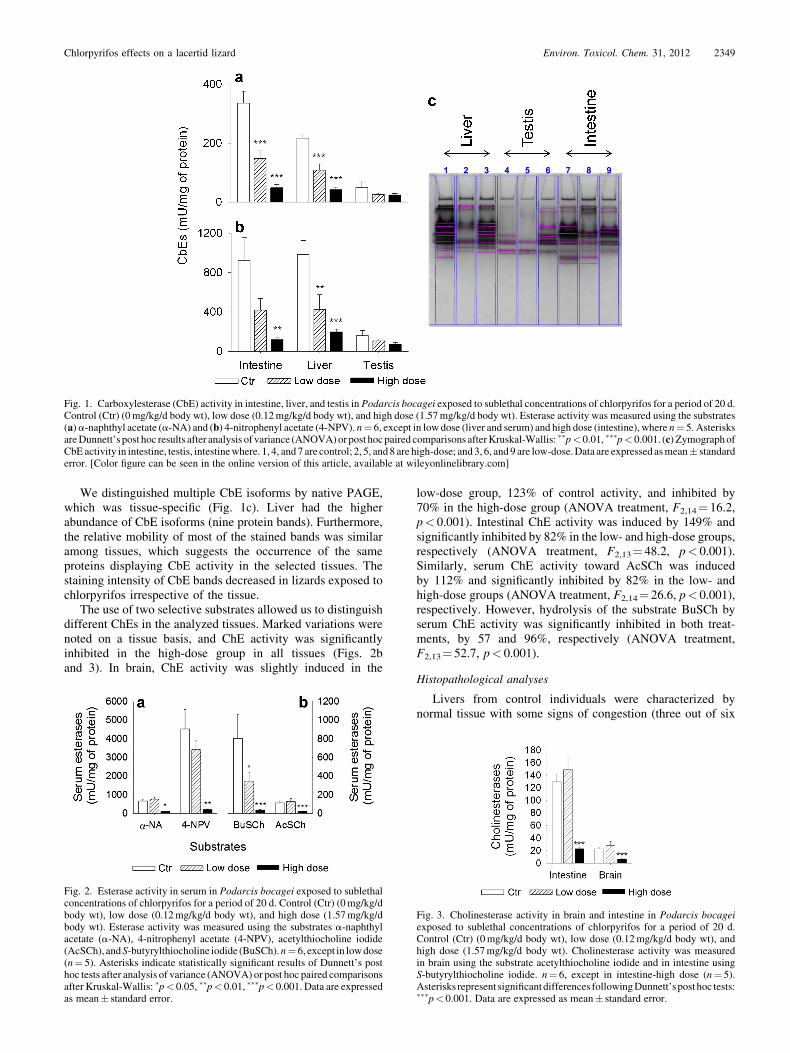
We distinguished multiple CbE isoforms by native PAGE,which was tissue-specific (Fig. 1c). Liver had the higherabundance of CbE isoforms (nine protein bands). Furthermore,the relative mobility of most of the stained bands was similaramong tissues, which suggests the occurrence of the sameproteins displaying CbE activity in the selected tissues. Thestaining intensity of CbE bands decreased in lizards exposed tochlorpyrifos irrespective of the tissue.
The use of two selective substrates allowed us to distinguishdifferent ChEs in the analyzed tissues. Marked variations werenoted on a tissue basis, and ChE activity was significantlyinhibited in the high-dose group in all tissues (Figs. 2band 3). In brain, ChE activity was slightly induced in the
low-dose group, 123% of control activity, and inhibited by70% in the high-dose group (ANOVA treatment, F2,14¼ 16.2,p< 0.001). Intestinal ChE activity was induced by 149% andsignificantly inhibited by 82% in the low- and high-dose groups,respectively (ANOVA treatment, F2,13¼ 48.2, p< 0.001).Similarly, serum ChE activity toward AcSCh was inducedby 112% and significantly inhibited by 82% in the low- andhigh-dose groups (ANOVA treatment, F2,14¼ 26.6, p< 0.001),respectively. However, hydrolysis of the substrate BuSCh byserum ChE activity was significantly inhibited in both treat-ments, by 57 and 96%, respectively (ANOVA treatment,F2,13¼ 52.7, p< 0.001).
Histopathological analyses
Livers from control individuals were characterized bynormal tissue with some signs of congestion (three out of six
Fig. 2. Esterase activity in serum in Podarcis bocagei exposed to sublethalconcentrations of chlorpyrifos for a period of 20 d. Control (Ctr) (0mg/kg/dbody wt), low dose (0.12mg/kg/d body wt), and high dose (1.57mg/kg/dbody wt). Esterase activity was measured using the substrates a-naphthylacetate (a-NA), 4-nitrophenyl acetate (4-NPV), acetylthiocholine iodide(AcSCh), andS-butyrylthiocholine iodide (BuSCh).n¼ 6, except in lowdose(n¼ 5). Asterisks indicate statistically significant results of Dunnett’s posthoc tests after analysis of variance (ANOVA) or post hoc paired comparisonsafter Kruskal-Wallis: �p< 0.05, ��p< 0.01, ���p< 0.001. Data are expressedas mean� standard error.
Fig. 3. Cholinesterase activity in brain and intestine in Podarcis bocageiexposed to sublethal concentrations of chlorpyrifos for a period of 20 d.Control (Ctr) (0mg/kg/d body wt), low dose (0.12mg/kg/d body wt), andhigh dose (1.57mg/kg/d body wt). Cholinesterase activity was measuredin brain using the substrate acetylthiocholine iodide and in intestine usingS-butyrylthiocholine iodide. n¼ 6, except in intestine-high dose (n¼ 5).Asterisks represent significantdifferences followingDunnett’spost hoc tests:���p< 0.001. Data are expressed as mean� standard error.
Fig. 1. Carboxylesterase (CbE) activity in intestine, liver, and testis in Podarcis bocagei exposed to sublethal concentrations of chlorpyrifos for a period of 20 d.Control (Ctr) (0mg/kg/d body wt), low dose (0.12mg/kg/d body wt), and high dose (1.57mg/kg/d body wt). Esterase activity was measured using the substrates(a)a-naphthyl acetate (a-NA) and (b) 4-nitrophenyl acetate (4-NPV). n¼ 6, except in low dose (liver and serum) and high dose (intestine), where n¼ 5. AsterisksareDunnett’s post hoc results after analysis of variance (ANOVA)or post hocpaired comparisons afterKruskal-Wallis: ��p< 0.01, ���p< 0.001. (c) ZymographofCbEactivity in intestine, testis, intestinewhere. 1, 4, and 7 are control; 2, 5, and 8 are high-dose; and3, 6, and 9 are low-dose.Data are expressed asmean� standarderror. [Color figure can be seen in the online version of this article, available at wileyonlinelibrary.com]
Chlorpyrifos effects on a lacertid lizard Environ. Toxicol. Chem. 31, 2012 2349
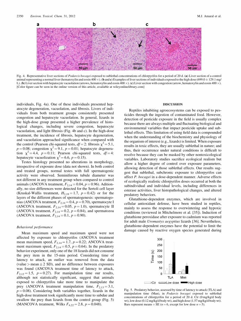
individuals, Fig. 4a). One of these individuals presented hep-atocyte degeneration, vacuolation, and fibrosis. Livers of indi-viduals from both treatment groups consistently presentedcongestion and hepatocyte vacuolation. In general, lizards inthe high-dose group presented a higher prevalence of histo-logical changes, including severe congestion, hepatocytevacuolation, and light fibrosis (Fig. 4b and c). In the high-dosetreatment, the incidence of fibrosis, hepatocyte degeneration,and vacuolation approached significance when compared withthe control (Pearson chi-squared tests, df¼ 2: fibrosis x2¼ 5.1,p¼ 0.08; congestion x2¼ 9.1, p¼ 0.01; hepatocyte degenera-tion x2¼ 4.4, p¼ 0.11; Pearson chi-squared tests, df¼ 4:hepatocyte vacuolization x2¼ 6.6, p¼ 0.15).
Testes histology presented no alterations in morphology,irrespective of exposure dose (data not shown). In both controland treated groups, normal testes with full spermatogenicactivity were observed. Seminiferous tubule diameter wasnot different in any treatment group when compared to controlanimals (ANCOVA treatment, F2,14¼ 0.04, p¼ 0.96). Adition-ally, no size differences were detected for the Sertoli cell layer(Kruskal-Wallis treatment, H2,18¼ 1.7, p¼ 0.42) or for thelayers of the different phases of spermatogenesis: spermatogo-nias (ANCOVA treatment, F2,14¼ 0.4, p¼ 0.70), spermatocyte I(ANCOVA treatment, F2,14¼ 0.05, p¼ 1.0), spermatocyte II(ANCOVA treatment, F2,14¼ 0.2, p¼ 0.84), and spermatozoa(ANCOVA treatment, F2,14¼ 0.1, p¼ 0.90).
Behavioral performance
Mean maximum speed and maximum speed were notaffected by exposure to chlorpyrifos (ANCOVA treatmentmean maximum speed, F2,14¼ 1.7, p¼ 0.22; ANCOVA treat-ment maximum speed, F2,14¼ 0.5, p¼ 0.64). In the predatorybehavior experiment, only one of the 18 lizards did not consumethe prey item in the 15-min period. Considering time oflatency to attack, an outlier was removed from the data(value>mean� 2 SD), and no difference between exposureswas found (ANCOVA treatment time of latency to attack,F2,12¼ 1.5, p¼ 0.27). For manipulation time our results,although not statistically significant, suggest that animalsexposed to chlorpyrifos take more time to manipulate theprey (ANCOVA treatment manipulation time, F2,13¼ 3.1,p¼ 0.08). Considering both variables together, lizards in thehigh-dose treatment took significantly more time to subdue andswallow the prey than lizards from the control group (Fig. 5)(MANCOVA treatment, Wilks F4,22¼ 2.8, p¼ 0.048).
DISCUSSION
Reptiles inhabiting agroecosystems can be exposed to pes-ticides through the ingestion of contaminated food. However,detection of pesticide exposure in the field is usually complexbecause there are always multiple and fluctuating biological andenvironmental variables that impact pesticide uptake and sub-lethal effects. This limitation of using field data is compoundedwhen the understanding of the biochemistry and physiology ofthe organism of interest (e.g., lizards) is limited. When exposureresults in toxic effects, they are usually sublethal in nature; andthus, their occurrence under natural conditions is difficult toresolve because they can be masked by other nontoxicologicalvariables. Laboratory studies sacrifice ecological realism butallow a higher degree of control over exposure parameters,allowing detection of those sublethal effects. Our results sug-gest that sublethal, subchronic exposure to chlorpyrifos canaffect P. bocagei in a dose-dependent manner. Adverse effectsof ecologically realistic chlorpyrifos doses occurred at both thesubindividual and individual levels, including differences inesterase activities, liver histopathological changes, and alteredpredatory behaviors.
Glutathione-dependent enzymes, which are involved incellular antioxidant defense, have been studied in reptiles,mainly to assess the response to overwintering and hypoxicconditions (reviewed in Mitchelmore et al. [35]). Induction ofglutathione peroxidase after exposure to cadmium was reportedfor adult male Uromastyx aegyptius lizards [36]. Nevertheless,glutathione-dependent enzymes have the potential to limit thedamage caused by reactive oxygen species generated during
Fig. 4. Representative liver sections of Podarcis bocagei exposed to sublethal concentrations of chlorpyrifos for a period of 20 d. (a) Liver section of a controlanimal representing a normal liver (hematoxylin and eosin 400�). (b and c) Examples of liver sections of individuals exposed to the high dose (699.0� 129.1mg/L). (b) Liver sectionwith hepatocyte vacuolation (arrows, hematoxylin and eosin 400�). (c) Liver sectionwith congestion (arrow, hematoxylin and eosin 400�).[Color figure can be seen in the online version of this article, available at wileyonlinelibrary.com]
Fig. 5. Predatory behavior, assessed by time of latency to attack (TLA) andmanipulation time (Man), in Podarcis bocagei exposed to sublethalconcentrations of chlorpyrifos for a period of 20 d. Ctr (0mg/kg/d bodywt), lowdose (0.12mg/kg/d bodywt), and high dose (1.57mg/kg/d bodywt).Bars represent meansþSE (n¼ 6, except for low dose n¼ 5).
2350 Environ. Toxicol. Chem. 31, 2012 M.J. Amaral et al.

pesticide-detoxification processes and have been used in severalstudies to assess pesticide exposure and effects (reviewed forfish in van der Oost et al. [37]). Lipid peroxidation has been usedas a quantitative measure of the extent of oxidative damagecaused by contaminants. In the present study, we found no signsof oxidative stress caused by chlorpyrifos exposure. This iscontrary to the results of a study in rats, for which a dose of6.75mg/kg body weight for a period of 28 d resulted in asignificant increase in serum lipid peroxidation and decreasedsignificantly the activity of serum GST [38]. Another rat studyalso reported accumulation of malondialdehyde in differenttissues and a dose-dependent decrease in antioxidant enzymesafter 1 d of exposure to 38.8mg/kg body weight [3]. In bothstudies, rats were orally exposed to a higher dosage of chlor-pyrifos than the higher one in the present study.
Chlorpyrifos exposure resulted in the inhibition of CbEsin all tissues analyzed in a dose-dependent manner, except forserum CbE, which was strongly depressed at the higher dose ofthe OP. Inhibition of CbE activity has been used as a biomarkerof OP exposure in different species, and several authors havesuggested that CbEs can have a protective role by bindingstoichiometrically to OP pesticides, decreasing OP concentra-tion and toxicity [39,40]. Thus, effects of pesticide exposure canbe alleviated in different tissues by the levels of CbE activity, itssensitivity to chlorpyrifos inhibition, and the presence of multi-ple isozymes [39]. Different studies have also suggested thatCbE activity could be a more sensitive biomarker than braincholinesterase activity, the most commonly used biomarker ofOP exposure [41]. It is widely accepted that CbE activityprovides a protective effect against OP exposure because ofits higher sensitivity to inhibition by these agrochemicalscompared to ChE activity [41]. Results in the present studysupport the aforementioned effect. Animals exposed to thelower dose of chlorpyrifos showed significant inhibition ofCbE activity in the liver and intestine, whereas this depressionwas not found in the brain and intestine ChE activities (Figs. 1and 3). Sanchez-Hernandez and Wheelock [40] suggested thatintestinal CbEs would have an important role in OP detoxifi-cation as one of the first surfaces of contact for feedingexposure. These results reinforce the need to study differenttissues and substrates as different isozymes seem to be involvedin the hydrolysis of the different substrates in multiple tissues.The only tissue where no significant differences were found forCbE activity was the testis, although a general trend of activitydepression with increased dose was observed. Several studieswith mammals and other organisms have demonstrated thatCbEs have an important role in reproduction by altering themetabolic routes of testosterone production [5]. In the presentstudy, enzymatic activity was dependent on tissue and substrate.The protective role of CbEs was not clear in serumCbE activity,but it was apparent in both liver and intestine CbE activities.Liver CbEs were strongly inhibited after exposure to both dosesof chlorpyrifos. Similarly, intestinal CbE activity was alsodepressed. However, we detected higher intestinal CbE activityin control than in liver with the a-Na substrate.
Cholinesterases, in particular AChE, have been intensivelystudied as biomarkers of pesticide exposure and effect, inparticular for OP and carbamate pesticides. In reptile brains,AChE is expected to represent >90% of total ChEs (S.R.Schmidt, 2003, Master’s thesis, Texas Tech University,Lubbock, TX, USA). The relationship between AChE inhibitionand death has been intensively debated. Reductions of 50 to80% in AChE activity have been related to mortality in differentvertebrate species (e.g., in birds [42]). In the present study,
inhibition levels of 70% of brain ChE activity recorded in thegroup exposed to the high dose did not result in the death ofany individual. These levels of inhibition are similar to thosedescribed for the lizard Gallotia gallotia after acute exposure toparathion, where ChE inactivation levels of 84 and 70% wereobserved without mortality [43].
Even if not lethal, chlorpyrifos can interfere with brain ChEsand the activity of the central nervous system, impairingessential behavioral functions, like predator–prey interactions[44]. Cholinesterases in other tissues, such as blood, have beensuggested to have, like CbEs, a protective role over brain ChE,detoxifying OPs before they reach the brain [41]. Differentstudies in lizards have further compared the levels of ChE inthose tissues (mainly serum) and found them to be correlatedwith those of brain ChE [43]. In the present study, this relation-ship could only be established with serum ChE (assessed withBuSCh substrate). In reptiles, 80% of the total ChE activity inserum is attributable to BChE activity [11,13]. Serum ChE(BuSCh substrate) was inhibited at the low dose before anyeffect was observed in brain ChE. Cholinesterase in intestineand serum (AcSCh substrate) was not inhibited at the low dose.At the high dose, ChEs were strongly inhibited in all tissues, soany protective role would be lost. Sanchez-Hernandez et al. [43]obtained similar results to the findings presented here, whichshow an exponential decay of AChE activity when serum BChEreached 90% of inhibition.
We detected a higher prevalence of liver histopathologicalchanges in animals exposed to chlorpyrifos. These changesoccurred mainly in the high-dose group and included fibrosis,hepatocyte degeneration, and vacuolation. Despite being stat-istically nonsignificant, these alterations might be indicativeof metabolic stress. Studies in rats have demonstrated a linkbetween exposure to chlorpyrifos and the appearance of similarlesions [38,45]. Toxic effects of chlorpyrifos on testicularfunction have also been noted in rats [5,9]. Nevertheless, inthe present study, no effects were noted. Normal testicularhistology with all the successive stages of spermatogenesiswas observed in the three groups.
If exposure to pesticides results in behavioral alterations,growth, reproductive success, and survival might be affected.Locomotor performance is probably the individual parameterthat has been employed most in studies with reptiles exposed toinsecticides. Nevertheless, these studies seem to indicate thatthis flight response is not a parameter sufficiently sensitive toaddress pesticide exposure [46,47]. In the present study, theresults similarly show this test to be an insensitive biomarker ofchlorpyrifos exposure, with no difference between control andtreated animals. In contrast, predatory behaviors provide a moresensitive indicator of locomotor impairment because theyinvolve multiple neuromuscular systems, rather than simplya sympathetically controlled, reflexive flight response. Bainet al. [13] observed that animals exposed to a high dose offenitrothion tended to make more attempts to catch prey.The authors speculated that this type of response could berelated to decreased visual acuity, changes in muscle activity,or decreased muscle coordination [13]. We found that afterexposure to chlorpyrifos, animals took more time to capture andswallow a prey item, despite prior habituation to the prey.Inhibition of ChEs, which was demonstrated in the sameanimals, provides a mechanistic explanation. Inhibition of AChEcauses accumulation of acetylcholine in synaptic junctions. Theaccumulation of acetylcholine can alter neuromuscular abilitiesand, thus, decrease the muscle coordination essential for themanipulation of prey.
Chlorpyrifos effects on a lacertid lizard Environ. Toxicol. Chem. 31, 2012 2351

We can presume that animals will recover when the exposureterminates. However, if we take into consideration the slow rateof esterase recovery that has been reported for reptiles [13,43],these results can represent a high exposure risk. Animals wouldbe impaired not only during the exposure period but also for asignificant period afterward. Insecticide application usuallyoccurs during spring and autumn, the period when animalsare acquiring energy for production and reproduction and tosurvive the winter. To fulfill energetic requirements, compro-mised individuals would have to spend more time foraging tocatch the same amount of prey. During foraging, lizards arepotentially at a higher risk of predation. Thus, we can speculatethat if an animal compensates for deficiencies in predatoryability by foraging for longer periods, it is increasing its risk ofpredation. On the other hand, if an animal does not compensate,then reduced feeding rates will have implications for itsenergetic balance.
Our exposure rates were under the median lethal concen-tration (LC50) values reported for several vertebrate speciesincluding birds (5–157mg/kg body wt) and mammals (151–1,000mg/kg body wt) [48]. For amphibians, the reported dosesare given as concentration in the aquatic medium and not easilycomparable, ranging from 2.4 to 66.2mg/L, depending on thespecies [48]. Given the lack of data, it is difficult to compare thetoxicity of chlorpyrifos to reptiles and other terrestrial verte-brates and, thus, evaluate if birds can be used in risk-assessmentprocesses as surrogate species.
CONCLUSIONS
In the present study, we demonstrated that a subchronicexposure to environmentally relevant doses of chlorpyrifos caninhibit esterases and impair predatory behaviors. It remains tobe seen if the biochemical and behavioral effects observed in thepresent study are likely to translate into any short- or long-termimplications for populations. Further work in the form of long-term mesocosm experiments will be needed to examine theseissues.
Acknowledgement—We appreciate the assistance of A. Kaliuntzopoulou, N.Sillero, andS. Freitas in animal collection.All animalswere collected under apermit issued by the Instituto da Conservacao da Natureza e Biodiversidade.This research was supported by FEDER through COMPETE-ProgramaOperacional Factores de Competitividade and by national funding throughFundacao para a Ciencia e Tecnologia (FCT), within the research projectLacertid Lizards as Bioindicators of Pesticide Exposure and Toxicity (LAB-PET) in intensivemarket garden agriculture (FCTPTDC/AMB/64497/2006)and through an FCT PhD grant to M. J. Amaral (SFRH/BD/31470/2006).
REFERENCES
1. Eurostat. 2007. The use of plant protection products in the EuropeanUnion:Data 1992–2003.Office forOfficial Publicationsof theEuropeanCommunities, Luxembourg, p. 222.
2. Casida JE. 2009. Pest toxicology: The primary mechanisms of pesticideaction. Chem Res Toxicol 22:609–619.
3. Ojha A, Yaduvanshi SK, Srivastava N. 2011. Effect of combinedexposure of commonly used organophosphate pesticides on lipidperoxidation and antioxidant enzymes in rat tissues. Pestic BiochemPhysiol 99:148–156.
4. YinX, ZhuG,LiX, LiuS. 2009.Genotoxicity evaluation of chlorpyrifosto amphibian Chinese toad (Amphibian: Anura) by comet assay andmicronucleus test. Mutat Res 680:2–6.
5. Joshi SC, Mathur R, Gulati N. 2007. Testicular toxicity of chlorpyrifos(an organophosphate pesticide) in albino rat. Toxicol Ind Health 23:439–444.
6. Tierney K, Casselman M, Takeda S, Farrell T, Kennedy C. 2007.The relationship between cholinesterase inhibition and two typesof swimming performance in chlorpyrifos exposed coho salmon(Oncorhynchus kisutch). Environ Toxicol Chem 26:998–1004.
7. Widder PD, Bidwell JR. 2008. Tadpole size, cholinesterase activity,and swim speed in four frog species after exposure to sub-lethalconcentrations of chlopryrifos. Aquat Toxicol (Amst) 88:9–18.
8. Giesy JP, Solomon KR, Coats JR. 1999. Chlorpyrifos: Ecological riskassessment in North American aquatic environments. Rev EnvironContam Toxicol 160:1–129.
9. Akhtar N, Srivastava MK, Raizada RB. 2009. Assessment ofchlorpyrifos toxicity on certain organs in rat, Rattus norvegicus.J Environ Biol 30:1047–1053.
10. Hall RJ, Clark DR. 1982. Responses of the iguanid lizard Anoliscarolinensis to four organophosphorus pesticides. Environ Pollut 28:45–52.
11. Sanchez-Hernandez JC. 2003. Evaluating reptiles exposure to chol-inesterase-inhibiting agrochemicals by serum butyrylcholinesteraseactivity. Environ Toxicol Chem 22:296–301.
12. Ozelmas U, Akay MT. 1995. Histopathological investigations of theeffects of malathion on dwarf lizards (Lacerta parva, Boulenger 1887).Bull Environ Contam Toxicol 55:730–737.
13. Bain D, Buttemer WA, Astheimer L, Fildes K, Hooper MJ. 2004.Effects of sublethal fenitrothion ingestion on cholinesterase inhibition,standard metabolism, thermal preference, and prey-capture ability inthe Australian central bearded dragon (Pogona vitticeps, Agamidae).Environ Toxicol Chem 23:109–116.
14. Weir SM, Suski JG, Salice CJ. 2010. Ecological risk of anthropogenicpollutants to reptiles: Evaluating assumptions of sensitivity andexposure. Environ Pollut 158:3596–3606.
15. Villeneuve DL, Garcia-Reyero N. 2011. Vision & strategy: Predictiveecotoxicology in the 21st century. Environ Toxicol Chem 30:1–8.
16. Ankley GT, Bennett RS, Erickson RJ, Hoff DJ, Hornung MW,Johnson RD, Mount DR, Nichols JW, Russom CL, Schmieder PK,Serrrano JA, Tietge JE, Villeneuve DL. 2010. Adverse outcomepathways: A conceptual framework to support ecotoxicology researchand risk assessment. Environ Toxicol Chem 29:730–741.
17. Kramer VJ, Etterson MA, Hecker M, Murphy CA, Roesijadi G,Spade DJ, Spromberg JA, Wang M, Ankley GT. 2011. Adverseoutcome pathways and ecological risk assessment: Bridging topopulation-level effects. Environ Toxicol Chem 30:64–76.
18. Carretero MA. 2004. From set menu to a la carte. Linking issuesin trophic ecology of Mediterranean lacertids. Ital J Zool 2:121–133.
19. Fossi MC, Sanchez-Hernandez JC, Diaz-Diaz R, Lari L, Garcia-Hernandez JE, Gaggi C. 1995. The lizard Gallotia galloti as abioindicator of organophosphorus contamination in the Canary Islands.Environ Pollut 87:289–294.
20. Mann RM, Sanchez-Hernandez JC, Serra EA, Soares AMVM. 2007.Bioaccumulation of Cd by a European lacertid lizard after chronicexposure to Cd-contaminated food. Chemosphere 68:1525–1534.
21. CardoneA,ComitatoR,Angelini F. 2008. Spermatogenesis, epididymismorphology and plasma sex steroid secretion in themale lizardPodarcissicula exposed to diuron. Environ Res 108:214–223.
22. Marsili L, Casini S, Mori G, Ancora S, Bianchi N, D’Agostino A,Ferraro M, Fossi MC. 2009. The Italian wall lizard (Podarcis sicula)as bioindicator of oil field activity. Sci Total Environ 407:3597–3604.
23. Brewer LW,McQuillenHL Jr,MayesMA, Stafford JM, Tank SL. 2003.Chlorpyrifos residue levels in avian food items following applicationsof a commercial EC formulation to alfalfa and citrus. Pest Manag Sci59:1179–1190.
24. BradfordMM. 1976. A rapid and sensitivemethod for the quantificationof microgram quantities of protein utilizing the principle of protein dyebinding. Anal Biochem 72:248–254.
25. Anastassiades M, Lehotay SJ, Stajnbaher D, Schenck FJ. 2003. Fast andeasymultiresiduemethod employing acetonitrile extraction/partitioningand ‘‘dispersive solid-phase extraction’’ for the determination ofpesticide residues in produce. J AOAC Int 86:412–431.
26. Habig WH, Pabst MJ, Jakoby WB. 1974. Glutathione S-transferases.J Biol Chem 249:7130–7139.
27. Ramos-Martinez JI, Bartolome TR, Pernas RV. 1983. Purification andproperties of glutathione reductase from hepatopancreas of Mytilusedulis L. Comp Biochem Physiol B Comp Biochem 75:689–692.
28. Hissin PJ, Hilf R. 1976. A fluorometric method for determinationof oxidized and reduced glutathione in tissues. Anal Biochem 74:214–226.
29. Ohkawa H, Ohishi N, Yagi K. 1979. Assay for lipid peroxides in animaltissues by thiobarbituric acid reaction. Anal Biochem 95:351–358.
30. Gomori G, Chessick RD. 1953. Esterases and phosphatases of the brain.A histochemical study. J Neuropathol Exp Neurol 12:387–396.
31. Bunyan PJ, Jennings DM, Taylor A. 1968. Organophosphorus poison-ing. J Agric Food Chem 16:326–331.
2352 Environ. Toxicol. Chem. 31, 2012 M.J. Amaral et al.

32. Carr RL, Chambers JE. 1991. Acute effects of the organophosphateparaoxon on schedule-controlled behavior and esterase activity in rats:Dose–response relationships. Pharmacol Biochem Behav 40:929–936.
33. EllmanGL, CourtneyKD,AndresV Jr, Feather-StoneRM. 1961.A newand rapid colorimetric determination of acetylcholinesterase activity.Biochem Pharmacol 7:88–95.
34. HolemRR,HopkinsWA,Talent LG. 2008. Effects of repeated exposureto malathion on growth, food consumption, and locomotor perfomanceof the western fence lizard (Sceloporus occidentalis). Environ Pollut152:92–98.
35. Mitchelmore CL, Rowe CL, Place AR. 2006. Tools for assessingcontaminant exposure and effects in reptiles. InGardnerSC,OberdorsterE, eds Toxicology of Reptiles. Taylor & Francis, Boca Raton, FL, USA,pp 63–122.
36. Al-Johany AM, Haffor AS. 2009. Effects of cadmium exposure on theultrastructural pathology of different pulmonary cells, leukocyte count,and activity of glutathione peroxidase and lactate dehydrogenase inrelation to free radical production in Uromastyx aegyptius. UltrastuctPathol 33:39–47.
37. van der Oost R, Beyer J, Vermeulen NPE. 2003. Fish bioaccumulationand biomarkers in environmental risk assessment: a review. EnvironToxicol Pharmacol 13:57–149.
38. Mansour SA, Mossa A-TH. 2010. Oxidative damage, biochemical andhistopathological alterations in rats exposed to chlorpyrifos and theantioxidant role of zinc. Pestic Biochem Physiol 96:14–23.
39. Wheelock CE, Eder KJ, Werner I, Huang H, Jones PD, Brammell BF,Elskus AA, Hammock BD. 2005. Individual variability in esteraseactivity and CYP1A levels in Chinook salmon (Oncorhynchustshawytscha) exposed to esfenvalerate and chlorpyrifos. Aquat Toxicol(Amst) 74:172–192.
40. Sanchez-Hernandez JC, Wheelock CE. 2009. Tissue distribution,isozyme abundance and sensitivity to chlorpyrifos-oxon of carboxy-lesterases in the earthworm Lumbricus terrestris. Environ Pollut 157:264–272.
41. Wheelock CE, Phillips BM, Anderson BS, Miller JL, Miller MJ,Hammock BD. 2008. Applications of carboxylesterase activity inenvironmentalmonitoringand toxicity identification evaluations (TIEs).Rev Environ Contam Toxicol 195:117–178.
42. Ludke J, Hill E, Dieter M. 1975. Cholinesterase (ChE) response andrelated mortality among birds fed ChE inhibitors. Arch Environ ContamToxicol 3:1–21.
43. Sanchez-Hernandez JC,FossiMC,Focardi S. 1997.SerumBesterasesasa nondestructive biomarker in the lizardGallotia galloti experimentallytreated with parathion. Environ Toxicol Chem 16:1954–1961.
44. Hart ADM. 1993. Relationships between behavior and the inhibition ofacetylcholinesterase in birds exposed to organophosphorus pesticides.Environ Toxicol Chem 12:321–336.
45. Tripathi S, Srivastav AK. 2010. Liver profile of rats after long-termingestion of different doses of chlorpyrifos. Pestic Biochem Physiol97:60–65.
46. DuRant SE, Hopkins WA, Talent LG. 2007. Impaired terrestrial andarboreal locomotor perfomance in the wester fence lizard (Sceloporusoccidentalis) after exposure to an AChE-inhibiting pesticide. EnvironPollut 149:18–24.
47. Holem RR, Hopkins WA, Talent LG. 2006. Effect of acute exposure tomalathion and lead on sprint performance of the western fence lizard(Sceloporus occidentalis). Arch Environ Contam Toxicol 51:111–116.
48. Christensen K, Harper B, Luukinen B, Buhl K, Stone D. 2009.Chlorpyrifos technical fact sheet.NationalPesticide InformationCenter,Oregon State University Extension Services, Corvallis, OR, USA.
Chlorpyrifos effects on a lacertid lizard Environ. Toxicol. Chem. 31, 2012 2353