THÈSE Georgios PAVLOU
176
THÈSE Pour obtenir le grade de DOCTEUR DE LA COMMUNAUTE UNIVERSITE GRENOBLE ALPES Spécialité : Chimie Physique Moleculaire et Structurale Arrêté ministériel : 25 mai 2016 Présentée par Georgios PAVLOU Thèse dirigée par Isabelle TARDIEUX, Directeur de Recherche, Institute for Advanced Biosciences, Grenoble dans l'École Doctorale Chimie et Sciences du Vivant Toxoplasma gondii, un champion de course et un redoutable envahisseur. Etude des forces motrices et invasives Toxoplasma gondii, a super fast runner and cell invader. Studying motion and forces Thèse soutenue publiquement le «22 June 2020», devant le jury composé de : Isabelle, TARDIEUX Directeur de Recherche, Institute for Advanced Biosciences, Directeur de these Mohamed-ali, HAKIMI Directeur de Recherche, Institute for Advanced Biosciences, Président Ana-Maria, LENNON-DUMENIL Directeur de Recherche, Institute Curie, Examinateur Friedrich, FRISCHKNECHT Professeur, Center for Infectious Diseases Heidelberg University Medical School, Rapporteur Serge, MOSTOWY Professeur, Department of Infection Biology London School of Hygiene & Tropical Medicine, Rapporteur Genevieve, MILON Directeur de Recherche Emerite, Institute Pasteur, Examinateur
-
Upload
khangminh22 -
Category
Documents
-
view
2 -
download
0
Transcript of THÈSE Georgios PAVLOU

THÈSE Pour obtenir le grade de
DOCTEUR DE LA COMMUNAUTE UNIVERSITE GRENOBLE ALPES Spécialité : Chimie Physique Moleculaire et Structurale
Arrêté ministériel : 25 mai 2016
Présentée par
Georgios PAVLOU
Thèse dirigée par Isabelle TARDIEUX, Directeur de Recherche, Institute for Advanced Biosciences, Grenoble
dans l'École Doctorale Chimie et Sciences du Vivant
Toxoplasma gondii, un champion de course et un redoutable envahisseur. Etude des forcesmotrices et invasives
Toxoplasma gondii, a super fast runner and cell invader. Studying motion and forces
Thèse soutenue publiquement le «22 June 2020»,devant le jury composé de :
Isabelle, TARDIEUX Directeur de Recherche, Institute for Advanced Biosciences, Directeur de these Mohamed-ali, HAKIMI Directeur de Recherche, Institute for Advanced Biosciences, Président Ana-Maria, LENNON-DUMENIL Directeur de Recherche, Institute Curie, Examinateur Friedrich, FRISCHKNECHT Professeur, Center for Infectious Diseases Heidelberg University Medical School, Rapporteur Serge, MOSTOWY Professeur, Department of Infection Biology London School of Hygiene & Tropical Medicine, Rapporteur Genevieve, MILON Directeur de Recherche Emerite, Institute Pasteur, Examinateur

Toxoplasma gondii, a super fast runner and cell invader
Studying Motion and Forces
PhD Thesis Georgios Pavlou
Toxoplasma from Greek τόξον (toxon, "arc, bow") and πλάσμα (plasma, "shape, form")
Under supervision of Dr Isabelle Tardieux
Institute for Advanced Biosciences
Grenoble, France

Toxoplasma gondii, a super fast runner and cell invader
Studying motion and forces
PhD Thesis Georgios Pavlou
This document begins with a short introduction describing major biological
features of the protozoan microbe named Toxoplasma gondii/T. gondii with
special reference to (1) its main structural attributes, (2) its life cycle as a parasite
of warm-blooded metazoans including with the diseases it can cause to the hosts,
and (3) its fast replicating developmental stage called tachyzoite, with a focus on
the importance of motile behaviors along its life. We will then consider in more
details the mechanisms evolved by the T. gondii tachyzoite to efficiently navigate
within extracellular matrices or in between cells and then invade host cells, two
steps that determine the success of the T. gondii parasitic lifestyle and which we
have successively investigated during the PhD period. To this end, we will
provide a brief context and question presentation before recapitulating the main
highlights of the work and insisting on the techniques we implemented while the
corresponding research article under evaluation or recently published will follow.
To further flesh out the discussion chapter of these manuscripts, major issues
emerging from this work will be listed and analyzed in an additional section with
respect to the concepts, tools and techniques we could design for better
knowledge in the future. Finally, we briefly conclude with a perspective
mentioning different aspects of application the PhD work may lead to including
in fields beyond parasitology, thereby strengthening the importance of cross
feeding between fields and expertise for optimal chance of innovation and
promising end-product delivery to the society.

Contents
List of abbreviations
1. Introduction
1.1. The Apicomplexa Toxoplasma gondii, a member of the Alveolata group is the causative agent
of toxoplasmosis………….................................................................................................................2
1.2. T. gondii life cycle…………………………………………………………………………………..4
1.3. The T. gondii tachyzoite stage…………………………………………..........................................6
1.4. The T. gondii tachyzoite motile behaviors………………………………………………………..7
1.5. The T. gondii tachyzoite invasive behavior……………………………………………………...10
1.6. The T. gondii tachyzoite forms progeny, which eventually displays an egress behavior…….12
2. Chapters
2.1. Chapter 1 - Exploring mechanistically the complex choreography of T. gondii tachyzoite
movement……………………………………………………………………………………….....17
2.1.1. Context and questions………………………………………………………………………..17
2.1.2. Implementing methods to study the movement of T. gondii tachyzoite……………………..23
2.1.3. Main highlights of the study……………………………………………….…………………31
2.1.4. Publications…………………………………………………………………………………..31
2.1.5. Discussion and perspectives………………………………………………………….............51
2.2. Chapter 2 – Investigating host cell invasion by the T. gondii tachyzoite with a focus on the
late steps of the process.…………………………………………………………………………..542.2.1. Context and questions…………………………………………………………......................542.2.2. Implementing methods to study the cell invasion by T. gondii tachyzoite…………………..602.2.3. Main highlights of the study………………………………………………………….............632.2.4. Publications…………………………………………………………......................................642.2.5. Discussion and perspectives…………………………………………………………...........113
3. Conclusion…………………………………………………............................................................116
4. Other publications……………………………………………....................................................118
5. Acknowledgements…………………………………………………………..............................161
6. References…………………………………………………………………………………………163

Abbreviations
ALIX ALG-2-interacting protein X
AMA Apical membrane antigen
CIN Cbl-interacting
DAG Diacylglycerol
DALYs Disability adjusted life years
DC Dendritic cell
DG Dense granules
DNM Dynamin
ECM Extracellular matrix
EDC 1-ethyl-3- [3-dimethylaminopropyl] carbodiimide
ELC Essential light chain
ESCRT Endosomal sorting complex required for transport
FCS Fetal calf serum
HA Hyaluronan
HAART Highly active antiretroviral therapy
IMC Inner membrane complex
IFN-γ Interferon gamma
IP3 Inositol triphosphate
F-actin Filamentous actin
GAC Glideosome-associated connector
GAP Gliding-associated protein
GAPM Glideosome-associated protein with multiple-membrane spans
GED GTPase effector domain
mC mCherry
MD Middle domain
MEF Mice embryonic fibroblasts
MIC Microneme protein
MIP Maximum intensity projection
MLC Myosin light chain
MTOC Microtubule organizing center
Myo Myosin
NK Natural killer
PAA Polyacrylamide
PEG poly(ethylene glycol)
PKA Protein kinase A
PKG Protein kinase G
PHD Pleckstrin homology domain
PLL-g-PEG poly(L-lysine)-graft-poly(ethylene glycol)
PM Plasma membrane
PRD Proline-rich domain
PV Parasitophorous vacuole

PVM Parasitophorous vacuole membrane
qFSM Quantitative fluorescent speckle microscopy
RB Residual body
RICM Reflection Interference Contrast Microscopy
ROM Rhomboid protease
RON RhOpry Neck Protein
TIRF Total internal reflection fluorescence
TFM Traction force microscopy
Tg Toxoplasma gondii
ZCJ Zoite cell junction

Introduction
1

1. Introduction
1.1 The Apicomplexa Toxoplasma gondii, a member of the Alveolata group is
the causative agent of toxoplasmosis
Toxoplasma gondii is a single-celled eukaryote and one of the thousands
members of the Apicomplexa phylum. This large and diverse phylum is part of a
higher order group of protozoans known as Alveolata, which is also composed of
the ciliates, a set of micropredators and the marine phytoplankton Dinoflagellates.
Alveolata are characterized by flattened vesicle-like structures lying beneath the
plasma membrane defined as cortical alveoli and in the case of Apicomplexa are
termed Inner Membrane Complex/IMC (see Figure 2 in the next section 1.3).
The Apicomplexa name stems from a unique complex localized at the apical pole
that typifies the motile stages, also called zoites, of these protozoans (Levine
1970). Another unifying trait across the Apicomplexan species is the parasitic
lifestyle they have evolved since they strictly rely on hosting cells to produce
progeny (Dubey, 1998). Among the Apicomplexa members already identified, T.
gondii is often seen as one of the most successful because of an impressive ability
to parasitize virtually all warm-blooded wild and domestic animals, and this in all
terrestrial ecosystems. In addition to be quite cosmopolitan and to accommodate a
broad range of hosts, T. gondii multiplies in a vast repertoire of cells from
different lineages in vivo and parasitizes all nucleated cells under in vitro
conditions. In line with these features and according to serological surveys, about
a third of the human population has been exposed to T. gondii (Halonen & Weiss,
2013) and the prevalence rate is even higher in areas of Europe and South
America. The serological markers used in the surveys inform on the proportion of
people that have been in contact with the fast replicating tachyzoite stage during
the acute but subsequently controlled expansion of the T. gondii population.
However, they are also considered as fair indicators for the presence of the slow
replicating, hence a long-term persisting parasite population, which takes over the
acute phase. The persistent population is represented by the T. gondii
developmental stage called bradyzoite that settle in tissue cells away from the
initial intestinal site of colonization, especially in cells from the nervous central
and peripheral systems, but also in skeletal and cardiac muscles.
At the clinical level, the acute phase of infection remains subclinical and the long-
term infection, often referred as to the chronic phase, is perceived as clinically
quasi silent with little or no direct harmful impact in healthy humans. Of note,
severe cases of toxoplasmosis in immunocompetent persons were reported from
South America and were assigned to several T. gondii atypical strains (Carme et
al, 2002). Of higher clinical concern are the transient or prolonged dysfunctions
of the immune system that can cause life-threatening or life-debilitating
toxoplasmosis- related diseases. As examples, when patients are under
2

immunosuppressive anti-cancer chemotherapy, anti-auto-immune therapy or anti-
graft rejection protocols post organ transplantation, the “silent” bradyzoites can
revert back to non-silent tachyzoites through a differentiation process termed “
parasite reactivation”. The massive replication of newly converted tachyzoites
can induce irreversible damages in sites of parasite persistence and lead to lethal
disseminated infection unless pharmacologically controlled (Dubey, 1996). Of
note, the reverse bradyzoite-tachyzoite conversion and its consequences were a
leading cause of the AIDS-related encephalitis in HIV infected
immunocompromised people (Luft & Remington, 1992) prior to the development
of Highly Active AntiRetroviral Therapy (HAART) but it still remains after
HAART a common cause of cerebral focal lesion in AIDS patients, and the third
most frequent AIDS-defining condition in developing countries (Colombo et al,
2005). In the same line, recent metadata analysis based on 74 studies that has
scanned in total 25 989 HIV-infected people from 34 countries, highlighted the
high prevalence of Toxoplasma-HIV co-infection (25 to 60%) especially in North
Africa and the Middle East (Wang et al, 2017).
Aside from pathology- or therapy-induced immunosuppressive situations,
pregnancy is another case in which the immune balance is shifted towards an
immunosuppressive state, compatible with the establishment of fetomaternal
tolerance, and thus development of the fetus in the uterus while the mother’s
immune system remains functional. While the fetus can be naturally protected
from harmful microbes thanks to the transfer of maternal antibodies across the
placenta throughout the umbilical cord, the same route can also promote fetus
colonization by microbes including T. gondii tachyzoites. Interestingly, the first
known photographic images of T. gondii were published in 1923 by Josef Janku
taken from the retina of an infant suffering from congenital toxoplasmosis
(McGovern & Wilson, 2013). In the last years, the global annual incidence of
congenital toxoplasmosis was estimated about 200.000 cases which corresponds
to a burden of 1.20 million Disability Adjusted Life Years (DALYs) with the
highest values seen in South America and some Middle Eastern and low-income
countries (Torgerson & Mastroiacovo, 2013). DALYs represent the total number
of years lost to illness, disability, or premature death within a given population
(Murray et al, 2012).
In this context, primo-infection with T. gondii during the early stages of
pregnancy is associated with (i) a low probability of fetus contamination because
of the restricted size of maternal-fetal placenta interface but (ii) severe
consequences including abortion and stillbirth or severe neurological defects. In
contrast when primo-infection occurs at later stages of pregnancy, the risk of
congenital toxoplasmosis for the fetus increases but the contamination leads to
less severe outcomes in particular chorioretinitis and uveitis (reviewed in Weiss
& Dubey, 2009). In addition, in the case of early infection (during the first
trimester of infection), the focal proliferation of tachyzoites was reported to
3

continue specifically in the fetal brain over time thus creating lethal damages
even when the maternal immune anti-Toxoplasma response has started to take
place and the maternal IgGs have crossed the placenta and reach the fetus
(Ferguson et al, 2013).
1.2 T. gondii life cycle
T. gondii, like many other Apicomplexa parasites, has a complex life cycle that
involves both asexually reproducing forms and sexual stages. Because these
stages are timely formed through unique differentiation processes - as already
mentioned for the tachyzoite and bradyzoite asexual stages during bi-directional
conversion- that occur in different host species, the T. gondii life cycle is
described as heteroxenous (Figure 1). By definition, the hosts in which parasitic
organisms can sexually reproduce are classified as definitive hosts and those
supporting only asexual reproduction are the intermediate hosts. T. gondii can
sexually reproduce only in felids (i.e. the definitive T. gondii hosts) while all the
other warm-blooded animals are intermediate hosts. Of note many intermediate
hosts including birds, rodents and other mammals are typical preys for the felids
and contribute to the T. gondii genetic diversity potentially generated in the felids.
Aside from the tachyzoite and bradyzoite stages mentioned above, the sporozoite
is the third infectious stage of T. gondii. This stage is formed in the protecting
oocyst which itself derives from the fertilization of female gamete and the
resulting zygote in the felid intestine. After fertilization, the zygote evolves in an
oocyst, which is released from the host enterocyte into the intestine lumen and
further shed in the environment. Upon appropriate aeration and temperature
conditions, the unsporulated oocyst converts into a sporulated oocyst, which
consists of two sporocysts and each of these contains four sporozoites (Dubey et
al, 1970).
Once a potential host (definitive or intermediate) ingests T. gondii oocysts
together with unwashed raw vegetable or contaminated water, the sporozoite is
next released from the oocyst following mechanical and enzymatic disruption of
the envelope, a process, which mainly takes place in the host stomach. Ending its
journey in the host intestinal tractus, the sporozoites invade enterocytes of the
ileum (small intestine) and differentiate into the tachyzoite stage that rapidly
amplifies the parasite population in cells from the gut mucosa. In this multi-
component mucosa, resident immune cells, in particular the CD8α+dendritic cells
(DCs) together with chemotactically recruited monocytes and neutrophils are
highly permissive to tachyzoite multiplication (Mowat & Agace, 2014).
Importantly some of these leucocytes are early sources of interleukin 12, a
cytokine that elicits among other cells, natural killer (NK) and CD4+and CD8
+T
4

cells to produce interferon gamma (IFN-γ) which orchestrates the host resistance
to T. gondii in hematopoietic and non-hematopoietic cells through the activation
of toxoplasmacidal activities (Hunter & Sibley, 2012). Remarkably, this
“vigorous innate immune response” ensures significant control of the replicating
parasite population. However, it concomitantly promotes the survival of a
subpopulation that exits from the inflamed site and reaches distant sites to further
reside in tissue cells protected from a too harsh immune surveillance, in particular
the brain and muscles. In these distant tissues, the tachyzoite undergoes
bradygenesis, which drives the formation of a cyst enclosing bradyzoites that
accounts for the long-term parasitism phase and was first described in 1908 by
Splendore (McGovern & Wilson, 2013). In a close scenario, once a potential
carnivorous host (definitive or intermediate) ingests food contaminated with T.
gondii cysts, and following cyst wall disruption in the stomach, the bradyzoites
colonize cells from the host small intestine and differentiate into tachyzoites. The
tachyzoite population expansion and its reduction concomitant to the formation
bradyzoite cysts parallels the scheme described for the sporozoite-mediated gut
infection. In conclusion, the relatively recent discoveries of the oral mode of
transmission by two distinct parasite stages, each protected by a cyst wall, were
achieved separately in the 1955 to 1970 years and can explain the wide
geographical distribution of T. gondii (Ferguson, 2009).
Figure 1. Schematic
representation of the T.
gondii life cycle.
Warm-blooded metazoans
ingest either encysted-
bradyzoites or sporozoites
of T. gondii. The two
parasite developmental
stages are released out of
their respective cyst
envelope in the host
stomach and infect the
epithelial cells from the
ileum part of the intestine.
They rapidly transform into
highly replicative
tachyzoites in the intestinal mucosa. The innate immune system further controls the
expansion of the tachyzoite population but also promotes the colonization of intestine-
distant tissues. In these tissues, tachyzoites differentiate as slow replicating bradyzoites
in long lasting cysts, which are taken up by new hosts through carnivorism and the same
5

cycle repeats. However, in felids that feed upon contaminated preys, gametes are formed
in the intestine through a complicated set of developmental stages and they produce
zygotes that evolve as oocysts, which are released in the environment with the felid
feces. There they sporulate and can be taken by herbivores while feeding or by both
carnivores and herbivores while drinking oocyst-contaminated water. Adapted from
Hunter & Sibley, 2012.
1.3 The T. gondii tachyzoite stage
Figure 2. The T. gondii tachyzoite. (A) Schematic representation showing the main
structural elements and organelles from left to right the conoid and the polar ring from
which emerge the cortical twisted microtubules, the microneme and the rhoptry secretory
vesicles, the dense granules scattered within the cytoplasm, the mitochondrion nearby the
apicoplast and the nucleus. The three layers of membrane that form the pellical and the
intramembrane particles connecting the formers are indicated. (B) Electron micrograph
showing an intracellular tachyzoite. (C) Scanning electron micrograph of an extracellular
tachyzoite with an extruded conoid. Adapted from Carmen et al, 2016. The three
parasites are oriented with the apical pole on the left side. (A) and (B) adapted from
Heintzelman, 2006.
T. gondii took its name from the Greek Toxo (τόξο = bow) and plasma (πλάσµα
= body) since it was initially described by Nicolle and Manceaux in 1908 who
observed in the rodent named “gundii”, extracellular and intracellular unicellular
organisms with a bow shape and measuring about 5-8 µm in length and 2-3 µm
in width. This description corresponds to the developmental stage called
tachyzoite years later in 1973 by Frenkel based on the rapid (tachos = ταχός, fast
in Greek), the biology of which has been since decades heavily studied. A major
characteristic of the tachyzoite lies in the robust polar architecture organized
around a distinct apical pole that houses a unique microtubule-made appendage
6

called conoid and two sets of secretory organelles named rhoptries, and
micronemes, respectively (Figure 2). A wealth of these products has already been
identified and a repertoire found involved to promote cell movement and host cell
invasion (Frénal et al, 2017). As such they will be highlighted in the “context and
question section” opening each result chapter of this document. Central to the
apical complex and connected to the conoid is the so-called apical polar ring that
serves as an atypical MicroTubule Organizing Center (MTOC) for the nucleation
of 22 unusually stable subpellicular MicroTubules (MTs) which elongate as
spirals until about two third of the parasite length. The MTs tightly subtend the
alveoli/IMC that lines most of the plasma membrane (Liu et al, 2016). The IMC
is made of flattened vesicles aligned in longitudinal rows and joined in a
patchwork fashion by sutures.
Briefly, other secretory vesicles called Dense Granules (DGs) populate the whole
cell cytoplasm together with the nucleus, the single mitochondria, the Golgi
apparatus and the Apicomplexa-restricted organelle called the apicoplast. The
apicoplast term was coined from APICOmplexan PLASTid since it was
recognized as a vestigial non-photosynthetic plastid homologous to the
chloroplasts of algae and plants acquired through endosymbiosis (McFadden &
Yeh, 2017).
Importantly the shape and “rigidity” characterizing the extra-cellular tachyzoite is
primarily determined by the apical complex together with the juxtaposed IMC
and the cortical MTs as revealed by the production of parasites expressing tubulin
or IMC mutants (Ma et al, 2007)(Barkhuff et al, 2011). Reinforcing this
conclusion is the recent identification of IMC integral proteins that typically carry
multiple membrane spans (the so-called GAPM) and which significantly
contribute to the MT stable curved shape (Harding et al, 2019).
1.4 The T. gondii tachyzoite motile behaviors
Motility is critical for T. gondii survival since once the tachyzoite stage emerges
in the intestinal cells through differentiation of the bradyzoite or the sporozoite
stages, it further rapidly expands its population in the intestinal mucosa prior to
spread in the draining lymph node likely via the lymphatic system but also distant
tissues. Therefore the tachyzoite needs to (i) navigate in the extracellular matrices
composing tissues to access a growth permissive host cell, (ii) invade a host cell
where to form progeny and (iii) egress from host cells, as schematized below
(Figure 3).
7

Figure 3. The intracellular-extracellular
life cycle of the T. gondii tachyzoite.
Adapted from Hortua Triana et al, 2018.
To initiate and sustain a motile behavior, the tachyzoite utilizes a very unique
actomyosin motor, which despite the notorious difficulties in visualizing the short
actin filaments, has been located within the 20- to 30-nm space between the IMC
and PM. Together with actin, the divergent class XIV of myosins that typifies the
Apicomplexa zoites but also specific scaffolding and regulatory molecules define
functional units called glideosomes (Soldati et al, 2004). Since these units are
known to govern force generation underpinning the tachyzoite motile and
invasive skills (Frénal et al, 2017), they will be more detailed in the specific
chapter introducing the study on motile forces.
Pioneer video-microscopic and kinematic studies using 2D conditions revealed
three main types of tachyzoite motile behaviors, all requiring attachment to a
substrate and all typically associated with body rotation along the long axis of the
cell (Frixione et al, 1996)(Håkansson et al, 1999). While the counterclockwise
helical and clockwise circular types of gliding are considered as productive since
they ensure forward cell migration, the third complex set of clockwise motions
grouped as twirling defines a non productive -i.e. stationary- motion. In contrast
to what has been initially proposed, helical motility is not the only behavior that
allows subsequent cell invasion. Actually, our team analyzed the preinvasive
behavior of several hundreds of tachyzoites and found that most frequently, the
tachyzoite executes only a short motion, referred to as “minimal impulse” that
associates with extrusion of conoid and concomitant body extension prior to
invade the cell (Bichet et al, 2014). At about the same time, two elegant settings
were designed by the G. Ward’ laboratory enabling for the first time to monitor
how tachyzoites move in 3D first in vitro using the matrigel, a reconstituted
extracellular matrix derived from a mouse sarcoma line and second, ex vivo in a
mouse earflat imaged by two-photon laser scanning microscopy (Leung et al,
2014). These live imaging studies demonstrate that under 3D conditions, the
tachyzoite moves only by making irregular corkscrew-like trajectories, which
closely recapitulate the 2D helical gliding (Figure 4).
8

Figure 4. T. gondii tachyzoite motility assay in 3 dimensions. Preparation of matrigel
and tachyzoites stained for nucleus with Hoechst 33342 inside the so-called “Pitta”
chambers. Maximum intensity projection (MIP) from the tracking of the nucleus
overtime illustrates the corkscrew-like movement of the tachyzoites. Adapted from
Leung et al, 2014.
Interestingly, a wealth of studies related to cell motility has been conducted in the
phylogenetically related Apicomplexa Plasmodium spp, the causative agents of
the devastating malaria in subtropical and tropical geographical areas. The
sporozoite is the developmental stage that is formed in the mosquito host, stored
in the insect salivary gland and eventually inoculated into the skin mammalian
host along with a blood feeding bite. The sporozoite has the ability to locomote in
a fairly similar way as the T. gondii tachyzoite and hence it shows fairly
conserved glideosome machinery which power gliding motility. However the
major sporozoite motile behavior on 2D substrates in vitro follows a circular
pattern whereas in the dermis, this motion is transformed to a complex non-linear
path (Amino et al, 2006)(Hellmann et al, 2011) which, over time shifts to
continuous circular pattern (Hopp et al, 2015). In contrast to the tachyzoite case,
no typical helical trajectory has been reported (Figure 5).
Figure 5. Plasmodium sporozoite
gliding pattern in vivo and in vitro.
Sporozoite motility within a time period
was monitored in salivary duct, skin,
hepatocyte and glass by reconstituting
the trajectory of the parasite. Adapted
from Münter et al, 2009.
9

1.5 The T. gondii tachyzoite invasive behavior
In line with the intracellular lifestyle, T. gondii has evolved strategies to invade a
host cell from which to steal the nutritive resources required to produce a progeny
within a growth permissive intracellular niche. Therefore, by securing the
offspring production, the invasion process appears as a crucial event.
To achieve active invasion of a metazoan cell in a minimal time frame, that does
not exceed a few tens of seconds, with a maximum chance of success, the parasite
relies on a tetrameric molecular complex of proteins pre-stored in the duct of the
rhoptry secretory organelles that abuts the conoid tip. The members of this
complex identified in 2009 are named RhOpry Neck Protein (RON2,4,5,8)
among which RON2 is the one that displays a trans-membrane domain (Besteiro
et al, 2009). The other RON members face the host cell cytoplasm and possibly
directly interact with components of the underlying cortical actin cytoskeleton
thereby promoting stabilization of the RON complex in site. As instances, RON8
was proposed to bind to the host F-actin cortical cytoskeleton and to provide the
necessary strength to anchor the invading tachyzoite because RON8-deficient
parasites do not seem to form stable grip and eventually fail to complete the
invasion process (Straub et al, 2011). The contribution of the host cortical actin
dynamics in the anchoring of the ring-shaped RON structure was supported by
the observation that de novo formation of actin polymerization through the
activation of the actin nucleating ARP2/3 complex is required for invasion to
proceed properly (Gonzalez et al, 2009).
In this scheme, the availability of G-actin monomers is an important parameter.
To fuel the local and transient actin polymerization reaction, it was proposed that
the rhoptry bulb-resident toxofilin protein a tiny pool of which is concomitantly
released with the RON complex indeed drives the production of free actin
monomers. As first evidence, the parasites genetically lacking toxofilin are
impaired in their ability to promote disassembly of the host cell cortical actin
when compared to those secreting toxofilin. These differences were noticed when
fixed samples of invading tachyzoites were analyzed by correlative light and
electron microscopy combined with electron tomography, followed by 3D
analysis (Delorme-Walker et al, 2012). The Δ toxofilin mutants show difficulties
to pass through the RON made ring, and frequent abortive invasion was observed
by live imaging. Finally, when recombinant toxofilin and fluorescent G-actin
were successively microinjected in cells prior to perform quantitative Fluorescent
Speckle Microscopy (qFSM), toxofilin was shown to enhance the dynamics of
actin at the leading edge by promoting the dissociation and the turnover of actin
filaments. Therefore toxofilin, which has the right function and is timely secreted,
is a primary candidate to drive the host cell remodeling that optimizes anchoring
of the RON complex to the host cell cortex at the site of entry.
Apart from RON8, the RON4 member was shown to bind to ALIX (ALG-2-
interacting protein X) an accessory member of the endosomal sorting complex
10

required for transport (ESCRT), and the adaptor protein CIN85 (Cbl-interacting
protein of 85kDa) which acts as a scaffold for a variety of endocytic accessory
proteins, including ALIX (Guérin et al, 2017). Guérin and collaborators propose
that CIN85 might also assist the actin cytoskeleton for cortical anchoring of the
nanodevice through the ability to bind cortactin, an activator of the ARP2/3 actin
nucleator complex, but these interesting possibilities await confirmation.
Importantly, the ring-shaped complex and the host cell potential partners already
mentioned, together defines the so-called junction between the two cells, which
we will hereafter refer to as Zoite-Cell Junction/ZCJ. The ZCJ serves not only
as a door of entry, but studies from our team have demonstrated that the ZCJ acts
as a strong anchor withstanding transmission of the invasive force (Bichet et al,
2014) and will be discussed in the chapter introducing the work on invasion.
Force transmission during cell invasion implies the transient formation of a
bridge between the RON-made ZCJ and the force generating apparatus of the
tachyzoite. Upon the release of the RON complex, the RON2 core partner not
only inserts into the plasma membrane of the host cell but also exposes a C-
terminal region to the outside. This short region of RON is highly structured and
can be captured into a high-affinity interaction with the micronemal protein called
AMA1 (Apical membrane antigen 1). AMA1 is a calcium-induced secreted
protein, which like other microneme proteins, is proteolytically cleaved after
apical secretion onto the parasite surface by the rhomboid class of proteases,
thereby releasing its ectodomain. It is also shed posteriorly from the surface of
invading parasites (Parussini et al, 2012). At the onset of invasion, binding of the
RON2 C-terminal peptide to the AMA1 groove induces AMA1conformation
change that strengthens the interaction. The co-structure of the AMA1-RON2
peptide has been solved for both Plasmodium and Toxoplasma zoites and strongly
supports a key function in the force transmission process (Tonkin et al, 2011).
The structural organization of the ZCJ and the contribution of toxofilin are
depicted in the following schematic and composite representation whereas more
mechanistic insights will be provided in the appropriate chapter (Figure 6).
11

Figure 6. Presentation of the tachyzoite invasive nanodevice as a key element for
successful entry. (A) Schematic representation of the multi subunit complex forming the
invasive nanodevice once released into the host cell plasma membrane and underlying
cortex. Adapted from Straub et al, 2011. (B) Representation of the RON2-AMA1
complex that connects the tachyzoite and the host cell during invasion. Adapted from
Tonkin et al, 2011. (C) Confocal images (top) and 3D images (bottom) of host cell F-
actin during invasion of T. gondii tachyzoite. Adapted from Gonzalez et al, 2009. (D)
Correlative light and electron microscopy images of T. gondii tachzoites with or without
toxofilin protein and the effect of them in the host cell plasma membrane respectively.
Tachyzoites genetically devoid of toxofilin in the process of cell invasion are not
associated with a sparse host cortical actin network whereas the wild type toxofilin +
tachyzoites induce the early dissolution of the actin network at the site of entry. Adapted
from Delorme-Walker et al, 2012.
1.6 The T. gondii tachyzoite forms progeny, which eventually displays an
egress behavior
Following invasion, the tachyzoite is enclosed in a sub-cellular compartment that
it dynamically remodels in large part by secreting products from the rhoptry and
the cytoplasmic DGs that inserts in the Parasitophorous Vacuole Membrane
(PVM) or assembles a tubular network connecting the parasite with the PVM. In
this intracellular niche, the tachyzoite starts to replicate by a process called
endodyogeny in which two daughter cells progressively bud within the mother
cell without loss of maternal cell shape and apical polarity. The daughter cell
assembly starts by the building of two nascent IMC scaffolds that each underlies
12

newly assembled cortical microtubules and encompasses the Golgi and
apicoplast, which have already divided. Meanwhile the nucleus profoundly
deforms showing a typical horseshoe-shaped bifurcation but the nuclear
membrane remains intact throughout the division cycle. The daughter IMC
complexes continue to grow and the two mature daughter cells acquire their
plasma membrane while budding from the mother and adopting her membrane,
therefore being ready for another round of endodyogeny replication cycle (Figure
7, 8).
Figure 7. Endodyogeny of T.
gondii tachyzoites. Subcellular
structures include, the conoid
(blacklines), IMC (light green
lines), rhoptries (turquoise),
micronemes (lavender), dense
granules (blue), apicoplast (pink),
mitochondrion (red), Golgi (gold)
and nucleus (grey), bordered by
endoplasmic reticulum (yellow).
Mid-way through daughter cell
formation steps show the
developing daughter IMC
scaffolds (dark green). Adapted
from Nishi et al, 2008.
Figure 8. F-actin during
production of the tachyzoite
progeny. F-actin forms a ring at
the RB that remains while the
actin network spreads between the
tachyzoites. Adapted from Periz
et al, 2017.
It is noteworthy that during the breakdown of the maternal material subsequently
recycled for the daughter cell needs, pieces of such material collapse at the
posterior end of the cells and organize as the Residual Body (RB). This RB
maintains the two offspring connected, thereby directing the similar polar
organization of the progeny, a feature that leads to the so-called rosettes of
13

tachyzoite progeny with aligned centripetal apical poles. The recent production of
a tachyzoite cell line that expresses a fluorescently tagged actin chromobody by
the M. Meissner laboratory has allowed for the first time to monitor F-actin
dynamics in live. These observations have highlighted that a F-actin network
assembles at the posterior pole of mature daughter cells and concentrated at the
RB where it builds a thick ring structure. When the progeny continues to expand
within the parasitophorous vacuole (PV), there are extensive actin networks that
likely bundle and connect the daughter cells with each other but spread as well
within the PV space (Periz et al, 2017). Periz and collaborators propose that
molecules are transported from one parasite to the neighbor via the actin-based
filamentous network (Figure 9).
After five to six cycles of endodyogeny multiplication which at 37°C in the
laboratory condition takes about approximately 42 hours, the vacuole containing
the mature progeny fills most of the hosting cell. The actin filaments between the
parasites and the ring break in a calcium dependent manner just prior to egress.
The network then collapses and when the tachyzoites eventually bud from the
mother and egress from the parasitophorous vacuole, they leave behind an
accumulation of actin patches in the RB. Mechanisms and signaling pathways
that drive tachyzoite egress have been dissected. While a timely calcium-
dependent secretion from micronemes, and accordingly the release of pore-
forming proteins, has been shown to be key to compromise the integrity of the PV
and host cell membrane, thereby enabling egress, multiple external stimuli have
also been identified upstream secretion to trigger microneme release in in vitro
cultures. Without going into details on the complex signaling recently dissected in
particular by the group of D. Soldati, these include a drop in external K+ (Moudy
et al, 2001) or pH (Roiko et al, 2014), and serum albumin (Brown et al, 2016). A
recent scheme accounting for egress induced by intracellular acidification
involves a complex cross talk between parasite cAMP-dependent Protein Kinase
A1 (PKA1) and the cGMP-dependent Protein Kinase G (PKG). PKG stimulates
Ca2+ mobilisation from tachyzoite intracellular stores (reticulum endoplasmic,
acidocalsiosome, mitochondrion…) and the phospholipase C activity which lead
to the production of the second messengers Inositol-1,4,5-triPhosphate (IP3) and
DiAcylGlycerol (DAG). These products induce microneme secretion and
eventually support the gliding behavior that is required for parasite egress (Jia et
al, 2017). Once released, the free tachyzoites continue to glide until they
successfully invade host cells to ensure amplification and dissemination of the T.
gondii tachyzoite population. The sequence of molecular events leading to
microneme secretion and the breakage of the F-actin connecting network are
depicted in a simplified schematic representation (Figure 9).
14

Figure 9. Egress process and microneme secretion. (A) Scanning electron micrograph
showing intracellular vacuoles that each contains the progeny of a single tachyzoite. Note
the rosette organization of the progeny. Adapted from Caldas & de Souza, 2018. (B)
Schematic representation of replicating tachyzoites that are connected by F-actin bundles
at the RB position. Egress from the host cell occurs concomitantly with the disruption of
the F-actin network. Adapted from Periz et al, 2017. (C) Schematic representation of the
tachyzoite apical part that shows the distribution of micronemes on the MTs and the
putative secretion of micronemal proteins at the apical tip (left), and the signaling
cascade upstream the release of micronemal proteins that activates the egress behavior of
the tachyzoite progeny (right). Adapted from Dubois & Soldati-Favre, 2019.
15

Chapter 1
16

2. Chapters
2.1 Chapter 1 - Exploring mechanistically the complex choreography of T.
gondii tachyzoite movement
2.1.1 Context and questions
Cell movement: a panel of strategies dictated by chemical and physical cues
While the Dutch scientist van Leeuwenhoek is universally acknowledged as the
father of microbiology, having discovered the world of protists and bacteria with
pioneering work in microscopy, he was also the first to observe cell movement in
1675, and describe the crawling behavior of cells across his microscope slide.
Yet, the molecular mechanisms behind cell movement have started to be
investigated and molecularly understood only in the past decades with decisive
advances achieved in fluorescence microscopy, molecular biology and
biochemistry. These experimental techniques have allowed identifying a wealth
of proteins primarily required for cell movement, and remarkably conserved
across different cell and organism systems, but it is the introduction of
biophysical studies to this framework that informed on (i) where forces and force-
generating elements are dynamically localized in cells as well as (ii) the
amplitude and contractile or non-contractile nature of these forces. Furthermore,
the theoretical studies and computational modeling have significantly helped
quantify how the forces generated at a molecular level are integrated to produce
whole cell movement.
Over this period, researchers have analyzed in details how their favorite single
cell or even group of cells move in space and time including under in vitro, ex
and in vivo conditions, hence they documented the key role of single cell and
“collective cell” migratory behaviors in promoting and maintaining cell and tissue
homeostasis in metazoans. As a matter of fact, motility dysfunctions were shown
to impair the fundamental and diverse functions of immune cells over processes
ranging from embryogenesis, cell/tissue renewal, or progress/resolution of
microbe-induced inflammatory responses among others. In addition, a hyper
migratory phenotype characterizes tumor cells that undergo metastasis, hence
they move out from the initial tumor site, enter the vascular compartments and
eventually colonize a distant site.
Most of the initial studies on motility of non-swimming cells were performed in
vitro in two dimensions (2D) where cells were plated on glass coverslips, coated
or not with any flat substrate. Under these conditions, the cells commonly adopt
the so-called “crawling” mode of migration. Also referred to as mesenchymal
type, this migratory mode requires the formation of a thin lamellipodial front
protrusion known to be driven by actin polymerization in conjunction with cell–
17

matrix adhesion and cytoskeletal contractility (Abercrombie et al,
1970a)(Abercrombie et al, 1970b). However, in in vitro 3D reconstituted
extracellular matrices, cells elicit more than one crawling strategy, and grouped
under the term amoeboid, these motion patterns share a constant change in cell
shape, which is mediated by the rapid protrusion and retraction of extensions
called pseudopods or ‘false feet’ (Lämmermann & Sixt, 2009)(Petrie & Yamada,
2012). A significant advance in our understanding of cell motility has come with
the demonstration that the physical properties of the extracellular matrix, the
degree of extracellular proteolysis and specific soluble signaling factors were key
parameters to dictate the “choice” of a cell to undergo a given 3D migratory mode
(Gupta et al, 2016). These observations imply that cells can shift from one to the
other modes while encountering in native ECM microenvironments a variety of
physical and chemical cues. Among main cues have been identified, the adhesive
ligands, the substrate degradability but also the stiffness, viscosity, geometry and
porosity (Yamada & Sixt, 2019).
In 2D but also 3D collagen rich environments, cells commonly migrate following
the mesenchymal pattern, a pattern which involves the coordinated assembly of
multi-molecular adhesion platforms that once mature are defined as focal
adhesions. These platforms are organized around pairs of integrins that physically
bridge the outside world, i.e. ECM ligands or cell surface-exposed ligands, to the
internal cytoskeleton, thereby providing anchorage to the substrate but also
enabling traction force transmission to the ECM while they can ideally convey bi-
directional signaling (Ridley et al, 2003).
In contrast, the different amoeboid motion types which are classified based on the
type of protrusions formed at the leading edge are generally characterized by
weak adhesions, low traction forces, high contractility as well as marked cell
body deformations which can be driven by actin protrusions or hydrostatic
membrane blebs (Yamada & Sixt, 2019) (Paluch & Raz, 2013). Using this
combination of actions, these cells can go significantly faster as they do not have
the same need to disassemble and detach from organized adhesion platforms than
cells undergoing crawling. Indeed leukocytes such as neutrophils and monocytes
can move respectively at speeds up to 18 µm/min and 5 ± 7 µm/min under
microfluidic devices and chemotactism (Butler et al, 2010). Although we
mentioned the gain in speed of cells using the amoeboid movement style, it is
worth noting the case of the wound-healing cells in the fish skin, which are called
keratocytes. Keratocytes require polymerization of actin and contraction of actin
stress fibers to expand a leading edge and retract the rear, hence they belong more
to the mesenchymal type, yet they reach speeds of 10-50 µm/min. This fast speed
feature together with the persistence of their elongated, half moonshape while
they move smoothly along a fairly straight track let to describe them as gliders.
Interestingly, 3D high-speed video-recording revealed that the stress fibers in
keratocytes, which typically surround the cytoplasm and organize perpendicularly
18

to the migration-direction, thereby connecting the left and right focal adhesions,
rotate like a wheel during the peculiar crawling-gliding mode of migration these
cells adopt (Okimura et al, 2018).
If we now enter the world of protozoan non-swimming motility, we can consider
T. gondii as one top leader in the class of fast motile cells, which surpass the
gliding keratocytes since tachyzoites reach speeds of approximately 3-5 µm/s
(Frixione et al, 1996). They also preserve their shape during motion and display
unidirectional but no persistent migration and indeed belong to the glider
descriptive category. In addition the gliding tachyzoite exclusively follows left-
handed corkscrew-like tracks in 3D conditions (Leung et al, 2014). It is worth
noticing that helical gliding is a descriptive trait of several bacteria devoid of
propulsive organelles on their surface, that move on substrates covered with only
a thin aqueous film. Using this smooth gliding, Myxococcus Xanthus bacteria,
which reach speeds of 4 to 5 µm/min.
Why would a tachyzoite gain to go faster than almost any cells? Let’s mention
that the tachyzoite must avoid spending too much time within the ECM where
immune cells endowed with high killing capacity, are also navigating.
Furthermore, a number of enzymes including proteases, lipases, DNAse are
released by tissue-resident cells in the surrounding ECM and could thus harm the
tachyzoite. In this context, it is expected that the motile skills of the tachyzoite
constituted a trait under selection, hence determine to which extent the microbe
population expands and locally disseminate prior to be controlled by the immune
system.
The tachyzoite peculiar motor machinery
To ensure rapid movement during navigation through tissues, host cell invasion
and egress, the tachyzoite relies on the interaction of short actin filaments (F-
actin) and the type XIV myosin TgMyoA housed in the parasite pellicle between
the PM and IMC. As already mentioned in the Introduction, the composite IMC
lies on the alveolin network and the underlying basket of cortical microtubules
(Figure 10). The TgMyoA was the founding member of the class XIV non
conventional myosins. They are typified by a single head myosin that carries the
actin binding motor domain, a neck domain containing ID motifs that interact
with light chains but lack the canonical C terminal tail domain, which is usually
implicated in interactions with partners that dictate the localization.
The TgMyoA is found tightly associated with the IMC in a hetero-oligomeric
complex that was then called glideosome (Opitz & Soldati, 2002). In addition to
TgMyoA, a myosin light chain (TgMLC1), two essential light chains (ELC1 and
ELC2) and three additional partners, TgGAP40, TgGAP45 and TgGAP50
19

compose the glideosome (Gaskins et al, 2004)(Frénal et al, 2014). A similar
complex has also been described in Plasmodium (Bosch et al, 2006). For net
gliding movement to occur, the glideosome must be immobilized within the plane
of the IMC membrane, an anchoring provided by the integral membrane GAP50
protein while being assisted by the GAP45 that spans the entire space between the
PM and IMC (reviewed in Heintzelman, 2015) (Figure 10).
Figure 10. Schematic representation of the MyoA
glideosome. Starting from the bottom with the host cell
membrane (or gliding permissive substrate) to the top with
the Alveolin that is connected to the microtubule network.
In between the PM and the IMC, the MyoA glideosome is
presented with all the identified partners of this machinery.
Adapted from Boucher & Bosch, 2015.
In addition, T. gondii possesses two orthologs of GAP45; GAP70 interacts with
the MyoA glideosome at the apical cap, while GAP80 is located at the basal pole
of the parasite along with another myosin motor (MyoC), and IMC-associated
protein. Overall, it is tempting to consider three glideosomes machineries
organized around three distinct myosins (Figure 11). The myosinH function is
restricted at the apical part of the parasite and more specifically in the conoid.
Upon initiation of movement and conoid extrusion, the myosinH which is thought
to interact with the tubulin fibers of the conoid, takes action until the apical polar
ring where it is relayed by the MyoA which connects to the IMC that emerges to
the vicinity of the polar ring (Jacot et al, 2016).
20

Figure 11. Schematic representation of the 3 identified T. gondii glideosomes.
Starting from the basal complex occupied by the MyoC glideosome, then the MyoA
glideosome along most of the body length, and at the apical pole, the MyoH glideosome.
Adapted from Frénal et al, 2014.
The Tg myosins are mechanoenzymes that use the free-energy change associated
with the binding and hydrolysis of ATP of its motor domains to generate force
and displacement along filamentous actin, as do the dimeric myosins II expressed
in most eukaryotes but Apicomplexa. Despite that MyoA belongs to the class of
much smaller myosins than myosin II, it is a fast motor, and its kinetics and
mechanical properties are close to the fast skeletal muscle myosin. MyosinA has
been shown to reach a speed around 3 µm/s in vitro on mammalian actin, but
interestingly, this speed corresponds to the tachyzoite gliding speed (Herm-Gotz,
2002). Of note TgActin as well as other Apicomplexan actins are also notoriously
divergent from canonical actins and have proven to be difficult to visualize in
vivo. There were indeed until recently no reliable reagents or tools for labeling
and visualizing the unconventional apicomplexan actin, whereas long filaments
have been visualized in Theileria (Kühni-Boghenbor et al, 2012). This limitation
was thought related to the high monomer:filament ratio and the short length,
transient nature and rapid turnover of the filaments (Schmitz et al, 2005)(Sahoo et
al, 2006)(Schmitz et al, 2010) but the recent introduction of a chromobody actin
in T. gondii allowed to ascertain an unexpected F-actin subpopulation in the
tachyzoite (Periz et al, 2017) as well as in the Plasmodium merozoite (Stortz et
al, 2019). In addition, while F-actin assembly in eukaryotes usually relies on a set
of actin nucleating factors, in the case of T. gondii, only the formin class is
expressed with three members. Actin polymerization is facilitated by the formins,
21
and specifically formin 1 is essential since can not be compensated by either
formin 2 or 3, and appears exclusively dedicated to promote the required
elongation of actin filaments during gliding motility. In contrast, formin 2 has
been shown to participate in apicoplast division whereas formin 3 was reported to
localize to the RB and contribute to a peculiar network of F-actin involved in cell-
cell communication (Tosetti et al, 2019).
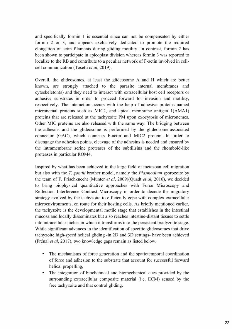
Overall, the glideosomes, at least the glideosome A and H which are better
known, are strongly attached to the parasite internal membranes and
cytoskeleton(s) and they need to interact with extracellular host cell receptors or
adhesive substrates in order to proceed forward for invasion and motility,
respectively. The interaction occurs with the help of adhesive proteins named
micronemal proteins such as MIC2, and apical membrane antigen 1(AMA1)
proteins that are released at the tachyzoite PM upon exocytosis of micronemes.
Other MIC proteins are also released with the same way. The bridging between
the adhesins and the glideosome is performed by the glideosome-associated
connector (GAC), which connects F-actin and MIC2 protein. In order to
disengage the adhesion points, cleavage of the adhesins is needed and ensured by
the intramembrane serine proteases of the subtilisins and the rhomboid-like
proteases in particular ROM4.
Inspired by what has been achieved in the large field of metazoan cell migration
but also with the T. gondii brother model, namely the Plasmodium sporozoite by
the team of F. Frischknecht (Münter et al, 2009)(Quadt et al, 2016), we decided
to bring biophysical quantitative approaches with Force Microscopy and
Reflection Interference Contrast Microscopy in order to decode the migratory
strategy evolved by the tachyzoite to efficiently cope with complex extracellular
microenvironments, en route for their hosting cells. As briefly mentioned earlier,
the tachyzoite is the developmental motile stage that establishes in the intestinal
mucosa and locally disseminates but also reaches intestine-distant tissues to settle
into intracellular niches in which it transforms into the persistent bradyzoite stage.
While significant advances in the identification of specific glideosomes that drive
tachyzoite high-speed helical gliding -in 2D and 3D settings- have been achieved
(Frénal et al, 2017), two knowledge gaps remain as listed below.
• The mechanisms of force generation and the spatiotemporal coordination
of force and adhesion to the substrate that account for successful forward
helical propelling,
• The integration of biochemical and biomechanical cues provided by the
surrounding extracellular composite material (i.e. ECM) sensed by the
free tachyzoite and that control gliding.
22

In the next section, we will develop in more details than in the publication (see
below) the techniques and assays we have introduced in this study.
2.1.2 Implementing methods to study the movement of T. gondii tachyzoite
- Reflection Interference Contrast Microscopy (RICM)
We have already highlighted the crucial need of dynamic interactions between the
tachyzoite and the substrate during gliding. Therefore, we decided to accurately
quantify the adhesion pattern of a tachyzoite when it undergoes an helical cycle
by measuring the distance between the latter and a flat substrate. To this end we
applied quantitative Reflection Interference Contrast Microscopy (qRICM).
RICM provides a way to measure the distance between an object and its substrate
under water with nanometric precision and milliseconds time resolution. In the
beginning, Interference Reflection Microscopy (IRM) was established as a
method to study interactions between surfaces by simply using the interference of
the light but only from a qualitative point of view. As the optics of surfaces
improved, several upgrades in this system made it possible for quantitative
measurement of the inter-surface distances using an improved contrast (qRICM).
The technique has been used in various scientific fields and it has major
applications in live Science since it allows comparing adhesion strength,
dynamics and spreading, as well as cell membrane fluctuations, reconstruction of
cell membrane conformations and others. A major improvement in the robustness
and accuracy of the technique came with the dual wave RICM where two
wavelengths are used instead of the single used before to provide accurate
measurement of the height between inter-surfaces. The general principle of this
technique is that a monochromatic incident ray Io (Figure 12) is reflected at the
glass surface to obtain the I1 and it reflects further to reach the surface of the
object-cell to give the I2. Then using these intensity values, the height (h) can be
accurately measured if image processing and analysis were carefully performed to
reduce the probability of errors. Among main sources of errors are the
heterogeneity of the background and the reference intensity or the movement of
organelles inside cells. Overall, RICM provides quantification of adhesion
patterns and dynamics without the need of labeling the cells. It requires fast
imaging but can be combined with other microscopy techniques.
23

Figure 12. Representation of light interferences
in the case of glass-membrane contact and
calculation formula. A monochromatic incident
ray I0 is first reflected at the glass/medium interface
to create the ray I1, the transmitted ray is reflected
further at the surface of the membrane and gives
rise to ray I2. I1 and I2 interfere and will result the
creation of the intensity I. Adapted from Limozin &
Sengupta, 2009.
The combination of RICM with Total Internal Reflection Fluorescence (TIRF)
microscopy is quite promising since it allows cross-validation of the current
models in membrane shape reconstruction and can provide time-lapse recording
of the membrane and related proteins during a variety of cell processes. New
improvements in the RICM signal have already started to be applied by using
anti-reflecting substrates that are easily reproducible in optical properties. One
current challenge in the RICM field is its application on the examination of cell
functions that occur in a micro and nano resolution scale on non-flat substrates.
RICM was recently coupled to microfluidic devices in order to get insights on the
interactions between thick glycocalyx-mimetics and cells/cell-mimetics under
flow (Davies et al, 2019) (Figure 13). Indeed in addition to distinguish the
distance between an object and a surface with nanometer precision, the RICM is
sensitive enough to detect in flow assays the arrests of particles interacting with a
surface by observation of particle height with respect to the surface, compared to
measurement of velocity alone. These achievements are of great interest to better
understand how a variety of cells, including leucocytes such as T lymphocytes or
neutrophils, but also stem or cancer cells can be “captured” by the endothelial
cells before they eventually cross the endothelium. These migrating cells engage
thanks to their surface-exposed CD44 molecule a critical interaction with the
polysaccharyde hyaluronan (HA), a major component of the glycocalix that lines
the luminal side and protrudes in the lumen of blood vessels. A combined RICM-
microfluidic setting appears particularly well suited for integrating key dynamics
and biomechanical features of the HA-enriched glycocalix as the CD44+ cells are
captured and further roll on the surface under flow.
24

Figure 13. Coupling RICM with a microfluidic
device. Mounting of the assembled flow chamber -
allowing laminar tuneable flow rates- on a multi-
modal inverted microscope with RICM capability
provides a method to examine bead height with
respect to HA films, in addition to velocity under
flow. HA chains were grafted through a biotin
functionality at their reducing end to the Lipid SAv
monolayer. Adapted from Davies et al, 2019.
- Traction Force Microscopy (TFM)
While the combination of high-speed videorecording and RICM allowed
unveiling a typical apical deformation of the tachyzoite at the onset of the helical
cycle, the next challenge was to get insights on the location and amplitude of
forces that are applied by the parasite during helical movement, and to thus verify
whether traction forces could be generated at the site of adhesion. Traction Force
Microscopy (TFM) had never been used for analyzing forces during T. gondii
movement but has been in contrast successfully developed and applied to the
slender Plasmodium sporozoite in the course of circular gliding (Münter et al,
2009).
Cellular traction was first demonstrated using compliant silicon-rubber substrate,
on which adherent non-muscle cells could produce wrinkling patterns.
Quantitative techniques were then developed, based on the principle that traction
forces can be extrapolated from substrate deformations and they commonly use
polyacrylamide (PAA) elastic gels and micropillars. The one using PAA gels is
conventionally called Traction Force Microscopy (TFM). TFM requires
fluorescent beads that are evenly embedded into the PAA gel (Figure 14) to be
used as fiduciary markers whereas cells of interest are plated on top of the gel and
both the beads and the cells are recorded overtime. In the end of the assay,
exposing the cells to the enzymatic activity of trypsin will give the opportunity to
obtain the relaxed position of the beads when the cells are no longer attached and
these bead positions serve for force calibration. Knowing the relaxed position of
the beads and tracking the beads overtime allows producing a displacement map
for the substrate, which is then used to solve the ‘inverse’ problem using the
Boussinesq’s formulation: an equation for the displacement data in infinite half-
space is derived into a convolution integral using Green’s tensor, discretized and
then inverted to determine traction forces (Gupta et al, 2016) (Figure 14).
Critically, the substrate stiffness value and stability are essential parameters to
allow proper calculations (Martiel et al, 2015).
25

Figure 14. Schematic representation of cell lying on top
of PAA gel embedded with beads and displacement-force
calculation approach. Left panel: A cell creates focal
adhesions and pulls the substrate in x-y direction. Right
panel: Calculation of the beads displacement overtime
(t0=black color, t1=grey color). Knowing the displacement,
force value can be also computed. In this approach it is
considered that the beads displacement in the z direction is zero or close to zero. Left
panel adapted from Schwarz & Soiné, 2015. Right panel adapted from Martiel et al,
2015.
Challenges and parameters to optimize for T. gondii motility:
• PAA gels coated with fibronectin
o Concentration of fibronectin
§ Trying two extreme conditions where we put much higher
concentration of fibronectin than the one that is commonly
used, didn’t affect the gliding proprieties.
o Stiffness of PAA gel
§ Starting with 5 kPa Young’s modulus stiffness PAA gels
coated with fibronectin, parasites could nicely glide in
these conditions but although beads displacement could be
observed, it was not enough convincing, as the
displacement length was very small. Testing different
stiffness we found out that 2 kPa was enough to have
gliding and bead displacement. The problem was that
reducing the stiffness, parasite motility percentage and
travel distance length were both decreased.
o TFM bead size and concentration
§ The concentration of beads used during the preparation of
the PAA gels (0.4 µl / 164 µl of PAA), was not good
enough to have separate beads that could cover all the area
underneath the parasite while it was moving. Increasing
26

this to approximately 0.8 µl / 164 µl of PAA, we managed
to have ideal conditions of bead displacement without lots
of aggregates and overlapping of beads. One other
suggestion that we had, was to use two different color
beads in order to cover the gaps but because we used
streaming option for recording (see after), we could not use
more than two channels overall (we need brightfield and
one fluorescence).
• Microscopy
o Recording timing
§ Fast recording – less quality
• 1 frame/second was not good enough to catch the
displacement of the beads for longer time. For this
reason, we utilize the streaming option of the
microscope with the help of the management
software MetaMorph in order to record at around 6
frames/second. Limitation of this was that you
decrease the exposure time and the number of lasers
that you can use during the acquisition.
§ Streaming option-only two laser channels
o No focus outside of the beads height (for example for cytoskeleton
proteins as researchers use with mammalian cells, they can
observe proteins’ behavior at the same time with the forces
dynamics).
By performing control experiments, we found out that reducing the stiffness
significantly affects the tachyzoite motile behavior which translated in a
decreased number of moving parasites for a given time and in the same line, a
reduction in the duration of the gliding behavior. Increasing the concentration of
TFM beads inside the gel two times more than the initial one and performing a
well-controlled sonication step to avoid aggregates of beads, have helped in
increasing the probability of having a visible bead displacement event. The more
uniform distribution of single (not clustered) beads provides the most accurate
detection bead displacement by the software during image analysis.
The next level of TFM approaches is to implement the z dimension in order to
have a more physiologically relevance with the in vivo situation. Some of them
(Legant et al, 2010) have been described but the limitations of a closely related to
the in vivo substrate is still missing. This is because the extracellular matrices are
a mix of different proteins that complicates the establishment of a “real” elastic
substrate with a stable relaxed position. To counteract this problem, hydrogels
which provide uniform and complete crosslinked networks have been used.
Although collagen (fibrillar) and matrigel gels work very nicely, it is impossible
to get stable stiffness value overtime, thus you cannot get force estimation.
27

- PRIMO micropatterning
Micropatterning techniques are widely used in the field of cell biology and
mechanobiology in order to interrogate for example how cells interact with each
other or, how tissue-like layers behave, how cells attach to a given substrate and
develop stress fibers, how these forces act during cell division or cell migration,
or phagocytosis (Senger et al, 2019). At the subcellular level, these approaches
have been applied to investigate how organelles or proteins deal with the
micropattern-driven constraint. In some cases, high precision in the designing
process is needed like in this study since we wanted to create a pattern where
there is a clear separation of the fibronectin coated area (gliding area) and the
PEG area (non gliding area). When we tried to make micropatterns manually
(without the use of PRIMO) we found out that there were always a few pieces of
fibronectin around the main design, therefore in a area supposed to remain non
adherent (Figure 15).
Figure 15. Schematic representation of the
micropatterning principle. UV light exposure
to PEG coated glass coverslip, through a
photomask with the specific pattern. Patterns will
be created in the PEG layer and then ECM
(fibronectin in our case) protein will cover the
patterns space. Making these patterns by hands,
we saw that some protein (red in last step) stays
next to the pattern position and it is not visible
enough to know the exact fibronectin region of
the pattern. Adapted from Thery, 2010.
This could not ensure the clear separation of the two substrates, which was
something very crucial for our experiment knowing that the size of the parasite is
5-7 µm. Therefore, we worked with the Alveole company (www.alveolelab.com)
that has been able to design using the PRIMO device (Figure 16) the design we
chose with a high precision. The design we needed includes a sharp demarcation
between permissive (fibronectin) and non permissive (PEG) gliding substrates so
we could examine how the tachyzoite behaves when the apical part of the parasite
body was “touching” the non permissive area while the posterior part was
“touching” the permissive fibronectin area.
28

Figure 16. Schematic representation of the micropatterning principle using
PRIMO. Solution of PLL-g-PEG is deposited on an air-plasma-activated glass coverslip.
After rinsing, photoinitiator is added and UV light is projected onto the surface through a
pattern mask. The UV activated photoinitiator molecules locally cleave the PEG chains.
Then the substrate becomes adhesive and at the last step, we have the protein coating.
Adapted from Strale et al, 2016.
In addition, micropatterning can be combined with other techniques such as
Traction Force Microscopy and Atomic Force Microscopy where researchers are
interested in the forces that are applied by a single cell, few cells or even whole
tissues.
- Nano/microtools to design innovative assays: Immobilizing the parasite on
a substrate by forcing adhesion of the posterior pole
Once cellular forces have been mapped during motion, the logical next step is to
attempt to locally interfere with the force application in order to bring definitive
conclusions on how these forces control the movement under study. To this end,
we have used the permissive-non permissive micropattern as explained above to
interfere with tachyzoite front adhesion with the substrate. However, we also
wanted to see the outcome of mechanically holding the parasite, the time that it
was ready to move. Immobilizing posteriorly the tachyzoite but keeping it alive
and potentially active was the main goal of this experiment. To this end we
incubated the parasite with sub-micrometer beads under conditions that would
statistically allow some parasites to be tightly bound to the bead at their posterior
while the bead itself would be trapped in interaction with the substrate made of
fibronectin layer. As the result the parasite would be indirectly but firmly
attached to the substrate so a “normal” front traction force would remain
inefficient at disengaging the posterior pole from the adhesion with the substrate.
29

Figure 17. Schematic of
immobilization techniques.
From left to right: physical
adsorption, entrapment and
covalent attachment.
Adapted from Mohamad et
al, 2015.
Figure 18. Schematic
representation of beads
activation with EDC.
Activation of the carboxyl
groups by EDC which then
forms reactive O-acylisourea
to account for covalent
binding between the beads
and the T. gondii membrane
proteins.
There are different techniques of molecule immobilization (Figure 17) such as (i)
physical adsorption (ii) embedding inside gels or (ii) covalently binding. We
chose commercially available carboxylated beads that were subsequently
activated using the manufactor protocol to promote covalent binding of the beads
surface to the parasite surface-exposed proteins (Figure 18). Activation of the
carboxyl groups on the beads surface was achieved with water-soluble 1-ethyl-3-
[3-dimethylaminopropyl] carbodiimide (EDC) and led to the formation of an
active intermediate (O-acylisourea). The latter quickly reacted with primary
amines (nucleophile) to form a stable amide bond. Given that tachyzoite PM is
covered by large amount of glycoproteins and proteins, it was expected that by
chemically activating beads, the latter would engage covalent interaction with the
amine of the surface-exposed tachyzoite molecules. At the same time, we hoped
that because active parasites would theoretically engage the surface-bound bead
in an antero-posterior capping process, the beads would end at the posterior pole
and there engage new interactions with the fibronectin substrate. As the result, we
expected that the parasite would be trapped by the posteriorly positioned beads
and incapable of disengaging from the substrate. To succeed in setting the
optimal experimental protocol, we had to readjust the protocol by reducing the
Fetal Calf Serum (FCS) concentration since this parameter was crucial to lower
the probability the parasite manages to escape from the posteriorly capped beads.
30

2.1.3 Main highlights of the study
Combining these approaches, we were able to explore the mechanisms of force
generation required for successful forward helical propelling of the tachyzoite.
While the results of a serie of assays are presented in the published article below,
the main highlights are summarized as follows:
• The tachyzoite establishes an apical strong adhesion with the substrate.
• The tachyzoite applies traction forces accounting for kink formation while
the kink release coincides with maximal forward helical acceleration.
• Apical and posterior attachment-detachment need to be coordinated for
successful engagement of the traction forces.
• Microtubule bending during the kink should account for energy storage
upon loading by the traction force.
2.1.4 Publications
Pavlou G, Touquet B, Vigetti L, Renesto P, Bougdour A, Debarre D, Balland M and Tardieux I. (2020) Coupling Polar Adhesion with Traction, Spring, and Torque Forces Allows High-Speed Helical Migration of the Protozoan Parasite Toxoplasma. ACS Nano acsnano.0c01893.https://pubs.acs.org/doi/10.1021/acsnano.0c01893
31

Coupling Polar Adhesion with Traction,Spring, and Torque Forces Allows High-SpeedHelical Migration of the Protozoan ParasiteToxoplasmaGeorgios Pavlou, Bastien Touquet, Luis Vigetti, Patricia Renesto, Alexandre Bougdour,Delphine Debarre, Martial Balland, and Isabelle Tardieux*
Cite This: https://dx.doi.org/10.1021/acsnano.0c01893 Read Online
ACCESS Metrics & More Article Recommendations *sı Supporting Information
ABSTRACT: Among the eukaryotic cells that navigate throughfully developed metazoan tissues, protozoans from theApicomplexa phylum have evolved motile developmental stagesthat move much faster than the fastest crawling cells owing to apeculiar substrate-dependent type of motility, known as gliding.Best-studied models are the Plasmodium sporozoite and theToxoplasma tachyzoite polarized cells for which motility is vitalto achieve their developmental programs in the metazoan hosts.The gliding machinery is shared between the two parasites and islargely characterized. Localized beneath the cell surface, itincludes actin filaments, unconventional myosin motors housedwithin a multimember glideosome unit, and apically secreted transmembrane adhesins. In contrast, less is known about theforce mechanisms powering cell movement. Pioneered biophysical studies on the sporozoite and phenotypic analysis oftachyzoite actin-related mutants have added complexity to the general view that force production for parasite forwardmovement directly results from the myosin-driven rearward motion of the actin-coupled adhesion sites. Here, we haveinterrogated how forces and substrate adhesion−de-adhesion cycles operate and coordinate to allow the typical left-handedhelical gliding mode of the tachyzoite. By combining quantitative traction force and reflection interference microscopy withmicropatterning and expansion microscopy, we unveil at the millisecond and nanometer scales the integration of a criticalapical anchoring adhesion with specific traction and spring-like forces. We propose that the acto-myoA motor directs thetraction force which allows transient energy storage by the microtubule cytoskeleton and therefore sets the thrust forcerequired for T. gondii tachyzoite vital helical gliding capacity.
KEYWORDS: cell migration, cell focal contact, 3D collagen, Toxoplasma, traction force microscopy,reflection interference contrast microscopy, expansion microscopy
A cross eukaryotic phyla, cell migration is an evolutionaryconserved function which appears vital for both freeliving protozoans and metazoans.1 In metazoans, cells
such as fibroblasts and leukocytes use a wide spectrum ofadhesion-dependent motility modes conjointly defined ascrawling and primarily driven by the dynamics of the cellactin−myosin cytoskeleton.2,3 These cells crawl with the helpof dynamic actin-powered membrane protrusions that fold atthe leading edge as a flat lamellipodium or as a variety ofpseudopodia-like membrane projections. Firmly attached tothe substrate upon coordinated assembly of integrin-drivenadhesive platforms, the lamellipodium is a hallmark of themesenchymal cell migration mode in 2D and 3D micro-
environments. These anchoring multimolecular platformsallow the development of actomyosin bundles that tunecontractile forces between the front and trailing margins andeventually lead to the release of trailing adhesions, henceallowing cell forward translocation.4,5 Likewise, metazoancellsas well as large free-living protozoanscan display a
Received: March 4, 2020Accepted: May 20, 2020Published: May 20, 2020
Article
www.acsnano.org
© XXXX American Chemical SocietyA
https://dx.doi.org/10.1021/acsnano.0c01893ACS Nano XXXX, XXX, XXX−XXX
Dow
nlo
aded
via
UN
IV G
RE
NO
BL
E A
LP
ES
on J
une
19, 2020 a
t 12:1
2:1
5 (
UT
C).
See
htt
ps:
//pubs.
acs.
org
/shar
ingguid
elin
es f
or
opti
ons
on h
ow
to l
egit
imat
ely s
har
e publi
shed
art
icle
s.
32

more versatile motility mode and cope with heterogeneous 3Dmicroenvironments. In addition to the lamellipodium style,they can adopt diversified pseudopodia mechanisms that,combined with a range of relative cell-matrix adhesion andactomyosin contractility, account for the rapid amoeboid typeof motility (∼10 to 20 μm/min versus less than 1 μm/min forthe lamellipodia mode).6 Alternatively, fibroblasts can utilizean amoeboid−mesenchymal hybrid type of 3D migrationcalled lopobodial during which a pressure-driven bleb-likemembrane protrusion stems at the leading margin from thepiston-like forward movement of the nucleus itself driven bythe cortical actomyosin contractility.7 Biophysical studies haveproved instrumental in mapping and measuring the develop-ment of forces in cells, and they brought insights on how cellsdifferentially regulate the adhesion−contractile balance tospatiotemporally control the adhesion assembly deassemblycycle and achieve productive forward movement in differentcontexts.8
Intriguingly, unicellular eukaryotic parasites from the ancientphylum of Apicomplexa have evolved an adhesion- and actin-dependent motility mode that differs from the mesenchymal,amoeboid, lobopodial repertoire and enables the microbes toglide within host tissues and across barriers to follow theircomplex stepwise developmental program. The severalthousand members of the Apicomplexa phylum includeamong the world’s most pre-eminent mammal-invasiveprotozoan parasites. In humans, Plasmodium spp. areinoculated in the vascularized dermis extracellular matrix(ECM) by a blood-feeding mosquito which hosts parasites insalivary glands. Plasmodium displays a prolonged asexualdevelopmental phase in red blood cells, which results in arange of clinical outcomes known as malaria, impacting humanpopulations in tropical and subtropical regions of the Earth. Itsrelative Toxoplasma gondii is present in a large assortment ofendotherm metazoans worldwide. Being ingested withcontaminated food and water, T. gondii is hosted quasi-silentlyby possibly up to a third of the human population in tissuessuch as the brain, retina, and heart and skeletal muscles.However, in case of immune dysfunction, T. gondii can re-emerge from these tissues and proliferate, causing life-threatening or debilitating complications.9
Throughout their life cycle in their respective hosts, bothPlasmodium and T. gondii ensure their perpetuation byundergoing multiple cell differentiation events that give riseto specific developmental stages with sizes of several microns,harboring a crescent shape and endowed with high motilityskills.10 Best studied are the Plasmodium slender maturesporozoite inoculated in the host skin and the T. gondii bulkyhighly replicative tachyzoite stages that glide 10 times fasterthan the amoeboid cells without folding any of the crawling-associated protrusions.10−12 Instead, they share a robustlongitudinal apico-basal polarity which persists during thegenerally unidirectional motile activity with the apex leading.13
Whereas the Plasmodium sporozoite moves on a circular pathin either counterclockwise (CCW) or clockwise (CW)directions with an irregular stop and go kinetics,13 it can alsooccasionally adopt a random motion or a corkscrew-likepattern in vivo.14,15 Instead a left-handed helix characterizes thepath the mosquito-restricted ookinete Plasmodium stage usedto move in Matrigel-based 3D matrices.16 The Toxoplasmatachyzoite can similarly glide CCW on a circular path under2D in vitro settings but often uses a CCW helical trajectory or asuccession of the two mechanisms over the same gliding
sequence.11,17 Another intermittent 2D movement termedtwirling accounts for the stationary CW rotation executed bythe upright-positioned tachyzoite around its posterior end.18
The corkscrew-like mode of gliding, which is the obligatemechanism in 3D reconstituted or in vivo environments wasproposed to relate to the 2D helical mode of gliding.19 Thelatter typically starts with a CW short motion, follows with a180° twist; meanwhile, the body flattens on the substratebefore twisting up and flipping on the side, which allowsstarting another helical cycle.11,18
To initiate and sustain all types of gliding activity, both thesporozoite and tachyzoite rely on the regulated secretion ofadhesive protein oligomers at the cell surface20,21 that, oncedelivered at the apical tip, can coengage with ECM proteinsand short cortical actin filaments organized beneath the plasmamembrane. Force production driving parasite movement haslong been considered to directly translate from the apico-basaltranslocation of the ECM-bound adhesins coupled to theflowing actin filaments and their enzymatic-mediated release ofthese adhesins at the trailing edge.22,23 It is the Apicomplexaspecific, fast, nonprocessive, single-headed motor myosin A(MyoA) that was assigned an essential powering role in pullingbackward the apically initiated actin filaments and the capturedadhesins.24,25 Upstream from MyoA however, the T. gondiitachyzoite additionally requires myosin H (MyoH) to propelactin filaments from the parasite apical tip formed by theconoid,26 a retractile appendage missing in Plasmodium. BothMyoA and MyoH molecules function within a multisubunitcomplex identified as glideosome22 that also comprises severalregulatory subunits and scaffolding partners.27−29 Specificallyfor MyoA, the glideosome associated proteins (GAPs) anchorthe motor machinery to the plasma membrane and the lininginternal bilayered membrane complex (IMC) which undersideis tethered to a cytoplasmic array of cortical microtubules(cMTs), the number and arrangement of which typify eachApicomplexa species and developmental stage.27,30
However, the recent introduction of force microscopy,reflection interference contrast microscopy (RICM) andoptical tweezers in conjunction with molecular genetics inthe Plasmodium field has added more complexity to theprevalent force production model.31 A key observation is thediscrepancy between the speed of the retrograde flow ofoptically trapped microbeads on the apical surface of thesporozoite and the speed achieved during typical circulargliding, the former being up to 50% faster than the latter.31 Inaddition, several distinct areas of contact between the movingsporozoite and the substrate were uncovered at the apex, base,and center over the circular path and were shown to displayspecific on−off dynamics,13 whereas the genetic loss of asurface-exposed adhesin reported to function during motilitywas found to trigger a speed increase in the retrograde flow anda concomitant decrease in force production.31 Collectively,these studies bring compelling evidence for the assembly ofdistinct force-generating adhesive structures which differ-entially control production of the force required for migration.By contrast, how forces and dynamic adhesions coordinate
in space and time and integrate with the impressive high-speedhelical path that features the T. gondii tachyzoite glidingmechanism has been much less investigated. Intriguingly, therearward capping of nanobeads bound to the tachyzoite surfacewas shown not to require an intact actomyosin system butrather a polarized secretory−endocytic cycle that would locallydrive membrane tension.32 On the other hand, qualitative
ACS Nano www.acsnano.org Article
https://dx.doi.org/10.1021/acsnano.0c01893ACS Nano XXXX, XXX, XXX−XXX
B
33

RICM allowed identifying an early tachyzoite contact with thesubstrate at the cell front and its rearward translocation which
was assumed to directly power the force-producing parasitemovement.33 In this study, we revisited the mechanisms
Figure 1. Tachyzoite pulls on the collagen fibers prior to helical acceleration. (A) Successive images from a time lapse of a tachyzoitenavigating within a 3D-meshwork of rat collagen1 fibers prelabeled with the CNA35-GFP collagen chimeric probe. Panels show images of(top) DIC, (middle) DIC-GFP over, giving yellow-colored fibers, (bottom) the fiber initial (t0, green) and over time (tx, magenta) positionswhen encountered by the moving tachyzoite. The overimposition of the green and magenta signals comes as white (white arrows) when thefiber maintains the t0 position, whereas the separation between the green (t0) and magenta (tx) signals attests two distinct positionsovertime. The backward displacement indicates the parasite pulls on the fiber (red arrow), whereas the graph (B) documents that thispulling coincides with the parasite low speed. Next the acceleration step coincides with the merge back of the green and magenta signals thatsigns for the release of fiber tension post motion. (C) Successive DIC images from a time lapse of a tachyzoite undergoing helical gliding onfibronectin-coated glass. (Top) back (green) and front (blue) tracks over three last frames (tx−2 to tx) are shown, (bottom) speed profile isshown from black to white scaling with increasing speeds. All white arrowheads point to the kink, all scale bars: 5 μm, time is in minutes/seconds. (D) Graph (left) plots the tachyzoite back−front distance (red line) and the speed based on the back tracking (light orange); notethat the value close to zero corresponds to the time the tachyzoite twirls, hence the apex is out of focus and the back-front distance notmeasurable (right) the xy displacement for both the back (green) and front (blue) sites over the helical sequence. Zoomed of a typical kinkstep for both graphs shown as inlays.
ACS Nano www.acsnano.org Article
https://dx.doi.org/10.1021/acsnano.0c01893ACS Nano XXXX, XXX, XXX−XXX
C
34

underpinning the helical gliding of T. gondii by combiningquantitative live imaging with traction force microscopy,quantitative RICM, micropatterning and expansion micros-copy. We provided evidence for a spatiotemporal coordinationof an actoMyoA-based traction force produced at a stableapical adhesion site and a spring-like force which, driven by the
left-handed cMT spiral orientation, directs straightening andhelical propelling of the T. gondii tachyzoite.
RESULTS/DISCUSSION
Tachyzoite Pulls on the Collagen Fibers in a 3DMeshwork Prior to Accelerating with a Helical Motion.
Figure 2. Helical cycle starts with an apical site of adhesion that constraints sliding of both the parasite and the parasite posteriorly shed trail.(A) Successive RICM images from a time lapse of a tachyzoite undergoing helical motion on a glass coverslip. Panels show (top)simultaneous bright-field (BF) and RICM images, (middle) blue and green RICM images superimposed and displayed as RGB, (bottom)segmentation of BF and RICM images showing the parasite outline (white), areas of close contact between the glass and the moving parasite(red), and material shed behind as trail (green). White arrows indicate the direction of the parasite movement. The dotted line marks theposition of the initial contact over one helical cycle and delineates the trajectory angular point. (B,C) Kymograph of the whole glidingsequence from (A) and for a single zoomed helical cycle (C) shows the evolution of the close contact area along the trajectory over time. (D)Successive three-color RICM images (shown as RGB) from a time lapse of a tachyzoite undergoing helical motion on a poly-L-lysine-coatedglass coverslip (top) and corresponding segmentation with the Weka FIJI plugin (bottom). The pink arrows indicate that over time thetachyzoite pulls on the trail that slides without breaking but follows the polygonal trajectory imposed by the initial anchoring sites. (E) Timeprojection of the segmented close contact areas over the gliding sequence on which (left) the longest-lived attachment points are brightest,(middle) the trails are green, and (right) adhesion and trails are overimposed. Note that angles in the polygonal path coincide with both thelong-lived close contacts and the kinks. All scale bars: 5 μm; time is in minutes/seconds.
ACS Nano www.acsnano.org Article
https://dx.doi.org/10.1021/acsnano.0c01893ACS Nano XXXX, XXX, XXX−XXX
D
35

Pioneering work on T. gondii 3D in vitro motility has relied onMatrigel-based matrices under rather elastic and homogeneousconditions.19 However, in vivo, most ECM microenvironmentsare enriched in a nonlinear, anisotropic, and dynamic fibril-richmeshwork. Because collagens are dominant ECM componentsthat form multiscale fibrils withstanding tensile forces,34 weanalyzed how the T. gondii tachyzoite moves within a confinedcollagen fibril-based matrix. Whereas the tachyzoite squeezedits body to pass through the meshwork, it first paused andbent, showing an apical deviation as it next accelerated,possibly with a rotation around its main axis, to escape fromthe collagen fibrils (Figure 1A,B and Supporting InformationFigure S1, Movie 1, and Movie 2). Using a fluorescent probeselectively associated with collagen fibrils,35 we applied atemporal color-code (green and magenta) and identified fibersencountered by the motile tachyzoite that shifted backwardbefore returning to initial position as the parasite propelledforward (Figure 1A, Supporting Information Movie 1). Thisfiber backward displacement was seen in all cases of movingtachyzoites encountering a fiber (n = 10), and qualitativelyindicated that the parasite exerted pulling forces on thesurrounding fibers.To get more mechanistic insights on the front-rear
spatiotemporal coordination over the helical gliding cycle, wefurther analyzed the changes in the tachyzoite shape with highspatiotemporal acuity using a 2D setting. We observed a yetundefined apical inward bending immediately post the initialclockwise arc motion and concomitant with the parasite pause(Figure 1C). Indeed, tracking the front and back revealed ashortening of the parasite that coincided with an apical flexureand thus an increased curvature, resembling the observationmade within the fibrous 3D matrix, and which we defined asthe kink step. Associating with the apical pole arrest wedetected the contraction of the cell body and retraction of theposterior pole. Next the apex re-extended and lifted up, a phasedefined as the kink release, while the tachyzoite straightenedand slid to further twisted in the typical CCW motion (Figure1C, Supporting Information Movie 3a and Movie 3b, slowmotion). Accordingly, not only the instantaneous speed wasabout null during the kink step and reached maximal value asthe latter relaxed back (Figure 1D), resulting in thecharacteristic periodic speed fluctuations documented under3D conditions.19 No such pattern was detected when theparasite performed the 2D-restricted CCW circular trajectory(Supporting Information Figure S1B,C and Movie 4). Of note,high temporal resolution live imaging of Plasmodiumsporozoites also reveals alternating periods of rapid movementin which they travel up to one body length and periods of slowmovement in which they more firmly adhere to the substrate,thereby indicating that distinct adhesion types operate duringmotility.13 Although such kinetics was associated with thesporozoite body stretching upon firm adhesion of its polar andcenter regions to the substrate and the subsequent thrust inspeed,13 no peculiar apical deformation that could resemble akink has been observed to initiate movement. We proposedthat the kink deformation and the subsequent body extensionmight reflect the tension built in the tachyzoite cytoskeleton toachieve the thrust force required for the helical twistingmotion.Apical Focal Adhesion Behaves as a Firm Long-Lived
Anchor That Constrains Sliding of the Parasite Body.Because the apical kink suggests the assembly of a specificcontact between the parasite front and the substrate, which
would be appropriate for building up tension in the T. gondiicytoskeleton, we first used qRICM to monitor the parasite-substrate distance. In contrast to a recent study,33 we usedhigh-speed (30 frames/s), multicolor RICM with simultaneoustransmitted-light imaging coupled with machine-learning-basedimage processing to achieve a precise segmentation of closecontact regions between the parasite and the substrate at hightemporal resolution (33 ms) over thousands of frames (Figure2 and Supporting Information Movie 5 and Movie 6). Inaddition to the low-distance patches detected between theparasite basal pole and the substrate even in absence ofmovement, we found that at the onset of a helical cycle, themembrane of the tachyzoite apex came in contact with theglass surface. Because these RICM-detected signals coexistedwith motion, we assumed they accounted for two polarparasite−substrate adhesive areas in agreement with Tosettiand collaborators33 and similarly to what was found for thePlasmodium sporozoite.13 Crucially, the apical contactremained static over the full length of the helical cycle (7.5± 0.9 s), and the posterior contact progressively expanded toaccount for ∼80% of the tachyzoite length along its major axis(Figure 2A−C, see kymographs, Supporting InformationMovie 5). Upon release of the kink, the parasite propelledforward and shifted orientation before it started a new frontattachment. For each helical cycle, the kink step could be aposteriori inferred through the mix of parasite-derivedmembrane hydrophobic droplets and surface-exposed pro-teins/glycoproteins left as a complex trail behind the cell edgeonto the substrate, a common feature of Apicomplexa motilestages (Figure 2A,D,E).11,36 Quantitative modeling of theRICM signal provides transverse dimensions for these trails of∼70 nm (isotropic) or 100 nm (width) and 40−60 nm(height). By comonitoring the parasite helical motion and thetrail assembly in real time, we were able to uncover that thetachyzoite not only slid on the adhesive tracks it built itself butcould also pull these, an observation that has not been reportedfor the RICM data collected on Plasmodium.13 Theseobservations suggest a continuous nature of the shed materialthat remained close but not firmly fixed on the substrate(Figure 2D and Supporting Information Movie 6), whichwould agree with membrane components including lipids aspreviously deduced from the analysis of fixed samples.11
Importantly, the sliding trajectories of the shed material oncepulled by the moving tachyzoite appeared constraint by theperiodically distributed firm anchors, which could occasionallybe stripped off from the substrate (Supporting InformationMovie 6). In further support of these periodic stable anchors,the time projection of the trail trajectory postimagesegmentation allowed positioning the longest-lived parasite−substrate contacts at each angular point of the trajectory,matching the initial apical anchoring sites (Figure 2E). Therelative homogeneity in the trail observed at our RICMresolution cannot inform on a specific distribution orarrangement in proteins and lipids upon specific secretoryevents or/and due to local physical constraints that wouldcompose the periodic anchors. The microneme-stored MIC2 isseen as the adhesion prototype that bridges the parasite motorsystem and ECM ligands during motility. Indeed partial or fullsilencing of the mic2 expression significantly impairs both thehelical and twirling motions.37,38 This double default arguesmore for a MIC2 contribution to the posterior adhesionforce,37 hence mic2 KO parasites show a reduced capacity tobind to host cells and collagen-coated surfaces.38 Consistently,
ACS Nano www.acsnano.org Article
https://dx.doi.org/10.1021/acsnano.0c01893ACS Nano XXXX, XXX, XXX−XXX
E
36

when the T. gondii tachyzoite expresses only residual levels ofthe rhomboid protease (ROM4) in charge of cleaving thesurface-exposed MIC2, MIC2 molecules accumulate at thesurface of the parasite that can no longer glide forward butinstead shows enhanced twirling motility.39 Similarly, whenPlasmodium berghei sporozoites express a mutant version of theMIC2 homologue TRAP that cannot be processed by therhomboid protease, the parasite can no longer disengage itsposterior pole from the substrate and move forward.36
Whereas we detected both SAG1 and MIC2 proteins inpatches over successive segments of the helical trails
postfixation and immunolabeling, we did not find MIC2-specific enrichment at the angular adhesion regions that wereaccurately identified using the RICM live sequences asrecognition pattern (Supporting Information Figure S2).Therefore, it is possible that another unknown adhesin(s),which would not integrate the retrograde flow, might modulateforce production at the parasite apex in line with the turnover-specific adhesion sites built by the Plasmodium sporozoiteduring circular gliding.13 In addition, monitoring the apico-basal flow of optically trapped beads pinned out that specificadhesions do not integrate the apico-basal capping process, yet
Figure 3. Helical motion includes the development of apical traction at the kink site and basal dragging forces. (A) Traction forcemicroscopy (TFM) images were taken at the time a parasite glides on a 2 kPa PAA gel in which fluorescent TFM beads are embeddedunderneath the gel surface itself coated with fibronectin (20 μg/mL). Panels show (top) bright-field (BF) and TFM beads composite images;white arrowheads indicate the apex of the tachyzoite, and white arrow marks the kink, (second row) beads alone, (third row) BF with forcevectors computed by the MATLAB TFM code. Red arrows indicate the force direction and value; black arrows have been manually added toease visualization of the two longitudinal forces and their orientation, (bottom) force heat map computed MATLAB TFM code. Scale bar: 5μm, time is in minutes/seconds; au for force heat map. (B) (Left) Speed (light orange line), the back to front body distance (red line) andthe traction force applied by the parasite over time (black line) are shown with a zoomed inlay (black dotted line) at the kink step time, and(right) the back and front displacement (green and blue trajectories, respectively) are shown with a zoomed inlay (black dotted line) at thekink time.
ACS Nano www.acsnano.org Article
https://dx.doi.org/10.1021/acsnano.0c01893ACS Nano XXXX, XXX, XXX−XXX
F
37

Figure 4. Front adhesion to and back disengagement from the substrate are both required for kink formation and traction forcedevelopment. (A) Percentage of tachyzoites that moved by at least one body length displacement over a 20 min period post contact withpoly-L-lysine (PLL) or poly(ethylene glycol) (PEG) chains grafted on PLL coating, unpaired t test, **p = 0.021. (B) Large view of the poly-L-lysine PEG−fibronectin composite micropattern shows that tachyzoites are selectively retained in the fibronectin area, which is fluorescentlylabeled by fibrinogen-Alexa647, scale bar: 15 μm. (C) Images from a sequence of two (top) or one (bottom) representative glidingtachyzoite(s) that fail to pass over the fibronectin−PLL−PEG demarcation line being incapable of interacting apically with PEG. Insteadthey elicit twirling around their posterior end bound to fibronectin and regain helical motion once apically sensing fibronectin. Blue and
ACS Nano www.acsnano.org Article
https://dx.doi.org/10.1021/acsnano.0c01893ACS Nano XXXX, XXX, XXX−XXX
G
38

they can optimize force production. Accordingly, the trap-likeprotein (TLP) was proposed to promote firmer adhesion tothe substrate by enabling specific actin remodeling underneaththe plasma membrane. Such function would explain why TLPcauses specific slowdown of the actin-dependent flow.31 Withthese in mind, we next interrogated whether the firm stableapical anchor formed by the moving tachyzoite and localized atthe kink could indeed serve as a platform for force transmissionby using traction force microscopy (TFM). These assays alsoallowed testing whether the kink-induced cell shortening couldbe associated with tension stored in the parasite that would bereleased to promote the thrust required for productive helicalmovement.Traction force Spatiotemporally Associates with the
Kink and Coincides with Body Dragging. To design theTFM assay, we first identified a substrate in which the stiffnesswas sufficient to allow helical gliding while being elastic enoughto measure substrate deformations via a displacement fieldanalysis performed using fluorescent beads embedded into thepolyacrylamide (PAA) hydrogel. Particle image velocity (PIV)combined with single-particle tracking allowed analyzingsubstrate deformation with a spatial resolution in the rangeof tenth of nanometers. Tracking in space and time thetachyzoite posterior pole and scoring as motile those thatperformed at least one body length displacement, we foundthat stiffer substrates such as glass (n = 73) and 40 kPa PAA (n= 55) correlated with optimal gliding skills, whereas loweringstiffness gradually decreased the frequency and duration of themotile behavior. Yet, the tachyzoites retained a short-lastingability to perform helical motion on the soft 2 kPa PAAsubstrate (n = 103) (Supporting Information Figure S3).Applying TFM40 to parasites gliding on 2 kPa fibronectin−PAA substrate, we obtained a traction vector map andidentified an early inward-oriented traction force stemmingat the cell front and exerted longitudinally in line with thebackward displacement of the in-gel beads (i.e., TFM beads)underneath the apical part of the parasite (Figure 3A andSupporting Information Movie 7a and Movie 7b, slowmotion). As the force progressively increased, concomitantdragging of the parasite body occurred and accounted for theTFM bead’s displacement underneath the posterior pole in thedirection of the parasite motion, in line with the previousdetection of trails being pulled by the tachyzoite (SupportingInformation Movie 6). Relaxation of the TFM beads back totheir initial position was recorded immediately after the kinkrelease and the parasite forward acceleration (Figure 3A,B).Pioneering TFM on the Plasmodium sporozoite has allowed
uncovering two longitudinal forces applied at each pole duringcircular gliding, alike what we found for the T. gondii tachyzoiteundergoing helical gliding. A large force localized at theposterior adhesion site peaked just prior sporozoite de-adhesion, whereas a lower pulling traction force at the apexpointed toward the center. In contrast to what we detected forthe tachyzoite, large forces were also found to pointorthogonally toward the sporozoite center,13 where theypromote body stretching and thus favor close contact of thewhole cell with the substratean obligate requirement forcircular glidingwithout contributing to force production.41
Therefore, although both the tachyzoite and sporozoite likelyuse a stalling force at the back, which upon rupture of theadhesive contact contributes to a thrust force, the spatiotem-poral distribution and intensity of the forces during theirspecific gliding cycle (i.e., helical versus circular) likely differ.Accordingly, the overall force during the helical cycle wascomputed to a few nanoNewtons (∼1 to 4 nN, n = 10) with anincrement over the kink step, therefore significantly higherthan the 100−200 pN range inferred for cortical forces usinglaser trap on nongliding T. gondii tachyzoites42 or glidingPlasmodium sporozoites.31 By comparison, the nanoNewtonrange characterize forces applied by both bacteria andmammalian cells on their surroundings.43
Toxoplasma Apical Focal Adhesion Is Essential toSwitch from Nonproductive Twirling to ProductiveHelical Motion. To demonstrate the functional contributionof the apical body adhesion to the subsequent helical gliding,we sought interfering with the parasite adhesive step at theapical traction force site. First, we screened for substrates notpermissive to parasite adhesion using several chemicals ofdistinct charge and wettability properties. We found thattachyzoites were unable to stabilize and glide on the denselypacked polymeric poly(ethylene glycol) (PEG) brushes graftedon poly-L-lysine (PLL) (Figure 4A) similarly to what wasreported for Plasmodium sporozoites.44 Therefore, compositesubstrate was photopatterned: the parasites underwentenergetic gliding on the fibronectin-coated area whenapproaching the demarcation line between the fibronectinand PLL−PEG-juxtaposed areas. The tachyzoites stoppedwhen facing PEG, sometimes sliding on it prior to steppingback in a reverse motion while still posteriorly bound tofibronectin (Figure 4B,C and Supporting Information Movie 8and Movie 9). They responded to the exclusive basalattachment by spinning around the main axis in a typicaltwirling nonproductive motion18 until the front region waseventually caught again in an interaction with fibronectin,
Figure 4. continued
cyan circles in the trajectories window indicate the initial and the final position of the tachyzoite over the several-minute gliding sequence.Trajectories were reconstituted by tracking the tachyzoite base, which unlike the apex stays in focus even during twisting and twirlingmotions. (D) TFM on a tachyzoite posteriorly immobilized on fibronectin through carboxylated microbeads (black circle). (Top) Selectiveinward displacement of TFM beads (green) beneath the apex of the immobilized parasite occurs during sustained contraction, whereas thebeads relax back to initial position upon parasite relaxation, (bottom) zoomed white square from top, showing the colored trajectories; notethe large yellow trajectory that attests significant bead inward displacement, the yellow dotted ellipsoid delineates the tachyzoite. (E)Confocal images showing a representative tachyzoite immunolabeled for the GAP45 protein (pink) that delineates the parasite peripheryand PI stained for DNA (yellow) prior ExM (ExM−) and after ExM (ExM+), maximal projection intensity are presented, compare the scalebars that attests the physical magnification of the ExM specifmen. The graph shows the ratio of the body and nucleus volumes after 3Dreconstruction prior (ExM−) and after ExM (ExM+), Kruskal−Wallis test, NS, p = 0,17. (F) ExM on tachyzoites posteriorly immobilized onbeads (cyan) and immunolabeled for the cortical MT-associated TLAP2 protein (pink) and PI stained for DNA (yellow), (top) nocontraction has yet started, (middle) contraction has started, note the apical humpy deformation in the cMT cytoskeleton indicated with awhite arrow, (bottom) cMT disruption at a latest time of tachyzoite sustained contraction. Scale bars: 5 μm unless indicated, time is inminutes/seconds. The ExM allowed reaching about a 70 nM xy resolution thanks to a ∼3−5-fold increase in the biological sample size.
ACS Nano www.acsnano.org Article
https://dx.doi.org/10.1021/acsnano.0c01893ACS Nano XXXX, XXX, XXX−XXX
H
39
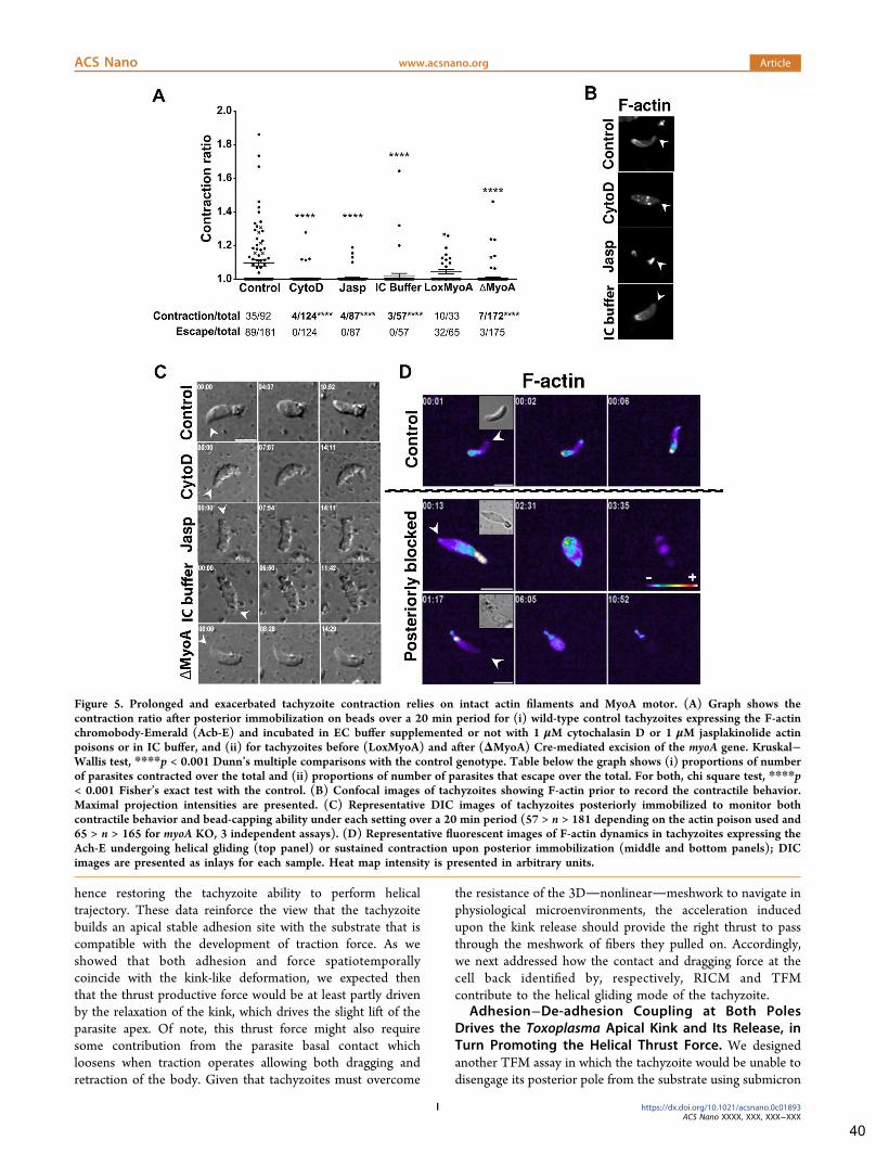
hence restoring the tachyzoite ability to perform helicaltrajectory. These data reinforce the view that the tachyzoitebuilds an apical stable adhesion site with the substrate that iscompatible with the development of traction force. As weshowed that both adhesion and force spatiotemporallycoincide with the kink-like deformation, we expected thenthat the thrust productive force would be at least partly drivenby the relaxation of the kink, which drives the slight lift of theparasite apex. Of note, this thrust force might also requiresome contribution from the parasite basal contact whichloosens when traction operates allowing both dragging andretraction of the body. Given that tachyzoites must overcome
the resistance of the 3Dnonlinearmeshwork to navigate inphysiological microenvironments, the acceleration inducedupon the kink release should provide the right thrust to passthrough the meshwork of fibers they pulled on. Accordingly,we next addressed how the contact and dragging force at thecell back identified by, respectively, RICM and TFMcontribute to the helical gliding mode of the tachyzoite.
Adhesion−De-adhesion Coupling at Both Poles
Drives the Toxoplasma Apical Kink and Its Release, in
Turn Promoting the Helical Thrust Force. We designedanother TFM assay in which the tachyzoite would be unable todisengage its posterior pole from the substrate using submicron
Figure 5. Prolonged and exacerbated tachyzoite contraction relies on intact actin filaments and MyoA motor. (A) Graph shows thecontraction ratio after posterior immobilization on beads over a 20 min period for (i) wild-type control tachyzoites expressing the F-actinchromobody-Emerald (Acb-E) and incubated in EC buffer supplemented or not with 1 μM cytochalasin D or 1 μM jasplakinolide actinpoisons or in IC buffer, and (ii) for tachyzoites before (LoxMyoA) and after (ΔMyoA) Cre-mediated excision of the myoA gene. Kruskal−Wallis test, ****p < 0.001 Dunn’s multiple comparisons with the control genotype. Table below the graph shows (i) proportions of numberof parasites contracted over the total and (ii) proportions of number of parasites that escape over the total. For both, chi square test, ****p< 0.001 Fisher’s exact test with the control. (B) Confocal images of tachyzoites showing F-actin prior to record the contractile behavior.Maximal projection intensities are presented. (C) Representative DIC images of tachyzoites posteriorly immobilized to monitor bothcontractile behavior and bead-capping ability under each setting over a 20 min period (57 > n > 181 depending on the actin poison used and65 > n > 165 for myoA KO, 3 independent assays). (D) Representative fluorescent images of F-actin dynamics in tachyzoites expressing theAch-E undergoing helical gliding (top panel) or sustained contraction upon posterior immobilization (middle and bottom panels); DICimages are presented as inlays for each sample. Heat map intensity is presented in arbitrary units.
ACS Nano www.acsnano.org Article
https://dx.doi.org/10.1021/acsnano.0c01893ACS Nano XXXX, XXX, XXX−XXX
I
40
sticky beads. As already reported in the context of membraneflow or cortical forces studies,32,42 the beads deposited on thetachyzoite surface underwent apico-basal translocation. How-ever, because in our setting the bead’s surface engaged covalentinteractions not only with the parasite but also with thefibronectin layer in a configuration that forced the former tostay posteriorly immobilized on the latter, the cell forwardprogression could be hindered (Figure 4D). Such posteriorlyconstraint tachyzoites never formed the apical kink (n = 0/92)and, instead, underwent a directional, minute time scale andrepetitive inward-oriented contraction visualized by the wholebody rounding (Figure 4D and Supporting Information Movie10). The TFM captured the displacement of in-gel beadssuitably positioned underneath the apex of the immobilizedparasite consistent with a sustained apical traction force, andtherefore a sustained adhesion between the parasite front andthe substrate (Figure 4D, zoomed panel, yellow bead andtrajectory). The prolonged force application induced pro-nounced shrinkage of the cell body along the longitudinal axisand eventually led to membrane damages to the extent thetachyzoite was likely wounded beyond repair. Meanwhile, weobserved that the in-gel beads shifted back to almost the initialposition, which we assumed to relate to the sudden relaxationof the cell “ghost” when internal tension fell (Figure 4D andSupporting Information Movie 10). Strong evidence for therupture of the plasma membrane upon excessive contraction ofthe YFP-expressing tachyzoite was provided by the acute lossof fluorescence that occurred immediately after maximal cellrounding and concomitantly with the relaxation of the cell(Supporting Information Figure S4). The relaxed tachyzoitecell looked mis-shapen for the rest of the recording time asexpected for a cell ghost, therefore attesting that theexacerbated and sustained contraction induced cell death. Tonext visualize the deleterious effect of the long-lastingcontraction on internal cytoskeletal structures at the nanoscaleresolution, we applied the expansion microscopy technique(ExM)45 and first validated the isotropic expansion of thespecimen with the 3D co-reconstruction of the tachyzoite bodyand nucleus prior and after expansion (Figure 4E). We thenanalyzed a tachyzoite line engineered to express a fluorescentversion of TLAP2, a protein aligned with the 22 cMTs,30 andconfirmed that during the prolonged contraction period, thecapped beads accumulated posteriorly while the spiral cMTsdeformed. We first observed an apical hump in the cMTnetwork (Figure 4F, middle panel), and eventually, the latterfell apart while the capped beads were redistributed over thedamaged surface (Figure 4F, bottom panel). Overall, thesedata support that the kink rose from the interplay between theapical adhesion/base de-adhesion and the forces generated atboth poles rather than from the apical traction force only. Theyalso highlight that creating unbalance between the front andback responses can result in a contractile force that imposes amechanical compression to the cMT network to which itcannot resist. The need for a balanced myosin-A-basedcontractile activity that stems at the tachyzoite apex andpromotes disengagement of the posterior pole can explain whythe genetically myosinA-deficient tachyzoites, which retainonly residual motility, do not any longer undergo helicalgliding.37,32
Tachyzoite Apico-Basal Contraction Requires IntactActin Filaments and Myosin A Motor and CorrelatesOnly Partially with the Requirements for RearwardMembrane Flow. Apart from the prolonged contraction and
fatal outcome described earlier, we also observed that thetachyzoites, which were posteriorly immobilized, could eitherrotate on the beads, slightly contract, and move forward, hencethose escaped (Supporting Information Movie 11) or remaininactive, hence those showed no contraction and stayedattached. To verify whether actin dynamics and MyoA motorthe function of which are required for motility25,46 would alsocontribute to the apically driven exacerbated contraction, wefirst defined a quantitative readout for measuring thecontraction extend over time. Using the ratio between theresting body length (i.e., no contraction) and the minimal bodylength (i.e., maximum contraction) as a contraction indicator,we found that nearly half of the wild-type (WT) controlparasites were able to escape from the immobilization setting,therefore attesting to the energetically active status of thespecimens under study. Approximately 40% of the second half(n = 35/92) contracted over a wide range of amplitude,whereas 20% of this contracting population actually showedsigns of irreversible damages (n = 7/35) (Figure 5A). Blockingactin dynamics with cytochalasin D (0.5 and 1 μM) orjasplakinolide (1 μM) that work through different mecha-nisms47,48 almost abolished both the escape (i.e., motile) andcontractile behaviors. Of note, the few (less than 5%)tachyzoites that displayed contraction never reached thestage of high and sustained body compression, hence noneof them died (Figure 5A). We confirmed the cytochalasin andjasplakinolide potency on tachyzoite actin as parasitesexpressing the fluorescent chromobody actin-Emerald (Acb-E)49 displayed the typical drug-induced redistribution of actinfilaments as, respectively, cytoplasmic aggregates and apicalacrosome-like.50 With the same readout, we also compared thecontractile behavior of parasites following or not MyoA genesilencing using the well-characterized DiCre Lox MyoAstrains.24 We observed that loss of MyoA (ΔMyoA)significantly altered the contractile capability of tachyzoitesimmobilized on their base when compared to the MyoA-expressing parental line (LoxMyoA) (ΔMyoA = 175, p <0.001) (Figure 5A). Finally, we checked if the contractileactivity was specific for tachyzoites metabolically ready forinitiating motility by measuring the contraction of WT controltachyzoites kept in the Endo buffer which mimics theintracellular buffer (herafter referred to as IC buffer) and isknown to drastically decrease microneme secretion (i.e.,adhesin delivery on the tachyzoite surface) and consequentlymotility.20 Those tachyzoites showed the typical actin posteriornetwork of intracellular tachyzoites51 and were significantlyinefficient at contracting under the posterior microbead setting(nIC = 57, p < 0.001 (Figure 5A−C and SupportingInformation Movie 12). Finally, we monitored actin filamentsin real time in Acb-E-expressing tachyzoites undergoing eitherhelical gliding or forced contraction during the immobilizationphase. The F-actin apical focus detected at the onset of thehelical cycle remained discrete and rapidly resolved into thelarge stable basal F-actin pool, as reported33,51 and in line withour force mapping assays. In contrast, an increasing F-actinsignal was detected within the whole body, in particular, in thecell front upon prolonged contraction of the immobilized androunding tachyzoites (Figure 5D and Supporting InformationMovie 13). Eventually, the F-actin signal vanished, which fitswith the loss of fluorescence observed with YFP-expressingtachyzoite artificially immobilized on the substrate throughtheir basal pole (Supporting Information Figure S4), therebyconfirming that the membrane had ruptured upon excessive
ACS Nano www.acsnano.org Article
https://dx.doi.org/10.1021/acsnano.0c01893ACS Nano XXXX, XXX, XXX−XXX
J
41

contractile activity, leaving a tachyzoite ghost, the apex ofwhich no longer attached to the substrate.As the retrograde membrane flow process has been shown to
operate in T. gondii tachyzoites in absence of the actomyosinsystem,32 we analyzed how this process would operate fortachyzoites artificially immobilized on their base. Unlikepreviously,32 we monitored in real time the bead-cappingprocess and found that the loss of contractility driven by actinpoisons and IC conditions did correlate with the inhibition ofthe rearward bead capping along the parasite surface (57 > n >181 depending on the condition) (Figure 5C and Figure 6Aand Supporting Information Movie 14), thus recapitulating thephenotype of formin 1(FRM1)-deficient tachyzoites33 andconsistent with the need of apically initiated actin filaments.However, on loss of myosinA,46 whereas the tachyzoitesshowed almost no ability to contract (Figure 5A), they
intriguingly maintained a bead-capping activity almost asefficient as tachyzoites from the parental line (65 > n > 165)(Figure 5C and Supporting Information Movie 12), inagreement with a MyoA contribution to force transmissionrather than production.32 Furthermore, along with these bead-translocation assays we observed in WT tachyzoites that thebead-capping process could precede productive helical move-ment (Figure 6A and Supporting Information Movie 14), andthat the bead apico-basal motion could follow the cMT helicaltrajectory. To confirm this observation, we applied ExM with arecently refined near-native expansion microscopy (U-ExM)protocol,52 and labeled the α,β-tubulin dimer that forms thecMT polymers taking advantage of the polyglutamylation post-translational modification of these isoforms.53 With this level ofresolution, we could measure the angle with the longitudinalaxis made by the cMTs when they emanate at the apical polar
Figure 6. Apico-basal translocation of surface-bound microbeads follows a helical path that approximates the spiral direction of thetachyzoite cMTs. (A) Representative images of apico-basal bead capping monitored in real time for tachyzoites posteriorly immobilized onbeads encircled with the dotted black line. The two surface-bound beads are indicated with white arrows, and their trajectory depicted inorange and cyan lines is shown on the fifth panel, before active escape of the tachyzoite from the immobilizing setting. In all panels, whitearrowheads mark the tachyzoite apex; all scale bars: 5 μm, time is in minutes/seconds. (B) Zoomed panels show images and schematics for(left) trajectory of two beads over the capping time taken from the sequence shown in A, which deviates from the body main axis (yellowline) of about a 11° angle, while (right) the spiral cMTs visualized on an extracellular tachyzoite treated for U-ExM and stained withantipolyglutamylated tubulin show about a 10° angle orientation. (Bottom) Plot of the different angles calculated for the cMTs; the meanwith SD is shown for each group (n = 10 tachyzoites, mean of 4 cMTs per tachyzoite) and for the capping bead flow (n = 9 tachyzoites).Please note the larger variability shown by the bead angles, which depends on the bead position at the initial time of capture and tracking.
ACS Nano www.acsnano.org Article
https://dx.doi.org/10.1021/acsnano.0c01893ACS Nano XXXX, XXX, XXX−XXX
K
42

Figure 7. Curvature of cortical microtubules changes during helical gliding suggesting a spring-like mechanism apically driven by myosin-based contractile forces. (A,B) Confocal images representative of extracellular tachyzoites following U-ExM and immunofluorescence co-staining of (A) tachyzoite cMTs using antipolyglutamylated tubulin antibodies (green) and nucleus (blue). Maximal projection intensities ofthe cMTs from the tachyzoite ventral side are shown for (top) a nonmotile tachyzoite, (middle and bottom) two motile tachyzoites. Note thepronounced torsion and compression (white arrowhead, middle panel) and the extension (white arrowhead, bottom panel) that likelyaccount for the spring like force underlying helical gliding. Right: Schematics of the cMTs for each stage show the highly curved cMTs at theapical region of the motile tachyzoites, drawn in red. (Bottom) Graph on the left shows a significant increase in mean curvature of the cMTs
ACS Nano www.acsnano.org Article
https://dx.doi.org/10.1021/acsnano.0c01893ACS Nano XXXX, XXX, XXX−XXX
L
43

ring while approximating the angle made by the microbeadsmonitored over the capping process (Figure 6B andSupporting Information Movie 14). Although we observedfor some beads that the angles were off the range of thosemade by the well-resolved cMTs, it is worth noting that thesegaps mainly correspond to situations where the microbeadswere tracked while they had already reached the minor axisplane of the ellipsoid tachyzoite. While traveling toward theposterior pole, the beads approached an area devoid of cMTsand often collided with the substrate before reaching theposterior pole, an observation already reported whenmonitoring the apico-basal trajectory of 0.6 μm beads deliveredby laser trap to the surface of resting (i.e., nonmotile)tachyzoites.42 Collectively, these data indicate that theactomyosin system mobilized during the rearward beadtranslocation process has aligned with the longitudinal cMTnetwork, this independently of the apical adhesion and thusbefore the apical traction force operates.Curvature of Cortical Microtubules Significantly
Changes during Helical Gliding, Suggesting a Spring-like Mechanism Apically Driven by Myosin-BasedContractile Forces. Triggered upon relaxation of the apicalkink, the extension of the tachyzoite body coincided with thethrust force while the parasite rotated with an orientationdictated by the MTs’ helical architecture (Figure 1C andFigure 3A and Supporting Information Movie 3a). Inmetazoans, the general ability of MTs to bear compressiveloads imposed by contractile forces translates into acoordinated bending of MTs and to drive a spring-like forcemechanism as exemplified with the rhythmically contractingcardiac myocytes.54 To interrogate whether the cMTs couldtransiently store energy upon actomyosin activation and acts asa torsional spring to promote the required thrust for helicalpropelling, we analyzed the behavior of cMTs during helicalmotion. Whereas the U-ExM allows nanoscale resolution, itdoes not permit to affirm that the tachyzoites underobservation were actually caught in motion. As under pro-gliding conditions, the tachyzoites displayed a statisticallysignificant elongation of the nucleus as compared to thesamples of either extracellular tachyzoites left in IC buffer ortachyzoites which had just invaded host cells (n > 75 for eachcondition, Supporting Information Figure S5A), we used thenucleus sphericity as first criterion. We next selected fortachyzoites associated with helical trails using the surface-exposed glycoprotein SAG1 known as robust trail marker,11
which also bore an elongated nucleus (p < 0.0001, n = 12motile helical and n = 15 intracellular) (SupportingInformation Figure S5B). To analyze whether individualcMTs could display a specific compression, relaxation andtorsion behavior in motile tachyzoites, we applied U-ExM andused both antiacetylated α-tubulin and antipolyglutamylateda,β-tubulin antibodies which have been widely used for labelingT. gondii cMTs.53 When quantifying the curvature of the cMTs
in nonmotile extracellular (Figure 7A, top panel) or nonmotileintracellular (Supporting Information Figure S5C, top panel)and motile parasites (Figure 7A, middle panel; SupportingInformation Figure S5C, bottom panel), we observed for themotile specimens, a highly significant increase in the meancurvature of the MTs as compared to the nonmotile ones(Figure 7A, polyglutamylated tubulin, p < 0,0001, n = 57nonmotile and n = 44 motile parasites) (SupportingInformation Figure S5B,C, acetylated tubulin, p < 0,0001, n= 24 nonmotile and n = 22 motile tachyzoites). Importantly,the curvature amplitude over the length of a single MT wasalso significantly enhanced in gliding when compared tointracellular parasites as assessed by the standard deviationvalues of the curvature profile over the full length MTs (Figure7A, polyglutamylated tubulin, p < 0,0001, SupportingInformation Figure S5B,C, acetylated tubulin, p < 0,0001).The decreased interdistance between single cMT nearby theapical ring (APR) from where they emanate together with theobserved nonuniform bending over the cMT length stronglyargue for a specific compression occurring at the tachyzoiteapex during cell movement. In addition, situations where thecMTs straightened at the apical region likely corresponded tothe time of the thrust force occurring upon kink release (Figure7A, bottom panel; see also schematics on the right). To furthertest the hypothesis of cooperation between the actoMyoA-based and the cMT-based cytoskeletons could contribute toforce production at the kink, we performed U-ExM imaging ofMyoA and cMTs simultaneously on either motile or nonmotiletachyzoites. To this end, we used a tachyzoite line thatexpresses a chimeric MyoA-HA protein under the endogenouspromoter and anti-HA antibodies. A clear co-alignment of theMyoA and the cMTs was detected in the apical region ofmotile tachyzoites together with a local enrichment in MyoAwhen compared to nonmotile parasites (Figure 7B). Althoughit is not possible to ascertain that this situation reflects the kinkstep, the alignment between the MyoA and the cMTs makesplausible an indirect connection and thus argue for an energytransfer between the two cytoskeletons. In addition, this co-alignment is consistent with the helical path taken by the beadsundergoing actin-based rearward capping. In metazoans cells, itwas found that the MTs were embedded in a viscoelasticmaterial provided by the surrounding medium and MT-associated proteinsin particular, kinesin motors55thatinfluence the MT ability to buckle and bear energy load.54
Therefore, it would be worth investigating whether some MTor/and IMC protein partners - including the APR-associatedkinesin,56 proteins that coat the cMT length and alveolinspositioned at the apical flexure site could behave as pro-elasticelements and thus optimize a cMT driven spring-like reaction.Of note the intrinsic stable left-handed curvature of≪resting≫ cMTs already sign for their ability to withstandlarge forces within the cytoplasm in agreement with theircontribution to shape maintenance.30,57 As the activation of
Figure 7. continued
for the motile tachyzoites in torsion, unpaired t test, ***p < 0.0001 n = 44, when compared to the nonmotile (n = 57); graph on the rightshows enhanced curvature amplitude over the length of single cMT for motile tachyzoites during the compression step (n = 47) whencompared to nonmotile (n = 57) tachyzoites, unpaired t test, ***p < 0.0001. (B) Co-staining of the cMTs (green) and MyoA-HA taggedusing anti-HA antibodies (purple) and the nucleus (blue). (Top) Nonmotile tachyzoite, (bottom) motile tachyzoite are shown. Note theclear MyoA signal enrichment at the apex of the motile parasite indicated with white arrowheads, which co-aligns with the MT signal. Theright frames show zoomed areas delineated in the merge frames with a white square. All scale bars: 5 μm except for the B zoomed frames: 2μm. The U-ExM protocol gave a 3−5-fold increase of the tachyzoite size and provided a ∼70 nM xy resolution. APR: Apical polar ring.
ACS Nano www.acsnano.org Article
https://dx.doi.org/10.1021/acsnano.0c01893ACS Nano XXXX, XXX, XXX−XXX
M
44

the actomyosin system translates into force-powering motility,it is plausible that the significant local increase in cMTcurvature of motile tachyzoites accounts for higher compres-sive loads on the cMTs in response to the increasedcontractility. Indeed, when the tachyzoite underwent exacer-bated actomyosin-driven contraction (Figure 4F) in a failedattempt to disengage its base from the adhesion site, the cMTcytoskeleton overcame its bending capacity and eventuallybroke up.
CONCLUSIONS
In this study, by combining surface sensitive biophysicaltechniques with high resolution-high speed 2D and 3D liveimaging and expansion microscopy, we provided insights onhow the T. gondii parasite couples cycles of adhesion−de-adhesion to the substrate with traction and spring-like forcegeneration to achieve helical gliding, a function which isrequired for perpetuating T. gondii populations in thehomeothermic hosts. Owing to its persistent structural polarityover the intracellular/extracellular cycles, the tachyzoitebypasses the canonical symmetry break most eukaryotic cellsmust undergo to start migrating.58 In addition, althoughlacking the typical actomyosin II motor commonly used togenerate traction in eukaryotes, Toxoplasma has evolved anApicomplexa-restricted unconventional myosinA motor that,immobilized within the glideosome unit and between themembrane layers forming the pellicle at the cell surface, workson noncanonical actin filaments to activate motility.29 Thepellicle and the subtending twisted 22 microtubules providethe tachyzoite with a peculiar crescent shape and flexibility,which has proposed to act as the primary driver of the helicalmotion. We now demonstrate that the T. gondii tachyzoitemust engage its apical region in a specific stable adhesion withthe substrate that is compatible with the development of abalanced traction force and drives, in turn, the loosening of theposterior contact point, allowing retraction of the parasite baseand dragging force. Therefore, similarly to what was found forthe Plasmodium motile sporozoite, real time analysis ofadhesion dynamics and force mapping supported by recentlyrefined high spatiotemporal resolution imaging approaches andbiophysics characterization applied to the Toxoplasma motiletachyzoite have challenged the classic model of Apicomplexagliding motility. In addition to the backward capping ofadhesion proteins at the cell surface, both parasite rely on adistinct class of adhesive contact that do not enter the rearwardflow but directly contribute to build tension in the parasitecytoskeleton and produce force by strengthening anchorspoints with a specific on−off dynamics, hence regulating themotile process. Although these studies confirm the need toshift paradigm for a comprehensive mechanistic understandingof Apicomplexa gliding motility, they raised key questions onthe nanoscale architecture of the singular adhesive platformsincluding the adhesin ligand identities on the extracellular side,the actin filament organization on the cytoplasmic side, butalso how they would transmit tensional forces from thecytoskeleton to the ECM with the yet to clarify contribution ofthe MyoA motor, and eventually, it would be worthinvestigating their putative contribution for mechanotransduc-tion. Providing indication of a spatiotemporally regulatedcooperation between the tachyzoite actomyoA-based contrac-tile activity and the helical flexible cMT ability to build tension,this study gives credit to pioneered kinematic analysis.18 Theseresults also highlight the necessity to delve deeper into the
compressive, torsional, and tensile strengths of Toxoplasmatachyzoite and also Plasmodium sporozoite cMTs as somedifferences between the two stages could in part account fortheir specific motile behavior.
METHODS/EXPERIMENTAL
Parasite Strain Maintenance and Preparation for AllMotility Live Assays. All media and products used for cell culturewere from Gibco-Life Technologies (St Aubin, France) unlessspecified. T. gondii strains were propagated on Mycoplasma-freehuman foreskin fibroblast monolayers (HFFs) and grown inDulbecco’s modified Eagle medium (DMEM) supplemented withglutamax, 10% heat-inactivated fetal calf serum (FCS), penicillin (100U/mL), and streptomycin (100 μg/mL) at 37 °C and 5% CO2. Thelaboratory type 1 RH strain was used as well as the YFP-expressingRH17 and the mutant deleted for the KU80 protein59 (gift from V.Carruthers, Michigan University, USA; see Molecular Cloningsection). The RH Δku80: DiCre/lox MyoA, RH Δku80: DiCre/ΔMyoA,60 and the RH cbactinEmFP49 expressing strains were givenby M. Meissner (MLU Munich University, Germany) and hereafterdesignated LoxMyoA, ΔMyoA, or Ach-E, respectively. For mostmotility assays, tachyzoites were collected within a few hoursfollowing spontaneous egress from the HFF monolayers, and ∼105
cells were centrifuged in HBSS++ supplemented with 0.2% FCS (0.2%HBSS). The pellet containing parasites was resuspended in 150 μL of1% FCS HBSS (1% HBSS) and adjusted at 1.6 mM of CaCl2 (1%HBSS-Ca2+). Typically, when using the 18 mm diameter glasscoverslip fitting chamber, 50 μL of the tachyzoite suspension wasadded to 100 μL of 1% FCS HBSS-Ca2+ covering the coverslip. Theamounts of FCS, CaCl2, and pharmacological reagents were adjustedupon need as described in each appropriate section.
Molecular Cloning. A mCherry (mC) tag was fused in frame withthe C-terminus of TLAP2 gene (ToxoDb.org, TGGT1_232130) inthe ΔKu80 strain using the ligation independent cloning strategy.59 A1.5 kb fragment corresponding to the 3′ end of the TLAP2 excludingthe stop codon was amplified by PCR with the forward primer 5′-GCGCCCTCCTTCAGTGTTCTTC-3′ and reverse primer 5′-TGCACCCTGCGGCACCGGCAG-3′. The PCR fragment andvector p-LIC-mCherry-HXG were digested with T4 DNA polymeraseand annealed, yielding plasmid pTLAP2mCherry-LIC-DHFR. Freshlyegressed Δku80 tachyzoites from a 1/2 T25 flask were transfectedwith 15 μg of the EcoRV-linearized plasmid. After one cycle withoutdrug, ΔKu80/TLAP2-mC transformants were selected with 2 μg/mLpyrimethamine and single-cell cloned by limiting dilution.
Endogenous tagging of MyoA with HA-tag was achieved using thepLIC-MyoA-HA-HXGPRT vector. To construct this plasmid thecoding sequence of MyoA was amplified using primers 5′-TACTTCCAATCCAATTTAGCAAAGCAGCAAACTGCCCA-GCGAAG-3 ′ and 5 ′ -TCCTCCACTTCCAATTTTAGC-GAACGCCGGCTGAACAGTCGCGGG-3′ and RH Δku80 genomicDNA as template. The PCR product was cloned to pLIC-mAID-HA-HXGPRT vector using the LIC cloning method as described above.61
RHku80 OsTir1-(Ty)3 parasites were electroporated with BstBI-linearized plasmid, and recombinant parasites were selected withmycophenolic acid (25 μg/mL) and xanthine (50 μg/mL). Stabletransgenic parasites were single-cell cloned by limiting dilution.
Video Microscopy. Time-lapse video microscopy was conductedin Chamlide chambers (LCI Corp., Seoul, Korea) accommodating 18mm diameter coverslips or 35 mm MatTek glass bottom dishes(Matek corporation) installed on an Eclipse Ti inverted confocalmicroscope (Nikon France Instruments, Champigny sur Marne,France) set up with a temperature and CO2-controlled stage andchamber (LCI Corp., Seoul, Korea). The microscope was equippedwith the sCMOS prime camera (Photometrics), a 60× objective, anda CSU X1 spinning disk (Yokogawa, Roper Scientific, Lisses, France).MetaMorph software was used for controlling the microscope(Universal Imaging Corporation, Roper Scientific, Lisses, France).All live microscopy was performed at 37 °C and 5% CO2.
ACS Nano www.acsnano.org Article
https://dx.doi.org/10.1021/acsnano.0c01893ACS Nano XXXX, XXX, XXX−XXX
N
45
Image Processing. For image processing, we combined the use ofImageJ, Icy, MATLAB, ChimeraX, and Amira-Avizo (Thermo-Fisher)software. Details of image processing are included in the appropriateprotocol sections. Slow motion videos were created using AdobePremiere with the optical flow module.Motility Assays in Collagen1 Fibrous Matrices. Collagen gels
were made using rat tail type I collagen (CellSystems). Nine parts ofcollagen dissolved in acidic medium were mixed with 1 part ofneutralization solution to reach the concentration of 4 mg/mL andpromote matrix formation. All solutions were kept in 4 °C duringpreparation. Then dilution of the gel at a concentration of 2 mg/mLin HBSS++ supplemented with 1% FCS was rapidly spread in themiddle of a MatTek glass bottom dish and incubated for 30 min at 37°C and 5% CO2. Labeling of the collagen fibers was achieved with therecombinant CNA35-EGFP produced in Escherichia coli (BL21-DE3strain) used at 2 μM final concentration according to the protocol.35
A 50 μL volume of the tachyzoite suspension prepared in 1% FCSHBSS-Ca2+ was deposited on top of the gel prior to being transferredin the microscopy stage for video microscopy. Once tachyzoites hadpenetrated within the mesh, images were captured at 1 frame/s. Fortracking the fibers during parasite navigation, we used Icy andvisualized with green the initial position of the fibers at t = 0 s whilewe duplicated this frame as many times as the number of the time-lapse frames. Then we merged this “time zero” stack with the originaltime lapse in which the fibers were colored in magenta. The whitishcolor accounts for the green and magenta overlay and indicates nodisplacement, whereas the detection of the green and magenta revealsthe fiber displacement during the sequence.Tachyzoite 2D Gliding Assays. Parasites were prepared as
described above, and assays were carried on 18 mm plasma-activatedglass coverslips coated with either fibronectin (20−50 μg/mL inphosphate-buffered saline (PBS)) or poly-L-lysine (150−300 kDaPLL, 50 μg/mL in PBS). Image analysis included tracking thetachyzoite back and front parts over the gliding time by using Icy asdescribed.62 “Manual tracking” and “Motion profiler” plugins wereused to extract the x−y position of the back and front spots over time.Having tracked the x−y positions over time, we used the “Distanceprofiler” plugin to calculate the distance between the two positions(back and front) over time.Reflection Interference Contrast Microscopy. RICM images
were acquired on an inverted microscope (IX71, Olympus) on whicha white-light source (HPLS 345, Thorlabs) was coupled using ahome-built illumination arm incorporating a diaphragm (SM1D12C,Thorlabs) controlling the illumination numerical aperture (NA = 0.46in all experiments shown here) and a triple-band spectral filter (FF01-457/530/628-25, Semrock) as described.63 The usual fluorescencecube in the microscope turret was replaced by polarization optics,allowing separation of the incoming and reflected light (polarizerWP25M-VIS, Thorlabs), polarizing beamsplitter cube (PBS251,Thorlabs), and achromatic imaging flat quarter waveplate (QWP;custom-made, Fichou, France). The sample was imaged with an oil-immersion objective (60XO UPLSApo, Olympus, Japan) onto asCMOS camera (Orca Flash 4 v2, Hamamatsu) after passing throughan autofocus device (CRISP system, ASI imaging, USA) and a home-built image-splitting setup incorporating a variable slit (VA100/M,Thorlabs), achromatic relay lenses (ITL200, Thorlabs), imaging flatdichroic mirrors (FF484-FDi01-25X36 and FF560-FDi01-25X36,Semrock) gently held through curable silicon paste (Sugru, Form-FormForm, UK), silver mirrors (PFSQ10-03-P01, Thorlabs) anddichroic filters (blue: Semrock FF01-452/45-25; green: FF01-531/46-25; red: FF01-629/56-25, Semrock). In addition, for bright-field (BF)imaging, a red colored glass filter (FGL610S, Thorlabs) was used tofilter the light for the microscope illumination arm that wassubsequently focused by a long-distance 0.3 NA condenser (allOlympus, Japan). BF and RICM images were acquired simulta-neously, in the red channel, and thus show a dim RICM imagesuperimposed. Finally, a custom-built thermostated box (Digital Pixel,UK) enclosing most of the microscope was used to control thetemperature at the sample. Image processing was done using ImageJ.Blue, green, and red stacks of images were first manually registered.
RICM images were subsequently filtered using the “FFT band-passfilter” plugin (1−100 pixels) to remove high-frequency noise anduneven illumination background. The two (in the case ofsimultaneous BF/RICM imaging) or three (in the case of onlyRICM imaging) RICM images were then superimposed andconverted into a RGB image. BF images and RGB RICM imageswere then segmented using the “trainable Weka segmentation”plugin.64 For BF images, the classifier was trained to segment thewhole parasite, and the outline of the resulting binary image wasextracted subsequently. For RICM images, the algorithm was used toseparate the “close contact” area defined by the first dark RICM fringeon the parasite surface, the trail (when relevant), the rest of the RICMsignal coming from the parasite (when no BF image was present), andthe rest of the image. Because the classifier could detect the seconddark RICM fringe manual correction of the segmented images wasperformed afterward to remove incorrect areas classified as “closecontact”. A binary image was obtained for the “close contact” area,whereas an 8-bit probability image was extracted for the trail. Analysisof the processed images was also performed using ImageJ. Thekymograph was obtained using the plugin “KymoResliceWide”. Formeasuring the speed of the parasite back end, the corresponding whiteline on the kymograph was isolated and the positions of the pixelswere exported to Origin. After smoothing, the position curve wasderived to obtain the velocity of the back end over time.
Traction Force Microscopy. TFM setting was adapted from ref65. The 2 kPa PAA gels were made and mixed with TFM beads(FluoSpheres carboxylate-modified microspheres, ThermoFisherScientific, 0.2 μm dark red fluorescent, λ: 660/680, 2% solid). Thegel was top-coated with fibronectin (20 μg/mL, 30 min, 23 °C)diluted in sodium bicarbonate pH 8.3 (100 mM). Typical sampleswere made from stocks of 0.7 μL beads mixed in 165 μL of gelsupplemented with 1 μL of both tetramethylethylenediamine(TEMED) and ammonium persulfate (APS) 10% using 20 μL ofbead-gel suspension for an 18 mm diameter silanized glass coverslip.A 50 μL volume of the tachyzoite suspension prepared in 1% HBSS-Ca2+ (see above) was deposited on top of the fibronectin-coated PAAgel already filled with 100 μL of 1% HBSS-Ca2+ in the appropriatechamber and video recording started upon tachyzoite contact with thesubstrate. MetaMorph streaming option was used for fast recording(<1 frame/s) in both the DIC and far-red channels. Analysis of theTFM beads displacement for extracting the force values carried outusing MATLAB. The TFM bead displacement was assessed using as areference bead position, the frame with a gel area free of parasite (i.e.,relaxed position of the gel). Then using the frames taken over time,the bead’s displacement was tracked from their initial relaxed position.To check the tachyzoite gliding behavior on a substrate of differentstiffness, gels of different PAA concentration and coated with a layerof fibronectin were made using the same protocol, omitting the beadaddition step, and the stiffness was controlled by atomic forcemicroscopy.
Micropatterned Devices. Micropatterned PEG-fibronectincomposite coverslips were engineered by Alveole using the PRIMOpatterning with specific area coated with a mix of fibronectin (100 μg/mL) and AlexaFluor647-coupled fibrinogen (10 μg/mL) and othercoated with PLL−PEG. Fifty microliters of the tachyzoite suspensionprepared in 1% HBSS-Ca2+ was deposited on top of the fibronectin-coated glass coverslip placed in the chamber and prefilled with 100 μLof buffer prior to video microscopy.
Rear-Blocked Parasite Assay. The 0.2 μm diameter beads(FluoSpheres carboxylate-modified microspheres, ThermoFisherScientific F8807, λ: 660/680, 2% solid) were activated using themicrosphere coupling−two-step EDC/Sulfo NHS covalent couplingprocedure for Estapor carboxyl-modified dyed microspheres protocol(MerckMillipore). Fifty microliters of the stock-activated beadsolution was diluted in 150 μL of MES buffer (50 mM, pH 6.0).Then 30 μL of the bead solution was added to 120 μL of thetachyzoite suspension in 0.15% FCS HBSS++ (0.15% HBSS). Fiftymicroliters of the parasite suspension (from either the RHΔKu80,ΔMyoA, RHΔKu80Acb-E, or YFP tachyzoite lines) was deposited onthe (50 μg/mL) fibronectin-coated glass coverslips in the microscopy
ACS Nano www.acsnano.org Article
https://dx.doi.org/10.1021/acsnano.0c01893ACS Nano XXXX, XXX, XXX−XXX
O
46

chamber, which was already filled with 200 μL of 0.15% HBSS++. Insome experiments, parasites were preincubated for 10 min with 0.5 or1 μM of either jasplakinolide or cytochalasin D (Sigma) and video-recorded in the presence of the compounds. For the intracellularmimicking conditions, tachyzoites collected by centrifugation in0.15% HBSS++ were rinsed in ENDO buffer referred to here as ICbuffer (145 mM KCl; 5 mM NaCl; 1 mM MgCl2; 15 mM MES; 15mM HEPES; pH 8.3)66 before use, and the activated beads wereprepared in IC buffer. The mixture prepared as mentioned above wasdeposited on the microscopy chamber, which was prefilled with 200μL of IC buffer. Images were acquired at 1 s intervals for 20 min. Todescribe the contractile potential of tachyzoites under eachexperimental condition, we calculated the contraction ratio bymeasuring two times the length of the parasite body starting fromthe back until its farther apical point but excluding the conoid to avoidfalse measurements in case of fluctuating extrusion. The first measurecorresponded to the relax position and the second to the maximalcontraction. Division of the relaxed versus the contracted lengthprovided the contraction ratio. When needed, both DIC andfluorescence channels were recorded at ∼1 frame/s.Bead Flow Assays. The assay was performed similarly to the rear
blocked parasite assay. For tracking the beads, visualizing their tracksand extracting their speed, “Spot detector” and “Track manager”plugins (Icy) were used.Expansion Microscopy. To analyze gliding tachyzoites, we
collected the parasites in either in prewarmed 1% FCS HBSS-Ca2+
or IC buffer, which were centrifuged at low speed to synchronize theirsedimentation and left to glide for 10 min on PLL (50 μg/mL)-coatedplasma-activated glass coverslips (37 °C, 5% CO2). Gliding wasstopped by addition of paraformaldehyde (PFA, 3.2%, 15 min). Thetachyzoite surface and trails were immunolabeled with anti-TgPSAG1antibodies (mAb, clone TP3), and the chimeric TLAP2 protein wasdetected using the rabbit anti-mCherry antibodies (Institut Curie,TAb-IP Paris, France) directed against the mC tag after cellpermeabilization with TritonX-100 (0.1% in PBS, 5 min). Thesamples were next incubated with appropriate secondary highly cross-adsorbed antibodies (4 μg/mL, 2h). The ExM protocol was adaptedfrom ref 45. Briefly Acryloyl-X was used at 0.1 mg/mL PBS for 2 h at23 °C prior to the gelation step, which was performed by placing thegelation solution (80 μL) on parafilm and the coverslip with the cellsample face down on top of it. Gelation was allowed to proceed at 37°C for 1 h followed by the digestion step (0.5% Triton X-100, 8 units/mLProteinase K) carried out overnight at 23 °C. A 4 mm diameter gelcore sample was collected with a puncher and immerged in 5 mL ofddH2O in a 50 mm diameter Petri dish to promote expansion, whilePI staining was concomitantly performed (2.5 μM in PBS). H2O wasexchanged every 20 min until maximal expansion. The piece of gelwas next gently transferred onto a PLL-coated glass coverslip in theminimal liquid volume to avoid gel drift. The 0.2 μm Z-image stacksof parasites in stages were captured using a spinning disk confocalEclipse-ti Nikon microscope.Ultrastructure Expansion Microscopy. Gliding assays were
performed with RHΔKu80MyoA-HA similarly to that for ExM.Additionally, for the invasion assay, HFF cells were plated on a poly-L-lysine-coated glass coverslip to obtain 80% cell confluence on thefollowing day. Parasites were settled on top of the cells by gentlecentrifugation (2 min, 250g) and left to invade for 1 h (37 °C, 5%CO2). For U-ExM, we followed the protocol developed by theGuichard laboratory,52 but to optimally preserve both free andintracellular tachyzoite shapes, we found it better to fix the samplesbefore expansion in a solution made of 0.7% formaldehyde and 1%acrylamide in PBS (4−5 h, 37 °C). Next, similar to the ExM protocol,gelation was carried out for 1 h (37 °C). Coverslips with gels werethen transferred into ∼2 mL of the denaturation buffer in a 6-wellplate for 15 min (23 °C). Gels were removed from the coverslips withtweezers and transferred to a 35 mm diameter glass Petri dish filledfresh denaturation buffer for a 60 min incubation at 95 °C. Afterdenaturation, gels were placed in a 150 mm diameter glass Petri dishfilled with ddH2O for the first expansion. Water was exchanged atleast twice every 30 min at room temperature, and gels were
incubated overnight in ddH2O. After limited shrinkage in PBS, several4 mm diameter pieces of gel were collected and deposited on 24-wellplates. Gels were then incubated for 3 h at 37 °C under shaking inprimary antibodies (antiacetylated α-tubulin, mAb, clone 6-11B-1) orrabbit α- polyglutamylated (mAb, clone GT335) and rat anti-HAantibodies (mAb, clone 3F10). Gels were washed in PBS-Tween 0.1%prior to incubation with appropriate secondary highly cross-adsorbedantibodies (4 μg/mL) for 150 min at 37 °C and washed again in PBS-Tween 0.1%. Expansion was achieved by controlled immersion inddH2O. The PI staining, final ddH2O wash, and gel mounting stepswere all performed as mentioned for ExM.
ExM and U-ExM Image Processing. 3D reconstruction wasachieved with the UCSF ChimeraX software from the raw data (i.e.,xyz files) processed with ImageJ to first crop the region of interest andthen to apply the “Iterative Deconvolve 3D” plugin for each channel.We used the ImageJ “3D Object counter” plugin to check for theisotropic physical expansion of the specimen by comparing the ratioof the parasite (SAG1 staining) and the nucleus (PI staining) volumesin controlled and expanded extracellular tachyzoite samples. Thenucleus sphericity index of motile, nonmotile, and intracellulartachyzoites was compared by thresholding each image object to create3D binary images, whereas the “3D Object counter” plugin was nextused to segment Z-stack binary images and retrieve the volume andsurface of the 3D objects (i.e., parasite nucleus and whole body). Thesphericity index was calculated using the formula where V is thevolume and S the surface:
π
Ψ =V
Ssphericity
(36 )2 1/3
The nucleus roundness was obtained by thresholding a Z-Maxprojection from the Z-stack images and using the “Particle analysis”plugin.
For the tubulin curvature analysis in expanded specimens, we usedthe “Kappa” plugin in Fiji on the Z-Max projection from Z-stackimages of the tachyzoite ventral cMTs (i.e., facing the substrate),which allowed tracing each MT shape and retrieving the meancurvature of each cMT as well as the curvature amplitude over thecMT length (standard deviation).
MIC2 and SAG1 Analysis in Helical Trail. Extracellulartachyzoites were let to glide for 15 min prior to be fixed with PFAas described in the ExM section. A double immunolabeling wasperformed sequentially using the anti-TgSAG1 protein (clone TP3,30 min, 23 °C) and the anti-TgMIC2 (clone 6D10, overnight, 4 °C)as primary antibodies, each followed by incubation with appropriatesecondary highly cross-adsorbed antibodies (4 μg/mL, 2 h). Imageswere acquired on the Axio Imager Z1 Zeiss microscope using theZeiss imaging software ZEN before being processed using Fijisoftware.
To determine the size and position of the adhesion point formed atthe early step of the helical cycle (i.e., coinciding with the kink) on theMIC2/SAG immunolabeled trails, we used the machine-learning-based segmentation of the real-time RICM sequence as a template.The region of interest for the trail trajectory, the angular area, and theparasite apical adhesion area were defined manually with Fiji: thedistance between the center of adhesion point and the angle point wasmeasured for six angles over a sequence of successive helical cycles,which allowed positioning the apical adhesion at about +0.170 μmdownstream the angle point, and the size of the adhesion point wasestimated around 0.5 μm. Applying these values on the MIC2/SAG1labeled trails, it was possible to accurately identify the adhesion andangle area over the profiles of fluorescence intensity measured for thewhole trails trajectories that in total defined 40 helical cyclesperformed by eight tachyzoites. The fluorescence intensity is given asa ratio by normalizing on the mean intensity value.
Quantification and Statistical Analysis. Data were statisticallyanalyzed, and plots were made using GraphPad Prism 6.0 software forWindows (La Jolla, CA, USA). Data are presented as mean ±
standard error of mean if not indicated otherwise. Figure legendsinclude the statistical test and resulting comparison when appropriate.
ACS Nano www.acsnano.org Article
https://dx.doi.org/10.1021/acsnano.0c01893ACS Nano XXXX, XXX, XXX−XXX
P
47

For Figure 4A, Figure 7A, and Figures S3A and S5B,C, an unpaired ttest was used with significance being represented as a p value <0.05,and the n indicated represents the parasite sample size or the numberof individual microtubules analyzed. For Figure 4E, Figure 5A, andFigure S5A, an unpaired Kruskal−Wallis test was performed, and then indicates the sample size. For the table in Figure 5A, a chi-squaretest followed by Fisher’s exact text was used, with the n showing theproportions analyzed.
ASSOCIATED CONTENT
*sı Supporting Information
The Supporting Information is available free of charge athttps://pubs.acs.org/doi/10.1021/acsnano.0c01893.
Helical and circular gliding by T. gondii tachyzoites;tachyzoite apical adhesion site is not enriched in MIC2and SAG1 proteins; frequency and duration of T. gondiitachyzoite gliding behavior depends on the substratestiffness; prolonged and exacerbated contraction inducedupon tachyzoite posterior immobilization leads tomembrane leakage; expansion microscopy allows un-covering cMT local selective compression and torsion ingliding tachyzoites (PDF)Movie 1: Parasite moving in 3D collagen fibers1 (AVI)Movie 2: Parasite moving 3D collagen fibers2 (AVI)Movie 3a: Parasite helical gliding in 2D (AVI)Movie 3b: Slow motion but same as movie 3a (AVI)Movie 4: Parasite circular gliding in 2D (AVI)Movie 5: RICM analysis of parasite helical gliding (AVI)Movie 6: RICM-pulling on or breaking the trail (AVI)Movie 7a: TFM analysis of parasite helical gliding (AVI)Movie 7b: Slow motion but same as movie 7a (AVI)Movie 8: Parasite helical gliding on micropatternAlveole1 (AVI)Movie 9: Parasite helical gliding micropattern Alveole2(AVI)Movie 10: Parasite base blocked by microbeads (AVI)Movie 11: Bead release from the base of a movingparasite (AVI)Movie 12: Parasite contraction in the presence of drugs(AVI)Movie 13: Actin dynamics in gliding or immobilizedparasites (AVI)Movie 14: Bead flow and bead release in tachyzoites(AVI)
AUTHOR INFORMATION
Corresponding AuthorIsabelle Tardieux − Institute for Advanced Biosciences (IAB),Team Membrane Dynamics of Parasite−Host Cell Interactions,CNRS UMR5309, INSERM U1209, Universite GrenobleAlpes, 38700 Grenoble, France; orcid.org/0000-0002-5677-7463; Email: [email protected]
AuthorsGeorgios Pavlou − Institute for Advanced Biosciences (IAB),Team Membrane Dynamics of Parasite−Host Cell Interactions,CNRS UMR5309, INSERM U1209, Universite GrenobleAlpes, 38700 Grenoble, France
Bastien Touquet − Institute for Advanced Biosciences (IAB),Team Membrane Dynamics of Parasite−Host Cell Interactions,CNRS UMR5309, INSERM U1209, Universite GrenobleAlpes, 38700 Grenoble, France
Luis Vigetti − Institute for Advanced Biosciences (IAB), TeamMembrane Dynamics of Parasite−Host Cell Interactions, CNRSUMR5309, INSERM U1209, Universite Grenoble Alpes,38700 Grenoble, France
Patricia Renesto − Institute for Advanced Biosciences (IAB),Team Membrane Dynamics of Parasite−Host Cell Interactions,CNRS UMR5309, INSERM U1209, Universite GrenobleAlpes, 38700 Grenoble, France; TIMC-IMAG UMR 5525 -UGA CNRS, 38700 Grenoble, France
Alexandre Bougdour − Institute for Advanced Biosciences(IAB), Team Host−Pathogen Interactions & Immunity toInfections, CNRS UMR5309, INSERM U1209, Universite Grenoble Alpes, 38700 Grenoble, France
Delphine Debarre − Laboratoire Interdisciplinaire de Physique,UMR CNRS, 5588, Universite Grenoble Alpes, Grenoble38402, France
Martial Balland − Laboratoire Interdisciplinaire de Physique,UMR CNRS, 5588, Universite Grenoble Alpes, Grenoble38402, France
Complete contact information is available at:https://pubs.acs.org/10.1021/acsnano.0c01893
Author Contributions
Conceptualization, I.T., M.B, G.P; Software, M.B., D.D, G.P.;Formal Analysis, G.P., B.T., D.D, L.V.; Funding acquisition,I.T.; Investigation; G.P., B.T., I.T., V.L., D.D., P.R., A.B.;Methodology, I.T., G.P., B.T., A.B.; Project administration,I.T.; Supervision, I.T., M.B., P.R.; Validation, I.T., G.P., M.B.,D.D.; Visualization, G.P., B.T., L.V.; Writing−original draft,I.T. Writing−review and editing, I.T., G.P., P.R., A.B., M.B.,D.D.
Funding
This work has been funded by the IAB department“Prevention and Therapy of Chronic Diseases (Grenoble,France) with an internal grant. The University Grenoble-Alpesand the Fondation pour la Recherche Medicale (FRMFDT201904008408) awarded to G.P. with a 3 year Ph.D.fellowship and 6 month Ph.D. extension fellowship, respec-tively.
Notes
The authors declare no competing financial interest.
ACKNOWLEDGMENTS
We thank T. Boudou for help with the collagen gels, T.Andersen, V. Petrolli, and A. Ruppel for help in setting andanalyzing the TFM assays. They all belong to the LaboratoireInterdisciplinaire de Physique, UMR CNRS, 5588, Universite Grenoble Alpes, France. We thank C. Guilluy for the AFMassays to probe gels, M. Bogeda for help in micropatterning(IAB, Grenoble), and the Alveole team for providing thecomposite micropatterns. We thank the therapeutic antibodies(Tab-IP) and recombinant antibodies platform from the CurieInstitute, Paris, France for providing the anti-mCherryantibodies.
REFERENCES
(1) Scarpa, E.; Mayor, R. Collective Cell Migration in Development.J. Cell Biol. 2016, 212, 143−155.(2) Friedl, P.; Weigelin, B. Interstitial Leukocyte Migration andImmune Function. Nat. Immunol. 2008, 9, 960−969.(3) Tschumperlin, D. J. Fibroblasts and the Ground They Walk On.Physiology 2013, 28, 380−390.
ACS Nano www.acsnano.org Article
https://dx.doi.org/10.1021/acsnano.0c01893ACS Nano XXXX, XXX, XXX−XXX
Q
48

(4) Raftopoulou, M.; Hall, A. Cell Migration: Rho GTPases Leadthe Way. Dev. Biol. 2004, 265, 23−32.(5) Gardel, M. L.; Schneider, I. C.; Aratyn-Schaus, Y.; Waterman, C.M. Mechanical Integration of Actin and Adhesion Dynamics in CellMigration. Annu. Rev. Cell Dev. Biol. 2010, 26, 315−333.(6) Yamada, K. M.; Sixt, M. Mechanisms of 3D Cell Migration. Nat.Rev. Mol. Cell Biol. 2019, 20, 738−752.(7) Petrie, R. J.; Koo, H.; Yamada, K. M. Generation ofCompartmentalized Pressure by a Nuclear Piston Governs CellMotility in a 3D Matrix. Science 2014, 345, 1062−1065.(8) Copos, C. A.; Walcott, S.; del Alamo, J. C.; Bastounis, E.;Mogilner, A.; Guy, R. D. Mechanosensitive Adhesion ExplainsStepping Motility in Amoeboid Cells. Biophys. J. 2017, 112, 2672−2682.(9) Robert-Gangneux, F.; Darde, M.-L. Epidemiology of andDiagnostic Strategies for Toxoplasmosis. Clin. Microbiol. Rev. 2012,25, 264−296.(10) Tardieux, I.; Baum, J. Reassessing the Mechanics of ParasiteMotility and Host-Cell Invasion. J. Cell Biol. 2016, 214, 507−515.(11) Håkansson, S.; Morisaki, H.; Heuser, J.; Sibley, L. D. Time-Lapse Video Microscopy of Gliding Motility in Toxoplasma GondiiReveals a Novel, Biphasic Mechanism of Cell Locomotion. Mol. Biol.Cell 1999, 10, 3539−3547.(12) Amino, R.; Thiberge, S.; Martin, B.; Celli, S.; Shorte, S.;Frischknecht, F.; Menard, R. Quantitative Imaging of PlasmodiumTransmission from Mosquito to Mammal. Nat. Med. 2006, 12, 220−224.(13) Munter, S.; Sabass, B.; Selhuber-Unkel, C.; Kudryashev, M.;Hegge, S.; Engel, U.; Spatz, J. P.; Matuschewski, K.; Schwarz, U. S.;Frischknecht, F. Plasmodium Sporozoite Motility Is Modulated by theTurnover of Discrete Adhesion Sites. Cell Host Microbe 2009, 6, 551−562.(14) Frischknecht, F.; Matuschewski, K. Plasmodium SporozoiteBiology. Cold Spring Harbor Perspect. Med. 2017, 7, a025478.(15) Amino, R.; Giovannini, D.; Thiberge, S.; Gueirard, P.; Boisson,B.; Dubremetz, J.-F.; Prevost, M.-C.; Ishino, T.; Yuda, M.; Menard, R.Host Cell Traversal Is Important for Progression of the MalariaParasite through the Dermis to the Liver. Cell Host Microbe 2008, 3,88−96.(16) Kan, A.; Tan, Y.-H.; Angrisano, F.; Hanssen, E.; Rogers, K. L.;Whitehead, L.; Mollard, V. P.; Cozijnsen, A.; Delves, M. J.; Crawford,S.; Sinden, R. E.; McFadden, G. I.; Leckie, C.; Bailey, J.; Baum, J.Quantitative Analysis of Plasmodium Ookinete Motion in ThreeDimensions Suggests a Critical Role for Cell Shape in theBiomechanics of Malaria Parasite Gliding Motility. Cell. Microbiol.2014, 16, 734−750.(17) Bichet, M.; Joly, C.; Hadj Henni, A.; Guilbert, T.; Xemard, M.;Tafani, V.; Lagal, V.; Charras, G.; Tardieux, I. The Toxoplasma-HostCell Junction Is Anchored to the Cell Cortex to Sustain ParasiteInvasive Force. BMC Biol. 2014, 12, 773.(18) Frixione, E.; Mondragon, R.; Meza, I. Kinematic Analysis ofToxoplasma Gondii Motility. Cell Motil. Cytoskeleton 1996, 34, 152−163.(19) Leung, J. M.; Rould, M. A.; Konradt, C.; Hunter, C. A.; Ward,G. E. Disruption of TgPHIL1 Alters Specific Parameters ofToxoplasma Gondii Motility Measured in a Quantitative, Three-Dimensional Live Motility Assay. PLoS One 2014, 9, No. e85763.(20) Carruthers, V. B.; Tomley, F. M. Receptor-Ligand Interactionand Invasion: Microneme Proteins in Apicomplexans. Subcell Biochem2008, 47, 33−45.(21) Lagal, V.; Binder, E. M.; Huynh, M.-H.; Kafsack, B. F. C.;Harris, P. K.; Diez, R.; Chen, D.; Cole, R. N.; Carruthers, V. B.; Kim,K. Toxoplasma Gondii Protease TgSUB1 Is Required for Cell SurfaceProcessing of Micronemal Adhesive Complexes and EfficientAdhesion of Tachyzoites: TgSUB1Microneme Protein Processing.Cell. Microbiol. 2010, 12, 1792−1808.(22) Opitz, C.; Soldati, D. The Glideosome: A Dynamic ComplexPowering Gliding Motion and Host Cell Invasion by ToxoplasmaGondii. Mol. Microbiol. 2002, 45, 597−604.
(23) Frenal, K.; Dubremetz, J.-F.; Lebrun, M.; Soldati-Favre, D.Gliding Motility Powers Invasion and Egress in Apicomplexa. Nat.Rev. Microbiol. 2017, 15, 645−660.(24) Andenmatten, N.; Egarter, S.; Jackson, A. J.; Jullien, N.;Herman, J.-P.; Meissner, M. Conditional Genome Engineering inToxoplasma Gondii Uncovers Alternative Invasion Mechanisms. Nat.Methods 2013, 10, 125−127.(25) Bichet, M.; Touquet, B.; Gonzalez, V.; Florent, I.; Meissner, M.;Tardieux, I. Genetic Impairment of Parasite Myosin Motors Uncoversthe Contribution of Host Cell Membrane Dynamics to ToxoplasmaInvasion Forces. BMC Biol. 2016, 14, 97.(26) Graindorge, A.; Frenal, K.; Jacot, D.; Salamun, J.; Marq, J. B.;Soldati-Favre, D. The Conoid Associated Motor MyoH IsIndispensable for Toxoplasma Gondii Entry and Exit from HostCells. PLoS Pathog. 2016, 12, No. e1005388.(27) Morrissette, N. S.; Sibley, L. D. Cytoskeleton of ApicomplexanParasites. Microbiol. Mol. Biol. Rev. 2002, 66, 21−38.(28) Frenal, K.; Polonais, V.; Marq, J.-B.; Stratmann, R.; Limenitakis,J.; Soldati-Favre, D. Functional Dissection of the ApicomplexanGlideosome Molecular Architecture. Cell Host Microbe 2010, 8, 343−357.(29) Mueller, C.; Graindorge, A.; Soldati-Favre, D. Functions ofMyosin Motors Tailored for Parasitism. Curr. Opin. Microbiol. 2017,40, 113−122.(30) Liu, J.; He, Y.; Benmerzouga, I.; Sullivan, W. J.; Morrissette, N.S.; Murray, J. M.; Hu, K. An Ensemble of Specifically TargetedProteins Stabilizes Cortical Microtubules in the Human ParasiteToxoplasma Gondii. Mol. Biol. Cell 2016, 27, 549−571.(31) Quadt, K. A.; Streichfuss, M.; Moreau, C. A.; Spatz, J. P.;Frischknecht, F. Coupling of Retrograde Flow to Force ProductionDuring Malaria Parasite Migration. ACS Nano 2016, 10, 2091−2102.(32) Whitelaw, J. A.; Latorre-Barragan, F.; Gras, S.; Pall, G. S.;Leung, J. M.; Heaslip, A.; Egarter, S.; Andenmatten, N.; Nelson, S. R.;Warshaw, D. M.; Ward, G. E.; Meissner, M. Surface Attachment,Promoted by the Actomyosin System of Toxoplasma Gondii IsImportant for Efficient Gliding Motility and Invasion. BMC Biol.2017, 15, 1.(33) Tosetti, N.; Dos Santos Pacheco, N.; Soldati-Favre, D.; Jacot,D. Three F-Actin Assembly Centers Regulate Organelle Inheritance,Cell-Cell Communication and Motility in Toxoplasma gondii. eLife2019, 8, e42669.(34) Mouw, J. K.; Ou, G.; Weaver, V. M. Extracellular MatrixAssembly: A Multiscale Deconstruction. Nat. Rev. Mol. Cell Biol. 2014,15, 771−785.(35) Aper, S. J. A.; van Spreeuwel, A. C. C.; van Turnhout, M. C.;van der Linden, A. J.; Pieters, P. A.; van der Zon, N. L. L.; de laRambelje, S. L.; Bouten, C. V. C.; Merkx, M. Colorful Protein-BasedFluorescent Probes for Collagen Imaging. PLoS One 2014, 9,No. e114983.(36) Ejigiri, I.; Ragheb, D. R. T.; Pino, P.; Coppi, A.; Bennett, B. L.;Soldati-Favre, D.; Sinnis, P. Shedding of TRAP by a RhomboidProtease from the Malaria Sporozoite Surface Is Essential for GlidingMotility and Sporozoite Infectivity. PLoS Pathog. 2012, 8,No. e1002725.(37) Huynh, M.-H.; Carruthers, V. B. Toxoplasma MIC2 Is a MajorDeterminant of Invasion and Virulence. PLoS Pathog. 2006, 2,No. e84.(38) Gras, S.; Jackson, A.; Woods, S.; Pall, G.; Whitelaw, J.; Leung, J.M.; Ward, G. E.; Roberts, C. W.; Meissner, M. Parasites Lacking theMicronemal Protein MIC2 Are Deficient in Surface Attachment andHost Cell Egress, but Remain Virulent In Vivo. Wellcome Open Res.2017, 2, 32.(39) Buguliskis, J. S.; Brossier, F.; Shuman, J.; Sibley, L. D.Rhomboid 4 (ROM4) Affects the Processing of Surface Adhesins andFacilitates Host Cell Invasion by Toxoplasma Gondii. PLoS Pathog.2010, 6, No. e1000858.(40) Martiel, J.-L.; Leal, A.; Kurzawa, L.; Balland, M.; Wang, I.;Vignaud, T.; Tseng, Q.; Thery, M. Measurement of Cell TractionForces with ImageJ. Methods Cell Biol. 2015, 125, 269−287.
ACS Nano www.acsnano.org Article
https://dx.doi.org/10.1021/acsnano.0c01893ACS Nano XXXX, XXX, XXX−XXX
R
49

(41) Hegge, S.; Uhrig, K.; Streichfuss, M.; Kynast-Wolf, G.;Matuschewski, K.; Spatz, J. P.; Frischknecht, F. Direct Manipulationof Malaria Parasites with Optical Tweezers Reveals Distinct Functionsof Plasmodium Surface Proteins. ACS Nano 2012, 6, 4648−4662.(42) Stadler, R. V.; White, L. A.; Hu, K.; Helmke, B. P.; Guilford, W.H. Direct Measurement of Cortical Force Generation and Polar-ization in a Living Parasite. Mol. Biol. Cell 2017, 28, 1912−1923.(43) Biais, N.; Higashi, D. L.; Brujic, J.; So, M.; Sheetz, M. P. Force-Dependent Polymorphism in Type IV Pili Reveals Hidden Epitopes.Proc. Natl. Acad. Sci. U. S. A. 2010, 107, 11358−11363.(44) Perschmann, N.; Hellmann, J. K.; Frischknecht, F.; Spatz, J. P.Induction of Malaria Parasite Migration by Synthetically TunableMicroenvironments. Nano Lett. 2011, 11, 4468−4474.(45) Asano, S. M.; Gao, R.; Wassie, A. T.; Tillberg, P. W.; Chen, F.;Boyden, E. S. Expansion Microscopy: Protocols for Imaging Proteinsand RNA in Cells and Tissues. Curr. Protoc. Cell Biol. 2018, 80,No. e56.(46) Egarter, S.; Andenmatten, N.; Jackson, A. J.; Whitelaw, J. A.;Pall, G.; Black, J. A.; Ferguson, D. J. P.; Tardieux, I.; Mogilner, A.;Meissner, M. The Toxoplasma Acto-MyoA Motor Complex IsImportant but Not Essential for Gliding Motility and Host CellInvasion. PLoS One 2014, 9, No. e91819.(47) Bubb, M. R.; Spector, I.; Beyer, B. B.; Fosen, K. M. Effects ofJasplakinolide on the Kinetics of Actin Polymerization. AnExplanation for Certain In Vivo Observations. J. Biol. Chem. 2000,275, 5163−5170.(48) Sampath, P.; Pollard, T. D. Effects of Cytochalasin, Phalloidin,and PH on the Elongation of Actin Filaments. Biochemistry 1991, 30,1973−1980.(49) Periz, J.; Whitelaw, J.; Harding, C.; Gras, S.; Del RosarioMinina, M. I.; Latorre-Barragan, F.; Lemgruber, L.; Reimer, M. A.;Insall, R.; Heaslip, A.; Meissner, M. Toxoplasma Gondii F-Actin Formsan Extensive Filamentous Network Required for Material Exchangeand Parasite Maturation. eLife 2017, 6, e24119.(50) Shaw, M. K.; Tilney, L. G. Induction of an Acrosomal Processin Toxoplasma Gondii: Visualization of Actin Filaments in a ProtozoanParasite. Proc. Natl. Acad. Sci. U. S. A. 1999, 96, 9095−9099.(51) Del Rosario, M.; Periz, J.; Pavlou, G.; Lyth, O.; Latorre-Barragan, F.; Das, S.; Pall, G. S.; Stortz, J. F.; Lemgruber, L.;Whitelaw, J. A.; Baum, J.; Tardieux, I.; Meissner, M. Apicomplexan F-Actin Is Required for Efficient Nuclear Entry during Host CellInvasion. EMBO Rep. 2019, 20, e48896.(52) Gambarotto, D.; Zwettler, F. U.; Le Guennec, M.; Schmidt-Cernohorska, M.; Fortun, D.; Borgers, S.; Heine, J.; Schloetel, J.-G.;Reuss, M.; Unser, M.; Boyden, E. S.; Sauer, M.; Hamel, V.; Guichard,P. Imaging Cellular Ultrastructures Using Expansion Microscopy (U-ExM). Nat. Methods 2019, 16, 71−74.(53) Plessmann, U.; Reiter-Owona, I.; Lechtreck, K.-F. Posttransla-tional Modifications of α-Tubulin of Toxoplasma Gondii. Parasitol.Res. 2004, 94, 386−389.(54) Brangwynne, C. P.; MacKintosh, F. C.; Kumar, S.; Geisse, N.A.; Talbot, J.; Mahadevan, L.; Parker, K. K.; Ingber, D. E.; Weitz, D.A. Microtubules Can. Bear Enhanced Compressive Loads in LivingCells Because of Lateral Reinforcement. J. Cell Biol. 2006, 173, 733−741.(55) Kabir, A. Md. R.; Inoue, D.; Afrin, T.; Mayama, H.; Sada, K.;Kakugo, A. Buckling of Microtubules on a 2D Elastic Medium. Sci.Rep. 2015, 5, 17222.(56) Leung, J. M.; He, Y.; Zhang, F.; Hwang, Y.-C.; Nagayasu, E.;Liu, J.; Murray, J. M.; Hu, K. Stability and Function of a PutativeMicrotubule-Organizing Center in the Human Parasite ToxoplasmaGondii. Mol. Biol. Cell 2017, 28, 1361−1378.(57) Harding, C. R.; Gow, M.; Kang, J. H.; Shortt, E.; Manalis, S. R.;Meissner, M.; Lourido, S. Alveolar Proteins Stabilize CorticalMicrotubules in Toxoplasma Gondii. Nat. Commun. 2019, 10, 401.(58) Cramer, L. P. Forming the Cell Rear First: Breaking CellSymmetry to Trigger Directed Cell Migration. Nat. Cell Biol. 2010,12, 628−632.
(59) Huynh, M.-H.; Carruthers, V. B. Tagging of Endogenous Genesin a Toxoplasma Gondii Strain Lacking Ku80. Eukaryotic Cell 2009, 8,530−539.(60) Andenmatten, N.; Egarter, S.; Jackson, A. J.; Jullien, N.;Herman, J.-P.; Meissner, M. Conditional Genome Engineering inToxoplasma Gondii Uncovers Alternative Invasion Mechanisms. Nat.Methods 2013, 10, 125−127.(61) Farhat, D. C.; Swale, C.; Dard, C.; Cannella, D.; Ortet, P.;Barakat, M.; Sindikubwabo, F.; Belmudes, L.; De Bock, P.-J.; Coute,Y.; Bougdour, A.; Hakimi, M.-A. A MORC-Driven TranscriptionalSwitch Controls Toxoplasma Developmental Trajectories and SexualCommitment. Nat. Microbiol. 2020, 5, 570−583.(62) Pavlou, G.; Tardieux, I. Phenotyping Toxoplasma Invasive Skillsby Fast Live Cell Imaging. Methods Mol. Biol. 2020, 2071, 209−220.(63) Davies, H. S.; Baranova, N. S.; El Amri, N.; Coche-Guerente,L.; Verdier, C.; Bureau, L.; Richter, R. P.; Debarre, D. An IntegratedAssay to Probe Endothelial Glycocalyx-Blood Cell Interactions underFlow in Mechanically and Biochemically Well-Defined Environments.Matrix Biol. 2019, 78−79, 47−59.(64) Arganda-Carreras, I.; Kaynig, V.; Rueden, C.; Eliceiri, K. W.;Schindelin, J.; Cardona, A.; Sebastian Seung, H. Trainable WekaSegmentation: A Machine Learning Tool for Microscopy PixelClassification. Bioinformatics 2017, 33, 2424−2426.(65) Martiel, J.-L.; Leal, A.; Kurzawa, L.; Balland, M.; Wang, I.;Vignaud, T.; Tseng, Q.; Thery, M. Measurement of Cell TractionForces with ImageJ. Methods Cell Biol. 2015, 125, 269−287.(66) Moudy, R.; Manning, T. J.; Beckers, C. J. The Loss ofCytoplasmic Potassium upon Host Cell Breakdown Triggers Egress ofToxoplasma Gondii. J. Biol. Chem. 2001, 276, 41492−41501.
ACS Nano www.acsnano.org Article
https://dx.doi.org/10.1021/acsnano.0c01893ACS Nano XXXX, XXX, XXX−XXX
S
50

2.1.5 Discussion and perspectives
In this study, we report for the first time on the actomyosin-based traction forces
the tachyzoite needs to apply against the substrate to propel forward. We
eventually propose that the chemical energy generated by the functional
actomyosin could be loaded on the stable, but not necessarily rigid cortical
microtubules. Indeed while early after the parasite starts to move, it bents inward
its apex, which leads to an increase in the body curvature and coincides with a
pause in the motile pattern before the parasite extends again, a step associated
with the typical phase of acceleration. As a result, the tachyzoite performs a
helical trajectory and finds its way to navigate inside fibrous and heterogeneous
matrices that it likely encounters before to reach host cells. We brought evidence
that the parasite made contact with the fiber and pulls on it whereas in 2D it also
builds an early contact with the substrate which can be further described as a
stable adhesion, a prerequisite for the properly coordinated front and back
attachment-detachment that, overall looks like a fine-tuned choreography. This
study, especially owing to the panel of biophysical approaches it uses, opens a
new window for further investigation in particular for identifying the components
and the nano-architecture of the front adhesion and for getting a comprehensive
understanding of how parasite material is deposited to build tracks underneath.
The preliminary quantitative modeling of the track would agree with a cylinder
that would be loosely glued to the surface and coupled to periodic firm anchors
but this warrants confirmation with high resolution imaging and would deserve to
be studied under 3D settings. It is well known that microneme secretion is
crucially required for triggering the gliding motile behavior: proteins that are
secreted are ideal candidates to be screened for their role in this strong early
adhesion. It would be very exciting if we could identify proteins specifically
positioned at these elusive adhesion platforms and then dissect how some of these
interact with varying surfaces while others connect to the actomyosin machinery.
It would also be exciting to follow their spatiotemporal assembly/disassembly
over the helical cycle and the molecular mechanisms of their force-transmitting
properties. A second investigation axis would be to decipher the bending
properties and flexural behavior of the cortical microtubules and their physical
connection with the actomyosin system at least if one considers that energy can
be transferred to the microtubule cytoskeleton during helical motility. It is quite
remarkable that these cortical microtubules keep their shape and show no cycle of
assembly-disassembly in free non-dividing, hence potentially motile parasites.
However this long-standing statement should be addressed in more details in the
future since recent work on Plasmodium shows that cytoplasmic microtubules
undergo some level of shrinkage and growth, although on the time scale of hours
to days rather than the seconds time scale observed in mammalian cells.
Furthermore, the same study points that sporozoites engineered to assemble
microtubules of reduced length and numbers show changes in their body
51

curvature that affects sporozoite gliding potential and in turn infectivity (Spreng
et al., 2019). Recently, researchers highlighted the connection of alveolar integral
proteins with the microtubule cortical network, which similarly to several
microtubule-associated proteins, have been shown to secure the stability of these
microtubules. Thus, the bending capacity of the network might be in part driven
by the alveoli structural organization. The interplay between the actomyosin and
the microtubules during the motile behavior is nicely evidenced by the bead
antero-posterior trajectory just prior and during movement, a trajectory that
follows the helical path of the cortical microtubules. Finally, an important issue
that we would like to address is the presence of a motor other than MyoA, which
may serve as a rear wheel to assist the twisting thrust during helical propelling.
Live imaging and image analysis speaking, this study could be used as an
example guide for tracking very fast cells, therefore implying the collection of
large and high-content image dataset and one perspective would be to bring
artificial intelligence software in the image analysis task. The migration and cell
invasion processes of T. gondii tachyzoites, which are small and rapid cells
compared to most metazoan cells, not only need fast recording but also accurate
monitoring of a single or several specific fluorescent reporters. Therefore the live
imaging assays often require capturing images using more than one laser channel,
a condition that results in a huge number of signals to analyze. For example we
needed the brightfield and a parasite fluorescent marker that was stable enough so
to reliably track the speed of the parasite during gliding or to follow the behavior
and fate of the zoite-cell junction during cell invasion. In some instances,
applying machine learning to dynamically detect specific shape and transient
shape changes could ease the tracking of a parasite, even in complex 3D contexts
and would even avoid the need for using a fluorescent marker. On other
circumstances, the segmentation of fluorescent signal over hundreds of images
can also greatly benefit from machine learning as we have showed in the RICM-
based sequence analysis.
In conclusion, this work only starts to give light to some biomechanical properties
of the T. gondii tachyzoite. However, it already proves that a number of “old” but
refined or recent imaging in conjunction with force microscopy techniques can be
applied to this small and rapid cell to get the desired information at the
millisecond and nanometer scales, thereby providing a new framework to
understand the complex and unique way evolved by this parasite to move.
52

Chapter 2
53
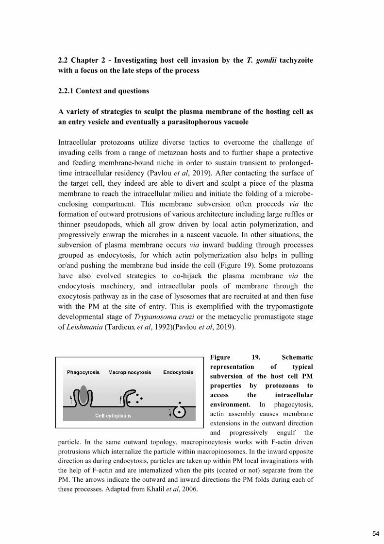
2.2 Chapter 2 - Investigating host cell invasion by the T. gondii tachyzoite
with a focus on the late steps of the process
2.2.1 Context and questions
A variety of strategies to sculpt the plasma membrane of the hosting cell as
an entry vesicle and eventually a parasitophorous vacuole
Intracellular protozoans utilize diverse tactics to overcome the challenge of
invading cells from a range of metazoan hosts and to further shape a protective
and feeding membrane-bound niche in order to sustain transient to prolonged-
time intracellular residency (Pavlou et al, 2019). After contacting the surface of
the target cell, they indeed are able to divert and sculpt a piece of the plasma
membrane to reach the intracellular milieu and initiate the folding of a microbe-
enclosing compartment. This membrane subversion often proceeds via the
formation of outward protrusions of various architecture including large ruffles or
thinner pseudopods, which all grow driven by local actin polymerization, and
progressively enwrap the microbes in a nascent vacuole. In other situations, the
subversion of plasma membrane occurs via inward budding through processes
grouped as endocytosis, for which actin polymerization also helps in pulling
or/and pushing the membrane bud inside the cell (Figure 19). Some protozoans
have also evolved strategies to co-hijack the plasma membrane via the
endocytosis machinery, and intracellular pools of membrane through the
exocytosis pathway as in the case of lysosomes that are recruited at and then fuse
with the PM at the site of entry. This is exemplified with the trypomastigote
developmental stage of Trypanosoma cruzi or the metacyclic promastigote stage
of Leishmania (Tardieux et al, 1992)(Pavlou et al, 2019).
Figure 19. Schematic
representation of typical
subversion of the host cell PM
properties by protozoans to
access the intracellular
environment. In phagocytosis,
actin assembly causes membrane
extensions in the outward direction
and progressively engulf the
particle. In the same outward topology, macropinocytosis works with F-actin driven
protrusions which internalize the particle within macropinosomes. In the inward opposite
direction as during endocytosis, particles are taken up within PM local invaginations with
the help of F-actin and are internalized when the pits (coated or not) separate from the
PM. The arrows indicate the outward and inward directions the PM folds during each of
these processes. Adapted from Khalil et al, 2006.
54

T. gondii performs differently since in contrast to the aforementioned cases, it is
the parasite itself that provides the force that eventually directs the budding of the
PM, and thereby the folding of an entry vesicle. As mentioned in the
Introduction, the T. gondii tachyzoite reproduces asexually by giving birth to
usually two offsprings but this process depends from nutrients that can only be
supplied by the hosting cell. In this context, the free tachyzoite displays an
impressive ability to actively invade virtually any type of nucleated cell and this
action mobilizes mainly two distinct machineries. The first one is made by the
RON protein complex and can be qualified as an invasive nanodevice since it is
delivered by the tachyzoite just before any attempt of active invasion, and the
second is the actomyosin motor system organized in the narrow space between
the PM and the IMC within the trilayer pellicle (Introduction 1.3). The secretion
of the nanodevice from the rhoptry is a tightly regulated event with still many
unknowns, in particular as regard to the upstream signal(s) required for secretion.
However, this event remains so far the first that can be visualized and thus
reliably informs on the on-going invasion attempt even before changes due to the
bud formation can be detected in the host cell surface. The two machineries
tightly work together during the invasion event since the invasive nanodevice
shapes a circular bridge between the two cells, namely the Zoite Cell Junction
(ZCJ), which our lab shows to act as a force anchor point to transmit the driving
force that propels the tachyzoite inside the cell. To briefly describe the invasion
sequence according to the prevailing model, let’s consider the succession of five
steps that are all directed by the invasive nanodevice. This sequence includes (i)
the release of the invasive nanodevice from the rhoptry to the tip of the conoid in
contact with the target cell surface, (ii) the insertion of the invasive nanodevice
into the target cell PM and underlying cortex, (iii) the pulling force applied by the
parasite on the static but somehow elastic invasive nanodevice, (iv) the closure of
the invasive nanodevice once the budding entry vesicle encloses the whole
tachyzoite body, and (v) the pinching off of the budding entry vesicle from the
plasma membrane to release a separate membrane bound compartment enclosing
the parasite and defined as the PV.
To collect dynamic insights possibly for these five steps, our lab has engineered a
tachyzoite in which the RON2 protein was replaced by a fluorescent RON2
chimeric protein using tagging at the C terminal part and which proved fully
functional (Huynh & Carruthers, 2009). As shown in Figure 6, the C terminal
region of RON2 remains exposed to the outside and contains a loop region that
fits in the cleft designed by the AMA1 protein but also accommodates the
addition of the fluorescent tag at the Cterminal end. The invasive behavior of
RON2-mCherry (RON2-mC) tachyzoites was then monitored by fast and high
resolution live imaging allowing to demonstrate that once the RON2-mC positive
nanodevice is delivered into the recipient PM, the parasite tracts itself in the tight
PM bud it directs (Bichet et al, 2014). Interestingly, it was possible for the first
55

time to follow the “one-shot” release of the RON2-mC material, which clearly
signs for the release of a fully functional complex and then its delivery into the
PM to further delineate the ZCJ.
When I arrived in the lab, the transgenic fluorescent RON2-mC parasite line and
a number of human and murine cell lines expressing a variety of fluorescent
reporters for the cell PM or cortex were available. I was given the charge to
develop quantitative live imaging for accurately monitoring and analyzing the
RON-invasive nanodevice behavior in conjunction with the local PM remodeling
that accounts for the budding entry vesicle in the target cell. One particular issue
left quasi untouched in the recent years concerns the mechanisms by which the
invasion event ends, namely the processes covering both the closure of the ring-
shaped RON-nanodevice and the separation of the PM-derived bud that contains
the invading parasite from the host cell PM.
A missing piece of the invasion puzzle: how does invasion give birth to a
tachyzoite-loaded intracellular parasitophorous vacuole?
As early as 1996, a pioneer patch-clamp study performed by G. Ward and
collaborators has provided definitive evidence that the parasite induces the target
cell PM to invaginate as the primary source of the membrane material needed to
fold the budding entry vesicle (Suss-Toby et al, 1996). Therefore, unlike
previously stated, the products released from the secretory organelles of the
parasite had only a minor if any contribution to this early step. This study also
provided the first timing of the PV birth upon bud pinch off and subsequent PV
release in the host cell cytoplasm. Performing electrophysiological studies on the
simian fibroblast-like cell (COS-1 cell line) into which the tachyzoite was
entering, the authors measured in time the capacitance electrical parameter as a
direct way to assess the net membrane surface area and thus they could detect any
change over the invasion event (Figure 20). They concluded on the absence of
significant increase in surface area during bud formation, invalidating a
significant role of parasite secreted material in this step. In contrast, they
monitored a drop in capacitance consistent with the reduction of surface area that
corresponded to the amount of membrane needed to enwrap the tachyzoite and
concomitant with the pinch off of the entry vesicle. Furthermore, the assay
allowed the detection of the tachyzoite-induced fission pore. A fission pore is the
small aqueous connection between a forming endocytic vacuole and the
extracellular medium that forms and constricts to closure during the final stages
of vacuole pinch off. The electrical parameter measured during the tachyzoite-
induced fission pore argues for (i) a process possibly related to the endocytic
fission and (ii) the maintenance of cell membrane integrity during pinch off.
56
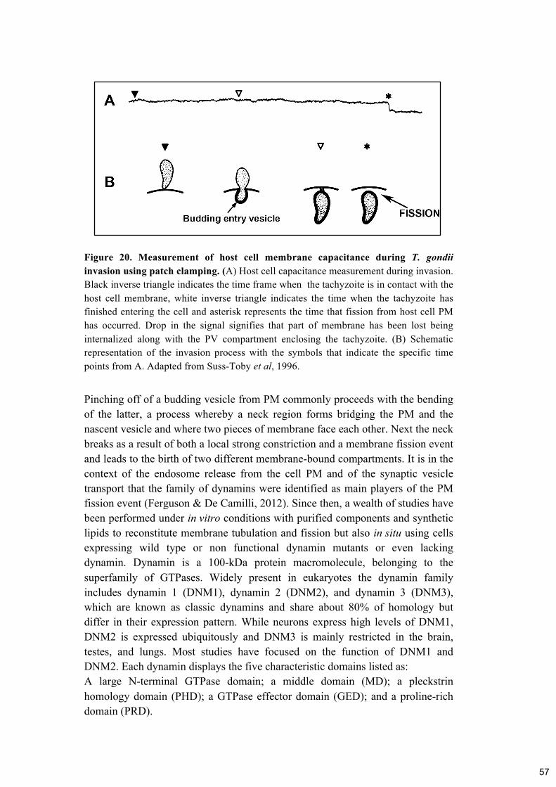
Figure 20. Measurement of host cell membrane capacitance during T. gondii
invasion using patch clamping. (A) Host cell capacitance measurement during invasion.
Black inverse triangle indicates the time frame when the tachyzoite is in contact with the
host cell membrane, white inverse triangle indicates the time when the tachyzoite has
finished entering the cell and asterisk represents the time that fission from host cell PM
has occurred. Drop in the signal signifies that part of membrane has been lost being
internalized along with the PV compartment enclosing the tachyzoite. (B) Schematic
representation of the invasion process with the symbols that indicate the specific time
points from A. Adapted from Suss-Toby et al, 1996.
Pinching off of a budding vesicle from PM commonly proceeds with the bending
of the latter, a process whereby a neck region forms bridging the PM and the
nascent vesicle and where two pieces of membrane face each other. Next the neck
breaks as a result of both a local strong constriction and a membrane fission event
and leads to the birth of two different membrane-bound compartments. It is in the
context of the endosome release from the cell PM and of the synaptic vesicle
transport that the family of dynamins were identified as main players of the PM
fission event (Ferguson & De Camilli, 2012). Since then, a wealth of studies have
been performed under in vitro conditions with purified components and synthetic
lipids to reconstitute membrane tubulation and fission but also in situ using cells
expressing wild type or non functional dynamin mutants or even lacking
dynamin. Dynamin is a 100-kDa protein macromolecule, belonging to the
superfamily of GTPases. Widely present in eukaryotes the dynamin family
includes dynamin 1 (DNM1), dynamin 2 (DNM2), and dynamin 3 (DNM3),
which are known as classic dynamins and share about 80% of homology but
differ in their expression pattern. While neurons express high levels of DNM1,
DNM2 is expressed ubiquitously and DNM3 is mainly restricted in the brain,
testes, and lungs. Most studies have focused on the function of DNM1 and
DNM2. Each dynamin displays the five characteristic domains listed as:
A large N-terminal GTPase domain; a middle domain (MD); a pleckstrin
homology domain (PHD); a GTPase effector domain (GED); and a proline-rich
domain (PRD).
57

Several models have been proposed with the main view that dynamin assembles
into contractile helical polymers that wrap around the neck of the budding vesicle
to direct fusion of the PM leading to hemifission and upon supercontraction, to
fission. As a result, the PM bud transforms into a membrane-bound intracellular
vesicle that can further move by passive or active transport within the cell
cytoplasm.
It is well established that the catalyzing membrane fission activity of dynamins
emerges from main properties (see also Figure 21). First, dynamins oligomerize
into lock-washer-like rings or cylindrical helices. This oligomerization reaction
explains the membrane tubulation activity and was proposed to promote
membrane curvature at the neck of budding vesicles such as endocytic pits.
Secondly dynamin oligomers constrict membrane in the presence of GTP.
Finally, the GTP hydrolysis of dynamin promotes “super” constriction and
oligomer disassembly that eventually cause the subunit release from the PM. The
Figure 21 depicts schematically the sequential activity of the dynamin oligomers.
58

Figure 21. Structure and assembly of dynamin in a membrane tube to mediate
fission. (A) Crystal-based structure of the dimer and assembly into tetramers that
surround the membrane tube in a helix formation. (B) Dynamin constricts the membrane
tube in a helical way. According to the original constriction model for dynamin-mediated
membrane fission, dynamin will first constrict the membrane tube to bring the bilayers
close enough (hemifission) and then more constriction occurs to account for fission.
Adapted from Antonny et al, 2016.
In the context of tachyzoite PV birth, it was proposed that the host cell dynamins
could be involved during pinching off of the nascent PV. These conclusions were
drawn based on the effect of dynasore, a widely used pharmacological inhibitor
of dynamins, which in the case of T. gondii was reported to significantly decrease
the rate of invasion (Caldas et al, 2009). The same authors reported that when
cells and parasites were exposed to dynasore, the tachyzoites could not complete
invasion and instead remained blocked at the periphery. However, recent
advances in molecular genetics allowed obtaining cells which were
simultaneously conditionally silenced for the three genes encoding dynamins.
Using these cells, dynasore was then shown to have several off target effects in
particular related to cholesterol changes in the cell membrane (Park et al, 2013).
Therefore these findings called into question the specificity of the dynasore effect
during cell invasion by tachyzoites. We chose to interrogate the mechanisms of
PV birth by addressing the following points.
• Could the nanodevice impose specific requirements for the sealing of the
budding entry vesicle and its release in the host cell cytoplasm?
• What could be the nanodevice contribution to the PV birth event?
• Could the host cell dynamins contribute to the process?
• Could the fission event serve as signal for down stream remodeling of the
PV?
59

2.2.2 Implementing methods to study the cell invasion of T. gondii tachyzoite
- Video microscopy with two fluorescent markers
We already referred to the importance of video microscopy for studying the
process of invasion by T. gondii tachyzoites. One of the main challenges is to
collect enough details over the whole process that lasts around 30 seconds, and
this implies to have a camera with fast imaging capabilities. Fast live imaging
using more than one laser is not obvious if one needs to apply longer time
exposure in order to optimize the signal to noise ratio of the fluorescent reporter.
We upgraded our original confocal spinning disk microscopy platform by shifting
to a camera with higher resolution imaging, sensitivity and image acquisition time
and we established protocols where we could acquire images from brightfield and
1-3 lasers (green, red, dark red) within 1 to 2 seconds depending on the goal of
the experiment. For most of our experiments, we needed the red channel open to
record the behavior of the RON2-mC protein within the invasive nanodevice,
while at the same time we were interested in monitoring the formation of the
budding entry vesicle and its development until it separates from the host cell
PM. This necessitated the use of the green channel to capture the fluorescence
associated with the PM reporter we chose. In other cases, we had dyes (variety of
fluorescent colors) in the medium, as instances to verify the viability of the
tachyzoite in particular when the process of invasion was artificially slowed down
or impaired. To interfere with the process of invasion, we design assays for which
fluorescent microbeads (dark red for instances) were used. So overall, at least two
lasers had to be active simultaneously and we did not have a dual view system
where two cameras allow simultaneous recording of two emission channels.
Therefore, we optimized the laser exposure time and power to our needs for the
best informative image. Last but not least, we rationalized the best upgrading we
could afford, in particular with the installation of a piezo objective that offers
great focus adjustment and high-speed Z-stack acquisition.
- Image processing to develop quantitative analysis
As we described before, we applied fast live imaging where acquisition of at least
three images per second was performed for two to three channels. This generated
large amount of images. Thus powerful computers were needed for the analysis
of these data. I would like to highlight the particle tracking techniques (using Icy
software) that were used for this study since they helped for describing the
twisting motion in an informative way. Using a variety of plugins and tools, we
managed not only to quantify parameters of motion characteristics of the twist but
also to clarify the visualization of the results (i.e. the parasite rotation). This
image analysis protocol has been included as a book chapter in the Toxoplasma
gondii book (Pavlou & Tardieux, 2020). Generally, image processing was
60

performed with ImageJ, Metamorph, ChimeraX and MATLAB software that are
widely used. I would like to separately refer to Icy software as I performed most
of the image analysis needed for this manuscript. It is an open source user-
friendly software that through all the years of my PhD, was my main software for
image analysis and I can admit that I am more than satisfied with the environment
and the tools that it offers for analyzing any data from microscopy experiment.
One other strong point of this software is the interactive forum that supports the
users and turned quite helpful when problems arose during my analyzing
experiments.
- Blocking invasion using nanotools/microbeads
We used the same method as explained in chapter 2.1.2 for immobilizing the
parasite in a substrate. In this case, we had the addition of the host cells in our
experiments because we wanted to block the tachyzoite while it tries to invade a
host cell using the capping behavior of microbeads pre-bound to the parasite
surface. The idea was that if we manage to keep the beads capped at the
tachyzoite posterior end, we could test the consequences of failed closure of the
invasive nanodevice. In some cases, the beads detached from the tachyzoite body
as explained in chapter 1 but in most cases, the beads stayed attached to the
posterior and therefore they did interfere with the torus closure. In control, we
confirmed that the host cells were not affected by the presence of the microbeads
by performing live imaging of host cells alone with microbeads.
- Utilization of /Engineering of mammalian cell lines expressing genetically
encoded markers
There was a need for engineering mammalian cell lines that express different PM
to monitor in live the membrane budding and fission. Stable cell lines with
different PM reporters engineered as described in Pavlou et al, 2018 because we
needed stable expression and 100% positive cells since invasion is not an event
that is easy to record. First, we needed to make sure that we produced healthy
active parasites and that we kept all the conditions to maintain their invasiveness
over the recording period. Then we wanted to ensure the stable protein expression
of all the potential host cells. Thus, colleagues in the lab used plasmids encoding
fluorescent markers of interest to transfect a variety of mammalian cell lines and
carried antibiotic resistance selection before isolating clones by limiting dilution.
For testing the importance of host cell dynamins during the fission process, we
were given by P. de Camilli a line of Mice Embryonic Fibroblasts (MEF) derived
from a tamoxifen-inducible triple conditional KO mouse (Dnm1loxP/loxP;
Dnm2loxP/loxP; Dnm3loxP/loxP; Cre-ER+/0). Using tamoxifen, we could
silence the expression of the three genes encoding dynamins as described in Park
61

et al, 2013 and the effectiveness of the treatment was controlled by visualizing
the block in endocytosis. When we tested the contribution of ALIX during
tachyzoite entry in MEFs, we benefited from MEFs derived from a Alix knock-
out mouse generated in the lab of colleagues at Grenoble (Laporte et al, 2017).
We needed however to compare with a MEF KO line genetically complemented
with a wild type copy of the ALIX encoding gene. To this end, the lab generated
a MEF line expressing a chimeric version of ALIX that carries the GFP protein at
the N terminal position, which we selected with puromycine and isolated by
limiting dilution.
Overall, we have been successful in providing highly informative image datasets
on the mechanisms by the T. gondii tachyzoite ends entry into the host cell
because we used (i) well-controlled and stable lines for both the tachyzoite and
the host (human/mouse) cells, but also (ii) we carefully chose the fluorescent
markers of interest, and finally (ii) we applied image processing for accurate
spatio-temporal signal visualization coupled with quantitative analysis.
62

2.2.3 Main highlights of the study
• T. gondii RhOptry Neck protein complex forms a torus-shaped invasive
device in the membrane of the target cell that stretches and shrinks to
accommodate the passage of the ellipsoid tachyzoite.
• T. gondi tachyzoite rotational twisting motion completes host invasion in 2D
but also 3D (spheroids) conditions and it works independently of host cell
dynamins.
• The budding entry vesicle pinches off of the host cell PM to give birth to a
parasitophorous vacuole through a fission process upstream the position of the
RON-nanodevice. The time of fission varied between parasites with two
distinct motion sequences of specific kinematical parameters.
• The first remodeling of the tachyzoite-loaded budding vacuole can be
detected immediately post-twist and reveals the assembly of specific
microdomains in the membrane of the parasitophorous vacuole.
• Artificially induced failure of torus closure impedes proper sealing of the
budding entry vesicle, promotes influx of extracellular Na2+ ions inside the
unclosed bud which eventually leads to osmotic driven swelling and
subsequent parasite death.
63

2.2.4 Publications
Pavlou G, Biesaga M, Touquet B, Lagal V, Balland M, Dufour A, Hakimi M &
Tardieux I (2018) Toxoplasma Parasite Twisting Motion Mechanically Induces
Host Cell Membrane Fission to Complete Invasion within a Protective Vacuole.
Cell Host & Microbe 24: 81-96.e5
https://linkinghub.elsevier.com/retrieve/pii/S1931312818303159
Pavlou G, Tardieux I (2018) The Toxoplasma Tour de Force to Unfold its
Intravacuolar Developmental Program. J Infectiology. 1(3): 13-18
https://www.infectiologyjournal.com/articles/the-toxoplasma-tour-de-force-to-
unfold-its-intravacuolar-developmental-program.pdf
Pavlou G, Milon G & Tardieux I (2019) Intracellular protozoan parasites: living
probes of the host cell surface molecular repertoire. Current Opinion in
Microbiology 52: 116–123
https://linkinghub.elsevier.com/retrieve/pii/S1369527419300256
Pavlou G & Tardieux I (2020) Phenotyping Toxoplasma Invasive Skills by Fast
Live Cell Imaging. In Toxoplasma gondii, Tonkin CJ (ed) pp 209–220. New
York, NY: Springer US http://link.springer.com/10.1007/978-1-4939-9857-9_12
64

Article
Toxoplasma Parasite Twisting Motion MechanicallyInduces Host Cell Membrane Fission to CompleteInvasion within a Protective Vacuole
Graphical Abstract
Highlights
d T. gondii/TgRhOptry Neck protein complex forms an invasive
device in target membrane
d Tg rotational twisting motion completes host invasion,
independent of host cell dynamins
d First remodeling of the Tg parasitophorous vacuole occurs
post-twist
d Failure to seal the vacuole prior to detachment promotes
swelling and parasite death
Authors
Georgios Pavlou, Mateusz Biesaga,
Bastien Touquet, ..., Alexandre Dufour,
Mohamed-ali Hakimi, Isabelle Tardieux
Correspondence
In Brief
During host cell invasion, the protozoan
parasite Toxoplasma gondii injects a
parasite-derived complex, or
nanodevice, that consists of the RhOptry
Neck proteins. This nanodevice opens
the host cell membrane and directs
budding of the parasitophorous vesicle.
Pavlou et al. uncover a parasite-directed
rotation that drives vesicle closure and
release.
Pavlou et al., 2018, Cell Host & Microbe 24, 1–16
July 11, 2018 ª 2018 Elsevier Inc.
https://doi.org/10.1016/j.chom.2018.06.003
65

Cell Host & Microbe
Article
Toxoplasma Parasite Twisting Motion MechanicallyInduces Host Cell Membrane Fissionto Complete Invasion within a Protective Vacuole
Georgios Pavlou,1 Mateusz Biesaga,1 Bastien Touquet,1 Vanessa Lagal,2 Martial Balland,3 Alexandre Dufour,4
Mohamed-ali Hakimi,5 and Isabelle Tardieux1,6,*1Institute for Advanced Biosciences (IAB), Team Membrane Dynamics of Parasite-Host Cell Interactions, CNRS UMR5309, INSERM U1209,
Universite Grenoble Alpes, Grenoble 38700, France2Unit of Malaria Infection and Immunity, Department of Parasites and Insect Vectors, Institut Pasteur, Paris 75015, France3Laboratoire Interdisciplinaire de Physique, UMR CNRS, 5588, Universite Grenoble Alpes, Grenoble 38402, France4Institut Pasteur, Bioimage Analysis Unit, CNRS UMR3691, Paris 75015, France5Institute for Advanced Biosciences (IAB), Team Host-Pathogen Interactions & Immunity to Infection, INSERM U1209, CNRS UMR5309,
Universite Grenoble Alpes, Grenoble 38700, France6Lead Contact
*Correspondence: [email protected]
https://doi.org/10.1016/j.chom.2018.06.003
SUMMARY
To invade cells, the parasite Toxoplasma gondii
injects a multi-unit nanodevice into the target
cell plasma membrane (PM). The core nanodevice,
which is composed of the RhOptry Neck (RON) pro-
tein complex, connects Toxoplasma and host cell
through a circular tight junction (TJ). We now report
that this RON nanodevice mechanically promotes
membrane scission at the TJ-PM interface, directing
a physical rotation driven by the parasite twisting
motion that enables the budding parasitophorous
vacuole (PV) to seal and separate from the host cell
PM as a bona fide subcellular Toxoplasma-loaded
PV. Mechanically impairing the process induces
swelling of the budding PV and death of the parasite
but not host cell. Moreover, this study reveals that
the parasite nanodevice functions as a molecular
trigger to promote PV membrane remodeling and
rapid onset of T. gondii to intracellular lifestyle.
INTRODUCTION
The several-micron-sized and ellipsoid-shaped protozoan
Toxoplasma gondii is an obligate intracellular parasite of warm-
blooded metazoan nucleated cells. The T. gondii replicative
stage, called tachyzoite, gains cellular entry within a mem-
brane-bound vesicle by subverting a piece of the cell plasma
membrane (PM). In contrast to those intracellular microbes that
hijack endocytic vesicles to enter host cells (Kumari et al.,
2010; Cossart and Helenius, 2014), the tachyzoite directs the
selection of PM components that flow into the budding entry
vesicle (Mordue et al., 1999). When the zoite entry is about com-
plete, the adjacent bilayer regions that connect the budding
vesicle to the PM merge and are severed, thus accounting for
the release in the host cell cytoplasm of a sealed vesicle, the
so-called parasitophorous vacuole (PV) that houses the parasite
and thereafter its progeny (Morisaki et al., 1995; Suss-Toby et al.,
1996). How the PM-derived bud seals and pinches off a PV re-
mains largely elusive.
To form a functional PV, the parasite secretes a multi-molecu-
lar complex that is pre-stored in its apical organelles called rhop-
tries (Besteiro et al., 2009; Straub et al., 2011; Bichet et al., 2014).
The four-member complex consists of RhOptry Neck RON 2, 4,
5, 8 proteins and represents an invasive nanodevice as it pro-
vides, in combination with other parasite-derived products (Bes-
teiro et al., 2009; Sidik et al., 2016), transient coupling of the two
cells by building a tight circular interface, also called Tight Junc-
tion (TJ). The TJ serves several functions: (1) promoting delivery
of the earliest parasite effectors into the host cell cytoplasm, in
particular toxofilin (Poupel et al., 2000; Lodoen et al., 2010),
which loosens the cortical actin meshwork and facilitates PM
invagination (Delorme-Walker et al., 2012), (2) defining an entry
door on which the tachyzoite applies invasive force (Bichet
et al., 2014), and (3) acting as a gatekeeper for the metazoan
cell PM proteins composing the budding entry vesicle (Mordue
et al., 1999; Charron and Sibley, 2004).
A pioneer patch-clamp study has highlighted similarities be-
tween T. gondii PV pinch-off and endocytic fusion pores, albeit
of slower kinetics (Suss-Toby et al., 1996). It then has been pro-
posed that the parasitophorous vacuole membrane (PVM) fuses
at the parasite rear to separate from the host cell PM (Charron
and Sibley, 2004) while the TJ-associated RONs were im-
muno-localized in young PVs (Besteiro et al., 2009), suggesting
that fission occurs at the PM. Moreover, the mechanoenzymes
dynamins, which drive vesicle scission during Clathrin-Mediated
(CME) or clathrin-independent endocytosis (Antonny et al.,
2016), were implicated during T. gondii PV release, based on
the effect of the pharmacological inhibitor dynasore (Caldas
et al., 2009). However, recent data challenged these conclusions
by reporting unspecific additional effects of dynasore on mem-
brane homeostasis (Park et al., 2013; Preta et al., 2015; Persaud
et al., 2018). Finally, among other drivers of PM fission is the
ESCRT (Endosomal Sorting Complex Required for Transport)
machinery whose cofactor Alix (ALG-2 Interacting protein X)
Cell Host & Microbe 24, 1–16, July 11, 2018 ª 2018 Elsevier Inc. 1
Please cite this article in press as: Pavlou et al., Toxoplasma Parasite Twisting Motion Mechanically Induces Host Cell Membrane Fission to Complete
Invasion within a Protective Vacuole, Cell Host & Microbe (2018), https://doi.org/10.1016/j.chom.2018.06.003
66

was detected at the T. gondii TJwhere it interactedwith the cyto-
plasmically exposed TJ member RON4 (Guerin et al., 2017). Yet,
Alix contribution to cell invasion was not directly assessed and
was proposed by the authors to be ESCRT-independent.
We raised the hypothesis that the RON nanodevice imposes
unique requirements for vesicle sealing and scission from the
host cell PM. In this study, we analyzed in depth the process
that gives birth to the unique T. gondii PV using high-speed
quantitative live fluorescence imaging in conjunction with kine-
matic modeling.
RESULTS
Genetic Ablation of Either the Mechanoenzymes
Dynamins or the ESCRT Cofactor Alix in Host Cells Does
Not Affect T. gondii Tachyzoite Invasiveness and
PV Birth
Pharmacological inhibition of host cell dynamins was reported to
drastically impair T. gondii tachyzoite invasiveness but also to
retain tachyzoites that achieved invasion at the cell periphery
(Caldas et al., 2009). This description resembles what is
observed for endocytic vesicles when dynamin-mediated
vesicle scission no longer operates (Ferguson and De Camilli,
2012). Using mouse embryonic fibroblasts (MEFs) derived from
mice harboring floxed alleles of all dynamin 1, 2, and 3 genes
(triple knockout [TKO]) and transgenic for 4-hydroxytamoxifen
(OHT)-inducible Cre recombinase (Park et al., 2013), we re-
examined the contribution of host cell dynamins during tachy-
zoite entry. We confirmed that dynamin expression in TKO
MEF cells treated with OHT was quasi-undetectable using anti-
bodies recognizing all three dynamin isoforms (Figure 1A, left
panel). Indeed, consistent with the endocytosis defect caused
by the lack of dynamins (Ferguson et al., 2009), the Arp2/3 com-
plex, visualized by Arp2 immunostaining, significantly accumu-
lated beneath the PM of the OHT-treated cells where it is ex-
pected to decorate the neck of endocytic vesicles (Figure 1B).
Next, using automatic image capture and processing tomeasure
the differential fluorescence of intracellular and extracellular
parasites, we quantified RON2-mCherry (RON2-mC) tachyzoite
invasiveness in wild-type (WT) and TKO MEF cells, as well as
in either WT U2OS or U2OS cells ectopically expressing low to
high amounts of a functional GFP-dynamin1 fusion protein (Per-
saud et al., 2018) (Figure 1A). Using clathrin light chain (CLC)-
monomeric red fluorescent protein (mRFP) to detect CME, we
observed a significant number of CLC-positive vesicles apposed
to GFP-dynamin1 dot-like structures at the PM of these cells
(Figure S1A), consistent with a functional GFP-dynamin1. In
large-scale invasion assays, we found no significant differences
in tachyzoite invasive capacities in MEFs or U2OS regardless
of dynamin expression as indicated by the relative invasion
rate on three independent assays (Figure 1C). In addition, we
did not observe any retention of newly formed PVs at the cell pe-
riphery (data not shown). Next, since Alix, which assists the
ESCRT machinery during membrane fission, was documented
to team with TJ RON4 (Guerin et al., 2017), we used MEFs
derived from Alix homozygous knockout (KO) mice (KO Alix)
(Mercier et al., 2016). Alix rescued KO (KO Alix GFP-Alix) as
well as U2OS overexpressing GFP-Alix cell lines were also
generated to assess the functional contribution of Alix during
pinch-off of the entry vesicle. We confirmed that no signal was
detected in the KOAlix cells by specific anti-Alix antibodies (Mer-
cier et al., 2016), whereas these unambiguously recognized both
the GFP-Alix fusion protein and the endogenous Alix in the rele-
vant MEF and U2OS cell lines, respectively (Figure 1D). In addi-
tion, GFP fluorescence was detected in all KO AlixGFP-Alix cells
(Figure 1E). We then compared invasion between Alix-deficient
and Alix rescued MEF cells, and found no statistical differences
in tachyzoite invasiveness (Figure 1F). Importantly, the detection
of GFP-Alix at the Ron2-mC-positive TJ site mostly at the end of
the invasion process (Figure S1B) confirmed the construct func-
tionality (Mercier et al., 2016) and the TJ-associated Alix localiza-
tion (Guerin et al., 2017). Finally, we observed that newly formed
PVs were not retained at the cell periphery (data not shown).
Collectively, these data indicate that neither dynamins nor Alix
from the host cells are required for T. gondii tachyzoite optimal
entry into a functional PV.
Closure of the Toroidal TJ Is Concomitant with a
Tachyzoite Anticlockwise Twisting Motion
Since we established that both dynamin and Alix were dispens-
able for pinch-off of tachyzoite-loaded budding vesicles, we hy-
pothesized that tachyzoites use their ownmechanical properties
to drive the biogenesis of a functional PV. To investigate this pos-
sibility, we analyzed the dynamics of the invasion sequence us-
ing high-speed confocal microscopy. We first engineered a set
of transgenic human U2OS cell lines ectopically expressing
PM markers fused to GFP and benefited from the previously
characterized transgenic tachyzoite line expressing the rhoptry
RON2-mC (Bichet et al., 2014). In U2OS cells expressing a
chimeric glycosylphosphatidylinositol (GPI) protein (i.e., GFP-
GPI), we visualized a discrete reorganization of the cell PM at
the site of contact with the parasite apex shortly prior to the
discharge of the RON2-mC complex into the recipient PM. Inser-
tion of RON2-mCwithin the PM coincided with the formation of a
highly curved PM invagination (Figure 2A; see also Video S1).
A significant stretch of the RON2-mC ring-shaped surface that
in 3D geometrically defines a torus (or anchor ring) and coin-
ciding with the TJ could then be tracked while it accommodated
the passage of the ellipsoid-shaped tachyzoite into the unfolding
PM (Figure 2A). When the tachyzoite had entered the cell half
way, the torus started to shrink and was eventually detected
as a thick dot behind the tachyzoite (Figure 2A; see also
Video S1). Using Icy software (de Chaumont et al., 2012) to auto-
matically track the different pools of RON2-mC, namely the (1)
apical rhoptries, (2) posteriorly positioned TJ, both delineated
by blue circles, and (3) internal spherical RON2-mC compart-
ments marked by green circles, allowed to define approxi-
mately a�90�–540� anticlockwise twist rotation of the tachyzoite
around its long axis which imposed a twisting motion on its basal
end (Figures 2B and 2C; see also Videos S2 and S3). When the
signal intensity of the tracked object was analyzed over the rota-
tion time, the profile showed periodic fluctuations as only ex-
pected for a continuous 360� rotation of the parasite. Indeed,
the weaker signals that followed the first bright spots detected
corresponded to the rotation times for which the tracked object
was slightly out of focus. Importantly the signal intensity came
back to the initial level of intensity when the object returned to
the initial in focus positions (Figure 2C). These datasets allow
2 Cell Host & Microbe 24, 1–16, July 11, 2018
Please cite this article in press as: Pavlou et al., Toxoplasma Parasite Twisting Motion Mechanically Induces Host Cell Membrane Fission to Complete
Invasion within a Protective Vacuole, Cell Host & Microbe (2018), https://doi.org/10.1016/j.chom.2018.06.003
67

Figure 1. The Host Cell Membrane Fission Mechanoenzymes Dynamins and ESCRT Cofactor Alix Are Not Required for Tachyzoite Entry
(A) Western blotting on WT MEFs, MEFs conditionally silenced for the three dynamin encoding genes (TKO), WT U2OS, and U2OS ectopically expressing low to
high number of dynamins.
(B) In situ detection of the MEF Arp2/3 complex before and after tamoxifen (Tmx) treatment. Scale bar, 20 mm.
(C) Quantitative large-scale invasion assays. Results show the relative invasion rate asmean ± SEM for three independent assays, each performed three (MEF) or
four (U2OS) times. The total number of intracellular tachyzoites and total number of human cells can be read in each column.
(D) Western blotting following SDS-PAGE separation of protein extracts from KO MEFs Alix and KO MEFs Alix GFP-Alix (rescued), WT U2OS and U2OS
ectopically expressing GFP-Alix.
(E) Confocal images of DIC-GFP channel overlays of KO MEF Alix and KO MEF Alix GFP-Alix (‘‘rescued’’) cells. Scale bar, 20 mm.
(F) Quantitative large-scale invasion assays with similar MOI for each host cell type. Results show the relative invasion rate when the rescued cells served
as reference as mean ± SEM for three independent assays, each performed in quadruplicate. (C and F) Raw data and p values (t test) are indicated. NS,
nonsignificant.
Cell Host & Microbe 24, 1–16, July 11, 2018 3
Please cite this article in press as: Pavlou et al., Toxoplasma Parasite Twisting Motion Mechanically Induces Host Cell Membrane Fission to Complete
Invasion within a Protective Vacuole, Cell Host & Microbe (2018), https://doi.org/10.1016/j.chom.2018.06.003
68

(legend on next page)
4 Cell Host & Microbe 24, 1–16, July 11, 2018
Please cite this article in press as: Pavlou et al., Toxoplasma Parasite Twisting Motion Mechanically Induces Host Cell Membrane Fission to Complete
Invasion within a Protective Vacuole, Cell Host & Microbe (2018), https://doi.org/10.1016/j.chom.2018.06.003
69

not only arguing for a continuous rotation but also controlling
for the absence of fluorescence bleaching over time. To confirm
the twisting motion, we also tracked the slightly eccentric
apicoplast using a fluorescent version of the Ferredoxin
NADP+oxidoReductase (FNR-mRFP) (Amiar et al., 2016). By
analyzing both particle motion and fluorescence intensity, we
unambiguously assessed the ongoing 360� rotation over several
revolutions prior to PV release (Figure 2E; see also Figure S2D
and Video S4 as well as Movie S1 on Mendeley Data: https://
doi.org/10.17632/fmhnprp66j.1). Finally, since a parasite 180�
twist should lead to an opposite position of its convex curvature,
we analyzed tachyzoites that expressed GFP in the cytoplasm
as a reliable marker of cell shape and confirmed that a twisting
motion of the parasite preceded PV release (Figure 2D; see
also Mendeley Movie S2).
To next better characterize the kinematic parameters of the
toroidal TJ at the time of closure and gain mechanistic insights,
we applied image processing methods along with kinematic
and mathematical modeling using MATLAB software. In 2D live
imaging of the invasion, two informative settings were distin-
guished: either (1) the invasion of a.U2OS expressing the fluores-
cent chimeric transmembrane domain of the PDGF receptor (i.e.,
GFP-TM PDGFR) occurred on the plane of the toroid section or
(2) the zoite entered anMEF cell in parallel to the recording plane,
which resulted in the appearance of the TJ as a cross-section
ring. In the first case, we carefully selected sequences in which
the tachyzoite did not reorient during the entry event by checking
that the apical tip of the rhoptry was not changing plane (Fig-
ure 2F; see also Figure S2B and Mendeley Movie S3). The dis-
placements of two extreme points of the toroid were tracked us-
ing MATLAB and revealed alternated motion with one edge
moving first (red trajectory) and the other edge following
(blue trajectory) (Figure 2F bottom panel; see also Mendeley
Movie S3) within the exact time of the tachyzoite twist that could
be determined by the apical ‘‘rhoptry’’ motion. While the first
track (red) could also signify the reorientation of the zoite result-
ing in the apparent faster displacement of one of the TJ edges,
the subsequent track (blue) confirmed that the edges were
symmetrically displaced but with a different time delay, which re-
sulted in observed alternating motion (Figure 2F). Moreover, the
speeds of the ring edge motion in relation to first detected posi-
tion were calculated (Figure 2F). In the second plane described,
we attempted to mathematically model the toroid-shaped TJ by
fitting an ellipse (Figure S2C; see also Mendeley Movie S4).
Although tracking of the ellipse evolution in time was limited by
the image resolution, it revealed an irregular type of closure.
Interestingly, while the TJ has a symmetric toroid shape and
could be expected to close as a result of a symmetric distribution
of inward-directed forces, the irregularity of the tracking of the
torus and the kinematic modeling possibly suggest asymmetric
distribution of forces applied during the twist motion. Finally,
it is worth mentioning that while the majority of tachyzoites
executed a quarter to half turn, we saw rare instances of multiple
rotations (Figure 2G; see also Figure S2A and Mendeley Movie
S5). Interestingly we found this multi-rotation characteristic to in-
vasion of host cells in metaphase (n = 15/15 analyzed cells) (Fig-
ure S2E; see also Mendeley Movie S6). Remarkably, when the
sister chromatids started separating as anaphase proceeded,
the tachyzoite stopped rotating. Therefore, since cell surface
tension of the mitotic cell is reported dramatically and transiently
increased during metaphase (Fischer-Friedrich et al., 2014), the
two determinants of surface tension, i.e., membrane in plane
tension and cortex contractibility, might control successful
membrane fission and PV pinch-off.
The Disconnection of Exogenous Material Attached to
the Tachyzoite Temporally Coincides with the Twisting
Motion Ending Invasion
Since we expected release of exogenous material loosely re-
tained at the posterior pole when the torus has closed, we
analyzed the final twisting motion of invading parasites that car-
ried posteriorly bound products. First, when host cell PM com-
ponents were attached to the rear of a tachyzoite, they were
pulled as filopodium-like projections by the moving parasite
that eventually invaded a cell. We observed that the host cell
filopodium detached from the parasite rear concomitantly with
the twisting motion and retracted quickly as tension dropped
(Figure 3A; see also Figure S3A, Video S5 and Mendeley
Movie S7). We next designed an assay where microbeads would
be similarly positioned at the posterior end of an invading
Figure 2. RON2-mC Is Delivered into the Host Cell PM as a Torus that Stretches and Shrinks Along with Tachyzoite Entry into the Budding
Vesicle
(A–G) The tachyzoite performs an anticlockwise twist that coincides with torus closure.
(A) Time lapse shows a RON2-mC tachyzoite (red) invading a.U2OS cell that expresses a GFP-GPI PM marker (green). The RON2-mC torus assembles
(arrowhead) within the host cell PM that locally remodels (arrowhead), further stretches and shrinks while the parasite penetrates in the highly curved PM-derived
budding vesicle (arrow). Tracking of the torus diameter during stretch (red line) and shrinkage (blue line) is shown on the graph.
(B andC) Time lapses of DIC (top) and fluorescent channel overlays (bottom) show the parasites prior to RON2-mC release and halfway inside a.U2OS-expressing
GFP-TM PDGFR (B) or anMEF (C) cell (see the torusmarked by arrowheads). Upon torus closure, automatic tracking of the RON2-mC pools at the parasite poles
is reported by blue circles and short blue trajectories, while the cytoplasmic RON2-mC circular object (middle, green circle) tracking identifies the twisting motion
(�180� for B,� 540� for C) of the parasite along itsmain axis (green bar between the two poles, see the circular arrows). Graphs (B, top C) indicate the speed of the
three objects tracked and (top C) the signal intensity profile of the tracked objects. Note (1) the higher speed of the internal reference and (2) the periodic
fluctuations in signal intensity that correlates with the position with regard to focal point (full focus is indicated by red arrows).
(D) Time lapse shows a yellow fluorescent protein (YFP)-tachyzoite invading an a.U2OS cell. The TJ site is delineated with arrows while arrowheads show the
position of the parasite convex side that alternates during the twisting motion.
(E) Time lapse shows DIC and fluorescent merged channels of an FNR-mRFP tachyzoite invading a GFP-PM U2OS cell (top). At the end of invasion, automatic
tracking of the FNR-mRFP object (white circle) identifies the twisting motion (�2 turns) and white arrows indicate the TJ site. Note in the graph the periodic
fluctuations in signal intensity that correlate with the position with regard to in-focus point (red arrows).
(F) Kinematicmodeling of the RON2-mC toroid section during closure. Edge pixel is tracked in respect to its initial position and speed of themotion is represented
on the graph. Note in enlargement of selected frames, the alternating motion of right edge (red track) that moves first and left edge (blue track) that follows.
(G) Histogram shows the distribution of the twist amplitude measured by the angle for n = 200 parasites. All scale bars, 5 mm; time is indicated in seconds.
Cell Host & Microbe 24, 1–16, July 11, 2018 5
Please cite this article in press as: Pavlou et al., Toxoplasma Parasite Twisting Motion Mechanically Induces Host Cell Membrane Fission to Complete
Invasion within a Protective Vacuole, Cell Host & Microbe (2018), https://doi.org/10.1016/j.chom.2018.06.003
70

(legend on next page)
6 Cell Host & Microbe 24, 1–16, July 11, 2018
Please cite this article in press as: Pavlou et al., Toxoplasma Parasite Twisting Motion Mechanically Induces Host Cell Membrane Fission to Complete
Invasion within a Protective Vacuole, Cell Host & Microbe (2018), https://doi.org/10.1016/j.chom.2018.06.003
71

tachyzoite and expected to detach when the torus closes. We
indeed observed that the twistingmotion of the tachyzoites coin-
cidedwith bead release from the zoite rear. A repetitive feature of
these sequences was the emergence of membrane ruffles or
tube-like projections emanating from the torus site and that en-
gulfed the beads prior to and often during separation (Figures 3B
and 3C; see also Figure S3B, Videos S6 and S7 and Mendeley
Movie S8), in line with the concept of host cell membrane dy-
namics operating at the TJ (Bichet et al., 2016). Of note, because
the beads stayed in place at the cell surface, theymarked the site
of entry enabling to follow the displacement of the PV post-twist.
Finally, we took advantage of the fact that the daughter tachy-
zoite cells formed within the PV remain linked at their rear ends
through a maternal Residual Body (RB), until they actively exit
the host cell. In some occasions, pairs of posteriorly conjoined
daughter cells can be released in the extracellular medium. We
observed that when one of the paired tachyzoites actively
entered the cell, it could be pulled back through the torus by
the motile partner provided the former has not yet performed
the final twist (Figure 3D; see also Mendeley Movie S9). Instead,
if a paired tachyzoite has achieved twisting motion, the active
conjoined partner could free itself from the joint by executing a
twirling motion, which did not impact the intracellular parasite
(Figure 3E; see also Mendeley Movies S10 and S11). Together,
these data indicate that the final twisting motion likely reports
on the closed status of the torus, a step that defines the ‘‘point
of no return’’ for invasion.
A Twisting Motion of the Tachyzoite Ends Entry into
Cells under 3D Spatial Constraints
The in vivo environment provides cells with a different dimen-
sionality as they experience neighboring cells on all sides and
extracellular matrix that all supply mechanical constraints in
addition to biochemical agonists. Accordingly, the PM lipid and
protein composition was found to differ between 2D and 3D cul-
ture systems, thus conferring distinct mechanophysical proper-
ties, in particular membrane and cortex elasticity (Cesarz and
Tamama, 2016), which likely influences membrane-associated
interface processes. We thus determined whether the invasive
behavior of tachyzoites, in particular the twist motion prior to
PV birth, was retained in a more physiological tissue-related
arrangement. To this end, we adjusted a protocol for 3D culture
of breast cancer cells (Lee et al., 2007), such as human foreskin
fibroblast (HFF) cells, each containing one to a fewRON2-mC ta-
chyzoites, that were mixed with uninfected GFP-GPI expressing
U2OS cells in a Matrigel-based matrix and deposited onto a
Matrigel layer. By monitoring the 3D culture over 4 days, we
observed, between 20 and 40 hr post embedding, mixed multi-
cellular spheroid aggregates that were composed of a majority
of round uninfectedU2OS cells and of few round fibroblasts filled
by growing tachyzoite rosettes (Figure 4A). With time, the tachy-
zoite-providing HFF cells were lost, whereas U2OS spheroids
grew in size and most of their constituent cells hosted tachy-
zoites, providing evidence for the viability of both parasites and
human cells over this period (Figure 4A; see also Mendeley
Movie S12). We observed that tachyzoites injected the RON de-
vice in the cell PM that locally remodeled and moved through a
static torus until the latter closed, a step concurring with a typical
twisting motion of the parasites (Figures 4B and 4C; see also
Mendeley Movies S13 and S14). Collectively, these data vali-
dated the view of the twisting as the mechanical mechanism ac-
counting for torus closure and subsequent PV birth in a more
physiologically relevant context.
Sustained Mechanical Impairment of the Torus Closure
Induces Impressive Osmotic Swelling of the Entry
Vesicle with Concomitant Tachyzoite Death
To clarify how closure of the torus contributes to the release of a
functional PV, we analyzed what would happen when invading
tachyzoites were arrested at the exact step prior to this event.
Therefore, we generated tachyzoites that carry microbeads
covalently bound to their rear end and imaged invasion in
U2OS cells expressing different PM markers. While the torus
formed and shrank normally, the tachyzoite could not perform
the typical final twist and instead stayed connected to the PM
at the TJ site. Meanwhile, the dark-red fluorescent beads re-
mained extracellularly positioned, juxtaposed to the torus and
surrounded by host cell membrane ruffles. Under this configura-
tion, the budding membrane invagination dilated but kept a
similar fluorescence intensity, indicating that the PM compo-
nents continued to selectively flow, hence preserving the usual
lipid enrichment of the nascent PVM over the PM (Figures 5A
and 5B; see also Video S8 and Mendeley Movie S15). The
swelling bud then frequently retracted and could then expand
as intracellular balloons that filled most of the cell cytoplasm.
Intriguingly, the growing balloons eventually separated from
the budding point and underwent massive vesiculation (Fig-
ure 5B). The addition of two cell impermeable RNA/DNA
Figure 3. Exogenous Material Attached to the Tachyzoite Rear End Is Released Concomitantly to the Twist Motion upon RON2-mC Torus
Closure
(A) A RON-2mC (red) tachyzoite invades an a.U2OS cell expressing GFP-TM PDGFR (green) while carrying on its back a PM-derived filopodium that elongates
along with forward movement. Note that the filopodium disconnects from the parasite (arrow) when the latter executes the anticlockwise twist (circular arrow and
arrowheads).
(B and C) Microbead clusters (blue) bound to the rear of invading tachyzoites are released (arrows) when they rotate to close the RON2-mC torus prior to PV
(green) birth. (C) Note also PM projections that form around the beads (arrowheads) shaped as a pedestal.
(D) An invading tachyzoite (blue arrow) is pulled backward by a posteriorly conjoined partner (yellow arrow) thus passing back through the RON2-mC torus. The
direction and length of arrows describes the successive motile activity of each partner. Note, the simultaneous retraction of the budding entry vesicle membrane
and the parasite (arrowhead).
(E) One partner of conjoined YFP-tachyzoites enters a.U2OS cell (white arrows show the constriction) and performs the final twisting motion as attested by the
alternate flips of the convex side marked with white arrowheads. Note that the extracellular bound partner actively detached (double white arrow) without
affecting the invaded partner.
Time lapses show DIC (C–F, top) and fluorescent channels (A, separate and merged channels; B–F, overlays) of entry sequences. All scale bars, 5 mm; time is
indicated in seconds.
Cell Host & Microbe 24, 1–16, July 11, 2018 7
Please cite this article in press as: Pavlou et al., Toxoplasma Parasite Twisting Motion Mechanically Induces Host Cell Membrane Fission to Complete
Invasion within a Protective Vacuole, Cell Host & Microbe (2018), https://doi.org/10.1016/j.chom.2018.06.003
72

intercalating agents of different spectral properties, i.e., SYTOX
Green (SG) and ethidium bromide (ETBR), into the extracellular
medium brought evidence for the tachyzoite death with first
cytoplasmic and then nuclear dye accumulation (Figure 5B). Dif-
ferential interference contrast (DIC) microscopy allowed visuali-
zation of the concomitant contraction of the parasite body
together with membrane blebs. Importantly, while these pro-
cesses occurred in other cell types (data not shown), none
were detected when the microbeads detached from the para-
sites and stayed at the host cell PM while the latter were en-
closed within PVs (Figure 5B). Next, to assess whether an inflow
of Na+ ions into the bud through a leaky torus/TJ could cause
swelling, we used the cell impermeant sodium indicator
CoroNa-Green. The selective accumulation of Na+ ions in the
Figure 4. A Twist Motion of the Tachyzoite
Ends Entry into Matrigel-Embedded U2OS
Spheroids
(A) HFF infected with a few RON2-mC tachyzoites
are co-cultivated with uninfected GFP-GPI ex-
pressing U2OS cells in Matrigel, and imaged at 20,
40, 65, and 82 hr post embedding. A single 0.5-mm
plane of DIC-mC image overlays are shown (left
column); maximal intensity projections of 50–100
images of z planes for GFP-mC channel overlays
are presented following image deconvolution
(medium column), the deconvolved images are
used for 3D representation (right column). Note that
RON2-mC tachyzoites multiply in HFFs and the
progeny expands in the spheroids. Scale bars,
20 mm.
(B and C) Time lapses show DIC (top) and fluo-
rescent merged (bottom) sequences of tachyzoite
entry in two different orientations. Note the PM
projections that form upon RON2-mC delivery
(arrow), the typical static torus through which the
tachyzoites squeeze (arrowheads), and the para-
site twist that accompanies torus closure. Scale
bars, 5 mm; time is indicated in seconds.
balloon and not in tight-fitting PVs (Fig-
ure 5C; see also Mendeley Movies S16
and S17) combined with the SYTOX stain-
ing of the parasite and not the host cell nu-
cleus confirmed (1) TJ leakiness during
unfinished invasion and (2) causal relation
between balloon formation and parasite
death (Figure 5D; see also Mendeley
Movie S18). Finally, 2D and 3D imaging
showed sustained opening of the torus
during bead-induced vesicle swelling (Fig-
ure S4) while beads alone did not trigger
any balloon at the site of contact with the
host cell nor did they cause nuclear accu-
mulation of the SYTOX dye (Mendeley
Movie S19). Interestingly failure in torus
closure and subsequent bulges of the en-
try vesicle were also observed under less
artificial conditions with conjoined para-
site pairs that remained trapped onto the
maternal RB. The latter when in contact
with the host cell PM was surrounded by PM protrusions, there-
fore recapitulating what was observed with the microbead
setting. Importantly, the partner was able to pull both the ar-
rested parasite and its entry vesicle out of the cell, without
causing apparent damage to the host cell (Figure 5E; see also
Video S9). Overall, these assays allowed demonstrating that
proper closure of the toroidal TJ is essential for the biogenesis
of a functional intracellular PV.
Two Distinct Motion Sequences of Specific Kinematical
Parameters Allow Establishing that Torus Closure Is
Essential for the Biogenesis of the Functional PV
The release of the PV compartment into the host cell cyto-
plasm is commonly described as occurring within a few minutes
8 Cell Host & Microbe 24, 1–16, July 11, 2018
Please cite this article in press as: Pavlou et al., Toxoplasma Parasite Twisting Motion Mechanically Induces Host Cell Membrane Fission to Complete
Invasion within a Protective Vacuole, Cell Host & Microbe (2018), https://doi.org/10.1016/j.chom.2018.06.003
73

(legend on next page)
Cell Host & Microbe 24, 1–16, July 11, 2018 9
Please cite this article in press as: Pavlou et al., Toxoplasma Parasite Twisting Motion Mechanically Induces Host Cell Membrane Fission to Complete
Invasion within a Protective Vacuole, Cell Host & Microbe (2018), https://doi.org/10.1016/j.chom.2018.06.003
74

post-closure, hence contrasting with the tens of seconds suffi-
cient for TJ assembly and tachyzoite entry into the budding entry
vesicle. Given the lack of PM and PVMmarkers, and the intrinsic
limitations of the patch-clamping approaches, the kinetics of
membrane severing and PV release have not been yet captured
in real time. We collected data on the time interval between the
closure, defined by a decrease of the parasite speed to a negli-
gible value, and the first PV movement (n = 214). We next used
MATLAB software to determine which distribution model fit the
dataset best, and found a bimodal shape, suggesting a dual na-
ture of the observed processes (i.e., two different unimodal dis-
tributions) (Figure 6A). The first distribution should account for
the process of PV birth per se and was achieved around 78.5 s
(SD=70.4 s) post-TJ closure for themajority of events (Figure 6A,
dash line). Indeed, when we tracked both the apical rhoptries
and the closed torus, we could observe a small displacement
of the posterior end of the PV (i.e., RON2-mC torus tracking,
green trajectory) but not of the anterior part (i.e., RON2-mC rhop-
try tracking, blue trajectory), a scheme compatible with amoder-
ate reorientation of the PVwhen fission was achieved (Figure 6C,
green trajectory). Within the following 6–10 min, both the
rhoptries and torus were seen to move in tandem over a longer
distance (Figure 6C, overlapping blue and green trajectories),
consistent with a cytoplasmic PVmovement defining the second
distribution component, which was centered at 263.6 s (SD =
112.9 s) post-torus closure (Figure 6A, dotted line). In other situ-
ations, the delay between the reorientation and the capture was
negligible and the tracking could not discriminate between them
(Figure 6D) or just one type of movement was distinguished (Fig-
ure 6E). Interestingly, we next analyzed the speed and the accel-
eration of the newly born PV (n = 32) and defined the first type
of motion as ‘‘slow’’ with an average speed around 100 nm/s
and an acceleration below 10 nm/s2 (mean = 5 nm/s2; SD =
2 nm/s2) (Figure 6B, red points) while the later motion had a
comparatively higher average speed around 300 nm/s and
higher acceleration (mean = 28 nm/s2; SD = 22 nm/s2) (Figure 6B,
blue points). Collectively, these data fit with the view of (1) the
first motion resulting from the actual separation of the entry
vesicle from the PM and the consecutive readjustment of the
PV position, and of (2) the second and ‘‘faster’’-defined motion
of broader kinetics and features but most typically nucleus-
directed. Indeed, 4D reconstruction provided an accurate
description of the disconnection between the PM and the newly
born PV within less than a minute, revealing a faster timescale
than previously estimated (Figure 6F; see also Mendeley
Movie S20). In addition, this analysis also unambiguously identi-
fied the site of membrane fusion/fission at the PM, since the
RON2-mC torus continued to be detected as a ‘‘torus scar’’ in
the PVM when the PV traveled toward the cell nucleus.
The Rapid Transition Toward the Tachyzoite
Intracellular Lifestyle Reflects PV Membrane
Remodeling Starting upon Torus Closure
The current model of PVM biogenesis around an invading tachy-
zoite acknowledges the predominant contribution of the host
cell PM while also positing that a minority of components could
derive from parasite lipid/protein-released products (Suss-Toby
et al., 1996) despite no parasite product delivery has ever been
monitored in real time. To examine whether changes in themem-
brane of the entry vesicle could be associated with torus closure,
we used U2OS cell lines expressing the GFP protein in fusion
with four different PM targeting/anchoring motifs and for which
the GFP reporter was incorporated into the membrane of the
budding entry vesicle. The TM of the PDGFR, and GPI motifs
drove GFP at the cell surface while the MyrPalm and CAAX
motifs promoted the localization of the GFP at the inner leaflet
of the host cell PM (Figure 7I). Given the resolution of imaging,
they were all detected as a quasi-uniform fluorescent signal
throughout the development of the nascent PV in line with no
significant addition of non-PM-related material during the pro-
cess. In contrast, immediately or shortly after torus closure,
we observed either a redistribution of fluorescence within the
PVM that generated GFP-enriched microdomains (Figures 7A
and 7C) or GFP signal dissolution (Figures 7E and 7G). For
instance, the GFP-TM PDGFR signal pattern shifted from
uniform to transient dot-like sparkles as soon as the torus had
closed and typically evolved with sustained enrichment of two
symmetrical patches positioned at the anterior part of the PVM
(Figure 7A; see also Video S10). In the case of theGFP-GPImem-
brane reporter, the pattern change was less prominent but char-
acterized by a bright dot that overlapped with the TJ-associated
RON2-mCpool in themembrane of the newly bornPV (Figure 7C;
Figure 5. SustainedMechanical Impairment of the Torus Closure Induces TJ Leakage Assessed by the Inflow of Na+ Ions and Swelling of the
Budding Entry Vesicle within Which the Tachyzoite Dies
(A–D) Time lapses showDIC (top) and fluorescent channel overlays (lower). Tachyzoites attached tomicrobeads (blue) (A and B), to both a bead and a partner (C),
to a partner through abnormal RB (D), enter a.U2OS cell expressingGFP-TMPDGFR (A), GFP-GPI (B), CAAX-GFP (E), or a.U2OSWT cell (C) but are arrested prior
to TJ closure. The double-headed arrows point to the RON2-mC torus (A–C), whereas the swelling budding vesicles are marked with arrows (A–D).
(A) The bead and the torus remain juxtaposed with PM ruffles surrounding them (arrowhead), while bulging of the vesicle starts a few minutes post-block. Note
that the fluorescence of the PM reporter does not decrease during bud swelling, consistent with a continuous PM component inflow in the bud membrane
(pseudo-colored bottom panel).
(B) At about 40 min post-block, SG and ETBR added to the extracellular milieu (as indicated with an arrow above the frame) are readily incorporated in the cell
debris in the medium and are detected in the tachyzoite nucleus, indicating loss of membrane integrity (arrowheads). Note that the extracellular beads are
surrounded by PM protrusions and with time the huge balloon (arrows) can separate from the parasite to undergo vesiculation.
(C) At about 45–60 min post-block, ETBR (arrowheads) and CoroNa-Green reagents (double arrowheads) are detected in the PV and the parasite, attesting torus
leakiness and subsequent tachyzoite death.
(D) SG specifically accumulates in the parasite posteriorly attached to a bead andwithin a developing balloon; note that the nucleus (dotted lines) of the host cell is
not stained attesting from the leakiness of the torus.
(E) The physical constraint imposed by the RB and the conjoined partner impedes torus closure and triggers a moderate osmotic response (arrow), which reverts
upon opposite pulling from the partner. Note the PM projections at the torus site (arrowhead).
All scale bars, 5 mm; time is indicated in seconds.
10 Cell Host & Microbe 24, 1–16, July 11, 2018
Please cite this article in press as: Pavlou et al., Toxoplasma Parasite Twisting Motion Mechanically Induces Host Cell Membrane Fission to Complete
Invasion within a Protective Vacuole, Cell Host & Microbe (2018), https://doi.org/10.1016/j.chom.2018.06.003
75

Figure 6. Two Types of PV Movements Are Characterized by Distinct Kinematical Parameters
(A) Delay time between torus closure and first PV movement is measured over n = 214 samples and reveals a bimodal distribution, delineated with dashed blue
and dotted blue lines, respectively.
(B) Speed of PV and its acceleration aremeasured over n = 32, uncovering that for early post-closuremotion the determined parameters are low (red) while for the
late motion they are comparatively higher and more dispersed (blue).
(C–E) Time lapses of DIC-mC overlays show during entry the tracking of the RON2-mC from the rhoptries (blue trajectories) and from the closed torus (E, green
trajectory) while corresponding graphs document the speed of the tracked objects. The three typical situations are presented: (C) first early PVmotion, where only
the RON2-mC torus moves, thus accounting for PV scission (green trajectory pointed with arrowhead and first peak in the graph, blue arrow), and second PV
(legend continued on next page)
Cell Host & Microbe 24, 1–16, July 11, 2018 11
Please cite this article in press as: Pavlou et al., Toxoplasma Parasite Twisting Motion Mechanically Induces Host Cell Membrane Fission to Complete
Invasion within a Protective Vacuole, Cell Host & Microbe (2018), https://doi.org/10.1016/j.chom.2018.06.003
76

see also Mendeley Movie S21). Redistribution of the GFP-TM
PDGFR and GFP-GPI markers were precisely monitored in 4D
(Figures 7B and 7D; see also Mendeley Movies S22 and S23)
and confirmed dynamic clustering over the whole PV volume.
In addition to these high-frequency patterns (Figure 7I), we also
noticed more subtle, apical, and dynamic membrane deforma-
tions on the PVM (Figure 7C). When the GFP was fused to either
the CAAX or MyrPalm PM targeting motifs, the protein faced the
cytoplasmic side of the PVM and fluorescence selectively van-
ished (Figures 7E and 7G; see also Mendeley Movies S24 and
S25), as indicated by the fluorescence signal intensity profile
along a transect overlapping both PVM and host cell cytoplasm
(lines in Figures 7E and 7G, and graphs in Figures 7F and 7H).
Moreover, these reorganizations correlated with an increased
in the length of the parasite anterior-posterior axis, consistent
with a whole parasite body relaxation, seen for each PM marker
studied (Figure 7J). Collectively these analyses uncover signifi-
cant early membrane dynamics in the PVM, likely to involve the
delivery of material to the PVM post-torus closure rather than
before, therefore at a time when extracellular tachyzoites shifted
to an intracellular replicative status.
DISCUSSION
In this study, we report that the release, in the host cell cyto-
plasm, of the T. gondii-hosting functional PV is mechanically
driven by the parasite that closes the nanodevice it first inserts
in the host cell PM to allow active invasion. Remarkably, we
found no involvement of the host cell master membrane fission
dynamin in the process.
A Parasite Mechanical Twist Imposes Spiral Motion on
the Tachyzoite Basal End and Guarantees PV Tightness
and Functionality
High-speed live imaging of the invading RON2-mC tachyzoite al-
lowed capturing the torus geometry of the RON nanodevice from
the time it inserted and stretched into the target PM to the time it
shrunk and capped the rear of the ellipsoid zoite, along with its
progression into the host cell cytoplasm, within a budding entry
vesicle. To reach this key developmental transition phase, the ta-
chyzoite executed a specific twist along its major axis which
amplitude varied between�90� to several continuous 360� rota-
tions. This twisting motion analyzed by automatic tracking and
quantification of two distinct fluorescent internal markers was
also detected during invasion of Matrigel-embedded host cell
spheroids. Importantly the twist imposed a spiral motion on the
parasite rear end, and was synchronous with torus closure. We
next provided independent evidence for the twisting motion as
part of the torus closure mechanism. First, by pulling a piece of
host cell PM material firmly attached at the posterior pole prior
to and during cell entry, the tachyzoite induced elongation of
a PM filopodium-like projection that retracted upon the tachy-
zoite twist. Secondly, when submicron-sized beads were weakly
bound to the rear pole of invading zoites, they neither prevented
twist nor vesicle sealing and pinch-off. Finally, invading tachy-
zoites could be mechanically withdrawn from the open but not
closed torus by a posteriorly conjoined partner. Of note, the
simultaneous retraction of the budding entry vesicle membrane
and the parasite suggests a physical coupling between the two
membranes, although the features of the interactions have still
to be defined.
Moreover, image data-driven mathematical modeling of the
torus behavior allowed describing an irregular closure, which
would conform well to the twist motion directing the closure
event. Future experiments should test whether the specific
myosins (Frenal et al., 2017) and actin filaments (F-actin) (Periz
et al., 2017) that populate the parasite basal pole, or/and the
host cell F-actin anchoring the TJ (Gonzalez et al., 2009) could
contribute to the force(s) driving torus closure. The transgenic
tachyzoites in which the myosin A encoding gene is deleted
(DMyoA) are known to assemble a TJ but to be driven into the en-
try vesicle by an alternative host cell-driven force (Egarter et al.,
2014; Bichet et al., 2016). These mutant parasites were able to
perform the typical 90� to 180� final twist prior to the release of
their PV, suggesting that other motor(s) than MyoA might be
involved during this step (n = 22, Figure S2F; see also Mendeley
Movie S26). Definitive evidence for the role of twisting motion
during torus closure and PV birth came with parasites arrested
just prior closure. Invading tachyzoites that carried covalently
bound microbeads at their rear, remained blocked in the
budding entry vesicle while being connected to the PM through
the RON2-mC torus. This caused the bud to transform into a
balloon-like inclusion, therefore indicating a defect in closure
with two main consequences. First, the vacuolar space became
accessible to the extracellular fluid, as evidenced by sodium
influx, which led to osmotic water flow from the cytoplasm fol-
lowed by ballooning and tachyzoite death. It is worth mentioning
the amazing amount of PM material, mostly lipids, that infolded
as a huge bud and filled up the cell which likely impacted the
whole host cell surface properties and mechanics. As balloons
that contained fluid and dead tachyzoite material continued to
develop, they eventually ruptured from the connecting PM and
immediately underwent fragmentation known as an invariable
consequence of acute swelling during cell injury.
To the PM Severing, Reflected by the Partition of
RON2-mC in the Budding PV Membrane, Succeeds the
Centripetal Displacement of the Tachyzoite-Hosting
Functional PV
Given that the mechanical twist of the zoite guarantees torus
closure independently of host cell fission machineries, we pro-
pose that the RON torus acts as a mechanical device with anal-
ogous properties of the GTPase dynamins. Similarly to the twist
of dynamin helical spirals that generate torsion forces on the
neck of an endocytic vesicle, the parasite twist would draw
together the PM bilayers at the boundary with the torus site,
motion, where both tracked points move simultaneously (green and blue trajectories, peaks in the two graphs labeled with color arrows); (D) PV late motion;
(E) short delay between the two PV motions post-TJ closure, which does not allow discrimination between them.
(F) 4D reconstruction of the entry sequence shows a top view (top) and side view (bottom) of a RON2-mC tachyzoite entering a GFP-GPI expressing U2OS cell.
Note that the delay between torus closure and PVMfission (arrow) is less than aminute. A 2D view frame indicates the scale bar. All scale bars, 5 mm; time is shown
in seconds.
12 Cell Host & Microbe 24, 1–16, July 11, 2018
Please cite this article in press as: Pavlou et al., Toxoplasma Parasite Twisting Motion Mechanically Induces Host Cell Membrane Fission to Complete
Invasion within a Protective Vacuole, Cell Host & Microbe (2018), https://doi.org/10.1016/j.chom.2018.06.003
77

(legend on next page)
Cell Host & Microbe 24, 1–16, July 11, 2018 13
Please cite this article in press as: Pavlou et al., Toxoplasma Parasite Twisting Motion Mechanically Induces Host Cell Membrane Fission to Complete
Invasion within a Protective Vacuole, Cell Host & Microbe (2018), https://doi.org/10.1016/j.chom.2018.06.003
78

hence favoring membrane severing upstream from the torus. In
support of this scheme was the typical partition of RON2-mC
into the membrane of the newly born PV (i.e., the PVM). To figure
out the kinetics of membrane severing and PV release with
respect to torus closure, we first identified a bimodal distribution
of the dataset reporting the time interval between torus closure
and PV intracellular displacement. This distribution fits with
two distinct motile events post-torus closure, which were then
revealed by dual tracking of the rhoptry/torus and 4D image
reconstruction. The PV first executed a space-limited displace-
ment of low speed and acceleration that proceeded with a
change of the angle or a forward-backward spring-like move-
ment, and could occur within only tens of seconds. Next, the
PV traveled at significantly higher speed and acceleration.
Whether the increased amplitude of the tachyzoite final twist to
several full rotations reflected less favorable conditions for mem-
brane fission is not yet clear. Interestingly, tachyzoite rotation
systematically stopped when the PV had separated from the
PM (i.e., fission has occurred). Taking into account that in vitro
membrane tension influences the kinetics, but not the rate of
membrane fission (Dar et al., 2015), exploring a role of mem-
brane tension in situ should be informative. Finally, the distinct
kinematical features of the second PV motion argue for another
force mechanism to control the cytoplasmic PV traveling, such
as those supported by microtubule and microfilament motors.
A Significant Burst of PVM Remodeling Occurs upon
Torus Closure and Coincides with the Transition to
Intracellular Lifestyle in a Functional PV
While the fluorescent signal of the budding entry vesicle was de-
tected as quasi-uniform until the torus closed, it became uneven
after closure was achieved, giving rise to specific patterns for
given PVMmarkers. The discharge of the ROP2 family members
(i.e., ROP2, 4, 5, and 7), as well as of theGRA proteins (i.e., GRA2
as first released GRAs) that insert in or associate with the PVM is
expected to promote PVM component, lipid and protein
included, redistribution. Numerous factors including proteolytic
activities released in the vacuolar space by the parasite, changes
in luminal pH or ionic composition of the PV or changes in phys-
ical constraints could also contribute to PVM remodeling. Finally,
we cannot exclude that the rhoptry-derived e-vacuoles secreted
into the cell (Hakansson et al., 2001) could modify the early PVM
composition. While future investigation will require character-
izing the distinct players along with PVM fluorescent pattern for-
mation, we propose that the torus closure may act as a signal for
the tachyzoite to start secreting a pool of proteins and shape a
functional intracellular niche. Such gain of PVM material would
also explain the usual relaxation of the tachyzoite body observed
upon PVM remodeling by causing an increase in PV volume
or/and PVM elasticity. Of note, when the torus was arrested
before it closed, as with conjoined parasites or microbeads,
we never observed membrane patterning of the budding vesicle
including before and during osmotic damages. These observa-
tions are consistent with the view of a parasite-derived signal
(possibly a combination signals) to regulate the early remodeling
of the newly formed PV.
In conclusion, this study highlights the multifunctional proper-
ties of a device evolved by the T. gondii tachyzoite: once inserted
in a limited domain of the host cell PM, thus initiating the invasive
process, this parasite device displays mechanical functions
that mimic the ones driven by dynamin mechano-enzymes.
The outputs of these mechanical functions culminate in the
pinching off of the tachyzoite-loaded budding vesicle. Upon
the closure of the latter and its transient membrane remodeling,
not only emerges a functional PV but also rapidly proceeds the
unfolding of the tachyzoite developmental program.
STAR+METHODS
Detailed methods are provided in the online version of this paper
and include the following:
d KEY RESOURCES TABLE
d CONTACT FOR REAGENT AND RESOURCE SHARING
d EXPERIMENTAL MODELS AND SUBJECT DETAILS
B Cell Culture
d METHOD DETAILS
B Cell Lines Engineered to Stably and Ectopically Ex-
press Fluorescent Reporters
B Western blots
B Immunofluorescence Labeling
B Image Processing, Kinematic and Mathematical
Modeling
B Manual Tracking with ImageJ Software
B Automatic Tracking with Icy Software
B Automatic Tracking and Mathematical Modeling with
MATLAB
B Videomicroscopy
B 3D/4D Reconstruction
B 3D Matrigel Co-culture Assays
B Microbead Assays
B Large-Scale Invasion Assay
Figure 7. A Burst of Membrane Remodeling and Deformation of the Unborn and Newly Born PVs Is Revealed by Different Host Cell PM
Reporters
(A–H) Time lapses of fluorescent channel overlays showing RON2-mC expressing zoites entering U2OS cells that express (A) GFP-TM PDGFR, (C) GFP-GPI,
(E) MyrPalm-GFP, and (G) CAAX-GFP. Dual-color inlays of GFP channel are shown in the lower left corner of selected frames. Note the dynamic clustering of the
GFP (A) and the PVM deformation sites marked with arrowheads (C). 4D reconstruction is shown (B and D) of deconvolved images from (A) and (C), respectively.
Both side (top) and top (bottom) orientations are shown as indicated with coordinates. The 2D view shows the scale bar. Dispersion of the PVM signal (E and G) is
observed post-torus closure as verified by the GFP signal intensity profiles over time of dissections (F and H) marked by white lines (E and G).
(I) Top shows cartoons of the GFP fusion position in the PM and the entry vesicle, bottom shows histogram of the frequency of characteristic PVM patterns; the
number of samples scored is indicated. Note that only clearly detectable patterns were considered positive.
(J) Graphs of representative examples for each PM marker, showing the evolution in parasite length post-torus closure. Length is automatically measured by
tracking the RON2-mC at the rhoptry and the torus. The parasite length values prior to and after PVM remodeling are presented as black squares and circles
respectively. The data are normalized relative to the parasite length at the time of torus closure.
All scale bars, 5 mm.
14 Cell Host & Microbe 24, 1–16, July 11, 2018
Please cite this article in press as: Pavlou et al., Toxoplasma Parasite Twisting Motion Mechanically Induces Host Cell Membrane Fission to Complete
Invasion within a Protective Vacuole, Cell Host & Microbe (2018), https://doi.org/10.1016/j.chom.2018.06.003
79
d QUANTIFICATION AND STATISTICAL ANALYSIS
B Dataset Distribution
B Quantitative Invasion Assay
d DATA AND SOFTWARE AVAILABILITY
SUPPLEMENTAL INFORMATION
Supplemental Information includes 4 figures and 10 videos and can be found
with this article online at https://doi.org/10.1016/j.chom.2018.06.003.
ACKNOWLEDGMENTS
We thank Prof. P. De Camilli (Yale University, New Haven, CT) for the TKO
dynamin MEFs, Prof. R. Sadoul (GIN, Grenoble, France) for the KO Alix
MEFs, Prof. G. Charras (UCL, London, UK) for the CAAX-GFP and MyrPalm-
GFP-encoding plasmids, and Dr. H. Ewers (Freie Universit€at, Berlin, Germany)
for the GFP-GPI plasmid. We thank the Foundation for Innovation in Infectiol-
ogy (FINOVI) for funding this research program, the University Grenoble-Alpes
for the AGIR PhD fellowship support to G.P., and Prof. P. Hainaut (IAB, Greno-
ble, France) for internship support to M. Biesaga. This work was supported by
the European Research Council (ERC Consolidator Grant no. 614880 Hosting
TOXO to M.-a.H.). V.L. performed the work in the team of I.T. when appointed
at the Institut Cochin (Paris, France).
AUTHOR CONTRIBUTIONS
Conceptualization, I.T.; Software, M. Biesaga, A.D.; Formal Analysis, G.P., M.
Biesaga, B.T., I.T., A.D.; Investigation, G.P., B.T., M. Biesaga, I.T., M. Balland;
Resources, B.T., V.L., M. Balland, I.T.; Writing – Original Draft, I.T.; Writing –
Review & Editing, I.T., G.P.; Visualization, G.P., M. Biesaga, B.T.; Supervision,
I.T.; Funding Acquisition, M.-a.H., I.T., M.Balland.
DECLARATION OF INTERESTS
The authors declare no competing of interests.
Received: January 12, 2018
Revised: March 27, 2018
Accepted: June 5, 2018
Published: June 28, 2018
REFERENCES
Amiar, S., MacRae, J.I., Callahan, D.L., Dubois, D., van Dooren, G.G., Shears,
M.J., Cesbron-Delauw,M.-F., Marechal, E., McConville, M.J., McFadden, G.I.,
et al. (2016). Apicoplast-localized lysophosphatidic acid precursor assembly
is required for bulk phospholipid synthesis in Toxoplasma gondii and relies
on an algal/plant-like glycerol 3-phosphate acyltransferase. PLoS Pathog.
12, e1005765.
Antonny, B., Burd, C., De Camilli, P., Chen, E., Daumke, O., Faelber, K., Ford,
M., Frolov, V.A., Frost, A., Hinshaw, J.E., et al. (2016). Membrane fission by dy-
namin: what we know and what we need to know. EMBO J. 35, 2270–2284.
Besteiro, S., Michelin, A., Poncet, J., Dubremetz, J.-F., and Lebrun, M. (2009).
Export of a Toxoplasma gondii rhoptry neck protein complex at the host
cell membrane to form the moving junction during invasion. PLoS Pathog. 5,
e1000309.
Bichet, M., Joly, C., Henni, A.H., Guilbert, T., Xemard, M., Tafani, V., Lagal, V.,
Charras, G., and Tardieux, I. (2014). The Toxoplasma-host cell junction is
anchored to the cell cortex to sustain parasite invasive force. BMC Biol.
12, 773.
Bichet, M., Touquet, B., Gonzalez, V., Florent, I., Meissner, M., and Tardieux, I.
(2016). Genetic impairment of parasite myosin motors uncovers the contribu-
tion of host cell membrane dynamics to Toxoplasma invasion forces. BMC
Biol. 14, 97.
Caldas, L.A., Attias, M., and de Souza, W. (2009). Dynamin inhibitor impairs
Toxoplasma gondii invasion. FEMS Microbiol. Lett. 301, 103–108.
Cesarz, Z., and Tamama, K. (2016). Spheroid culture of mesenchymal stem
cells. Stem Cells Int. 2016, 9176357.
Charron, A.J., and Sibley, L.D. (2004). Molecular partitioning during host cell
penetration by Toxoplasma gondii. Traffic 5, 855–867.
de Chaumont, F., Dallongeville, S., Chenouard, N., Herve, N., Pop, S.,
Provoost, T., Meas-Yedid, V., Pankajakshan, P., Lecomte, T., Le Montagner,
Y., et al. (2012). Icy: an open bioimage informatics platform for extended repro-
ducible research. Nat. Methods 9, 690–696.
Cossart, P., and Helenius, A. (2014). Endocytosis of viruses and bacteria.
Cold Spring Harb. Perspect. Biol. 6, https://doi.org/10.1101/cshperspect.
a016972.
Dar, S., Kamerkar, S.C., and Pucadyil, T.J. (2015). A high-throughput platform
for real-time analysis of membrane fission reactions reveals dynamin function.
Nat. Cell Biol. 17, 1588–1596.
Delorme-Walker, V., Abrivard, M., Lagal, V., Anderson, K., Perazzi, A.,
Gonzalez, V., Page, C., Chauvet, J., Ochoa, W., Volkmann, N., et al. (2012).
Toxofilin upregulates the host cortical actin cytoskeleton dynamics, facilitating
Toxoplasma invasion. J. Cell Sci. 125, 4333–4342.
Egarter, S., Andenmatten, N., Jackson, A.J., Whitelaw, J.A., Pall, G., Black,
J.A., Ferguson, D.J.P., Tardieux, I., Mogilner, A., and Meissner, M. (2014).
The Toxoplasma Acto-MyoA motor complex is important but not essential
for gliding motility and host cell invasion. PLoS One 9, e91819.
Ferguson, S.M., and De Camilli, P. (2012). Dynamin, a membrane-remodeling
GTPase. Nat. Rev. Mol. Cell Biol. 13, 75–88.
Ferguson, S.M., Ferguson, S., Raimondi, A., Paradise, S., Shen, H.,Mesaki, K.,
Ferguson, A., Destaing, O., Ko, G., Takasaki, J., et al. (2009). Coordinated ac-
tions of actin and BAR proteins upstream of dynamin at endocytic clathrin-
coated pits. Dev. Cell 17, 811–822.
Fischer-Friedrich, E., Hyman, A.A., J€ulicher, F., M€uller, D.J., and Helenius, J.
(2014). Quantification of surface tension and internal pressure generated by
single mitotic cells. Sci. Rep. 4, 6213.
Frenal, K., Jacot, D., Hammoudi, P.-M., Graindorge, A., Maco, B., and Soldati-
Favre, D. (2017). Myosin-dependent cell-cell communication controls syn-
chronicity of division in acute and chronic stages of Toxoplasma gondii. Nat.
Commun. 8, 15710.
Goddard, T.D., Huang, C.C., Meng, E.C., Pettersen, E.F., Couch, G.S., Morris,
J.H., and Ferrin, T.E. (2017). UCSF ChimeraX: meeting modern challenges in
visualization and analysis. Protein Sci. 27, 14–25.
Gonzalez, V., Combe, A., David, V., Malmquist, N.A., Delorme, V., Leroy, C.,
Blazquez, S., Menard, R., and Tardieux, I. (2009). Host cell entry by apicom-
plexa parasites requires actin polymerization in the host cell. Cell Host
Microbe 5, 259–272.
Guerin, A., Corrales, R.M., Parker, M.L., Lamarque, M.H., Jacot, D., El Hajj, H.,
Soldati-Favre, D., Boulanger, M.J., and Lebrun, M. (2017). Efficient invasion
by Toxoplasma depends on the subversion of host protein networks. Nat.
Microbiol. 2, 1358–1366.
Hakansson, S., Charron, A.J., and Sibley, L.D. (2001). Toxoplasma evacuoles:
a two-step process of secretion and fusion forms the parasitophorous vacu-
ole. EMBO J. 20, 3132–3144.
Kumari, S., Mg, S., and Mayor, S. (2010). Endocytosis unplugged: multiple
ways to enter the cell. Cell Res. 20, 256–275.
Lee, G.Y., Kenny, P.A., Lee, E.H., and Bissell, M.J. (2007). Three-dimensional
culture models of normal andmalignant breast epithelial cells. Nat. Methods 4,
359–365.
Lodoen, M.B., Gerke, C., and Boothroyd, J.C. (2010). A highly sensitive FRET-
based approach reveals secretion of the actin-binding protein toxofilin during
Toxoplasma gondii infection. Cell. Microbiol. 12, 55–66.
Mercier, V., Laporte, M.H., Destaing, O., Blot, B., Blouin, C.M., Pernet-Gallay,
K., Chatellard, C., Saoudi, Y., Albiges-Rizo, C., Lamaze, C., et al. (2016). ALG-2
interacting protein-X (Alix) is essential for clathrin-independent endocytosis
and signaling. Sci. Rep. 6, 26986.
Mordue, D.G., Desai, N., Dustin, M., and Sibley, L.D. (1999). Invasion by
Toxoplasma gondii establishes a moving junction that selectively excludes
Cell Host & Microbe 24, 1–16, July 11, 2018 15
Please cite this article in press as: Pavlou et al., Toxoplasma Parasite Twisting Motion Mechanically Induces Host Cell Membrane Fission to Complete
Invasion within a Protective Vacuole, Cell Host & Microbe (2018), https://doi.org/10.1016/j.chom.2018.06.003
80
host cell plasma membrane proteins on the basis of their membrane
anchoring. J. Exp. Med. 190, 1783–1792.
Morisaki, J.H., Heuser, J.E., and Sibley, L.D. (1995). Invasion of Toxoplasma
gondii occurs by active penetration of the host cell. J. Cell Sci. 108 (Pt 6),
2457–2464.
Park, R.J., Shen, H., Liu, L., Liu, X., Ferguson, S.M., and De Camilli, P. (2013).
Dynamin triple knockout cells reveal off target effects of commonly used dyna-
min inhibitors. J. Cell Sci. 126, 5305–5312.
Periz, J., Whitelaw, J., Harding, C., Gras, S., Del Rosario Minina, M.I., Latorre-
Barragan, F., Lemgruber, L., Reimer, M.A., Insall, R., Heaslip, A., et al. (2017).
Toxoplasma gondii F-actin forms an extensive filamentous network required
for material exchange and parasite maturation. Elife 6, https://doi.org/10.
7554/eLife.24119.
Persaud, A., Cormerais, Y., Pouyssegur, J., and Rotin, D. (2018). Dynamin
inhibitors block activation of mTORC1 by amino acids independently of dyna-
min. J. Cell Sci. 131, https://doi.org/10.1242/jcs.211755.
Poupel, O., Boleti, H., Axisa, S., Couture-Tosi, E., and Tardieux, I. (2000).
Toxofilin, a novel actin-binding protein from Toxoplasma gondii, sequesters
actin monomers and caps actin filaments. Mol. Biol. Cell 11, 355–368.
Preta, G., Cronin, J.G., and Sheldon, I.M. (2015). Dynasore - not just a dynamin
inhibitor. Cell Commun. Signal. 13, 24.
Sidik, S.M., Huet, D., Ganesan, S.M., Huynh, M.-H., Wang, T., Nasamu, A.S.,
Thiru, P., Saeij, J.P.J., Carruthers, V.B., Niles, J.C., et al. (2016). A genome-
wide CRISPR screen in Toxoplasma identifies essential apicomplexan genes.
Cell 166, 1423–1435.e12.
Straub, K.W., Peng, E.D., Hajagos, B.E., Tyler, J.S., and Bradley, P.J. (2011).
The moving junction protein RON8 facilitates firm attachment and host cell in-
vasion in Toxoplasma gondii. PLoS Pathog. 7, e1002007.
Suss-Toby, E., Zimmerberg, J., and Ward, G.E. (1996). Toxoplasma invasion:
the parasitophorous vacuole is formed from host cell plasma membrane and
pinches off via a fission pore. Proc. Natl. Acad. Sci. USA 93, 8413–8418.
16 Cell Host & Microbe 24, 1–16, July 11, 2018
Please cite this article in press as: Pavlou et al., Toxoplasma Parasite Twisting Motion Mechanically Induces Host Cell Membrane Fission to Complete
Invasion within a Protective Vacuole, Cell Host & Microbe (2018), https://doi.org/10.1016/j.chom.2018.06.003
81

STAR+METHODS
KEY RESOURCES TABLE
REAGENT or RESOURCE SOURCE IDENTIFIER
Antibodies
Mouse monoclonal Anti-P30 Novocastra Clone TP3 Leica Cat#NCL-TG; RRID:AB_564038
Rabbit homemade polyclonal Anti-Alix Covalab Cat#pab0204; RRID:AB_2716810
Monoclonal mouse Anti-Dynamin (Clone41) BD Biosciences Cat#610245; RRID:AB_397640
Rabbit polyclonal Anti-p34-Arc/ARPC2 Millipore Cat#07-227; RRID:AB_310447
Mouse monoclonal Anti-GAPDH Ambion Cat#AM4300; RRID:AB_437392
Chemicals, Peptides, and Recombinant Proteins
4-Hydroxytamoxifen Sigma-Aldrich H6278
Deposited Data
Additional supplemental videos and legends Mendeley Data Tardieux, Isabelle (2018), ‘‘Pavlou et al.
Cell Host Microbe Supplemental Data,’’
Mendeley Data, v1 https://doi.org/
10.17632/fmhnprp66j.1
Experimental Models: Cell Lines
U2OS (female) ATCC Cat#HTB-96; RRID:CVCL_0042
U2OS GFP-Flag-Alix (female) This paper N/A
U2OS GFP-GPI (female) This paper N/A
U2OS GFP-TMPDGFR (female) This paper N/A
U2OS MyrPalm-GFP (female) This paper N/A
U2OS CAAX-GFP (female) This paper N/A
HFF (male) ATCC Cat# SCRC-1041; RRID:CVCL_3285
MEFs isolated from Alix KO mice provided
by R. Sadoul (who did not reveal the sex)
Mercier et al., 2016 N/A
MEFs isolated from Alix KO mice provided
by R. Sadoul (who did not reveal the sex)
transfected with GFP-Alix
This paper N/A
MEFs isolated from TKO mice provided by
P. De Camilli (who did not reveal the sex)
Park et al., 2013 N/A
MEFs isolated from parental mice provided
by P. De Camilli (who did not reveal the sex)
Park et al., 2013 N/A
U2OS GFP-Dynamin1(Low expression) (female) This paper N/A
U2OS GFP-Dynamin1(High expression) (female) This paper N/A
Experimental Models: Organisms/Strains
T.gondii:RH DKU80 RON2-mC (DHFR) Bichet et al., 2014 N/A
T.gondii:RH YFP Laboratory of David Roos N/A
T.gondii:RH-TATi FNR-mRFP Amiar et al., 2016 N/A
T.gondii:RH DKU80 DMyoA Bichet et al., 2016 N/A
Software and Algorithms
MATLAB R2016a Mathworks RRID:SCR_001622
ImageJ 1.51j8 https://imagej.nih.gov/ij/ RRID:SCR_003070
Icy 1.9.5.1 http://icy.bioimageanalysis.org/ RRID:SCR_010587
UCSF ChimeraX 0.1 https://www.rbvi.ucsf.edu/chimerax/ RRID:SCR_015872
Graphpad Prism 6.01 Graphpad RRID:SCR_002798
MetaMorph 7.7.0.0 Molecular devices RRID:SCR_002368
ScanR 2.7.1 Olympus N/A
(Continued on next page)
Cell Host & Microbe 24, 1–16.e1–e5, July 11, 2018 e1
Please cite this article in press as: Pavlou et al., Toxoplasma Parasite Twisting Motion Mechanically Induces Host Cell Membrane Fission to Complete
Invasion within a Protective Vacuole, Cell Host & Microbe (2018), https://doi.org/10.1016/j.chom.2018.06.003
82

CONTACT FOR REAGENT AND RESOURCE SHARING
Further information and requests for reagents may be directed to and will be fulfilled by the Lead Contact, Isabelle Tardieux (isabelle.
EXPERIMENTAL MODELS AND SUBJECT DETAILS
No vertebrate animals were used in this study.
Cell Culture
Human cell lines expressing fluorescent reporters of interest (i.e., targeting the host cell PM and the PVM aswell asmembrane fission
players such as dynamin and Alix proteins) were obtained from human female bone-derived U2OS cells (ATCC. no. HTB-96) or from
MEF cells. The MEF TKO cell line isolated from dynamin TKO mice was given by P. De Camilli (Yale University, New Haven, CT)
who did not reveal the sex. This cell line was derived in De Camilli laboratory from mice harboring floxed alleles of all Dynamin 1,
2, and 3 genes (triple KO: TKO) and transgenic for 4-hydroxytamoxifen (OHT)-inducible Cre recombinase (Park et al., 2013). Briefly,
DeCamilli laboratory obtained the TKOmice by interbreeding 129SV/J andC57Bl6mice first with dynamin 1 and 2 conditional double
KO with the dynamin 3 conditional KO and with mice transgenic for Cre-ER to generate tamoxifen-inducible triple conditional KO
mice (Dnm1loxP/loxP; Dnm2loxP/loxP; Dnm3loxP/loxP; Cre-ER+/0).
MEFs isolated from Alix KOmice were provided by R. Sadoul (GIN, Grenoble) who did not reveal the sex. This line has been gener-
ated in R. Sadoul laboratory from 3 individual E12.5 embryos of Alix homozygous KOmouse. To obtain the Alix komice, exon 2 of alix
(chromosome 9) was targeted by LoxP sites and the floxed mice was backcrossed with the CMV-Cre mouse (Mercier et al., 2016).
To silence the MEF TKO cells, we adapted the protocol from (Ferguson et al., 2009) such as cells were incubated with 4 mM 4-hy-
droxytamoxifen for 3 days and left on 0.4 mMOHT until use, resulting in dynamin depletion at 5-6 days from the start of the treatment
period. For all assays, silenced cells were not used after 9 days post start of OHT exposure and they were systematically checked for
dynamin depletion by western blot prior to video-recording sessions.
Both U2OS andMEF cell lines were certified asMycoplasma free and grown in high glucose Dulbecco’s Modified Eagle’s Medium
Glutamax (DMEM) supplemented with 10% fetal bovine serum (FBS), 10 mM 4-(2-HydroxyEthyl)-1-PiperazineEthaneSulfonic acid
(HEPES), 100 units/mL penicillin, and 100 mg/mL streptomycin (i.e., complete medium) at 37�C with 95% air and 5% CO2.
Toxoplasma Gondii
The type 1 T. gondii strains (RH-DKU80-RON2-mCherry, and RH-YFP (Bichet et al., 2014, 2016) (RH- FNR-mRFP; (Amiar et al., 2016)
were propagated in vitro by serial passages on human foreskin fibroblasts (HFF-1,ATCC. no.SCRC-1041) monolayers grown in com-
plete medium at 37�C with 95% air and 5% CO2. HFF cells were certified as Mycoplasma free. Invasive parasites were prepared in
Continued
REAGENT or RESOURCE SOURCE IDENTIFIER
Other
FluoSpheres Carboxylate-Modified Microspheres,
0.2 mm, dark red fluorescent (660/680), 2% solids
ThermoFisher Scientific F8807
CoroNa Green, cell impermeant ThermoFisher Scientific C36675
SYTOX Green Nucleic Acid Stain ThermoFisher Scientific S7020
BD Matrigel Matrix BD 356234
Cell Tracker CMAC blue dye Life Technologies C2110
Chamlide chambers LCI Corp., Seoul, Korea CM-B18-1
HBSS++ Life Technologies 24020117
DMEM Life Technologies 10566016
Penicillin/Streptomycin Life Technologies 15140122
HEPES Life Technologies 15630080
Protease inhibitors Roche 11697498001
Poly-L-lysine Sigma-Aldrich P8920
Fibronectin Sigma-Aldrich F1141-5MG
MatTek glass bottom dishes MatTek P35G-1.5-14-C
MES Sigma-Aldrich M3671-1KG
Lipofectamine LTX Invitrogen 15338100
Neon transfection system kit Invitrogen MPK1096
e2 Cell Host & Microbe 24, 1–16.e1–e5, July 11, 2018
Please cite this article in press as: Pavlou et al., Toxoplasma Parasite Twisting Motion Mechanically Induces Host Cell Membrane Fission to Complete
Invasion within a Protective Vacuole, Cell Host & Microbe (2018), https://doi.org/10.1016/j.chom.2018.06.003
83

Motility Buffer (HBSS++ supplemented with 0.2 to 1% FCS) for immediate live imaging of invasion sequences of human cells previ-
ously plated on poly-L-Lysine (50 mg/mL) or fibronectin (50 mg/mL) coated glass coverslips.
METHOD DETAILS
Cell Lines Engineered to Stably and Ectopically Express Fluorescent Reporters
To engineer stable cells line expressing chimeric GFP-PM reporters that include the transmembrane domain of the PDGFR, GPI,
CAAX and MyrPalm domain or chimeric GFP-dynamin1 or chimeric GFP-Alix, lipofectamin LTX was used for U2OS cell DNA trans-
fection and the Neon transfection system was preferred for MEFs. Lipofectamin-mediated transfection was conducted with a com-
bination of plasmids encoding the chimeric protein of interest (1 mg) and a puromycine resistance cassette (0.16 mg). MEFs were elec-
troporated, (www.thermofisher.com). Puromycin was added � 68 hr following DNA transfection at 1 mg/mL final dilution and fresh
medium containing antibiotic was changed every 3 days. Once GFP fluorescent cell colonies were detected they were isolated
and expanded. Alternatively independent stable transfectant clones were obtained by serial dilution in 96-well plates.
Western blots
Cultures of Alix KO and rescued Alix KOGFP-Alix MEF cells as well as of U2OSWT andU2OS expressing GFP-dynamin1 or GFP-Alix
were harvested using trypsin-EDTA followed by addition of complete medium. Cell suspensions were collected by centrifuged in a
large volume of PBS and were lysed in 10 mM Tris-HCl, pH 7.4, 100 mM NaCl, 1 mM EDTA, 1mM EGTA, 1% Triton X-100, 0.1 SDS,
0.5% deoxycholate, 10% glycerol-containing buffer supplemented with a cocktail of protease inhibitors (30 min, 4�C). After clarifi-
cation (13.000g, 10min, 4�C), the protein extracts from23 105 cells were separated on 4-12%gradient pre-cast SDS-PAGE gels and
transferred onto a polyvinylidene fluoride membrane by liquid transfer. Membranes were probed using appropriate primary anti-
bodies - mouse anti-dynamin (clone 41, 1:2500) or rabbit anti-Alix (1:3000) - followed by phosphatase-conjugated goat appropriate
secondary antibodies (1:7000). Signals were detected using NBT-BCIP. The same membranes were also probed with rabbit anti-
GAPDH antibodies (1:7000), to control for protein loads.
Immunofluorescence Labeling
Immunofluorescence labeling of MEF TKO dynamin cells before and after OHT treatment was performed following cell fixation in
3.7% paraformaldehyde (PFA) in PBS, pH 7.5 (20 min, 23�C) and the quenching of residual PFA with NH4+Cl (50 mM) in PBS
(10 min). Fixed cells were permeabilized with 0.2% Triton X-100 in PBS for 10 min and incubated in 2% BSA as a saturating buffer
(30 min) and all reagents subsequently used were diluted in this buffer. Polyclonal anti-p34-Arc/ARPC2 antibodies (1:150) were used
(2 hr, 23�C) followed by Alexa Fluor 488 anti-rabbit cross-adsorbed IgG [H+L] antibodies (1:1000). Samples were mounted in Mowiol
l4-88 and analyzed by laser scanning confocal microscopy. For the invasion assays (see below), immunolabeling was performed
without cell permeabilization to allow discriminating between the extracellular and intracellular parasites. Extracellular tachyzoites
were selectively stained using antibodies raised against the surface exposed TgP30 protein (1:800) followed by Alexa-594-coupled
anti-mouse cross-adsorbed IgG (H+L) (1:1000) antibodies while SYBR Green-i-nucleic acid (1:20,000) was added to detect all cell
nuclei. The 96-well plates were then scanned as detailed in the invasion assay section.
Image Processing, Kinematic and Mathematical Modeling
All samples processed were randomly selected by researchers that were not blinded during the study. Images from x,y,z,t stacks for
every event of interest were prepared and annotated with time, bar scale and sometimes, graphic labels, using MetaMorph (www.
moleculardevices.com) software from the raw image data file. Image processing methods along with kinematic and mathematical
modelling was subsequently applied using ImageJ (https://imagej.nih.gov/ij/), Icy (http://Icy.bioimageanalysis.org), MATLAB
(www.mathworks.com), and ChimeraX (https://www.cgl.ucsf.edu/chimerax/docs/credits.html) software.
Manual Tracking with ImageJ Software
Manual tracking plugin was applied on DIC-mC channel overlay images obtained with MetaMorph software to describe in time the
spatial xy positions of both the parasite RON2-mC rhoptries and closed torus as a way tomonitor the PVmotion in the cytoplasm and
examine fission delay time.
Automatic Tracking with Icy Software
Automatic tracking was applied on the images retrieved from the mC channel of the video-recording assays to describe the tachy-
zoite twisting motion. We first used the ‘‘Median filter’’ to remove noise, and the tool ‘‘Spot detector’’ for selecting-filtering size and
intensity values and for automatic detection of the RON2-mC spots. Next, the tool ‘‘Track Manager’’ allowed tracking of the detected
mC spots, while the ‘‘Track Painter’’ allowed painting of the tracks and ‘‘Track Processor Time Clip’’ the editing of the timing of the
track visualization. Finally, the speed values were provided with the ‘‘Track Processor Instant Speed.’’ Automatic tracking combined
with ‘‘Track Processor intensity profile’’ was also used to analyze signal intensity of the tracked object during parasite rotation. To
measure the change in parasite length once intracellular and enclosed in the newly formed PV, both the front and back RON2-
Cell Host & Microbe 24, 1–16.e1–e5, July 11, 2018 e3
Please cite this article in press as: Pavlou et al., Toxoplasma Parasite Twisting Motion Mechanically Induces Host Cell Membrane Fission to Complete
Invasion within a Protective Vacuole, Cell Host & Microbe (2018), https://doi.org/10.1016/j.chom.2018.06.003
84

mC spots were tracked and the ‘‘Distance Profiler’’ plugin was used to calculate the distance between the two spots overtime. Data
obtained either by manual or automatic tracking were exported in Excel files and then visualized using Prism (www.graphpad.com)
and Excel software.
Automatic Tracking and Mathematical Modeling with MATLAB
In order to track and model the torus shrinkage, images were preprocessed in MATLAB by applying medfilt2 function and binarized
over a manually chosen threshold. For the two-point model, the bwareafilt function was used to extract two biggest objects. Conse-
quently, two most-distanced pixels were selected for each timeframe and tracked over time. Speed was calculated on the basis of
displacement in relation to first point. For the ellipse model, fit_ellipse function was used to fit the ellipse equation. For the ellipse
model, the ellipse Equation 1 was fit with the least squares method.
ax2 + bxy + cy2 + dx + ey + f = 0 (Equation 1)
Videomicroscopy
Time-lapse video microscopy was conducted in chamlide chambers (LCI Corp., Seoul, Korea) installed on an Eclipse Ti inverted
confocal microscope (Nikon France Instruments, Champigny sur Marne, France) with a temperature and CO2-controlled stage
and chamber (LCI Corp., Seoul, Korea), equipped with a coolsnap HQ2 camera and a CSU X1 spinning disk (Yokogawa, Roper
Scientific, Lisses, France). Themicroscope was piloted using MetaMorph software (Universal Imaging Corporation, Roper Scientific,
Lisses, France). Similar parameters for image acquisition (i.e., exposure time, gain, laser power, etc) and image processing (i.e., with
ScanR, deconvolution, etc, see below) were used throughout each independent experiment.
3D/4D Reconstruction
For 3D reconstruction, the raw data (i.e., xyz files) obtained with MetaMorph software were processed with Icy to crop the sequence
and region of interest and of z stack from each channel were deconvoluted using the ‘‘Iterative Deconvolve 3D’’ plugin (ImageJ
software). For 4D reconstruction, the x,y,z,t raw data were similarly processedwith Icy to extract each fluorescent channel separately
and to save it as multiple files. We then used the TIF converter (MATLAB, M. Biesaga) to create one file for all the z stacks in the spe-
cific time point and then used the plugin ‘‘DeconvolutionBatchProcessing’’ (ImageJ, M. Biesaga) to deconvolute the z stacks over
time. Except for the Matrigel assays (see next section), z step was 0.3 mm. The UCSF ChimeraX software (Goddard et al., 2017)
was used for both 3D and 4D reconstruction of processed images.
3D Matrigel Co-culture Assays
HFF cells infected by 1 to 2 Ron2-mC tachyzoites and GFP-GPI U2OS cells were harvested with trypsin-EDTA, further diluted in
excess of complete medium and numerated. � 2 3 105 cells of each cell line were combined and centrifuged (5 min, 500g) before
being collected in 200 mL of a pre-chilled solution containing EHS (BDMatrigel Basement Membrane Matrix Growth Factor Reduced
Source: Engelbreth-Holm-Swarm (EHS)Mouse Tumor) and complete DMEM (ratio = 1/3.6). Themixture of cells and EHS/DMEMwas
gently deposited with cold prechilled pipettes on a MatTek glass bottom surface pre-coated with a thin layer of EHS/DMEMmixture
(15-30 min at 37�C, 5% CO2). The whole setting was incubated 1 hr at 37�C, 5% CO2 to allow EHS to gel before addition of 2 mL of
complete medium which was replaced every 48 hr. The 3D cultures were monitored over a 96 hr time period to control for egress
(starting � 40-48 hr post embedding) and U2OS cell spheroid development. Z-image stacks (n= 50-100, z step = 0,5 mm) and
time-lapse acquisition of sequences showing the progeny egress from HFF cells and the invasion of U2OS cells were captured using
the spinning disk confocal Eclipse microscope as mentioned above. Iterative deconvolution followed by 3D reconstruction was
applied to image stacks as explained in a previous section. This 3D culture protocol is adapted from (Lee et al., 2007) but the
EHS/DMEM ratio used appeared as a critical parameter to allow tachyzoite egress from HFFs.
Microbead Assays
0.2 mm diameter carboxylate-modified dark-red fluospheres were activated using the ‘‘Microsphere Coupling – Two-step EDC/Sulfo
NHS Covalent Coupling Procedure for Estapor Carboxyl-modified Dyed Microspheres’’ protocol (www.merckmillipore.com). 50 mL
of stock suspensionwere diluted in 150 mLMESbuffer (50mM, pH 6.0) following activation. Then 5 mL beads solutionweremixedwith
�105 tachyzoites in 150 mL of MB 0.2% FCS. 40 mL of the mix were added to the chamber containing the U2OS adherent cells plated
on coverslips in 100 mL ofMB. In specific assays, ETBR (2mMstock solution in DMSO), SYTOX green (5mMstock solution in DMSO),
and CoroNa green (1 mM stock solution in DMSO) were perfused in the chamber setting to reach 0.5 to 1 mM final concentration of
each reagent.
Large-Scale Invasion Assay
U2OS (WT, GFP-Alix, GFP-dynamin1) and MEF (WT, TKO, KO Alix and KO Alix GFP-Alix i.e., ‘‘Rescue KO Alix’’) were seeded at a
density of 104 or 2 3 104 cells per well, respectively, into 96-well plates and cultivated for 24 hr at 37�C and 5% CO2 to allow cell
reaching sub-confluency. Cells from wells that serve as controls were stained with the nuclear dye Hoechst 33342 (1 mg/mL)
and automatically scanned under microscope to quantify the average number of cells per well prior the invasion assay. � 5 3 106
e4 Cell Host & Microbe 24, 1–16.e1–e5, July 11, 2018
Please cite this article in press as: Pavlou et al., Toxoplasma Parasite Twisting Motion Mechanically Induces Host Cell Membrane Fission to Complete
Invasion within a Protective Vacuole, Cell Host & Microbe (2018), https://doi.org/10.1016/j.chom.2018.06.003
85

to 107 parasites were collected upon spontaneous egress from synchronously infectedHFFmonolayers and next loadedwith the Cell
Tracker CMAC blue dye (15 mM final dilution, stock solution 10 mM in DMSO) in 10 mL of complete medium (20 min at 37�C). Dye-
loaded tachyzoites were dispersed in large amount of complete medium before centrifugation (900g, 7 min) to remove non-internal-
ized excess dye. The parasites were gently resuspended in 10mL of complete medium and numerated. The amount of tachyzoites to
add to each well was carefully adjusted such as the 20:1 MOI wasmaintained similar among the human cell types under study taking
into consideration their respective density per well (see above). To optimize invasion, tachyzoites were gently centrifuged (1 min,
100 g) and immediately incubated for 45 min at 37�C and 5% CO2 until invasion was stopped by PFA addition. After immunofluores-
cence labeling (see above), samples were automatically scanned at a magnification of 3 20 under an Olympus ScanR automated
inverted microscope. Images were acquired for 3 (MEF TKO dynamins) and 4 (U2OS GFP-dynamin, KO MEF Alix and U2OS
GFP-Alix) wells per condition for each invasion assay, with 20 randomly scanned fields for each well and further processed with
ScanR software. ScanRAnalysis includes successive algorithms to provide automated cell nuclei segmentation following signal-
to-noise ratio optimization and accurate cell surface mask definition. To identify intracellular (blue cytoplasm, P30 negative) over
extracellular (blue cytoplasm + surface P30 positive, red) parasites, image subtraction from each channel was automatically
obtained. Data collected allowed determining the total number of intracellular tachyzoites over the total number of host cells for
each well.
QUANTIFICATION AND STATISTICAL ANALYSIS
Dataset Distribution
Bimodality: Bin width was chosen arbitrarily as 30 s to emphasise the distribution features and to correspond to the timescale of
observed events. A Gaussian function, sum of two and three of them was fitted using MATLAB command fit with fitType argument
set to ’gauss1’, ’gauss2’ and ’gauss3’ respectively.
With increasing number of Gaussian functions the fit was better as assessed by comparing R-square error. However, for the fit
characterized by the smallest error (sum of three Gaussians) one component was significantly smaller as its amplitude constituted
29.2% of the biggest component amplitude and was considered as a subpopulation. Hence, the data were assessed as bimodal and
fitted with sum of two Gaussian functions.
Quantitative Invasion Assay
3 independent assays were conducted for analyzing the effect of conditional silencing of dynamins in MEFs, and on the overexpres-
sion of dynamin1 in U2OS.With the same protocol, 3 independent invasion assays were carried out on both KOAlix and ‘‘recued’’ Alix
(i.e., expressing GFP-Alix) MEFs as well as on U2OS WT and U2OS ectopically expressing GFP-Alix. Following image processing,
data from triplicates or quadruplicate (see section above) were statistically analyzed using GraphPad Prism software. Where appro-
priate, figure legends define the statistical test and associated parameters used to analyze data are displayed in the corresponding
Figure. For Figures 1C and 1F, statistical analyses were performed using a Student’s t test (Prism), with significance being repre-
sented as a p value <0.05’’. For Figure 2G, the n indicated represents the total number of parasites, which were checked for the
twisting motion. For Figures 6A and 6B, n values represent the number of parasites while for Figure 7I the number indicated on
the top of each bar represents the parasite samples tested for remodeling of the PVM post-twisting motion.
DATA AND SOFTWARE AVAILABILITY
Additional supplemental videos and legend files are available on Mendeley Data: Tardieux, Isabelle (2018), ‘‘Pavlou et al. Cell Host
Microbe Supplemental Data,’’ Mendeley Data, v1 https://doi.org/10.17632/fmhnprp66j.1.
Number of Gaussians 1 2 3
R-square 0.9214 0.9836 0.9867
RMSE 2.4216 1.1583 1.0928
Cell Host & Microbe 24, 1–16.e1–e5, July 11, 2018 e5
Please cite this article in press as: Pavlou et al., Toxoplasma Parasite Twisting Motion Mechanically Induces Host Cell Membrane Fission to Complete
Invasion within a Protective Vacuole, Cell Host & Microbe (2018), https://doi.org/10.1016/j.chom.2018.06.003
86

Pavlou G, Tardieux I. The Toxoplasma Tour de Force to Unfold its Intravacuolar Developmental Program.
J Infectiology. 2018; 1(3): 13-18
Journal of Infectiology
Mini review Open Access
Page 13 of 18
Journal of
Infectiology
The Toxoplasma Tour de Force to Unfold its Intravacuolar Developmental Program
Georgios Pavlou, Isabelle Tardieux* Institute for Advanced Biosciences (IAB), Team Membrane dynamics of parasite-host cell interactions, CNRS UMR5309, INSERM U1209, Université Grenoble
Alpes, Grenoble, France
Article Info
Article Notes
Received: September 10, 2018
Accepted: October 20, 2018
*Correspondence:
Dr. Isabelle Tardieux, Institute for Advanced Biosciences
(IAB), Team Membrane dynamics of parasite-host cell
interactions, CNRS UMR5309, INSERM U1209, Université
Grenoble Alpes, Grenoble, France;
Email: [email protected]
© 2018 Tardieux I. This article is distributed under the terms of
the Creative Commons Attribution 4.0 International License.
Keywords:
Toxoplasma gondii
Invasive nano-device
Host cell Invasion
Membrane fissionDynamins
Vacuole biogenesis
Original Article Citation: Pavlou G, Biesaga M, Touquet
B, Lagal V, Balland M, Dufour A, Hakimi MA, Tardieux I.
Toxoplasma Parasite Twisting Motion Mechanically Induces
Host Cell Membrane Fission to Complete Invasion within a
Protective Vacuole. Cell Host Microbe. 2018 Jul 11;24(1):81-
96.e5. doi: 10.1016/j.chom.2018.06.003. Epub 2018 Jun 28.
PMID:30008293
Abstract
Toxoplasma gondii is an obligate intracellular single-celled eukaryotic parasite with an impressive ability to invade virtually all nucleated cells from
all warm-blooded animals, within a second time-scale. The invasive T. gondii
tachyzoite achieves this feat by injecting a multi-unit nanodevice in the plasma membrane and underlying cortical cytoskeleton of the targeted cell that serves as an anchor point to withstand the parasite invasive force. Whether this nanodevice could also contribute at the latest step of invasion when the
budding entry vesicle pinches off of the plasma membrane as a parasitophorous vacuole had not been yet addressed. Using fluorescent versions of both a parasite nanodevice component and a reporter for the target plasma
membrane in conjunction with quantitative high-resolution live imaging, Pavlou et al. characterized the nanodevice toroidal shape once inserted in the membrane as well as its stretching and shrinking when accommodating the passage of the several micron-sized ellipsoid shaped tachyzoite. Tracking in real time the motion of internal eccentric markers allowed defining the tachyzoite final rotation along the long axis which imposes a twisting motion on its basal pole and directs closure of the torus hence promoting both sealing and release of the entry vesicle. Monitoring distinct host cell plasma markers allowed Pavlou et al. to propose that the twisting motion could also act as an initial mechanical trigger for the transition to the intracellular lifestyle. Their publication therefore brings evidence for a key new contribution of the nanodevice to end the high-speed multi-step invasion process.
Introduction
The protozoan Toxoplasma gondii belongs to the phylum of
Apicomplexa that exceeds 6000 members, most of which depend on
hosting cells to achieve their life cycle1,2. T. gondii is considered as
one of the most successful Apicomplexa because of its worldwide
distribution, its host spectrum that comprises most warm-blooded
vertebrates, human included, and its amazing ability to colonize
virtually any type of nucleated cells in only a few tens of seconds.
The T. gondii developmental stage called tachyzoite is a model of
choice for investigating the mechanisms of cell invasion because
this polarized cell has high invasive and replicative skills and is tractable to genetic modifications owing to recent conceptual and technological advances in molecular genetics3,4,5. About a decade
ago, the discovery of a protein complex stored in the neck of apical
secretory organelles typifying the phylum, namely the rhoptries,
has been a major breakthrough in the Toxoplasma-cell invasion field of investigation. This protein complex is first released at the tachyzoite apical tip and injected into the target cell surface - Plasma Membrane (PM) and cortex - after a still molecularly undefined interaction. Made of four RhOptry Neck proteins - RON2, 4, 5, 8 -
87

Pavlou G, Tardieux I. The Toxoplasma Tour de Force to Unfold its Intravacuolar Developmental Program. J
Infectiology. 2018; 1(3): 13-18Journal of Infectiology
Page 14 of 18
and defined as the RON complex6,7 , this complex is seen as
the main component of the Zoite-Cell circular Junction (ZCJ)
often named Tight Junction (TJ) that bridges the two cells
during the invasion process8,9,10. While in the 1980-1990s,
the ZCJ was described as an electron dense close contact or
a moving junction for several species of zoites invading their
respective host cells, recent live imaging on T. gondii (strain RH type 1) tachyzoites that express a fluorescent tagged version of RON2 - which spans the target cell PM - allowed
demonstrating the anchor point function of the ZCJ. This
platform was indeed shown to be anchored to the target cell
cortex11 and withstand a tachyzoite actin myosin (myosinA-
myoA)-based traction force12,13 driving the parasite into a
budding Entry Vesicle (EV). At about the same time genetic
evidence raised the possibility that the traction force model
does not fully explain the motile and invasive potential of
tachyzoites. Motor-independent mechanisms, in particular
relying on viscoelastic properties of the tachyzoite cell were first proposed14,15. Then a compensatory function by myoC
for the genetic loss of myoA has been claimed16, but real time imaging of ∆myoA parasites argues rather for an alternative mode of entry depending on the host cell actin when the
parasite is unable to apply its own traction force on the ZCJ17.
During uptake by non-phagocytic and phagocytic
host cells, a majority of intracellular microbes subvert
the endocytosis pathway18,19. The process that promotes
scission of endocytic buds (or pits) from the cell PM
accounts for the endosome birth and involves the activity of
ubiquitous mechano-enzymes that belong to the dynamin
family. Dynamins assemble as helical oligomers wrapping
around the neck of budding vesicles and applying torsional
and contractile forces on the two membranes apposed at the neck region hence directing membrane fission20,21.
Interestingly, early electrophysiology studies on tachyzoite
entry into cells highlighted shared features with the
endocytic fusion pore during endosome formation22 while
pharmacological and in situ imaging assays suggested
the functional contribution of host cell dynamins during
the release of the tachyzoite-loaded EV23,24. However, in
absence of direct proof for such contribution and because of the recent identification of off target effects for dynamin inhibitors, Pavlou et al. decided to examine the features of
the RON complex at the time of closure and immediately
post-closure. Their working hypothesis was that the RON
complex positioned at the interface between the host cell PM and the budding EV could influence the process leading to the pinch off of the vacuole-containing tachyzoite,
namely the Parasitophorous Vacuole (PV). Their main findings are discussed below.The twisting motion of the T. gondii tachyzoite from
the ZCJ platform, coincides with the RON torus
closure
An important limitation in dissecting the multi-step
sequence that defines a successful entry event relates to the second-scale dynamics of the process, hence difficult to accurately characterize. To provide the best framework
for assessing the invasive skills of the parasite, the Tardieux
laboratory has in the recent past engineered a T. gondii
line (RH type I strain) in which the gene encoding RON2 is replaced by a chimeric sequence encoding a fluorescent RON2-mCherry (RON2-mC) as well as human cell lines
expressing a set of constructs to target the GFP protein
at the cell PM13. Using spinning-disk laser scanning live
microscopy with high spatio-temporal resolution, Pavlou
et al. could resolve the early secretion of the RON2-mC-
containing complex that organizes a toroidal ZCJ platform,
which progressively stretches and shrinks as the ellipsoidal
zoite passes through and forces the budding of an EV.
More excitingly, they observed that once the tachyzoite
basal end is about to reach the torus, the parasite executes
an anticlockwise rotation that coincides with closure
of the torus (Figure 1). The closed status of the torus
was convincingly assessed by the disconnection of any
extra material tightly bound posteriorly on the invading
tachyzoite occurring concomitantly with the zoite twisting motion. Next, the definition of a long axis with the two RON-mC signals from the rhoptries at the apex and the closed
torus at the basal pole respectively, together with the detection of a set of internal eccentric fluorescent spherical markers, allowed automatic “xyt” tracking of the three particles. This unambiguously identified a rotation of the internal marker around the long axis, a rotation imposed
by the twisting motion of the tachyzoite. In addition, when
the tracking was coupled with the analysis of the signal
intensity of the internal particle, it was possible to detect periodic fluctuations in pixel intensity which agrees with a continuous rotation of the internal marker, hence of the
parasite. Moreover, several lines of evidence suggest that
the host cell cortical tension and contractibility underneath the torus could significantly determine the number of 360° rotation of the internal particle around the axis, and
therefore the number of parasite twists “required” prior to
PV release.
PV release requires the parasite twisting motion but
not the major host cell membrane fission players namely dynamins and Alix
The authors interrogated whether the key host cell membrane fission players, namely dynamins, could assist the process of PV release. Quantifying and monitoring
invasion in MEF cells genetically silenced for the three
dynamin encoding-genes25 they found that dynamins are
not required for full invasiveness of tachyzoites unlike
previously thought. Importantly in the mutant cells lacking
dynamins, the invasion scenario is identical with those in
dynamin–expressing cells and includes the typical twisting
motion prior to the pinch off of the PM that terminates
88

Pavlou G, Tardieux I. The Toxoplasma Tour de Force to Unfold its Intravacuolar Developmental Program. J
Infectiology. 2018; 1(3): 13-18Journal of Infectiology
Page 15 of 18
the process. Besides the dynamin machinery, the Endosomal
Sorting Complexes Required for Transport (ESCRT) family of
proteins has also been characterized as driving membrane
scission. The ESCRTs, in particular the ESCRT-III known to
form helical polymers and their cofactors, control the release
of enveloped viral particles in the extracellular milieu, the
cytokinetic abscission of mammalian cells as well as the
biogenesis of multivesicular bodies26,27. Since the ESCRT-III co-
factor Alix has been detected at the ZCJ during cell invasion28,
the authors directly assessed whether the lack of Alix in cells derived from ∆Alix mice could impact the PV scission process.
In disagreement with this study, they found no quantitative
or qualitative difference of the whole tachyzoite cell invasion process in Alix-positive or -deficient cells. Importantly while the tachyzoite applies torque on the RON torus independently
of host cell dynamins, Pavlou et al. do not exclude that dynamin
partners in particular actin or other host cell component
could collaborate with the twisting motion to drive membrane fission upstream the torus.
Closure of the torus is required for PV biogenesis
and could serve as an initiating mechanical signal
for transition to unfold and complete T. gondii
developmental program
To functionally correlate the tachyzoite twisting motion
with the mechanism of torus closure, Pavlou et al. designed
an innovative assay in which the invading tachyzoite
carries microbeads at the basal pole, which makes it unable to properly close the torus at the ZCJ. Under this “artificial” situation, while they did not observe twisting motion, they monitored the unusual inflow of extracellular sodium ions that progressively filled the budding EV space. The osmotic unbalance between the bud and the host cell led to
a balloon-like expansion of the former and the death of the
tachyzoite, presumably due to an increased pressure in the balloon (Figure 2). Interestingly, based on the fluorescence stability of the balloon membrane, the authors concluded
that despite the ZCJ site showed leakiness, the ZCJ preserved
Figure 1: The twisting motion of the T. gondii tachyzoite from the ZCJ platform, coincides with the RON torus closure(A, B) Time lapses of DIC (top) and fluorescent channel overlays (bottom) show the parasites prior to RON2-mC release and halfway inside a U2OS (b) or a MEF(c) cell (see the torus marked by arrowheads). Upon torus closure, automatic tracking of the RON2-mC pools at the parasite poles is reported by blue circles and short blue trajectories while the cytoplasmic RON2-mC circular object (middle, green circle) tracking identifies the twisting motion (~ 180° for b, ~ 540° for c) of the parasite along its main axis (green bar between the two poles, see the circular arrows). Graphs (A, top B) indicate the speed of the three objects tracked and bottom B shows the signal intensity profile of the tracked objects. Note (i) the higher speed of the internal reference and (ii) the periodic fluctuations in signal intensity that correlates with the position with regard to focal point (full focus is indicated by red arrows).
89

Pavlou G, Tardieux I. The Toxoplasma Tour de Force to Unfold its Intravacuolar Developmental Program. J
Infectiology. 2018; 1(3): 13-18Journal of Infectiology
Page 16 of 18
its ability to sieve the molecular components of the PM
entering into the composition of the enlarging bud. Indeed,
if the balloon had resulted from the stretching of the initial bud membrane, a proportional loss of fluorescence would have characterized the balloon membrane. In contrast changes in the membrane fluorescence can be visualized in the unborn PV immediately post twisting motion and post
torus closure. These changes reveal that when the parasite
has lost contact with the extracellular milieu - i.e., the EV
is sealed - lipids and proteins composing the membrane of
the EV ready to pinch already reassemble as new micro-
domains. This dynamic feature of the PVM suggests import
or/and removal of material signing for the early maturation
of the PV niche.
ConclusionBenefiting from recent advances in both live imaging that provide significant gain in spatio-temporal resolution
and T. gondii molecular genetics that eases the introduction of fluorescent markers in the genome, Pavlou et al. have
collected highly informative images on the invasion process. More specifically, image processing with open source and home made algorithms provided an accurate
qualitative and quantitative description of the tachyzoite
behavior at the end of the process (see Graphical abstract,
Figure 3). Data analysis led the authors uncovering a
mechanism by which the extracellular parasite actively
accesses the intracellular environment within a cell PM-
derived bud. While the nano-scale architecture of the RON
torus within the recipient PM in the course of entry remains
puzzling and deserves to be investigated in depth, this
work unveiled a new functional contribution of the RON
complex. Qualifying this complex as invasive nanodevice,
these authors document that a parasite-derived torque
applied on the nanodevice can indeed account for proper
sealing and subsequent release of a membrane-bound PV
Figure 2: Closure of the torus is required for PV biogenesis and mechanical impairment of the process causes parasite death due to osmotic damages (A, B) Time lapses show DIC (top) and fluorescent channel overlays (lower). Tachyzoites attached to microbeads (blue) and to both a bead and a partner (B). (A) At about 40 min post-block, SYTOX green and ETBR added to the extracellular milieu (as indicated with an arrow above the frame) are readily incorporated in the cell debris in the medium and are detected in the tachyzoite nucleus indicating loss of membrane integrity (arrowheads). Note that the extracellular beads are surrounded by PM protrusions and with time the huge balloon (arrows) can separate from the parasite to undergo vesiculation. (B) At about 45-60 min post-block, ETBR (arrowheads) and Corona green reagents (double arrowheads) are detected in the PV and the parasite, attesting torus leakiness and subsequent tachyzoite death.
90

Pavlou G, Tardieux I. The Toxoplasma Tour de Force to Unfold its Intravacuolar Developmental Program. J
Infectiology. 2018; 1(3): 13-18Journal of Infectiology
Page 17 of 18
while preserving the host cell PM integrity. In addition, the authors monitored for the first time how dynamic is the PV compartment: whether the early remodeling contributes to
protect the PV from host cell endocytic processing and build
a niche where to expand a replicating-prone tachyzoite
progeny is the next question to address. This study also
leads to ask fascinating follow-up questions on the forces
underlying the torque and the identity of host cell factors
other than dynamins that could possibly assist the process. Of note, a common requirement for membrane fission under different topologies is the need for a contractile ring
and spiral around adjacent membranes which is provided
Figure 3: Schematic representation of the host cell invasion process by T. gondii.
The invasive tachyzoite stage injects an invasive nanodevice into the Plasma Membrane (PM) of the target cell that organises as a torus and promotes the formation of a tight Zoite-Cell Junction (ZCJ). The nanodevice successively stretches and shrinks while the parasite enters within a budding entry vesicle derived from the PM. Closure of the nanodevice and vesicle sealing behind the parasite are critical for the release of the parasite protective vacuole in the host cell cytoplasm. This study unveils a twisting motion of the tachyzoite imposing rotation on its basal end and likely constriction/torsion of the entry vesicle neck that facilitates membrane fusion and fission upstream the torus to release a Parasitophorous Vacuole (PV) whose membrane (PVM) is rapidly remodelled. When closure is mechanically prevented by microbeads that stay tightly bound to the posterior end of the tachyzoite, the leaky ZCJ causes an osmotic response from the host cell leading to parasite lysis. This work suggests a functional similarity between the mode of action of (i) the GTPase dynamins that form helices around the neck of vesicles (endocytic buds) at the PM to apply torsion and constriction forces that mediate fission (i.e. release of endosomes) and (ii) the torque applied by the tachyzoite on the toroidal nanodevice to ensure PV birth and intracellular development.
91

Pavlou G, Tardieux I. The Toxoplasma Tour de Force to Unfold its Intravacuolar Developmental Program. J
Infectiology. 2018; 1(3): 13-18Journal of Infectiology
Page 18 of 18
by helical polymers of proteins such as dynamins or ESCRT-
III21,29. Intriguingly the protozoan Toxoplasma has evolved a
torus positioned at the interface between the bud neck and
the donor PM, which is twisted and constricted immediately
prior to membrane rupture upstream.
References
1. Gubbels MJ, Duraisingh MT. Evolution of apicomplexan secretory
organelles. Int J Parasitol. 2012; 42: 1071–1081.
2. Seeber F, Steinfelder S. Recent advances in understanding
apicomplexan parasites. F1000Res. 2016; 5.
3. Huynh MH, Carruthers VB. Tagging of endogenous genes in a Toxoplasma
gondii strain lacking Ku80. Eukaryotic Cell. 2009; 8: 530–539.
4. Fox BA, Ristuccia JG, Gigley JP, et al. Efficient gene replacements in Toxoplasma gondii strains deficient for nonhomologous end joining. Eukaryotic Cell. 2009; 8: 520–529.
5. Sidik SM, Huet D, Lourido S. CRISPR-Cas9-based genome-wide
screening of Toxoplasma gondii. Nat Protoc. 2018; 13: 307–323.
6. Besteiro S, Michelin A, Poncet J, et al. Export of a Toxoplasma gondii
rhoptry neck protein complex at the host cell membrane to form the
moving junction during invasion. PLoS Pathog. 2009; 5: e1000309.
7. Straub KW, Peng ED, Hajagos BE, et al. The moving junction proteinRON8 facilitates firm attachment and host cell invasion in Toxoplasma gondii. PLoS Pathog. 2011; 7: e1002007.
8. Bannister LH, Butcher GA, Dennis ED, et al. Structure and invasive
behaviour of Plasmodium knowlesi merozoites in vitro. Parasitology.
1975; 71: 483–491.
9. Aikawa M, Miller LH, Johnson J, et al. Erythrocyte entry by malarial
parasites. A moving junction between erythrocyte and parasite. J Cell
Biol. 1978; 77: 72–82.
10. Dubey JP, Lindsay DS, Speer CA. Structures of Toxoplasma gondii
tachyzoites, bradyzoites, and sporozoites and biology and
development of tissue cysts. Clin Microbiol Rev. 1998; 11: 267–299.
11. Gonzalez V, Combe A, David V, et al. Host cell entry by apicomplexa
parasites requires actin polymerization in the host cell. Cell Host
Microbe. 2009; 5: 259–272.
12. Sibley LD. How apicomplexan parasites move in and out of cells. Curr
Opin Biotechnol. 2010; 21: 592–598.
13. Bichet M, Joly C, Henni AH, et al. The toxoplasma-host cell junction
is anchored to the cell cortex to sustain parasite invasive force. BMC
Biol. 2014; 12: 773.
14. Egarter S, Andenmatten N, Jackson AJ, et al. The toxoplasma Acto-
MyoA motor complex is important but not essential for gliding
motility and host cell invasion. PLoS ONE. 2014; 9: e91819.
15. Whitelaw JA, Latorre-Barragan F, Gras S, et al. Surface attachment,
promoted by the actomyosin system of Toxoplasma gondii isimportant for efficient gliding motility and invasion. BMC Biol. 2017; 15: 1.
16. Frénal K, Marq JB, Jacot D, . Plasticity between MyoC- and MyoA-
glideosomes: an example of functional compensation in Toxoplasma
gondii invasion. PLoS Pathog. 2014; 10: e1004504.
17. Bichet M, Touquet B, Gonzalez V, et al. Genetic impairment of parasite
myosin motors uncovers the contribution of host cell membrane
dynamics to Toxoplasma invasion forces. BMC Biol. 2016; 14: 97.
18. Cossart P, Helenius A. Endocytosis of viruses and bacteria. Cold Spring
Harb Perspect Biol. 2014; 6.
19. Colonne PM, Winchell CG, Voth DE. Hijacking Host Cell Highways:
Manipulation of the Host Actin Cytoskeleton by Obligate Intracellular
Bacterial Pathogens. Front Cell Infect Microbiol. 2016; 6: 107.
20. Antonny B, Burd C, De Camilli P, et al. Membrane fission by dynamin: what we know and what we need to know. EMBO J. 2016; 35: 2270–
2284.
21. Colom A, Redondo-Morata L, Chiaruttini N, et al. Dynamic remodeling
of the dynamin helix during membrane constriction. Proc Natl Acad
Sci USA. 2017; 114: 5449–5454.
22. Suss-Toby E, Zimmerberg J, Ward GE. Toxoplasma invasion: the
parasitophorous vacuole is formed from host cell plasma membraneand pinches off via a fission pore. Proc Natl Acad Sci USA. 1996; 93: 8413–8418.
23. Caldas LA, Attias M, de Souza W.. Dynamin inhibitor impairs
Toxoplasma gondii invasion. FEMS Microbiol Lett. 2009; 301: 103–
108.
24. Caldas LA, Soares LL, Henrique Seabra S, et al. Monitoring of dynamin
during the Toxoplasma gondii cell cycle. Pathog Dis. 2016; 74.
25. Park RJ, Shen H, Liu L, et al. Dynamin triple knockout cells reveal off
target effects of commonly used dynamin inhibitors. J Cell Sci. 2013;
126: 5305–5312.
26. Chiaruttini N, Redondo-Morata L, Colom A, et al. Relaxation of Loaded
ESCRT-III Spiral Springs Drives Membrane Deformation. Cell. 2015;
163: 866–879.
27. Votteler J, Sundquist WI. Virus Budding and the ESCRT Pathway. Cell
Host Microbe. 2013; 14.
28. Guérin A, Corrales RM, Parker ML, et al. Efficient invasion by Toxoplasma depends on the subversion of host protein networks. Nat
Microbiol. 2017; 2: 1358–1366.
29. Lafaurie-Janvore J, Maiuri P, Wang I, et al. ESCRT-III assembly
and cytokinetic abscission are induced by tension release in the
intercellular bridge. Science. 2013; 339: 1625–1629.
92

Intracellular protozoan parasites: living probes of thehost cell surface molecular repertoireGeorgios Pavlou1, Genevieve Milon2 and Isabelle Tardieux1
Intracellular protozoans co-evolved with their mammalian host
cells a range of strategies to cope with the composite and
dynamic cell surface features they encounter during migration
and infection. Therefore, these single-celled eukaryotic
parasites represent a fascinating source of living probes for
precisely capturing the dynamic coupling between the
membrane and contractile cortex components of the cell
surface. Such biomechanical changes drive a constant re-
sculpting of the host cell surface, enabling rapid adjustments
that contribute to cellular homeostasis. As emphasized in this
review, through the design of specific molecular devices and
stratagems to interfere with the biomechanics of the
mammalian cell surface these parasitic microbes escape from
dangerous or unfavourable microenvironments by breaching
host cell membranes, directing the membrane repair machinery
to wounded membrane areas, or minimizing membrane assault
using discretion and speed when invading host cells for
sustained residence.
Addresses1 Institute for Advanced Biosciences (IAB), Team Membrane Dynamics
of Parasite-Host Cell Interactions, CNRS UMR5309, INSERM U1209,
Universite Grenoble Alpes, Grenoble 38700, France2 Institut Pasteur, Paris 75015, France
Corresponding author: Tardieux, Isabelle ([email protected])
Current Opinion in Microbiology 2019, 52:116–123
This review comes from a themed issue on Host–microbe interac-
tions: parasites
Edited by Norma Andrews and Dario Zamboni
https://doi.org/10.1016/j.mib.2019.06.007
1369-5274/ã 2019 Elsevier Ltd. All rights reserved.
IntroductionOver the last few decades, researchers have shed light on
how micron-size single-celled eukaryotes – that is, proto-
zoan microbes – utilize diverse tactics to overcome the
challenge of invading cells from a range of mammalian
hosts. In close partnership with either ectotherm or warm-
blooded mammalian hosts, those protozoan microbes
sculpt the dynamic protective and feeding niches to
sustain transient to prolonged-time intracellular resi-
dence. Regardless of the ‘manoeuvres’ co-evolved by
each pair of invasive protozoan–host cell to build hetero-
typic associations of distinct life-time and fate, the
parasites must inevitably cope with the structural and
functional complexity of the host cell surface. This com-
plexity is dictated by the host cell identity alongside a
micro-environment that usually consists of scaffolding
bioactive matrices and, in the context of barrier tissues,
of connecting neighbouring cells. In this short review only
a few examples have been selected to illustrate how
Trypanosomatid and Apicomplexan intracellular proto-
zoan parasites make use of their target cell properties to
generate selective paths towards the intracellular world,
where they unfold developmental programs responsible
for further producing transmissible populations.
What invasive protozoan microbes face/perceive when meeting the surface oftargeted mammalian cellsWhen confronted with the surface of the mammalian
permissive host cell, pre-invading protozoan parasites
encounter a continuous, non-uniform interface that
behaves as a dual-active composite [1]. While a fluid
bilayer predominantly composed of phospholipids and
loaded with hundreds of proteins and protein modules is
positioned at the extracellular interface – namely the
Plasma Membrane (PM) –, underneath is tethered a thin
ActoMyosin Contractile Meshwork (AMCM) known as
cortex [2]. The dynamic coupling that operates between
the two composite entities over a range of spatiotemporal
scales is key for ensuring vital contributions and adjust-
ments of the PM, which concurrently maintain cellular
homeostasis. Through the control of nano-domain to
micro-domain assembly, PM-AMCM active coupling
allows ‘out–in’ signals to be sensed, amplified and con-
veyed before promoting a choreography of events that
reshape the cell surface architecture. Recently, in vitro
minimal composite models revealed how cortex contrac-
tile myosins [1] (including mammalian myosin 2 isoforms,
myosin 1 and myosin 17) [3] can drive mini-filament
assembly alongside the actin architecture to precisely
regulate cortical tension gradients, thus sculpting the cell
surface into distinct shapes [2].
In this context, when protozoans and host cells come into
contact with each other, signals emitted by the single-
celled eukaryotic parasite – either through surface-exposed
or secreted ligands – are expected to be captured by host
cell surface through lectin-carbohydrate, protein–protein
or protein–lipid molecular interactions and processed to
regulate PM-AMCM dynamics in a way compatible with
Available online at www.sciencedirect.com
ScienceDirect
Current Opinion in Microbiology 2019, 52:116–123 www.sciencedirect.com
93

the safe intracellular installation of the parasitic microbes.
However, to reach the selected residence cell/tissue alive,
some protozoans need first to take tough measures to deal
with barriers/hurdles impeding their intruding progression.
The extreme case: the protozoan parasitedisrupts the mammalian cell surface, en routeto safer residence in bona fide hosting cellsA somehow ‘drastic’ strategy was reported for parasites
encountering a continuous host cell layer before reach-
ing a growth-permissive cell/tissue. Such layers called
endothelia or epithelia are composed of type-specific
cells that collectively develop robust apico-basal polarity
through engagement with specialized cell–cell and cell–
substratum molecular anchors, forming a physical barrier
to the progression of non-parasitic intruders [4]. As a way
to circumvent this obstacle, some parasitic intruders such
as protozoan parasites can literally cross cells by causing
the PM-AMCM to irreversibly breach, imposing a rapid
exit of the invader from the sacrificed host cell. This cell
traversal (CT) scheme seems to be a quasi-obligate path
for the malaria-causing Plasmodium spp. Sporozoites,
which are delivered into the dermis of warm-blooded
mammals by blood-feeding anopheline mosquitoes. A
fraction of these sporozoites eventually initiate the intra-
hepatocyte phase of the Plasmodium spp. intracellular
development, provided that the parasites first achieve
traversal of endothelial cells of the dermal blood micro-
vascular bed, of the liver sinusoid endothelial cells, as
well as resident Kupffer macrophages [5–7]. Relying on
Plasmodium gene disruption, the latter teams provided
insights on a required discharge of stage-specific pore-
forming or putative pore-forming proteins by sporozoites
(e.g. sporozoite-restricted SPECT1 and SPECT2/PLP1,
sporozoite/ookinete-restricted CelTOS) that enable cell
traversal. Upon exocytosis from a calcium-responsive
secretory organelle called microneme, the SPECT1
and SPECT2 plasmodial proteins were proposed to
promote destabilization of the PM by forming trans-
membrane channels in Kupffer macrophages and fenes-
trated sinusoidal endothelial cells, ultimately promoting
PM rupture and parasite translocation across the cell
layer adjacent to hepatocytes. Tandem cooperation
between the PM-inserted channels and yet undefined
sporozoite effectors has also been speculated as a way to
impact the cell cortex integrity, contributing to the
extent of PM wounds and facilitating CT. However,
in vivo live imaging of pore formation in endothelial cells
and macrophages targeted by sporozoites in
experimentally relevant <hosts – mice or humanized
mice – remains a challenge. These limitations preclude
definitive conclusions on which membrane domains,
apical versus basal, are targeted by the plasmodial mole-
cules, and how the sporozoite intracellular gliding
behavior contributes to achieve CT. To add complexity
to the CT issue, it was recently reported that CT can
occur without detectable lesions at the site of entry,
whereas the SPECT2/PLP1-triggered membrane dis-
ruption actually accounts only for exit out of entry
vesicles and host cells to complete rapid CT [8]. Of
note, this study interrogated CT of rodent-
associated Plasmodium spp. in human – possibly not fully
polarized – hepatocytes, mammalian cells that typically
support both rapid parasite CT and progeny production.
Thus, this scenario might not be valid for CT of macro-
phages and endothelial cells. Interestingly, comparative
analysis of CT and productive invasion processes using
hepatoma cell lines [8] established a link with a large
body of work performed in several Apicomplexa under-
scoring the crucial post-entry role of a large family of
canonical pore-forming proteins {excellently reviewed in
Ref. [9]}. Finally, an intriguing structure depicting a
4-helix bundle connected to a helical hook was identified
between the rodent-associated Plasmodium berghei
SPECT1 and the mammalian sperm protein Izumo,
the latter being proposed as a critical member of an
egg-sperm fusogenic complex in mice [10]. This finding
raised even more perplexity about the mechanisms by
which SPECT 1 could perhaps cooperate with the
pore-forming SPECT2 [5] and other partners to orches-
trate CT [11,9].
Do invasive protozoan microbes designselective paths within permissive mammalianhost cells by benefiting from PM-AMCMdynamic features?In contrast to Plasmodium sporozoites that cause crude,
deadly cell surface attack when confronted with a non-
growth permissive cellular barrier, a large number of pro-
tozoan parasites from different lineages have evolved
developmental stages that tackle the cell surface in a more
subtle way to ensconse the parasite intracellularly. They
have elaborated an array of unrelated procedures that all (i)
serve the same goal – that is, preserve cell viability for
longer pairing – and (ii) obey a common principle – that is,
benefit from PM-AMCM dynamic features for designing
selective paths of entry. Depending on the mode of entry,
the intracellular microbe subsequently either escapes from
or reconfigures the PM-derived entry compartment to
satisfy its metabolic needs and to perceive and counteract
any endogenous cell defences. Because these crucial deter-
minants for successful parasitism operate post-cell
invasion, they are beyond the scope of this review and
have been discussed elsewhere [12,13].
Taking advantage of PM-AMCM dynamicsdriving multiple types of endocytosis to gainintracellular residenceAmong the predominant strategies elaborated to preserve
PM integrity are those relying on (i) parasite surface-
exposed ligands – such as glycoproteins, proteins and lipids
– that recognize surface-exposed receptor molecule(s) of
the target cell or (ii) specific molecular sets of parasite
delivery systems and effectors, the first directing the second
Protozoans as living probes of mammalian cell surfaces Pavlou, Milon and Tardieux 117
www.sciencedirect.com Current Opinion in Microbiology 2019, 52:116–123
94

to differentially target host cell elements. Both designs
modulate signalling pathways, most commonly PIP3 phos-
phoinositide-centred pathways, that in turn promote the
formation of PM outward protrusions through processes
derived from the ancestral feeding mode of free phago-
trophic predators and broadly dedicated to nutrient uptake
in mammalian cells (for recent reviews see Refs. [14,15]).
While these processes typify a large set of closely related
endocytic events, they invariably proceed at the expense of
PM regions that shape ruffles, filopodia and blebs or pseu-
dopods during macropinocytosis and phagocytosis, respec-
tively. Driven by a variety of stimuli that promote the
activation of cortical F-actin assembly, these PM folds
emerge and expand to engulf micrometer sized droplets
of medium or/and solid materials within a PM-derived bud
that is subsequently removed from the cell surface through
the action of a multi-component and multistep fission force
machinery [16]. The PM-AMCM dynamic coupling
driving macropinocytosis is so robust that it allows tissue-
patrolling immune cells to both overcome hydraulic resis-
tance and clear undesired materials [17]. As an efficient way
to safely access the cytoplasm of mammalian cells, a range of
medically relevant parasitic protozoans – among them the
Trypanosomatids and Apicomplexans discussed in Ref.
[18] – but mostly non-protozoan invasive microbes – includ-
ing viruses, bacteria and fungi – have exploited facets of the
endocytic potential of their hosts [19,20]. Likewise, func-
tional reprogramming of macrophages from receptor-medi-
ated phagocytosis to macropinocytosis has been observed
upon activation by non-protozoan microbes, leading to
increased bacterial killing in the case of Mycobacterium
[21]. In the case of the protozoan Leishmania amazonensis
(Trypanosomatids), amastigotes in the progeny released
from reprogrammed macrophages display phosphatidylser-
ine lipid on their outer leaflet, which serves as a signal to
promote uptake via macropinocytosis and subsequent rapid
amastigote cycling [22]. These examples underline that
alternative uptake modes may be used by a parasite devel-
opmental stage population that is not as homogeneous as
often perceived, considering the inflammatory/counter-
inflammatory milieu operating in the hosting tissue(s).
Future investigation should continue documenting the
contrasting immune-metabolic features of two clinically
silent tissues – the dermis and skin-draining lymph nodes
– populated with (i) a large number of reprogrammed
macrophages hosting rapidly cycling Leishmania amasti-
gotesand (ii) a smallnumberof thefollowingreprogrammed
populations: monocyte-derived macrophages, monocyte-
derived dendritic cells and fibroblasts hosting persistent
Leishmania amastigotes.
Promoting cooperation between PM-AMCMdynamics and endo-exocytic organellefunctions of mammalian cells to shape ahybrid entry compartmentInvasive stratagems that mitigate the cell surface com-
posite contribution have been evolutionary retained by
some protozoan developmental stages. For instance,
Leishmania spp. and Trypanosoma spp. have evolved slen-
der, flagellated invasive parasite stages of up to a few
tenths of a micron in length that do not exclusively rely on
macropinocytic or phagocytotic routes to access their host
cells. Indeed, when targeting non-professional vertebrate
phagocytic cells, these ‘actively swimming stages’ are
endowed with the capacity to subvert intracellular mem-
brane pools from the host cell endo-exocytic pathway as a
way to assist the PM in generating a nascent entry
compartment while alleviating the risk of irreversible
damage to the PM. First uncovered for fibroblast or
epithelial cell invasion by the trypomastigote stage of
the Chagas disease-causing Trypanosoma cruzi and
recently demonstrated for fibroblast invasion by the
metacyclic promastigote stage of L. amazonensis [23], this
path of entry is proposed to rely at least in part on parasite
mechanical forces to actually inflict local PM injury to the
target cell. In response to such microbe-induced cell
surface lesions and initiated by the resulting calcium
influx, a local disruption of the cortex promotes exocytosis
of a specific subset of PM-associated (T. cruzi) or
PM-redistributed (T. cruzi and L. amazonensis) lysosomes
at the mammalian cell surface. Explored in the T. cruzi
model and still to be investigated for L. amazonensis, the
concomitant release of acid sphingomyelinase to the PM
outer leaflet was identified as the trigger for massive
caveolae endocytosis [24]. This PM remodelling event
drives removal of the wounded cell surface area, hence
heading off any threats of prolonged PM leakage. To
achieve efficient PM lesion mending, a contribution of
the Exocyst complex to direct cytoplasmic <vesicles –
lysosomes and/or endosomes – to the cell surface was also
observed during T. cruzi trypomastigote entry [25] and it
awaits confirmation in the case of invading Leishmania
promastigotes. In addition to protecting the PM integrity,
this ordered sequence of events provides the required
extra membrane to enable host cell entry by both
parasites in a PM-endosome-lysosome hybrid/mosaic
compartment, and independently of any target cell
AMCM-generated force. In this peculiar invasive sce-
nario that unifies two groups of Trypanosomatidae, the
early event(s) responsible for PM rupture of the target
cells are not yet fully characterized. How the parasites
mechanically assault the cell surface and how the inter-
play between parasite-mammalian cell surface molecules
[26] could act as additive mechanisms for ensuring cal-
cium-mediated effector functions to ensure parasite entry
are issues to be addressed.
Designing a protozoan molecular device totarget the PM-AMCM and to allow tractioninto a non-fusogenic entry compartment withminimal disturbanceIn contrast to the aforementioned parasites, some
invading Apicomplexa parasites can perform discreet
space and time minimally imposing, although not silent,
118 Host–microbe interactions: parasites
Current Opinion in Microbiology 2019, 52:116–123 www.sciencedirect.com
95

remodelling of the PM-AMCM. Achieved in only a few
tens of seconds, this entry process is not associated with (i)
large membrane ruffling; hence it does not co-opt with
any of the ‘uptaking’cortical forces driven by the mam-
malian cell PM-AMCM, nor with (ii) significant surface
damage, not relying on endocytic/exocytic sources of
membrane to support formation of an entry compartment.
Falling in this category are the well-studied Plasmodium
merozoite and Toxoplasma gondii tachyzoite developmen-
tal stages. Lacking motile appendages these parasites
show a robust polarized architecture, primarily featured
by an apical complex whose functions govern the parasite
invasive skills. Indeed, within the club-shaped rhoptry
organelles that populate the zoite apex, is stored a very
unique invasive nanodevice designed to timely direct
both the development of actomyosin-based invasive
forces and the formation of a PM bud containing the
invading zoites. The discovery of the RhOptry Neck
(RON) multi-subunit complex and its regulated dis-
charge once the zoite is apically positioned on the cell
surface [27–29] were key clues for solving these parasites’
invasive stratagem.
Despite notorious differences between the molecular
composition and biomechanical properties of the
PM-AMCM in the a-nucleated and organelle-free eryth-
rocyte – the only cell targeted by the Plasmodium ovoid
merozoite – and the wide repertoire of non-erythrocyte
nucleated cells targeted by the Toxoplasma ellipsoidal
tachyzoite, the two zoites still initiate invasion per se by
releasing the rhoptry-derived RON complex into the
mammalian cell surface. Likewise, while several specific
duos of merozoite ligand-erythrocyte receptor molecules
have been characterized and their timely interaction
shown to initiate specific changes in architecture of the
very peculiar erythrocyte PM-AMCM before RONs’
discharge [30], the upstream molecular events directing
the latter event remain less defined for the invasive
tachyzoite. However, a first clue on the identity of a
molecular trigger for RONs’ delivery was provided by
the recent discovery of Ferlin2, a calcium sensor strictly
required for this step. This was an important finding that
also provided insights into the mode of RON delivery into
the target PM [31].
Visualizing the Toxoplasma nanodevicefunctions after assembly in the mammaliancell surfaceGenetically transformed T. gondii tachyzoites engineered
to express a fluorescent version of RON2, a member of
the complex that spans the mammalian cell PM, com-
bined to genetically transformed mammalian cells expres-
sing chimeric fluorescent proteins of interest and high
speed live cell imaging allowed assessment in real time
the ‘one shot process’ of RON material release, and its
assembly as a toroidal platform in the recipient
PM-AMCM composite [32,33]. Such approaches have
proven particularly fruitful to dissect salient features of
the tachyzoite invasive stratagem, taking advantage of the
5–7 mm size of the tachyzoite, its wide host cell repertoire
and its genetic tractability. The toroidal nanodevice can –
within a few seconds – stretch and shrink in coordination
with forces applied by the tachyzoite, which rapidly
moves its body through the device into a budding portion
of the PM [32]. Such a force, which is sensed by the
nanodevice, is generally thought to arise from a divergent
parasite actomyosin motor housed in the space between
the parasite PM and the Internal Membrane Complex
(IMC) underneath the PM [34]. Transmission of this
force across the torus occurs through an evolutionary
conserved structural interaction between the ectodomain
of RON2 and the surface-exposed AMA1 protein [35,36],
a surface-exposed protein that is secreted from the cigar-
shaped, apically positioned microneme organelles.
Although the intrinsic dynamic properties of Plasmodium
and Toxoplasma actin have been extensively debated –
and are not discussed here –, let us mention that the
original model for motion implies that the myosin motor is
fixed in place on the IMC and pulls actin filaments toward
the basal end of the zoite [37]. Alternative, related [38] or
more divergent [39] models assign force generation or
force anchorage to the parasite actomyosinA system dur-
ing both gliding and invasive motions. Overall it is clear
that the actomyosinA motor drives tachyzoite gliding in
2D and 3D conditions [39] as well as during cell invasion
[40]: the visualization of F-actin dynamics using a
T. gondii tachyzoite line expressing a fluorescent chromo-
body actin chimera [41] confirmed that a F-actin ring is
co-aligned with the nanodevice site in most invading
parasites (Figure 1). Recent biophysical approaches have
allowed detecting and measuring of gliding force at the
PM for Plasmodium sporozoites [42] while laser trap-
devices revealed a typical reorganization of the motor
machinery along the main axis only under conditions
promoting tachyzoite directional motility [43]. However,
directly observing the invasive force, which might not be
simply overimposed to the motile force of the free gliding
tachyzoite, remains a challenge considering the second-
scale time frame of interest. The most convincing data-
sets in support of a tachyzoite traction force applied on the
torus during invasion was brought on by tracking the xyt
trajectory of the apex of the parasite and the nanodevice,
while the former experienced resistance to forward pro-
gression by hitting an obstacle ahead from it [32]. In this
setting, the torus together with the PM into which it is
inserted were seen to translocate backward. Even stron-
ger evidence is now available thanks to a peculiar
situation where invading conjoined parasites are simulta-
neously pulled back by each other, resulting in both being
impeded in their forward motion (Figure 2). As recently
attributed to force application on the nanodevice, this
peculiar parasite arrangement allowed visualization of the
mechanism, by which the budding entry vesicle actually
separates from the PM it is derived from, to give birth to a
Protozoans as living probes of mammalian cell surfaces Pavlou, Milon and Tardieux 119
www.sciencedirect.com Current Opinion in Microbiology 2019, 52:116–123
96

Parasitophorous Vacuole (PV) enclosing the tachyzoite in
the mammalian cell cytoplasm. Unlike previously
thought, this fission step does not require PM fission
specialized mechanoenzymes, namely dynamins, but
instead relies on a parasite twisting motion that imposes
a spiral motion of the tachyzoite basal end. The
compressive forces applied on the nanodevice co-drive
mechanical closure of the torus and tight sealing of
the bud [33]. The authors of this study proposed that
torsion and constriction during torque achieve PM fission
upstream of the nanodevice, similar to what is commonly
performed in concert by dynamins, actin and associated
proteins to complete fission of endocytic vesicles [16]. As
a result, the nanodevice incorporates in the cytoplasmic
PV, thereby being removed from the cell surface.
Whether the host cell PM is left totally unscathed during
the torus functions has not been formerly investigated
(see below), but it already became clear that unusual
PM-AMCM reorganizations including PM defects are
associated with the minute-range persistence of the
nanodevice in the cell surface in the case of impaired
invasion events [40,33].
Optimal function of the nanodevice requiresPM specificities and AMCM anchoring points:can these requirements be associated withcell surface damage, hence activatingmembrane repair mechanisms?To sustain insertion of the nanodevice and the resulting
tensile stress force application while avoiding excessive
alterations in local PM tension, proper anchoring of the
RON nanodevice on the host cell is expected. RON2
partners (RON4, 8) that are retained in the cortex through
interactions with the RON2 N terminal region were
reported to selectively interact with the F-actin [44] and
microtubule cytoskeletons [45], suggesting how the torus
might acquire the ability to handle the mechanical load
imposed by AMA1–RON2 during invasion. Although
details on molecular links between specific RON proteins
and the cytoskeleton await confirmation, anchoring rein-
forcement through local assembly of cortical actin is sup-
ported by the transient recruitment of the ARP2/3 actin
nucleator complex and its regulatory partner cortactin to
the site of parasite entry [46]. Two novel mammalian
proteins that bind directly to different regions of RON4
were recently identified:ALIX,anaccessorymember of the
endosomal sorting complex required for transport
(ESCRT), and the adaptor protein CIN85 (Cbl-interacting
protein of 85 kDa) which acts as a scaffold for a variety of
endocytic accessory proteins, including ALIX [47]. How-
ever, ALIX-associated ESCRT components required for
membrane fission and repair were not detected at the
nanodevice site [47] and the invasion process remains
unaltered in ALIX null fibroblasts [33]. Interestingly,
Poupel et al. proposed that ALIX and CIN85 might assist
the actin cytoskeleton for cortical anchoring of the nano-
device through their ability to bind cortactin, an activator of
the ARP2/3 actin nucleator complex. In addition, the
rhoptry effector toxofilin protein was shown to directly
impact the AMCM actin dynamics [48]. Delivered by
the tachyzoite upon apical contact with the host cell surface
[49], toxofilin promotes severing of actin filaments, causing
local release of actin monomers [50]. Therefore, the actin
monomer pool could potentially fuel the ARP2/3 complex-
driven actin assembly seen at the nanodevice site. It is still
unclear if toxofilin-mediated loosening of the cortex,
known to facilitate formation of the tachyzoite entry com-
partment, can also facilitate attachment of the RON nano-
device to the F-actin cortex. Major challenges to be
overcome include deciphering the spatial organization of
the RON members within an active torus, and better
understanding the torus capacity to withstand the parasite
invasive force when bridged to specific components of both
parasite and mammalian cells. Similarly, there is a need to
clarify the unique sieving properties of the invasive nano-
device, which allows selection of PM components that
120 Host–microbe interactions: parasites
Figure 1
Transmission Tg cbactin-Emerald HeLaCAAX-mCherry
Early
Mid
End
Current Opinion in Microbiology
T. gondii tachyzoite F-actin re-localizes nearby the RON nanodevice
insertion site in the metazoan target cell surface during the entry
process.
Evidence has accumulated that the tachyzoite relies on its actomyosin
motor to propel itself into a unique nascent parasitophorous vacuole,
but the parasite F-actin has remained difficult to visualize in the
parasite. The recent availability of a tachyzoite line expressing a
chimeric F-actin chromobody-Emerald GPF has allowed visualization
of F-actin dynamics during invasion in real time. The movie stills show
the tachyzoite at early (top panel), mid (middle panel) and end (bottom
panel) stages of the invasion event. The columns show the two cells
by transmission light (left), the actin-chromobody-Emerald GPF in the
tachyzoite (middle) and the CAAX-mCherry construct expressed in
HeLa cells and targeted to the plasma membrane (right). White arrows
point to the enrichment of F-actin aligning with the site where the
nanodevice is inserted, and from where emerges the plasma
membrane-derived bud that allows parasite entry. Scale bar: 5 mm.
Current Opinion in Microbiology 2019, 52:116–123 www.sciencedirect.com
97

incorporate into the parasite-driven PM bud. By retaining
most PM-associated proteins outside from this bud, the
nanodevice controls the emergence of a lipid-enriched and
fluid membrane around the invading tachyzoite, probably
easing the invasion process. In addition, the PVM is rapidly
remodeled post PV fission [33] and remains excluded from
the endocytic pathway thereby provide a safe growth-
permissive residence to the parasite [51].
ConclusionsThe diversity of invasive stratagems evolved by intracel-
lular protozoans to access mammalian cells are obviously
not restricted to the situations depicted in this review. For
instance, some medically important Apicomplexa includ-
ing Cryptosporidum spp. in humans or Theileria spp. in non-
human animals do not rely on the RON nanodevice to
remodel the PM-AMCM when they infect target cells.
Here we have highlighted recent advances in molecular
and imaging technologies that have brought insights on
the fascinating ‘molecular language’ repertoire unfolding
at the cell surface composite of a range of mammalian
cells, as they interact with Apicomplexan and Trypano-
somatidae protozoans. Yet, the distinct mammalian cell
lineages operating as either host cells or parasite-shuttling
cells are durably or transiently anchored to basement
membranes or the extracellular matrix (ECM), so how
the dynamic ECM features contribute to the molecular
language repertoire remains understudied. We foresee
that these gaps may be approached by (i) implementing
biomechanics and mechanotransduction analysis to deter-
mine how bilateral force generation and cellular responses
are integrated during microbe-induced PM-AMCM cou-
pling, and (ii) engineering organoids of metazoan host
cells where single-celled eukaryotic parasites can deploy
the complex stepwise developmental programs on which
their fitness relies.
Conflict of interestNothing declared.
Protozoans as living probes of mammalian cell surfaces Pavlou, Milon and Tardieux 121
Figure 2
(a) (b)
1
2
3
4
5
00.000 03.213 12.695
27.01225.41023.827 34.948 63.550 93.769
22.24617.46015.874
F1 = F2
Current Opinion in Microbiology
The tachyzoite applies an invasive force on the RONs nanodevice to propel itself into the metazoan host cell.
Evidence has accumulated that the nanodevice acts as a traction point for tachyzoite invasion of the host cell. A unique demonstration of this is shown
with two Toxoplasma tachyzoites attached at their posterior ends through the maternal residual body. Both parasites simultaneously assemble a
nanodevice and start to invade the same cell, thereby pulling on each other and imposing reciprocal resistance. Because the two invasion events are
precisely synchronized, both parasites attempt to apply traction force that translates into a symmetrical rearward displacement and closure of their
junctions, eventually isolating each in a distinct vacuole (unpublished data). Movie stills are shown in (a) schematic is shown in (b).
1_ Constrained extracellular motility: posteriorly connected tachyzoites extrude their conoids and attempt to move forward in opposite directions on
top of a Ptk cell monolayer; by applying similar force this results in almost no net displacement of the parasites, which remain attached to each other.
2_ Torus assembly: both tachyzoites simultaneously release RONs (visualized by RON2-mCherry) that assemble a torus in the host cell surface.
3_ Force engagement on the torus: both tachyzoites pull simultaneously on the torus inserted in the PM and impose resistance to forward movement
on each other. This situation leads to a symmetrical rearward displacement of the torus, suggesting that the forces applied by both zoites are of similar
intensity.
4_ Membrane sealing: complete translocation of the torus to the posterior pole coincides in both tachyzoites, leading to concomitant closure of the PM
at the torus site.
5_ Intracellular vacuole: each tachyzoite ends up in their respective vacuoles.
www.sciencedirect.com Current Opinion in Microbiology 2019, 52:116–123
98

Appendix A. Supplementary dataSupplementary material related to this article can be
found, in the online version, at doi:https://doi.org/10.
1016/j.mib.2019.06.007.
AcknowledgementsWe thank the Foundation for Innovation in Infectiology (FINOVI) forfunding our research program (France), the University Grenoble-Alpes forthe AGIR PhD fellowship support to Georgios Pavlou, and Prof. P. Hainaut(IAB, Grenoble, France) for internship support to the IT team. We thankDr MA. Hakimi for sharing discussions along the writting of this review.
References
1. Koster DV, Husain K, Iljazi E, Bhat A, Bieling P, Mullins RD, Rao M,Mayor S: Actomyosin dynamics drive local membranecomponent organization in an in vitro active composite layer.Proc Natl Acad Sci U S A 2016, 113:E1645-E1654.
2. Chugh P, Clark AG, Smith MB, Cassani DAD, Dierkes K, Ragab A,Roux PP, Charras G, Salbreux G, Paluch EK: Actin cortexarchitecture regulates cell surface tension. Nat Cell Biol 2017,19:689-697.
3. Chugh P, Paluch EK: The actin cortex at a glance. J Cell Sci2018, 131 jcs186254.
4. Luscinskas FW, Leick M, Newton G, Nusrat A: Introduction forthe special issue on “Tissue Barriers in Inflammation.”. TissueBarriers 2015, 3:e1015825.
5. Ishino T, Chinzei Y, Yuda M: A plasmodium sporozoite proteinwith a membrane attack complex domain is required forbreaching the liver sinusoidal cell layer prior to hepatocyteinfection: role of a MACPF-related protein in the malarial lifecycle. Cell Microbiol 2004, 7:199-208.
6. Tavares J, Formaglio P, Thiberge S, Mordelet E, Van Rooijen N,Medvinsky A, Menard R, Amino R: Role of host cell traversal bythe malaria sporozoite during liver infection. J Exp Med 2013,210:905-915.
7. Yang ASP, O’Neill MT, Jennison C, Lopaticki S, Allison CC,Armistead JS, Erickson SM, Rogers KL, Ellisdon AM,Whisstock JC et al.: Cell traversal activity is important forplasmodium falciparum liver infection in humanized mice. CellRep 2017, 18:3105-3116.
8. Risco-Castillo V, Topc u S, Marinach C, Manzoni G, Bigorgne AE,Briquet S, Baudin X, Lebrun M, Dubremetz J-F, Silvie O: Malariasporozoites traverse host cells within transient vacuoles. CellHost Microbe 2015, 18:593-603.
9. Guerra Alfredo, Carruthers Vern: Structural features ofapicomplexan pore-forming proteins and their roles inparasite cell traversal and egress. Toxins 2017, 9:265.
10. Inoue N, Hagihara Y, Wright D, Suzuki T, Wada I: Oocyte-triggered dimerization of sperm IZUMO1 promotes sperm–egg fusion in mice. Nat Commun 2015, 6.
11. Nishimura K, Han L,Bianchi E, Wright GJ,de SanctisD, Jovine L:Thestructure of sperm izumo1 reveals unexpected similarities withPlasmodium invasion proteins. Curr Biol 2016, 26:R661-R662.
12. David Sibley L: Invasion and intracellular survival by protozoanparasites: parasite invasion strategies. Immunol Rev 2011,240:72-91.
13. Walker DM, Oghumu S, Gupta G, McGwire BS, Drew ME,Satoskar AR: Mechanisms of cellular invasion by intracellularparasites. Cell Mol Life Sci 2014, 71:1245-1263.
14. King JS, Kay RR: The origins and evolution ofmacropinocytosis. Philos Trans R Soc B Biol Sci 2019, 37420180158.
15. Bloomfield G, Kay RR: Uses and abuses of macropinocytosis. JCell Sci 2016, 129:2697-2705.
16. Antonny B, Burd C, De Camilli P, Chen E, Daumke O, Faelber K,Ford M, Frolov VA, Frost A, Hinshaw JE et al.: Membrane fissionby dynamin: what we know and what we need to know. EMBOJ 2016, 35:2270-2284.
17. Moreau HD, Blanch-Mercader C, Attia R, Maurin M, Alraies Z,Sanseau D, Malbec O, Delgado M-G, Bousso P, Joanny J-F et al.:Macropinocytosis overcomes directional bias in dendriticcells due to hydraulic resistance and facilitates spaceexploration. Dev Cell 2019, 49:171-188.e5.
18. de Carvalho TMU, Barrias ES, de Souza W: Macropinocytosis: apathway to protozoan infection. Front Physiol 2015, 6.
19. Fernandes MC, Flannery AR, Andrews N, Mortara RA: Extracellularamastigotes of Trypanosoma cruzi are potent inducers ofphagocytosis in mammalian cells: T. cruzi amastigotes inducephagocytosis. Cell Microbiol 2013, 15:977-991.
20. Mercer J, Helenius A: Virus entry by macropinocytosis. Nat CellBiol 2009, 11:510-520.
21. BoseDasgupta S, Pieters J: Inflammatory stimuli reprogrammacrophage phagocytosis to macropinocytosis for the rapidelimination of pathogens. PLoS Pathog 2014, 10:e1003879.
22. Wanderley JLM, Deolindo P, Carlsen E, Portugal AB, DaMatta RA,Barcinski MA, Soong L: CD4+ T cell-dependent macrophageactivation modulates sustained PS exposure on intracellularamastigotes of Leishmania amazonensis. Front Cell InfectMicrobiol 2019, 9.
23. Cavalcante-Costa VS, Costa-Reginaldo M, Queiroz-Oliveira T,Oliveira ACS, Couto NF, dos Anjos DO, Lima-Santos J,Andrade LO, Horta MF, Castro-Gomes T: Leishmaniaamazonensis hijacks host cell lysosomes involved in plasmamembrane repair to induce invasion in fibroblasts. J Cell Sci2019, 132 jcs226183.
24. Fernandes MC, Cortez M, Flannery AR, Tam C, Mortara RA,Andrews NW: Trypanosoma cruzi subverts thesphingomyelinase-mediated plasma membrane repairpathway for cell invasion. J Exp Med 2011, 208:909-921.
25. Fernandes MC, Corrotte M, Miguel DC, Tam C, Andrews NW: Theexocyst is required for trypanosome invasion and the repair ofmechanical plasma membrane wounds. J Cell Sci 2015,128:27-32.
26. Maeda FY, Cortez C, Yoshida N: Cell signaling duringTrypanosoma cruzi invasion. Front Immunol 2012, 3.
27. Lebrun M, Michelin A, El Hajj H, Poncet J, Bradley PJ, Vial H,Dubremetz JF: The rhoptry neck protein RON4 re-localizes atthe moving junction during Toxoplasma gondii invasion. CellMicrobiol 2005, 7:1823-1833.
28. Alexander DL, Mital J, Ward GE, Bradley P, Boothroyd JC:Identification of the moving junction complex of Toxoplasmagondii: a collaboration between distinct secretory organelles.PLoS Pathog 2005, 1:e17.
29. Besteiro S, Michelin A, Poncet J, Dubremetz J-F, Lebrun M:Export of a Toxoplasma gondii rhoptry neck protein complexat the host cell membrane to form the moving junction duringinvasion. PLoS Pathog 2009, 5:e1000309.
30. Weiss GE, Gilson PR, Taechalertpaisarn T, Tham W-H, deJong NWM, Harvey KL, Fowkes FJI, Barlow PN, Rayner JC,Wright GJ et al.: Revealing the sequence and resulting cellularmorphology of receptor-ligand interactions duringPlasmodium falciparum invasion of erythrocytes. PLOS Pathog2015, 11:e1004670.
31. Coleman BI, Saha S, Sato S, Engelberg K, Ferguson DJP,Coppens I, Lodoen MB, Gubbels M-J: A member of the ferlincalcium sensor family is essential for Toxoplasma gondiirhoptry secretion. mBio 2018, 9:14.
32. Bichet M, Joly C, Henni AH, Guilbert T, Xemard M, Tafani V,Lagal V, Charras G, Tardieux I: The toxoplasma-host celljunction is anchored to the cell cortex to sustain parasiteinvasive force. BMC Biol 2014, 12:773.
33. Pavlou G, Biesaga M, Touquet B, Lagal V, Balland M, Dufour A,Hakimi M-A, Tardieux I: Toxoplasma parasite twisting motion
122 Host–microbe interactions: parasites
Current Opinion in Microbiology 2019, 52:116–123 www.sciencedirect.com
99

mechanically induces host cell membrane fission to completeinvasion within a protective vacuole. Cell Host Microbe 2018,24:81-96.e5.
34. Meissner M, Schluter D, Soldati D: Role of Toxoplasma gondiimyosin A in powering parasite gliding and host cell invasion.Science 2002, 298:837-840.
35. Tonkin ML, Roques M, Lamarque MH, Pugniere M, Douguet D,Crawford J, Lebrun M, Boulanger MJ: Host cell invasion byapicomplexan parasites: insights from the co-structure ofAMA1 with a RON2 peptide. Science 2011, 333:463-467.
36. Vulliez-Le Normand B, Tonkin ML, Lamarque MH, Langer S, Hoos S,Roques M, Saul FA, Faber BW, Bentley GA, Boulanger MJ et al.:Structural and functional insights into the malaria parasitemoving junction complex. PLoS Pathog 2012, 8:e1002755.
37. Heintzelman MB: Gliding motility in apicomplexan parasites.Semin Cell Dev Biol 2015, 46:135-142.
38. Tardieux I, Baum J: Reassessing the mechanics of parasitemotility and host-cell invasion. J Cell Biol 2016, 214:507-515.
39. Whitelaw JA, Latorre-Barragan F, Gras S, Pall GS, Leung JM,Heaslip A, Egarter S, Andenmatten N, Nelson SR, Warshaw DMet al.: Surface attachment, promoted by the actomyosinsystem of Toxoplasma gondii is important for efficient glidingmotility and invasion. BMC Biol 2017, 15:1.
40. Bichet M, Touquet B, Gonzalez V, Florent I, Meissner M, Tardieux I:Genetic impairment of parasite myosin motors uncovers thecontribution of host cell membrane dynamics to Toxoplasmainvasion forces. BMC Biol 2016, 14:97.
41. Periz J, Whitelaw J, Harding C, Gras S, Del Rosario Minina MI,Latorre-Barragan F, Lemgruber L, Reimer MA, Insall R, Heaslip Aet al.: Toxoplasma gondii F-actin forms an extensivefilamentous network required for material exchange andparasite maturation. eLife 2017, 6.
42. Quadt KA, Streichfuss M, Moreau CA, Spatz JP, Frischknecht F:Coupling of retrograde flow to force production during malariaparasite migration. ACS Nano 2016, 10:2091-2102.
43. Stadler RV, White LA, Hu K, Helmke BP, Guilford WH: Directmeasurement of cortical force generation and polarization in aliving parasite. Mol Biol Cell 2017, 28:1912-1923.
44. Straub KW, Peng ED, Hajagos BE, Tyler JS, Bradley PJ: Themoving junction protein RON8 facilitates firm attachment andhost cell invasion in Toxoplasma gondii. PLoS Pathog 2011, 7:e1002007.
45. Takemae H, Sugi T, Kobayashi K, Gong H, Ishiwa A, Recuenco FC,Murakoshi F, Iwanaga T, Inomata A, Horimoto T et al.:Characterization of the interaction between Toxoplasmagondii rhoptry neck protein 4 and host cellular b-tubulin. SciRep 2013, 3.
46. Gonzalez V, Combe A, David V, Malmquist NA, Delorme V,Leroy C, Blazquez S, Menard R, Tardieux I: Host cell entry byapicomplexa parasites requires actin polymerization in thehost cell. Cell Host Microbe 2009, 5:259-272.
47. Guerin A, Corrales RM, Parker ML, Lamarque MH, Jacot D, ElHajj H, Soldati-Favre D, Boulanger MJ, Lebrun M: Efficientinvasion by Toxoplasma depends on the subversion of hostprotein networks. Nat Microbiol 2017, 2:1358-1366.
48. Poupel O, Boleti H, Axisa S, Couture-Tosi E, Tardieux I: Toxofilin,a novel actin-binding protein from Toxoplasma gondii,sequesters actin monomers and caps actin filaments. Mol BiolCell 2000, 11:355-368.
49. Lodoen MB, Gerke C, Boothroyd JC: A highly sensitive FRET-based approach reveals secretion of the actin-binding proteintoxofilin during Toxoplasma gondii infection. Cell Microbiol2010, 12:55-66.
50. Delorme-Walker V, Abrivard M, Lagal V, Anderson K, Perazzi A,Gonzalez V, Page C, Chauvet J, Ochoa W, Volkmann N et al.:Toxofilin upregulates the host cortical actin cytoskeletondynamics, facilitating Toxoplasma invasion. J Cell Sci 2012,125:4333-4342.
51. Charron AJ, Sibley LD: Molecular partitioning during host cellpenetration by Toxoplasma gondii. Traffic 2004, 5:855-867.
Protozoans as living probes of mammalian cell surfaces Pavlou, Milon and Tardieux 123
www.sciencedirect.com Current Opinion in Microbiology 2019, 52:116–123
100

Chapter 12
Phenotyping Toxoplasma Invasive Skills by Fast Live CellImaging
Georgios Pavlou and Isabelle Tardieux
Abstract
Host cell invasion by Toxoplasma gondii/T. gondii tachyzoites is an obligate but complex multistep processoccurring in second-scale. To capture the dynamic nature of the whole entry process requires fast and high-resolution live cell imaging. Recent advances in T. gondii/host cell genome editing and in quantitative livecell imaging—image acquisition and processing included—provide a systematic way to accurately pheno-type T. gondii tachyzoite invasive behaviour and to highlight any variation or default from a standardscenario. Therefore, applying these combined strategies allows gaining deeper insights into the complexmechanisms underlying host cell invasion.
Key words T. gondii tachyzoite, Host cell invasion, Live cell microscopy, Image processing
1 Introduction
Once it reaches warm-blooded metazoan tissues, the tachyzoitedevelopmental stage of Toxoplasma gondii/T. gondii strictlydepends on nutrients supplied by hosting cells where to eitherexpand a replicating-prone progeny or where to differentiate asslow replicating bradyzoite developmental stage. This conditionimposes to first achieve entry of the target cell by a mechanismthat allows rapid biogenesis of an intracellular niche in which thetachyzoite further multiplies. Once the mature progeny fills up thehosting cell, it actively moves out to undergo a transient extracellu-lar phase prior to new successive phases of cell invasion and intra-cellular multiplication. In this context, the invasive skills ofT. gondii determine to which extent the parasite populationexpands, and accordingly the amount of tissue damages whichimpacts on clinical outcomes. Therefore, a comprehensive andmolecular understanding of how tachyzoites access the intracellularniche remains actively investigated with the perspective of design-ing anti-invasive strategies to combat or prevent the disease.
Christopher J. Tonkin (ed.), Toxoplasma gondii: Methods and Protocols, Methods in Molecular Biology, vol. 2071,https://doi.org/10.1007/978-1-4939-9857-9_12, © Springer Science+Business Media, LLC, part of Springer Nature 2020
209
101

Since decades, such productive invasion event is described as ashort lasting but also as a well-coordinated multistep sequence(Fig. 1) [1]. Despite recent advances in the identification of keymolecules of both T. gondii—a single-celled eukaryotic parasite—and the metazoan host cell, the whole set of players and themechanisms that control the transition from an extracellular motileto an no more motile intracellular immotile/replicative tachyzoite,remain unresolved in large part. A major limitation clearly relates tothe second-scale dynamic of the entry process, hence difficult tocapture and accurately characterize. Indeed, although recentadvances in molecular genetics have provided unprecedentedtools with the design of genetic screens from which have beengenerated—and will continue to be—several T. gondii mutantspotentially disabled in invasive skills [2, 3], the optimal assessmentof these is still difficult. However conclusive evidence for anyinvasion-defective trait that would translate into a specific pheno-type can greatly benefit from fast live microscopy settings inparticular when combined with multiple markers for the T. gondiitachyzoite or/and the targeted cell.
Here we provide basic guidelines to set up an optimized exper-imental framework enabling to collect high-content image datasetsfrom which accurate information can be retrieved using commercialand open source software. By tracking defined objects of interest intime and space, key parameters of the invasion process have been/can be identified and quantified, and have proven/prove to bringinformative mechanistic insights. Importantly, as exemplifiedbelow, introducing new variables and upgrading the settings allowautomatic analysis of successive steps of the whole invasion eventand provide unbiased high-resolution analysis of the invasive
Fig. 1 Schematic depicting the four main steps of the invasion process defined
by the RON complex fate. RON stands for T. gondii RhOptry Neck complex
210 Georgios Pavlou and Isabelle Tardieux
102
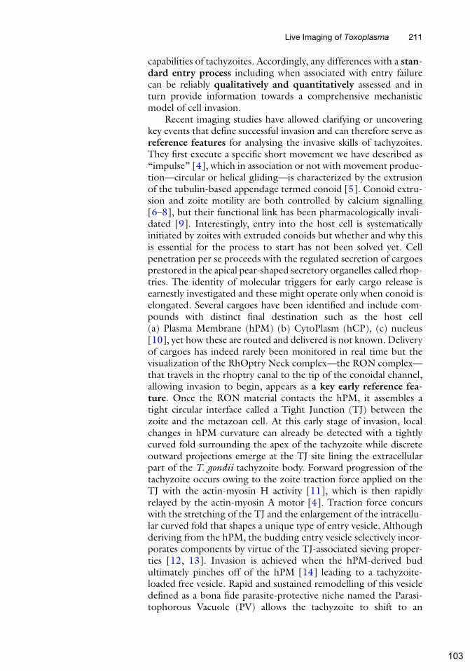
capabilities of tachyzoites. Accordingly, any differences with a stan-dard entry process including when associated with entry failurecan be reliably qualitatively and quantitatively assessed and inturn provide information towards a comprehensive mechanisticmodel of cell invasion.
Recent imaging studies have allowed clarifying or uncoveringkey events that define successful invasion and can therefore serve asreference features for analysing the invasive skills of tachyzoites.They first execute a specific short movement we have described as“impulse” [4], which in association or not with movement produc-tion—circular or helical gliding—is characterized by the extrusionof the tubulin-based appendage termed conoid [5]. Conoid extru-sion and zoite motility are both controlled by calcium signalling[6–8], but their functional link has been pharmacologically invali-dated [9]. Interestingly, entry into the host cell is systematicallyinitiated by zoites with extruded conoids but whether and why thisis essential for the process to start has not been solved yet. Cellpenetration per se proceeds with the regulated secretion of cargoesprestored in the apical pear-shaped secretory organelles called rhop-tries. The identity of molecular triggers for early cargo release isearnestly investigated and these might operate only when conoid iselongated. Several cargoes have been identified and include com-pounds with distinct final destination such as the host cell(a) Plasma Membrane (hPM) (b) CytoPlasm (hCP), (c) nucleus[10], yet how these are routed and delivered is not known. Deliveryof cargoes has indeed rarely been monitored in real time but thevisualization of the RhOptry Neck complex—the RON complex—that travels in the rhoptry canal to the tip of the conoidal channel,allowing invasion to begin, appears as a key early reference fea-ture. Once the RON material contacts the hPM, it assembles atight circular interface called a Tight Junction (TJ) between thezoite and the metazoan cell. At this early stage of invasion, localchanges in hPM curvature can already be detected with a tightlycurved fold surrounding the apex of the tachyzoite while discreteoutward projections emerge at the TJ site lining the extracellularpart of the T. gondii tachyzoite body. Forward progression of thetachyzoite occurs owing to the zoite traction force applied on theTJ with the actin-myosin H activity [11], which is then rapidlyrelayed by the actin-myosin A motor [4]. Traction force concurswith the stretching of the TJ and the enlargement of the intracellu-lar curved fold that shapes a unique type of entry vesicle. Althoughderiving from the hPM, the budding entry vesicle selectively incor-porates components by virtue of the TJ-associated sieving proper-ties [12, 13]. Invasion is achieved when the hPM-derived budultimately pinches off of the hPM [14] leading to a tachyzoite-loaded free vesicle. Rapid and sustained remodelling of this vesicledefined as a bona fide parasite-protective niche named the Parasi-tophorous Vacuole (PV) allows the tachyzoite to shift to an
Live Imaging of Toxoplasma 211
103

intracellular lifestyle [15]. Each of these events—that is, (1) conoidextrusion/RON secretion, (2) RON insertion/TJ assemblyinto the hPM, (3) TJ stretching, (4) TJ shrinking, (5) TJ closure,and (6) PV biogenesis—constitute six steps that can now serve asreference features for scoring tachyzoite invasive skills in the con-text of any mutation or drug effect under study (Fig. 2).
2 Material
2.1 T. gondii Cells The parasite lines ideally carry one or more fluorescent markers thatcould include a RONTJmarker to accurately assess the dynamics ofinvasion and/or any other marker of interest (http://toxodb.org/toxo/). Multiple genetic endogenous tagging can now be easilyobtained in both type I and II T. gondii strains engineered to lackthe gene encoding the DNA double-strand break repair Ku80protein hence showing drastic increase in the rate of homologousrecombination [16, 17]. In conjunction with these breakthroughsin molecular genetics, LIC-mediated and CRISPR/Cas9 knock-instrategies [2, 3] have undoubtedly eased the introduction of avariety of fluorescent markers thus allowing any laboratory mem-bers to image parasites for specific investigation. While care mustthen be taken to verify expression and functionality of the fusion
Fig. 2 Host cell invasion is a multistep process that can be decomposed in six steps (I–VI) using key
markers from the invading parasite (RON2-mC, red) and the host cell plasma membrane (GFP-GPI, green).
Scale bar: 5 μm
212 Georgios Pavlou and Isabelle Tardieux
104

constructs, a number of these have allowed precise description ofpreinvasive and invasive T. gondii tachyzoite behaviors. As such, wehave generated RH (i.e., type 1) ΔKu80 T. gondii strains thatexpress a functional chimeric fluorescent version of the rhoptry-stored RON2 protein which enable visualizing in real time thewhole multistep invasion event from the early RON complexrelease to the TJ closure and the subsequent birth of the PV[4]. Of note the CRISPR/Cas9 knock-in procedure has proveduseful for genome editing—gene deletion and point mutations[18, 19]—while conditional silencing systems have allowed analys-ing loss of function mutants for the so-called “essential” genes [20]and the impact of all those modifications for T. gondii invasion-related gene candidates can now be accurately examined in theRON2-mC-expressing tachyzoite lines. Indeed, dynamic imagingof mutants deleted for theMyoA gene revealed an alternative modeof entry into host cells that relies on hPM actin-rich protrusionsforcing the parasite into a hPM-derived bud [21].
2.2 Metazoan Cells The human foreskin fibroblasts (HFF-1, ATCC no. SCRC-1041,certified mycoplasma free) have been used to propagate theT. gondii tachyzoites in vitro by serial passages in high glucoseDulbecco’s Modified Eagle’s Medium GlutaMAX (DMEM) sup-plemented with 10% fetal bovine serum (FBS), 100 units/ml peni-cillin, and 100 mg/ml streptomycin (i.e., complete medium) at37 �C with 95% air and 5% CO2.
The host cell lines used for analyzing invasive skills are diversedue to the particularly broad spectrum of warm-blooded metazoanadherent cells permissive to T. gondii tachyzoites. We recommendchoosing cells that are easy to transfect for transient or stabletransgene expression and that preferentially spread thin and largelamellipodium/lamella area to ease the recording. However inva-sion of polarized epithelial or endothelial cell monolayers—whichdisplay rather columnar architectures—can also be used. In ourhands the human U2OS sarcoma cell line (ATCC, Cat#HTB-96;RRID: CVCL_0042) has been convenient for assessing—thereforecomparing—invasive behaviors. We can derive from the U2OS cellline a large number of transgenic sublines expressing GFP,mCherry, or any other fluorescent tags in fusion with a variety ofconstructs targeting the hcPM [4] (i.e., using a transmembranedomain of PM-exposed receptors such as the PDGF receptor or alipid covalent anchor/tether such as glycosyl-phosphatidylinositol,myristate, and palmitate) or/and the hc cortical cytoskeleton(using lifactin, etc.) (For details, see [4]) [15, 21, 22]. During theprocedure to obtain the U2OS (or other) cell line of interest, weusually select clones that express low amounts of the fusion con-struct to limit the risk of mis-localisation or physiologicalimpairment due to transgene overexpression.
Live Imaging of Toxoplasma 213
105

2.3 Microscope
Platform
We are working with a spinning disk (Yokogawa CSU-X) laserconfocal microscope (Nikon Eclipse), a platform that fulfills thespeed, sensitivity and resolution requirements for imaging(1) release of the RON material, (2) TJ assembly and functionalong with (3) parasite penetration. To achieve efficient resolution,we also use the X63 oil objective with a numerical aperture of 1.4.Because of the need to capture emitted signals from distinct fluor-ophores—exited with distinct laser beams—within only few sec-onds, fast frame rate video-recording is compulsory. In additionbecause we also want to capture as many events as possible in thesame session to further perform quantitative analysis and applystatistics (i.e., on the same field of view for the appropriate objec-tive), we usually extend the recording periods over two tens ofminutes. Therefore, particular care must be taken to limit (1) pho-totoxicity due to ROS generated from high/long excitation of thefluorophore and (2) photobleaching (see Note 1). Although pho-totoxicity might not be apparent for short-imaging time windowsas during invasion assay, and clearly depends on the fluorophoreand the subcellular localization of the marker of interest. For eachmarker we therefore optimize the emission filter for a maximalsignal and test the best illumination conditions aiming to limitexcitation to the level that provides both satisfying signal to noiseratio and spatiotemporal resolution.
Several alternatives to counteract both the phototoxicity andphotobleaching exist, including the use of other illumination tech-niques (Total internal reflection fluorescence—TIRF—light sheetillumination), but increasing the quantum efficiency of the emittedlight detector—that is, improving the sensitivity of the camera—iscertainly a good start. With these concerns, we use either theEM-CCD (Photometrics CoolSNAP HQ2, Andor iXon) or thesCMOS (Photometrics Prism BSI) cameras having a preferencefor the second one.
2.4 Additional
Microscope Settings
To keep the physiological conditions required for long term liveimaging of the T. gondii invasion in metazoan cells, a 37 �C tem-perature and 5% CO2 gas as well as humidity need to be providedthroughout the video session. Homemade and commercial micro-scope environmental chambers as well as heat and CO2 flow ratecontrollers need to be used. As regards the heat, because theimmersion objectives act as heat sink thereby creating a decreaseof temperature at the interface between the objective and thesample, it is important to use an objective heater in the environ-mental top stage chamber while a stage heater is also good. We hadalso positive experience using the compact Chamlide incubator topstage system from Live Cell Instrument, which accommodates arange of magnetic or nonmagnetic holder settings (i.e., glass bot-tom dishes, chambers holding glass coverslips or well plates of
214 Georgios Pavlou and Isabelle Tardieux
106

various dimensions) compatible or not with liquid perfusion atcontrolled flow rates.
2.5 Software MetaMorph 7.7.0.0 Molecular devices.Icy 1.9.5.1 http://icy.bioimageanalysis.org/ImageJ 1.51j8 https://imagej.nih.gov/ij/.UCSF ChimeraX 0.1 https://www.rbvi.ucsf.edu/chimerax/
3 Methods
3.1 Plating Cells
on Coated Glass-
Coverslips
Twenty-four to forty-eight hours prior to recording invasionevents, adherent cells are plated by default on poly-L-Lysine(100 μg/ml dH2O) -coated glass coverslips or MatTek glass bot-tom dishes or any other systems fitting the cell chamber holders(overnight 4 �C). For specific needs, collagen (rat tail collagen1, 50–100 μg/ml 0.02 M acetic acid), fibronectin (50 μg/mlPBS), gelatin (0.2% dH2O) or specific extracellular matrix compo-nents can be used with adapted coating protocols. At the time ofthe invasion assay, cells must have reached ~80% confluence. Cellsare washed with HBSS++ supplemented with 0.2–1% FCS (i.e., lowamounts of FCS can help to limit autofluorescence) and calcium to1.6 mM final concentration immediately prior transfer into the cellholder setting within the microscope chamber. The so-calledMotil-ity Buffer (MB), which allows maintaining proper osmolarity, is ourreference buffer used for all comparative preinvasive and invasivebehaviors. However if we extend the period of live recording post-invasion—to monitor tachyzoite replication for instances—we thenreplace the MB by complete medium but lacking phenol red toavoid nonspecific fluorescence in particular in the blue-yellowspectrum.
3.2 Collecting
T. gondii Tachyzoites
for Invasion Recording
The quality of the parasite preparation is essential to sample enoughevents in a short amount of time, thereby limiting the photosensi-tivity problems (see Note 1). To this end, we synchronize theparasite culture and recover the progeny within 2–4 h post–spon-taneous egress in the culture medium. In the case of tachyzoitemutants impaired in their egress potential (i.e., actin knockout,[23]), we scrap the cells from the flask and mechanically disruptthem by a few passages through a 26 gauge syringe needle. Theamount of parasites needed for each session is washed in the MB bygentle centrifugation, resuspended in a small volume of MB andrapidly added to the target cells under the microscope. Because welimit the volume in the cell chamber, parasite sedimentation is fastallowing to rapidly proceed for image acquisition. For the 20 minregular video sessions, we do not need to add more volume but forlonger imaging periods care is taken to prevent evaporation thatwould change the osmolarity by filling the cell chamber with
Live Imaging of Toxoplasma 215
107

medium and keeping saturated humidity in the vicinity of the cellculture setting (i.e., in the compact or wide top-stage chambers).Importantly our priority is to maintain parasites “healthy” by limit-ing CO2 and temperature fluctuations to obtain reliable phenotypeanalysis.
3.3 Image
Acquisition
and Analysis
Images are usually acquired in DIC and two fluorescent channels atoptimal speeds to achieve the efficient spatiotemporal resolution.UsingMetaMorph, we systematically go below 1 s per frame and tothis end, for reasons mentioned earlier, we select low laser powerand low time exposure while in case the camera has a “denoise”setting, we switch off the option. Once sequences have been cap-tured over routine 20 min video-recording sessions, the sequencesof interest are cropped using MetaMorph and ImageJ software.Then using the open software Icy, the researcher can access a varietyof parameters of static or moving defined “fluorescent” object(s)using tools such as (see Notes 2 and 3):
l Tracking of object(s):
– Simultaneous tracking of multiple moving and static objects.
– Combination of different fluorescent markers’ tracking.
– Extraction of values depending on the tracking object(s) suchas:
Speed.
Displacement.
Surface area.
Size measurement (perimeter, etc.).
Circularity.
Distance tracking between two different objects.
Fluorescence intensity, etc.
– Wide variety of visualization options for the tracks.
l Deformability of static object(s) overtime:
– Size measurement (perimeter, etc.).
– Fluorescence intensity.
– Circularity.
– Surface area, etc.
l 3D/4D reconstruction:– Volume area.
– Fluorescence Intensity, etc.
All the data are exported in either the .xls or the .csv file whichallows easy import and processing by all common software for data
216 Georgios Pavlou and Isabelle Tardieux
108

analysis. Moreover, Icy can create graphical plots of your data ofinterest (see Note 2).
To finalize the 3D and 4D reconstruction, we use the opensource software ChimeraX.
Example: A tachyzoite that expresses RON2 fused to themCherry tag (red) invades a host cell which expresses the hPMreporter fused to GFP (green). This dual fluorescence allowsdetecting and monitoring the (1) release of the RON complexprecisely defining the t0 point of entry, (2) stretching and shrinkingof the circular TJ that accommodates parasite progression into aninward hPM-derived bud that forms concomitantly, (3) the closureof the TJ ring, and (4) the birth of the PV when the bud hasseparated from the hPM therefore precisely defining the final time(ft). Image processing reveals that the parasite executes a rotationaround its long axis which imposes a twisting motion on its basalpole, hence ensuring TJ closure and bud sealing while promotingmembrane fission upstream the TJ. Interestingly, we observe aftertwisting the formation of micropatterns in the PV membrane(PVM), while after fission we measure a relaxation phase of theparasite body coupled with the stretching of the tightly adjustedPVM. We focus here on how image processing of the invadingRON2-mC tachyzoite has allowed analyzing the final twistingmotion (Fig. 3, for details, see [15]).
Rotation can be defined by the movement of one object aroundthe axis that connects two objects.
In our case, we assess the rotation using the following protocol:
1. Detection of the objects in the red fluorescent channel usingthe Spot detector plugin overtime. The RON2-mC pools corre-spond to the rhoptry and the closing/close TJ at each pole ofthe parasite respectively and define the main axis of theT. gondii tachyzoite.
2. Tracking of these objects overtime using the Spot trackingplugin.
Fig. 3 Schematic representation of the object tracking that allows quantifying the twisting motion accounting
for TJ closure. Right top panel shows the overlay image (DIC-RON2-mC, red) and right bottom panel shows the
three objects (rhoptry, TJ, and internal vesicle) tracked using Icy software, scale bar: 5 μm
Live Imaging of Toxoplasma 217
109

3. After step 2, the Track manager plugin window opens auto-matically providing the possibility to activate different subplu-gins (track processors) that can export values for speed,displacement, etc. of all the tracked objects. In our case weuse the track processor Instant speed which shows that thereference objects at each pole of the tachyzoite have almostzero speed and the intermediate object moves with a significantspeed around the main axis. Track processor Intensity speed isused to assess whether the object rotates around the axis (i.e.,continuous 360� rotation) (left panel Fig. 3) rather than a flip-flop like movement (i.e., 180 �C back and forward rotations).In more details, the intensity of the tracked object over timedecreases when the object gets out of focus and reincreaseswhen back in focus, thereby demonstrating the continuousrotation process.
By applying filters and threshold on the images in the fluores-cent channel of interest, we clearly observe the dynamics of thehPM-derived bud that tightly surrounds the invading parasite. Icyhas by default the widely used filters such as Median filter and theinterface of the intensity brightness control panel is quite useful. Byusing the Intensity profile plugin we can access the intensity distri-bution of the forming and newly formed PV and therefore detectany abnormal process of PV biogenesis.
We describe the relaxation of the parasite body as follows:
1. Using the same “stable” spots that have been tracked in therotation protocol.
2. Using the track processor Distance profiler, the distance betweenthe two stable spots could be recorded over time. Resultsshowed that there was an increase in the length of the axisthat connects these two spots.
4 Notes
1. T. gondii tachyzoites have been found sensitive to phototoxicdamages when they express high amounts of soluble protein infusion with GFP or YFP.Mammalian cells also show differentialsensitivity to illumination-induced ROS. Overall, we advise toreduce the peak intensity of the laser beam even if that meansextending the exposure time provided it is compatible with thetemporal resolution needed to capture the multistep sequenceof cell invasion. Ideally fluorescent markers should be carefullytested and compared with available alternatives. On the sameline, caution is necessary to ensure that cells are maintained ingood condition and are functional while on the microscope
218 Georgios Pavlou and Isabelle Tardieux
110

stage with illumination in the presence of synthetic fluoro-phores or fluorescent proteins.
2. Icy is an open source software that gives the ability to the userto search for similar tasks or solutions to possible problemsthrough an online interactive forum. For example, the user cancreate a new thread with his/her problem and then other usersor the Icy software engineers can provide advice/help.
3. Not only the best phenotypic description of a parasite preinva-sive and invasive skills clearly gains from a careful comparativeanalysis of the dynamic behavior with the current referencescheme (i.e., as described earlier), but it can also improve themodel itself by highlighting specific steps within “normal” or“abnormal” sequences. It is important to be aware of continualadvances in live imaging techniques/platforms, in the design offluorescent probes and computationally efficient algorithms toincrease the accuracy of the image information content we cancapture and process.
References
1. Tardieux I, Baum J (2016) Reassessing themechanics of parasite motility and host-cellinvasion. J Cell Biol 214:507–515. https://doi.org/10.1083/jcb.201605100
2. Sidik SM, Huet D, Ganesan SM, Huynh M-H,Wang T, Nasamu AS, Thiru P, Saeij JPJ, Car-ruthers VB, Niles JC, Lourido S (2016) Agenome-wide CRISPR screen in Toxoplasmaidentifies essential apicomplexan genes. Cell166:1423–1435.e12. https://doi.org/10.1016/j.cell.2016.08.019
3. Sidik SM, Huet D, Lourido S (2018) CRISPR-Cas9-based genome-wide screening of Toxo-plasma gondii. Nat Protoc 13:307–323.https://doi.org/10.1038/nprot.2017.131
4. Bichet M, Joly C, Henni AH, Guilbert T,Xemard M, Tafani V, Lagal V, Charras G, Tar-dieux I (2014) The Toxoplasma-host cell junc-tion is anchored to the cell cortex to sustainparasite invasive force. BMC Biol 12:773.https://doi.org/10.1186/s12915-014-0108-y
5. Hu K, Johnson J, Florens L, Fraunholz M,Suravajjala S, DiLullo C, Yates J, Roos DS,Murray JM (2006) Cytoskeletal componentsof an invasion machine--the apical complex ofToxoplasma gondii. PLoS Pathog 2:e13.https://doi.org/10.1371/journal.ppat.0020013
6. Mondragon R, Frixione E (1996) Ca(2+)-dependence of conoid extrusion in Toxoplasmagondii tachyzoites. J Eukaryot Microbiol43:120–127
7. Lourido S, Tang K, Sibley LD (2012) Distinctsignalling pathways control Toxoplasma egressand host-cell invasion. EMBO J31:4524–4534. https://doi.org/10.1038/emboj.2012.299
8. Lourido S, Moreno SNJ (2015) The calciumsignaling toolkit of the apicomplexan parasitesToxoplasma gondii and Plasmodium spp. CellCalcium 57:186–193. https://doi.org/10.1016/j.ceca.2014.12.010
9. Carey KL, Westwood NJ, Mitchison TJ, WardGE (2004) A small-molecule approach tostudying invasive mechanisms of Toxoplasmagondii. Proc Natl Acad Sci U S A101:7433–7438. https://doi.org/10.1073/pnas.0307769101
10. Hakimi M-A, Olias P, Sibley LD (2017) Toxo-plasma effectors targeting host signaling andtranscription. Clin Microbiol Rev30:615–645. https://doi.org/10.1128/CMR.00005-17
11. Graindorge A, Frenal K, Jacot D, Salamun J,Marq JB, Soldati-Favre D (2016) The conoidassociated motor MyoH is indispensable forToxoplasma gondii entry and exit from hostcells. PLoS Pathog 12:e1005388. https://doi.org/10.1371/journal.ppat.1005388
12. Mordue DG, Desai N, Dustin M, Sibley LD(1999) Invasion by Toxoplasma gondii estab-lishes a moving junction that selectivelyexcludes host cell plasma membrane proteinson the basis of their membrane anchoring. JExp Med 190:1783–1792
Live Imaging of Toxoplasma 219
111

13. Charron AJ, Sibley LD (2004) Molecular par-titioning during host cell penetration by Toxo-plasma gondii. Traffic 5:855–867. https://doi.org/10.1111/j.1600-0854.2004.00228.x
14. Suss-Toby E, Zimmerberg J, Ward GE (1996)Toxoplasma invasion: the parasitophorous vac-uole is formed from host cell plasma membraneand pinches off via a fission pore. Proc NatlAcad Sci U S A 93:8413–8418
15. Pavlou G, Biesaga M, Touquet B, Lagal V,Balland M, Dufour A, Hakimi M-A, Tardieux I(2018) Toxoplasma parasite twisting motionmechanically induces host cell membrane fissionto complete invasion within a protective vacu-ole. Cell Host Microbe 24:81–96.e5. https://doi.org/10.1016/j.chom.2018.06.003
16. Fox BA, Ristuccia JG, Gigley JP, Bzik DJ(2009) Efficient gene replacements in Toxo-plasma gondii strains deficient for nonhomolo-gous end joining. Eukaryot Cell 8:520–529.https://doi.org/10.1128/EC.00357-08
17. Huynh M-H, Carruthers VB (2009) Taggingof endogenous genes in a Toxoplasma gondiistrain lacking Ku80. Eukaryot Cell8:530–539. https://doi.org/10.1128/EC.00358-08
18. Sidik SM, Hackett CG, Tran F, Westwood NJ,Lourido S (2014) Efficient genome engineer-ing of Toxoplasma gondii using CRISPR/Cas9.PLoS One 9:e100450. https://doi.org/10.1371/journal.pone.0100450
19. Shen B, Brown KM, Lee TD, Sibley LD (2014)Efficient gene disruption in diverse strains of
Toxoplasma gondii using CRISPR/CAS9.MBio 5:e01114–e01114. https://doi.org/10.1128/mBio.01114-14
20. Andenmatten N, Egarter S, Jackson AJ,Jullien N, Herman J-P, Meissner M (2013)Conditional genome engineering in Toxo-plasma gondii uncovers alternative invasionmechanisms. Nat Methods 10:125–127.https://doi.org/10.1038/nmeth.2301
21. Bichet M, Touquet B, Gonzalez V, Florent I,Meissner M, Tardieux I (2016) Geneticimpairment of parasite myosin motorsuncovers the contribution of host cell mem-brane dynamics to Toxoplasma invasion forces.BMC Biol 14:97. https://doi.org/10.1186/s12915-016-0316-8
22. Rhee JM, Pirity MK, Lackan CS, Long JZ,Kondoh G, Takeda J, Hadjantonakis A-K(2006) In vivo imaging and differential localiza-tion of lipid-modified GFP-variant fusions inembryonic stem cells and mice. Genesis44:202–218. https://doi.org/10.1002/dvg.20203
23. Egarter S, Andenmatten N, Jackson AJ, White-law JA, Pall G, Black JA, Ferguson DJP,Tardieux I, Mogilner A, Meissner M (2014)The Toxoplasma Acto-MyoA motor complexis important but not essential for gliding motil-ity and host cell invasion. PLoS One 9:e91819.https://doi.org/10.1371/journal.pone.0091819
220 Georgios Pavlou and Isabelle Tardieux
112

2.2.5 Discussion and perspectives
In this study, we report the importance of mechanical forces generated by the T.
gondii tachyzoite to end the invasion sequence in mammalian host cells and
initiate the modeling of an intracellular niche compatible with the production of
progeny. When compared to what we knew about the early steps of cell invasion,
the mechanisms underlying the birth of a subcellular compartment in which the
tachyzoite could safely adopt an intracellular lifestyle were quite elusive. While
the functions of the RON invasive nanodevice as a traction force platform and a
molecular sieve were already established and shown to support the subversion of
a component-selective piece of PM during tachyzoite entry, we were curious to
investigate if the presence of this nanodevice could also dictate the way the
budding entry vesicle actually pinches off of the PM it derives from. To this end,
we combined quantitative live imaging, T. gondii and mammalian cell molecular
genetics and nanotools. We were able to detect a final twisting motion of the
parasite that imposes rotation on the basal pole and therefore on the ZCJ and
appears critical for finalizing the safe entry of the parasite while preserving the
hosting cell from membrane damages. Another fascinating observation came
when we imaged simultaneously the fluorescent invasive RON-nanodevice and
the fluorescence associated with the host cell PM membrane. We were able not
only to detect a homogeneous signal in the budding membrane when the
tachyzoite entered but also the changes in the bud fluorescence that occurred only
post twisting motion and led to a specific fluorescent patchy pattern in the
membrane of the PV. Given the fact that when the PV emerges post-pinching off,
the tachyzoite next must rapidly adjust to its new microenvironnemt and organize
a niche appropriate for growth. These requirements are satisfied thanks to a
number of proteins and lipids the tachyzoite secretes, in particular from the
heterogeneous population of dense granules, which promote the assembly of an
intricate network of tubular structures that ensure the import and the export of
material beyond the PV membrane. Apart from dense granule products, the
rhoptries also secrete components into the vacuolar space and it would be
interesting to verify if the clustering of PV-derived fluorescent material can
reflect the integration of known transmembrane proteins such as the ROP2
superfamily members, but also for instances MYR2 and 3 that are thought to be
essential part of a translocon system underpinning the export of GRA proteins
across the PVM (Arrizabalaga, 2019). Many more proteins have been identified
and shown to associate with either side of the PVM and they could theoretically
contribute even without spanning the elusive PVM at applying force to the PVM
and trigger some redistribution of molecules in the PVM. Indeed, mechanical
forces on the PVM could be expected with the rapid recruitment of ER and
mitochondria that interact with the young PV.
As regard to the twisting motion, we hypothesized that the torque applied onto the
the nanodevice forces the two upstream membranes facing each other to follow
113

the same movement and this is in line with the observation that the RON2-mC
signal actually partitions into the PV. Therefore this implies that any expected
fusion/fission event(s) must take place immediately upstream the nanodevice. It is
tempting to compare the pinch off process with a topologically similar event
occurring for the release of endocytic vesicle from the coated pits during clathrin-
mediated endocytosis. In this case, the dynamins play a major role by directing
the constriction and the torsion of the two membranes forming the neck of the
nascent endocyctic vesicle. Thanks to cells engineered to tamoxifen induce triple
knockout of dynamins 1,2 and 3, we proved that the tachyzoite achieves invasion
similarly in cells expressing or lacking dynamins. With the same approach, we
disproved the functional involvement of ALIX during cell invasion, a protein
partner of the ESCRT-I machinery which was recently reported but using indirect
assays and based on (i) the in situ localization of a pool of ALIX at the ZCJ and
(ii) the physical interaction established between ALIX and RON4 (Guérin et al,
2017).
However, we do not exclude that other molecules from the host cell could assist
the torus closure and membrane fission processes. An important candidate to
investigate is actin, which is known to team with dynamins for force production
during endosome formation. F-actin is clearly present at the ZCJ site, which led
our team to propose a role of a de novo assembled F-actin pool in the anchorage
of the nanodevice-made platform. The same situation holds for the actin ARP2/3
nucleating factor (Gonzalez et al, 2009), therefore positioning in space and time
the actin and actin polymerization machinery to the ZCJ over the process of
invasion. Finally, other host cell molecules could deserve attention to molecularly
understand the fission process among which lipids that are known to determine
the fusogenic capacities of membranes and that could facilitate fusion upstream
the torus upon twisting motion. In terms of the fission process itself, the
remarkable preservation of cell membrane integrity is a fascinating aspect of the
process and it still might be important to delve deeper in the potential
contribution of membrane repair mechanism.
Overall, this study highlights the cleverness of the tachyzoite to engineer a multi-
functional unique “invasive nanodevice” to achieve amazing high-speed invasion
of many different cell types. It allows suggesting that, with this nanodevice and
the ability to apply force, the tachyzoite has evolved a dynamin like machinery to
force constriction and torsion of the two facing membrane segments at the “neck”
of the budding entry vesicle. With this strategy, the tachyzoite actively closes the
entry door made by the toroidal invasive nanodevice and mechanically induces
separation from the plasma membrane of the hosting cell to eventually reside in
the intracellular niche and adopt the intracellular lifestyle.
114

Conclusion
115

Conclusion
During this thesis period, we wished
to create a map of the forces that
contribute to Toxoplasma motility
and host cell invasion. By
highlighting the key role of
mechanical forces that drive cell
movement in a 3D matrix or into a
cell, this work opens a new era of
investigation with the objective of
an in-depth understanding of the
peculiar forces, including torque
and contraction over these
processes. It is quite clear how
unique is this cell by using a variety
of mechanisms not only to migrate
but also to invade and parasitize a
host cell to fill their metabolic
needs. We could make use of its
“secret weapons” beyond the field
of parasitology starting with the ever- increasing range of applications for
nanotechnologies including in medicine, and reaching even the
macrotechnologies as for example in architecture. We could take inspiration from
the invasive nanodevice and the invasion mechanisms to engineer new generation
of carriers with specific surface-exposed molecules favoring their capture by
given cells, such as cancer cells, and then delivery of specific cargo including
pharmacological drugs or other compounds to promote destruction for tumor cells
or in some other cases to eliminate the microbial threats with the minimum
damage during the delivery process. As for the field of architecture and in
particular the subfield of biomimicry in architecture design of which I am a big
fan, one can imagine that architects could use mechanical properties of the rigid
and flexible Toxoplasma to build objects or even buildings! Coming from a
mathematics background where I love geometry, I would be very inspired by the
unique cytoskeleton of the tachyzoite and especially the network of twisted
microtubules and the elastic properties that this network seems to have, but also
the amazing construction of connections bridging the membranous elements of
the pellicle to the cytoskeleton. But it seems that a famous architect named
Norman Foster had already been inspired by nature to build the Toxo-like
Gherkin building in London… He was inspired from a different organism but
why not from Toxoplasma in the future.
116

Other publications
117

Other publications
Del Rosario M, Periz J, Pavlou G, Lyth O, Latorre-Barragan F, Das S, Pall GS,
Stortz JF, Lemgruber L, Whitelaw JA, Baum J, Tardieux I & Meissner M (2019)
Apicomplexan F-actin is required for efficient nuclear entry during host cell
invasion. EMBO Rep 20: Available at:
https://onlinelibrary.wiley.com/doi/abs/10.15252/embr.201948896
Siebert C, Villers C, Pavlou G, Touquet B, Yakandawala N, Tardieux I &
Renesto P (2020) Francisella novicida and F. philomiragia biofilm features
conditionning fitness in spring water and in presence of antibiotics. PLoS ONE
15: e0228591 Available at: https://dx.plos.org/10.1371/journal.pone.0228591
118

Article
Apicomplexan F-actin is required for efficient
nuclear entry during host cell invasion
Mario Del Rosario1 , Javier Periz1, Georgios Pavlou2, Oliver Lyth3, Fernanda Latorre-Barragan1,4 ,
Sujaan Das1, Gurman S Pall1, Johannes Felix Stortz1 , Leandro Lemgruber1, Jamie A Whitelaw5,
Jake Baum3 , Isabelle Tardieux2 & Markus Meissner1,6,*
Abstract
The obligate intracellular parasites Toxoplasma gondii and Plas-
modium spp. invade host cells by injecting a protein complex
into the membrane of the targeted cell that bridges the two
cells through the assembly of a ring-like junction. This circular
junction stretches while the parasites apply a traction force to
pass through, a step that typically concurs with transient
constriction of the parasite body. Here we analyse F-actin
dynamics during host cell invasion. Super-resolution microscopy
and real-time imaging highlighted an F-actin pool at the apex
of pre-invading parasite, an F-actin ring at the junction area
during invasion but also networks of perinuclear and posteriorly
localised F-actin. Mutant parasites with dysfunctional acto-
myosin showed significant decrease of junctional and perinu-
clear F-actin and are coincidently affected in nuclear passage
through the junction. We propose that the F-actin machinery
eases nuclear passage by stabilising the junction and pushing
the nucleus through the constriction. Our analysis suggests that
the junction opposes resistance to the passage of the parasite’s
nucleus and provides the first evidence for a dual contribution
of actin-forces during host cell invasion by apicomplexan
parasites.
Keywords actin; Apicomplexa; host cell invasion; myosin; nucleus
Subject Categories Cell Adhesion, Polarity & Cytoskeleton; Microbiology,
Virology & Host Pathogen Interaction
DOI 10.15252/embr.201948896 | Received 21 July 2019 | Revised 21 August
2019 | Accepted 11 September 2019
EMBO Reports (2019) e48896
Introduction
The phylum Apicomplexa consists of more than 5,000 species, most
of them obligate intracellular parasites, including important human
and veterinary pathogens, such as Plasmodium (malaria) or Toxo-
plasma (toxoplasmosis).
During their complex life cycles, apicomplexan parasites move
through different environments to disseminate within and between
hosts and to invade their host cell [1]. Therefore, the invasive
stages, called zoites, evolved a unique invasion device, consisting of
unique secretory organelles and the parasites’ acto-myosin system,
the Glideosome, localised in the narrow space (~30 nm) between
the plasma membrane and the inner membrane complex (IMC) [2].
Zoites actively enter the host cell by establishing a tight junctional
ring (TJ) at the point of contact between the two cells. The TJ is
assembled by the sequential secretion of unique secretory organelles
(micronemes and rhoptries), leading to the insertion of rhoptry neck
proteins (RONs) into the host cell plasma membrane (PM) and
underneath [3]. On the extracellular side, the exposed domain of the
RON2 member binds the micronemal transmembrane protein AMA1
exposed on the parasite surface, resulting in the formation of a
stable, junctional complex [3]. The TJ is further anchored to the
host cell cortex by de novo formation of F-actin through the recruit-
ment of actin-nucleating proteins [4,5]. During host cell invasion,
the parasites use their acto-myosin motor to pass through the TJ.
However, the exact role and orientation of the parasite’s acto-
myosin system is still under debate [6] and intriguingly, mutants for
key component of this system show residual motile and invasive
capacities [7–9], the latter reflecting in large part an alternative and
host cell actin-dependant mode of entry [10]. According to the
Glideosome model, the force generated for motility and invasion
relies exclusively on F-actin polymerised at the apical tip of the
parasite by the action of Formin-1 and translocated within the
narrow space (~30 nm) between the IMC and PM of the parasite
[11]. However, recent studies suggest that the parasite can also use
other motility systems, such as a secretory-endocytic cycle that
produces retrograde membrane flow [12], similar to the fountain
flow model suggested for other eukaryotes [13,14].
In support of the linear motor model, was the detection of para-
site F-actin underneath the junction formed by invading parasites
1 Wellcome Centre For Integrative Parasitology, Institute of Infection, Immunity & Inflammation, Glasgow Biomedical Research Centre, University of Glasgow, Glasgow, UK2 Institute for Advanced Biosciences, CNRS, UMR5309, INSERM U1209, Université Grenoble Alpes, Grenoble, France3 Department of Life Sciences, Imperial College London, London, UK4 Faculty of Science, Food Engineering and Biotechnology, Technical University of Ambato, Ambato, Ecuador5 CRUK Beatson Institute, Glasgow, UK6 Experimental Parasitology, Department for Veterinary Sciences, Ludwig-Maximilians-University Munich, Munich, Germany
*Corresponding author. Tel: +49 89 21803622; E-mail: [email protected]
ª 2019 The Author. Published under the terms of the CC BY 4.0 license EMBO reports e48896 | 2019 1 of 18119

when using an antibody preferentially recognising apicomplexan
F-actin. Furthermore, the detection of cytosolic locations, predomi-
nantly around the nucleus [15], suggests additional roles of this
cytoskeletal protein during invasion.
While it was assumed a major role of F-actin in driving Apicom-
plexa zoite gliding motility and cell invasion, recent studies demon-
strated the pivotal role of F-actin in multiple other processes, such
as apicoplast inheritance [16], dense granule motility [17] and likely
nuclear functions through the control of expression of virulence
genes in malaria parasites [18]. However, building a comprehensive
model for F-actin dynamics, localisation and function in apicom-
plexan parasites has been hampered for decades by the lack of tools
enabling reliable F-actin detection. Interestingly, several studies
suggested that F-actin is interacting with subpellicular microtubules
[19] and/or the subpellicular matrix of the IMC [20,21]. Further-
more, components of the Glideosome, such as GAPM proteins [22]
or the invasion-critical myosin, MyoH were demonstrated to interact
with microtubules [23], suggesting a coordinated action of the actin
and microtubule cytoskeleton during host cell invasion.
With the adaptation of nanobodies specifically recognising
F-actin, it is now possible to analyse F-actin dynamics in apicom-
plexan parasites [24,25] leading to the identification of distinct
cytosolic networks of dynamic actin in both T. gondii and P. falci-
parum [11,24,25]. We originally identified two F-actin polymerisa-
tion centres from where most of the F-actin flow occurs in
intracellular parasites [24], later expanded to three based on further
studies [11,25]. One polymerisation centre can be found at the
apical tip, corresponding well to the described location of Formin-1,
and one close to the parasite’s Golgi complex, corresponding to the
location of Formin-2 [11,25]. In T. gondii, these three polymerisa-
tion centres of F-actin correlate well with the location of Formins-1,
2 and 3 in T. gondii [11]. Formins-2 and 3 appear to have overlap-
ping functions during the intracellular development of the parasite
for material exchange and formation of an intravacuolar network in
intracellular parasites, while Formin-1 (FH-1) has been explicitly
implicated in the apical polymerisation of F-actin required for glid-
ing motility and cell invasion [11]. In this study, although not
directly shown, the authors suggested that FH-1 polymerises F-actin
at the apical tip of the parasite that is exclusively transported by the
action of the Glideosome within the subalveolar space to the poste-
rior pole. Interestingly, at least in intracellular parasites, the major-
ity of F-actin dynamics appears to occur within the cytosol of the
parasite close to the parasite’s Golgi, forming a continuous dynamic
flow from centre to the periphery of the parasite and from the apical
to the basal pole [24,25]. In addition, measurement of peripheral F-
actin flow demonstrated that it can occur bidirectional from the
apical to the posterior pole and vice versa [25].
In good agreement with a cytosolic oriented F-actin system,
previous reports demonstrated parasite F-actin to be associated with
the subpellicular microtubules that are connected to and stabilise
the IMC [19–21,26]. Furthermore, gliding-associated proteins identi-
fied via co-immunoprecipitation with the Glideosome were recently
found to play important roles in stabilising the IMC and directly
connecting it to the subpellicular microtubules [22,27], suggesting
that a close connection exists between parasite F-actin, the Glideo-
some and the subpellicular network.
Here we compared F-actin dynamics in WT and mutant parasites
for the acto-myosin system and correlated its dynamic location with
the phenotypic consequences during host cell invasion. Interest-
ingly, in unstimulated parasites, F-actin flow in extracellular para-
sites appears to be bidirectional, with the majority of events
occurring in a retrograde direction, which is similar to measure-
ments performed on intracellular parasites [25]. The activation of
calcium signalling results in a shift towards retrograde F-actin flow,
resulting in F-actin accumulation at the posterior pole. Comparison
of F-actin dynamics in parasite mutants for the acto-myosin system
helped to identify the nucleus as a major obstacle for efficient host
cell invasion that needs to be squeezed through the junction using
F-actin dynamics to translocate and potentially deform the nucleus
through this process.
Our analysis strongly suggests a push-and-pull mechanism for
nuclear entry during host cell invasion, in analogy to the dynamics
observed during migration of other eukaryotes through a constricted
environment [28].
Results
Rational for selecting parasite mutants in order to analyse
F-actin dynamics during host cell invasion
Over recent years, conditional mutagenesis systems have been
employed to generate and characterise a whole assortment of para-
site mutants affected in host cell invasion. These mutants can be
affected in different, unrelated pathways, such as microneme secre-
tion, formation of the tight junction or host cell entry. Since we were
interested in analysing F-actin dynamics during the invasion
process, we chose parasite mutants that were previously charac-
terised to be significantly affected, but not blocked in host cell entry
and linked to the parasites acto-myosin system. We excluded all
mutants that have been described to be completely blocked in the
initiation of invasion, such as MyoH [23] or the micronemal protein
MIC8 [29], since no invasion event can be expected that can be
analysed. Similarly, we also excluded mutants involved in actin
dynamics that have not been implicated in host cell invasion, such
as FH-2,3 [11] or myosins not involved in host cell invasion, such
as MyoI [30].
Therefore, we chose parasite actin [7,9], the actin depolymerisa-
tion factor ADF [31] and myosin A (MyoA), which has been used
previously to successfully identify the contribution of the host cell
for parasite invasion [10].
F-actin flow is mainly organised within the cytosol of
extracellular parasites and is modulated by Calcium and
cGMP signalling
We previously imaged F-actin flow in intracellular parasites and
were able to discriminate two major polymerisation centres, one at
the apical tip and one close to the apicoplast, where F-actin is
formed and subsequently transported to the posterior end of the
parasite [24]. Indeed, recent studies on Toxoplasma and Plasmod-
ium demonstrated that Formin-2 is localised to the apicoplast of the
parasite and required for most of the intracellular F-actin dynamic
[11,25], while Formin-1 is localised at the apical tip, where it is
thought to be exclusively required for parasite motility and inva-
sion. In good agreement, in intracellular parasites, F-actin appears
2 of 18 EMBO reports e48896 | 2019 ª 2019 The Author
EMBO reports Mario Del Rosario et al
120

to be formed at two polymerisation centres, localised at the apical
tip and close to the Golgi region of the parasite, indicating that FH-1
and FH-2 are acting as nucleators during intracellular parasite devel-
opment. Intriguingly, the majority of F-actin dynamics in intracellu-
lar parasites occurs within the cytosol of the parasite with F-actin
flow occurring in a bidirectional manner (retrograde and antero-
grade) [24,25]. We were interested to determine if during the tran-
sient extracellular life and at the time of cell invasion, the patterns
of actin dynamics differ from the intracellular ones and how they
were impacted upon disruption of the Glideosome or factors criti-
cally involved in F-actin regulation.
Since transient transfections are typically associated with overex-
pression of the target protein hence with significant uncontrolled
impact on a dynamic equilibrium [24], we generated T. gondii lines
stably expressing Cb-EmeraldFP. We ensured that the expression of
the chromobody was comparable between the different parasite lines
and ensured that its expression does not lead to phenotypic alter-
ations and misleading analysis. We established transgenic parasites
expressing Cb-EmeraldFP in WT parasites, a null mutant for myosin
A (MyoA), the core motor of the Glideosome [16] and a conditional
mutant for the critical actin regulator, actin depolymerisation factor
ADF, adfcKD [31]. Using live 3D-structured illumination microscopy
(3D-SIM), we compared F-actin dynamics and found two discernible
F-actin polymerisation centres (Fig 1A) as previously seen in intra-
cellular parasites [24]. Time-lapse analysis demonstrated that the
majority of F-actin dynamics occurs in the cytosol of the parasite
with some F-actin flow detectable at the periphery occurring in retro-
grade and anterograde orientation, similar to intracellular parasites
(see also [25]). In good agreement with the localisation of FH-1 and
FH-2, two polymerisation centres can be detected. F-actin flow starts
from the apical tip (1st polymerisation centre, yellow arrow in
Fig 1A) to the second polymerisation centre close to the Golgi (red
arrow, Fig 1A) to the posterior pole of the parasite (Fig 1A, Movie
EV1). While disruption of myoA did not result in significant changes
of F-actin localisation or dynamics in resting parasites (Fig 1A,
Movie EV1), depletion of ADF completely abrogated actin dynamics,
F-actin accumulation being observed at both apical and posterior
parasite poles (Fig 1A, Movie EV1).
To map the actin structures visualised with the chromobody, we
employed skeletonisation processing [32], which converts F-actin
signals into individual pixels, that can be traced to obtain informa-
tion about the localisation and dynamics of F-actin. The resulting
skeletonised movie can be projected to show where the majority of
(detectable) F-actin dynamics occur during the timeline of the movie
(Fig 1B, Movie EV1). This analysis confirmed that parasite F-actin
formed at the apical tip (yellow arrowhead) and Golgi region (red
arrowhead), converge within the cytosol of the parasite and reach
the basal pole of the parasite. Interestingly, both peripherical and
cytoplasmic flow appear to be connected and do not occur indepen-
dently from each other (Fig 1B). Note that both WT and myoAKO
parasites displayed similar signals, whereas the ADF—conditional
mutant did not.
Next, kymograph analysis was performed to analyse individual
F-actin flow events using KymographClear and KymographDirect
[33] confirming the presence of a continuous dynamic F-actin
network in cytosolic location that connects the apical pole, Golgi
area and basal pole of the parasite, in good agreement with the loca-
tion of the 3 Formins previously described in T. gondii [11].
KymographClear is a FIJI plugin that generates a 3 colour-coded
kymograph, where each colour labels either forward movement
(red), backward movement (green) or static (blue) as shown
(Fig 1C and Appendix Fig S1). This kymograph can then be read
into the KymographDirect software, which uses automatic detection
to trace trajectories of moving particles in the image. Values are
then assigned to these trajectories based on both intensity and speed
[33]. The intensity can be assessed in specified tracks over time as
the average intensity over time shown in Fig 1C. Both the RH and
myoA KO presented dynamic signals connecting the apical pole
(close to the point 0), Golgi region (between 2 and 3 lm) and basal
pole. In the case of the adf KD, only static signals could be detected
at the apical and basal pole of the parasite, confirming the lack of
F-actin dynamics in these mutants.
Next, we used this analysis to investigate intensity profiles of
actin signal travelling in the periphery of the parasite (Appendix Fig
S1B–D). Interestingly, actin dynamics along the periphery is similar
between WT and myoA KO with bidirectional actin flow events as
revealed by the kymographs, while confirming previous results with
the adf KD mutants, which shows a lack of F-actin dynamics across
the entire parasites body (first half (�), Appendix Fig S1A–C).
Together, the data presented in this work point that similarly to
intracellular parasites, the F-actin is highly dynamic in extracellular
parasites displaying two major polymerisation centres, one at the
apical tip and another at the Golgi region of the parasite. Of note,
this analysis underlines that there is actin flow coming from the
cytoplasm towards the periphery and shaping a highly dynamic
network that connects the apical and basal pole of the parasite,
therefore independently of any F-actin network positioned between
the zoite’s PM and IMC.
Tosetti et al [11] suggested that the activation of a Ca2+-signal-
ling cascade would lead to increased retrograde transport of F-actin,
eventually accumulating posteriorly. However, we found that the
treatment of parasites with the calcium ionophore A23187 induced
anterior accumulation of F-actin in wild-type parasites in the major-
ity of cases (Fig 1D–F; Appendix Fig S1A; Movie EV2), while BIPPO
triggered a predominantly basal F-actin accumulation (Fig 1D–F;
Appendix Fig S1A; Movie EV2). BIPPO is a drug capable of inhibit-
ing 30,50-cyclic nucleotide phosphodiesterases (PDEs) responsible in
blocking the breakdown of cyclic nucleotides such as cAMP and
cGMP. This mechanism is believed to be responsible in the activa-
tion of microneme secretion and egress by modulating the signal
pathway of PKG-dependent processes [34].
Once MyoA null mutants were submitted to treatments with
A23187 or BIPPO, the F-actin remained rather evenly distributed
within the cytoplasm albeit with some peripheral enrichment in the
first half of the parasite closer to the apical end (Fig 1D–F; Movie
EV2). In contrast, depletion of ADF led to the expected phenotype
with F-actin accumulating at the apical and basal pole of the para-
site, independently of A23187 or BIPPO addition (Fig 1D–F; Movie
EV2). Together, these data suggest that calcium signalling regulates
F-actin dynamics, potentially by stimulating the activity of Formin-1
followed by F-actin transport to the posterior pole of the parasite by
the action of MyoA.
In conclusion, similar to intracellular parasites, extracellular
parasites show impressive F-actin dynamics within the cytosol. In
parasite mutants for ADF, this dynamic is abolished, and F-actin
accumulates at the apical and basal pole, independent of triggering
ª 2019 The Author EMBO reports e48896 | 2019 3 of 18
Mario Del Rosario et al EMBO reports
121

RH
Cb
-Em
era
ldF
P
myoA
KO
Cb
-Em
era
ldF
P
adf
cKD
Cb
-Em
era
ldF
P
0:00 0:10 0:21 0:31 TgActin
0:00 0:10 0:21 0:31
0:00 0:10 0:21 0:31
RH
Cb
-Em
era
ldF
P
myoA
KO
Cb
-Em
era
ldF
P
adf
cKD
Cb
-Em
era
ldF
P
0:00 0:10 0:21 0:31 TgActin
0:00 0:10 0:21 0:31
0:00 0:10 0:21 0:31
RH
Cb
-Em
era
ldF
P
0 2 4 6 80
50000
100000
150000
200000
250000
Position [µm]
Inte
nsit
y [
a.u
.]
Average Maximum Minimum
08
myoA
KO
Cb
-Em
era
ldF
P
2 4 6 8
-100000
0
100000
200000
300000
Position [µm]
Inte
nsit
y [
a.u
.]
Average Maximum Minimum
0
8
adf
cKD
Cb
-Em
era
ldF
P
2 4 6 8
-50000
0
50000
100000
150000
Position [µm]
Inte
nsit
y [
a.u
.]
Average Maximum Minimum
08
0:11
myoA
SN
AP
Cb
-Em
era
ldF
P
(-) Calcium Ionophore
(2uM)
(-)
Cy
toch
ala
sin
DD
MS
O
myoA
KO
Cb
-Em
era
ldF
P
adf
cKD
Cb
-Em
era
ldF
P
TgActinMyoA
TgActinMyoA
TgActinMyoA
TgActin
TgActin
BIPPO
(5uM)
TgActin
TgActin
TgActin
TgActin
myoA
SN
AP
Cb
-Em
era
ldF
P
adf cK
D
Cb
-Em
era
ldF
P
myoA
KO
Cb
-Em
era
ldF
P
Actin apical accumulation
Actin peripherical accumulation
close to the apical end
No accumulation
No accumulation
Actin accumulation both ends
(-) Calcium Ionophore
(2uM)
Actin peripherical accumulation
close to the apical end
Actin accumulation both ends Actin accumulation both ends
Posterior apical accumulation
BIPPO
(5uM)TgActinMyoA
TgActinMyoA
TgActinMyoA
TgActinMyoA
TgActinMyoA
TgActinMyoA
A B
C
D E
F
Actin accumulation on apical end
Peripherical accumation close to the apical end
No accumulation
Actin accumulation on posterior end
RH
RH C
ytD
RH D
MSO
Myo
AK
O
adfc
KD
adfc
KD 9
6h A
Tc+ R
H
RH C
ytD
RH D
MSO
Myo
AKO
adfc
KD
adfc
KD 9
6h A
Tc+
0
20
40
60
80
100
120
Perc
en
tag
e [
%]
no treatment Calcium Ionophore 2µM
****
****
****
****
ns
RH
RH C
ytD
RH D
MSO
Myo
AK
O
adfc
KD
adfc
KD 9
6h A
Tc+ R
H
RH C
ytD
RH D
MSO
Myo
AKO
adfc
KD
adfc
KD 9
6h A
Tc+
0
20
40
60
80
100
120
Perc
en
tag
e [
%]
no treatment BIPPO 5µM
****
****
****
****
ns
Figure 1.
4 of 18 EMBO reports e48896 | 2019 ª 2019 The Author
EMBO reports Mario Del Rosario et al
122

a calcium-signalling cascade. In contrast, disruption of MyoA results
in normal F-actin dynamics, but upon calcium signalling, F-actin
remains more diffusely localised within the cytosol of the parasite
with some accumulation at the periphery.
The acto-myosin system is required to ensure efficient invasion
To investigate whether this impressive cytoplasmic F-actin dynamics
could assist the parasite over the invasion of the target cell, namely
when it fits through the junction and constricts (Fig 2A and B), we
first measured the average diameter of parasites prior and during
invasion. We observed a significant decrease at mid-invasion, (prior:
2.5–3 lm and mid-invasion 1.5–2 lm) that attests significant body
deformation and compression in order to fit through the junction
(Fig 2A and B). In good agreement, analysis of the average diameter
of the parasite’s nucleus indicated that this organelle suffers a strik-
ing deformation, up to ~50% of its normal diameter (i.e. < 1 lm)
while passing through the junction (Fig 2A and B). Since the acto-
myosin system generates traction force at the junction to account for
the invasive force, we investigated if interference causes changes,
that is differences in the diameter of the TJ and/or stronger deforma-
tions of the parasite. While a slight reduction in the average TJ diam-
eter was observed during invasion of myoA KO parasites (Fig 2C),
the junctional ring appears remarkably constant upon interference
with the acto-myosin system of the parasite.
Previously, it was demonstrated that mutants for the acto-myosin
system enter the host cell in a stop-and-go manner, leading to a
prolonged invasion process with different features [7,9,31]. While
mutant parasites could be driven through the TJ by the host cell
membrane outward projections in an uncompleted micropinocyto-
sis-like process [10], they could in some cases remain trapped in the
TJ and die, probably due to compressive forces exerted on the
embedded part of the parasite by the host cell membrane protru-
sions [10]. We infer from these observations that the tachyzoite
likely needs to counteract compressive forces at the TJ to secure
entry of its bulky organelles, such as the nucleus and hypothesised
that the parasite acto-myosin is critically involved in nuclear entry.
We compared the invasion process of WT and mutant parasites
in detail using time-lapse analysis (Fig 2D and E, Appendix Fig S2,
Movie EV3 and EV4). Interestingly, the invasion process of WT
parasites proceeded with an initial rapid invasion step, followed by
a short pause when ~1/3 of the body was inside the host cell, and
another acceleration until the parasite was fully inside the host cell
(Fig 2D and E, Movie EV3). In contrast, while initial invasion steps
appeared to be comparable between WT parasites and mutants for
the acto-myosin system (act1cKO, mlc1cKO, myoAKO), an extended
period then followed, where parasites stalled (up to 8 min), before
invasion continued (Fig 2D and E, Appendix Fig S2, Movie EV4). In
some cases, we also observed abortive invasion events, where the
invading parasites terminally stalled before being pushed out from
the TJ (Fig 2F, Movie EV5). Interestingly, this stalling occurred
approximately when the nucleus reached the TJ suggesting that in
addition to providing the traction force that promotes entry through
the TJ [30], F-actin also assists the passage of the nucleus through
the TJ, akin to other eukaryotes when migrating through constricted
environments [35].
F-actin accumulates at the posterior pole and in most cases at
the junction
To correlate F-actin dynamics with the invasive behaviour of para-
sites, we stably expressed F-actin chromobodies in fusion with
Emerald FP in T. gondii [24] WT and mutant parasites and
compared the localisation and dynamics of F-actin when they
◀Figure 1. F-actin, formed at two major nucleation centres, forms a dynamic, continuous network within the cytosol of the parasite.
A Stills depicting actin flow (see Movie EV1) in extracellular RH, myoA KO and adf KD parasites expressing Cb-EmeraldFP. Continuous F-actin flow can be seen from the
apical tip, to the Golgi region towards the posterior pole of the parasite. No difference can be observed between RH and myoA KO. For adf KD, the flow is completely
abrogated. Red arrowhead marks the Golgi region, while the yellow arrowhead marks the apical nucleation centre. The time lapse is presented in Movie EV1.
B Skeletonisation processing of (A) depicting areas where F-actin dynamics/flow is prevalent. Individual signals for F-actin form a continuous, dynamic network that
connects apical and posterior pole of the parasite. Red arrowhead marks the Golgi region nucleation centre, while the yellow arrowhead marks the apical nucleation
centre. The images represent a projection of frames for each timepoint to provide a better overview of the dynamics of F-actin. The time lapse is presented in Movie EV1.
C Kymograph analysis of (A) and (B). The kymograph was generated by tracing a line in the middle of the parasite’s body (red line). The colour-coded kymograph
represents forward movement (red), backward movement (green) and static F-actin (blue). The results demonstrate continuous exchange of F-actin from the apical to
the posterior pole of the parasite within the cytosol of the parasite. This cytosolic exchange is similar in RH and myoA KO parasites, while completely abolished in the
case of adf KD.
D Top three rows: Parasites expressing Cb-EmeraldFP along with myoA-SNAP before and after Ca2+ ionophore (A23187) or BIPPO treatment. The parasites were also
treated with 2 lM of cytochalasin D or DMSO (as control) for 30 min. After the addition of A23187, preferential relocalisation of actin can be observed at the apical
tip, while the addition of BIPPO caused F-actin accumulation at the basal end. 4th row: myoAKO parasites expressing Cb-EmeraldFP before and after treatment with
A23187 and BIPPO. While no apical or basal accumulation of F-actin is observed, some peripheral location of F-actin occurs in the presence of A23187 or BIPPO,
preferentially in the apical half of the parasite. Bottom row: adf cKD parasites expressing Cb-EmeraldFP before and after treatment with Ca2+ ionophore or BIPPO. No
apparent change in F-actin localisation can be seen upon treatment. The time lapse is presented in Movie EV2.
E Skeletonisation of movies shown in (D). Before the addition of A23187 or BIPPO, RH and myoA KO parasites behave similar with no accumulation of F-actin at the
apical tip. After treatment with calcium ionophore A23187, preferential accumulation of F-actin at the apical tip can be observed for RH (red arrowhead), while BIPPO
presented preferential accumulation in the basal end (red arrowhead). In myoA KO, some relocalisation of actin is observed at the periphery of the parasite (yellow
arrowhead). In the case of adf cKD, no relocalisation occurs. The images represent a projection of frames for each timepoint to provide a better overview of the
dynamics of F-actin.
F Quantification of actin accumulation before and after treatment with Ca2+ ionophore or BIPPO. The parasites were counted for F-actin accumulation after adding
Ca2+ ionophore or BIPPO. Numbers were generated by counting total number of parasites and then number of parasites with actin accumulation on the apical or
basal tip. A minimum of 200 parasites were counted upon 3 biological replicates. Two-way ANOVA was used for statistical analysis and Tukey’s multiple comparison
test. ****P < 0.0001. Error bars represent standard deviation.
Data information: Numbers indicate minutes:seconds, and scale bars represent 5 lm.
Source data are available online for this figure.
ª 2019 The Author EMBO reports e48896 | 2019 5 of 18
Mario Del Rosario et al EMBO reports
123

A B C
D
F
E
Figure 2. The nucleus is a limiting factor for host cell invasion.
A Wild-type RH parasites mid-invasion. Tight junction assay was done by allowing freshly egressed parasite to invade for 5 min, fixed with 4% PFA before labelling the
TJ with anti-Ron2 (yellow) and DAPI (magenta) for nuclear staining. White arrow points at invasion direction.
B Graph showing deformation of the whole parasite and its nucleus between invading and non-invading parasites. 100 parasites found mid-invasion were counted in
triplicate and compared to extracellular, freshly egressed parasites. One-way ANOVA was used for statistical analysis. ****P < 0.0001.
C Graph showing deformation of the whole parasite and its nucleus between invading and non-invading parasites. Indicated parasites were analysed. For adf cKD,
induced and non-induced conditions were compared, as indicated. 30 parasites were counted for each condition in triplicate. One-way ANOVA was used for statistical
analysis and Tukey’s multiple comparison test. ****P < 0.0001. ***P < 0.0009.
D Time-lapse microscopy analysis of indicated parasites invading HFFs cells. Time was determined from the onset of invasion. The time lapse is presented in Movie EV4.
White arrow points at invasion direction; yellow arrow points at tight junction; red dot follows the apical pole of the parasite.
E Speed profiles of invading parasites shown in (D). The relative displacement of the apical tip over time allowed speed calculations. Individual stages were named in
regard to the invasion step (penetration initiation, speed decrease and finalisation). Invasion was analysed with Icy Image Processing Software (Pasteur Institut) using
the Manual Tracking plugin by analysing each movie 10 times and averaging the measurements (for additional speed profiles, see Appendix Fig S2).
F Time-lapse analysis demonstrating abortive invasion of indicated parasites. Time was determined from the onset of invasion. The time lapse is presented in Movie
EV5. White arrow points at invasion direction; yellow arrow points at tight junction; red dot follows the apical pole of the parasite.
Data information: Error bars represent standard deviation. Numbers indicate minutes:seconds, and scale bars represent 5 lm.
Source data are available online for this figure.
6 of 18 EMBO reports e48896 | 2019 ª 2019 The Author
EMBO reports Mario Del Rosario et al
124

invaded the host cells. To ensure specificity of Cb-EmeraldFP for
F-actin during the invasion process, we used a previously validated
antibody raised against P. falciparum actin that preferentially
recognises F-actin [15] and compared the staining obtained with this
antibody on WT parasites with the staining obtained with Cb Emer-
ald-expressing parasites. In both cases, F-actin accumulation at the
junction and the posterior pole was apparent (Appendix Fig S3).
Interestingly, while posterior accumulation of F-actin could always
(100% of all analysed images) be detected, it was not systematic for
the F-actin localised at the junction site (Fig 3A, E and F). We also
ensured that the expression of Cb Emerald did not cause any signifi-
cant differences in the behaviour of parasites during invasion
(Appendix Fig S3, see also [24]. Combining live cell and super-
resolution microscopy, we were able to identify two situations in
WT parasites: in the majority of cases (76%), we detected F-actin
forming a ring coinciding with the TJ, while in ~24% of all cases, no
significant accumulation was detectable, as confirmed by intensity
plots (Fig 3A–C, Appendix Fig S4, Movie EV6). Importantly, genetic
interference with actin dynamics led to constrictions/TJ devoid of
F-actin at the junction, as seen in the case of adf cKD (Fig 3A and B,
Movie EV6), demonstrating that F-actin accumulation at the junc-
tion requires correctly regulated F-actin dynamics, as suggested
previously [31]. Previously, it was shown that WT parasites can
enter the host cell through static or capped TJ depending on
whether the TJ is respectively firmly or loosely anchored [4]. Analy-
sis of parasites entering the host cell via capping shows F-actin at
the TJ (Fig 3A and B, Appendix Fig S4, Movie EV6), confirming the
finding that both invasion scenarios rely on the parasite F-actin-
based traction force [4].
Although we were unable to accurately quantify F-actin accumu-
lation at the junction, we consistently observed that it increased
during the invasion event (Fig 3A, Movie EV6). At the onset of inva-
sion, only a relatively faint F-actin signal could be detected at the
apical tip of the parasite, its intensity increasing during invasion,
culminating when the parasite invaded to ~2/3. Collectively, while
these observations fit well with the contribution of F-actin to the
traction force applied at the TJ, the amount of invasion for which
we could not detect F-actin at the TJ (25% show no F-actin ring) is
close to the fraction of residual invasiveness detected for mutants
for the acto-myosin system, such as shown in independent studies
for mutants for act1, mlc1, myoA, myoH, GAP45 and adf [11,36–38].
Together, these observations suggest that interference with either
MyoA or actin dynamics results in a reduction of parasite invasion
in line with the absence of an F-actin ring at the entry point.
While time-lapse analysis of invasion events allowed documenting
the dynamics of F-actin at the junction and posterior pole of the para-
site, this analysis remained limited by insufficient resolution. There-
fore, we decided to obtain higher resolution images using fixed
assays to confirm the observed variability in F-actin accumulation at
the junction. We quantified F-actin at the junction and the posterior
pole using a modified red-green assay based on differential immuno-
labelling [39]. Invading parasites expressing Cb-EmeraldFP were
labelled with SAG1 (non-invaded part of the parasite) and Ron2 (TJ-
marker). We confirmed that in all cases, parasites entered the host
cell via a TJ, as indicated by positive Ron2 staining (Fig 3D).
In good correlation with live imaging analysis, we detected a
F-actin ring at the TJ in ~80% of RH parasites, leaving ~20% of
the events where the presence of an F-actin ring remained
questionable in fixed samples (Fig 3D and E). In contrast, in all
invading parasites, a posterior accumulation of F-actin was appar-
ent (orange arrow, Fig 3D and F). In the case of adf cKD, which
showed a reduced invasion rate of ~20% compared to WT para-
sites, no F-actin ring at the junction was obvious in any invasion
event (red arrowhead, adf cKD panel, Fig 3D and E), while the
posterior accumulation was still maintained (orange arrow, Fig 3D
and F). As expected, no F-actin at the junction or at the posterior
pole of the parasite was detectable in act1 cKO parasites and only a
cytosolic signal for Cb-EmeraldFP was apparent (Fig 3A, D, E and
F). Similar to adfcKD, an overall invasion rate of ~20% compared
to WT parasites was detected for act1cKO parasites, as described
previously [9], despite the posterior, aberrant morphology described
previously [7,9] (green arrow, Fig 3D). Interestingly, in the case of
myoAKO parasites, we found (similar to adfKD and act1cKO) an
invasion rate of ~20% compared to WT parasites, confirming previ-
ous results [9,16,40]. However, disruption of myoA resulted in loss
of F-actin accumulation at both the junction site and the posterior
pole, demonstrating that MyoA is the main motor required for retro-
grade F-actin flow during host cell invasion. Interestingly detectable
F-actin appears to be equally distributed within the cytosol of the
parasite and at the periphery of the parasite (Fig 3D–F), confirming
the data obtained for extracellular parasites (Fig 1).
Together, these data confirm that an intact acto-myosin system is
required for efficient host cell invasion and that the accumulation of
F-actin at the posterior pole can occur independently of its accumu-
lation at the junction, suggesting two independent processes, both
dependent on MyoA-motor activity and F-actin dynamics. At this
point, our analysis does not allow to pinpoint how F-actin dynamics
driven by a Glideosome localised between the IMC and PM is coor-
dinated and connected with the changes in F-actin dynamics
observed within the cytosol of the parasite. One potential explana-
tion could be that both processes are regulated by the same signal-
ling cascades, which is supported by the actin flow analysis shown
above (Fig 1).
In analogy to other eukaryotes migrating through a constricted
space, where the nucleus represents a major obstacle that needs to
be deformed, squeezed and pushed through the constriction [35],
we speculated that the nucleus of the parasite represents a major
limitation for invasion that needs to be forced through the junction.
Posterior localised F-actin assists nuclear entry
We next simultaneously visualised F-actin dynamics and the posi-
tion and shape of the nucleus using time-lapse imaging (Fig 4). We
confirmed that the accumulation of F-actin at the junction became
most prominent, once the nucleus reached the junction (orange
arrow, Fig 4A, Movie EV7). Interestingly, in the vicinity of F-actin
located at the parasite posterior pole, we observed a cup-like mesh-
work around the nucleus close to the junctional F-actin ring (blue
arrow, Fig 4A, Movie EV7). Once the nucleus reached the junction,
it became constricted and deformed when the F-actin ring is visible,
(see red arrowhead 0:15–0.21, Fig 4A and B). When no F-actin ring
was detectable (Fig 4B), the shape of the nucleus remained rela-
tively constant (red arrows, Fig 4D and E). However, independently
of F-actin ring formation, the posterior pole of the parasite
contracted (blue arrows in Fig 4A and D; Movie EV7) prior or coin-
ciding with nuclear entry, which was also confirmed by measuring
ª 2019 The Author EMBO reports e48896 | 2019 7 of 18
Mario Del Rosario et al EMBO reports
125

the diameter of the posterior pole of the parasite during the invasion
process (Fig 4C and F).
We then imaged the nucleus of myoAKO parasites over their
residual host cell invasion (Fig 4G). Since the MyoA-motor-complex
is known to generate the required traction force at the TJ [10],
we interpret these data as if optimal entry by WT parasites
results from a coordinated mechanism, requiring the Glideosome,
acting below the surface of the parasite [6], together with
A
D E
F
B C
Figure 3. F-actin dynamics during Toxoplasma gondii invasion during fixed and live imaging.
A Time-lapse analysis depicting invading RH adfcKD parasites expressing Cb-EmeraldFP. A F-actin ring can be observed at the TJ in the majority of cases (red
arrowheads, first panel). Invasion can also occur without formation of an F-actin ring (second panel). During capped invasion, an F-actin ring is formed (third panel).
Invasion of adf cKD parasites occurs without formation of an F-actin ring (fourth panel). F-actin accumulation is observed at the posterior pole of the parasite in all
cases (orange arrows). Green segmented lines depict the area that was measured for generation of intensity plot profiles shown in (B). Yellow and purple dotted lines
indicate the extra- and intracellular part of the parasite, respectively. The time lapse is presented in Movie EV6.
B Intensity plot profiles of the movies shown in (A). Depicted plots correlate to half-invaded parasites. In the case of F-actin ring formation, two distinct peaks are
detected, correlating to the F-actin ring at the TJ (red arrowhead) and the posterior pole of the parasite (orange arrow). In the case of no F-actin ring, no
accumulation at the TJ can be observed; only the peak correlating to posterior F-actin can be detected. For adf cKD, only one peak can be seen corresponding to the
posterior pole of the parasite.
C Quantification of F-actin accumulation at the TJ for RH Cb-EmeraldFP parasites as detected in time-lapse analysis. The values are expressed as percentage. 28 total
parasite invasion events were captured across three different biological replicates. Two-tailed unpaired t-test analysis was performed. ****P < 0.0001.
D Representative immunofluorescence assays on indicated parasite strains during host cell penetration. Parasites were labelled with indicated antibodies. SAG1 staining
was performed prior to permeabilisation to stain only the extracellular portion of the parasite. Antibodies against Ron2 were used to stain the TJ. Red arrowhead:
position of the TJ. Orange arrow: posterior pole of the parasite.
E Tight junction assay on fixed samples (as shown in (D)) depicting invasion rates of indicated parasites. While in the majority of control parasites, F-actin can be
detected at the junction (~80%), no junctional ring can be detected in ~20% of all invasion events. In contrast, adf cKD, act1 cKO or myoA KO only shows an overall
invasion rate of ~20% compared to controls. In these cases, junctional F-actin accumulation could never be detected. Note that the number of total invasion events
for adf cKD, act1cKO and myoAKO correlates to the number of invasion events, seen for RH without F-actin ring formation.
F Quantification of posterior F-actin accumulation during invasion of indicated parasite lines.
Data information: Error bars represent standard deviation. White arrows point to direction of invasion. Scale bars represent 5 lm. One-way ANOVA was performed for
each graph in (E, F). ****P < 0.0001.
Source data are available online for this figure.
8 of 18 EMBO reports e48896 | 2019 ª 2019 The Author
EMBO reports Mario Del Rosario et al
126

cytosolic localised F-actin accumulating at the posterior pole and
surrounding the nucleus. In good support, when no F-actin was
detectable at the junction, indicating independence of traction
force generated at the junction, posterior accumulation of F-actin
adjacent to the nucleus was still seen during invasion (Fig 4D–F,
Movie EV7).
A
B
G
C
D
E F
Figure 4. Time-lapse analysis of F-actin and nuclear dynamics during host cell invasion.
A Time-lapse analysis of invading RH Cb-EmeraldFP parasites. During penetration, an F-actin ring is formed at the TJ (orange arrow). The nucleus (purple) is squeezed
through the TJ (red arrowhead), and posterior F-actin appears to be directly connected to the nucleus and TJ (blue arrow). The time lapse is presented in Movie EV7.
B Analysis of nucleus deformation during live imaging shown in (A). The nucleus constricts while it is passing through the TJ (red arrowheads).
C The posterior end of the parasite deforms during the invasion process. F-actin is accumulated during invasion, with the posterior end contracting (blue arrow).
D Time lapse stills depicting RH Cb-EmeraldFP parasites invading in the absence of detectable F-actin at the junction (orange arrow). The nucleus (purple) is squeezed
through the TJ (red arrowhead) during invasion. Actin accumulation at the posterior pole of the parasite is strongly detected in all cases (blue arrow). The time lapse
is presented in Movie EV7.
E Analysis of nucleus deformation during live imaging shown in (D). The nucleus appears to not show constriction once it is passing through the TJ when the F-actin
ring is not present.
F The posterior end of the parasite also deforms during the invasion process.
G Z-stack gallery depicting mid-invading myoA KO parasites. Note that the nucleus is located at the back during invasion events.
Data information: Numbers indicate minutes:seconds. Scale bar represents 5 lm. White arrow points to direction of invasion.
Source data are available online for this figure.
ª 2019 The Author EMBO reports e48896 | 2019 9 of 18
Mario Del Rosario et al EMBO reports
127

F-actin forms a continuous network between the junction and
the posterior accumulation that surrounds the nucleus
We applied super-resolution structured illumination microscopy
(SR-SIM) to analyse the distribution of F-actin in WT parasites that
were at the mid of invasion and found consistently that the nucleus
of the parasite is surrounded by F-actin that formed a continuous
meshwork connected to the posterior pole of the parasite (Fig 5A–
D). Importantly, this meshwork could be observed independently of
F-actin accumulation at the junction but broke down upon disrup-
tion of adf and was significantly reduced in the case of myoAKO
parasites (Fig 5A and D Movie EV8). The perinuclear F-actin mesh-
work formed a continuum with the posterior accumulation (Fig 5B).
Finally, 3D models demonstrated that F-actin is perinuclear and can
be observed in ~60% of all cases in WT parasites, while it is signifi-
cantly reduced in myoAKO (~20%) and absent in adfKD parasites
(Fig 5D).
In most migrating cells, the nucleus is positioned in the back,
which requires the coordinated action of posterior F-actin as well as
anterior microtubules, resulting in positioning of the nucleus by a
pull-and-push mechanism driven by the interplay of an acto-myosin
complex, actin-nucleus interface proteins and other cytoskeletal struc-
tures [28]. In the case of T. gondii, the subpellicular microtubules are
polymerised from microtubule organising centres at the apical tip,
aligned on the cytosolic side of the IMC, where they directly interact
with gliding-associated membrane proteins, which are components of
the Glideosome (GAPMs [22]). This subpellicular microtubules form
a basket that covers ~2/3 of the parasite length. Furthermore, it was
previously suggested that parasite F-actin can interact with the
subpellicular microtubules [19,21], leading to the question if the
basally localised meshwork consisting of F-actin might interact with
the apically localised subpellicular microtubules during invasion.
Therefore, we assessed the location of microtubules (MTs) during
invasion by the use of SiR-tubulin [41]. The MTs cover 2/3 of the
parasite body creating a rigid structure starting from the apical tip
(Fig 5E–M; Movie EV9). During invasion, this MT structure appears
to be significantly deformed, with several examples detected, where
MTs seem to colocalise in a positive correlation with junctional F-
actin (white arrows in Fig 5; Pearson’s coefficient and white marker
in 3D rendering Appendix Fig S5). Furthermore, MTs appear
frequently bent and distorted at the junction. Interestingly, this polari-
sation with F-actin at the posterior end and microtubules at the lead-
ing edge is maintained throughout the invasion process, from early to
late stage (Fig 5). In some cases, the MTs extended far beyond the 2/
3 parasite body, aligning the nucleus (white arrow, Fig 5). The micro-
tubules closely align to the part of the nucleus that enters through the
TJ (orange arrowheads), while F-actin closely aligns to the posterior
part of the nucleus (green arrowheads), resulting in a highly polarised
cytoskeletal organisation around the nucleus (Appendix Fig S5;
Movie EV9). It is plausible that akin to other eukaryotes, a coordi-
nated action of microtubule and F-actin-based mechanisms supports
nuclear entry and deformation through the TJ (Appendix Fig S6A and
B). In good agreement with this hypothesis, a positive correlation
between microtubules and actin colocalisation during the invasion
process is observed for WT parasites, but not myoAKO or adfcKO
(Figs 5 and Appendix Fig S6A). This basic organisation is also main-
tained in those cases, where no F-actin accumulation is detectable at
the junction (Fig 5G).
Upon interference with actin dynamics (adfcKO) or the Glideo-
some (myoAKO), this organisation is disrupted (Appendix Fig
S6B). In case of ADF depletion, posterior accumulation of F-actin
is still apparent (green arrowheads, Fig 5K–M), but no F-actin
meshwork around the nucleus, connecting to subpellicular micro-
tubules, is detectable (orange arrowheads, Fig 5K–M), while micro-
tubules and actin colocalise at the apical tip of the parasite
(Appendix Fig S6B). In contrast, disruption of MyoA results in a
dominant arrangement of F-actin throughout the cytosol with no
accumulation at the posterior pole of the parasite or formation of
perinuclear F-actin (Fig 5H–J), again suggesting that there is a
tight coordination of Glideosome function in between the PM and
IMC and the formation of the cytosolic localised meshwork around
the nucleus. It is also noteworthy that F-actin accumulates in the
region of the TJ (green arrowheads) when compared to the rest of
the cell, where it shows a good colocalisation with microtubules
(Appendix Fig S6B).
In summary, the combined data from fixed invasion assays and
SR-SIM imaging allow documenting that the parasite’s nucleus is
surrounded by a F-actin meshwork and that its formation and coor-
dination might depend on subpellicular microtubules. Here, F-actin
appears to adapt a cytoskeletal conformation designed to protect,
deform and push the nucleus inside the host cell and we propose
that during invasion, the Glideosome provides the traction force for
general movement of the parasite through the TJ, while perinuclear
actin is important in order to facilitate nuclear entry.
Perinuclear F-actin is detected during invasion of
Plasmodium merozoites
While T. gondii is a relevant model to address general questions
in apicomplexan biology, we previously observed differences in
the role of F-actin during the asexual life cycles of T. gondii and
P. falciparum, the causative agent of human malaria. For
instance, in case of P. falciparum, invasion is dependent on para-
site F-actin [42], while in the case of T. gondii, residual invasion
can be observed. To analyse the role of F-actin during merozoite
invasion, we expressed CB-Halo and Cb-EmeraldFP in P. falci-
parum and validated their efficiency to analyse F-actin localisa-
tion and dynamics in this parasite (see also [25]). Invasion
assays and localisation analysis of F-actin during merozoite inva-
sion showed a similar distribution of F-actin at the junction,
around the nucleus and at the posterior pole of the parasite in
100% of all analysed images (n > 100) (Fig 6A and C). We also
analysed F-actin localisation during the invasion of P. knowlesi
merozoites using a previously described antibody [15] that prefer-
entially recognises P. falciparum F-actin and obtained identical
results (Fig 6B). Fixed immunofluorescence imaging of merozoites
in the process of invasion was analysed for the distribution of
F-actin. As with T. gondii, F-actin can be detected at the junction,
at the onset of invasion (Fig 6) pointing to a similar strategy
between apicomplexan parasites. Importantly, super-resolution
imaging of F-actin in merozoites in different stages of invasion
clearly highlights the accumulation of F-actin at the posterior pole
and in direct contact with the nucleus (Fig 6C). Quantification of
invasion events demonstrated that in over 80% of cases, a close
actin–nucleus association can be detected (Fig 6D) and, in over
90% of cases, actin was accumulated at the TJ (Fig 6D).
10 of 18 EMBO reports e48896 | 2019 ª 2019 The Author
EMBO reports Mario Del Rosario et al
128

Discussion
Host cell invasion by apicomplexan parasites occurs through a tight
junctional ring (TJ), established by the parasite upon secretion of its
rhoptry content [3]. Previous studies demonstrated that the TJ is
anchored to the host cell cortex via its interaction with host cell
factors [5,43] thereby acting as an anchor for traction force exerted
by the parasites acto-myosin system [10]. In addition, compressive
A
B C
D
E
F
G
H
I
J
K
L
M
Figure 5. Super-resolution microscopy demonstrates formation of a F-actin cage around the nucleus during invasion.
A SR-SIM images depicting invading parasites with and without the presence of the F-actin ring at the tight junction and invading adf cKD Cb-EmeraldFP parasites.
SAG1 staining (in red) was performed prior to permeabilisation to specifically label extracellular part of the parasite. Note the accumulation of actin (blue arrow)
around the nucleus and the posterior pole of the parasite, irrespective of F-actin ring formation at the TJ (orange arrowhead). 3D rendering is shown for each
presented case (inlet). 3D rendering is presented in Movie EV8.
B SR image of an invading parasite before nuclear entry. F-actin forms a continuous meshwork around the nucleus that appears connected to the posterior pole.
C F-actin localisation in relation to the TJ during invasion. F-actin (white arrow) is in close vicinity to the Ron2 ring (light blue arrow) during invasion.
D Quantification of perinuclear actin in indicated parasites. Parasites were counted by assessing Cb-EmeraldFP signal as an actin marker around or in the vicinity of
the nucleus. Still images to the right represent what was considered as F-actin–nucleus association. The orange arrowhead points to F-actin surrounding the
nucleus in the posterior end of the parasite, and the red and yellow arrowhead shows F-actin in the vicinity of the nucleus, hinting at a close interaction between
the two. Three biological replicates were analysed with a minimum of 25 parasites per replicate.
E–G SR-SIM images showing three stages of invading wt parasites. SiR-tubulin staining for microtubules (in red) was performed prior to fixation to specifically label
microtubules in the parasite. During invasion, the microtubules (MT) are deformed at the TJ area (white arrowhead). The accumulation of actin (green arrow) at
the posterior forms a F-actin meshwork. The nucleus is deformed when it enters the TJ area (orange arrowhead). These images are expanded with additional
panels in Appendix Fig S5. 3D rendering is presented in Movie EV9.
H–J In the case of the myoA KO strain, the MTs also deform when passing through the TJ (white arrowhead). However, F-actin is not concentrated at the posterior pole
or the TJ, but evenly distributed within the cytosol of the parasite, with some peripheral location.
K–M In case of adfKD, MTs are deformed at the TJ area (white arrowhead). F-actin is accumulated at both ends of the parasite (green arrowheads), with no
accumulation at the TJ.
Data information: Error bars represent standard deviation. Scale bar represents 5 lm for SR-SIM images and 1 lm for 3D models. White arrow points to direction of
invasion.
Source data are available online for this figure.
ª 2019 The Author EMBO reports e48896 | 2019 11 of 18
Mario Del Rosario et al EMBO reports
129

A
C a b
c d
a
b
c
d
D
B
Figure 6. Plasmodium F-actin localisation during invasion.
A Different invasion stages of P. falciparum merozoites expressing Cb-EmeraldFP. DAPI labels the nucleus (DNA), Cb-EmeraldFP labels actin filaments (CB-EME, green),
and anti-RON4 labels the junction (red). Left panel: onset of invasion. Middle panel: a merozoite in the middle of invasion; note the constriction of the nucleus (blue)
as it passes through the RON4 junction and F-actin colocalising with it (green). Right panel: a merozoite completing invasion; note the F-actin ring beyond the
RON4-junction and actin filaments surrounding the nucleus.
B IFA showing various stages of invasion of erythrocytes by P. knowlesi merozoites. DAPI labels the nucleus (DNA), anti-actin antibody labels actin filaments (anti-actin,
green), and the invasion junction is marked with a C-terminally tagged PkRON2 mCherry-HA (red). Left panel: a merozoite beginning invasion; note the formation of
an F-actin ring (green) behind the PkRON2 junction (red). Middle panel: a merozoite in the middle of the invasion event; note the constriction of the nucleus (blue) as
it passes through the PkRON2 junction and F-actin colocalising with it (green). Right panel: a merozoite completing the process of invasion; note the F-actin ring
beyond the PkRON2-junction and actin filaments colocalising with the nucleus.
C Super-resolution images showing various stages of invasion of erythrocytes by P. falciparum merozoites expressing Cb-EmeraldFP. Left panel: DAPI labels the nucleus
(blue), Cb-EmeraldFP labels actin filaments (CB-EME, green), and the invasion junction is marked with an anti-RON4 antibody (red). Brightfield images have been
marked with red dots depicting the junction, black dots depicting merozoite boundary still outside the erythrocyte and white dots depicting a merozoite that has
penetrated the host cell. Top panel shows an attached merozoite beginning the process of invasion, the second panel during the process of invasion, and the bottom
two panels show merozoites completing the process of invasion. Right panel: schematic depicting merozoite invasion into red blood cells. Letters depict the
corresponding event in the model (left) and microscopy pictures (right). White arrows depict actin filaments that colocalise with the nucleus.
D Quantification of Plasmodium falciparum invasion events categorising actin-TJ and actin-nucleus association events. 121 events were counted in total.
Data information: Error bars represent standard deviation. Scale bar represents 1 lm for Plasmodium falciparum and 3 lm for Plasmodium knowlesi.
Source data are available online for this figure.
12 of 18 EMBO reports e48896 | 2019 ª 2019 The Author
EMBO reports Mario Del Rosario et al
130

forces, caused by the host cell at the TJ, require the deformation of
the parasite during invasion, which appears to be counterbalanced
to ensure integrity of the parasite [10]. In fact, the invasion of host
cells through the TJ appears in many aspects very similar to the
migration of eukaryotic cells through a constricted environment,
with impressive morphological changes observed at the point of
constriction [44]. Here, the nucleus represents a physical barrier for
migration that requires to be modulated, deformed and protected in
order to be squeezed through the constriction [35]. During migra-
tion through a constricted space, rapid actin nucleation around the
nucleus facilitates nuclear deformation and protection. Furthermore,
proteins that interface actin and the nucleus such as the family of
LINC (Linker of Nucleoskeleton and Cytoskeleton) proteins allow
the formation of intricate perinuclear F-actin structures connected to
microtubules and other cytoskeletal elements that support nucleus
translocation, mechanotransduction and protection [45,46].
In apicomplexan parasites, a similar function of F-actin was initi-
ally ruled out based on the assumption that F-actin acts exclusively
within the narrow space between the PM and IMC, where the
Glideosome is localised [2]. However, the finding that the majority
of F-actin dynamics appears to occur within the cytosol of the para-
site in extracellular parasites, and the fact that host cell invasion is
significantly slowed down once the parasite’s nucleus reaches the
TJ and is usually blocked at this stage in the case of mutants for the
acto-myosin system, led us to the hypothesis that parasite F-actin
serves a second role during invasion, namely the facilitation of
nuclear entry, akin to other eukaryotes, where a squish and squeeze
model has been proposed [28].
To test this hypothesis, we performed a careful analysis of WT
parasites and mutants for core components of the acto-myosin system
and correlated their phenotypes with the observed changes in F-actin
dynamics in extracellular and invading parasites, using quantitative
imaging approaches allowing us to describe the dynamics of F-actin
during host cell invasion. We used a combination of real-time and
super-resolution microscopy to demonstrate that the nucleus is indeed
a major obstacle that needs to be overcome for successful invasion. In
good agreement, mutants of the acto-myosin system demonstrate an
extended pause during invasion, as soon as the nucleus reaches the
junction. In all cases, parasites accumulate F-actin at the posterior
pole during invasion, which forms a continuous meshwork with
perinuclear F-actin. In contrast, F-actin accumulation at the junction is
not always observed during the invasion process, suggesting that the
Glideosome activity is fine-tuned, depending on the force required for
host cell penetration. Importantly, F-actin accumulation at the poste-
rior pole can occur independently from F-actin accumulation at the
junction, suggesting the action of two independent pools of F-actin
that contribute to invasion and nuclear entry. Finally, we demonstrate
that parasite F-actin and subpellicular microtubules form a highly
polarised cell during invasion, with the subpellicular microtubules at
the leading edge and F-actin at the posterior pole, raising the question
if a tight and controlled coordination of these two cytoskeletal
elements facilitates host cell invasion.
While the interplay between the F-actin and microtubule
cytoskeleton of the parasite needs to be further analysed, it is inter-
esting to note that deletion of myoA, the core motor of the Glideo-
some [40], results in complete loss of F-actin polarity and an even
distribution of F-actin in the cytosol of the parasite, with some
peripheral accumulation during motility and invasion. This indicates
a tight interlinkage of F-actin dynamics occurring in between the
PM and IMC and controlled by the action of the Glideosome with
the F-actin dynamics observed within the cytosol of the parasite.
This is also reflected, by the fact that F-actin dynamics originating
from the apical tip of the parasite, potentially by Formin-1 and
translocated via MyoH [11] and close to the Golgi by Formin-2
[11,25] forms a continuous network that appears to be interlinked
and demonstrates bidirectional F-actin flow. Interestingly, stimula-
tion of calcium signalling with either A23187 or BIPPO resulted in
preferential F-actin flow towards the basal end of the parasite.
Surprisingly, F-actin dynamics in unstimulated myoAKO parasites is
identical to WT parasites, but upon stimulation, F-actin relocation
to the basal pole appears to be blocked and remains “stuck” in the
first half of the parasite, demonstrating that the Glideosome is
required for efficient relocation of the whole F-actin pool and that
both processes are tightly interlinked. Although a more in depth
analysis of the myosin machinery could provide more evidence of
the interplay of actin dynamics, only MyoA and MyoH have been
implicated in invasion. However, MyoH mutants were shown to be
unable to initiate invasion [23], making it impossible to study the
role of this myosin for parasite and nuclear entry.
Analysis of host cell invasion demonstrated that F-actin can be
found at the TJ in most cases and—as expected—disruption of
MyoA, ADF or Act1 results in highly reduced invasion rates, where
no F-actin can be detected at the TJ. It remains to be seen, if the F-
actin ring is formed in order to stabilise the junction to counteract
pressure exerted by the host cell [10] or if the activity of the Glideo-
some can be modulated, depending on the necessity to provide the
traction force to facilitate entry of the parasite. Collectively, while
these observations fit well with the contribution of F-actin to trac-
tion force applied at the TJ, the amount of invasion for which we
could not detect F-actin at the TJ (25% show no F-actin ring)
equalled the fraction of residual invasiveness detected for mutants
for the acto-myosin system, such as shown in independent studies
for mutants for act1, mlc1, myoA, myoH, GAP45 and adf
[7,9,11,31,36–38,40].
A similar situation is seen for the formation of filamentous actin
structures in close proximity to the nucleus during host cell inva-
sion, with most parasites demonstrating the formation of an F-actin
meshwork around the nucleus appearing as perinuclear actin struc-
tures. Again, disruption of the acto-myosin system causes disruption
of this meshwork, demonstrating a tight interplay between Glideo-
some function and formation of perinuclear actin. In contrast, in all
cases, F-actin is seen to accumulate at the basal pole of the parasite
during invasion, which is connected to the perinuclear meshwork
(if formed during invasion). Finally, live imaging analysis indicates
that the basal end of the parasite contracts during host cell entry,
potentially forcing the nucleus through the junction.
In most migrating cells, the nucleus is positioned in the back,
which requires the coordinated action of posterior F-actin as well as
anterior microtubules, resulting in positioning of the nucleus by a
pull-and-push mechanism driven by the interplay of an acto-myosin
complex, actin-nucleus interface proteins and other cytoskeletal struc-
tures such as intermediate filaments and microtubules [28]. In the
case of T. gondii, the subpellicular microtubules are polymerised
from microtubule organising centres at the apical tip, aligned on the
cytosolic side of the IMC, where they directly interact with compo-
nents of the Glideosome, such as the gliding-associated membrane
ª 2019 The Author EMBO reports e48896 | 2019 13 of 18
Mario Del Rosario et al EMBO reports
131
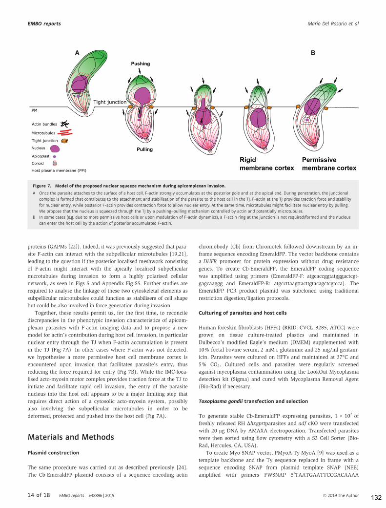
proteins (GAPMs [22]). Indeed, it was previously suggested that para-
site F-actin can interact with the subpellicular microtubules [19,21],
leading to the question if the posterior localised meshwork consisting
of F-actin might interact with the apically localised subpellicular
microtubules during invasion to form a highly polarised cellular
network, as seen in Figs 5 and Appendix Fig S5. Further studies are
required to analyse the linkage of these two cytoskeletal elements as
subpellicular microtubules could function as stabilisers of cell shape
but could be also involved in force generation during invasion.
Together, these results permit us, for the first time, to reconcile
discrepancies in the phenotypic invasion characteristics of apicom-
plexan parasites with F-actin imaging data and to propose a new
model for actin’s contribution during host cell invasion, in particular
nuclear entry through the TJ when F-actin accumulation is present
in the TJ (Fig 7A). In other cases where F-actin was not detected,
we hypothesise a more permissive host cell membrane cortex is
encountered upon invasion that facilitates parasite’s entry, thus
reducing the force required for entry (Fig 7B). While the IMC-loca-
lised acto-myosin motor complex provides traction force at the TJ to
initiate and facilitate rapid cell invasion, the entry of the parasite
nucleus into the host cell appears to be a major limiting step that
requires direct action of a cytosolic acto-myosin system, possibly
also involving the subpellicular microtubules in order to be
deformed, protected and pushed into the host cell (Fig 7A).
Materials and Methods
Plasmid construction
The same procedure was carried out as described previously [24].
The Cb-EmeraldFP plasmid consists of a sequence encoding actin
chromobody (Cb) from Chromotek followed downstream by an in-
frame sequence encoding EmeraldFP. The vector backbone contains
a DHFR promoter for protein expression without drug resistance
genes. To create Cb-EmeraldFP, the EmeraldFP coding sequence
was amplified using primers (EmeraldFP-F: atgcaccggtatgggactcgt-
gagcaaggg and EmeraldFP-R: atgccttaagttacttgtacagctcgtcca). The
EmeraldFP PCR product plasmid was subcloned using traditional
restriction digestion/ligation protocols.
Culturing of parasites and host cells
Human foreskin fibroblasts (HFFs) (RRID: CVCL_3285, ATCC) were
grown on tissue culture-treated plastics and maintained in
Dulbecco’s modified Eagle’s medium (DMEM) supplemented with
10% foetal bovine serum, 2 mM L-glutamine and 25 mg/ml gentam-
icin. Parasites were cultured on HFFs and maintained at 37°C and
5% CO2. Cultured cells and parasites were regularly screened
against mycoplasma contamination using the LookOut Mycoplasma
detection kit (Sigma) and cured with Mycoplasma Removal Agent
(Bio-Rad) if necessary.
Toxoplasma gondii transfection and selection
To generate stable Cb-EmeraldFP expressing parasites, 1 × 10⁷ of
freshly released RH ∆hxgprtparasites and adf cKO were transfected
with 20 lg DNA by AMAXA electroporation. Transfected parasites
were then sorted using flow cytometry with a S3 Cell Sorter (Bio-
Rad, Hercules, CA, USA).
To create Myo-SNAP vector, PMyoA-Ty-MyoA [9] was used as a
template backbone and the Ty sequence replaced in frame with a
sequence encoding SNAP from plasmid template SNAP (NEB)
amplified with primers FWSNAP 50TAATGAATTCCGACAAAA
A B
Figure 7. Model of the proposed nuclear squeeze mechanism during apicomplexan invasion.
A Once the parasite attaches to the surface of a host cell, F-actin strongly accumulates at the posterior pole and at the apical end. During penetration, the junctional
complex is formed that contributes to the attachment and stabilisation of the parasite to the host cell in the TJ. F-actin at the TJ provides traction force and stability
for nuclear entry, while posterior F-actin provides contraction force to allow nuclear entry. At the same time, microtubules might facilitate nuclear entry by pulling.
We propose that the nucleus is squeezed through the TJ by a pushing–pulling mechanism controlled by actin and potentially microtubules.
B In some cases (e.g. due to more permissive host cells or upon modulation of F-actin dynamics), a F-actin ring at the junction is not required/formed and the nucleus
can enter the host cell by the action of posterior accumulated F-actin.
14 of 18 EMBO reports e48896 | 2019 ª 2019 The Author
EMBO reports Mario Del Rosario et al
132

TGGACAAAGACTGCGAAATGA30 and RVSNAP 50TAATATGCATAC
CCAGCCCAGGCTTGCCCAG30.
Merozoite isolation
PkRON2*mCherry-HA A1-H.1 merozoites were isolated as
described [47]. In brief, ring-stage parasites were tightly synchro-
nised to a 3.5 h and allowed to develop to schizont stage. Schi-
zonts were separated from uninfected erythrocytes by MACS
magnet separation (CS column, Miltenyi Biotec). Parasites were
returned to complete media at 37°C and incubated with 10 lM E64
(Sigma-Aldrich) for < 4 h. Schizonts were pelleted (850 g, 5 min)
and resuspended in incomplete media and merozoites purified by
3 lm filtration (Whatman) immediately followed by 2 lm filtration
(Whatman).
Inducing the conditional LoxPact1 cKO
The inducible act1 cKO was obtained by the addition of 50 nM rapa-
mycin to the parental LoxPAct1 strain for 4 h at 37°C, 5% CO2 and
cultured as described previously [9].
Inducing the conditional Cas9act1 cKO
The act1 cKO parasite strain was obtained by addition of 50 nM of
rapamycin to the parental lines. The strain was incubated for 1 h at
37°C and 5% CO2 and cultured as described previously [9]. To
decrease the population of un-induced parasites, the culture media
was replaced by DMEM complete supplemented with 2.5% dextran
sulphate after 24 h to inhibit re-invasion of WT parasites.
MyoA-SNAP staining
Parasites expressing MyoA-SNAP were incubated with a TMR ligand
for the SNAP tag [48] (1:10,000) for 30 min. The cells were then
washed three times with PBS prior analysis.
SiR-tubulin staining
Prior invasion, parasites were incubated with 1,000 nM SiR-tubulin
[41] (1:1,000) for 1 h. The cells were then washed three times with
PBS prior analysis.
Light microscopy
Widefield images were acquired in z-stacks of 2 lm increments and
were collected using an Olympus UPLSAPO 100× oil (1.40NA)
objective on a Delta Vision Core microscope (Applied Precision, GE)
attached to a CoolSNAP HQ2 CCD camera. Deconvolution was
performed using SoftWoRx Suite 2.0 (Applied Precision, GE). Video
microscopy was conducted with the Delta Vision Core microscope
as above. Normal growth conditions were maintained throughout
the experiment (37°C; 5% CO2). Further image processing was
performed using FIJI [49] and Icy Image Processing Software (Insti-
tut Pasteur) [50].
Super-resolution microscopy (SR-SIM) was carried out using
an ELYRA PS.1 microscope (Zeiss) as described previously [24].
3D rendering and model views were generated using Imaris
software (Bitplane, Oxford Instruments) using the acquired SR-
SIM files.
Flow analysis time-lapse microscopy
To discriminate parasite orientation, 500 nm of SiR-tubulin was
added to parasite cultures 1 h before the experiment. To prepare
tachyzoites for the assay, 1 × 10⁶ mechanically freshly released
parasites per live dish were washed three times with PBS and incu-
bated a minimum of 10 min in serum-free DMEM media. Cytocha-
lasin D was added 30 min before imaging in a concentration of
2 lM. The samples were then imaged by widefield microscopy
using a Delta Vision Core microscope or super-resolution micro-
scopy (SR-SIM) using an ELYRA PS.1 microscope.
Parasite treatment with A23187 and BIPPO
Calcium ionophore A23187 was added to parasite live dishes using
with a final concentration of 2lM. BIPPO was similarly used with a
final concentration of 5 lM. The parasites were imaged by light
microscopy immediately after adding A23187 or BIPPO.
Tight Junction assay
For the assay 3 × 10⁶, mechanically freshly released parasites per
well were incubated in 200 ll Endo buffer (44.7 mM K2SO4, 10 mM
MgSO4, 106 mM sucrose, 5 mM glucose, 20 mM Tris–H2SO4,
3.5 mg/ml BSA, pH 8.2) for 10 min at 37°C. The parasites were then
allowed to settle into confluent layer of HFFs for 3 min at room
temperature. This step was followed by 20-min incubation at 37°C.
The supernatant was carefully removed and replaced by pre-
warmed culture media. The samples were further incubated for
5 min to allow invasion.
This was followed by fixing the samples using two different
buffers: cytoskeleton buffer (CB1) (MES pH 6.1 10 mM, KCL
138 mM, MgCl 3 mM, EGTA 2 mM, 5% PFA) and cytoskeleton
buffer (CB2) (MES pH6.1 10 mM, KCL 163.53 mM, MgCl
3.555 mM, EGTA 2.37 mM, sucrose 292 mM). These buffers were
mixed in a 4:1 ratio, respectively, and subsequently used for fixation
for 10 min. The samples were then treated with a PFA quenching
solution (NH4CL 50 mM) for 10 min followed by PBS washing for
three times. A minimum total of 40 parasites were counted for each
sample in three biological replicates. The tight junction assay
involving several mutants and wt in Fig 3E and F employed a multi-
ple comparison 2-way ANOVA using Tukey’s multiple comparison
test with a P-value of < 0.0001.
Cell deformation analysis
Samples were captured using SR-SIM and widefield microscopy as
previously described [24]. The deformation was then analysed with
Zen blue software (Zeiss) by measuring the size of the parasite body
and nucleus when going through a tight junction. A total of 100
parasites per biological replicate were measured for each condition
for RH in Fig 1B, and a total of 30 parasites per biological replicate
were measured for the mutant in Appendix Fig S1A. Statistical anal-
ysis was carried out by ordinary one-way ANOVA, using Tukey’s
multiple comparison test with a P-value of < 0.0001.
ª 2019 The Author EMBO reports e48896 | 2019 15 of 18
Mario Del Rosario et al EMBO reports
133

Colour-coded kymogram generation for particle
dynamics analysis
Fourier-filtered colour-coded kymograms were generated using the
KymographClear plugin on ImageJ and published in Mangeol et al
[33]. This plugin allows to trace a path of interest on a collapse t-
stack to follow particle movement. The plugin is able to generate a
three colour-coded kymogram using Fourier transformation to filter
different populations of particles by the orientation of the trajectory.
The colour coding is represented by forward movement as red,
backward movement as green and no movement as blue. These data
are then exported to the stand-alone software KymographDirect to
establish time-average intensity profiles. A more detailed protocol
can be found on the author’s website: https://sites.google.com/site/
kymographanalysis/.
Skeletonisation analysis
The skeletonisation process was done in FIJI using the Skeletonisa-
tion processing plugin [51]. The sample was binarised by threshold-
ing. A skeletonisation algorithm is then employed which converts
the thresholded signal into pixels, reducing the total width but not
the length. The time-lapse profile was collapsed (Z-Project) to define
the F-actin structures and their dynamics over the course of the time
lapse. The plugin can be accessed by: https://imagej.net/Analyze
Skeleton.
Live cell invasion
Parasites were mechanically released using 23 G needle and filtered
prior to inoculation on a confluent layer of HFFs, grown on glass
bottom dishes (MaTek). The dish was then transferred to the DV Core
microscope (Applied Precision, GE) and maintained under standard
culturing conditions. Images were captured at 1.4 frames per second
in DIC using an Olympus apochromat 60× oil objective. Images were
analysed using the Icy Image Processing software (Institut Pasteur)
[50]. Penetration speed profiles were obtained for 18 independent
invasion events for RH Cb-EmeraldFP and Cb-EmeraldFP parasites.
The statistical analysis of RH wt Invasion events in Fig 2C was done
by unpaired two-tailed t-test in 28 total parasite invasion events that
were captured across three different biological replicates.
Penetration speed profile analysis
Movies obtained were analysed using Icy Image Processing software
(Institut Pasteur) [50] with the Manual Tracking plugin. The
methodology used consists on tracking the apical end of the parasite
through the duration of penetration. The resulting data were then
exported to GraphPad PRISM7 Software for posterior analysis.
Plasmodium assays
The Cb-EmeraldFP sequence was cloned and transfected into
P. falciparum 3D7 parasites, and parasites were cultured in RPMI
1640 supplemented with Albumax (Invitrogen). Schizonts were
collected on a bed of 70% Percoll as previously described [42]. The
schizonts were allowed to egress in a medium containing blood at
1% haematocrit and merozoites at various stages of invasion were
fixed in 4% paraformaldehyde with 0.0075% glutaraldehyde as
previously described [42]. The junction was stained with a RON4
antibody and the nucleus stained by DAPI. Cb-EmeraldFP-labelled
actin filaments were fluorescent in the green range. For image acqui-
sition, z-stacks were collected using a UPLSAPO 100× oil (1.40 NA)
objective on a Delta Vision Core microscope (Applied Precision, GE)
attached to a CoolSNAP HQ2 CCD camera. Deconvolution was
performed using SoftWoRx Suite 2.0 (Applied Precision, GE). An
Elyra S1 microscope with super-resolution structured illumination
microscopy (SR-SIM) (Zeiss) was used for super-resolution dissec-
tion of RON4 staining on the merozoite surface.
Plasmodium knowlesi A1-H.1 parasites were cultured as
described previously [47] in human O+ RBCs in RPMI-HEPES media
supplemented with 2.3 g/l sodium bicarbonate, 2 g/l dextrose,
0.05 g/l hypoxanthine, 0.025 g/l gentamicin, 0.292 g/l L-glutamine,
5 g/l Albumax II (Gibco) and 10% (v/v) equine serum (Gibco).
Parasites were cultured at 37°C with a gas mixture of 90% N2, 5%
O2 and 5% CO2.
PkRON2*mCherry-HA A1-H.1 merozoites were incubated with
fresh erythrocytes, shaken at 1,000 rpm, 37°C for 90 s and samples
fixed by addition of an equal volume of 2× fixative (8%
paraformaldehyde/0.015% glutaraldehyde, Sigma-Aldrich). Cells
were permeabilised with 0.1% Triton X-100/PBS for 10 min and
blocked with 3% BSA/PBS. Cells were incubated with 1:500 mouse
anti-actin [47] and 1:1,000 rat anti-HA (3F10, Roche) followed by
1:1,000 anti-mouse Alexa 488 (Invitrogen) and 1:1,000 anti-rat
Alexa 594 (Invitrogen) secondary antibodies. Cells were smeared on
slides and mounted in VectaShield (Vector Laboratories) with
0.1 ng/ml DAPI to label the parasite nucleus. Widefield fluorescent
microscopy images were acquired with a Nikon plan apo 100×/1.45
oil immersion lens on a Nikon eclipse Ti microscope and images
processed with NIS Elements (Nikon).
Data availability
All imaging data and genetic material used for this paper are avail-
able from the authors on reasonable request.
Expanded View for this article is available online.
Acknowledgments
We acknowledge the assistance of the Institute of Infection, Immunity and
Inflammation Imaging Platform at the University of Glasgow. We also want to
thank Dr. Philip Thompson for providing us with the reagent BIPPO and Dr.
Maryse Lebrun for anti-RON2 antibodies. This work was supported by an ERC-
Starting grant (ERC-2012-StG 309255-EndoTox), a Wellcome 087582/Z/08/Z
Senior Fellowship to MM, Wellcome 100993/Z/13/Z Investigator Award to JB and
a National Secretariat for Higher Education, Sciences, Technology and Innova-
tion of Ecuador (SENESCYT) PhD scholarship (IFTH-GBE-2015-0475-M) to MD.
The Wellcome Centre for Molecular Parasitology is supported by core funding
from the Wellcome (085349). The funders had no role in study design, data
collection and analysis, decision to publish, or preparation of the manuscript.
Author contributions
MDR, JB, IT and MM conceived, designed or planned the study. MDR, JP, JAW,
GP, OL, SD, GSP, JFS, LL and FL-B contributed to the acquisition, analysis or
interpretation of the data. MM drafted the manuscript. All authors critically
16 of 18 EMBO reports e48896 | 2019 ª 2019 The Author
EMBO reports Mario Del Rosario et al
134

reviewed or revised the manuscript for intellectual content and approved the
final version.
Conflict of interest
The authors declare that they have no conflict of interest.
References
1. Meissner M, Ferguson DJP, Frischknecht F (2013) Invasion factors of
apicomplexan parasites: essential or redundant? Curr Opin Microbiol 16:
438 –444
2. Frénal K, Dubremetz JF, Lebrun M, Soldati-Favre D (2017) Gliding motil-
ity powers invasion and egress in Apicomplexa. Nat Rev Microbiol 15:
645 – 660
3. Besteiro S, Dubremetz JF, Lebrun M (2011) The moving junction of
apicomplexan parasites: a key structure for invasion. Cell Microbiol 13:
797 – 805
4. Bichet M, Joly C, Hadj Henni A, Guilbert T, Xémard M, Tafani V, Lagal V, Char-
ras G, Tardieux I (2014) The toxoplasma-host cell junction is anchored to the
cell cortex to sustain parasite invasive force. BMC Biol 12: 773
5. Guérin A, Corrales RM, Parker ML, Lamarque MH, Jacot D, El Hajj H,
Soldati-Favre D, Boulanger MJ, Lebrun M (2017) Efficient invasion by
Toxoplasma depends on the subversion of host protein networks. Nat
Microbiol 2: 1358 – 1366
6. Tardieux I, Baum J (2016) Reassessing the mechanics of parasite motility
and host-cell invasion. J Cell Biol 214: 507 – 515
7. Egarter S, Andenmatten N, Jackson AJ, Whitelaw JA, Pall G, Black JA,
Ferguson DJP, Tardieux I, Mogilner A, Meissner M (2014) The toxoplasma
acto-myoA motor complex is important but not essential for gliding
motility and host cell invasion. PLoS One 9: e91819
8. Gras S, Jackson A, Woods S, Pall G, Whitelaw J, Leung JM, Ward GE,
Roberts CW, Meissner M (2017) Parasites lacking the micronemal
protein MIC2 are deficient in surface attachment and host cell egress,
but remain virulent in vivo. Wellcome Open Res 2: 32
9. Whitelaw JA, Latorre-Barragan F, Gras S, Pall GS, Leung JM, Heaslip A,
Egarter S, Andenmatten N, Nelson SR, Warshaw DM et al (2017)
Surface attachment, promoted by the actomyosin system of Toxoplasma
gondii is important for efficient gliding motility and invasion. BMC Biol
15: 1
10. Bichet M, Touquet B, Gonzalez V, Florent I, Meissner M, Tardieux I
(2016) Genetic impairment of parasite myosin motors uncovers the
contribution of host cell membrane dynamics to Toxoplasma invasion
forces. BMC Biol 14: 97
11. Tosetti N, Dos N, Pacheco S, Soldati-favre D, Jacot D (2019) Three
F-actin assembly centers regulate organelle inheritance, cell-cell
communication and motility in Toxoplasma gondii. Elife 8: e42669
12. Gras S, Jimenez-Ruiz E, Klinger CM, Schneider K, Klingl A, Lemgruber L,
Meissner M (2019) An endocytic-secretory cycle participates in Toxo-
plasma gondii in motility. PLoS Biol 17: e3000060
13. O’Neill PR, Castillo-Badillo JA, Meshik X, Kalyanaraman V, Melgarejo K,
Gautam N (2018) Membrane flow drives an adhesion-independent
amoeboid cell migration mode. Dev Cell 46: 9 – 22
14. Tanaka M, Kikuchi T, Uno H, Okita K, Kitanishi-Yumura T, Yumura S
(2017) Turnover and flow of the cell membrane for cell migration. Sci
Rep 7: 12970
15. Angrisano F, Riglar DT, Sturm A, Volz JC, Delves MJ, Zuccala ES, Turnbull
L, Dekiwadia C, Olshina MA, Marapana DS et al (2012) Spatial
localisation of actin filaments across developmental stages of the
malaria parasite. PLoS One 7: e32188
16. Andenmatten N, Egarter S, Jackson AJ, Jullien N, Herman J-P, Meissner
M (2013) Conditional genome engineering in Toxoplasma gondii uncov-
ers alternative invasion mechanisms. Nat Methods 10: 125 – 127
17. Heaslip AT, Nelson SR, Warshaw DM (2016) Dense granule trafficking in
Toxoplasma gondii requires a unique class 27 myosin and actin fila-
ments. Mol Biol Cell 27: 2080 – 2089
18. Zhang Q, Huang Y, Zhang Y, Fang X, Claes A, Duchateau M, Namane A,
Lopez-Rubio JJ, Pan W, Scherf A (2011) A Critical role of perinuclear fila-
mentous Actin in spatial repositioning and mutually exclusive expression
of virulence genes in malaria parasites. Cell Host Microbe 10: 451–463
19. Patrón SA, Mondragón M, González S, Ambrosio JR, Guerrero BAL,
Mondragón R (2005) Identification and purification of actin from the
subpellicular network of Toxoplasma gondii tachyzoites. Int J Parasitol 35:
883 – 894
20. Hliscs M, Millet C, Dixon MW, Siden-Kiamos I, Mcmillan P, Tilley L
(2015) Organization and function of an actin cytoskeleton in Plasmod-
ium falciparum gametocytes. Cell Microbiol 17: 207 – 225
21. Yasuda T, Yagita K, Nakamura T, Endo T (1988) Immunocytochemical
localization of actin in Toxoplasma gondii. Parasitol Res 75: 107 – 113
22. Harding CR, Gow M, Kang JH, Shortt E, Manalis SR, Meissner M, Lourido
S (2019) Alveolar proteins stabilize cortical microtubules in Toxoplasma
gondii. Nat Commun 10: 401
23. Graindorge A, Frénal K, Jacot D, Salamun J, Marq JB, Soldati-Favre D
(2016) The conoid associated motor myoh is indispensable for Toxo-
plasma gondii entry and exit from host cells. PLoS Pathog 12:
e1005388
24. Periz J, Whitelaw J, Harding C, Gras S, Minina MIDR, Latorre-Barragan F,
Lemgruber L, Reimer MA, Insall R, Heaslip A et al (2017) Toxoplasma
gondii F-actin forms an extensive filamentous network required for
material exchange and parasite maturation. Elife 6: e24119
25. Stortz JF, Del Rosario M, Singer M, Wilkes JM, Meissner M, Das S (2019)
Formin-2 drives polymerisation of actin filaments enabling segregation
of apicoplasts and cytokinesis in Plasmodium falciparum. eLife 8: e49030
26. Shaw MK, Compton HL, Roos DS, Tilney LG (2000) Microtubules, but not
actin filaments, drive daughter cell budding and cell division in Toxo-
plasma gondii. J Cell Sci 113(Pt. 7): 1241 – 1254
27. Harding CR, Egarter S, Gow M, Jiménez-Ruiz E, Ferguson DJP, Meissner
M (2016) Gliding associated proteins play essential roles during the
formation of the inner membrane complex of Toxoplasma gondii. PLoS
Pathog 12: e1005403
28. McGregor AL, Hsia C-R, Lammerding J (2016) Squish and squeeze — the
nucleus as a physical barrier during migration in confined environ-
ments. Curr Opin Cell Biol 40: 32 – 40
29. Kessler H, Herm-Gotz A, Hegge S, Rauch M, Soldati-Favre D, Frischknecht
F, Meissner M (2008) Microneme protein 8 – a new essential invasion
factor in Toxoplasma gondii. J Cell Sci 121(Pt. 7): 947 – 956
30. Frénal K, Jacot D, Hammoudi PM, Graindorge A, Maco B, Soldati-Favre D
(2017) Myosin-dependent cell-cell communication controls synchronicity
of division in acute and chronic stages of Toxoplasma gondii. Nat
Commun 8: 15710
31. Mehta S, Sibley LD (2011) Actin depolymerizing factor controls actin
turnover and gliding motility in Toxoplasma gondii. Mol Biol Cell 22:
1290 – 1299
32. Arganda-Carreras I, Fernández-González R, Muñoz-Barrutia A, Ortiz-De-
Solorzano C (2010) 3D reconstruction of histological sections: application
to mammary gland tissue. Microsc Res Tech 73: 1019 – 1029
ª 2019 The Author EMBO reports e48896 | 2019 17 of 18
Mario Del Rosario et al EMBO reports
135

33. Mangeol P, Prevo B, Peterman EJG (2016) KymographClear and Kymo-
graphDirect: two tools for the automated quantitative analysis of molecu-
lar and cellular dynamics using kymographs. Mol Biol Cell 27: 1948 – 1957
34. Howard BL, Harvey KL, Stewart RJ, Azevedo MF, Crabb BS, Jennings IG,
Sanders PR, Manallack DT, Thompson PE, Tonkin CJ et al (2015) Identifi-
cation of potent phosphodiesterase inhibitors that demonstrate cyclic
nucleotide-dependent functions in apicomplexan parasites. ACS Chem
Biol 10: 1145 – 1154
35. Thiam HR, Vargas P, Carpi N, Crespo CL, Raab M, Terriac E, King MC,
Jacobelli J, Alberts AS, Stradal T et al (2016) Perinuclear Arp2/3-driven
actin polymerization enables nuclear deformation to facilitate cell
migration through complex environments. Nat Commun 7: 10997
36. Frénal K, Polonais V, Marq JB, Stratmann R, Limenitakis J, Soldati-Favre
D (2010) Functional dissection of the apicomplexan glideosome molecu-
lar architecture. Cell Host Microbe 8: 343 – 357
37. Frénal K, Soldati-Favre D (2015) Plasticity and redundancy in proteins
important for toxoplasma invasion. PLoS Pathog 11: e1005069
38. Plattner F, Yarovinsky F, Romero S, Didry D, Carlier M-F, Sher A, Soldati-
Favre D (2008) Toxoplasma profilin is essential for host cell invasion
and TLR11-dependent induction of an interleukin-12 response. Cell Host
Microbe 3: 77 – 87
39. Huynh MH, Carruthers VB (2006) Toxoplasma MIC2 is a major determi-
nant of invasion and virulence. PLoS Pathog 2: e84
40. Meissner M, Schlüter D, Soldati D (2002) Role of Toxoplasma gondii
myosin a in powering parasite gliding and host cell invasion. Science
298: 837 – 840
41. Lukinavi�cius G, Reymond L, D’Este E, Masharina A, Göttfert F, Ta H,
Güther A, Fournier M, Rizzo S, Waldmann H et al (2014) Fluorogenic
probes for live-cell imaging of the cytoskeleton. Nat Methods 11:
731 – 733
42. Das S, Lemgruber L, Tay CL, Baum J, Meissner M (2017) Multiple essen-
tial functions of Plasmodium falciparum actin-1 during malaria blood-
stage development. BMC Biol 15: 70
43. Gonzalez V, Combe A, David V, Malmquist NA, Delorme V, Leroy C, Blaz-
quez S, Ménard R, Tardieux I (2009) Host cell entry by Apicomplexa
parasites requires actin polymerization in the host cell. Cell Host
Microbe 5: 259 – 272
44. Petrie RJ, Yamada KM (2016) Multiple mechanisms of 3D migration: the
origins of plasticity. Curr Opin Cell Biol 42: 7 – 12
45. Guilluy C, Burridge K (2015) Nuclear mechanotransduction: forcing the
nucleus to respond. Nucleus 6: 19 – 22
46. Guilluy C, Osborne LD, Van Landeghem L, Sharek L, Superfine R, Garcia-
Mata R, Burridge K (2014) Isolated nuclei adapt to force and reveal a
mechanotransduction pathway in the nucleus. Nat Cell Biol 16:
376 – 381
47. Lyth O, Vizcay-Barrena G, Wright KE, Haase S, Mohring F, Najer A,
Henshall IG, Ashdown GW, Bannister LH, Drew DR et al (2018) Cellular
dissection of malaria parasite invasion of human erythrocytes using
viable Plasmodium knowlesi merozoites. Sci Rep 8: 10165
48. Keppler A, Gendreizig S, Gronemeyer T, Pick H, Vogel H, Johnsson K
(2003) A general method for the covalent labeling of fusion proteins
with small molecules in vivo. Nat Biotechnol 21: 86 – 89
49. Schindelin J, Arganda-Carreras I, Frise E, Kaynig V, Longair M, Pietzsch T,
Preibisch S, Rueden C, Saalfeld S, Schmid B et al (2012) Fiji: an
open-source platform for biological-image analysis. Nat Methods 9:
676 – 682
50. De Chaumont F, Dallongeville S, Chenouard N, Hervé N, Pop S, Provoost
T, Meas-Yedid V, Pankajakshan P, Lecomte T, Le Montagner Y et al
(2012) Icy: an open bioimage informatics platform for extended repro-
ducible research. Nat Methods 9: 690 – 696
51. Doube M, Klosowski MM, Arganda-Carreras I, Cordelières FP, Dough-
erty RP, Jackson JS, Schmid B, Hutchinson JR, Shefelbine SJ (2010)
BoneJ: free and extensible bone image analysis in ImageJ. Bone 47:
1076 – 1079
License: This is an open access article under the
terms of the Creative Commons Attribution 4.0
License, which permits use, distribution and reproduc-
tion in any medium, provided the original work is
properly cited.
18 of 18 EMBO reports e48896 | 2019 ª 2019 The Author
EMBO reports Mario Del Rosario et al
136

RESEARCH ARTICLE
Francisella novicida and F. philomiragia biofilmfeatures conditionning fitness in spring waterand in presence of antibiotics
Claire Siebert1, Corinne Villers1,2, Georgios Pavlou3, Bastien Touquet3,
Nandadeva Yakandawala4, Isabelle TardieuxID3, Patricia RenestoID
1*
1 TIMC-IMAGUMR 5525—UGACNRS, Grenoble Cedex 9, France, 2 Universite de Caen Normandie,EA4655 U2RM, Caen, France, 3 Institute for Advanced Biosciences (IAB), TeamMembrane Dynamics of
Parasite-Host Cell Interactions, CNRSUMR 5309, INSERMU1209, Universite Grenoble Alpes, Grenoble,France, 4 Kane Biotech, Inc., Winnipeg, Canada
Abstract
Biofilms are currently considered as a predominant lifestyle of many bacteria in nature.
While they promote survival of microbes, biofilms also potentially increase the threats to ani-
mal and public health in case of pathogenic species. They not only facilitate bacteria trans-
mission and persistence, but also promote spreading of antibiotic resistance leading to
chronic infections. In the case of Francisella tularensis, the causative agent of tularemia,
biofilms have remained largely enigmatic. Here, applying live and static confocal micros-
copy, we report growth and ultrastructural organization of the biofilms formed in vitro by
these microorganisms over the early transition from coccobacillary into coccoid shape dur-
ing biofilm assembly. Using selective dispersing agents, we provided evidence for extracel-
lular DNA (eDNA) being a major and conserved structural component of mature biofilms
formed by both F. subsp. novicida and a human clinical isolate of F. philomiragia. We also
observed a higher physical robustness of F. novicida biofilm as compared to F. philomiragia
one, a feature likely promoted by specific polysaccharides. Further, F. novicida biofilms
resisted significantly better to ciprofloxacin than their planktonic counterparts. Importantly,
when grown in biofilms, both Francisella species survived longer in cold water as compared
to free-living bacteria, a trait possibly associated with a gain in fitness in the natural aquatic
environment. Overall, this study provides information on survival of Francisella when
embedded with biofilms that should improve both the future management of biofilm-related
infections and the design of effective strategies to tackle down the problematic issue of bac-
teria persistence in aquatic ecosystems.
Introduction
Francisella tularensis is a non-motile Gram-negative coccobacillus and the causative agent of
zoonotic tularemia disease. Due to a remarkable infectivity (< 10 bacteria) which is associated
with high mortality and morbidity and a good genetic tractability, the US Centers for Disease
PLOSONE | https://doi.org/10.1371/journal.pone.0228591 February 5, 2020 1 / 23
a1111111111a1111111111a1111111111a1111111111a1111111111
OPEN ACCESS
Citation: Siebert C, Villers C, Pavlou G, Touquet B,
Yakandawala N, Tardieux I, et al. (2020) Francisella
novicida and F. philomiragia biofilm features
conditionning fitness in spring water and in
presence of antibiotics. PLoS ONE 15(2):
e0228591. https://doi.org/10.1371/journal.
pone.0228591
Editor:Mohamed Ali Hakimi, Centre National de la
Recherche Scientifique, FRANCE
Received:December 4, 2019
Accepted: January 17, 2020
Published: February 5, 2020
Copyright:© 2020 Siebert et al. This is an open
access article distributed under the terms of the
Creative Commons Attribution License, which
permits unrestricted use, distribution, and
reproduction in any medium, provided the original
author and source are credited.
Data Availability Statement: All relevant data are
within the manuscript and its Supporting
Information files.
Funding: This work was supported by the FINOVI
Foundation (Grant AO12-02) and by the ANR
ASTRID (grant ANR-17-ASTR-0024). NY
Yakandawala was employed by the company Kane
Biotech, Inc. (Winnipeg, Canada). The authors
declare that the research was conducted in the
absence of any commercial or financial
137

Control and Prevention (CDC) has classified F. tularensis as category A bioterrorism agent [1].
The ability of Francisella strains to form biofilms in vitro was first reported in 2009 with F.
novicida [2] and later observed with other Francisella species including highly virulent [3] and
environmental strains [4]. Given the importance of biofilms in infectious diseases [5–10], the
regulation of Francisella biofilm assembly was investigated and led to identifying the Sec secre-
tion system [3] as well as of the transcription factor qseB [11] as main players of the biofilm
regulatory cascade [12]. Although not demonstrated experimentally, and considering the role
of Francisella chitinases in biofilm formation, it has been hypothesized that biofilms would sig-
nificantly contribute to Francisella persistence in both aquatic habitats and mosquito hosting
vectors [3, 13, 14]. The correlation between Francisella biofilm dispersion and an increased
bacterial susceptibility towards biocides has been reported [14, 15] and we have recently
brought first genetic evidence that the biofilm growth mode provides F. tularensis with a
reduced susceptibility towards fluoroquinolones [16].
Tularemia is associated with several clinical forms whose severity depends not only on the
Francisella subspecies, but also on the route of infection [17]. The more severe disease in
human is caused by inhalation of F. tularensis subsp. tularensis (Type A) leading to a pneu-
monic form of tularemia [18]. Although the epidemiology of this disease is still not fully under-
stood, risk factors include exposure to arthropods, especially ticks which are the most
important vectors of the terrestrial life cycle of Type A Francisella [1, 19]. The two unusual out-
breaks of pneumonic tularemia reported from the island of Martha’s Vineyard [20, 21]
together with the observed prolonged survival of Type A F. tularensis in brackish-water col-
lected on this island [22] allowed hypothesizing that the aquatic environment could serve as a
reservoir for F. tularensis [13, 23]. F. tularensis subsp. holarctica (Type B) being widespread in
the Northern hemisphere is the other subspecies of major clinical importance although the dis-
ease is rarely fatal in humans [24]. Clinical and epidemiological data have highlighted a close
relationship between outbreaks of F. tularensis subsp. holarctica with water sources while
infected mosquitoes were described as major vectors of these bacteria, in particular in Scandi-
navia [1, 25–27]. F. tularensis subsp. novicida (herein F. novicida) is described as a rare oppor-
tunistic human pathogen despite being responsible for severe pathologies in either immuno-
or medically weakened patients [28]. In healthy individuals, the consumption of contaminated
water was identified as a major source of acute F. novicida infection following near-drowning
events and ingestion of icy water [29–31]. Being the most genetically tractable Francisella sub-
species, F. novicida has largely been used as an experimental relevant model to study F. tularen-
sis pathogenesis [28, 32, 33]. F. philomiragia (previously Yersinia philomiragia) is another
Francisella species which is commonly found in soil, water and aerosol samples [34]. While
only a few cases of human infection with F. philomiragia have been described, they correlated
with pneumonia in healthy individuals undergoing near-drowning accidents or in immuno-
compromised people. Overall, water appears as an important reservoir for some human patho-
genic Francisella strains [23].
To gain insights on the development and composition of Francisella biofilms, a prerequisite
for designing anti-biofilm efficient strategies to target the bacteria biofilms, we compared the
main features of the biofilm dynamics and composition for both F. novicida and F. philomira-
gia. F. novicida strain U112 was used as relevant model for the highly pathogenic Francisella
subsp. tularensis [28] and the F. philomiragia strain CHUGA-FP47 isolated from a patient [35]
was used as a clinically relevant isolate. We visualized in real time the biofilm formation and
analyzed the extracellular polymeric substances (EPS) of mature biofilms using selective dis-
persing agents. Further, the behavior of both planktonic and biofilm forms of Francisella was
assessed both under antibiotics and cold water exposure.
Analysis of Francisella biofilms
PLOSONE | https://doi.org/10.1371/journal.pone.0228591 February 5, 2020 2 / 23
relationships that could be construed as a potential
conflict of interest. The funders had no role in
study design, data collection and analysis, decision
to publish, or preparation of the manuscript.
Competing interests: The authors have declared
that no competing interests exist.
138

Materials andmethods
Bacterial strains and growth conditions
F. novicida (U112) CIP56.12 (Centre de Ressources Biologiques de l’Institut Pasteur, Paris,
France) and F. philomiragia (CHUGA-FP47) were grown either on solid or in liquid media, as
indicated. The solid medium corresponds to pre-made Polyvitex-enriched chocolate agar
(PVX-CHA) plates (Ref#43109 BioMerieux, France) incubated at 37˚C in a 5% CO2 incubator
with humidified atmosphere and used both to revive glycerol stocks and for CFU counting.
Liquid cultures were carried out in Modified Mueller-Hinton broth (MMH) at 37˚C under
shaking. MMH corresponds to Mueller Hinton Broth (Ref#275730 Grosseron) supplemented
with NaCl (Sigma; 5 g/L), BactoProteose peptone (Ref#211693 BD Biosciences; 5 g/L), Bacto-
Tryptone (Ref#211705 BD Biosciences 5 g/L) and L-cysteine (Ref#C7477 Sigma; 1 g/L).
Biofilm quantification
The static biofilm was grown in 96-well plates and detected by crystal violet staining [36].
Briefly, liquid bacterial cultures (absorbance around 0.8 at OD600 nm) were diluted in 37˚C
pre-warmed MMH and 200 μL of bacterial suspensions were dispensed per well of flat-bottom
polystyrene 96-well plates (Falcon1). To avoid the edge effect resulting from evaporation, the
peripheral wells were filled with 200 μL PBS. Plates were incubated for 24 h to 72 h at 37˚C in a
humidified atmosphere containing 5% CO2. The OD600 nm was measured using Tecan Plate
reader to normalize bacterial growth and planktonic bacteria were gently aspirated. F. novicida
biofilms were washed twice with sterile PBS while F. philomiragia biofilms were washed only
once to ensure minimum to negligible amount of loss of bacteria as assessed by count of the
colony forming unit (CFU) from washings. Microtiter plates were then incubated for 1 h at
70˚C, stained with 200 μL of 0.1% (w/v) crystal violet/well (Merck) for 15 min and washed
twice with 200 μL H2O. Biofilms were solubilized in 200 μL 30% acetic acid per well and quan-
tified by measuring optical density at 595 nm.
Fluorescence microscopy
For imaging, 1 mL of bacteria in MMH (1x10⁷/mL for F. novicida and 5x10⁷/mL F. philomira-
gia) were distributed in 24-well plates containing poly-L-lysine-coated glass coverslips. Sam-
ples were fixed at different time points with freshly made 4% paraformaldehyde in PBS for 20
minutes at room temperature. The supernatant containing planktonic cells was gently
removed allowing the biofilm at the air-liquid interface to adhere to the coverslip. The wells
were washed twice with PBS to remove residual planktonic cells and incubated in 50 mM
NH4Cl in PBS for 5 min to quench auto-fluorescence caused by free aldehydes. Samples were
stored in PBS supplemented with 0.02% (w/v) sodium azide at 4˚C until staining. For staining
of both bacterial membranes and biofilm glycoproteins, fixed samples were incubated in 5 μg/
mL FM11-43FX membrane probe (Life Technologies) and 200 μg/mL concanavalin (ConA)-
FITC (Life Technologies) for 30 min at RT and washed 3 times with PBS. Nucleic acids were
then stained with 0.5 μg/mL DAPI fluorescent dye for 5 min and the coverslips were mounted
in Mowiol mounting medium. In some experiments, the presence of eDNA was visualized on
unfixed biofilms and using the cell-impermeant DITO™-1 (2 μM; AAT Bioquest), a chemical
analog to TOTO1 together with Hoechst 33342 (1:5,000). The viability of bacteria within bio-
films was also assessed from unfixed samples stained for 30 min at room temperature with the
membrane impermeant dye propidium iodide (PI) (3 μM, Molecular Probes) that does not
penetrate intact cells. Slides were imaged under Zeiss Apo Tome microscope with 63x/1.4 oil-
immersion objective using Zeiss Zen software. Images were further processed using ImageJ
Analysis of Francisella biofilms
PLOSONE | https://doi.org/10.1371/journal.pone.0228591 February 5, 2020 3 / 23
139

[37] and Adobe Photoshop software. 3D reconstruction of images was achieved by processing
raw data (i.e., xyz files) using ImageJ software to crop the sequence and region of interest.
When needed, z stack from each channel was deconvoluted using the “Iterative Deconvolve
3D” plugin with a z-step of 0.3 μm. Zen software was used for 3D reconstruction and to pro-
duce movies from processed images.
Time-lapse microscopy and image processing
Time-lapse video microscopy was performed with bacteria diluted in MMH and placed in
Chamlide™ chambers (LCI Corp., Seoul, Korea) installed on an Eclipse Ti inverted confocal
microscope (Nikon France Instruments, Champigny sur Marne, France) with a temperature
(37˚C) and CO2 (5%)-controlled stage. Images were collected every 15 minutes with a z-step of
0.3 μm using a CMOS camera (Photometrics, Tucson, AZ, USA) and a CSU X1 spinning disk
(Yokogawa, Roper Scientific, Lisses, France). Analysis was performed using MetaMorph soft-
ware (http://www.moleculardevices.com) from the raw image data files.
Dispersion of Francisella biofilms
The effect of dispersants was evaluated by using 24 h and 48 h-old biofilms of F. novicida and
F. philomiragia biofilms, respectively. F. novicida biofilms were grown in 96-well polystyrene
plates (Falcon1) starting from an inoculum of 1x10⁷ CFU/mL (2x10⁶ bacteria/well), while for
F. philomiragia 5x10⁷ CFU/m (107 bacteria/well) were used. After one (F. philomiragia) or two
(F. novicida) gentle PBS washes, the MMH was replaced with 200 μL fresh medium supple-
mented with increasing concentrations of dispersing agents including DNase I (Roche),
EDTA (Sigma-Aldrich), chitinase from Streptomyces griseus (E.C. 3.2.1.14) (Sigma-Aldrich),
cellulase (Sigma-Aldrich), proteinase K (Invitrogen) or Dispersin B (Kane Biotech Inc. Can-
ada). Microtiter plates were incubated at 37˚C for 24 h under 5% CO2 and biofilm was quanti-
fied as described elsewhere. Data were expressed as percentage of remaining biofilm calculated
in comparison to untreated biofilm under the same experimental conditions.
Evaluation of antimicrobial resistance of planktonic and biofilm bacteria
The antibiotic resistance of F. novicida and F. philomiragia was evaluated under the same con-
ditions as the dispersion assays but using MMH supplemented with increasing concentrations
of antibiotics. Following 24 h incubation at 37˚C the bacterial viability was evaluated using
both the resazurin assay detailed above and the CFU method, as previously described and
detailed below [16]. The antibiotic susceptibility of planktonic bacteria was assessed in parallel
using the same culture medium and starting from a bacterial suspension containing an equal
amount of bacteria as those present in biofilms. The growth of planktonic bacteria was evalu-
ated by OD600nmmeasurement and their viability was tested by the metabolic rezasurin test
(see below). The minimal inhibitory concentrations (MICs) of gentamicin (Panpharma) and
ciprofloxacin (Sigma-Aldrich) used as reference values were obtained using the broth-dilution
method and in line with the CLSI guidelines [38]. For F. novicida, they were of 1 mg/L and
0.064 mg/L, respectively, these values being of 0.5 mg/L and 0.032 mg/L for F. philomiragia,
Survival of planktonic or biofilm bacteria in water
F. novicida and F. philomiragia biofilms were grown and washed as described for dispersal
experiments. After washings, the wells were filled with 200 μL of water collected from a spring
around Grenoble and sterilized by filtration using 0.45 μm then 0.22 μm filters. The microtiter
plates were stored at 4˚C, and bacteria survival was assessed by CFU assay at each time point.
Analysis of Francisella biofilms
PLOSONE | https://doi.org/10.1371/journal.pone.0228591 February 5, 2020 4 / 23
140

In parallel, the viability of planktonic bacteria was determined starting from exponential
growth phase bacteria. The bacterial suspension was centrifuged and washed with PBS and the
resulting pellet was resuspended in a volume of spring water such as the bacterial density was
close to that found within biofilms (5x10⁷ bacteria/200 μL).
CFU counting
Biofilms previously disrupted to release cells by vigorous pipetting with PBS and planktonic
cell suspensions were serially diluted in PBS. For each sample 100 μL of at least four different
dilutions were plated on PVX-CHA plates, incubated for 24–48 h at 37˚C under 5% CO2, and
CFU were counted.
Measurement of bacterial viability by resazurin assay
This assays was done with resazurin (Sigma-Aldrich), a fluorescent indicator of mitochondrial
function conveniently used for the evaluation of cell viability in of several bacterial species [39,
40] including Neisseria gonorrhoeae [41] and Francisella LVS strains [16]. In this metabolic
assay, the incubation time was significantly lower (1–2 h vs 24h) than the conditions for which
this compound was depicted as being an antimicrobial agent towards Francisella and Neisseiria
species [42]. Viable bacteria reduce blue non-fluorescent resazurin dye (λmax = 600 nm) to the
pink fluorescent compound resorufin (max absorbance λmax = 570 nm). Antibiotic treated or
untreated biofilms were washed with PBS to remove planktonic cells, supplemented with
200 μL/well of a 0.02 mg/mL resazurin (Sigma-Aldrich) solution in MMH, and incubated for 2
h at 37˚C under static conditions. Cell viability was estimated by using absorbance was mea-
sured at (OD570nm-OD600nm). Planktonic cells (200 μL) were supplemented with 20 μL of resa-
zurin (0.2 mg/mL), incubated for 1 h at 37˚C, and cell viability was evaluated and expressed as
percent viability in comparison to untreated cells.
Statistical analysis
Statistical analysis was performed using the GraphPad PRISM software and Student’s t- tests.
Data correspond to means ± standard errors of the means (SEM) of at least three independent
replicates from independent experiments, as depicted. P values less than 0.05 were considered
statistically significant.
Results
Quantification of biofilm formation by F. novicida and F. philomiragia
The ability of F. novicidaU112 and F. philomiragia strain CHUGA-FP47 to produce biofilm
was first compared starting from increasing concentrations of the bacterial inoculum incu-
bated under static conditions in MMH broth at 37˚C. The biofilm biomass was quantified by
crystal violet staining, a basic dye that binds non-specifically to negatively charged surface
including polysaccharides and eDNA present into the extracellular matrix. As shown Fig 1A,
increasing F. novicida inoculum size translated in a higher bacterial density in the wells after a
24 h incubation time, while the biofilm biomass remained unchanged (Fig 1B). The largest
amount of biofilm was observed for an inoculum of 106−107 bacteria/mL while its production
estimated by the ratio biofilm biomass/bacteria number was seen to significantly drop beyond
108 bacteria/mL (P<0.05; n = 12). The recovery of viable bacteria evaluated by CFU counting
from a 24 h biofilm formed with 107 bacteria/mL in the starting starting inoculum was of 4.95
x107 ± 0.7x10⁷ bacteria/well (n = 13). While the F. novicida biofilm biomass measured after 48
h incubation was close to that observed at 24 h (not shown), the number of recovered bacteria
Analysis of Francisella biofilms
PLOSONE | https://doi.org/10.1371/journal.pone.0228591 February 5, 2020 5 / 23
141

was significantly increased 8.15 x10⁷ ± 2.4 x10⁷ bacteria/well (n = 4). In contrast to F. novicida,
the F. philomiragia 24 h-old biofilm was found particularly fragile since it detached easily from
the bottom of the microtiter plate and sheared despite gentle washing procedures (not shown).
The quantification of F. philomiragia biofilm formation was thus analyzed over a 48 h incuba-
tion period. Under such conditions, the optimal inoculum for the F. philomiragia CHU-
GA-FP47 strain was of 5x10⁷ bacteria/mL (Fig 1C and 1D) leading to a recovery of 5.49 x10⁷ ±
1.38 x10⁷ viable bacteria from a 96 well-biofilm (n = 12).
Visualization and microscopic analysis of Francisella biofilm growth
The formation of both F. novicida and F. philomiragia biofilms was then monitored during
several hours by real time confocal laser scanning microscopy (CLSM) at 37˚C (Fig 2 and S1
Movie). These observations confirmed the greater capacity of F. novicida to replicate and to
spatially organize as biofilms. The biofilm evolution over time was also imaged using Struc-
tured Illumination Microscopy SIM (Figs 3 and 4). Over the 15 min post-inoculation period,
no bacteria were observed on the coverslips used to retain the floating biofilm formed at the
air-liquid interface after gentle aspiration of planktonic cells. To have a reference point prior
to biofilm formation, we examined the bacterial suspension from the inoculum. For both
strains, bacteria displayed an uniform coccobacillary shape visualized by the dual labeling of
membrane and surface-exposed carbohydrates using the styryl fluorescent dye and the ConA
lectin, respectively (Figs 3A and 4A). Over the few following hours, confocal images attest
from cell-to-cell juxtaposition of bacteria secreting large amounts of glycoconjugates (Fig 3B–
3E and Fig 4B–4D). The floating colonies progressively developed in a more robust biofilm
Fig 1. Quantitative measurement of biofilm formation by F. novicida and F. philomiragia. Biofilm formation wasquantified in 96-well plates inoculated with increasing concentrations of bacteria and incubated under staticconditions. Following 24 h (F. novicida) or 48 h (F. philomiragia) incubation at 37˚C the bacterial growth (A, C) wasquantified by OD600nm and biofilm biomass (B, D) was estimated by crystal violet staining. The data correspond to themean ± SEM of three or more independent experiments with n = 6 replicates for each. �P<0.05 and ��� P<0.0005.
https://doi.org/10.1371/journal.pone.0228591.g001
Analysis of Francisella biofilms
PLOSONE | https://doi.org/10.1371/journal.pone.0228591 February 5, 2020 6 / 23
142

with bacteria embedded in EPS. A striking morphological feature of both strains was the
spherical shape they adopted in course of the biofilm formation, an effect observed as early as
Fig 2. Kinetics of biofilm formation by F. novicida and F. philomiragia. Confocal images were taken at each ofindicated time points from bacterial suspension grown at 37˚C without agitation in Chamlide™ chambers installed onan Eclipse Ti inverted confocal microscope as detailed in Materials and Methods. The scale bar indicates 5μM.
https://doi.org/10.1371/journal.pone.0228591.g002
Analysis of Francisella biofilms
PLOSONE | https://doi.org/10.1371/journal.pone.0228591 February 5, 2020 7 / 23
143

Fig 3. Fluorescence microscopy analysis of F. novicida biofilm formation. (A) Planktonic exponential phase bacteria and (B-D) biofilms formedupon incubation of bacteria for different time intervals were fixed and stained with DAPI (nucleic acids—blue), FM11-43FX (FM; bacterial membranes
Analysis of Francisella biofilms
PLOSONE | https://doi.org/10.1371/journal.pone.0228591 February 5, 2020 8 / 23
144

the 2 first hours for F. novicida (Fig 3B). During the 24 h of biofilm assembly, these bacteria
continued to proliferate within the matrix and the thickness was estimated around 9 μm (Fig
3E). In agreement with the biomass quantification data, a delayed biofilm assembly was
observed for F. philomiragia, with packed bacteria communities that clustered within a ~
4.5 μm-thick EPS after 48 h (Fig 4D).
Treatment of mature Francisella biofilms with potential dispersing agents
Given that main components of the bacterial biofilm EPS include eDNA, proteins and polysac-
charides which adhere to each other and maintain microbial cells together [6], the capacity of
enzymes or reagents targeting these specific compounds to induce biofilm dispersal was investi-
gated. Taking in consideration biofilm biomass, thickness and the number of biofilm embedded
bacteria, these assays were carried out using 24 h and 48 h-old biofilms of F. novicida and F. phi-
lomiragia, respectively. Results obtained showed that, following 24 h exposure, the DNase I
induced a concentration-dependent dispersion of F. novicida biofilm with a maximum level of
around 70% reached at 50 μg/mL (Fig 5A). The role of eDNA in F. novicida biofilm stabilization
was further suggested using the chelating agent EDTA (Fig 5B) that similarly affected the struc-
tural integrity of the EPS matrix but by impacting the electrostatic interactions between divalent
cations and negatively charged DNA [43]. Biofilm dispersion was almost complete (87.93% ±
0.8%; n = 12) following treatment with 0.5 μg/mL proteinase K, indicating that proteinaceous
structures largely contribute to F. novicida biofilm structure and integrity (Fig 5C). This biofilm
also contains polysaccharides and more specifically poly-β(1,4)-N-acetylglucosamine hydrolyzed
by chitinase, leading to more than 80% dispersion when used at 300 μg/ml (Fig 5D). Another
polysaccharide identified as a key component of F. novicida biofilm is the ß(1,4)-linked D glu-
cose homopolymer cellulose (Fig 5E) which displays the same coupling as the acetylamine group
with chitin. In contrast, dispersin B which targets poly-β-(1,6)-linked N-acetylglucosaminogly-
cans [44, 45] rather induced a moderate decrease of the biofilm biomass (Fig 5F). The capacity of
DNase I, proteinase K and cellulase to dissociate F. novicida biofilms has been reported but
under incubation time and concentration conditions different from our assays [14].
Interestingly, the composition of the biofilm matrix appeared strikingly different for F. phi-
lomiragia (Table 1). Importantly, proteinase K did not impact the F. philomiragia biofilm bio-
mass, even used at a concentration as high as 200 μg/mL, whereas dispersin B treatment led to
a greater biofilm detachment than observed toward F. novicida. Taking into account these data
together with the chitinase and cellulase inefficiency seen on F. philomiragia biofilm under-
scores the differences in EPS-secreted polysaccharides between both bacterial species. In con-
trast, and as observed with F. novicida, both DNase I and EDTA treatment significantly
affected the biofilm formed by F. philomiragia, albeit at a lesser extent. Accordingly, eDNA
seems to be a common and essential constituent of Francisella EPS, a point further confirmed
from biofilms treated or not with DNase I prior staining and imaging using the selective
DITO™-1 eDNAmarker (Fig 6).
Antimicrobial susceptibility of planktonic and biofilm-embeddedFrancisella
Because bacteria embedded within biofilm matrix were often described extremely resistant
to antibiotics [7, 8], we then compared the antibiotic susceptibility of F. novicida and F.
—red), ConA-FITC (ConA; EPS sugar residues—green). The scale bar represents 10 μm. (E) Z maximal projection of the 24 h biofilm. The zoomedpanels show the volume of the region of interest under two different orientations; note that the thickness of the biofilm reaches approximately 9 μm.
https://doi.org/10.1371/journal.pone.0228591.g003
Analysis of Francisella biofilms
PLOSONE | https://doi.org/10.1371/journal.pone.0228591 February 5, 2020 9 / 23
145

Fig 4. Fluorescence microscopy analysis of F. philomiragia biofilm formation. Same legend as in Fig 3 with a biofilm thickness at 48 h around 4.5 μm.
https://doi.org/10.1371/journal.pone.0228591.g004
Analysis of Francisella biofilms
PLOSONE | https://doi.org/10.1371/journal.pone.0228591 February 5, 2020 10 / 23
146

Fig 5. Disruption of F. novicida biofilms. F. novicida biofilms (24 h-old) were incubated with different concentrationsof (A)DNase I, (B) EDTA, (C) proteinase K, (D) chitinase, (E) cellulase and (F) dispersin B. After 24 h incubation at37˚C, biofilm biomass was estimated by crystal violet staining. Values were expressed as percentage of data obtained inabsence of dispersing agents and correspond to mean ± SEM from at least three different experiments with fourreplicates for each. � P<0.05, ��P<0.005 and ���P<0.0005.
https://doi.org/10.1371/journal.pone.0228591.g005
Table 1. Effect of various components on F. novicida and F. philomiragia biofilms.
Dispersant Concentration % of remaining biomass after treatment P value
F. novicida F. philomiragia
DNase I 100 μg/mL 30.2 ± 1.1 60.8 ± 4.4 0.0001
EDTA 30 mg/mL 32.8 ± 3.3 50.7 ± 3.3 0.0005
Proteinase K 200 μg/mL 12.2 ± 2.1 116.3 ± 6.9 0.0001
Chitinase 300 μg/mL 18.9 ± 1.7 104.2 ± 5.7 0.0001
Cellulase 50 mg/mL 19.9 ± 0.9 121.1 ± 10.7 0.0001
Dispersin B 100 μg/mL 81.7 ± 1.4 49,02 ± 3.6 0.0005
Same legend as in Fig 5 using 24 h and 48 h-old biofilms for F. novicida and F. philomiragia, respectively.
https://doi.org/10.1371/journal.pone.0228591.t001
Analysis of Francisella biofilms
PLOSONE | https://doi.org/10.1371/journal.pone.0228591 February 5, 2020 11 / 23
147

philomiragia embedded in biofilms with that of planktonic cells. Two bactericidal antimicrobi-
als commonly approved for the treatment of tularemia were used, i.e. ciprofloxacin and genta-
micin [46]. The growth of planktonic cells exposed to antibiotics was determined from
OD600nm while their metabolic activity was estimated through the reduction of resazurin [39,
40]. This approach also conveniently enabled assessing the viability of biofilm bacteria whose
survival was evaluated in parallel by CFU counting, as recently described for F. tularensis [16].
The results obtained demonstrated that F. novicida became significantly less susceptible to
Fig 6. Evidence of eDNA in Francisella biofilms. (A) Brightfield and fluorescence images showing typical biofilm of F. novicida after 24 h growth. Whole bacterialpopulation was stained with Hoescht (blue) and eDNA was visualized using DITOTM-1, a cell-impermeant, high-affinity nucleic acid stain (green). (B) Staining of 24 h-old F. novicida and (C) 48 h-old F. philomiragia biofilms following 1 h incubation with 100 μg/mL DNase 1 (lower panels) which efficiently reduced the amount ofdetected eDNA. The scale bars correspond to 10 μm.
https://doi.org/10.1371/journal.pone.0228591.g006
Analysis of Francisella biofilms
PLOSONE | https://doi.org/10.1371/journal.pone.0228591 February 5, 2020 12 / 23
148

ciprofloxacin when growing within biofilms (Fig 7). Thus, approximately 40-times more cip-
rofloxacin was required to induce 80% reduction of the metabolic activity in biofilms relative
to planktonic cultures (from 5x to 200x MIC) (Fig 7B and 7C). Strikingly, as few as 9.2% ±
2.4% bacteria (n = 3) were recovered from biofilms treated with the ciprofloxacin MIC (Fig
7D), whereas the bacterial viability evaluated in parallel through metabolic activity measure-
ment was of 91.1% ± 2.7% (n = 3; P<0.0005) (Fig 7C).
These data suggested that, in addition to the increased biofilm-specific antibiotic resistance,
ciprofloxacin treatment induced a viable but non culturable (VBNC) state in F. novicida when
organized as biofilm, similarly to what was reported for F. tularensis LVS [16]. The assumption
that F. novicida enter a VBNC state in biofilm exposed to ciprofloxacin was further confirmed
using the cell non-permeant DNA intercalating dye propidium iodide (Fig 8).
This protective effect of F. novicida biofilm toward antibiotic was found specific for cipro-
floxacin since gentamicin killed biofilm bacteria almost as efficiently as planktonic ones (Fig
9). Moreover, a significant correlation between the metabolic activity and CFU counting
recovered from F. novicida biofilms exposed to gentamicin was observed (80.06% ± 3.2% vs
68.33% ± 9.8% respectively for 2.5x MIC; n = 3; P> 0.05), therefore confirming the reliability
of resazurin fluorescence signal to quantitatively assess bacteria viability (Fig 9C and 9D).
Interestingly, when embedded in biofilms, F. philomiragia did not acquire higher resistance
than planktonic cells toward neither ciprofloxacin nor gentamicin (Figs 10 and 11).
Fig 7. Ciprofloxacin susceptibility of F. novicidaU112. (A, B) Planktonic or (C, D) 24 h-old biofilm bacteria were incubated for 24 h withconcentrations of ciprofloxacin ranging from 1-time to 200-times the MIC (0.064 mg/L). (A) Bacterial replication in liquid media calculatedfrom the OD600nm (B, C)metabolic activity estimated using the resazurin reduction assay (D) amount of viable bacteria present in biofilms asdetermined by CFU counting. Values were expressed as percentage of data obtained without antibiotics and correspond to mean ± SEM of 6replicates. Similar data were obtained in four to six experimental repeats.�P<0.05 ��P<0.005 ��� P< 0.0005.
https://doi.org/10.1371/journal.pone.0228591.g007
Analysis of Francisella biofilms
PLOSONE | https://doi.org/10.1371/journal.pone.0228591 February 5, 2020 13 / 23
149

Survival of planktonic and biofilm-embedded Francisella in water
By providing the capacity of bacteria to survive in nutrient-limited aquatic environment, the
biofilms could also contribute to the environmental persistence and thus indirectly promote
the transmission of Francisella to hosts [13, 23]. To adress this question, Francisella biofilms
formed in microtiter plates were incubated in water at 4˚C and survival of bacterial was evalu-
ated over time by CFU counting until no bacteria were recovered. These viability scores were
compared with those of exponential growth phase (planktonic) bacteria at similar density in
water and thus exposed to the same conditions. Interestingly, the obtained CFU decay curve of
planktonic F. novicida in spring water (Fig 12A) was found comparable to that observed by
Berrada and Telford when incubating the same strain in brackish-water [22]. Importantly, our
results showed that both strains survived longer to cold water exposure when embedded in
biofilms. Thus, while planktonic F. novicida persisted no more than nine weeks, at this stage
1.18 x10⁴ ± 0.5 x10⁴ (n = 6) colonies were still enumerated from the biofilm. Actually, F. novi-
cida survived two times longer in biofilm than their planktonic counterparts (52 vs 98 days)
(Fig 12A). Importantly, the biofilm was found to extend even more significantly the survival of
the clinical strain F. philomiragia CHUGA-FP47 from three to seven weeks in cold water (Fig
12B).
Discussion
In this study, we bring new insights on Francisella biofilms using the strains F. novicida and F.
philomiragia for which human infections have been mainly associated with environmental
Fig 8. F. novicida in biofilms acquire a VBNC state after exposure to ciprofloxacin. Viability of F. novicida was analyzed in 24 h-old biofilms washed and incubated(A) in fresh MMH or (B) in presence of ciprofloxacin (0.32 mg/L; MIC 5x). Live and dead cells were visualized following 24 h incubation at 37˚C by staining with theFilmTracer™ FM1 1–43 green biofilm cell stain (Molecular probes) and PI. The proportion of dead bacteria with damaged membranes exhibiting red fluorescence issimilar in both conditions.
https://doi.org/10.1371/journal.pone.0228591.g008
Analysis of Francisella biofilms
PLOSONE | https://doi.org/10.1371/journal.pone.0228591 February 5, 2020 14 / 23
150

contaminations including near-drowning events and use of outdoor ice machines [29–31]. In
contrast to the highly virulent F. tularensis strains that can only be assayed under BSL-3 condi-
tions, these strains can be handled under standard BSL-2 laboratory, being thus more tractable
to live imaging. The selection of these strains in this study was also driven by their capacity to
form higher biofilm biomasses than the F. tularensis subspecies tularensis (SCHU S4) [3] or F.
tularensis subspecies holarctica LVS [3, 16], hence being more relevant to assess how dispers-
ing agents impact on bacteria viability.
To ensure reproducible biofilm assays, we first evaluated the formation of the biofilm bio-
mass relative to the initial number of bacteria. To assess the biofilm matrix composition while
reducing potential bias due to heterogeneity in nutrient availability, a limitation already
described for Staphylococcus aureus [47] and also recently for Francisella [48], both strains
were grown in MMH at 37˚C. Under such conditions, we determined that the biofilm develop-
ment by F. novicida was optimal starting from a bacterial inoculum of 107 CFU/mL, this value
being of 5.107 bacteria/mL for F. philomiragia. Live and static images analyzed over time con-
firmed a slower kinetic of bacterial replication and assembly of F. philomiragia biofilm as com-
pared to F. novicida as previously observed when comparing other F. novicida and F.
philomiragia isolates grown at 37˚C [4]. It can be hypothesized that different capacity of both
strains to form biofilms could result from a differential activation of genes encoding two-com-
ponents system or of quorum sensing involved in the maturation and disassembly of biofilms
[49, 50]. Immunofluorescence confocal microscopy also provided structural insights of the
Fig 9. Gentamicin susceptibility of F. novicida U112. (A, B) Planktonic or (C, D) 24 h-old biofilm bacteria were incubated for 24 h withconcentrations of gentamicin ranging from 1-time to 200-times the MIC (1 mg/L). (A) Bacterial replication in liquid media calculated from theOD600nm (B, C)metabolic activity estimated using the resazurin reduction assay (D) amount of viable bacteria present in biofilms as determinedby CFU counting. Values were expressed as percentage of data obtained without antibiotics and correspond to mean ± SEM of 6 replicates.Similar data were obtained in four to six experimental repeats.�P<0.05 ��P<0.005 ��� P< 0.0005.
https://doi.org/10.1371/journal.pone.0228591.g009
Analysis of Francisella biofilms
PLOSONE | https://doi.org/10.1371/journal.pone.0228591 February 5, 2020 15 / 23
151

mature biofilms. Interestingly, as early as 2 h of growth under static conditions, the cocobacil-
lus shape typically observed for exponentially growing F. novicida was lost and the bacteria
shifted to a coccoid form. Changes of bacterial shapes are not accidental but are rather
described as biologically relevant [51]. Indeed the coccoid morphology previously observed for
F. novicida biofilms grown on chitin surfaces [3], but never reported before for F. philomiragia,
could reflect lower nutrient requirements and slow-growth conditions resulting from oxygen
limitation under static conditions. Both strains, which secreted EPS containing glucose and
mannose residues, as shown by ConA-FITC binding, later organized into a several micron-
sized 3D structure. The thickness of F. novicida biofilm was quantified by CLSM by others [3,
14, 48], but the comparison of results obtained across different laboratories is challenging
when considering differences in nutrient availability, temperature, or the flow conditions [52].
In this study, a slight discrepancy between biofilm thickness and their biomass assessed with
crystal violet staining was noticed and could result from the growth devices respectively used
(coated glass coverslips or polystyrene microtiter plates) [52]. Of note, the detachment or ero-
sion of the structurally fragile F. philomiragia biofilm provoked by extensive washings during
immunostaining procedure cannot be excluded.
Based on the resulting biofilm morphology, 24 h-old F. novicida and 48 h-old F. philomira-
gia biofilms were respectively used to determine their composition and to investigate their
potential protective effect against chemical stresses or cold water exposure mimicking the
aquatic environmental niche of these microorganisms. Results obtained highlighted marked
differences in the composition of the biofilm between F. novicidaU112 and F. philomiragia
CHUGA-FP47. Only DNase I and EDTA significantly reduced the biofilm biomass of both
Fig 10. Ciprofloxacin susceptibility of F. philomiragia CHUGA-FP47. Same legend as in Fig 7 using 48h-old biofilm with a ciprofloxacin MICvalue of 0.032 mg/L.
https://doi.org/10.1371/journal.pone.0228591.g010
Analysis of Francisella biofilms
PLOSONE | https://doi.org/10.1371/journal.pone.0228591 February 5, 2020 16 / 23
152

strains, suggesting that eDNA is a major common structural component of Francisella bio-
films, similarly to several species of bacteria [53]. Surprisingly, the biofilm produced by F. novi-
cida was found more susceptible than the F. philomiragia biofilm to almost all the dispersing
agents tested despite a much higher mechanical resistance to fluids during washing proce-
dures. Even more striking was the proteinase K resistance of the F. philomiragia-associated
EPS that exceeds about four hundred times the F. novicida-associated matrix. Since proteinase
K is a potent and broad-spectrum serine protease, these data indicated that the stabilization of
F. philomiragia biofilm does not require proteinaceous proteinase K-sensitive adhesins
although the contribution of proteins exhibiting another proteolytic profile [54] or resistant to
proteases, like amyloid fibers [55], cannot be excluded. In addition to nucleic acids and pro-
teins, exopolysaccharides including cellulose and polymeric β-1,4-linked N-acetylglucosamine
were also identified within the biofilm matrix of F. novicida. These EPS components appear to
be missing in F. philomiragia biofilm, which indeed remained intact after cellulase or chitinase
treatments. The observed mechanical fragility of F. philomiragia biofilms fits well with the
absence of the rigid β-1,4-linked polymers, notably cellulose [56]. Such « viscous hence non
rigid mass » biofilms were demonstrated to be ecologically advantageous to Pseudomonas
strains in a static liquid microcosm [56], a point supporting water source as potential reservoir
for the clinical strain CHUGA-PFP47. Another phenotypic difference between both biofilms
relies on their distinct sensitivity to dispersin B. Interestingly, the negative correlation between
the dispersal efficacy of proteinase K and dispersin B that characterizes F. philomiragia biofilm
is also shared for different isolates of S. aureus [47, 54].
Fig 11. Gentamicin susceptibility of F. philomiragia CHUGA-FP47. Same legend as in Fig 8 using 48h-old biofilm with a gentamicin MIC valueof 0.5 mg/mL.
https://doi.org/10.1371/journal.pone.0228591.g011
Analysis of Francisella biofilms
PLOSONE | https://doi.org/10.1371/journal.pone.0228591 February 5, 2020 17 / 23
153

Over the last decade, it became increasingly clear that the formation of biofilm was impli-
cated in resistance towards antimicrobials and caused persistent infections for many bacterial
species [5, 7, 8, 10]. Accordingly, in recent years several biofilm-targeting approaches were
developed as emerging therapeutic strategies [57]. This bacterial lifestyle usually results in a
lower efficacy of antimicrobials that need to penetrate the dense biofilm matrix, coupled to a
slow or even stopped bacteria growth, hence protecting microbes from the detrimental effects
of both antibiotics and the host immune system. Considering the diffusion barrier limit
imposed by the EPS in the biofilms, the protection provided is intrinsincally linked to its bio-
chemical composition as examplified by the interactions between gentamicin and alginate, the
major component of mucoid P. aeruginosa biofilms [58]. Variations in the components ensur-
ing the structural integrity of biofilms formed by different isolates of a bacterial strain such as
S. aureus [47, 59] or Propionibacterium acnes [60] were previously reported. Accordingly, the
Fig 12. Survival of planktonic and biofilm bacteria in water. Exponential phase (black circles) or biofilm bacteria(white circles) were incubated in spring water at 4˚C. Samples were taken every 24 h then every week and plated ontoPVX-CHA plates to estimate the number of bacterial CFU. Data correspond to mean ± SEM of five independentcultures.
https://doi.org/10.1371/journal.pone.0228591.g012
Analysis of Francisella biofilms
PLOSONE | https://doi.org/10.1371/journal.pone.0228591 February 5, 2020 18 / 23
154

heterogeneous composition of biofilms formed by F. novicida or F. philomiragia observed here
is not surprising. This biofilm structural heterogeneity was found associated with specific and
unexpected responses towards exogenous stressors including exposure to antibiotics and cold
water. Indeed F. novicida biofilms displayed a significantly lower susceptibility towards cipro-
floxacin than planktonic bacteria, hence raising important concerns about current therapies as
this antibiotic belongs to the first-line agents to treat tularemia [1, 46]. Such a ciprofloxacin-
protected lifestyle was also recently observed with a F. tularensis LVS isolate [16]. In contrast,
the clinical strain F. philomiragia CHUGA-FP47 showed unaltered antibiotic resistance
regardless of the free or biofilm-associated bacteria status.
Besides protecting bacteria against antibiotics, the biofilm formation by Francisella spp. was
hypothesized to be key mechanism of environmental survival and persistence, specifically in
aquatic habitats which constitute an important ecological niche of these microorganisms [13,
23, 61]. In line with this scheme, we now report that both strains growing in biofilms were far
more resistant to harsh water-based conditions than free-living bacteria, an effect particularly
pronounced for the clinical strain of F. philomiragia CHUGA-FP47.
In summary, our findings pave the way for further in-depth investigations to optimize the
design of new therapeutic approaches against tularemia in particular with the combined use of
antibiotics and dispersal agents or antibiotic adjuvants [7]. In this context, the precise role of
eDNA, a common feature of Francisella EPS, in biofilm formation and stability should be thor-
oughly addressed. However, our data already indicate that treatment of these biofilms with
DNase I and EDTA, two compounds already approved for human use [62, 63] may enhance
the ability of antibiotics to clear infections, especially when ciprofloxacin is used. Finally, this
study suggests that the functional role of biofilms could be intrinsically linked to their EPS
composition with a proteinase K resistant and carbohydrate-rich matrix [54] hence more
prone to promote the survival of Francisella spp. in aquatic environments.
Supporting information
S1 Movie. Formation of biofilm by F. novicida and F. philomiragia. Videorecording of bac-
terial suspension grown at 37˚C 5% CO2 without agitation in Chamlide™ chambers installed
on an Eclipse Ti inverted. DIC images were taken at 15 min interval over a 17h50 and 21h20
period for F. novicida and F. philomiragia, respectively. Scale bar: 5 μM.
(AVI)
Acknowledgments
The authors gratefully acknowledge Pr Max Maurin (CNR Francisella, CHU Grenoble-Alpes)
for supplying Francisella strains and Salima Kamche (TIMC-IMAG UMR 5525, Grenoble) for
her technical assistance.
Author Contributions
Conceptualization: Isabelle Tardieux, Patricia Renesto.
Formal analysis: Claire Siebert, Georgios Pavlou, Bastien Touquet, Isabelle Tardieux.
Funding acquisition: Patricia Renesto.
Investigation: Claire Siebert, Corinne Villers, Georgios Pavlou, Bastien Touquet, Isabelle Tar-
dieux, Patricia Renesto.
Methodology: Claire Siebert, Georgios Pavlou, Bastien Touquet, Isabelle Tardieux.
Analysis of Francisella biofilms
PLOSONE | https://doi.org/10.1371/journal.pone.0228591 February 5, 2020 19 / 23
155

Project administration: Patricia Renesto.
Resources: Nandadeva Yakandawala.
Supervision: Patricia Renesto.
Validation: Isabelle Tardieux, Patricia Renesto.
Writing – original draft: Claire Siebert, Isabelle Tardieux, Patricia Renesto.
Writing – review & editing: Georgios Pavlou, Nandadeva Yakandawala.
References1. Oyston PC, Sjostedt A, Titball RW. Tularaemia: bioterrorism defence renews interest in Francisella
tularensis. Nat Rev Microbiol. 2004; 2(12):967–78. https://doi.org/10.1038/nrmicro1045 PMID:15550942.
2. Dean RE, Ireland PM, Jordan JE, Titball RW, Oyston PC. RelA regulates virulence and intracellular sur-vival of Francisella novicida. Microbiology. 2009; 155(Pt 12):4104–13. https://doi.org/10.1099/mic.0.031021-0 PMID: 19762448.
3. Margolis JJ, El-Etr S, Joubert LM, Moore E, Robison R, Rasley A, et al. Contributions of Francisellatularensis subsp. novicida chitinases and Sec secretion system to biofilm formation on chitin. Appl Envi-ron Microbiol. 2010; 76(2):596–608. https://doi.org/10.1128/AEM.02037-09 PMID: 19948864; PubMedCentral PMCID: PMC2805214.
4. Verhoeven AB, Durham-Colleran MW, Pierson T, Boswell WT, Van Hoek ML. Francisella philomiragiabiofilm formation and interaction with the aquatic protist Acanthamoeba castellanii. Biol Bull. 2010; 219(2):178–88. https://doi.org/10.1086/BBLv219n2p178 PMID: 20972262.
5. Costerton JW, Stewart PS, Greenberg EP. Bacterial biofilms: a common cause of persistent infections.Science. 1999; 284(5418):1318–22. https://doi.org/10.1126/science.284.5418.1318 PMID: 10334980.
6. Flemming HC. EPS-Then and Now. Microorganisms. 2016; 4(4). https://doi.org/10.3390/microorganisms4040041 PMID: 27869702; PubMed Central PMCID: PMC5192524.
7. Lebeaux D, Ghigo JM, Beloin C. Biofilm-related infections: bridging the gap between clinical manage-ment and fundamental aspects of recalcitrance toward antibiotics. Microbiol Mol Biol Rev. 2014; 78(3):510–43. https://doi.org/10.1128/MMBR.00013-14 PMID: 25184564; PubMed Central PMCID:PMC4187679.
8. Davies D. Understanding biofilm resistance to antibacterial agents. Nat Rev Drug Discov. 2003; 2(2):114–22. https://doi.org/10.1038/nrd1008 PMID: 12563302.
9. Balcazar JL, Subirats J, Borrego CM. The role of biofilms as environmental reservoirs of antibiotic resis-tance. Frontiers in microbiology. 2015; 6:1216. https://doi.org/10.3389/fmicb.2015.01216 PMID:26583011; PubMed Central PMCID: PMC4628128.
10. Hall-Stoodley L, Costerton JW, Stoodley P. Bacterial biofilms: from the natural environment to infectiousdiseases. Nat Rev Microbiol. 2004; 2(2):95–108. https://doi.org/10.1038/nrmicro821 PMID: 15040259.
11. Durham-Colleran MW, Verhoeven AB, van Hoek ML. Francisella novicida forms in vitro biofilms medi-ated by an orphan response regulator. Microb Ecol. 2010; 59(3):457–65. https://doi.org/10.1007/s00248-009-9586-9 PMID: 19763680.
12. Zogaj X, Wyatt GC, Klose KE. Cyclic di-GMP stimulates biofilm formation and inhibits virulence of Fran-cisella novicida. Infect Immun. 2012; 80(12):4239–47. https://doi.org/10.1128/IAI.00702-12 PMID:22988021; PubMed Central PMCID: PMC3497427.
13. van Hoek ML. Biofilms: an advancement in our understanding of Francisella species. Virulence. 2013; 4(8):833–46. https://doi.org/10.4161/viru.27023 PMID: 24225421; PubMed Central PMCID:PMC3925715.
14. ChungMC, Dean S, Marakasova ES, Nwabueze AO, van Hoek ML. Chitinases are negative regulatorsof Francisella novicida biofilms. PLoS One. 2014; 9(3):e93119. https://doi.org/10.1371/journal.pone.0093119 PMID: 24664176; PubMed Central PMCID: PMC3963990.
15. Milton ME, Allen CL, Feldmann EA, Bobay BG, Jung DK, Stephens MD, et al. Structure of the Franci-sella response regulator QseB receiver domain, and characterization of QseB inhibition by antibiofilm 2-aminoimidazole-based compounds. Mol Microbiol. 2017; 106(2):223–35. https://doi.org/10.1111/mmi.13759 PMID: 28755524; PubMed Central PMCID: PMC5630518.
16. Siebert C, Lindgren H, Ferre S, Villers C, Boisset S, Perard J, et al. Francisella tularensis: FupAmuta-tion contributes to fluoroquinolone resistance by increasing vesicle secretion and biofilm formation.
Analysis of Francisella biofilms
PLOSONE | https://doi.org/10.1371/journal.pone.0228591 February 5, 2020 20 / 23
156

Emerg Microbes Infect. 2019; 8(1):808–22. https://doi.org/10.1080/22221751.2019.1615848 PMID:31164053.
17. Sjostedt A. Tularemia: history, epidemiology, pathogen physiology, and clinical manifestations. Ann N YAcad Sci. 2007; 1105:1–29. https://doi.org/10.1196/annals.1409.009 PMID: 17395726.
18. Kugeler KJ, Mead PS, Janusz AM, Staples JE, Kubota KA, Chalcraft LG, et al. Molecular Epidemiologyof Francisella tularensis in the United States. Clin Infect Dis. 2009; 48(7):863–70. https://doi.org/10.1086/597261 PMID: 19245342.
19. Akimana C, Kwaik YA. Francisella-arthropod vector interaction and its role in patho-adaptation to infectmammals. Frontiers in microbiology. 2011; 2:34. https://doi.org/10.3389/fmicb.2011.00034 PMID:21687425; PubMed Central PMCID: PMC3109307.
20. Teutsch SM, MartoneWJ, Brink EW, Potter ME, Eliot G, Hoxsie R, et al. Pneumonic tularemia on Mar-tha’s Vineyard. N Engl J Med. 1979; 301(15):826–8. https://doi.org/10.1056/NEJM197910113011507PMID: 481515.
21. Feldman KA, Enscore RE, Lathrop SL, Matyas BT, McGuill M, Schriefer ME, et al. An outbreak of pri-mary pneumonic tularemia on Martha’s Vineyard. N Engl J Med. 2001; 345(22):1601–6. https://doi.org/10.1056/NEJMoa011374 PMID: 11757506.
22. Berrada ZL, Telford Iii SR. Survival of Francisella tularensis Type A in brackish-water. Arch Microbiol.2011; 193(3):223–6. https://doi.org/10.1007/s00203-010-0655-8 PMID: 21136042; PubMed CentralPMCID: PMC3962107.
23. Hennebique A, Boisset S, Maurin M. Tularemia as a waterborne disease: a review. Emerg MicrobesInfect. 2019; 8(1):1027–42. https://doi.org/10.1080/22221751.2019.1638734 PMID: 31287787;PubMed Central PMCID: PMC6691783.
24. Maurin M, GyuraneczM. Tularaemia: clinical aspects in Europe. Lancet Infect Dis. 2016; 16(1):113–24.https://doi.org/10.1016/S1473-3099(15)00355-2 PMID: 26738841.
25. Backman S, Naslund J, ForsmanM, Thelaus J. Transmission of tularemia from a water source by trans-stadial maintenance in a mosquito vector. Sci Rep. 2015; 5:7793. https://doi.org/10.1038/srep07793PMID: 25609657; PubMed Central PMCID: PMC4302321.
26. Thelaus J, Andersson A, Broman T, Backman S, Granberg M, Karlsson L, et al. Francisella tularensissubspecies holarctica occurs in Swedish mosquitoes, persists through the developmental stages of lab-oratory-infected mosquitoes and is transmissible during blood feeding. Microb Ecol. 2014; 67(1):96–107. https://doi.org/10.1007/s00248-013-0285-1 PMID: 24057273; PubMed Central PMCID:PMC3907667.
27. Broman T, Thelaus J, Andersson AC, Backman S, Wikstrom P, Larsson E, et al. Molecular Detection ofPersistent Francisella tularensis Subspecies holarctica in Natural Waters. Int J Microbiol. 2011;2011.https://doi.org/10.1155/2011/851946 PMID: 20885922; PubMed Central PMCID: PMC2946586.
28. Kingry LC, Petersen JM. Comparative review of Francisella tularensis and Francisella novicida. FrontCell Infect Microbiol. 2014; 4:35. https://doi.org/10.3389/fcimb.2014.00035 PMID: 24660164; PubMedCentral PMCID: PMC3952080.
29. Brett M, Doppalapudi A, Respicio-Kingry LB, Myers D, Husband B, Pollard K, et al. Francisella novicidabacteremia after a near-drowning accident. J Clin Microbiol. 2012; 50(8):2826–9. https://doi.org/10.1128/JCM.00995-12 PMID: 22692740; PubMed Central PMCID: PMC3421515.
30. Whitehouse CA, Kesterson KE, Duncan DD, Eshoo MW,Wolcott M. Identification and characterizationof Francisella species from natural warm springs in Utah, USA. Letters in applied microbiology. 2012;54(4):313–24. https://doi.org/10.1111/j.1472-765X.2012.03214.x PMID: 22283482.
31. Brett ME, Respicio-Kingry LB, Yendell S, Ratard R, Hand J, BalsamoG, et al. Outbreak of Francisellanovicida bacteremia among inmates at a louisiana correctional facility. Clin Infect Dis. 2014; 59(6):826–33. https://doi.org/10.1093/cid/ciu430 PMID: 24944231.
32. Meunier E, Wallet P, Dreier RF, Costanzo S, Anton L, Ruhl S, et al. Guanylate-binding proteins promoteactivation of the AIM2 inflammasome during infection with Francisella novicida. Nat Immunol. 2015; 16(5):476–84. https://doi.org/10.1038/ni.3119 PMID: 25774716; PubMed Central PMCID: PMC4568307.
33. Ziveri J, Tros F, Guerrera IC, Chhuon C, Audry M, Dupuis M, et al. The metabolic enzyme fructose-1,6-bisphosphate aldolase acts as a transcriptional regulator in pathogenic Francisella. Nature communica-tions. 2017; 8(1):853. https://doi.org/10.1038/s41467-017-00889-7 PMID: 29021545; PubMed CentralPMCID: PMC5636795.
34. Petersen JM, Carlson J, Yockey B, Pillai S, Kuske C, Garbalena G, et al. Direct isolation of Francisellaspp. from environmental samples. Letters in applied microbiology. 2009; 48(6):663–7. https://doi.org/10.1111/j.1472-765X.2009.02589.x PMID: 19413814.
Analysis of Francisella biofilms
PLOSONE | https://doi.org/10.1371/journal.pone.0228591 February 5, 2020 21 / 23
157

35. Kreitmann L, Terriou L, Launay D, Caspar Y, Courcol R, Maurin M, et al. Disseminated Infection Causedby Francisella philomiragia, France, 2014. Emerg Infect Dis. 2015; 21(12):2260–1. https://doi.org/10.3201/eid2112.150615 PMID: 26583375; PubMed Central PMCID: PMC4672438.
36. Dean SN, ChungMC, van Hoek ML. Burkholderia Diffusible Signal Factor Signals to Francisella novi-cida To Disperse Biofilm and Increase Siderophore Production. Appl Environ Microbiol. 2015; 81(20):7057–66. https://doi.org/10.1128/AEM.02165-15 PMID: 26231649; PubMed Central PMCID:PMC4579433.
37. Schneider CA, RasbandWS, Eliceiri KW. NIH Image to ImageJ: 25 years of image analysis. Nat Meth-ods. 2012; 9(7):671–5. https://doi.org/10.1038/nmeth.2089 PMID: 22930834; PubMed Central PMCID:PMC5554542.
38. Barry AL, Craig W.A., Nadler H., Reller L.B., Sanders C.C., Swenson J.M. Methods for DeterminingBactericidal Activity of Antimicrobial Agents; Approved Guideline. CLSI, editor. Wayne, USA: Clinicaland Laboratory Standards Institute; 1999.
39. Pettit RK, Weber CA, Kean MJ, Hoffmann H, Pettit GR, Tan R, et al. Microplate Alamar blue assay forStaphylococcus epidermidis biofilm susceptibility testing. Antimicrob Agents Chemother. 2005; 49(7):2612–7. https://doi.org/10.1128/AAC.49.7.2612-2617.2005 PMID: 15980327; PubMed CentralPMCID: PMC1168683.
40. Peeters E, Nelis HJ, Coenye T. Comparison of multiple methods for quantification of microbial biofilmsgrown in microtiter plates. J Microbiol Methods. 2008; 72(2):157–65. https://doi.org/10.1016/j.mimet.2007.11.010 PMID: 18155789.
41. Foerster S, Desilvestro V, Hathaway LJ, Althaus CL, UnemoM. A new rapid resazurin-based microdilu-tion assay for antimicrobial susceptibility testing of Neisseria gonorrhoeae. J Antimicrob Chemother.2017; 72(7):1961–8. https://doi.org/10.1093/jac/dkx113 PMID: 28431096; PubMed Central PMCID:PMC5890744.
42. Schmitt DM, O’Dee DM, Cowan BN, Birch JW, Mazzella LK, Nau GJ, et al. The use of resazurin as anovel antimicrobial agent against Francisella tularensis. Front Cell Infect Microbiol. 2013; 3:93. https://doi.org/10.3389/fcimb.2013.00093 PMID: 24367766; PubMed Central PMCID: PMC3853850.
43. Das T, Sehar S, Koop L, Wong YK, Ahmed S, Siddiqui KS, et al. Influence of calcium in extracellularDNAmediated bacterial aggregation and biofilm formation. PLoS One. 2014; 9(3):e91935. https://doi.org/10.1371/journal.pone.0091935 PMID: 24651318; PubMed Central PMCID: PMC3961253.
44. Itoh Y, Wang X, Hinnebusch BJ, Preston JF 3rd, Romeo T. Depolymerization of beta-1,6-N-acetyl-D-glucosamine disrupts the integrity of diverse bacterial biofilms. J Bacteriol. 2005; 187(1):382–7. https://doi.org/10.1128/JB.187.1.382-387.2005 PMID: 15601723; PubMed Central PMCID: PMC538831.
45. Kaplan JB, Ragunath C, Ramasubbu N, Fine DH. Detachment of Actinobacillus actinomycetemcomi-tans biofilm cells by an endogenous beta-hexosaminidase activity. J Bacteriol. 2003; 185(16):4693–8.https://doi.org/10.1128/JB.185.16.4693-4698.2003 PMID: 12896987; PubMed Central PMCID:PMC166467.
46. Boisset S, Caspar Y, Sutera V, Maurin M. New therapeutic approaches for treatment of tularaemia: areview. Front Cell Infect Microbiol. 2014; 4:40. https://doi.org/10.3389/fcimb.2014.00040 PMID:24734221; PubMed Central PMCID: PMC3975101.
47. Sugimoto S, Sato F, Miyakawa R, Chiba A, Onodera S, Hori S, et al. Broad impact of extracellular DNAon biofilm formation by clinically isolated Methicillin-resistant and -sensitive strains of Staphylococcusaureus. Sci Rep. 2018; 8(1):2254. https://doi.org/10.1038/s41598-018-20485-z PMID: 29396526;PubMed Central PMCID: PMC5797107.
48. Champion AE, Catanzaro KCF, Bandara AB, Inzana TJ. Formation of the Francisella tularensis Biofilmis Affected by Cell Surface Glycosylation, Growth Medium, and a Glucan Exopolysaccharide. Sci Rep.2019; 9(1):12252. https://doi.org/10.1038/s41598-019-48697-x PMID: 31439876; PubMed CentralPMCID: PMC6706388.
49. van Hoek ML, Hoang KV, Gunn JS. Two-Component Systems in Francisella Species. Front Cell InfectMicrobiol. 2019; 9:198. https://doi.org/10.3389/fcimb.2019.00198 PMID: 31263682; PubMed CentralPMCID: PMC6584805.
50. Solano C, Echeverz M, Lasa I. Biofilm dispersion and quorum sensing. Curr Opin Microbiol. 2014;18:96–104. https://doi.org/10.1016/j.mib.2014.02.008 PMID: 24657330.
51. Young KD. The selective value of bacterial shape. Microbiol Mol Biol Rev. 2006; 70(3):660–703. https://doi.org/10.1128/MMBR.00001-06 PMID: 16959965; PubMed Central PMCID: PMC1594593.
52. Azeredo J, Azevedo NF, Briandet R, Cerca N, Coenye T, Costa AR, et al. Critical review on biofilmmethods. Crit Rev Microbiol. 2017; 43(3):313–51. https://doi.org/10.1080/1040841X.2016.1208146PMID: 27868469.
Analysis of Francisella biofilms
PLOSONE | https://doi.org/10.1371/journal.pone.0228591 February 5, 2020 22 / 23
158

53. Gloag ES, Turnbull L, Huang A, Vallotton P, Wang H, Nolan LM, et al. Self-organization of bacterial bio-films is facilitated by extracellular DNA. Proc Natl Acad Sci U S A. 2013; 110(28):11541–6. https://doi.org/10.1073/pnas.1218898110 PMID: 23798445; PubMed Central PMCID: PMC3710876.
54. Chaignon P, Sadovskaya I, Ragunah C, Ramasubbu N, Kaplan JB, Jabbouri S. Susceptibility of staphy-lococcal biofilms to enzymatic treatments depends on their chemical composition. Applied microbiologyand biotechnology. 2007; 75(1):125–32. https://doi.org/10.1007/s00253-006-0790-y PMID: 17221196.
55. Larsen P, Nielsen JL, DueholmMS,Wetzel R, Otzen D, Nielsen PH. Amyloid adhesins are abundant innatural biofilms. Environmental microbiology. 2007; 9(12):3077–90. https://doi.org/10.1111/j.1462-2920.2007.01418.x PMID: 17991035.
56. Koza A, Hallett PD, Moon CD, Spiers AJ. Characterization of a novel air-liquid interface biofilm of Pseu-domonas fluorescens SBW25. Microbiology. 2009; 155(Pt 5):1397–406. https://doi.org/10.1099/mic.0.025064-0 PMID: 19383709.
57. Koo H, Allan RN, Howlin RP, Stoodley P, Hall-Stoodley L. Targeting microbial biofilms: current and pro-spective therapeutic strategies. Nat Rev Microbiol. 2017; 15(12):740–55. https://doi.org/10.1038/nrmicro.2017.99 PMID: 28944770; PubMed Central PMCID: PMC5685531.
58. Heriot M, Nottelet B, Garric X, D’Este M, Richards GR, Moriarty FT, et al. Interaction of gentamicin sul-fate with alginate and consequences on the physico-chemical properties of alginate-containing biofilms.International journal of biological macromolecules. 2019; 121:390–7. https://doi.org/10.1016/j.ijbiomac.2018.10.025 PMID: 30304700.
59. Rohde H, Burandt EC, Siemssen N, Frommelt L, Burdelski C, Wurster S, et al. Polysaccharide intercel-lular adhesin or protein factors in biofilm accumulation of Staphylococcus epidermidis and Staphylococ-cus aureus isolated from prosthetic hip and knee joint infections. Biomaterials. 2007; 28(9):1711–20.https://doi.org/10.1016/j.biomaterials.2006.11.046 PMID: 17187854.
60. Okuda KI, Nagahori R, Yamada S, Sugimoto S, Sato C, Sato M, et al. The Composition and Structureof Biofilms Developed by Propionibacterium acnes Isolated from Cardiac Pacemaker Devices. Frontiersin microbiology. 2018; 9:182. https://doi.org/10.3389/fmicb.2018.00182 PMID: 29491850; PubMedCentral PMCID: PMC5817082.
61. Telford SR 3rd, Goethert HK. Ecology of Francisella tularensis. Annu Rev Entomol. 2020; 65:351–72.https://doi.org/10.1146/annurev-ento-011019-025134 PMID: 31600457.
62. Cavaliere R, Ball JL, Turnbull L, Whitchurch CB. The biofilmmatrix destabilizers, EDTA and DNaseI,enhance the susceptibility of nontypeable Hemophilus influenzae biofilms to treatment with ampicillinand ciprofloxacin. Microbiologyopen. 2014; 3(4):557–67. https://doi.org/10.1002/mbo3.187 PMID:25044339; PubMed Central PMCID: PMC4287182.
63. Belfield K, Bayston R, Hajduk N, Levell G, Birchall JP, Daniel M. Evaluation of combinations of putativeanti-biofilm agents and antibiotics to eradicate biofilms of Staphylococcus aureus and Pseudomonasaeruginosa. J Antimicrob Chemother. 2017; 72(9):2531–8. https://doi.org/10.1093/jac/dkx192 PMID:28859444.
Analysis of Francisella biofilms
PLOSONE | https://doi.org/10.1371/journal.pone.0228591 February 5, 2020 23 / 23
159

Acknowledgements
160

Acknowledgments
First of all, I would like to thank my supervisor Isabelle for the super amazing
interaction that we had all these years. Isabelle was not only my supervisor but
also a second mother for me. Scientifically, with her teaching and research
experience, she passed the researcher spirit-way of thinking to me. She showed
me that with hard work, there is no limit in what you can achieve. Life speaking,
she is and will be part of my family. Thank you for everything!
Then, I would like to thank my lovely parents and sister for supporting me all the
years that I am far away from them since I started my studies in Greece, Scotland
and then France. I have strong values inside me that because of my family I
managed to follow and keep them as guidance during my PhD life!
I would like to thank Bastien for being so patient with me even if I speak a lot.
You are a real friend and big brother to me!
I would like to thank also all the Bachelor and Master students that we worked
together! Mateusz and Luis for being part of my first two research stories!
I would like to thank Ali and his group for all the interaction and support during
my PhD. Ali you are a big brother to me. Thank you for everything!
I would like to thank all the people that I worked with, Patricia Renesto, the
group of Martial Balland and his colleagues at Liphy for our collaborations!
I would like to thank also Geneviève for the lovely interaction and support during
my PhD!
I would like to thank my family members and friends in Greece for supporting me
during these beautiful but also difficult years. Also Tomas, Vanni, Marila,
Christopher and Céline!
Dayana and Sheena I don’t have to say something. You know my feelings for
both of you!
I would like to thank also my “Amorgos” group of friends (Iasonas, Giannis,
Dimitris, Iakovos) for all the nice summer vacations that we had during my PhD
years!
A big thanks to Giannis for the friendship and support during my PhD and
generally my life since I moved in France!
Last but not least, I would like to thank the University Grenoble Alpes, the
Fondation Recherche Médicale and the Institute for Advanced Biosciences!
I would like to have this manuscript in the honor of all my grandparents!
They were so proud of me that this is the minimum that I can do for them.
161

References
162

References
Abercrombie M, Heaysman JE & Pegrum SM (1970a) The locomotion of fibroblasts in
culture. I. Movements of the leading edge. Exp. Cell Res. 59: 393–398
Abercrombie M, Joan E, Heaysman M & Pegrum SM (1970b) The locomotion of
fibroblasts in culture: II. “Ruffling”. Experimental Cell Research 60: 437–444
Amino R, Thiberge S, Martin B, Celli S, Shorte S, Frischknecht F & Ménard R (2006)
Quantitative imaging of Plasmodium transmission from mosquito to mammal. Nat Med
12: 220–224
Antonny B, Burd C, De Camilli P, Chen E, Daumke O, Faelber K, Ford M, Frolov VA,
Frost A, Hinshaw JE, Kirchhausen T, Kozlov MM, Lenz M, Low HH, McMahon H,
Merrifield C, Pollard TD, Robinson PJ, Roux A & Schmid S (2016) Membrane fission
by dynamin: what we know and what we need to know. EMBO J 35: 2270–2284
Arrizabalaga G (2019) Neighbors Working Together: a Toxoplasma Rhoptry Protein
That Facilitates Dense Granule Protein Translocation into the Host Cell. mSphere 4:
e00523-19, /msphere/4/4/mSphere523- 19.atom
Barkhuff WD, Gilk SD, Whitmarsh R, Tilley LD, Hunter C & Ward GE (2011)
Targeted Disruption of TgPhIL1 in Toxoplasma gondii Results in Altered Parasite
Morphology and Fitness. PLOS ONE 6: e23977
Besteiro S, Michelin A, Poncet J, Dubremetz J-F & Lebrun M (2009) Export of a
Toxoplasma gondii Rhoptry Neck Protein Complex at the Host Cell Membrane to Form
the Moving Junction during Invasion. PLoS Pathog 5: e1000309
Bichet M, Joly C, Henni AH, Guilbert T, Xémard M, Tafani V, Lagal V, Charras G &
Tardieux I (2014) The toxoplasma-host cell junction is anchored to the cell cortex to
sustain parasite invasive force. BMC Biol 12: 1– 21
Bosch J, Turley S, Daly TM, Bogh SM, Villasmil ML, Roach C, Zhou N, Morrisey JM,
Vaidya AB, Bergman LW & Hol WGJ (2006) Structure of the MTIP-MyoA complex, a
key component of the malaria parasite invasion motor. Proceedings of the National
Academy of Sciences 103: 4852–4857
Boucher LE & Bosch J (2015) The apicomplexan glideosome and adhesins – Structures
and function. Journal of Structural Biology 190: 93–114
Brown KM, Lourido S & Sibley LD (2016) Serum Albumin Stimulates Protein Kinase
G-dependent Microneme Secretion in Toxoplasma gondii. J. Biol. Chem. 291: 9554–
9565
Butler KL, Ambravaneswaran V, Agrawal N, Bilodeau M, Toner M, Tompkins RG,
Fagan S & Irimia D (2010) Burn injury reduces neutrophil directional migration speed
in microfluidic devices. PLoS ONE 5: e11921
163

Caldas L & de Souza W (2018) A Window to Toxoplasma gondii Egress. Pathogens 7:
69
Caldas LA, Attias M & de Souza W (2009) Dynamin inhibitor impairs Toxoplasma
gondii invasion. FEMS Microbiology Letters 301: 103– 108
Carme B, Bissuel F, Ajzenberg D, Bouyne R, Aznar C, Demar M, Bichat S, Louvel D,
Bourbigot AM, Peneau C, Neron P & Dardé ML (2002) Severe acquired toxoplasmosis
in immunocompetent adult patients in French Guiana. J. Clin. Microbiol. 40: 4037–
4044
Carmen MG del, Calvo LC & Mondragón R (2016) Induction of conoid extrusion is
asociated to MIC secretion in Toxoplasma gondii tachyzoites. In European Microscopy
Congress 2016: Proceedings pp 242–243. American Cancer Society
Colombo FA, Vidal JE, Oliveira ACP de, Hernandez AV, Bonasser-Filho F, Nogueira
RS, Focaccia R & Pereira-Chioccola VL (2005) Diagnosis of Cerebral Toxoplasmosis
in AIDS Patients in Brazil: Importance of Molecular and Immunological Methods
Using Peripheral Blood Samples. J Clin Microbiol 43: 5044–5047
Davies HS, Baranova NS, El Amri N, Coche-Guérente L, Verdier C, Bureau L, Richter
RP & Débarre D (2019) An integrated assay to probe endothelial glycocalyx-blood cell
interactions under flow in mechanically and biochemically well-defined environments.
Matrix Biology 78–79: 47–59
Delorme-Walker V, Abrivard M, Lagal V, Anderson K, Perazzi A, Gonzalez V, Page C,
Chauvet J, Ochoa W, Volkmann N, Hanein D & Tardieux I (2012) Toxofilin
upregulates the host cortical actin cytoskeleton dynamics, facilitating Toxoplasma
invasion. J. Cell. Sci. 125: 4333– 4342
Dubey JP (1996) Toxoplasma Gondii. In Medical Microbiology, Baron S (ed)
Galveston (TX): University of Texas Medical Branch at Galveston
Dubey JP (1998) Advances in the life cycle of Toxoplasma gondii. International
Journal for Parasitology 28: 1019–1024
Dubey JP, Miller NL & Frenkel JK (1970) THE TOXOPLASMA GONDII OOCYST
FROM CAT FECES. J Exp Med 132: 636–662
Dubois DJ & Soldati-Favre D (2019) Biogenesis and secretion of micronemes in
Toxoplasma gondii. Cellular Microbiology 21: e13018
Ferguson DJP (2009) Identification of faecal transmission of Toxoplasma gondii: Small
science, large characters. Int. J. Parasitol. 39: 871–875
Ferguson DJP, Bowker C, Jeffery KJM, Chamberlain P & Squier W (2013) Congenital
toxoplasmosis: continued parasite proliferation in the fetal brain despite maternal
immunological control in other tissues. Clin. Infect. Dis. 56: 204–208
164

Ferguson SM & De Camilli P (2012) Dynamin, a membrane-remodelling GTPase. Nat
Rev Mol Cell Biol 13: 75–88
Frénal K, Dubremetz J-F, Lebrun M & Soldati-Favre D (2017) Gliding motility powers
invasion and egress in Apicomplexa. Nat Rev Microbiol 15: 645–660
Frénal K, Marq J-B, Jacot D, Polonais V & Soldati-Favre D (2014) Plasticity between
MyoC- and MyoA-Glideosomes: An Example of Functional Compensation in
Toxoplasma gondii Invasion. PLoS Pathog 10: e1004504
Frixione E, Mondragón R & Meza I (1996) Kinematic analysis of Toxoplasma gondii
motility. Cell Motil. Cytoskeleton 34: 152–163
Gaskins E, Gilk S, DeVore N, Mann T, Ward G & Beckers C (2004) Identification of
the membrane receptor of a class XIV myosin in Toxoplasma gondii. The Journal of
Cell Biology 165: 383–393
Gonzalez V, Combe A, David V, Malmquist NA, Delorme V, Leroy C, Blazquez S,
Ménard R & Tardieux I (2009) Host Cell Entry by Apicomplexa Parasites Requires
Actin Polymerization in the Host Cell. Cell Host & Microbe 5: 259–272
Guérin A, Corrales RM, Parker ML, Lamarque MH, Jacot D, El Hajj H, Soldati- Favre
D, Boulanger MJ & Lebrun M (2017) Efficient invasion by Toxoplasma depends on the
subversion of host protein networks. Nat Microbiol 2: 1358–1366
Gupta M, Doss B, Lim CT, Voituriez R & Ladoux B (2016) Single cell rigidity sensing:
A complex relationship between focal adhesion dynamics and large-scale actin
cytoskeleton remodeling. Cell Adhesion & Migration 10: 554–567
Håkansson S, Morisaki H, Heuser J & Sibley LD (1999) Time-Lapse Video Microscopy
of Gliding Motility in Toxoplasma gondii Reveals a Novel, Biphasic Mechanism of Cell
Locomotion. Mol Biol Cell 10: 3539–3547
Halonen SK & Weiss LM (2013) TOXOPLASMOSIS. Handb Clin Neurol 114: 125–
145
Harding CR, Gow M, Kang JH, Shortt E, Manalis SR, Meissner M & Lourido S (2019)
Alveolar proteins stabilize cortical microtubules in Toxoplasma gondii. Nature
Communications 10: 1–14
Heintzelman MB (2006) Cellular and Molecular Mechanics of Gliding Locomotion in
Eukaryotes. In International Review of Cytology pp 79–129. Elsevier
Heintzelman MB (2015) Gliding motility in apicomplexan parasites. Seminars in Cell &
Developmental Biology 46: 135–142
Hellmann JK, Münter S, Kudryashev M, Schulz S, Heiss K, Müller A-K, Matuschewski
K, Spatz JP, Schwarz US & Frischknecht F (2011) Environmental Constraints Guide
Migration of Malaria Parasites during Transmission. PLoS Pathog 7: e1002080
165

Herm-Gotz A (2002) Toxoplasma gondii myosin A and its light chain: a fast, single-
headed, plus-end-directed motor. The EMBO Journal 21: 2149–2158
Hopp CS, Chiou K, Ragheb DRT, Salman AM, Khan SM, Liu AJ & Sinnis P (2015)
Longitudinal analysis of Plasmodium sporozoite motility in the dermis reveals
component of blood vessel recognition. Elife 4:
Hortua Triana MA, Márquez-Nogueras KM, Vella SA & Moreno SNJ (2018) Calcium
signaling and the lytic cycle of the Apicomplexan parasite Toxoplasma gondii. Biochim
Biophys Acta Mol Cell Res 1865: 1846– 1856
Hunter CA & Sibley LD (2012) Modulation of innate immunity by Toxoplasma gondii
virulence effectors. Nat Rev Microbiol 10: 766– 778
Huynh M-H & Carruthers VB (2009) Tagging of Endogenous Genes in a Toxoplasma
gondii Strain Lacking Ku80. Eukaryotic Cell 8: 530–539
Jacot D, Tosetti N, Pires I, Stock J, Graindorge A, Hung Y-F, Han H, Tewari R,
Kursula I & Soldati-Favre D (2016) An Apicomplexan Actin-Binding Protein Serves as
a Connector and Lipid Sensor to Coordinate Motility and Invasion. Cell Host &
Microbe 20: 731–743
Jia Y, Marq J-B, Bisio H, Jacot D, Mueller C, Yu L, Choudhary J, Brochet M &
Soldati-Favre D (2017) Crosstalk between PKA and PKG controls pH-
dependent host cell egress of Toxoplasma gondii. EMBO J. 36: 3250– 3267
Khalil IA, Kogure K, Akita H & Harashima H (2006) Uptake Pathways and Subsequent
Intracellular Trafficking in Nonviral Gene Delivery. Pharmacol Rev 58: 32–45
Kühni-Boghenbor K, Ma M, Lemgruber L, Cyrklaff M, Frischknecht F, Gaschen V,
Stoffel M & Baumgartner M (2012) Actin-mediated plasma membrane plasticity of the
intracellular parasite Theileria annulata: Membrane plasticity of the intracellular
parasite Theileria annulata. Cell Microbiol 14: 1867–1879
Lämmermann T & Sixt M (2009) Mechanical modes of ‘amoeboid’ cell migration.
Curr. Opin. Cell Biol. 21: 636–644
Laporte MH, Chatellard C, Vauchez V, Hemming FJ, Deloulme J-C, Vossier F, Blot B,
Fraboulet S & Sadoul R (2017) Alix is required during development for normal growth
of the mouse brain. Sci Rep 7: 44767
Legant WR, Miller JS, Blakely BL, Cohen DM, Genin GM & Chen CS (2010)
Measurement of mechanical tractions exerted by cells within three- dimensional
matrices. Nat Methods 7: 969–971
Leung JM, Rould MA, Konradt C, Hunter CA & Ward GE (2014) Disruption of
TgPHIL1 alters specific parameters of Toxoplasma gondii motility measured in a
quantitative, three-dimensional live motility assay. PLoS ONE 9: e85763
166

Limozin L & Sengupta K (2009) Quantitative reflection interference contrast
microscopy (RICM) in soft matter and cell adhesion. Chemphyschem 10: 2752–2768
Liu J, He Y, Benmerzouga I, Sullivan WJ, Morrissette NS, Murray JM & Hu K (2016)
An ensemble of specifically targeted proteins stabilizes cortical microtubules in the
human parasite Toxoplasma gondii. Mol Biol Cell 27: 549–571
Luft BJ & Remington JS (1992) Toxoplasmic encephalitis in AIDS. Clin. Infect. Dis.
15: 211–222
Ma C, Li C, Ganesan L, Oak J, Tsai S, Sept D & Morrissette NS (2007) Mutations in α-
Tubulin Confer Dinitroaniline Resistance at a Cost to Microtubule Function. Mol Biol
Cell 18: 4711–4720
Martiel J-L, Leal A, Kurzawa L, Balland M, Wang I, Vignaud T, Tseng Q & Théry M
(2015) Measurement of cell traction forces with ImageJ. In Methods in Cell Biology pp
269–287
McFadden GI & Yeh E (2017) The apicoplast: now you see it, now you don’t. Int. J.
Parasitol. 47: 137–144
McGovern KE & Wilson EH (2013) Dark side illuminated: imaging of Toxoplasma
gondii through the decades. Parasites Vectors 6: 334
Mohamad NR, Marzuki NHC, Buang NA, Huyop F & Wahab RA (2015) An overview
of technologies for immobilization of enzymes and surface analysis techniques for
immobilized enzymes. Biotechnology & Biotechnological Equipment 29: 205–220
Moudy R, Manning TJ & Beckers CJ (2001) The Loss of Cytoplasmic Potassium upon
Host Cell Breakdown Triggers Egress of Toxoplasma gondii. J. Biol. Chem. 276:
41492–41501
Mowat AM & Agace WW (2014) Regional specialization within the intestinal immune
system. Nat. Rev. Immunol. 14: 667–685
Münter S, Sabass B, Selhuber-Unkel C, Kudryashev M, Hegge S, Engel U, Spatz JP,
Matuschewski K, Schwarz US & Frischknecht F (2009) Plasmodium Sporozoite
Motility Is Modulated by the Turnover of Discrete Adhesion Sites. Cell Host &
Microbe 6: 551–562
Murray CJL, Vos T, Lozano R, Naghavi M, Flaxman AD, Michaud C, Ezzati M,
Shibuya K, Salomon JA, Abdalla S, Aboyans V, Abraham J, Ackerman I, Aggarwal R,
Ahn SY, Ali MK, Alvarado M, Anderson HR, Anderson LM, Andrews KG, et al (2012)
Disability-adjusted life years (DALYs) for 291 diseases and injuries in 21 regions,
1990-2010: a systematic analysis for the Global Burden of Disease Study 2010. Lancet
380: 2197–2223
Nishi M, Hu K, Murray JM & Roos DS (2008) Organellar dynamics during the cell
cycle of Toxoplasma gondii. J. Cell. Sci. 121: 1559–1568
167

Okimura C, Taniguchi A, Nonaka S & Iwadate Y (2018) Rotation of stress fibers as a
single wheel in migrating fish keratocytes. Sci Rep 8: 10615
Opitz C & Soldati D (2002) ‘The glideosome’: a dynamic complex powering gliding
motion and host cell invasion by Toxoplasma gondii. Mol. Microbiol. 45: 597–604
Paluch EK & Raz E (2013) The role and regulation of blebs in cell migration. Curr
Opin Cell Biol 25: 582–590
Park RJ, Shen H, Liu L, Liu X, Ferguson SM & De Camilli P (2013) Dynamin triple
knockout cells reveal off target effects of commonly used dynamin inhibitors. Journal
of Cell Science 126: 5305–5312
Parussini F, Tang Q, Moin SM, Mital J, Urban S & Ward GE (2012) Intramembrane
proteolysis of Toxoplasma apical membrane antigen 1 facilitates host-cell invasion but
is dispensable for replication. PNAS
Pavlou G, Biesaga M, Touquet B, Lagal V, Balland M, Dufour A, Hakimi M &
Tardieux I (2018) Toxoplasma Parasite Twisting Motion Mechanically Induces Host
Cell Membrane Fission to Complete Invasion within a Protective Vacuole. Cell Host &
Microbe 24: 81- 96.e5
Pavlou G, Milon G & Tardieux I (2019) Intracellular protozoan parasites: living probes
of the host cell surface molecular repertoire. Current Opinion in Microbiology 52: 116–
123
Pavlou G & Tardieux I (2020) Phenotyping Toxoplasma Invasive Skills by Fast Live
Cell Imaging. In Toxoplasma gondii, Tonkin CJ (ed) pp 209– 220. New York, NY:
Springer US
Periz J, Whitelaw J, Harding C, Gras S, Del Rosario Minina MI, Latorre- Barragan F,
Lemgruber L, Reimer MA, Insall R, Heaslip A & Meissner M (2017) Toxoplasma
gondii F-actin forms an extensive filamentous network required for material exchange
and parasite maturation. eLife 6: e24119
Petrie RJ & Yamada KM (2012) At the leading edge of three-dimensional cell
migration. J Cell Sci 125: 5917–5926
Quadt KA, Streichfuss M, Moreau CA, Spatz JP & Frischknecht F (2016) Coupling of
Retrograde Flow to Force Production During Malaria Parasite Migration. ACS Nano 10:
2091–2102
Ridley AJ, Schwartz MA, Burridge K, Firtel RA, Ginsberg MH, Borisy G, Parsons JT
& Horwitz AR (2003) Cell migration: integrating signals from front to back. Science
302: 1704–1709
Roiko MS, Svezhova N & Carruthers VB (2014) Acidification Activates Toxoplasma
gondii Motility and Egress by Enhancing Protein Secretion and Cytolytic Activity.
PLOS Pathogens 10: e1004488
168

Sahoo N, Beatty W, Heuser J, Sept D & Sibley LD (2006) Unusual Kinetic and
Structural Properties Control Rapid Assembly and Turnover of Actin in the Parasite
Toxoplasma gondii. Mol Biol Cell 17: 895–906
Schmitz S, Grainger M, Howell S, Calder LJ, Gaeb M, Pinder JC, Holder AA & Veigel
C (2005) Malaria Parasite Actin Filaments are Very Short. Journal of Molecular
Biology 349: 113–125
Schmitz S, Schaap IAT, Kleinjung J, Harder S, Grainger M, Calder L, Rosenthal PB,
Holder AA & Veigel C (2010) Malaria Parasite Actin Polymerization and Filament
Structure. J. Biol. Chem. 285: 36577– 36585
Schwarz US & Soiné JRD (2015) Traction force microscopy on soft elastic substrates:
A guide to recent computational advances. Biochimica et Biophysica Acta (BBA) -
Molecular Cell Research 1853: 3095–3104
Senger F, Pitaval A, Ennomani H, Kurzawa L, Blanchoin L & Théry M (2019) Spatial
integration of mechanical forces by α-actinin establishes actin network symmetry. J Cell
Sci 132: jcs236604
Soldati D, Foth BJ & Cowman AF (2004) Molecular and functional aspects of parasite
invasion. Trends Parasitol. 20: 567–574
Spreng B, Fleckenstein H, Kübler P, Di Biagio C, Benz M, Patra P, Schwarz US,
Cyrklaff M & Frischknecht F (2019) Microtubule number and length determine cellular
shape and function in Plasmodium. EMBO J 38:e100984
Stortz JF, Del Rosario M, Singer M, Wilkes JM, Meissner M & Das S (2019) Formin-2
drives polymerisation of actin filaments enabling segregation of apicoplasts and
cytokinesis in Plasmodium falciparum. eLife 8: e49030
Strale P-O, Azioune A, Bugnicourt G, Lecomte Y, Chahid M & Studer V (2016)
Multiprotein Printing by Light-Induced Molecular Adsorption. Adv. Mater. 28: 2024–
2029
Straub KW, Peng ED, Hajagos BE, Tyler JS & Bradley PJ (2011) The moving junction
protein RON8 facilitates firm attachment and host cell invasion in Toxoplasma gondii.
PLoS Pathog. 7: e1002007
Suss-Toby E, Zimmerberg J & Ward GE (1996) Toxoplasma invasion: the
parasitophorous vacuole is formed from host cell plasma membrane and pinches off via
a fission pore. Proceedings of the National Academy of Sciences 93: 8413–8418
Tardieux I, Webster P, Ravesloot J, Boron W, Lunn JA, Heuser JE & Andrews NW
(1992) Lysosome recruitment and fusion are early events required for trypanosome
invasion of mammalian cells. Cell 71: 1117–1130
Thery M (2010) Micropatterning as a tool to decipher cell morphogenesis and functions.
Journal of Cell Science 123: 4201–4213
169

Tonkin ML, Roques M, Lamarque MH, Pugnière M, Douguet D, Crawford J, Lebrun M
& Boulanger MJ (2011) Host cell invasion by apicomplexan parasites: insights from the
co-structure of AMA1 with a RON2 peptide. Science 333: 463–467
Torgerson PR & Mastroiacovo P (2013) The global burden of congenital toxoplasmosis:
a systematic review. Bull. World Health Organ. 91: 501–508
Tosetti N, Dos Santos Pacheco N, Soldati-Favre D & Jacot D (2019) Three F- actin
assembly centers regulate organelle inheritance, cell-cell communication and motility in
Toxoplasma gondii. eLife 8: e42669
Wang Z-D, Wang S-C, Liu H-H, Ma H-Y, Li Z-Y, Wei F, Zhu X-Q & Liu Q (2017)
Prevalence and burden of Toxoplasma gondii infection in HIV- infected people: a
systematic review and meta-analysis. Lancet HIV 4: e177–e188
Weiss LM & Dubey JP (2009) Toxoplasmosis: A history of clinical observations. Int. J.
Parasitol. 39: 895–901
Yamada KM & Sixt M (2019) Mechanisms of 3D cell migration. Nature Reviews
Molecular Cell Biology 20: 738–752
170