The utrophin promoter A drives high expression of the transgenicLacZ gene in liver, testis, colon,...
12
THE JOURNAL OF GENE MEDICINE RESEARCH ARTICLE J Gene Med 2005; 7: 237–248. Published online 10 November 2004 in Wiley InterScience (www.interscience.wiley.com). DOI: 10.1002/jgm.651 The utrophin promoter A drives high expression of the transgenic LacZ gene in liver, testis, colon, submandibular gland, and small intestine Joji Takahashi 1,2 Yuka Itoh 1 Keita Fujimori 3 Michihiro Imamura 1 Yoshihiro Wakayama 2 Yuko Miyagoe-Suzuki 1 Shin’ichi Takeda 1 * 1 Department of Molecular Therapy, National Institute of Neuroscience, National Center of Neurology and Psychiatry, 4-1-1 Ogawa-higashi, Kodaira, Tokyo 187-8502, Japan 2 Department of Neurology, Showa University Fujigaoka Hospital, 1-30 Fujigaoka, Aoba-ku, Yokohama 227-8501, Japan 3 Department of Ophthalmology, Akita University School of Medicine, 1-1-1 Hondo, Akita 010-8543, Japan *Correspondence to: Shin’ichi Takeda, Department of Molecular Therapy, National Institute of Neuroscience, National Center of Neurology and Psychiatry, 4-1-1 Ogawa-higashi, Kodaira, Tokyo 187-8502, Japan. E-mail: [email protected] Received: 30 March 2004 Revised: 31 May 2004 Accepted: 1 June 2004 Abstract Background Duchenne muscular dystrophy (DMD) is caused by the absence of the muscle cytoskeletal protein dystrophin. Utrophin is an autosomal homologue of dystrophin, and overexpression of the protein is expected to compensate for the defect of dystrophin. The utrophin gene has two promoters, A and B, and promoter A of the utrophin gene is a possible target of pharmacological interventions for DMD because A-utrophin is up-regulated in dystrophin-deficient mdx skeletal and cardiac muscles. To investigate the utrophin promoter A activity in vivo, we generated nuclear localization signal- tagged LacZ transgenic mice, where the LacZ gene was driven by the 5-kb flanking region of the A-utrophin gene. Methods Four transgenic lines were established by mating four independent founders with C57BL/6J mice. The levels of mRNA for β -galactosidase in several tissues were examined by RT-PCR. Cryosections from several tissues were stained with hematoxylin and eosin (H&E) and with 5-bromo-4-chloro- 3-indolyl-β -D-galactopyranoside (X-Gal). Results The 5-kb upstream region of the A-utrophin gene showed high transcriptional activity in liver, testis, colon, submandibular gland, and small intestine, consistent with the endogenous expression of utrophin protein. Surprisingly, the levels of both β -gal protein and mRNA for the transgene in cardiac and skeletal muscles were extremely low, even in nuclei near the neuromuscular junctions. These results indicate that the regulation of the utrophin gene in striated muscle is different from that in non-muscle tissues. Conclusions Our results clearly showed that the utrophin A promoter is not sufficient to drive expression in muscle, but other regulatory elements are required. Copyright 2004 John Wiley & Sons, Ltd. Keywords Duchenne muscular dystrophy; dystrophin; utrophin; promoter; β - galactosidase Introduction Duchenne muscular dystrophy (DMD) is an X-linked lethal disor- der caused by a defect in the DMD gene, which encodes a large cytoskeletal protein, dystrophin [1]. Dystrophin is normally expressed on the subsarcolemma and interacts with dystrophin-associated-proteins (DAPs) [2–5]. These proteins link the cytoskeleton of myofibers to the extracellular matrix and maintain the integrity of muscle fibers. The Copyright 2004 John Wiley & Sons, Ltd.
-
Upload
independent -
Category
Documents
-
view
1 -
download
0
Transcript of The utrophin promoter A drives high expression of the transgenicLacZ gene in liver, testis, colon,...

THE JOURNAL OF GENE MEDICINE R E S E A R C H A R T I C L EJ Gene Med 2005; 7: 237–248.Published online 10 November 2004 in Wiley InterScience (www.interscience.wiley.com). DOI: 10.1002/jgm.651
The utrophin promoter A drives high expressionof the transgenic LacZ gene in liver, testis, colon,submandibular gland, and small intestine
Joji Takahashi1,2
Yuka Itoh1
Keita Fujimori3
Michihiro Imamura1
Yoshihiro Wakayama2
Yuko Miyagoe-Suzuki1
Shin’ichi Takeda1*
1Department of Molecular Therapy,National Institute of Neuroscience,National Center of Neurology andPsychiatry, 4-1-1 Ogawa-higashi,Kodaira, Tokyo 187-8502, Japan2Department of Neurology, ShowaUniversity Fujigaoka Hospital, 1-30Fujigaoka, Aoba-ku, Yokohama227-8501, Japan3Department of Ophthalmology,Akita University School of Medicine,1-1-1 Hondo, Akita 010-8543, Japan
*Correspondence to:Shin’ichi Takeda, Department ofMolecular Therapy, NationalInstitute of Neuroscience, NationalCenter of Neurology and Psychiatry,4-1-1 Ogawa-higashi, Kodaira,Tokyo 187-8502, Japan. E-mail:[email protected]
Received: 30 March 2004Revised: 31 May 2004Accepted: 1 June 2004
Abstract
Background Duchenne muscular dystrophy (DMD) is caused by the absenceof the muscle cytoskeletal protein dystrophin. Utrophin is an autosomalhomologue of dystrophin, and overexpression of the protein is expectedto compensate for the defect of dystrophin. The utrophin gene has twopromoters, A and B, and promoter A of the utrophin gene is a possible targetof pharmacological interventions for DMD because A-utrophin is up-regulatedin dystrophin-deficient mdx skeletal and cardiac muscles. To investigate theutrophin promoter A activity in vivo, we generated nuclear localization signal-tagged LacZ transgenic mice, where the LacZ gene was driven by the 5-kbflanking region of the A-utrophin gene.
Methods Four transgenic lines were established by mating four independentfounders with C57BL/6J mice. The levels of mRNA for β-galactosidase inseveral tissues were examined by RT-PCR. Cryosections from several tissueswere stained with hematoxylin and eosin (H&E) and with 5-bromo-4-chloro-3-indolyl-β-D-galactopyranoside (X-Gal).
Results The 5-kb upstream region of the A-utrophin gene showed hightranscriptional activity in liver, testis, colon, submandibular gland, and smallintestine, consistent with the endogenous expression of utrophin protein.Surprisingly, the levels of both β-gal protein and mRNA for the transgenein cardiac and skeletal muscles were extremely low, even in nuclei near theneuromuscular junctions. These results indicate that the regulation of theutrophin gene in striated muscle is different from that in non-muscle tissues.
Conclusions Our results clearly showed that the utrophin A promoter is notsufficient to drive expression in muscle, but other regulatory elements arerequired. Copyright 2004 John Wiley & Sons, Ltd.
Keywords Duchenne muscular dystrophy; dystrophin; utrophin; promoter; β-galactosidase
Introduction
Duchenne muscular dystrophy (DMD) is an X-linked lethal disor-der caused by a defect in the DMD gene, which encodes a largecytoskeletal protein, dystrophin [1]. Dystrophin is normally expressedon the subsarcolemma and interacts with dystrophin-associated-proteins(DAPs) [2–5]. These proteins link the cytoskeleton of myofibers to theextracellular matrix and maintain the integrity of muscle fibers. The
Copyright 2004 John Wiley & Sons, Ltd.
238 J. Takahashi et al.
lack of dystrophin in DMD prevents the assembly of DAPson the sarcolemma and leads to massive muscle necrosis,resulting in cardiomyopathy and early death.
Utrophin is a 395-kDa cytoskeletal protein with a highdegree of amino acid identity with dystrophin [6,7]. It isubiquitously expressed in most tissues. In embryonic andneonatal skeletal muscles, it is expressed both synapticallyand extra-synaptically. In adult skeletal muscle, it is foundonly at the postsynaptic membrane of the neuromuscularjunction (NMJ) and the myotendinous junction [8,9].During muscle regeneration, it transiently reappears atthe sarcolemma of immature muscle fibers [10].
Previous transgenic experiments showed that overex-pression of utrophin at the sarcolemma compensates forthe lack of dystrophin function and ameliorates dystrophicphenotypes in dystrophin-deficient mdx mice [11–13].Similarly, it has been shown that adenovirally inducedutrophin improves dystrophic changes in mdx mice [14].We previously reported that adenovirally transferred β-galactosidase (β-gal) expression evoked up-regulation ofendogenous utrophin, resulting in partial amelioration ofdystrophic phenotypes of dystrophin-deficient mdx mus-cle [15]. These findings suggest that up-regulation ofendogenous utrophin is an alternative strategy for DMDtherapy.
Transcriptional regulation of the utrophin gene is morecomplicated than previously pictured. There are twofull-length utrophin mRNAs, A and B, with differentN termini, which are driven by two distinct promoters[16,17]. Further, it has been shown that transcription of A-utrophin is augmented by an intronic enhancer [18]. BothA- and B-utrophin mRNA are expressed in a tissue-specificmanner, and an immunohistochemical study showed thatA-utrophin is expressed at the NMJ, choroid plexus, piamater, and renal glomerulus and tubule, and is up-regulated in regenerating muscle and in mdx muscle[19,20]. On the other hand, B-utrophin is expressed invascular endothelial cells [20]. Several short utrophinisoforms have also been reported, as found in dystrophin[21]. Importantly, there is some evidence suggestingthat the expression of utrophin is regulated at multiplelevels, including transcription, targeting of transcripts andregulation of mRNA stability. In particular, the stabilityof transcripts is regulated mainly by the interaction of the3′-untranslated region (3′-UTR) of utrophin mRNA withsequence-specific RNA binding proteins [22,23].
We previously reported that local administration ofrecombinant interleukin-6 (rIL-6) elevated the levelof utrophin at the sarcolemma in dystrophin-deficientmdx muscle, although the effects continued for only ashort time [24]. To further elucidate the transcriptionalregulation of utrophin in skeletal muscle, we generatedtransgenic mice in which the LacZ gene is driven by the5320-bp 5′-flanking region containing the utrophin A corepromoter. Here we show that the 5-kb upstream region ofA-utrophin drives strong expression of nuclear localizationsignal (nls)-tagged β-gal expression in cells adjacentto basal lamina in liver, testis, colon, submandibular
gland, and small intestine, but shows extremely lowtranscriptional activity in skeletal and cardiac muscles.
Materials and methods
Construction of the transgene andgeneration of transgenic mice
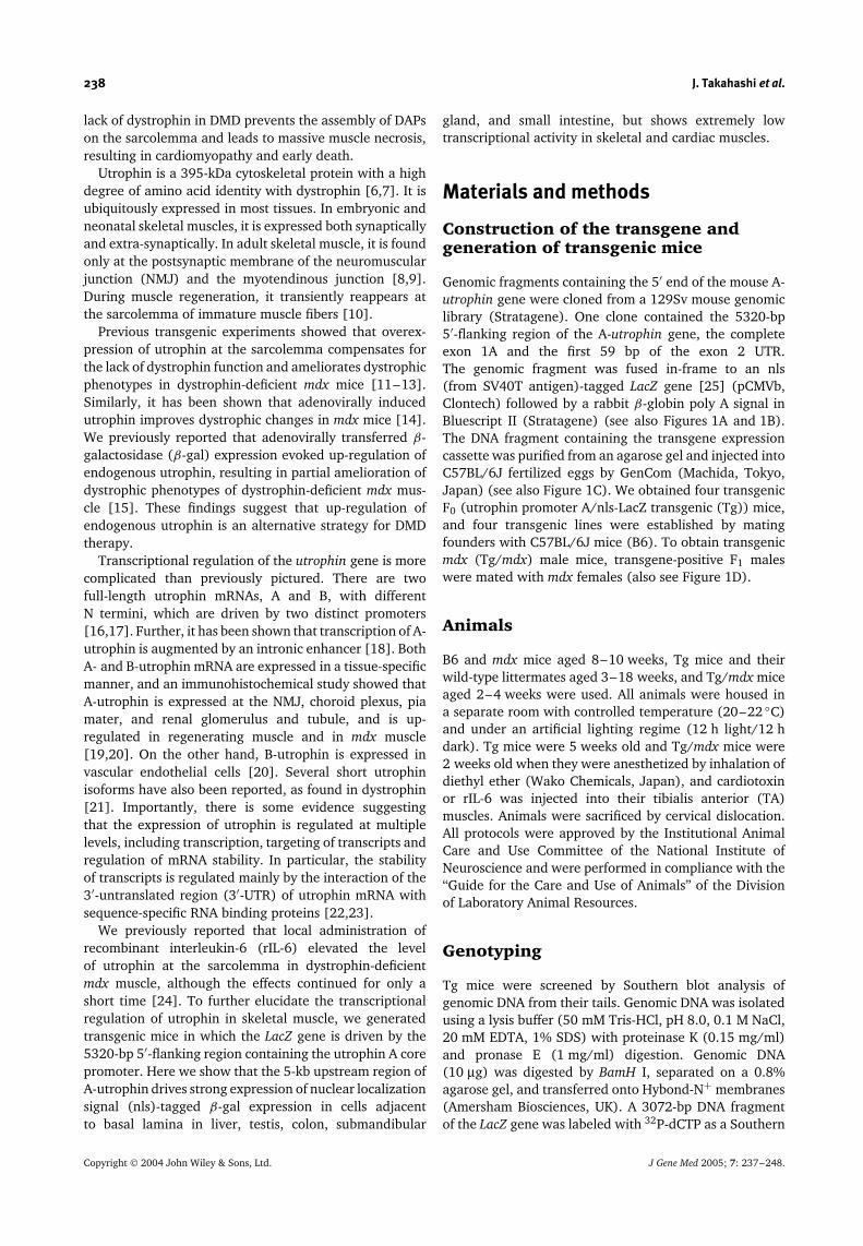
Genomic fragments containing the 5′ end of the mouse A-utrophin gene were cloned from a 129Sv mouse genomiclibrary (Stratagene). One clone contained the 5320-bp5′-flanking region of the A-utrophin gene, the completeexon 1A and the first 59 bp of the exon 2 UTR.The genomic fragment was fused in-frame to an nls(from SV40T antigen)-tagged LacZ gene [25] (pCMVb,Clontech) followed by a rabbit β-globin poly A signal inBluescript II (Stratagene) (see also Figures 1A and 1B).The DNA fragment containing the transgene expressioncassette was purified from an agarose gel and injected intoC57BL/6J fertilized eggs by GenCom (Machida, Tokyo,Japan) (see also Figure 1C). We obtained four transgenicF0 (utrophin promoter A/nls-LacZ transgenic (Tg)) mice,and four transgenic lines were established by matingfounders with C57BL/6J mice (B6). To obtain transgenicmdx (Tg/mdx) male mice, transgene-positive F1 maleswere mated with mdx females (also see Figure 1D).
Animals
B6 and mdx mice aged 8–10 weeks, Tg mice and theirwild-type littermates aged 3–18 weeks, and Tg/mdx miceaged 2–4 weeks were used. All animals were housed ina separate room with controlled temperature (20–22 ◦C)and under an artificial lighting regime (12 h light/12 hdark). Tg mice were 5 weeks old and Tg/mdx mice were2 weeks old when they were anesthetized by inhalation ofdiethyl ether (Wako Chemicals, Japan), and cardiotoxinor rIL-6 was injected into their tibialis anterior (TA)muscles. Animals were sacrificed by cervical dislocation.All protocols were approved by the Institutional AnimalCare and Use Committee of the National Institute ofNeuroscience and were performed in compliance with the‘‘Guide for the Care and Use of Animals’’ of the Divisionof Laboratory Animal Resources.
Genotyping
Tg mice were screened by Southern blot analysis ofgenomic DNA from their tails. Genomic DNA was isolatedusing a lysis buffer (50 mM Tris-HCl, pH 8.0, 0.1 M NaCl,20 mM EDTA, 1% SDS) with proteinase K (0.15 mg/ml)and pronase E (1 mg/ml) digestion. Genomic DNA(10 µg) was digested by BamH I, separated on a 0.8%agarose gel, and transferred onto Hybond-N+ membranes(Amersham Biosciences, UK). A 3072-bp DNA fragmentof the LacZ gene was labeled with 32P-dCTP as a Southern
Copyright 2004 John Wiley & Sons, Ltd. J Gene Med 2005; 7: 237–248.
Utrophin Promoter A-LacZ Transgenic Mice 239
Figure 1. Generation of the A-utrophin promoter/nls LacZ transgenic mice. (A) Structure of the 5′ end of the utrophin gene.The utrophin gene is transcribed by two A and B promoters (white lines) that give rise to two transcripts, A-utrophin mRNAand B-utrophin mRNA, respectively. The two utrophin transcripts have different N-termini. Gray boxes indicate the 5′-UTR. Blackboxes indicate the translated region of the exon. Scale bar indicates 100 bp. (B) Diagram of the transgene used in this study. Thegenomic fragment (12.4 kb), which contained the 5320-bp 5′-flanking region of the A-utrophin gene, exon 1A, and the first 59 bpof exon 2 UTR, was fused in-frame to a nls-tagged LacZ gene. Black bar indicates the Southern probe used to determine genotypes.(C) Summary of generation of transgenic mice. (D) Generation of transgenic mdx mice. Bold box indicates transgenic mdx malemice used in this study
probe, and hybridized with membranes at 65 ◦C overnight.The membranes were then washed extensively (2× SSPE,0.1% SDS; 1× SSPE, 0.1% SDS; 0.1× SSPE, 0.1% SDS)at 65 ◦C and analyzed by BAS 2500 (Fuji Film, Japan).
Isolation of total RNA
Three-week-old Tg mice and wild-type littermates weresacrificed, and tissues were isolated and rapidly frozenin liquid nitrogen. Total RNA was isolated fromfrozen tissues using TRIzol reagent (Invitrogen LifeTechnologies, USA) according to the manufacturer’sprotocol. Finally, total RNA was treated with 1 U of
RNase-free DNase I (Invitrogen Life Technologies) permicrogram of RNA at room temperature for 15 min, andDNase I was inactivated by the addition of 25 mM EDTA(pH 8.0) and heating at 65 ◦C for 10 min.
Reverse transcription (RT)-PCR
RT was performed with 3.5 µg of total RNA usingSuperScript II (Invitrogen Life Technologies) accord-ing to the manufacturer’s protocol. PCR was per-formed using the utrophin exon 1A sense (5′-GGCGTTTCCAATCGGGTGTC-3′) and the LacZ gene anti-sense (5′-GCGGGCCTCTTCGCTATTAC-3′) primers for 35
Copyright 2004 John Wiley & Sons, Ltd. J Gene Med 2005; 7: 237–248.

240 J. Takahashi et al.
cycles (denaturation at 95 ◦C for 1 min, annealing at59 ◦C for 30 s, and extension at 72 ◦C for 1 min). Asa control for the generation of PCR products due toresidual contamination of genomic DNA, an equivalentamount of RNA that had not been treated with RT wasalso tested in parallel. The RT-PCR products of all sam-ples were compared with the levels for a housekeepinggene, 18s rRNA, amplified with the following primerpair: sense primer (5′-TACCCTGGCGGTGGGATTAAC-3′)and anti-sense primer (5′-CGAGAGAAGACCACGCCAAC-3′). Amplification of A-utrophin was performed withthe following primer pair: the utrophin gene exon1A sense primer (5′-GGCGTTTCCAATCGGGTGTC-3′)and the utrophin gene exon 2 anti-sense primer (5′-CGTTCTGCCCATCATCAGGC-3′).
5′ RACE analysis
5′ RACE was carried out using the 5′ RACE sys-tem for rapid amplification of cDNA ENDs, V.2 kit(Invitrogen Life Technologies) following the manu-facturer’s protocol. Total RNA was reverse-transcribedusing the LacZ gene-specific primer (GSP)-1 (5′-CGGATTGACCGTAATGGGATA-3′). The anchor-ligatedcDNA was then PCR-amplified using the abridged anchorprimer provided and the LacZ gene anti-sense primer,which was designed upstream of GSP-1, GSP-2 (5′-GCGGGCCTCTTCGCTATTAC-3′). In addition, the pri-mary PCR product was amplified using the abridgeduniversal amplification primer provided and the nls anti-sense primer, GSP-3 (5′-CGCTCATGATGCACGGTCTG-3′)designed upstream of GSP-2. PCR products were clonedinto a pCR2.1 vector (Invitrogen Life Technologies) andsequenced using a Thermo Sequenase cycle sequencingkit (Amersham Biosciences).
Histochemical analysis
After Tg and wild-type mice had been sacrificed, cere-brum, cerebellum, submandibular gland, lung, kidney,liver, small intestine, colon, testis, pancreas, spleen, TAand soleus muscles, diaphragm, and heart were isolatedand frozen in liquid nitrogen cooled isopentane. Cryosec-tions (10 µm) from several tissues were stained withhematoxylin and eosin (H&E) and with 5-bromo-4-chloro-3-indolyl-β-D-galactopyranoside (X-Gal; Wako Chemicals,Japan) as described elsewhere [26].
Immunohistochemistry
Serial transverse cryosections (6 µm) from different tis-sues were placed on a slide, then dried and fixed inacetone for 6 min at −20 ◦C. We carried out immuno-histochemical analysis with a rabbit polyclonal antibodyagainst human utrophin (UT-2), which recognizes aminoacid positions 1768–2078 [27]. The primary antibodieswere detected with Alexa 488-labeled goat anti-rabbit IgG
(Molecular Probes, USA). The nucleus was stained withTOTO-3 iodide (Molecular Probes). The NMJ was stainedwith Alexa 594-labeled α-bungarotoxin (α-BTX) (Molec-ular Probes). Signals were recorded photographicallywith a laser-scanning confocal imaging system (TCSSP,Leica).
Cardiotoxin injection
To cause muscle degeneration, we injected 100 µl ofcardiotoxin (CTX) of Naja naja atra venom (10 µMin saline, Wako Chemicals) into the right TA muscleof 5-week-old Tg mice with a 29-gauge needle. Theconcentration of CTX was determined according to aprevious report [28]. CTX-injected Tg mice were sacrificed1–7 days after CTX injection. The CTX-injected andcontralateral non-injected TA muscles were isolated andfrozen in liquid nitrogen cooled isopentane. Cryosections(10 µm) were stained with X-Gal. At the same time, serialcryosections (6 µm) were stained with UT-2 together withAlexa 594-labeled α-BTX. Total RNA was isolated fromthese frozen tissues.
Administration of rIL-6 to Tg/mdxmouse muscles
To investigate the effect of IL-6 on utrophin promoterA activation, we daily injected rIL-6 (800 ng in 6 µlof phosphate-buffered saline (PBS)/day) (R&D Systems,USA) for 5 days into the TA muscles of 2-week-old Tg/mdxmice. rIL-6-injected Tg/mdx mice were sacrificed 2 daysafter the final injection.
Results
Generation of four mouse lines withvarying numbers of copies of thepromoter A/nlsLacZ transgene
To investigate the utrophin promoter A activity, we firstscreened a 129Sv mouse genomic library and obtaineda genome clone that contained the 5320-bp 5′-flankingregion of the A-utrophin gene and exon 1A, intron 1, andexon 2. We then subcloned it into a Bluescript II vector.Subsequently, an nls-tagged LacZ gene was inserteddownstream of the exon 2 UTR, as shown in Figure 1B.The nls was tagged to the LacZ gene to clarify the nucleiin which the utrophin promoter A was activated. Fourtransgenic F0 ‘founder’ mice were identified by Southernblotting using a LacZ probe, and four transgenic lineswere established (Figure 1). The approximate number oftransgene copies was determined by Southern blotting inall four lines (data not shown). Line 1 contained about13 copies, lines 2 and 3 contained 5–6 copies, and line 4contained 1 copy of the transgene.
Copyright 2004 John Wiley & Sons, Ltd. J Gene Med 2005; 7: 237–248.

Utrophin Promoter A-LacZ Transgenic Mice 241
mRNA levels of the transgene inseveral organs of Tg mice
The level of the transgene expression in several organsof Tg mice was determined by RT-PCR. Representativeresults from line 2 are shown in Figure 2. High levels oftransgene mRNA expression were detected in liver, testis,colon, and submandibular gland. The signal was weaklydetected in small intestine and lung, and an extremelylow signal was detected in kidney, cerebrum, cerebellum,and TA muscle.
Determination of the 5′ end of β-galmRNA
To investigate whether the transcription start site of thetransgene corresponds to the authentic transcription start
site of A-utrophin, we carried out 5′ RACE using totalRNA isolated from testis of Tg mice (line 2). We obtaineda single PCR product, indicative of a single transcriptionstart site in testis (Figure 3). We sequenced the PCRproduct (543 bp) and confirmed the transcription startsite of the transgene in testis, which was the same as thatpreviously reported [16].
β-Gal expression in liver, testis, colon,submandibular gland, and smallintestine of Tg mice
To identify the nuclei that express the transgene,cryosections were stained with X-Gal. At the same time,serial cryosections were stained with H&E or with UT-2, a polyclonal antibody against human utrophin [27](Figure 4). UT-2 recognizes only the full-length form
Figure 2. RT-PCR analysis of transgene mRNA from several tissues of transgenic mice. Total RNA was extracted from several tissuesof 3-week-old line 2 Tg mice, and RT-PCR was performed with the transgene-specific primers described in Materials and methods.PCR products were electrophoresed on a 2% agarose gel. The expected size of the PCR product was 414 bp (transgene; top). As acontrol, 18s rRNA (297 bp; bottom) was co-amplified
Figure 3. 5′ RACE analysis of transcription initiation sites. (A) 5′ RACE analysis with total RNA (1.5 µg) of the testis from 3-week-oldline 2 Tg mice was performed using the gene-specific primers described in Materials and methods. The final PCR products wereseparated on a 2% agarose gel. (B) Sequencing products confirmed that the transgene was transcribed from the authentic initiationsite of the utrophin A promoter (data not shown)
Copyright 2004 John Wiley & Sons, Ltd. J Gene Med 2005; 7: 237–248.

242 J. Takahashi et al.
(a)
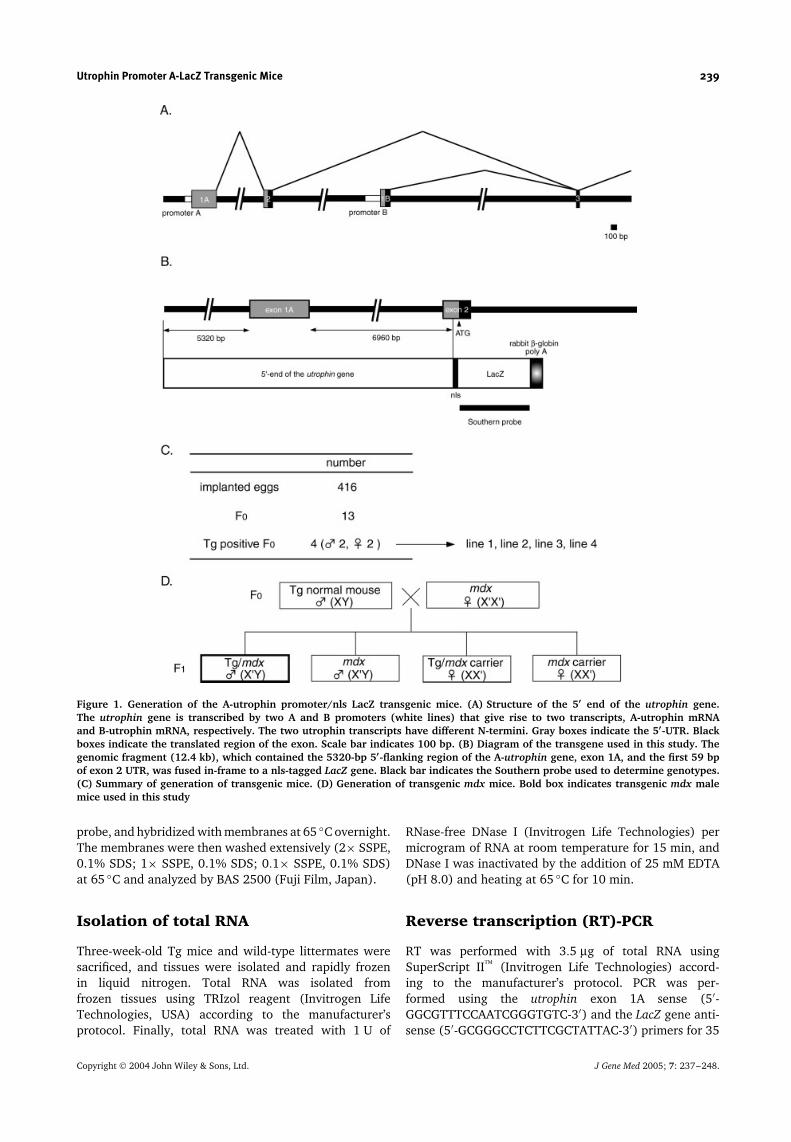
Figure 4. Histological and immunohistochemical analysis of transgenic mice. Cryosections (10 µm) were prepared from tissues(liver, testis, colon, submandibular gland, and small intestine (A), kidney and lung (B), cerebrum, cerebellum, heart, and TA muscle(C)) from 8-week-old line 2 Tg mice and stained with H&E (top) or X-Gal (middle). Serial 6 µm cryosections were stained witha polyclonal antibody against utrophin (UT-2; green). Nuclei were stained with TOTO-3 (blue). The NMJs were identified withα-BTX (red) (bottom). Shown are representative results obtained from line 2 Tg mice. Identical staining patterns were seen in alltransgenic lines. V, terminal hepatic venule; Si, sinusoid; H, hepatocyte; ST, seminiferous tubule; L, Leydig cell; S, Sertoli cell; C,capillary; A, arteriole; MM, muscularis mucosa; CM, inner circular muscle layer; LM, outer longitudinal muscle layer; G, gobletcell; SD, striated duct; Sc, serous secretory cell; SA, serous acinus; Vi, villus; Cr, crypt; P, Paneth cell; T, cortical renal tubule;Gl, glomerulus; Al, alveolar cell; PV, pulmonary veinule; CP, choroid plexus; PM, pia mater; CC, cerebral cortex; N, neuron; ML,molecular layer; GL, granular layer; P, Purkinje cell; ID, intercalated disk; NMJ, neuromuscular junction. Bar, 100 µm
that includes both A-utrophin and B-utrophin [27].We examined β-gal expression in all tissues in whichA-utrophin has been reported to be expressed [20],and found that β-gal expression coincided well withendogenous A-utrophin expression in liver, testis, colon,submandibular gland, and small intestine.
In the liver, the nuclei of hepatocytes, but not sinu-soid lining cells, were strongly stained with X-Gal, whileendogenous utrophin protein was detected in the mar-gins of hepatocytes along sinusoids and terminal hepaticvenules.
In the testis, β-gal activity was found in Sertoli cellsin the basal compartment of the seminiferous tubules,but not in the adluminal compartment, and in Leydigcells in the interstitial supporting tissues between theseminiferous tubules. Consistent with this observation,endogenous utrophin signals were found along thebasement membrane of the seminiferous tubules andLeydig cells.
In the colon, β-gal-positive nuclei were found in gobletcells in large intestinal glands. Endogenous utrophinsignals were found along the basement membrane oflarge intestinal glands and muscularis mucosa.
In the submandibular gland, the nuclei of both serousand mucous secretory cells were clearly stained with X-Gal. The striated duct epithelia lacked the β-gal signal.Endogenous utrophin was detected along the basement
(b)
Figure 4. (Continued)
membrane of serous and mucous acini, but not of striatedducts.
In the small intestine, β-gal-positive nuclei were foundin goblet cells and epithelia of the bases of villi and crypts.
Copyright 2004 John Wiley & Sons, Ltd. J Gene Med 2005; 7: 237–248.
Utrophin Promoter A-LacZ Transgenic Mice 243
(c)
Figure 4. (Continued)
β-Gal signals were stronger in a subset of cells, and theircharacteristic distribution suggests that these cells werePaneth cells. Endogenous utrophin signals were foundalong the basement membrane of villi and crypts and inmuscularis mucosa.
In the kidney, β-gal-positive nuclei were found inthe epithelia of cortical renal tubules, but not inglomeruli. It is not clear whether β-gal-positive nucleiwere present in proximal convoluted tubules, distalconvoluted tubules, collecting tubules, or collecting ducts.Endogenous utrophin was found along the basementmembrane of cortical renal tubules, collecting ducts of therenal medulla and Bowman’s capsules, and in glomerularcapillaries.
In the lung, β-gal-positive nuclei were found in alveoli,but not in terminal bronchiole epithelia. It is not clearwhether β-gal-positive nuclei were present in type I ortype II pneumocytes. Endogenous utrophin was foundin alveolar cells and terminal bronchiole epithelia. Thus,in these tissues, endogenous utrophin protein seems tolocalize at the plasma membranes of cells adjacent to thebasement membranes.
Inconsistent results of LacZ expressionamong the four transgenic lines
In the pancreas, β-gal-positive nuclei were clearly foundin exocrine cells of lines 1 and 2, but not at all in lines3 and 4 (data not shown). Because of this inconsistencyof β-gal expression patterns among the four Tg lines, wecould not determine the relationship between β-gal andendogenous utrophin expression in pancreas.
β-Gal-positive nuclei were ubiquitously found in thecerebrum of line 1, but not in other lines (Table 1).
The A-utrophin protein has been shown to be expressedin the pia mater and choroid plexus of the brain [20].Nevertheless, our results indicate that the A-utrophinpromoter was not active in the pia mater and choroidplexus in our Tg mice (Figure 4C).
In the cerebellum, β-gal-positive nuclei were found ingranular cells of lines 1 and 3, but not at all in lines 2and 4 (Figure 4C, Table 1). The A-utrophin protein hasbeen shown to be expressed in the pia mater of thecerebellum [20]. Nevertheless, our results indicate thatthe A-utrophin promoter was not active in the pia mater ofthe cerebellum in our Tg mice (Figure 4C). We speculatethat this discrepancy is due to the positional effects of thetransgene integration into the mouse genome.
In the spleen, β-gal-positive nuclei were not found inany of the four lines, although endogenous utrophin wasfound in endothelial cells of venous sinuses in red pulp(data not shown). It seems that β-gal expression was notdetected in spleen of our Tg lines because B-utrophin wasobserved in endothelia of blood vessels [20].
β-Gal-positive nuclei were not found incardiac, skeletal, or vascular smoothmuscle
A previous study using antibodies specific to A- and B-utrophins showed that, in skeletal muscle, A-utrophin isexpressed in the NMJ, peripheral nerves, and larger bloodvessels [20]. However, we detected β-gal expression inneither the NMJ nor peripheral nerves of Tg skeletalmuscles. As expected, β-gal expression was not foundin blood vessels of Tg mouse tissues, where B-utrophinhas been reported to be expressed. Unexpectedly, we did
Copyright 2004 John Wiley & Sons, Ltd. J Gene Med 2005; 7: 237–248.
244 J. Takahashi et al.
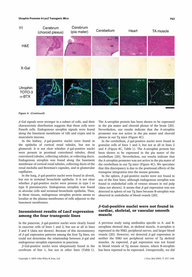
Table 1. Summary of β-gal expression in four transgenic lines. Tg mice (lines 1–4) were sacrificed at 3 weeks old and 8 weeks old andβ-gal expression in several tissues was examined. No β-gal-positive nuclei were found in non-transgenic littermates. β-Gal expressionlevel is shown as follows: −, none; ±, trace; +, weak; ++, moderate; +++, strong
Young (3–5w) Adult (8–18w)
line 1 line 2 line 3 line 4 line 1 line 2 line 3 line 4
Cerebrum ++ − − − ++ − − −Cerebellum + − + − ++ − + −Heart − − − − − − − −TA muscle − − − − − − − −Soleus muscle − − − − − − − −Diaphragm − − − − − − − −Arteriole − − − − − − − −Small intestine + + + − + + − −Colon + ++ ++ ++ ++ ++ + +Testis +++ +++ ++ − +++ +++ ++ −Liver ++ ++ ± ± ++ +++ ± ±Submandibular gland +++ +++ ++ − +++ +++ ++ −Kidney + ± ± ± ± ± − −Lung ++ + − + + − −Pancreas ++ + − − ++ + − −Spleen − − − − − − − −
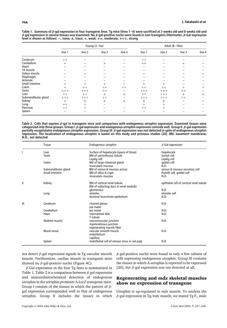
Table 2. Cells that express β-gal in transgenic mice and comparison with endogenous utrophin expression. Examined tissues werecategorized into three groups. Group I: β-gal expression and endogenous utrophin expression coincide well. Group II: β-gal expressionpartially recapitulates endogenous utrophin expression. Group III: β-gal expression was not detected in spite of endogenous utrophinexpression. The localization of endogenous utrophin is based on this study and previous studies [20]. BM, basement membrane;N.D., not detected
Tissue Endogenous utrophin β-Gal expression
I Liver Surface of hepatocyte (space of Disse) hepatocyteTestis BM of seminiferous tubule Sertoli cell
Leydig cell Leydig cellColon BM of large intestinal gland goblet cell
muscularis mucosa N.D.Submandibular gland BM of serous & mucous acinus serous & mucous secretory cellSmall intestine BM of villus & crypt Paneth cell, goblet cell
muscularis mucosa N.D.
II Kidney BM of cortical renal tubule epithelial cell of cortical renal tubuleBM of collecting duct in renal medullaglomerulus N.D.
Lung alveolus alveolar cellterminal bronchiole epithelium N.D.
III Cerebrum choroid plexus N.D.pia mater
Cerebellum pia mater N.D.Heart intercalated disk N.D.
T tubuleSkeletal muscle neuromuscular junction N.D.
myotendinous junctionregenerating muscle fiber
Blood vessel vascular smooth muscle N.D.endotheliumcapillary
Spleen endothelial cell of venous sinus in red pulp N.D.
not detect β-gal expression signals in Tg vascular smoothmuscle. Furthermore, cardiac muscle in transgenic miceshowed no β-gal-positive nuclei (Figure 4C).
β-Gal expression in the four Tg lines is summarized inTable 1. Table 2 is a comparison between β-gal expressionand immunohistochemical detection of endogenousutrophin in the utrophin promoter A-LacZ transgenic mice.Group I consists of the tissues in which the pattern of β-gal expression corresponded well to that of endogenousutrophin. Group II includes the tissues in which
β-gal-positive nuclei were found in only a few subsets ofcells expressing endogenous utrophin. Group III containsthe tissues in which A-utrophin is reported to be expressed[20], but β-gal expression was not detected at all.
Regenerating and mdx skeletal musclesshow no expression of transgene
Utrophin is up-regulated in mdx muscle. To analyze theβ-gal expression in Tg/mdx muscle, we mated Tg F1 male
Copyright 2004 John Wiley & Sons, Ltd. J Gene Med 2005; 7: 237–248.

Utrophin Promoter A-LacZ Transgenic Mice 245
Figure 5. Effects of recombinant IL-6 and CTX injection into TA muscle of transgenic mice on transgene expression (β-gal).Cryosections of TA muscle from mdx mice, Tg mdx male mice (Tg/mdx), CTX-injected Tg TA muscle (Tg + CTX), and rIL-6-injectedTg mdx TA muscle (Tg/mdx + rIL-6) were stained with X-Gal (top) or stained with a polyclonal antibody against utrophin (UT-2)(green) and Alexa 594-labeled α-BTX (red) (bottom). rIL-6-injected Tg/mdx TA muscles were obtained 2 days after the final injectionof rIL-6, and CTX-injected Tg TA muscles were obtained 5 days after CTX injection. * indicates NMJ. Bar, 100 µm
Figure 6. Effects of muscle regeneration on expression of transgene mRNA. Total RNA was extracted from TA muscle, soleus muscle,diaphragm, and cardiac muscle of 4-week-old Tg and Tg/mdx mice (A), and from CTX-injected 5-week-old Tg TA muscles at theindicated days after the injection (B). RT-PCR was performed with transgene-specific primers described in Materials and methods.PCR products were electrophoresed on 2% agarose gel. The expected size of PCR products was 414 bp (transgene; top) and 309 bp(A-utrophin; middle). As a control, 18s rRNA (297 bp; bottom) was co-amplified
mice of line 2 with mdx female mice (Figure 1D). Utrophinprotein was overexpressed along the sarcolemma of mdxmuscle fibers, but all myonuclei were negative for β-gal staining (Figure 5). Next, we investigated the mRNAlevels of A-utrophin and transgene by RT-PCR. To thisend, A-utrophin mRNA was elevated in TA muscle, soleusmuscle, diaphragm, and cardiac muscle of Tg/mdx mice,but transgene mRNA was not detected in these muscles(Figure 6A). In addition, there was no difference in β-gal
expression in various tissues other than muscle betweenTg/mdx male mice and parental C57BL/6J-Tg mice (datanot shown).
Utrophin is up-regulated in regenerating muscle.Recently, Galvagni et al. reported that the transcriptionof A-utrophin was elevated in regenerating muscle [19].Therefore, we next injected CTX into TA muscle of line2 Tg mice to damage muscle fibers, and analyzed β-gal expression during muscle regeneration (Figure 5).
Copyright 2004 John Wiley & Sons, Ltd. J Gene Med 2005; 7: 237–248.

246 J. Takahashi et al.
Figure 5 shows a section of transgenic skeletal muscleisolated 5 days after CTX injection, chosen because theutrophin protein signal was detected the most stronglyin it of all tissues collected after CTX injection. Utrophinwas overexpressed along the sarcolemma of regeneratingsmall muscle fibers, but all myonuclei were negative for β-gal staining. Furthermore, A-utrophin mRNA was elevated3 days after CTX injection, but transgene mRNA was notdetected in CTX-injected muscle (Figure 6B).
Administration of rIL-6 did not inducethe expression of the transgene inskeletal muscle
We have previously reported that rIL-6 induced overex-pression of endogenous utrophin in mdx skeletal muscle[24]. Subsequently, we found that rIL-6 elevated mainlyA-utrophin mRNA (Itoh et al., unpublished data). There-fore, we injected rIL-6 into the TA muscles of Tg/mdxmice daily for 5 days and analyzed the effect of rIL-6 onthe transgene expression (Figure 5). Without doubt, rIL-6induced overexpression of utrophin in Tg/mdx muscles,but not expression of the transgene (Figure 5). Further-more, A-utrophin mRNA was up-regulated 2 days afterthe final injection, but transgene mRNA was not detected(data not shown).
Discussion
Previous transgenic studies showed that forced expressionof utrophin at the sarcolemma of mdx muscle dramaticallyimproved dystrophic phenotypes [11–13]. Furthermore,an adenovirally induced utrophin gene ameliorated thedystrophic changes in mdx mice [14]. On the other hand,we have previously reported that adenovirus vector-mediated expression of β-gal up-regulated endogenousutrophin at the sarcolemma in mdx muscle, whereβ-gal-positive muscle fibers were protected from thedegeneration process [15]. Thus, up-regulation ofutrophin stabilizes dystrophin-deficient muscle membraneand protects muscle fibers from degeneration caused bymechanical stress.
There are two full-length transcripts: A-utrophin andB-utrophin [16,17]. A-Utrophin localizes to the NMJ andin peripheral nerves [20]. The upstream promoter A isCpG-rich, TATA-less, and contains a consensus N-box,which is critical for synaptic expression of utrophin. Incontrast, B-utrophin is observed in endothelial capillariesand vessels [20]. Further, A-utrophin, but not B-utrophin,is up-regulated in regenerating muscle fibers [19] or inmdx muscle fibers [20]. Therefore, we first examinedpromoter A activity using transgenic techniques. The LacZgene was tagged by nls to clarify the nuclei in whichpromoter A is active. Contrary to our expectation, analysisof both transcripts for the transgene and β-gal stainingshowed that the 5320-bp 5′-flanking region of the A-utrophin gene is highly and constantly active in liver,
testis, colon, submandibular gland, and small intestine,slightly active in kidney and lung, but not at all in striatedmuscles.
Intriguingly, detailed examination at the cellular levelshowed that promoter A is active in secretory cells. Forexample, in the Tg mouse liver, β-gal-positive nucleiwere found in hepatocytes, which synthesize and secretebile. In the Tg mouse testis, β-gal-positive nuclei werefound in Sertoli cells and Leydig cells. Sertoli cells ofthe fetal testis produce and secrete Mullerian-inhibitingsubstance (MIS), which suppresses the development ofthe reproductive tract and results in regression of theMullerian duct in the female. MIS is also producedin the adult gonads and plays a role in maintainingtestosterone homeostasis [29]. Leydig cells synthesizeand secrete testosterone, which induces male secondarysexual characteristics. In the Tg mouse colon, β-gal-positive nuclei were found in goblet cells, which producemucin. In the submandibular gland, nuclei of serousor mucous secretory cells, which secrete saliva, wereclearly stained with X-Gal, although the Tg mouse striatedduct epithelia, which secrete lysozyme and IgA, lackedβ-gal expression. In the Tg mouse small intestine, β-gal-positive nuclei were found in goblet cells and Panethcells. Goblet cells secrete mucus, and Paneth cells secretemicrobicidal α-defensins when exposed to bacteria orbacterial antigens [30]. Previous studies showed thatutrophin is found at high levels in secretory tissuesincluding pituitary, thyroid, adrenal glands, and choroidplexus epithelia, which secrete glucocorticoid hormones,the iodine-containing hormones tri-iodothyronine andthyroxine, steroid and catecholamine hormones, andcerebrospinal fluid, respectively [31,32]. We detected A-utrophin expression in the secretory cells of our Tg mice.We speculate that utrophin may have other unknownfunctions as well as supporting secretory cells.
In the Tg mouse kidney, endogenous utrophin wasexpressed in glomerular tufts, cortical renal tubules, andcollecting ducts in the renal medulla, and A-utrophinprotein has been shown to be expressed in glomerulartufts and tubules of the renal cortex [20]. However, β-galsignals were found in only a few epithelia of cortical renaltubules. On the other hand, endogenous utrophin signalswere found along the basement membrane of corticalrenal tubules, possibly assembling the DAP complex atthe plasma membrane and interacting with laminin in thebasement membrane. The reason β-gal-positive nucleiwere not found in glomeruli is unknown. In the Tg mouselung, a small number of β-gal-positive nuclei were foundin a few type I or type II pneumocytes. The reason for thisdiscrepancy is unclear.
In the cerebrum and the cerebellum of Tg mice,transgene mRNA was detected at extremely low levels,while β-gal-positive nuclei were not detected at all inthe choroid plexus and pia mater. In these tissues, thetranscriptional level was also low, and the stability oftransgene mRNA may be lower than the transcriptionallevel, as in skeletal muscle.
Copyright 2004 John Wiley & Sons, Ltd. J Gene Med 2005; 7: 237–248.

Utrophin Promoter A-LacZ Transgenic Mice 247
In this study, unexpectedly, we detected extremelylow levels of transgene mRNA in skeletal muscle and noβ-gal staining in myonuclei even in the vicinity of theNMJs. Likewise, the expression of the transgene wasnot observed in regenerating muscle of the utrophinpromoter A/LacZ transgenic mice. Further, we previouslyreported that IL-6 induced overexpression of utrophinin neonatal mdx muscle [24]. However, the expressionof β-gal was not observed in rIL-6-injected Tg/mdxmuscle. Briefly, the promoter of the A-utrophin geneis not active in cardiac and skeletal muscle, and thereare several possibilities for a regulatory mechanism ofA-utrophin expression in skeletal and cardiac muscle.First, the transgene construct used in this study containedthe 5′-flanking region (5320 bp) of the A-utrophin gene,but lacked the intronic enhancer (downstream utrophinenhancer, DUE) within the second intron, which hasrecently been reported [18]. Therefore, it is reasonableto speculate that the intronic enhancer is indispensablefor the expression of utrophin in both skeletal muscle andcardiac muscle. Second, the 5320 bp of the 5′-flankingregion or first intron might contain a silencer regionfor the expression of A-utrophin in skeletal and cardiacmuscle, since Galvagni et al. reported that LacZ expressionwas detected in muscle cell transfected with the promoteralone [19]. Third, important roles for the 3′-UTR ofutrophin mRNA in stabilization have been suggested, andtwo RNA-binding proteins have been reported [22]. Forexample, Gramolini et al. reported ∼42- kDa and 90-kDaproteins that bind utrophin 3′-UTR and are expressedmore abundantly in extensor digitorum longus musclesthan in soleus muscles. Hence, higher levels of utrophinmRNA in soleus muscles suggest that these proteinsdestabilize utrophin mRNA [22]. Furthermore, Gramoliniet al. reported that the utrophin 3′-UTR is responsiblefor targeting utrophin mRNA to cytoskeletal-boundpolysomes, binding actin, and controlling transcriptstability [23]. We previously reported that adenovirusvector-mediated gene transfer into skeletal muscle evokedrobust expression of endogenous utrophin via immuneresponse [15], in which both A-utrophin and B-utrophinmRNA levels were continuously elevated (Itoh et al.,unpublished data). These results suggest that utrophinoverexpression is induced by one or more inflammatorycytokines, and most of the effects seem to be due to post-transcriptional regulation. These observations and ourresults suggest that the expression of utrophin in skeletalmuscle might be regulated at the post-transcriptionallevel via 3′-UTR. Finally, it is also important to note thatthe utrophin protein seems to be relatively stable at thesarcolemma in skeletal muscle [22]. Once the utrophinprotein forms the dystrophin–glycoprotein complex at thesarcolemma, it might be extremely stable in muscle fiberscompared with β-gal.
In summary, we showed here that the 5-kb flankingregion of the A-utrophin gene containing the A-utrophin core promoter drives high levels of A-utrophintranscription in liver, testis, colon, submandibular gland,and small intestine, but not in skeletal and cardiac muscle,
indicating that A-utrophin expression in striated muscleis greatly dependent on other regulatory elements.
Acknowledgements
We thank Dr. Hirata and Dr. Yokota for technical instructions. Wealso thank S. Masuda and A. Fukase for technical assistance, andcolleagues in our laboratory for useful discussion and suggestionson this work. This work is supported by Grants-in-Aid for Centerof Excellence (COE), Research on Nervous and Mental Disorders(10B-1, 13B-1), and Health Science Research Grants for Researchon the Human Genome and Gene Therapy (H10-genome-015,H13-genome-001), for Research on Brain Science (H12-Brain-028, H15-Brain-021) from the Ministry of Health, Labor andWelfare, Grants-in Aids for Scientific Research (10 557 065,11 470 153, 11 170 264, 14 657 158 and 15 390 281) from theMinistry of Education, Culture, Sports, Science and Technology,and a Research Grant from the Human Frontier Science Project.
References
1. Koenig M, Hoffman EP, Bertelson CJ, et al. Complete cloning ofthe Duchenne muscular dystrophy (DMD) cDNA and preliminarygenomic organization of the DMD gene in normal and affectedindividuals. Cell 1987; 50: 509–517.
2. Ahn AH, Kunkel LM. The structural and functional diversity ofdystrophin. Nat Genet 1993; 3: 283–291.
3. Tinsley JM, Blake DJ, Zuellig RA, et al. Increasing complexity ofthe dystrophin-associated protein complex. Proc Natl Acad SciU S A 1994; 91: 8307–8313.
4. Campbell KP. Three muscular dystrophies: loss of cytoskeleton-extracellular matrix linkage. Cell 1995; 80: 675–679.
5. Ozawa E, Yoshida M, Suzuki A, et al. Dystrophin-associatedproteins in muscular dystrophy. Hum Mol Genet 1995; 4:1711–1716.
6. Pearce M, Blake DJ, Tinsley JM, et al. The utrophin anddystrophin genes share similarities in genomic structure. HumMol Genet 1993; 2: 1765–1772.
7. Grady RM, Teng H, Nichol MC, et al. Skeletal and cardiacmyopathies in mice lacking utrophin and dystrophin: a modelfor Duchenne muscular dystrophy. Cell 1997; 90: 729–738.
8. Khurana TS, Watkins SC, Chafey P, et al. Immunolocalizationand developmental expression of dystrophin related protein inskeletal muscle. Neuromuscul Disord 1991; 1: 185–194.
9. Ohlendieck K, Ervasti JM, Matsumura K, et al. Dystrophin-related protein is localized to neuromuscular junctions of adultskeletal muscle. Neuron 1991; 7: 499–508.
10. Wilson LA, Cooper BJ, Dux L, et al. Expression of utrophin(dystrophin-related protein) during regeneration andmaturation of skeletal muscle in canine X-linked musculardystrophy. Neuropathol Appl Neurobiol 1994; 20: 359–367.
11. Tinsley JM, Potter AC, Phelps SR, et al. Amelioration of thedystrophic phenotype of mdx mice using a truncated utrophintransgene. Nature 1996; 384: 349–353.
12. Deconinck N, Tinsley JM, De Backer F, et al. Expression oftruncated utrophin leads to major functional improvementsin dystrophin-deficient muscles of mice. Nat Med 1997; 3:1216–1221.
13. Tinsley JM, Deconinck N, Fisher R, et al. Expression of full-length utrophin prevents muscular dystrophy in mdx mice. NatMed 1998; 4: 1441–1444.
14. Gilbert R, Nalbantoglu J, Petrof BJ, et al. Adenovirus-mediatedutrophin gene transfer mitigates the dystrophic phenotype ofmdx mouse muscles. Hum Gene Ther 1999; 10: 1299–1310.
15. Yamamoto K, Yuasa K, Miyagoe Y, et al. Immune response toadenovirus-delivered antigens upregulates utrophin and resultsin mitigation of muscle pathology in mdx mice. Hum Gene Ther2000; 11: 669–680.
16. Dennis CL, Tinsley JM, Deconinck AE, et al. Molecular andfunctional analysis of the utrophin promoter. Nucleic Acids Res1996; 24: 1646–1652.
Copyright 2004 John Wiley & Sons, Ltd. J Gene Med 2005; 7: 237–248.
248 J. Takahashi et al.
17. Burton EA, Tinsley JM, Holzfeind PJ, et al. A second promoterprovides an alternative target for therapeutic up-regulation ofutrophin in Duchenne muscular dystrophy. Proc Natl Acad SciU S A 1999; 96: 14025–14030.
18. Galvagni F, Oliviero S. Utrophin transcription is activated by anintronic enhancer. J Biol Chem 2000; 275: 3168–3172.
19. Galvagni F, Cantini M, Oliviero S. The utrophin gene istranscriptionally up-regulated in regenerating muscle. J BiolChem 2002; 277: 19106–19113.
20. Weir AP, Burton EA, Harrod G, et al. A- and B-utrophin havedifferent expression patterns and are differentially up-regulatedin mdx muscle. J Biol Chem 2002; 277: 45285–45290.
21. Jimenez-Mallebrera C, Davies KE, Putt W, et al. A study of shortutrophin isoforms in mice deficient for full-length utrophin.Mamm Genome 2003; 14: 47–60.
22. Gramolini AO, Belanger G, Thompson JM, et al. Increasedexpression of utrophin in a slow vs. a fast muscle involvesposttranscriptional events. Am J Physiol Cell Physiol 2001; 281:C1300–C1309.
23. Gramolini AO, Belanger G, Jasmin BJ. Distinct regions in the 3′untranslated region are responsible for targeting and stabilizingutrophin transcripts in skeletal muscle cells. J Cell Biol 2001;154: 1173–1183.
24. Fujimori K, Itoh Y, Yamamoto K, et al. Interleukin-6 inducesoverexpression of the sarcolemmal utrophin in neonatal mdxskeletal muscle. Hum Gene Ther 2002; 13: 509–518.
25. Kalderon D, Roberts BL, Richardson WD, et al. A short aminoacid sequence able to specify nuclear location. Cell 1984; 39:499–509.
26. Ishii A, Hagiwara Y, Saito Y, et al. Effective adenovirus-mediatedgene expression in adult murine skeletal muscle. Muscle Nerve1999; 22: 592–599.
27. Imamura M, Ozawa E. Differential expression of dystrophinisoforms and utrophin during dibutyryl-cAMP-inducedmorphological differentiation of rat brain astrocytes. Proc NatlAcad Sci U S A 1998; 95: 6139–6144.
28. Couteaux R, Mira JC, d’Albis A. Regeneration of muscles aftercardiotoxin injury. I. Cytological aspects. Biol Cell 1988; 62:171–182.
29. Teixeira J, Fynn-Thompson E, Payne AH, et al. Mullerian-inhibiting substance regulates androgen synthesis at thetranscriptional level. Endocrinology 1999; 140: 4732–4738.
30. Ayabe T, Satchell DP, Wilson CL, et al. Secretion of microbicidalalpha-defensins by intestinal Paneth cells in response to bacteria.Nat Immunol 2000; 1: 113–118.
31. Schofield J, Houzelstein D, Davies K, et al. Expression of thedystrophin-related protein (utrophin) gene during mouseembryogenesis. Dev Dyn 1993; 198: 254–264.
32. Knuesel I, Bornhauser BC, Zuellig RA, et al. Differentialexpression of utrophin and dystrophin in CNS neurons: anin situ hybridization and immunohistochemical study. J CompNeurol 2000; 422: 594–611.
Copyright 2004 John Wiley & Sons, Ltd. J Gene Med 2005; 7: 237–248.