The use of muscle biopsy in the diagnosis of undefined ataxia with cerebellar atrophy in children
9
Original article The use of muscle biopsy in the diagnosis of undefined ataxia with cerebellar atrophy in children Alessandra Terracciano a,1 , Florence Renaldo b,1 , Ginevra Zanni a , Adele D’Amico a , Anna Pastore a , Sabina Barresi a , Enza Maria Valente c , Fiorella Piemonte a , Giulia Tozzi a , Rosalba Carrozzo a , Massimiliano Valeriani a , Renata Boldrini d , Eugenio Mercuri f , Filippo Maria Santorelli a,e , Enrico Bertini a, * a Unit of Neuromuscular and Neurodegenerative Disorders, Lab of Molecular Medicine and Unit of Neurology, Dept of Neuroscience, Bambino Gesu ` Childrens Hospital, Rome, Italy b Service de Neurope ´diatrie, Ho ˆpital Armand Trousseau, Paris, France c Research Institute Mendel, Laboratory of Neuroscience, Rome, Italy d Unit of Pathology, Bambino Gesu ` Childrens Hospital, Rome, Italy e IRCCS-Stella Maris, Calambrone, Pisa, Italy f Institute of Neuropsychiatry, Catholic University, Rome, Italy article info Article history: Received 4 April 2011 Received in revised form 21 July 2011 Accepted 24 July 2011 Keywords: Inherited cerebellar ataxias Marinesco-Sjogren syndrome Coenzyme Q10 deficiency abstract Childhood cerebellar ataxias, and particularly congenital ataxias, are heterogeneous disorders and several remain undefined. We performed a muscle biopsy in patients with congenital ataxia and children with later onset undefined ataxia having neuroimaging evidence of cerebellar atrophy. Significant reduced levels of Coenzyme Q10 (COQ10) were found in the skeletal muscle of 9 out of 34 patients that were consecutively screened. A mutation in the ADCK3/Coq8 gene (R347X) was identified in a female patient with ataxia, seizures and markedly reduced COQ10 levels. In a 2.5-years-old male patient with non syndromic congenital ataxia and autophagic vacuoles in the muscle biopsy we identified a homozygous nonsense mutation R111X mutation in SIL1 gene, leading to early diagnosis of Marinesco-Sjogren syndrome. We think that muscle biopsy is a valuable procedure to improve diagnostic assesement in children with congenital ataxia or other undefined forms of later onset childhood ataxia associated to cerebellar atrophy at MRI. ª 2011 European Paediatric Neurology Society. Published by Elsevier Ltd. All rights reserved. 1. Introduction Inherited cerebellar ataxias (ICA) in children are an extremely heterogeneous group of disorders. According to inheritance, inherited cerebellar ataxias can be classified into autosomal recessive, autosomal dominant, X-linked and maternally inherited forms. 1,2 Autosomal recessive (AR) ataxias are the most frequent group of inherited ataxias with onset in childhood, particu- larly Friedreich ataxia and Ataxia-telangectasia. Different * Corresponding author. Unit of Neuromuscular and Neurodegenerative Disorders, Laboratory of Molecular Medicine, Department of Neurosciences, Bambino Gesu ` Childrens Research Hospital, P.za S. Onofrio, 4, 00165 Rome, Italy. Tel.: þ39 0668502105; fax: þ39 0668592024. 1 These authors equally contributed to this work. Official Journal of the European Paediatric Neurology Society european journal of paediatric neurology 16 (2012) 248 e256 1090-3798/$ e see front matter ª 2011 European Paediatric Neurology Society. Published by Elsevier Ltd. All rights reserved. doi:10.1016/j.ejpn.2011.07.016
Transcript of The use of muscle biopsy in the diagnosis of undefined ataxia with cerebellar atrophy in children
e u r o p e a n j o u r n a l o f p a e d i a t r i c n e u r o l o g y 1 6 ( 2 0 1 2 ) 2 4 8e2 5 6
Official Journal of the European Paediatric Neurology Society
Original article
The use of muscle biopsy in the diagnosis of undefined ataxiawith cerebellar atrophy in children
Alessandra Terracciano a,1, Florence Renaldo b,1, Ginevra Zanni a, Adele D’Amico a,Anna Pastore a, Sabina Barresi a, Enza Maria Valente c, Fiorella Piemonte a, Giulia Tozzi a,Rosalba Carrozzo a, Massimiliano Valeriani a, Renata Boldrini d, Eugenio Mercuri f,Filippo Maria Santorelli a,e, Enrico Bertini a,*aUnit of Neuromuscular and Neurodegenerative Disorders, Lab of Molecular Medicine and Unit of Neurology, Dept of Neuroscience,
Bambino Gesu Childrens Hospital, Rome, Italyb Service de Neuropediatrie, Hopital Armand Trousseau, Paris, FrancecResearch Institute Mendel, Laboratory of Neuroscience, Rome, ItalydUnit of Pathology, Bambino Gesu Childrens Hospital, Rome, Italye IRCCS-Stella Maris, Calambrone, Pisa, Italyf Institute of Neuropsychiatry, Catholic University, Rome, Italy
a r t i c l e i n f o
Article history:
Received 4 April 2011
Received in revised form
21 July 2011
Accepted 24 July 2011
Keywords:
Inherited cerebellar ataxias
Marinesco-Sjogren syndrome
Coenzyme Q10 deficiency
* Corresponding author. Unit of NeuromuscNeurosciences, Bambino Gesu Childrens R0668592024.
1 These authors equally contributed to thi1090-3798/$ e see front matter ª 2011 Europdoi:10.1016/j.ejpn.2011.07.016
a b s t r a c t
Childhood cerebellar ataxias, and particularly congenital ataxias, are heterogeneous
disorders and several remain undefined. We performed a muscle biopsy in patients with
congenital ataxia and children with later onset undefined ataxia having neuroimaging
evidence of cerebellar atrophy. Significant reduced levels of Coenzyme Q10 (COQ10) were
found in the skeletal muscle of 9 out of 34 patients that were consecutively screened. A
mutation in the ADCK3/Coq8 gene (R347X) was identified in a female patient with ataxia,
seizures and markedly reduced COQ10 levels. In a 2.5-years-old male patient with non
syndromic congenital ataxia and autophagic vacuoles in the muscle biopsy we identified
a homozygous nonsense mutation R111X mutation in SIL1 gene, leading to early diagnosis
of Marinesco-Sjogren syndrome. We think that muscle biopsy is a valuable procedure to
improve diagnostic assesement in children with congenital ataxia or other undefined
forms of later onset childhood ataxia associated to cerebellar atrophy at MRI.
ª 2011 European Paediatric Neurology Society. Published by Elsevier Ltd. All rights
reserved.
1. Introduction recessive, autosomal dominant, X-linked and maternally
Inherited cerebellar ataxias (ICA) in children are an extremely
heterogeneous group of disorders. According to inheritance,
inherited cerebellar ataxias can be classified into autosomal
ular and Neurodegeneratesearch Hospital, P.za S
s work.ean Paediatric Neurology
inherited forms.1,2
Autosomal recessive (AR) ataxias are the most frequent
group of inherited ataxias with onset in childhood, particu-
larly Friedreich ataxia and Ataxia-telangectasia. Different
ive Disorders, Laboratory of Molecular Medicine, Department of. Onofrio, 4, 00165 Rome, Italy. Tel.: þ39 0668502105; fax: þ39
Society. Published by Elsevier Ltd. All rights reserved.

e u r o p e a n j o u r n a l o f p a e d i a t r i c n e u r o l o g y 1 6 ( 2 0 1 2 ) 2 4 8e2 5 6 249
criteria have been used to classify these forms.2e4 Palau and
Espinos (2006), in a pathogenic and clinically-oriented classifi-
cation, established five subgroups of autosomal recessive
ataxia including childhood and adult onset forms and in this
classification they incorporated the metabolic ataxias,
a growing group of genetically defined disorders.5 Clinical
criteria based on age at onset can distinguish inherited cere-
bellar ataxias (ICA) with onset in childhood as following: a)
congenital ataxias (CA), characterized by neonatal hypotonia,
developmental delay and early-onset ataxia, and b) later onset
childhood ataxias (CHA). Moreover all these conditions can be
progressive or non progressive forms, and syndromic or non
syndromic ataxias. Syndromic ataxias have associated symp-
toms such as dysmorphia, oculomotor apraxia, peripheral
neuropathy, deafness, optic atrophy, congenital cataracts, pig-
mented retinopathy, Lebers’ amaurosis, microcephaly, recur-
rent infections, immunodeficiency, that besides are helpful key
signs to define the various conditions and address diagnosis.
Because ICA are neurological disorders resulting from
degeneration or abnormal development of the cerebellum,
brain MRI is of pivotal importance for the subclassification of
cerebellar abnormalities (dysgenesis, hypoplasia and/or
atrophy) adding the potential association with supratentorial
abnormalities.6 In cases of cerebellar atrophy, only few main
pathogenetic categories have been defined7,8: metabolic, DNA
repair defects and neurodegenerative, often responsible for
congenital or later onset childhood ataxias.
In a series of patients affected by non syndromic congenital
or latereonset childhood ataxia with neuroradiological
evidence of cerebellar atrophy in whom known forms of
childhood onset ataxia were ruled out by extensive metabolic,
neurophysiological and laboratory examinations, we per-
formedsystematically amusclebiopsy ina selectedcohort of 34
consecutive patients in order to analyze muscle morphology,
and the mitochondrial respiratory chain enzyme activities
togetherwith the determination of CoQ10 levels inmuscle. Our
studies led to a definitive diagnosis in two patients: Marinesco-
Sjogren syndrome in one sporadic patient with apparently non
syndromic congenital ataxia, andanother sporadicpatientwith
a childhood onset non syndromic ataxia with a primary defect
of CoQ10 biogenesis.
2. Materials and methods
2.1. Patients
We have evaluated 68 consecutive children with ataxic
syndromes that were referred to our centre from 1998 to 2008
and we selected a cohort of 34 unrelated patients with unde-
termined cause who showed MRI evidence of cerebellar
atrophy. A group of patients (14 patients, 41%) were classified
as affected by a congenital non syndromic ataxia (CA)whereas
most of them (20 patients, 59%) had undetermined ataxia with
later onset in childhood (CHA). In all patients, family history
was unremarkable for ataxia or other relevant genetic disor-
ders. The brain MRI showed isolated cerebellar atrophy and
extensive laboratory investigations including metabolic
screening tests (isoelectric focusing of serum transferrin,
serum and urine aminoacid chromatography, urinary organic
acid chromatography MS, serum lactate, alpha-fetoprotein),
and echocardiography were negative. Neurophysiologic
examinations excluded a peripheral sensory motor neurop-
athy and other cranial nerve involvement. Patients were fol-
lowed up for at least 5 years. All patients were submitted to
a skeletal muscle biopsy for measuring mitochondrial respi-
ratory chain enzymes and coenzyme Q10 levels. The proce-
dure of the muscle biopsy was approved by or local Ethics
committee. In addition the neurological examination ruled
out other relevant associated symptoms and confirmed that
most (85%) patients with a congenital onset had early-onset
strabismus. Thirthy three patients of our series have been
clinically followed up in a range of 4e12 years and have not
shown any substantial progression of the disease. Only one
patient followed for 3 years and diagnosed with a Marinesco-
Sjogren syndrome has a slowly progressive ataxia.
2.2. Muscle biopsy
After obtaining an informed written consent, open muscle
biopsies were performed in all patients.
Frozen muscle sections were stained using standard
histochemical and histoenzymatic methods, and when
appropriate were selected for ultrastructural examination.
Mitochondrial respiratory chain enzymes were analyzed
spectrophotometrically in all muscle biopsies. Spectrophoto-
metric measurements of mitochondrial respiratory chain
enzyme activities were carried out as reported with modifica-
tions.9 Briefly, approximately 50 mg of muscle biopsy were
homogenized in Tris HCl/KCl (pH 7.4), centrifuged at 800� g for
10minandtheenzymeactivitiesassayedonthesupernatants in
aUVdouble-beam spectrophotometer. The rotenoneesensitive
Complex I activitywasmeasured by following the rate of NADH
oxidation at 340 nm for 1 min. Complex III specific activity was
measured by monitoring the reduction of cytochrome c at
550nmand the reaction startedbyadding reducedDB. Complex
IV was assayed by following the oxidation of reduced cyto-
chrome c at 550 nm. SDHwas assessed by following the reduc-
tion of 2,6-dichlorophenolindophenol at 600 nm for 1min in the
presence of succinate. Complex II activity was measured in
the same reaction mixture by adding 50 mMDB andmonitoring
the enzymekinetic for 3min. The coupledComplex IIþ III assay
wasalsodeterminedby starting the reactionwith succinate and
measuring the reduction of cytochrome c at 550 nm. Complex
II þ III was performed only in patients who received a muscle
biopsy after 2001.In allmuscle extracts the levels of CoQ10were
measured using the method developed in our laboratory.10
Summarizing approximately 2 mg of �80 �C frozen fragments
of muscle biopsy specimens were homogenized with 250 mL
methanol and 500 mL hexane (containing 50 mL of 100 nmol/L
CoQ9 as internal standard) in a Potter-Elvehjem type homoge-
nizer and a 5 mL aliquot of hexane extract was immediately
injected intoa150� 4.6mmHypersil-ODScolumn.Reducedand
oxidized CoQ10 was isocratic eluted at a flow rate of 1 mL/min
and the retention times for each analyte was calculated using
external standards at five different concentrations.
CoQ10 levels were measured with a HPLC-system by an
Agilent 1100 Series Liquid Chromatograph with a coulometric
electrochemical detector (Coulochem� II) equipped with
a Model 5020 Guard Cell (�600 mV) and a Model 5011
e u r o p e a n j o u r n a l o f p a e d i a t r i c n e u r o l o g y 1 6 ( 2 0 1 2 ) 2 4 8e2 5 6250
Analytical Cell (first electrode operating at �150 mV; second
electrode operating at þ600 mV). Data obtained are analyzed
by the ChemStation for LC program of Agilent Technologies.
Results were considered as significantly low CoQ10 levels
when they were below 20 mmol/g tissue (se Results for
criteria used to define significantly abnormal low CoQ10
concentrations).
2.3. Mutation analysis
In patient CA096, the 9 coding exons of SIL1 and their flanking
intronic sequences, were amplified by PCR from genomic DNA
isolated from blood lymphocyte of the patients, according to
standard procedures. PCR products where directly sequenced
on both strands using BigDye 3.1 chemistry (Applied Bio-
systems, Foster City CA, USA) on an ABI3130xl automatic
sequencer (Applied Biosystems, Foster City CA, USA).
In all the 9 patients with significant reduction of Coenzyme
Q10 in muscle we systematically performed molecular analysis
of the human genes involved in ubiquinone synthesis. Based on
published data we sequenced eleven known human genes
(PDSS1, PDSS2, COQ2, COQ3, COQ4, COQ5, COQ6, COQ7, ADCK3,
COQ9, ADCK2) encoding COQ10 biosynthetic proteins, and
prioritizinggenes typically involved in cerebellar sub-phenotype
(ADCK3). Exons and flanking intronic regions of 50nt was PCR
amplified using intronic primers. PCR products were directly
sequenced with the BigDye v 3.1 sequencing Kit (Applied Bio-
systems, Foster City CA, USA) on an ABI3130xl automatic
sequencer (Applied Biosystems, Foster City CA, USA).
3. Results
Muscle morphology showed no relevant abnormalities in all
patients except one with a CA (patient CA096) who demon-
strated numerous rimmed vacuoles (see detailed report below
in the Case reports and Fig. 1). We found a significant reduc-
tion of CoenzymeQ10 in themuscle biopsy of 9 patients (Table
2). In this group, 5 patients had the clinical presentation of
a congenital ataxia while the remaining 4 started showing
ataxic symptoms after the 3rd year of life and so far in the first
decade of life (childhood onset ataxia). We defined as signifi-
cative reduction of Coenzyme Q10 those values that were
related to the arbitrary range of Coenzyme Q10 levels
(0.79e12.5) obtained from our past experience in 3 patients
with confirmedmutations in genes involved in CoenzymeQ10
biogenesis, adding a fourth mutated patient CHA987 in this
series who was mutated in the ADCK3 gene. Three of these
patients were mutated in COQ2, two of which have been
published11; details of patient CHA987 are reported in the case
reports below. Thus significative reduced values that allegedly
predicted a primary Coenzyme Q10 deficiency were consid-
ered those under 20 mg/g.
In the 9 patients with significative reduction of Coenzyme
Q10 we also found abnormalities of the mitochondrial respira-
tory chain enzymes in some, particularly Complex IIþ complex
III activity was reduced in 2 patients and was normal in addi-
tional 2 patients (Table 2) but no mutations were found in 8
patients after sequencing 11 genes involved in ubiquinone
biosynthesis. In patient CHA987 we found a homozygous
c.1042C > T, p.R348X (see below for details on the clinical
report). Notably, in this patient Coenzyme Q10 values and
Complex IIþ III enzyme activitywas the lowest of all the series.
In the 8 patients in which we did not find any mutations in
known genes of Coenzyme Q10 biogenesis, the supplementa-
tionofCoenzymeQ10biogenesisat thedoseof 5mg/kg/daywas
delivered and in the follow-up from 4 to 12 years we did not
observe neither improvement not worsening of the ataxic
syndrome similarly to what we observed in the sub-group of
patients with CA or CHA that had normal biochemical results.
Patient with a defect in Coenzyme Q10 biogenesis and genetic
confirmation (CHA987) will be described in detail below.
3.1. Case reports
3.1.1. Patient CA096This boy was born from non consanguineous healthy parents
both originating from South Italy. The baby had two healthy
elder twin-brothers. Pregnancy had been complicated by
transient polyhydramnios and poor fetal movements.
Prenatal karyotype was normal. A caesarean section was
programmed at 39þ 4 GW. Birthweight was 2890 g. During the
neonatal period moderate axial hypotonia was noticed. Clin-
ical examination at age 3 months revealed global hypotonia,
psychomotor delay, convergent bilateral strabismus and
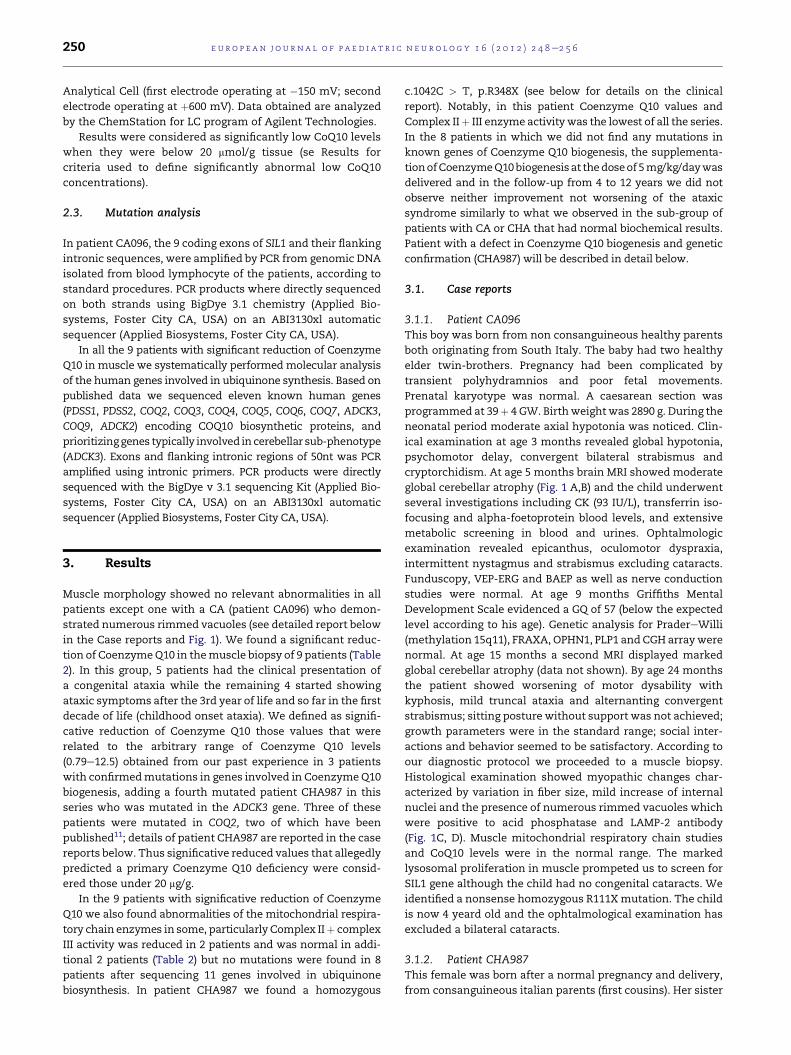
cryptorchidism. At age 5 months brain MRI showed moderate
global cerebellar atrophy (Fig. 1 A,B) and the child underwent
several investigations including CK (93 IU/L), transferrin iso-
focusing and alpha-foetoprotein blood levels, and extensive
metabolic screening in blood and urines. Ophtalmologic
examination revealed epicanthus, oculomotor dyspraxia,
intermittent nystagmus and strabismus excluding cataracts.
Funduscopy, VEP-ERG and BAEP as well as nerve conduction
studies were normal. At age 9 months Griffiths Mental
Development Scale evidenced a GQ of 57 (below the expected
level according to his age). Genetic analysis for PradereWilli
(methylation 15q11), FRAXA, OPHN1, PLP1 and CGH arraywere
normal. At age 15 months a second MRI displayed marked
global cerebellar atrophy (data not shown). By age 24 months
the patient showed worsening of motor dysability with
kyphosis, mild truncal ataxia and alternanting convergent
strabismus; sitting posture without support was not achieved;
growth parameters were in the standard range; social inter-
actions and behavior seemed to be satisfactory. According to
our diagnostic protocol we proceeded to a muscle biopsy.
Histological examination showed myopathic changes char-
acterized by variation in fiber size, mild increase of internal
nuclei and the presence of numerous rimmed vacuoles which
were positive to acid phosphatase and LAMP-2 antibody
(Fig. 1C, D). Muscle mitochondrial respiratory chain studies
and CoQ10 levels were in the normal range. The marked
lysosomal proliferation in muscle prompeted us to screen for
SIL1 gene although the child had no congenital cataracts. We
identified a nonsense homozygous R111Xmutation. The child
is now 4 yeard old and the ophtalmological examination has
excluded a bilateral cataracts.
3.1.2. Patient CHA987This female was born after a normal pregnancy and delivery,
from consanguineous italian parents (first cousins). Her sister

Fig. 1 e AeD. Fig A (T1 weighted, saggittal) and B (inversion recovery, coronal) are neuroimages of patient CA096 with
a Marinesco-Sjogren s. and SIL1 mutations showing the presence of a mild cerebellar atrophy at the age of 5 months. Fig. 1D
shows the morphology of the muscle biopsy performed at age 2 years showing increased immunofluorescence for LAMP-2
in relation to a control sample (Fig. 1C).
e u r o p e a n j o u r n a l o f p a e d i a t r i c n e u r o l o g y 1 6 ( 2 0 1 2 ) 2 4 8e2 5 6 251
was healthy. Psycomotor development was reportedly normal
until the age of 6 years when she started to have partial
seizures for which she was referred to our Hospital. Seizures
were controlled by AEDs but she developed slowly progressive
ataxic syndrome characterized bymild ataxic gait, intentional
tremor, dysmetria and dysarthria. Brain MRI performed at age
7 years showed mild global isolated cerebellar atrophy (Fig. 2
A, B). Later a second MRI at age 13 years, after 5 years of
CoQ10 supplementation, showed increased atrophy which
was limited to the vermis and excluding other brain abnor-
malities (Fig. 2 C, D).Neuropsychological evaluation at age 8
years evidenced a mild cognitive delay. Laboratory tests (CK,
vitamin E, alpha-foetoprotein, immunoglobulin electropho-
resis) and metabolic investigations (transferrin isofocusing,
serum lactate, serum and urine aminoacid chromatography,
urine organic acid chromatography) were normal, as were
neurophysiological examinations (BAEP, SEP, ERG, VEP).
Ophthalmologic examination excluded a retinopathy. At the
age of 8 years the child underwent a left quadriceps muscle
biopsy that did not show any relevant changes, but mito-
chondrial respiratory chain enzymes in muscle extracts
revealed decreased activities for complex II þ III suggesting
a CoQ10 defect. Indeed CoQ10 muscle levels were markedly
reduced (2.9 mg/g) in this patient. We started CoQ10 supple-
mentation (10 mg/kg/d) and within 6 months we observed
clear improvement of cerebellar ataxia. A serial brain MRI at
age 14 years did not reveal any progression of the cerebellar
atrophy compared to the neuroimage performed one year
before. Attempt to stop AEDs at age 11 failed, and now at the
age of 17 the epileptic syndrome is well controlled by AEDs
and CoQ10 supplementation, and the ataxic syndrome is
persistently stable. She is able to walk independently and she
is autonomous in daily life. The girl is attending school with
some support.
Clinical features of this patient, the reduced amounts of
ubiquinone in its muscle and the very low activities of mito-
chondrial complex I þ III and II þ III, prompted us to analyze
ADCK3/COQ8 gene with priority. We found a homozygous
nonsense mutation (c.1042C > T, p.R348X) that was hetero-
zygous in both healthy parents.
4. Discussion
Inherited cerebellar ataxias (ICA) in children are extremely
heterogeneous disorders and ataxia is a frequent and a non-
specific sign in many conditions.12 Most of the autosomal
recessive conditions associated with ataxia are summarized
in Table 1 and were excluded in our patients. Clinical criteria
together with neuroimaging findings are crucial to establish
a preliminary differential diagnosis of these conditions.
Considering age of onset, ICA in children can be divided in 2

Fig. 2 e AeD. Brain MRI of patient CHA987 performed at age 7 (Fig. 2 A,B) and 13 years. In Fig. 2 A (saggittal, T1 weighted) and
2 B (coronal, T2 weighted) the atrophy is very mild while it is clearly evident and prominent at the vermis at a later age in
Fig. 2C (saggital, T1 weighted) and 2 D (coronal, T2 weighted).
e u r o p e a n j o u r n a l o f p a e d i a t r i c n e u r o l o g y 1 6 ( 2 0 1 2 ) 2 4 8e2 5 6252
main groups: 1) congenital ataxia (CA), and 2) childhood onset
ataxia (CHA). CA is characterized by neonatal hypotonia and
developmental delay while patients with CHA show later
onset ataxia. CA is frequently non progressive10 while CHA
most frequently has a progressive course. In addition ICA can
be distinguished in syndromic or non syndromic ataxias.
Furthermore MRI is a valuable complementary tool for
differential diagnosis of ICA and is capable of defining
a possible brain involvement or simply of a cerebellar and
brainstem malformation or a cerebellar atrophy.7
Non syndromic CA or CHA associated to apparently “pure”
cerebellar atrophy are clinical entities in which the genetic
background has been defined in only very few known
diseases.8,13 Currently a specific diagnosis ismostly available in
syndromic ICA of childhood such as ataxia-telangectasia
syndrome, autosomal dominant ataxia type 2, CDG syndrome
for which some clinical and laboratory markers are known.
Following the participation of some of us to a preliminary
collaborative study thatmeasured the levels of CoenzymeQ10
in muscle in a series of patients affected by ICA with onset in
childhood of unknown cause,14 after exclusion of known
causes of ICA we decided to systematically carry out a muscle
biopsy in patients with non syndromic CA or CHA associated
to “pure” cerebellar atrophy in order to verify the impact of the
muscle biopsy examination in the diagnosis of this group of
disorders.
In our series of patients, muscle biopsy led to a definitive
genetic diagnosis in two patients (5.5%) out of 34. One patient
had clinical features of CA (patient A) while the second patient
(patient B) was consistent with the diagnosis of non syn-
dromic CHA.
In patient A with clinical featuires of CA the muscle biopsy
was useful to formulate an early diagnosis of Marinesco-
Sjogren syndrome (MSS). This sporadic patient had an
unusual presentation of MSS lacking cardinal features of
congenital or early-onset cataracts and normal CK levels in
blood. Although cataracts is considered as a pathognomonic
sign of MSS, it may seldom appear later in the course of the
disease.15 Our patients is now 4-years-old and does not show
any sign of bilateral cataracts. Muscular involvement has been
reported since early descriptions of MSS16,17 and myopathic
changes with proliferation of autophagic vacuoles at muscle
biopsy is a constant feature of MSS carrying SIL1 muta-
tions.18,19 In our patients typical muscle changes were detec-
ted as early as 2 years of age and were pivotal for addressing
the diagnosis. The homozygous R111X change found in our
patient has already been reported as recurrentmutation in the
mediterranean population and southern Italy.20

Table 1 e Summary of genetic conditions related to childhood onset autosomal recessive ataxias correlated with thepresence or absence of cerebellar atrophy at neuroimaging.
Autosomal Recessive Ataxia Cerebellaratrophy
Congenital ataxia Cayman ataxia þJoubert syndrome �
Metabolic ataxia Vitamin E deficiency �Abeta-lipoproteinemia �Refsum disease �Late-onset Tay-Sachs þNiemann-Pick C þCDG1a þCerebrotendinous xanthomatosis þNeuronal ceroid lipofuscinoses þ3-methylglutaconic aciduria þMevalonate kinase deficiency þADK3 mutaions and CoQ10 deficiency þLeucodystrophy: L-2-hydroxyglutaric aciduria þ/�Menkes disease þ/�Autosomal recessive mitochondrial ataxias:
AR-CPEO, MIRAS, SANDO, SCAE, AHS, IOSCA, LBSL,
Pyruvate decarboxylase deficiency, PDH deficiency
þ
Friedreich ataxia �DNA repair defects Ataxiatelangectasia (AT) þ
AT-like disorder þAOA1 þAOA2 þSpinocerebellar ataxia with axonal neuropathy þXeroderma pigmentosum þCockayne syndrome þ
Degenerative Spastic ataxia of Charlevoix-Saguenay þMarinesco-Sjogren syndrome þInfantile neuroaxonal dystrophy þLeucodystrophy: CACH syndrome �Hypomyelination: Salla disease, Pelizaeus-Merzbacher disease (PM),
PM-like, leucoencephalopathy with ataxia,
hypodontia and hypomyelination,
þ
Hypomyelination and atrophy of basal ganglia and cerebellum þ
e u r o p e a n j o u r n a l o f p a e d i a t r i c n e u r o l o g y 1 6 ( 2 0 1 2 ) 2 4 8e2 5 6 253
Moreover, with the systematic application of a muscle
biopsy to patients with undefined childhood ataxias with
cerebellar atrophy we were also able to genetically detect
a primary defect of COQ10 deficiency with a homozygous
mutation in ADK3 in a sporadic CHA patient (CHA987). This
homozygous nonsense mutation (c.1042C > T, p.R348X) has
been recently detected in an informative Dutch family21 only
after linkage analysis. The COQ10 levels in muscle were very
low (3.69 mg/g) in this patient besides a normal appearance of
light microscopy and ultrastructural morphology.
Supplementation of Coenzyme Q10 has improved and
probably stabilized ataxia in this patient but from serial neu-
roimaging we did not observe any reversal of cerebellar
atrophy. We have no explanation for this phenomenon at the
moment also because pathogenesis of cerebellar atrophy in
this disorder is currently not known. Additional serial MRIs
are in program to monitor possible improvement of cerebellar
atrophy in this patient. From our experience we can conclude
that CHA due to mutations in ADK3 is a very rare condition
because we have detected only one patient out of 20 with
undetermined CHA. Once again a patient with a ADK3 muta-
tion has the clinical pattern of a CHA rather that CA con-
firming the same clinical presentation that has been reported
so far in this condition.22,23 Thus a clinical clue to suspect
children with ataxia harboring mutations in the ADK3 gene is
that ataxia does not have a congenital onset and conceivably
occurs in the CHA category. Currently, patients with ICA and
primary defect of COQ10 deficiency with mutations in ADK3
can be suspected by determining levels of CoQ10 in a muscle
biopsy or fibroblasts or can be assumed by clinical and neu-
roimaging associated signs, because no other clues are avali-
able. The determination of CoQ10 levels in the muscle biopsy
and or in cultured fibroblasts is a rapid procedure rather that
measuring CoQ10 levels of CoQ10 biogenesis in skin fibro-
blasts that warrants a highly skilled laboratory.24 A needle
biopsy may also be sufficient to measure levels in Coenzyme
Q10 in muscle reducing the more invasive open biopsy.
Moreover we detected 8 additional patients with a CA or
CHA phenotype that showed significant reduction of COQ10
together with a reduction of complex II þ III in the mito-
chondrial respiratory chain enzyme activity only in some
patients. Nonetheless these patients did not show any muta-
tions in the known disease genes (COQ2, ADCK3, PDSS1 and
PDSS2, COQ9) and in additional 6 genes (COQ3, COQ4, COQ5,
COQ6, COQ7, ADCK2) encoding for COQ10 biosynthetic
proteins. The COQ10 muscle levels of these patients were

Table 2 e Summary of patients with either CA or CHA. Legends: CA: congenital ataxia; CHA: childhood onset ataxia; &: published in Diomedi-Camassei et al., J Am SocNephrol 2007, 18: 2773e2780; # the mean and SD value includes the value of patient CHA987. Only abnormal values of mitochondrial respiratory chain enzymes arereported; ND: not done; NL: normal; mt: mitochondrial.
Patients Diagnosis Coenzyme Q10 levels in muscle (mmol/g tissue) Mitochondrial Respiratorychain enzymes
(nmol/min/mg prot.)Mean SD Range
Total 24 Normal controls 37.4 18.5 20e79 Normal ranges
Patients with CA (9) or CHA (16)
ataxia (total 25)
Normal CoQ10 levels 34.6 16 20e77.2 Normal ranges
CA096 CA; Marinesco-Sjogren S. 40 Normal ranges
Patients with CA (5) and CHA (4)
ataxia and significatively
reduced CoQ10 levels (total 9)
13.1 2.6 9.24e17.25
CA982 CA 10.2 Complex II þ III: ND
CHA995 CHA 14.38 Complex II þ III: 0.02
CHA016 CHA 15.1 Complex II þ III: NL
CA028 CA 17.25 Complex II þ III: ND
CHA034 CHA 10 Complex II þ III: ND
CA056 CA 15 Complex II þ III: NL
CA073 CA 14.2 Complex II þ III: ND
CA978 CA 9.24 Complex II þ III: ND
CHA987 CHA; ADK3 mutations 3.69 Complex II þ III: 0.006
Additional patients with
confirmed mutations in genes
of CoQ10 biogenesis
7.345# 6# 0.79e12.5
VA/07 Leigh syndrome 0.79 Other myx enzymes NL Complex II þ III: 0.013
CB/09 Myopathy þ Encephalopathy 12.4 Other mtx enzymes NL Complex II þ III: 0.018
CV/06 Myopathy þ Encephalopathy 12.5 Other mtx enzymes NL Complex II þ III: 0.020
european
journalofpaedia
tric
neurology
16
(2012)248e256
254

e u r o p e a n j o u r n a l o f p a e d i a t r i c n e u r o l o g y 1 6 ( 2 0 1 2 ) 2 4 8e2 5 6 255
clearly low, although most of them had values above 10 mg/g
(Table 2) and complex II þ III enzyme activity was within
normal ranges in some. We found COQ10 muscle levels
around 12 mg/g in two patients with genetically confirmed
mutations in COQ225 so we cannot exclude that some of these
patients may be affected by a primary COQ10 biosynthetic
defect carrying mutations in genes that have not been char-
acterized so far. These 8 patients had Coenzyme Q10 supple-
mentation for several years and we did not observe any
improvement of ataxia that has remained stable, similarly to
the sub-group of our patients series that had normal levels of
Coenzyme Q10 in muscle. Patients with CA or CHA and
significative reduction of Coenzyme Q10 in muscle were also
reported in the first description associating heterogeneous
forms of ataxic syndromes with a COQ10 deficiency in
muscle.14 This sub-group of patients with significative
reduction of Coenzyme Q10 in muscle but without showing
mutations in known genes of Coenzyme Q10 biogenesis
mostly have clinical presentation of a CA rather than CHA.
The muscle biopsy of all patients with significative reduction
of Coenzyme Q10 showed no morphological clue abnormali-
ties, including our patient CHA987 with ADK3 mutations,
although proliferation of lysosomes and autophagic vacuoles
have been reported in the fibroblasts of one patient.25 In
contrast, muscle morphological abnormalities have been
reported in some myopathic forms of secondary CoQ10 defi-
ciency with ETFDH deficiency26 or other encephalomyopathic
forms in which the genetic basis are unknown27e29 showing
mitochondrial proliferation and lipid storage. Moreover we
have described a reduction of SDH staining in the muscle
biopsies of patients with CoQ10 defciency in muscle and
mutations in COQ2.11
Finally, it has been described that some patients with
ataxia may have low levels of COQ10 in muscle but this may
not be related to a primary COQ10 biosynthetic defect as has
been shown in conditions such as patients harboring apra-
taxin mutations.30
In conclusion in our series of patients with ICA, muscle
biopsy led to genetic diagnosis in two patients (5.5%) and gave
helpful indications for therapeutic advise in additional 8
patients that were treated with CoQ10 supplementation.
Following these studies, we think that muscle biopsy is
a valuable diagnostic approach and should be considered in
the panel of investigations to enhance diagnostic chances in
children with early-onset ataxia or genetically undiagnosed
ataxia associated to cerebellar atrophy, after excluding other
known conditions. ADK3 mutations should be suspected in
ICA patients with a clinical presentation of CHA rather that
CA. In these patients there is markedly reduced levels of
CoQ10 in muscle (at least under 10 mg/g in our series) and in
fibroblasts as already reported.31,32 In addition the finding of
a relative reduction of COQ10 inmuscle (levels between 10 and
20 mg/g) in patients which are negative for ADK3 mutations
offers a possible clue for subgrouping conditions of CA and
CHA of undetermined cause. However, increasing knowledge
on the underlying genetic cause of these latter conditions is
necessary to define whether supgrouping ICA patients with
the finding of a relative reduction of CoQ10 in the muscle
biopsy is a useful procedure for any preliminary diagnostic
approach.
Acknowledgments
This research was supported in part by grants from the Italian
Ministry of Health, and the Telethon Foundation Onlus
(Project GGP10225B on Autophagy to EB and GGP08145 on
Joubert syndrome to EB, EMV and GZ).
r e f e r e n c e s
1. Finsterer J. Ataxias with autosomal, X-chromosomal ormaternal inheritance. Can J Neurol Sci 2009;36:409e12.
2. Harding AE. Clinical features and classification of inheritedataxias. In: Harding AE, editor. Hereditary ataxias and relateddisorders. Edinburgh: Churchill-Livingstone; 1984. Adv Neurol.1993; 61: 1e14.
3. Koenig M. Rare forms of autosomal recessiveneurodegenerative ataxia. Semin Pediatr Neurol 2003;10:183e92.
4. De Michele G, Coppola G, Cocozza S, Filla A. A pathogeneticclassification of hereditary ataxias: is the time ripe? J Neurol2004;251:913e22.
5. Palau F, Espinos. Autosomal recessive cerebellar ataxias.Orphanet J Rare Dis 2006;17(1):47.
6. Boddaert N, Desguerre I, Bahi-Buisson N, Romano S,Valayannopoulos V, Saillour Y, Seidenwurm D, et al. Posteriorfossa imaging in 158 children with ataxia. J Neuroradiol 2010;37:220e30.
7. Boltshauser E. Cerebellar hypoplasias. Handb Clin Neurol 2007;87:115e27.
8. Poretti A, Wolf NI, Boltshauser E. Differential diagnosis ofcerebellar atrophy in childhood. Eur J Paediatr Neurol 2008;12:155e67.
9. Zheng XX, Shoffner JM, Voljavec AS, Wallace DC. Evaluationof procedures for assaying oxidative phosphorylation enzymeactivities in mitochondrial myopathy muscle biopsies. ReviewBiochim Biophys Acta 1990;1019:1e10.
10. Pastore A, Giovamberardino GD, Bertini E, Tozzi G, Gaeta LM,Federici G, Piemonte F. Simultaneous determination ofubiquinol and ubiquinone in skeletal muscle of pediatricpatients. Anal Biochem 2005;342:352e5.
11. Diomedi-Camassei F, Di Giandomenico S, Santorelli FM,Caridi G, Piemonte F, Montini G, et al. COQ2 nephropathy:a newly described inherited mitochondriopathy with primaryrenal involvement. J Am Soc Nephrol 2007;18:2773e80.
12. Garcıa-Cazorla A, Wolf NI, Serrano M, Perez-Duenas B,Pineda M, Campistol J, et al. Inborn errors of metabolism andmotor disturbances in children. J Inherit Metab Dis 2009;32:618e29.
13. Institut Cochin, Universite Paris Descartes, CNRS (UMR 8104)Paris, France Zanni G, Bertini E, Bellcross C, Nedelec B,Froyen G, Neuhauser G, et al. X-linked congenital ataxia:a new locus maps to Xq25-q27.1. Am J Med Genet A 2008;146A:593e600.
14. Lamperti C, Naini A, Hirano M, De Vivo DC, Bertini E,Servidei S, et al. Cerebellar ataxia and coenzyme Q10deficiency. Neurology 2003;60:1206e8.
15. Takahata T, Yamada K, Yamada Y, Ono S, Kinoshita A,Matsuzaka T, et al. Novel mutations in the SIL1 gene ina Japanese pedigree with the Marinesco-Sjogren syndrome. JHum Genet 2010;55:142e6.
16. Herva R, von Wendt L, von Wendt G, Saukkonen AL, Leisti J,Dubowitz V. A syndrome with juvenile cataract, cerebellaratrophy, mental retardation and myopathy. Neuropediatrics1987;18:164e9.
e u r o p e a n j o u r n a l o f p a e d i a t r i c n e u r o l o g y 1 6 ( 2 0 1 2 ) 2 4 8e2 5 6256
17. Goto Y, Komiyama A, Tanabe Y, Katafuchi Y, Ohtaki E,Nonaka I. Myopathy in Marinesco-Sjogren syndrome: anultrastructural study. Acta Neuropathol 1990;80:123e8.
18. Senderek J, Krieger M, Stendel C, Bergmann C, Moser M,Breitbach-Faller N, et al. Mutations in SIL1 cause Marinesco-Sjogren syndrome, a cerebellar ataxia with cataract andmyopathy. Nat Genet 2005;37:1312e4.
19. Mahjneh I, Anttonen AK, Somer M, Paetau A, Lehesjoki AE,Somer H, et al. Myopathy is a prominent feature inMarinesco-Sjogren syndrome: a muscle computedtomography study. J Neurol 2006;253:301e6.
20. Annesi G, Aguglia U, Tarantino P, Annesi F, De Marco EV,Civitelli D, et al. SIL1 and SARA2 mutations in Marinesco-Sjogren and chylomicron retention diseases. Clin Genet 2007;71:288e9.
21. Gerards M, van den Bosch B, Calis C, Schoonderwoerd K, vanEngelen K, et al. Nonsense mutations in CABC1/ADCK3 causeprogressive cerebellar ataxia and atrophy. Mitochondrion 2010;10:510e5.
22. Lagier-Tourenne C, Tazir M, Lopez LC, Quinzii CM, AssoumM,Drouot N, et al. ADCK3, an ancestral kinase, is mutated ina form of recessive ataxia associated with coenzyme Q10deficiency. Am J Hum Genet 2008;82:661e72.
23. Mollet J, Delahodde A, Serre V, Chretien D, Schlemmer D,Lombes A, et al. CABC1 gene mutations cause ubiquinonedeficiency with cerebellar ataxia and seizures. Am J Hum Genet2008;82:623e30.
24. Quinzii C, Naini A, Salviati L, Trevisson E, Navas P, Dimauro S,et al. A mutation in para-hydroxybenzoate-polyprenyl
transferase (COQ2) causes primary coenzyme Q10 deficiency.Am J Hum Genet 2006 Feb;78:345e9.
25. Rodrıguez-Hernandez A, Cordero MD, Salviati L, Artuch R,Pineda M, Briones P, et al. Coenzyme Q deficiency triggersmitochondria degradation by mitophagy. Autophagy 2009;5:19e32.
26. Gempel K, Topaloglu H, Talim B, Schneiderat P, Schoser BG,Hans VH, et al. The myopathic form of coenzyme Q10deficiency is caused by mutations in the electron-transferring-flavoprotein dehydrogenase (ETFDH) gene. Brain2007;130:2037e44.
27. Di Giovanni S, Mirabella M, Spinazzola A, Crociani P,Silvestri G, Broccolini A, et al. Coenzyme Q10 reversespathological phenotype and reduces apoptosis in familialCoQ10 deficiency. Neurology 2001;57:515e8.
28. Gironi M, Lamperti C, Nemni R, Moggio M, Comi G, Guerini FR,et al. Late-onset cerebellar ataxia with hypogonadism andmuscle coenzyme Q10 deficiency. Neurology 2004;62:818e20.
29. Sobreira C, Hirano M, Shanske S, Keller RK, Haller RG,Davidson E, et al. Mitochondrial encephalomyopathy withcoenzyme Q10 deficiency. Neurology 1997;48:1238e43.
30. Quinzii CM, Kattah AG, Naini A, Akman HO, Mootha VK,DiMauro S, et al. Coenzyme Q deficiency and cerebellar ataxiaassociated with an aprataxin mutation. Neurology 2005;64:539e41.
31. DiMauro S, Quinzii CM, Hirano M. Mutations in coenzymeQ10 biosynthetic genes. J Clin Invest 2007;117:587e9.
32. Quinzii CM, Lopez LC, Naini A, DiMauro S, Hirano M. HumanCoQ10 deficiencies. Biofactors 2008;32:113e8.


![[Posterior cortical atrophy]](https://static.fdokumen.com/doc/165x107/6331b9d14e01430403005392/posterior-cortical-atrophy.jpg)