The structure and catalytic mechanism of a poly(ADP-ribose) glycohydrolase
13
The structure and catalytic mechanism of a poly(ADP-ribose) glycohydrolase Dea Slade a,b,* , Mark S. Dunstan c,* , Eva Barkauskaite a , Ria Weston a , Pierre Lafite d , Neil Dixon c , Marijan Ahel e , David Leys c , and Ivan Ahel a,1 a Cancer Research UK, Paterson Institute for Cancer Research, University of Manchester, Wilmslow Road, Manchester M20 4BX, UK b Université de Paris-Descartes, Faculté de Médecine, INSERM U1001, 156 rue de Vaugirard, 75015 Paris, France c Manchester Interdisciplinary Biocentre, Princess Street 131, M1 7DN, Manchester, UK d ICOA – UMR CNRS 6005 Université d’Orléans Rue de Chartres, F-45067 Orléans, France e Rudjer Boskovic Institute, Bijenicka 54, HR-10000 Zagreb, Croatia Abstract Posttranslational modification of proteins by poly(ADP-ribosyl)ation regulates many cellular pathways that are critical for genome stability, including DNA repair, chromatin structure, mitosis and apoptosis 1 . Poly(ADP-ribose) (PAR) is composed of repeating ADP-ribose units linked via a unique glycosidic ribose-ribose bond, and is synthesised from NAD by poly(ADP-ribose) polymerases (PARPs) 1,2 . Poly(ADP-ribose) glycohydrolase (PARG) is the only protein capable of specific hydrolysis of the ribose-ribose bonds present in PAR chains; its deficiency leads to cell death 3,4 . Here we show that filamentous fungi and a number of bacteria possess a divergent form of PARG that exhibits all the main characteristics of the human PARG enzyme. We present the first PARG crystal structure (derived from the bacterium Thermomonospora curvata), which reveals that the PARG catalytic domain is a distant member of the ubiquitous ADP-ribose-binding macro domain family 5,6 . High resolution structures of T. curvata PARG in complexes with ADP- ribose and the PARG inhibitor ADP-HPD, complemented by biochemical studies, allow us to propose a model for PAR binding and catalysis by PARG. Our insights into the PARG structure and catalytic mechanism should greatly improve our understanding of how PARG activity controls reversible protein poly(ADP-ribosyl)ation and potentially of how the defects in this regulation link to human disease. While the mechanism and the functional aspects of poly(ADP-ribose) synthesis have been extensively characterised, the present understanding of the PAR-degradation pathways catalysed by PARG is comparatively poor. To date, the catalytic fold and the mechanism of 1 To whom correspondence should be addressed. [email protected] . * These authors contributed equally to this work Author Contributions D.S. performed biochemical and in vivo experiments, prepared proteins for crystallisation, analysed data and wrote the manuscript. M.D. performed structural/biophysical studies and analysed data. E.B. performed biochemical and in vivo experiments; R.W. performed supporting studies. M.A. and N.D. performed LC/MS analyses; P.L. performed molecular modelling studies. I.A. and D.L. wrote the manuscript, designed experiments and analysed data. Supplementary Information is linked to the online version of the paper at www.nature.com/nature. Author Information Reprints and permissions information is available at www.nature.com/reprints. The authors declare no competing financial interests. Atomic coordinates and structure factors have been deposited with the Protein Data Bank (PDB ID codes 3SIG, 3SIH, 3SII and 3SIJ). UKPMC Funders Group Author Manuscript Nature. Author manuscript; available in PMC 2012 March 29. Published in final edited form as: Nature. ; 477(7366): 616–620. doi:10.1038/nature10404. UKPMC Funders Group Author Manuscript UKPMC Funders Group Author Manuscript
-
Upload
independent -
Category
Documents
-
view
1 -
download
0
Transcript of The structure and catalytic mechanism of a poly(ADP-ribose) glycohydrolase

The structure and catalytic mechanism of a poly(ADP-ribose)glycohydrolase
Dea Sladea,b,*, Mark S. Dunstanc,*, Eva Barkauskaitea, Ria Westona, Pierre Lafited, NeilDixonc, Marijan Ahele, David Leysc, and Ivan Ahela,1
aCancer Research UK, Paterson Institute for Cancer Research, University of Manchester,Wilmslow Road, Manchester M20 4BX, UKbUniversité de Paris-Descartes, Faculté de Médecine, INSERM U1001, 156 rue de Vaugirard,75015 Paris, FrancecManchester Interdisciplinary Biocentre, Princess Street 131, M1 7DN, Manchester, UKdICOA – UMR CNRS 6005 Université d’Orléans Rue de Chartres, F-45067 Orléans, FranceeRudjer Boskovic Institute, Bijenicka 54, HR-10000 Zagreb, Croatia
AbstractPosttranslational modification of proteins by poly(ADP-ribosyl)ation regulates many cellularpathways that are critical for genome stability, including DNA repair, chromatin structure, mitosisand apoptosis1. Poly(ADP-ribose) (PAR) is composed of repeating ADP-ribose units linked via aunique glycosidic ribose-ribose bond, and is synthesised from NAD by poly(ADP-ribose)polymerases (PARPs)1,2. Poly(ADP-ribose) glycohydrolase (PARG) is the only protein capable ofspecific hydrolysis of the ribose-ribose bonds present in PAR chains; its deficiency leads to celldeath3,4. Here we show that filamentous fungi and a number of bacteria possess a divergent formof PARG that exhibits all the main characteristics of the human PARG enzyme. We present thefirst PARG crystal structure (derived from the bacterium Thermomonospora curvata), whichreveals that the PARG catalytic domain is a distant member of the ubiquitous ADP-ribose-bindingmacro domain family5,6. High resolution structures of T. curvata PARG in complexes with ADP-ribose and the PARG inhibitor ADP-HPD, complemented by biochemical studies, allow us topropose a model for PAR binding and catalysis by PARG. Our insights into the PARG structureand catalytic mechanism should greatly improve our understanding of how PARG activitycontrols reversible protein poly(ADP-ribosyl)ation and potentially of how the defects in thisregulation link to human disease.
While the mechanism and the functional aspects of poly(ADP-ribose) synthesis have beenextensively characterised, the present understanding of the PAR-degradation pathwayscatalysed by PARG is comparatively poor. To date, the catalytic fold and the mechanism of
1To whom correspondence should be addressed. [email protected] .*These authors contributed equally to this workAuthor Contributions D.S. performed biochemical and in vivo experiments, prepared proteins for crystallisation, analysed data andwrote the manuscript. M.D. performed structural/biophysical studies and analysed data. E.B. performed biochemical and in vivoexperiments; R.W. performed supporting studies. M.A. and N.D. performed LC/MS analyses; P.L. performed molecular modellingstudies. I.A. and D.L. wrote the manuscript, designed experiments and analysed data.Supplementary Information is linked to the online version of the paper at www.nature.com/nature.Author Information Reprints and permissions information is available at www.nature.com/reprints.The authors declare no competing financial interests.Atomic coordinates and structure factors have been deposited with the Protein Data Bank (PDB ID codes 3SIG, 3SIH, 3SII and 3SIJ).
UKPMC Funders GroupAuthor ManuscriptNature. Author manuscript; available in PMC 2012 March 29.
Published in final edited form as:Nature. ; 477(7366): 616–620. doi:10.1038/nature10404.
UKPM
C Funders G
roup Author Manuscript
UKPM
C Funders G
roup Author Manuscript
PARG-mediated hydrolysis remain unknown. Our homology searches revealed that themajority of fungal genomes lack close mammalian/canonical PARG homologues, butinstead possess a divergent PARG-like protein, annotated as DUF2263. DUF2263 proteinsequences contain the PARG signature (GGG-X6-8-QEE)7, which includes the previouslyidentified key residues: two consecutive glutamates7 (Fig. 1a, black asterisk) and a glycine8
(Fig. 1a, grey asterisk). In addition, a tyrosine residue (Fig. 1a, black cross) implicated inbinding to the PARG inhibitor ADP-HPD9 is also present in DUF2263. The DUF2263orthologue is found also in other eukaryotes lacking canonical PARGs such as the rotiferAdineta vaga (Fig. 1b), where it is fused to several copies of the poly(ADP-ribose)-bindingzinc finger10. As both filamentous fungi and A. vaga have functional PARP orthologues andan active poly(ADP-ribose) metabolism11,12, we postulated that DUF2263 was likely to be afunctional PARG in these organisms. Surprisingly, although bacteria are historically thoughtto be devoid of poly(ADP-ribose) metabolism, certain bacterial species possess both PARP(closely related to PARP113) and DUF2263 genes, while some only contain the DUF2263homologue (Fig. 1a, b). To analyse the biochemical activities of the corresponding bacterialPARP and PARG proteins, we purified His6-tagged Herpetosiphon aurantiacus (HA) PARPand DUF2263. HA PARP was able to synthesise poly(ADP-ribose) from NAD, as revealedby antibodies recognising PAR (Fig. 1c) and by the analysis of PAR ladders usingsequencing gels (Supplementary Fig. 1). Analogous to the human PARP1 enzyme, HAPARP was sensitive to the PARP inhibitor KU-0058948 and required DNA for its activation(Fig. 1c). Strikingly, poly(ADP-ribosyl)ation produced by HA PARP is efficientlyhydrolysed by the HA DUF2263 protein, demonstrating that DUF2263 is a functionalPARG (Fig. 1c) and suggesting the presence of functional PAR metabolism in bacteria.
We extended our analyses to other bacterial and fungal PARGs (Fig 1a, b andSupplementary Fig. 2) using established PAR substrates formed by the activity of the well-characterised human PARP1 enzyme. Canonical human and Entamoeba dispar PARGsserved as controls. The in vitro activity of the purified PARG enzymes was evaluated in fiveways: (i) an assay which measures the loss of PAR from PARP1-modified histones; (ii) aWestern-blot analysis of the hydrolysis of PAR from the auto-modified human PARP1 usinganti-PAR antibodies; (iii) an SDS-PAGE analysis of the [32P]-PAR auto-modified humanPARP1; (iv) an analysis of the release of ADP-ribose from PAR either by thin-layerchromatography (TLC), and (v) liquid chromatography-mass spectrometry (LC/MS) (Fig.2a-d, Supplementary Fig. 4). The quantification of PAR hydrolysis by the PARG assayshows that bacterial and fungal PARGs are highly active in hydrolysing PAR in vitro; theircatalytic potency is comparable to the E. dispar and human PARG enzymes (Fig. 2a).Moreover, the mode of action of these proteins and mutant versions on poly(ADP-ribosyl)ated PARP1 shows the same pattern observed for the canonical PARGs: (i) all areinactivated by the mutation of a key glutamate residue (Fig. 2b, d), (ii) and by the treatmentwith the PARG inhibitor ADP-HPD (Fig. 2c), and (iii) they are active on ribose-ribosebonds between the two ADP-ribose units, but they are not efficient on mono(ADP-ribosyl)ated protein substrates with the ADP-ribose unit linked directly to the PARP1 (Fig.2c) or the PARP10 protein (Supplementary Fig. 3). Finally, TLC and LC/MS analysesconfirmed that the main product of DUF2263-type PARGs is ADP-ribose (Fig. 2d andSupplementary Fig. 4). Collectively, these results demonstrate that bacterial and fungalPARG proteins possess genuine PARG activity in vitro.
To test whether bacterial and fungal PARG proteins can act as PARGs in vivo, we used theSaccharomyces cerevisiae system expressing human PARP114. Yeast cells, naturallydeficient in PAR metabolism, can readily express human PARP1, resulting in poly(ADP-ribosyl)ation of various protein substrates14 (Fig. 2e). When divergent PARG proteins wereco-expressed with human PARP1, we observed that the poly(ADP-ribosyl)ation levels in
Slade et al. Page 2
Nature. Author manuscript; available in PMC 2012 March 29.
UKPM
C Funders G
roup Author Manuscript
UKPM
C Funders G
roup Author Manuscript
yeast extracts diminished, indicating an efficient hydrolysis of PAR in vivo (Fig. 2e,Supplementary Fig. 5).
Although the first PARG gene was cloned in 199715, to date there are no structural dataavailable for a PARG enzyme. To gain an insight into the PARG structure and mechanism,we solved the T. curvata PARG ligand-free crystal structure to 1.5 Å in addition to ADP-ribose- and ADP-HPD inhibitor-bound structures (Supplementary Table 1). Comparisonwith available structures reveals that T. curvata PARG consists of an ADP-ribose-bindingmacro domain fold with an N-terminal extension (residues 3-67, Fig. 3c). Similar macrodomain structures are found in the E. coli YmdB protein (1SPV, Z score 16.2, r.m.s.d. 2.1 Åover 161 Cα atoms, 19% identity), an Archaeoglobus fulgidus macro domain protein (2BFQ,Z score 15.6, r.m.s.d. 2.8 Å over 164 Cα atoms, 16% identity)5 and the histone variantMACROH2A1.1 (1ZR3, Z score 15.0, 2.3 Å r.m.s.d. over 157 Cα atoms, 12% identity)16.The diphosphate-binding loop that flanks one side of the ADP-ribose binding cavity ishighly conserved between PARG and other macro domain structures (in magenta, Fig. 3a,c). In contrast, the opposite side of the PARG ADP-ribose binding cavity is lined by astretch of amino acids corresponding to the PARG-specific GGG-X6–8-QEE signaturesequence7 (in yellow, Fig. 3a-c). The structural alignment reveals this PARG-specific loop isinserted into the macro domain fold to accommodate the Glu115 side chain that projects intothe PARG active site (Fig. 3c, d). Due to the PARG-specific loop, it appears that onlyPARGs but not other macro domain proteins can hydrolyse PAR (Supplementary Fig. 6).
The ligand-PARG complex structures reveal that ADP-α-ribose occupies a position similarto that observed in other ADP-ribose-macro domain complex structures5 (Fig. 3d andSupplementary Fig. 7). A minor rearrangement can be observed for Val226-Phe227 uponADP-ribose binding. The Phe227 side chain is in close contact with the ribose” moiety ofADP-ribose, ensuring a close distance between the ribose” O4 and one of the phosphategroups within the constraints of the active site. The ribose” moiety also forms hydrogenbonds with Glu114 (via the 2′-OH group) and Glu115 (via the 1′-OH group) (Fig. 3d). Withthe exception of water-mediated polar contacts, few direct contacts can be observed with theadenosine moiety. In contrast to the ribose” moiety, the adenosine ribose’ moiety issignificantly less accessible. The presence of any substituents on the ribose’ 2′-OH group(such as another ADP-ribose) would require a drastic reorientation of the PARG C-terminalα-helix (see Fig. 4a) with concomitant exposure of the hydrophobic core of the macrodomain. This suggests that bacterial PARG binds the PAR terminus and acts as an exo-glycohydrolase.
The steric restraints imposed by the PARG active site form a basis for the modelling of anα(1″→2′) O-glycosidic linkage with an additional ADP-ribose group to provide furtherinsight into PAR binding (Fig. 4a, b). Our models suggest that the (n-1) adenine moiety ispartially enclosed by Ala110, Ala112 and Val226 while the (n-1) ribose’ is in close contactwith Ser98 and Gly104 (Fig. 4b). However, MD simulations suggest that the enzyme lacksany obvious binding sites for the (n-1) phosphate and ribose” moieties, as exemplified bysignificant conformational freedom of the latter with respect to the terminal ADP-ribose andthe associated (n-1) ribose’. This suggests that bacterial PARG does not specifically bindadditional PAR elements.
The T. curvata PARG complex with ADP-ribose (Fig. 3d) and the corresponding PAR-PARG model (Fig. 4b) allow us to propose a mechanism for PARG catalysis (Fig. 4c).Binding of the PAR terminus positions the key ribose-ribose O-glycosidic linkage in directhydrogen bonding contact with Glu115, while constraining the conformation of the terminalribose”. Formation of a putative oxocarbenium intermediate is supported by the protonationof the (n-1) PAR ribose’ 2′-OH leaving group via Glu115, and by the stabilization of the
Slade et al. Page 3
Nature. Author manuscript; available in PMC 2012 March 29.
UKPM
C Funders G
roup Author Manuscript
UKPM
C Funders G
roup Author Manuscript
positively charged oxocarbenium through close proximity with the terminal diphosphategroup. A water molecule is ideally positioned to attack the oxocarbenium intermediate,activated through concomitant deprotonation by Glu115. This leads to the release of ADP-β-ribose” and (n-1) PAR.
This mechanism is supported by mutagenesis and binding studies. Although mutations ofboth Glu114 and Glu115 render the enzyme inactive without disruption to the overall PARGfold (Fig. 4d, Supplementary Fig. 8), ITC ADP-ribose binding studies reveal that Glu115Adoes not affect binding, while Glu114A leads to an approximate 10-fold decrease in bindingaffinity (Supplementary Fig. 9). This supports the notion that the role of Glu114 is confinedto substrate binding/orientation as opposed to the acid-base catalysis proposed for Glu115.The mutation of Phe227 implicated in positioning the terminal ribose” also renders theenzyme inactive (Fig. 4d). Mutations of Ser98 and Val226, implicated by our model inbinding the (n-1) ADP-ribose, greatly diminish T. curvata PARG activity (Fig. 4d).Importantly, mutations of the corresponding catalytic residues in the human PARG have asimilar effect on the enzymatic activity, which suggests a universal catalytic mechanism forbacterial and mammalian PARGs (Fig. 4d).
These data provide the first insight into PARG structure and mechanism, and improve ourunderstanding of the biochemical strategies regulating reversible poly(ADP-ribosyl)ation.Our results define existence of bacterial PAR metabolism and show that PARG is a newmember of the widespread macro domain protein family5,6, which is known to be implicatedin genome stability and regulated by poly(ADP-ribosyl)ation17-19. Our data confirm thatPARGs are evolutionary unrelated to ARH3 proteins, another class of enzymes with theability to cleave PAR20,21 (Supplementary Information). We believe that our findings willprovide the groundwork for future studies that might ultimately lead to the development ofsmall, cell-permeable PARG inhibitors and the potential to manipulate physiology of healthand disease by interfering with PAR metabolism.
METHODS SUMMARYProteins with His6-tags were purified from E. coli. A colorimetric PARG Assay Kit(Trevigen) was used to measure the loss of biotinylated PAR from histones. Auto-modifiedpoly(ADP-ribosyl)ated human and bacterial PARP substrates were made by incubation withNAD10. In vivo activity of PARGs was tested in a yeast system with inducible expression ofhuman PARP1. The T. curvata PARG structure was solved by means of single-wavelengthanomalous diffraction on Pt-soaked crystals. Ligand-enzyme complexes were obtained bythe soaking of PARG crystals. The PAR-PARG model was created using MD simulations ofthe ADP-α-ribose dimer in complex with PARG.
METHODSPlasmids and proteins
Bacterial and fungal PARG proteins and the E. dispar PARG were expressed from thepET28a vector (Novagen). Human PARG with an N-terminal truncation (Δ1-455) wasexpressed from the pColdTF vector (Takara). The A. fumigatus Af293 gene(AFUA_4G11940), the D. radiodurans R1 ATCC13939 gene (DR_B0099) and the E.dispar SAW760 gene (EDI_110590) were amplified from genomic DNA, the humanPARP1 and PARG gene (Q86W56) were amplified from HeLa cDNA, while the T. curvataDSM 43183 gene (Tcur_1721) and the A. variabilis ATCC 29413 gene (Ava_4013) weresynthesised according to the database sequence (GenScript USA Inc.). The H. aurantiacusATCC 23779 PARG (Haur_1618) and PARP (Haur_4763) genes were amplified fromgenomic DNA extracted from a dried culture purchased from DSMZ. All proteins bear an
Slade et al. Page 4
Nature. Author manuscript; available in PMC 2012 March 29.
UKPM
C Funders G
roup Author Manuscript
UKPM
C Funders G
roup Author Manuscript

N-terminal his tag with the exception of the A. variabilis PARG, which has a C-terminal histag. Mutations were introduced using the QuickChange II site-directed mutagenesis kit(Stratagene). Proteins were expressed in E. coli Rosetta2(DE3) cells (Novagen).Recombinant proteins were purified on Ni-NTA beads according to standard procedure. Forcrystallisation studies, the T. curvata PARG was purified by FPLC on a HisTrap HP columnfollowed by Superdex 200 (GE Healthcare). The catalytic fragment of human PARP10(amino acids 818–1025) was expressed as a GST-tagged protein and purified on GlutathioneSepharose columns.
PARP activity assayPARP auto-modification activity of the recombinant H. aurantiacus (HA) PARP wasassayed in a 30-minute room-temperature reaction containing 100 nM HA PARP, 200 μMNAD (Trevigen), 50 mM Tris pH 7.5 and 50 mM NaCl with or without activated DNA(Trevigen), and with or without the PARP inhibitor KU-0058948 (10 μM). After thereactions were stopped with the PARP inhibitor, 500 nM HA PARG and its catalyticglutamate mutants were added to the reaction for 30 min. The reactions were analysed byWestern blotting with rabbit anti-PAR antibodies as described under ‘PARG activityassays’.
PARG activity assaysPARG activity of recombinant proteins was quantified with a colorimetric PARG assay kit(Trevigen), which measured the loss of biotinylated PAR from histones generated byPARP1. 3 nM PARG enzymes were used for this assay. For Western-blot analysis of PARGactivity, poly(ADP-ribose) was synthesised by the automodification of PARP1 in a reactionmixture containing 2 units of PARP1 (Trevigen), 200 μM NAD (Trevigen), activated DNA(Trevigen), 50 mM Tris pH 7.5 and 50 mM NaCl at room temperature. Reactions werestopped after 30 min by the addition of the PARP inhibitor KU-0058948. 500 nM PARGswere added to the reaction and incubated for another 30 min. The reactions were run on4-12% SDS-PAGE gels and blotted onto a nitrocellulose membrane. PAR hydrolysis wasdetected by rabbit polyclonal anti-PAR antibodies (1:1000; Trevigen). Western blots wereanalysed densitometrically by GeneTools (SynGene). For a direct visualisation of PARGactivity via SDS-PAGE, [32P]-labelled NAD (GE Healthcare) was added to the abovereaction to generate radioactively labelled poly(ADP-ribosyl)ated or mono(ADP-ribosyl)ated PARP substrates. 500 nM PARGs were subsequently added with or without thePARG inhibitor ADP-HPD (50 μM). The reaction products were analysed by SDS-PAGEand visualised by autoradiography. For thin layer chromatography (TLC) analysis, thereactions were stopped by the addition of 50 mM ADP-ribose, spotted ontopolyethyleneimine (PEI)-cellulose plates (Macherey-Nagel, Polygram CEL 300 PEI/UV254)and developed in 0.15 M LiCl and 0.15 M formic acid. Dried plates were exposed on X-rayfilm or visualised by UV254 shadowing.
Expression of heterologous proteins in S. cerevisiaePlasmids used to introduce human PARP1, A. fumigatus PARG and D. radiodurans PARGinto S. cerevisiae were created via the Gateway (Invitrogen) site-specific recombinationsystem using yeast-specific destination vectors (Addgene)22. hPARP1 was introduced intopAG303GAL-ccdB (HIS3), while AF PARG and DR PARG were introduced intopAG423GAL-ccdB (URA3). The host strain W303-1a (MATa, ade2-1, ura3-1, his3-11,15,trp1-1, leu2-3,112, can1-100) was grown in rich YPD medium at 30°C. The strain was firsttransformed with the hPARP1-harboring plasmid resulting in his+ transformants withchromosomally integrated hPARP1. Subsequent transformation with the PARG-harboringplasmids yielded his+ura+ transformants with episomally maintained PARG. To induce theexpression of PARP1 and PARG from the GAL1 promoter, the strains were grown overnight
Slade et al. Page 5
Nature. Author manuscript; available in PMC 2012 March 29.
UKPM
C Funders G
roup Author Manuscript
UKPM
C Funders G
roup Author Manuscript

in minimal SC medium with 2% glucose. The overnight cultures were washed, resuspendedin SC+2% raffinose at 7×10⁶ cells/ml and grown for one cell generation. Cells werecentrifuged before resuspending in SC+2% galactose medium and grown overnight toinduce the expression of hPARP1 and PARG from the GAL1 promoter. Yeast proteinextracts were analysed by immunoblotting. Rabbit polyclonal anti-PAR antibody (Trevigen)and rabbit anti-PARP1 antibody (Abcam) were used at 1:1000 dilution, while ratmonoclonal anti-alpha-tubulin antibody (Abcam) was used at 1:5000 dilution. Rabbit anti-DRPARG and anti-AFPARG polyclonal antibodies were raised against the respective full-length proteins purified from E. coli (Eurogentec) and applied at 1:1000 dilution.
Crystallisation, refinement and model buildingInitial T. curvata PARG crystallisation conditions were identified using the JCSG+ matrixscreen (Molecular dimensions). Crystals suitable for diffraction experiments were obtainedby sitting drop vapour diffusion in 4 μL drops containing equal volumes of protein and asolution containing 10% PEG 6K and 0.1 M Bicine buffer (pH 9.0). The crystals werederivatised by soaking with the same solution supplemented with 1 mM potassiumtetracyanoplatinate (II) trihydrate for 10 minutes. For the ADP-ribose and ADP-HPDcomplexes, native crystals were soaked with 1 mM ligand for 1-2 hrs. The crystals werecryoprotected with the addition of 15% PEG 200 to the mother liquor and flash cooled inliquid nitrogen. Data was collected on beamline I04 at the Diamond Light Source Facilityand reduced and scaled with the X-ray Detector Software suite (XDS)23.
The ligand-free PARG crystal structure was determined by single-wavelength anomalousdiffraction on a Pt-soaked crystal using Solve and Resolve as implemented in Phenix24. Theinitial electron density map was of sufficient quality for Resolve to build 70% of the model.Further automated model building with ARPwARP25 successfully constructed the entiremodel, with the exception of a small loop region incorporating residues 58-66, for which nocorresponding density could be observed. The model was completed by iterative cycles ofmanual model building and real space refinement using the program Coot26 andcrystallographic refinement using Phenix.refine24. Validation of the structure was performedwith Molprobity. The processing, phasing and final refinement statistics are presented inSupplementary Table 1.
Isothermal Titration CalorimetryThe thermodynamic ligand binding properties of wild-type T. curvata and two mutantproteins (E114A and E115A) were measured using a VP-ITC microcalorimeter. Protein andligand concentrations were 20 μM and 200 μM respectively in 50 mM Tris (pH 7.5), 100mM NaCl. Titration curves were fitted using a non-linear least squares method in MicrocalOrigin software. A model that was indicative of a single binding site was found to give thebest fit in each case; this model was used to obtain the thermodynamic parameters shown inSupplementary Fig. 9.
Thermal shift assayIn each 50 μL reaction 15 μL of 300× Sypro Orange (Sigma), 2 μL of water and 33 μL ofprotein (4 mg/ml) in 25 mm Tris pH 7.5, 50 mM NaCL, 10% glycerol and 1 mM DTT wereadded to the wells of a 96-well thin-wall PCR plate (Bio-Rad). For thermal shift assays inthe presence of ADP-ribose, 2 μL of 25 mM ADP-ribose was added instead of water.Samples were placed in a CFX96 Real Time PCR thermal cycler (Bio-Rad) and slowlyheated from 15 to 95 °C with sample fluorescence recorded every 0.2 °C. Fluorescence wasmonitored at 575 nm (emission), using 490 nm for excitation.
Slade et al. Page 6
Nature. Author manuscript; available in PMC 2012 March 29.
UKPM
C Funders G
roup Author Manuscript
UKPM
C Funders G
roup Author Manuscript
Analysis of poly(ADP-ribose) chains by sequencing gels[32P]-labelled poly(ADP-ribose) chains produced by PARPs were resolved in 10%sequencing gels in TBE buffer. Reactions were stopped by the addition of 600uL 20% ice-cold trichloroacetic acid (TCA). The precipitated material was washed twice with 5% TCA.The pellet was then incubated with 70 μL of 10 mM Tris and 1mM EDTA buffer (pH 12)for 2 hours at 60 oC to detach PAR chains. PAR was phenol-chloroform extracted, dried andresuspended in 2 mM EDTA-formamide buffer before loading. The products werevisualised by autoradiography.
Computational simulationsNAMD software1 was used to perform all molecular dynamics (MD) simulations of theADP-α-ribose dimer in complex with PARG. The topology and parameters files for theligand were obtained using the Antechamber program2 and AM1-BCC charges3. Theadditional ADP-α-ribose monomer was added to the ADP- α -ribose bound to PARG bycreating an α (1″→2′) O-glycosidic bond. Five conformers of the added monomer weretaken as starting points for MD simulations. The PARG:ADP-α-ribose dimer complexmodel was then immersed in a periodic water box (TIP3), as half of the ligand was incontact with the bulk solvent. Then, for each conformer, several simulations (0.5 ns) wererun with variable parameters (200 K or 310 K, peptidic backbone fixed, restrained or free).During these MD simulations, snapshots (100 in total for clarity) were periodically extractedand energy-minimized, to represent the conformational flexibility of the ADP-α-ribosedimer.
Analyses by ultrahigh-performance liquid chromatography (UHPLC) coupled to quadruple-time-of-flight mass spectrometry (QTOFMS)
The poly(ADP-ribosyl)ated PARP1 was treated with T. curvata PARG, and the mixture wasfiltered using centricons (30 kD cutoff). The analysis of the filtrate was performed using amodified procedure by Coulier et al.4, which employed ion-pair chromatography for theseparation of nucleotides and negative electrospray ionization for mass spectrometricdetection. All analyses were performed using a Waters Acquity UPLC system (WatersCorp., Milford, MA, USA), equipped with a binary solvent delivery system andautosampler. The chromatographic separations employed a column (50 mm × 2.1 mm) filledwith a 1.7 μm BEH C18 stationary phase (Waters Corp., Milford, MA, USA). Binarygradients at a flow rate of 0.4 mL/min were applied for the elution. The eluent A was watercontaining 5 mmol/L of pentylamine, while the pH value was adjusted to 6.5 using aceticacid. A fast elution gradient was applied, which allowed an efficient separation of NAD+and ADP-ribose within 5 min. The gradient started at 2 % B and then the percentage of Blinearly increased to 25 % in 5 min.
The mass spectrometry was performed on a QTOF Premier instrument (Waters Micromass,Manchester, UK) using an orthogonal Z-spray-electrospray interface. The drying gas and thenebulizing gas was nitrogen. The desolvation gas flow was set to 700 L/h at a temperature of300 °C. The cone gas flow was adjusted to 25 L/h, and the source temperature to 120 °C.The capillary and cone voltages were 3200 V and 30 V respectively. The instrument wasoperated in V mode with TOFMS data being collected between m/z 100–1000, applying acollision energy of 4 eV. All spectra were recorded using the extended dynamic range(DRE) option in order to correct for possible peak saturations, the data were collected in thecentroid mode with a scan time of 0.08 s and interscan time of 0.02 s. In order to ensuremaximum accuracy and reproducibility of the system, all acquisitions were carried out usingan independent reference spray via the lock spray interface. Leucine enkephalin was appliedas a lock mass in negative ionization mode (m/z 554.2615).
Slade et al. Page 7
Nature. Author manuscript; available in PMC 2012 March 29.
UKPM
C Funders G
roup Author Manuscript
UKPM
C Funders G
roup Author Manuscript

The chromatograms, recorded in the total ion current (TIC) mode, were systematicallyexamined by manually generating mass spectra of each visible individual peak using thebackground-subtraction option and searching for target components expected to occur in themixture, in particular ADP-ribose and NAD+, using extracted ion chromatograms. Thetarget analyses were based on the accurate mass feature of the instrument, applying a masswindow of 50 mDa. Elemental composition of the selected ions was calculated using theMassLynx software incorporated in the instrument. The main criterion for selecting theelements to be included in search parameters for the calculation of the molecular formulawas the elemental composition of the presumed parent compounds. According to theinstrument specifications, the acceptable deviation from the theoretical m/z values was set at5 mDa.
Supplementary methods references27. Phillips JC, et al. Scalable molecular dynamics with NAMD. J Comput Chem. 2005; 26:1781–802.
[PubMed: 16222654]28. Cornell WD, et al. A Second Generation Force Field for the Simulation of Proteins, Nucleic Acids,
and Organic Molecules. J Am Chem Soc. 1995; 117:5179–5197.29. Jakalian A, Jack DB, Bayly CI. Fast, efficient generation of high-quality atomic charges. AM1-
BCC model: II. Parameterization and validation. J Comput Chem. 2002; 23:1623–41. [PubMed:12395429]
30. Coulier L, et al. Simultaneous quantitative analysis of metabolites using ion-pair liquidchromatography-electrospray ionization mass spectrometry. Anal Chem. 2006; 78:6573–82.[PubMed: 16970336]
Supplementary MaterialRefer to Web version on PubMed Central for supplementary material.
AcknowledgmentsWe thank G. Clark for genomic DNA from E. dispar, G. Smith for PARP inhibitor, M. Rossi for purified proteins,and R. Thorough for editing English. We thank D. Ahel, A. Jordan, D. Ogilvie, S. Terzic, D. Neuhaus and S.Eustermann for helpful discussions. We are grateful to B. Lüscher for the gift of PARP10 expression plasmid, andK. Labib and the members of his lab for advice with yeast work. This work was funded by Cancer Research UK.D.S. holds an AXA Research Fund post-doctoral fellowship. D.L. is a Royal Society University Research Fellow.Access to Diamond beamlines is gratefully acknowledged.
References1. Hakme A, Wong HK, Dantzer F, Schreiber V. The expanding field of poly(ADP-ribosyl)ation
reactions. ‘Protein Modifications: Beyond the Usual Suspects’ Review Series. EMBO Rep. 2008;9:1094–100. [PubMed: 18927583]
2. D’Amours D, Desnoyers S, D’Silva I, Poirier GG. Poly(ADP-ribosyl)ation reactions in theregulation of nuclear functions. Biochem J. 1999; 342:249–68. [PubMed: 10455009]
3. Koh DW, et al. Failure to degrade poly(ADP-ribose) causes increased sensitivity to cytotoxicity andearly embryonic lethality. Proc Natl Acad Sci U S A. 2004; 101:17699–704. [PubMed: 15591342]
4. Hanai S, et al. Loss of poly(ADP-ribose) glycohydrolase causes progressive neurodegeneration inDrosophila melanogaster. Proc Natl Acad Sci U S A. 2004; 101:82–6. [PubMed: 14676324]
5. Karras GI, et al. The macro domain is an ADP-ribose binding module. Embo J. 2005; 24:1911–20.[PubMed: 15902274]
6. Till S, Ladurner AG. Sensing NAD metabolites through macro domains. Front Biosci. 2009;14:3246–58. [PubMed: 19273270]
7. Patel CN, Koh DW, Jacobson MK, Oliveira MA. Identification of three critical acidic residues ofpoly(ADP-ribose) glycohydrolase involved in catalysis: determining the PARG catalytic domain.Biochem J. 2005; 388:493–500. [PubMed: 15658938]
Slade et al. Page 8
Nature. Author manuscript; available in PMC 2012 March 29.
UKPM
C Funders G
roup Author Manuscript
UKPM
C Funders G
roup Author Manuscript
8. Panda S, Poirier GG, Kay SA. tej defines a role for poly(ADP-ribosyl)ation in establishing periodlength of the arabidopsis circadian oscillator. Dev Cell. 2002; 3:51–61. [PubMed: 12110167]
9. Koh DW, et al. Identification of an inhibitor binding site of poly(ADP-ribose) glycohydrolase.Biochemistry. 2003; 42:4855–63. [PubMed: 12718526]
10. Ahel I, et al. Poly(ADP-ribose)-binding zinc finger motifs in DNA repair/checkpoint proteins.Nature. 2008; 451:81–5. [PubMed: 18172500]
11. Kothe GO, Kitamura M, Masutani M, Selker EU, Inoue H. PARP is involved in replicative agingin Neurospora crassa. Fungal Genet Biol. 2010; 47:297–309. [PubMed: 20045739]
12. Semighini CP, Savoldi M, Goldman GH, Harris SD. Functional characterization of the putativeAspergillus nidulans poly(ADP-ribose) polymerase homolog PrpA. Genetics. 2006; 173:87–98.[PubMed: 16510786]
13. Hassa PO, Hottiger MO. The diverse biological roles of mammalian PARPS, a small but powerfulfamily of poly-ADP-ribose polymerases. Front Biosci. 2008; 13:3046–82. [PubMed: 17981777]
14. Tao Z, Gao P, Liu HW. Studies of the expression of human poly(ADP-ribose) polymerase-1 inSaccharomyces cerevisiae and identification of PARP-1 substrates by yeast proteome microarrayscreening. Biochemistry. 2009; 48:11745–54. [PubMed: 19877712]
15. Lin W, Ame JC, Aboul-Ela N, Jacobson EL, Jacobson MK. Isolation and characterization of thecDNA encoding bovine poly(ADP-ribose) glycohydrolase. J Biol Chem. 1997; 272:11895–901.[PubMed: 9115250]
16. Kustatscher G, Hothorn M, Pugieux C, Scheffzek K, Ladurner AG. Splicing regulates NADmetabolite binding to histone macroH2A. Nat Struct Mol Biol. 2005; 12:624–5. [PubMed:15965484]
17. Ahel D, et al. Poly(ADP-ribose)-dependent regulation of DNA repair by the chromatin remodelingenzyme ALC1. Science. 2009; 325:1240–3. [PubMed: 19661379]
18. Gottschalk AJ, et al. Poly(ADP-ribosyl)ation directs recruitment and activation of an ATP-dependent chromatin remodeler. Proc Natl Acad Sci U S A. 2009; 106:13770–4. [PubMed:19666485]
19. Timinszky G, et al. A macrodomain-containing histone rearranges chromatin upon sensing PARP1activation. Nat Struct Mol Biol. 2009; 16:923–9. [PubMed: 19680243]
20. Mueller-Dieckmann C, et al. The structure of human ADP-ribosylhydrolase 3 (ARH3) providesinsights into the reversibility of protein ADP-ribosylation. Proc Natl Acad Sci U S A. 2006;103:15026–31. [PubMed: 17015823]
21. Oka S, Kato J, Moss J. Identification and characterization of a mammalian 39-kDa poly(ADP-ribose) glycohydrolase. J Biol Chem. 2006; 281:705–13. [PubMed: 16278211]
22. Alberti S, Gitler AD, Lindquist S. A suite of Gateway cloning vectors for high-throughput geneticanalysis in Saccharomyces cerevisiae. Yeast. 2007; 24:913–9. [PubMed: 17583893]
23. Kabsch W. Evaluation of single-crystal X-ray diffraction data from a position-sensitive detector. JAppl Cryst. 1988; 21:916–24.
24. Adams PD, et al. PHENIX: a comprehensive Python-based system for macromolecular structuresolution. Acta Crystallogr D Biol Crystallogr. 2010; 66:213–21. [PubMed: 20124702]
25. Perrakis A, Morris R, Lamzin VS. Automated protein model building combined with iterativestructure refinement. Nat Struct Biol. 1999; 6:458–63. [PubMed: 10331874]
26. Emsley P, Cowtan K. Coot: model-building tools for molecular graphics. Acta Crystallogr D BiolCrystallogr. 2004; 60:2126–32. [PubMed: 15572765]
Slade et al. Page 9
Nature. Author manuscript; available in PMC 2012 March 29.
UKPM
C Funders G
roup Author Manuscript
UKPM
C Funders G
roup Author Manuscript

Figure 1. Phylogeny and functional relationship between DUF2263 and canonical-type PARGsa, Multiple sequence alignment of different DUF2263 and PARG proteins fromThermomonospora curvata (Tcu), Herpetosiphon aurantiacus (Hau), Deinococcusradiodurans (Dra), Aspergillus fumigatus (Afu), Homo sapiens (Hsa), Bos taurus (Bta) andEntamoeba dispar (Edi). The two catalytic glutamates, a conserved glycine and tyrosine aremarked with black asterisks, grey asterisk and black cross respectively. Secondary structureelements from the Tcu PARG structure are indicated above. b, YmdB-rooted phylogenetictree of PARGs implied by the neighbour-joining method. Organisms devoid of PARP aremarked in grey. c, H. aurantiacus (HA) PARP and PARG enzymes are active as shown byWestern blotting with anti-PAR antibodies.
Slade et al. Page 10
Nature. Author manuscript; available in PMC 2012 March 29.
UKPM
C Funders G
roup Author Manuscript
UKPM
C Funders G
roup Author Manuscript
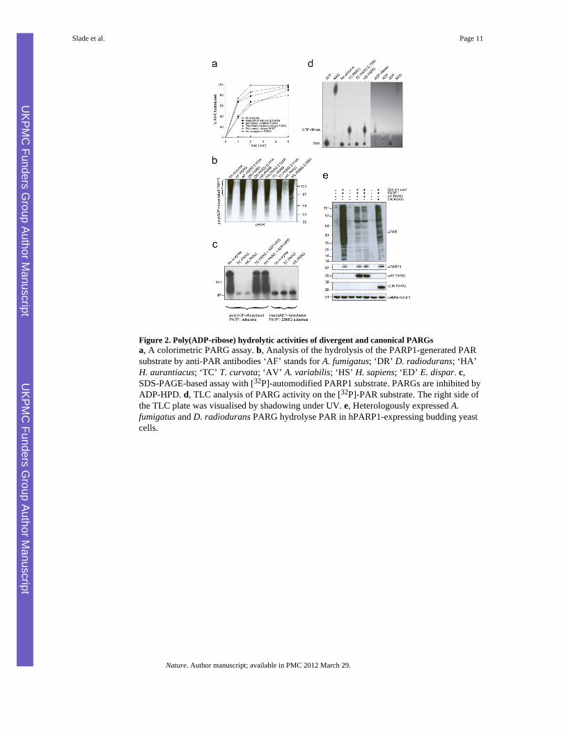
Figure 2. Poly(ADP-ribose) hydrolytic activities of divergent and canonical PARGsa, A colorimetric PARG assay. b, Analysis of the hydrolysis of the PARP1-generated PARsubstrate by anti-PAR antibodies ‘AF’ stands for A. fumigatus; ‘DR’ D. radiodurans; ‘HA’H. aurantiacus; ‘TC’ T. curvata; ‘AV’ A. variabilis; ‘HS’ H. sapiens; ‘ED’ E. dispar. c,SDS-PAGE-based assay with [32P]-automodified PARP1 substrate. PARGs are inhibited byADP-HPD. d, TLC analysis of PARG activity on the [32P]-PAR substrate. The right side ofthe TLC plate was visualised by shadowing under UV. e, Heterologously expressed A.fumigatus and D. radiodurans PARG hydrolyse PAR in hPARP1-expressing budding yeastcells.
Slade et al. Page 11
Nature. Author manuscript; available in PMC 2012 March 29.
UKPM
C Funders G
roup Author Manuscript
UKPM
C Funders G
roup Author Manuscript

Figure 3. T. curvata PARG crystal structure in complex with ADP-ribosea, b, Overall fold of PARG with α-helices depicted in red, β-sheets in blue and bound ADP-ribose in green spheres. The PARG-specific catalytic loop is shown in yellow, and thediphosphate-binding loop in magenta. c, Overlay of the T. curvata PARG structure with arepresentative macro domain structure (PDB code 2BFQ, in cyan). The PARG-specific N-terminal extension and additional PARG structural elements are highlighted in red.Structural features that are similar to 2BFQ and other macro domains are shown in gray. d,Detailed view of ADP-ribose bound in the PARG active site. ADP-ribose is shown with thecorresponding 2Fo-Fc omit-map density contoured at 1.2 σ in blue. Key active site residuesare represented in atom coloured sticks with hydrogen bonds indicated by black dotted lines.
Slade et al. Page 12
Nature. Author manuscript; available in PMC 2012 March 29.
UKPM
C Funders G
roup Author Manuscript
UKPM
C Funders G
roup Author Manuscript
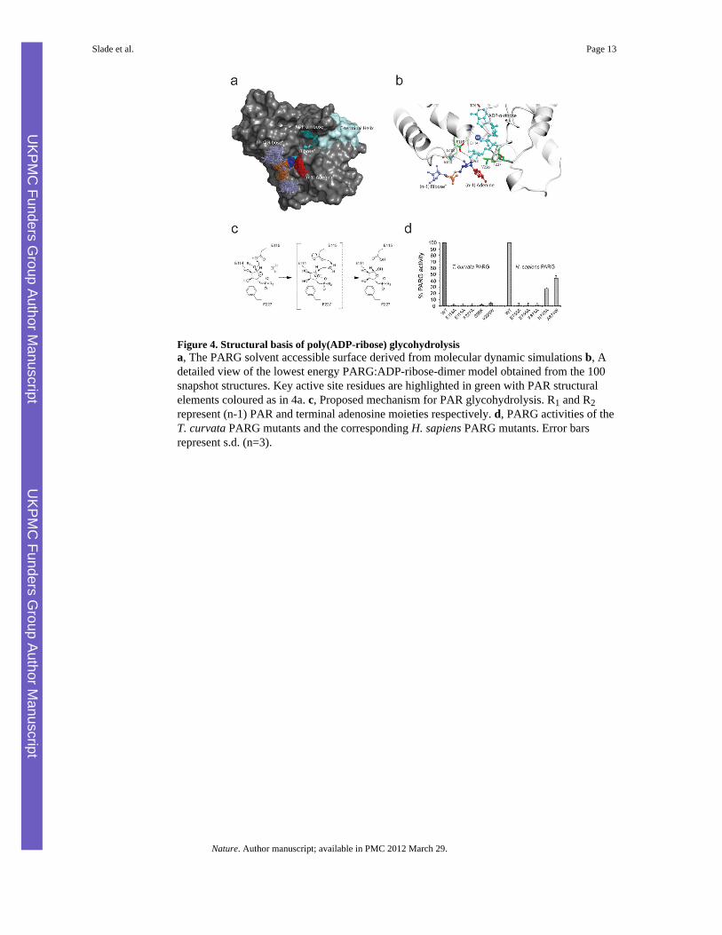
Figure 4. Structural basis of poly(ADP-ribose) glycohydrolysisa, The PARG solvent accessible surface derived from molecular dynamic simulations b, Adetailed view of the lowest energy PARG:ADP-ribose-dimer model obtained from the 100snapshot structures. Key active site residues are highlighted in green with PAR structuralelements coloured as in 4a. c, Proposed mechanism for PAR glycohydrolysis. R1 and R2represent (n-1) PAR and terminal adenosine moieties respectively. d, PARG activities of theT. curvata PARG mutants and the corresponding H. sapiens PARG mutants. Error barsrepresent s.d. (n=3).
Slade et al. Page 13
Nature. Author manuscript; available in PMC 2012 March 29.
UKPM
C Funders G
roup Author Manuscript
UKPM
C Funders G
roup Author Manuscript


![Synthesis, [18F] radiolabeling, and evaluation of poly (ADP-ribose) polymerase-1 (PARP-1) inhibitors for in vivo imaging of PARP-1 using positron emission tomography](https://static.fdokumen.com/doc/165x107/6335c3a302a8c1a4ec01e906/synthesis-18f-radiolabeling-and-evaluation-of-poly-adp-ribose-polymerase-1.jpg)