The structural molecular biology network of the State of São Paulo, Brazil
13
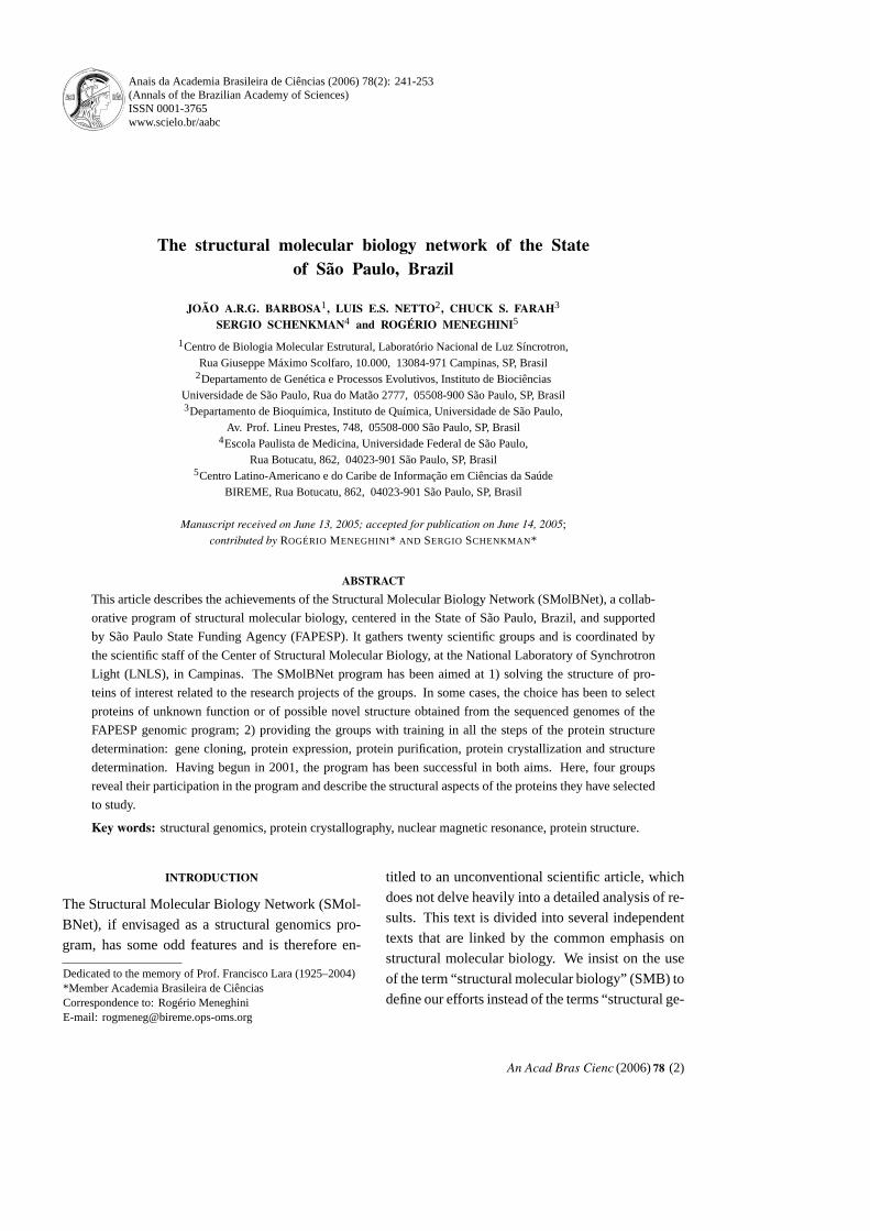
Anais da Academia Brasileira de Ciências (2006) 78(2): 241-253 (Annals of the Brazilian Academy of Sciences) ISSN 0001-3765 www.scielo.br/aabc The structural molecular biology network of the State of São Paulo, Brazil JOÃO A.R.G. BARBOSA 1 , LUIS E.S. NETTO 2 , CHUCK S. FARAH 3 SERGIO SCHENKMAN 4 and ROGÉRIO MENEGHINI 5 1 Centro de Biologia Molecular Estrutural, Laboratório Nacional de Luz Síncrotron, Rua Giuseppe Máximo Scolfaro, 10.000, 13084-971 Campinas, SP, Brasil 2 Departamento de Genética e Processos Evolutivos, Instituto de Biociências Universidade de São Paulo, Rua do Matão 2777, 05508-900 São Paulo, SP, Brasil 3 Departamento de Bioquímica, Instituto de Química, Universidade de São Paulo, Av. Prof. Lineu Prestes, 748, 05508-000 São Paulo, SP, Brasil 4 Escola Paulista de Medicina, Universidade Federal de São Paulo, Rua Botucatu, 862, 04023-901 São Paulo, SP, Brasil 5 Centro Latino-Americano e do Caribe de Informação em Ciências da Saúde BIREME, Rua Botucatu, 862, 04023-901 São Paulo, SP, Brasil Manuscript received on June 13, 2005; accepted for publication on June 14, 2005; contributed by ROGÉRIO MENEGHINI * AND SERGIO SCHENKMAN* ABSTRACT This article describes the achievements of the Structural Molecular Biology Network (SMolBNet), a collab- orative program of structural molecular biology, centered in the State of São Paulo, Brazil, and supported by São Paulo State Funding Agency (FAPESP). It gathers twenty scientific groups and is coordinated by the scientific staff of the Center of Structural Molecular Biology, at the National Laboratory of Synchrotron Light (LNLS), in Campinas. The SMolBNet program has been aimed at 1) solving the structure of pro- teins of interest related to the research projects of the groups. In some cases, the choice has been to select proteins of unknown function or of possible novel structure obtained from the sequenced genomes of the FAPESP genomic program; 2) providing the groups with training in all the steps of the protein structure determination: gene cloning, protein expression, protein purification, protein crystallization and structure determination. Having begun in 2001, the program has been successful in both aims. Here, four groups reveal their participation in the program and describe the structural aspects of the proteins they have selected to study. Key words: structural genomics, protein crystallography, nuclear magnetic resonance, protein structure. INTRODUCTION The Structural Molecular Biology Network (SMol- BNet), if envisaged as a structural genomics pro- gram, has some odd features and is therefore en- Dedicated to the memory of Prof. Francisco Lara (1925– 2004) *Member Academia Brasileira de Ciências Correspondence to: Rogério Meneghini E-mail: [email protected] titled to an unconventional scientific article, which does not delve heavily into a detailed analysis of re- sults. This text is divided into several independent texts that are linked by the common emphasis on structural molecular biology. We insist on the use of the term “ structural molecular biology” (SMB) to define our efforts instead of the terms “ structural ge- An Acad Bras Cienc (2006) 78 (2)
-
Upload
independent -
Category
Documents
-
view
0 -
download
0
Transcript of The structural molecular biology network of the State of São Paulo, Brazil
Anais da Academia Brasileira de Ciências (2006) 78(2): 241-253(Annals of the Brazilian Academy of Sciences)ISSN 0001-3765www.scielo.br/aabc
The structural molecular biology network of the Stateof São Paulo, Brazil
JOÃO A.R.G. BARBOSA1, LUIS E.S. NETTO2, CHUCK S. FARAH3
SERGIO SCHENKMAN4 and ROGÉRIO MENEGHINI5
1Centro de Biologia Molecular Estrutural, Laboratório Nacional de Luz Síncrotron,Rua Giuseppe Máximo Scolfaro, 10.000, 13084-971 Campinas, SP, Brasil
2Departamento de Genética e Processos Evolutivos, Instituto de BiociênciasUniversidade de São Paulo, Rua do Matão 2777, 05508-900 São Paulo, SP, Brasil3Departamento de Bioquímica, Instituto de Química, Universidade de São Paulo,
Av. Prof. Lineu Prestes, 748, 05508-000 São Paulo, SP, Brasil4Escola Paulista de Medicina, Universidade Federal de São Paulo,
Rua Botucatu, 862, 04023-901 São Paulo, SP, Brasil5Centro Latino-Americano e do Caribe de Informação em Ciências da Saúde
BIREME, Rua Botucatu, 862, 04023-901 São Paulo, SP, Brasil
Manuscript received on June 13, 2005; accepted for publication on June 14, 2005;contributed by ROGÉRIO MENEGHINI* AND SERGIO SCHENKMAN*
ABSTRACT
This article describes the achievements of the Structural Molecular Biology Network (SMolBNet), a collab-
orative program of structural molecular biology, centered in the State of São Paulo, Brazil, and supported
by São Paulo State Funding Agency (FAPESP). It gathers twenty scientific groups and is coordinated by
the scientific staff of the Center of Structural Molecular Biology, at the National Laboratory of Synchrotron
Light (LNLS), in Campinas. The SMolBNet program has been aimed at 1) solving the structure of pro-
teins of interest related to the research projects of the groups. In some cases, the choice has been to select
proteins of unknown function or of possible novel structure obtained from the sequenced genomes of the
FAPESP genomic program; 2) providing the groups with training in all the steps of the protein structure
determination: gene cloning, protein expression, protein purification, protein crystallization and structure
determination. Having begun in 2001, the program has been successful in both aims. Here, four groups
reveal their participation in the program and describe the structural aspects of the proteins they have selected
to study.
Key words: structural genomics, protein crystallography, nuclear magnetic resonance, protein structure.
INTRODUCTION
The Structural Molecular Biology Network (SMol-
BNet), if envisaged as a structural genomics pro-
gram, has some odd features and is therefore en-
Dedicated to the memory of Prof. Francisco Lara (1925– 2004)*Member Academia Brasileira de CiênciasCorrespondence to: Rogério MeneghiniE-mail: [email protected]
titled to an unconventional scientific article, which
does not delve heavily into a detailed analysis of re-
sults. This text is divided into several independent
texts that are linked by the common emphasis on
structural molecular biology. We insist on the use
of the term “ structural molecular biology” (SMB) to
define our efforts instead of the terms “ structural ge-
An Acad Bras Cienc (2006) 78 (2)

242 JOÃ O A.R.G. BARBOSA et al.
nomics” (since many investigators may be involved
in SMB without using genomic data) or “ structural
biology” (in homage to classic structural biologists
who operate with optical or electronic microscopes
in the micron to millimicron scale whereas SMB
deals with atomic resolution).
Structural genomics, in the strict sense, is SMB
applied on the genomic scale and as such, if not
explanatory, is etymologically adequate. With the
advent of genomics, the annotation of thousands of
known and putative genes became and continues to
become available. This technological breakthrough
has provided a great impulse to different fields
of biology, including SMB. Before the era of large-
scale genome sequencing SMB had already been
experiencing enormous progress with the elucida-
tion of the 3D-atomic structure of several hundred
proteins thanks to the use of two modern technolo-
gies: X-ray crystallography, using X-ray beam-lines
of synchrotron radiation facilities, and high field
nuclear magnetic resonance (NMR). In this way, a
somewhat astonishing perception has come to light:
the number of ways proteins may fold into defined
3D atomic structures in the universe of all proteins
in the biosphere is limited. As more 3D structures
of proteins were solved the rarer it became to find
novel folding modes. Therefore one may envisage
a future where virtually all new structures solved
may be classified into a previously defined family.
Although large, the number of folding topologies is
probably limited to one or a few thousand (Govin-
darajan et al. 1999). Furthermore, if the structure of
one member of a protein family that share signifi-
cant primary structure similarity is known, feasible
molecular models of all the remaining members may
be constructed by homology modeling.
After the advent of the genomics era, the struc-
tural genomics approach was launched. In Brazil,
the ONSA program (Organization for Nucleotide
Sequencing and Analysis, FAPESP) was responsi-
ble for the sequencing of the first plant pathogen
genome, that of the Xyllela fastidiosa (Xf ), which
has 2900 predicted genes (Simpson et al. 2000), a
significant fraction of which has no known function.
The path of structural genomics goes as follow: de-
fine a criterion by which to select specific protein
targets, for example proteins of unknown function
and/or unknown structure. Then: (i) clone the se-
lected target genes into expression vectors, (ii) ex-
press the corresponding proteins in an heterologous
system, (iii) purify the proteins, (iv) submit them
to crystallization trials, (v) collect X-ray diffraction
data usually at a synchrotron radiation facility and
(vi) solve the structure. Alternatively, from step (iv)
obtain a high concentration protein solution and col-
lect data at an NMR facility. The latter is an attrac-
tive option for those many cases in which the pro-
teins are refractory to form crystals. This sequence
is a funnel, for each step has a limited chance of
success. Therefore, most structural genomics ef-
forts adopt a high throughput approach: if one of the
steps of the pipeline poses a difficult obstacle for a
particular target, that target is disposed of. Hence,
only a small percent of the chosen proteins have their
3D-structure resolved (typically 1-5%). Dozens of
worldwide structural genomics projects are under-
way. (http://www.rcsb.org/ pdb.strucgen.html).
Of course, not all SMB projects are high
throughput. It is often the case that a group is inter-
ested in resolving the structure of one or a few pro-
teins related to a specific biological question. Here,
there is no thoughtless disposal of the protein tar-
gets if difficulties in the pipeline arise. The main
purpose is not structural per se; rather, solving these
structures may shed light on functional aspects of
interest.
The central focus of the SMolBNet program
was to bring together a wide variety of biomedical
and biological research groups with an interest in
SMB. In 1999, twenty groups of the State of São
Paulo entered the program, almost all of them with
no previous experience in SMB. Most of them had
in mind the use of SMB to study proteins that were
already targeted to be of interest in their specific line
of study. However, some were interested in embark-
ing on a structural genomics program, even though
it was not directly linked to a traditional line of re-
search, to a great extent influenced by the success of
An Acad Bras Cienc (2006) 78 (2)

STRUCTURAL MOLECULAR BIOLOGY NETWORK 243
473 genes cloned, ~ 6 proteins extracted and ~ 10 synthetic peptides
396 proteins expressed
305 proteins purified
NMR
– 14 peptides solved
– 2 proteins solved
X-ray Crystallography
– 114 proteins submitted to
crystallization trials
– 24 structures solved
14 homology models constructed
14 SAX structures
Fig. 1 – The SMolBNet pipeline showing the general achievements at the end of the fourth year.
FAPESP genomics projects.
The program was coordinated by the Center
for Structural Molecular Biology (CeBiME) of the
Brazilian Synchrotron Light Laboratory (LNLS), in
Campinas, São Paulo, which includes several staff
investigators with expertise in protein crystallo-
graphy and protein NMR. The Center facili-
ties include NMR spectrometers, mass spectrom-
eters, light spectrometers, protein purification and
crystallization, and molecular biology laboratories,
and a crystallography beamline connected to the
1.37 GeV circular electron accelerator that gener-
ates synchrotron radiation. In addition, a new MAD
crystallography beamline is coming up at the end of
2005. This set of facilities is unique in the southern
hemisphere.
The selected groups started working in 1999,
under the assistance of the CeBiME staff. Judg-
ing from the results accumulated up until now we
can conclude that the SMolBNet program was suc-
cessful in providing an important impetus for the
advancement of SMB in Brazil. The pipeline in Fig-
ure 1 summarizes the current state of the program at
each stage of the production pipeline. The program
is intended to terminate by the end of 2006.
Among the five authors contributing to this
work, four summarize here the results of their
participation. JARGB is a crystallographer and a
CeBiME-LNLS staff member. He reports his par-
ticipation in collaborating with and training other
members of the network. Not included in this re-
port is his own work determining the structures of
some Xf hydrolases and some hypothetical gene
products from Xanthomonas axonopodis pv. citri
(Xac). CSF is a biochemist who has also chosen to
use both NMR and X-ray crystallography to study
some Xac conserved hypothetical proteins and
proteins important for Xac pathogenicity. LESN
is a biochemist who has been studying functional
aspects of antioxidant proteins for some years and
has picked several of these proteins from Saccha-
romyces cerevisiae (Sc) and Xf for crystallographic
studies. SS is a biochemical parasitologist in-
terested in the infectious mechanisms employed by
protozoa and their insect vectors. He focused his
structural investigations on proteins from the pro-
tozoa Trypanosoma cruzi, Plasmodium vivax and
falciparum and a protein from the insect vector
Triatoma infestans, using both X-ray crystallogra-
phy and NMR.
An Acad Bras Cienc (2006) 78 (2)

244 JOÃ O A.R.G. BARBOSA et al.
CRYSTALLOGRAPHIC EFFORTS IN THE SMolBNet
Within the SMolBNet framework, CeBiME had the
challenge of working with the various groups of the
network in a way as to provide the expertise neces-
sary to solve peptide or protein structures. Further,
this expertise should be passed to the groups, allow-
ing them to gain as much independency as possible.
Crystallization experiments are the first step
in trying to obtain a crystallographic structure of
a protein. It usually demands that enough quantities
(10 mg) of high grade purified and properly folded
protein are available. This procedure has a low suc-
cess rate and is a tough test of the researchers’ will.
The vast majority of the crystallization experiments
were carried out using the hanging- or sitting-drop
vapor diffusion methods. Here, the equilibration of
two different solutions leads to a very slow deple-
tion of water in the solution that contains the protein
and consequently concentrates it. As the protein
concentrates, it tends to aggregate in an amorphous
or crystalline precipitate. In Figure 2 and Table I
it is possible to see the crystals obtained for sev-
eral proteins. Each of these crystals demanded a
unique crystallization condition and represents the
mid-point to success mark in the path to structure so-
lution when we consider all the pre-crystallographic
work.
The next step was X-ray diffraction data col-
lection at the LNLS beamline. This was a much
expected part of the work because the researchers
would finally be sure that their crystals would be
useful to structure solution and also because they
would have contact with the synchrotron in-
strumentation. From this point onwards there is a
clear division in the type of work carried out by
the groups: until now they were working in the
wet lab as molecular biologists and biochemists,
now their work will be carried out on computers.
Another issue is that the diffraction experiment is
within the domain of physics, and while physicists
and chemists might be familiar with it, biologists,
who were the majority, felt uncomfortable. The stu-
dents and/or PIs carried out data processing of the
oscillation images of those crystals that had inter-
pretable diffraction with close and extensive super-
vision. They were taught not only how to run the
programs, but also the ideas behind them: notions of
real and reciprocal space, the physics of diffraction,
reflection’s intensities and indices, the concepts of
unit cell, asymmetric unit, space group, integration,
scaling, merging, etc.
Phase determination / structure solution was
usually a straightforward procedure since most of
the structures were solved by molecular replace-
ment. The only exception was a hypothetical pro-
tein (“ f” in Table I) that was solved by multiwave-
length anomalous dispersion (MAD). This rather
interesting case demanded extra input from the par-
ticular group such as growing selenium methionine
protein crystals and is described in another part of
this manuscript. Structure building and refinement
is a procedure that still demands weeks and perhaps
months of work. This was accomplished by having
the people from the network coming over to the Ce-
BiME for these periods of time. This part helped
the students to learn about the protein databank for-
mat, atom coordinates, R factor, temperature factor,
electron density and many other concepts. Structure
analysis would be the final step and as the groups
moved towards this, it could be foreseen that dif-
ficulties arose when trying to explore completely
unrelated structures. Here, there is no clear path to
be followed, except for comparing the structures to
their homologues when they are present. In most
cases some unique features of the proteins will need
to be addressed, such as: why is the lysozyme (“ b”
in Table I) capable of working in a basic pH, what
is the information that the hypothetical protein (“ f”)
can give about its function or what makes the infestin
4 (“ j”) such a good inhibitor of blood coagulation-
cascade factor XIIa.
It is clear that throughout this project all of the
groups gained experience in diverse techniques used
in structural biology. Now that the project has been
extended for two years and shall finish in 2006, it is
time to consolidate the results and think about how
the groups shall move to the next step: become inde-
An Acad Bras Cienc (2006) 78 (2)

STRUCTURAL MOLECULAR BIOLOGY NETWORK 245
Fig. 2 – Crystals obtained by four different groups of the SMolBNet. The letters “ a” to “ j” correlate to the information for each
individual protein presented in Table I. Their sizes vary approximately from a few µm to one mm.
pendent of CeBiME. It is not realistic to expect that
all of the groups will grow and incorporate struc-
tural biology. One solution for everybody’s effort to
flourish all its potential lies in new positions opened
for structural biologists at the Departments to which
SMolBNet groups are affiliated.
STRUCTURAL AND FUNCTIONAL ASPECTS OFPEROXIDE DETOXIFICATION PATHWAYS
The mechanisms by which cells protect themselves
against the toxic effects of free radicals and related
species have been the subject of study in our labo-
ratory (LESN). Cells possess multiple pathways to
decompose peroxides such as catalases and thiol de-
pendent peroxidases, including glutathione peroxi-
dases. Emphasis has been given to a new class of
antioxidant enzymes named peroxiredoxins, which
includes thioredoxin peroxidases. In spite of the
fact that peroxiredoxins and glutathione peroxidases
have similar biochemical activity (thiol-dependent
peroxidases), they do not share amino acid sequence
similarity. Interestingly, however, both glutathione-
dependent peroxidases and peroxiredoxins belong
to the superfamily thioredoxin that comprises pro-
teins possessing the thioredoxin fold characterized
by a central core of four stranded mixed beta-sheet,
flanked by three alpha-helices. (Copley et al. 2004).
The yeast S. cerevisiae, which has been used
as model for higher eukaryotes, possesses five per-
oxiredoxins among two catalases and three phos-
pholipid glutathione peroxidases. Our studies have
demonstrated that although all five peroxiredoxins
have the same biochemical activity (thioredoxin
peroxidase), their functions are not completely re-
An Acad Bras Cienc (2006) 78 (2)

246 JOÃ O A.R.G. BARBOSA et al.
TABLE I
Name of the proteins, organism, maximum diffraction resolution and current researchstage of the crystals shown from Figure 1. The letters “a” to “h” on the left of theprotein’s column correspond to the letters in Figure 2.
Protein Organism Resolution (Å) Stage
a Beta-glicosidaseSpodoptera
2.8Crystallization,
frugiperda data collection
b LysozymeMusca
1.9 Analysisdomestica
cMaltose-binding Xanthomonas ∼ 4.0
Crystallization,
protein – MalE axonopodis pv. citri data collection
dMerozoite surface Plasmodium sp. ∼ 10.0
Crystallization,
proteins 3α data collection
e Xac2355/SufEXanthomonas
1.9 Refinementaxonopodis pv. citri
fHypothetical Xanthomonas
1.9 RefinementXac2396 axonopodis pv. citri
gMolibdate-binding Xanthomonas
1.7 Refinementprotein – ModA axonopodis pv. citri
hThioredoxin Saccharomyces
2.2Crystallization,
peroxidase III cerevisiae data collection
iInfestin 1 Triatoma infestans
2.5 Refinement– trypsin Bos taurus
j Infestin 4 Triatoma infestans 1.8 Analysis
dundant. For example, cytosolic thioredoxin pero-
xidase I (cTPxI/Tsa1/YML028W) is specifically im-
portant for the defense of yeast with dysfunctional
mitochondria (Demasi et al. 2001), whereas mito-
chondrial thioredoxin peroxidase I (PrxI/YBL064C)
is more active in conditions where yeast obtain ATP
preferentially by respiration (Monteiro et al. 2002,
Monteiro and Netto 2004). Finally, cytosolic thio-
redoxin peroxidase II (cTPxII/Tsa2/YDR453C) ap-
pears to be an important backup for cTPxI for the
defense against organic peroxides, independently of
the functional state of mitochondria (Munhoz and
Netto 2004).
We are also studying antioxidant proteins from
other organisms as a consequence of our partici-
pation in X. fastidiosa Genome Project. In this re-
gard, we were able to characterize for the first time
the biochemical role of a new class of antioxidant
enzymes conserved in several pathogenic bacteria:
Ohr (“ Organic Hydroperoxide Resistance protein)
is a dithiol-dependent peroxidase (Cussiol et al.
2003).
When we joined SMolBNet, we decided to in-
vestigate structural aspects of thiol-dependent per-
oxidases and related proteins in a hope to better
understand mechanisms of peroxide detoxification
in cells. Several recombinant proteins from yeast
and bacteria were submitted to crystallization tri-
als. Since thiol-dependent peroxidases can assume
different redox states, we decided to pre-treat pro-
teins with various oxidants and reductants at differ-
ent concentrations in an attempt to obtain a homoge-
nous preparation. This appeared to be a successful
approach since several crystals from different pro-
An Acad Bras Cienc (2006) 78 (2)

STRUCTURAL MOLECULAR BIOLOGY NETWORK 247
teins were obtained.
In the case of Ohr from X. fastidiosa, several
crystals were obtained from proteins treated with
different redox compounds using the hanging-drop
vapor diffusion method in the presence of PEG
4000 as precipitant (Oliveira et al. 2004). The crys-
tal structure was solved by molecular replacement
methods and the protein possesses a homodimeric
quaternary structure. Two Ohr structures were ob-
tained at two different molecular proportions (16
tert-butylhydroperoxide: 1 Ohr and 1 tert-butyl-
hydroperoxide: 1 Ohr), which will be described
in detail elsewhere. In summary, the protein pos-
sesses two domains: the N-terminal domain com-
posed of three antiparallel β strands (β1, β2 and
β3) that forms a β sheet and a short helix (H1) and
the C-terminal domain composed of an antiparallel
β sheet (β4-β6) and three α-helices (H2-H4). Con-
trary to glutathione peroxidases and peroxiredoxins,
two other classes of thiol-dependent peroxidases,
Ohr does not possess the thioredoxin fold. Further-
more, the mechanism by which the reactive cysteine
is stabilized appeared to be different among the three
classes of thiol-dependent peroxidases. Therefore,
Ohr is a new class of antioxidant proteins. The reac-
tive cysteine is in a sulfonate form (R-SO3H−) in the
structure obtained from Ohr treated with very high
doses of peroxide, indicating that these enzymes can
only be inactivated by very harsh conditions (M.A.
Oliveira et al., unpublished data).
Other proteins with redox properties from S.
cerevisiae also had their structures solved by our
group. One of them was glutaredoxin 2, which is
the main glutathione-dependent oxidoreductase of
yeast. This ubiquitously distributed enzyme is in-
volved in many cellular processes including protein
folding, regulation of protein activity and repair of
oxidatively damaged proteins (Rietsch and Beck-
with 1998). Glutaredoxin 2 structure was solved by
molecular replacement using a homologous protein
from Sus scrofa as a model (Discola et al. 2005).
Thioredoxin reductase is a pyridine nucleotide-di-
sulfide oxidoreductase capable to reduce redox ac-
tive disulfide bond of thioredoxins at the expense
of NADPH. The thioredoxin system (NADPH/thio-
redoxin reductase/thioredoxin) is involved in the re-
duction of disulfide bonds in several targets. Crys-
tals of thioredoxin reductase 1 from S. cerevisiae
were obtained from proteins pretreated with H2O2,
using the hanging-drop method. X-ray diffraction
data were collected to a maximum resolution of
2.4 Å using a synchrotron radiation source and the
structure was also solved by molecular replacement
using a homologous protein from Arabidopsis tha-
liana (Oliveira et al., unpublished data). Structures
of glutaredoxin 2 and thioredoxin reductase 1 are
under refinement and we hope that their elucida-
tion will contribute to the better understanding of
their catalytic mechanisms. In summary, participa-
tion in the SMolBNet was very important because
it made possible the establishment of structural and
functional relationships, which represented a con-
siderable improvement from my previous work.
STRUCTURAL DETERMINATION OF XANTHOMONASPROTEINS OF UNKNOWN FUNCTION
The sequencing of the genome of the citrus pathogen
Xanthomonas axonopodis pv. citri (Xac) revealed
the existence of many macromolecular systems im-
portant for pathogenesis (da Silva et al. 2002, Van
Sluys et al. 2002, Moreira et al. 2004) and several
Brazilian laboratories are studying their functions.
In addition to the identification of specific proteins
of interest due to their homology with those found in
other organisms, the Xac genome codes for a large
number of proteins that have not yet been character-
ized in any way and some for which orthologs have
not yet been identified (da Silva et al. 2002). Our
laboratory began its work in the SMolBNet project
focusing on the determination of the structure of a
number of such Xac proteins.
The reining philosophy with respect to struc-
tural studies of uncharacterized (sic “ hypothetical”)
proteins resides on the following two premises:
firstly that this class of proteins may hide previously
un-contemplated biological functions and secondly
that high resolution structural studies of these pro-
teins may in many cases be the most efficient means
An Acad Bras Cienc (2006) 78 (2)

248 JOÃ O A.R.G. BARBOSA et al.
by which these functions are revealed. Several re-
cent examples have demonstrated the feasibility of
obtaining functional information through structure
(for example see Bhattacharyya et al. 2002). The
Xac genome codes for 1658 hypothetical and con-
served hypothetical proteins (da Silva et al. 2002)
and we began by attacking the problem of target se-
lection within this group for structural studies.
NMR – BASED ASSAY TO SCREEN TARGETS FOR
STRUCTURAL STUDIES
As the success of a structural study will ultimately
depend on the protein being well folded and mono-
disperse, we attempted to obtain information in this
respect before large-scale production and purifica-
tion. To do so, we selected a set of 35 proteins
based on size (79– 330 residues), methionine content
(>1.1% for future selenomethionine incorporation),
and the absence of homologs of known function in
the Protein Data Bank. Of these, 31 were success-
fully expressed and 19 were found to be soluble. We
selectively 15N labeled these proteins during het-
erologous expression in E. coli and, in collaboration
with the protein NMR group of Centro Nacional de
Ressonância Magnética Nuclear (Universidade Fed-
eral do Rio de Janeiro), analyzed the bacterial lysates
by rapid 1H-15N HSQC NMR analysis. The chemi-
cal shift dispersion, line-width and number of peaks
observed in these 2-D spectra allowed us to classify
these 19 soluble candidates into 3 groups – good
(8 proteins), promising (6) and poor (5) in terms
of “ foldedness” (Galvão-Botton et al. 2003). The
“ good and “ promising” groups became” our initial
objects of study by NMR and X-ray diffraction and
our progress in this respect will be outlined briefly
below.
INITIAL CRYSTALLIZATION STUDIES
Three of the “ good” candidates were selected for
initial crystallization trials by the sparse matrix
sampling approach. Of these, two were found to
form crystals. The structure of one, YaeQ
(XAC2396), was determined by the multiple wave-
length anomalous dispersion (MAD) technique
(Guzzo et al. 2005). The YaeQ structure, deter-
mined to a resolution of 1.9 Å has no homologs in
the structural databases and represents a new protein
fold (C.R. Guzzo et al., unpublished data). The sec-
ond protein to produce crystals, YajQ (XAC3671),
diffracted only to low resolution (4 angstrons) and
assays to refine the crystallization conditions are
still in progress. The third (XAC1223) has so far
resisted crystallization. A fourth good candidate,
XAC2355, was recently submitted to crystallization
tests with positive results. It diffracted to 1.9 Å at
the crystallography beam line of the LNLS, initial
experimental phases were calculated by molecular
replacement using the recently determined E. coli
SufE structure as an initial model (pdb 1MZG, 36%
identity) and an interpretable electron density map
was obtained.
INITIAL NMR STUDIES
Two “ good”(ApaG/XAC0862 and ClpS/XAC2000)
and “ one” promising (XACb0070) candidates were
initially selected for structure determination by
NMR in collaboration with the LNLS protein NMR
laboratory led by Alberto Spisni. Through the use
of triple resonance experiments the backbone and
side chain assignments of all three proteins have
been completed (Katsuyama et al. 2003). Through
the use of distance and torsion angle restraints ob-
tained from NOEs and 3J coupling constants, the so-
lution structure of ApaG was determined. This 124
residue protein adopts a beta cup structure in which
the planes of two beta sheets are oriented at a small
angle creating a deep central hydrophobic pocket.
ApaG has a topology similar, though not identical, to
several proteins that utilize this groove to bind to the
hydrophobic lipid moieties of lipoproteins: Human
GM2-Activator protein, Human PDE – subunit of
rod-specific cGMP Phosphodiesterase e rhoG Dis-
sociation Inhibitor (Pertinhez et al. manuscript in
preparation). While the function of ApaG homologs
in bacteria is not known, ApaG homology domains
are encountered in a specific subclass (FBA) of F-
box proteins that form part of the E3-ubiquitin lig-
ase complex involved in the recognition of specific
An Acad Bras Cienc (2006) 78 (2)

STRUCTURAL MOLECULAR BIOLOGY NETWORK 249
proteins destined for ubiquitination and subsequent
degradation (Ilyin et al. 2000).
The XACb0070 protein structure was also solv-
ed in a similar manner. This protein is a dimer in
solution and has a topology similar to that of proteins
of the Arc-MetJ family of DNA binding proteins
(A. Gallo et al., unpublished data).
The third protein of this group, ClpS, was an
uncharacterized protein at the beginning of this
project but has since been characterized as an adapter
protein that binds to the N-terminal domain of
the ClpA chaperone ATPase that forms part of
the ClpAP bacterial proteosome and its structure in
complex with the ClpA N-domain has been deter-
mined by others. Backbone assignments, chemical
shift index analysis and H/D exchange experiments
indicate that the ClpS fold topology in solution is
very similar to that of the crystal structure (L.M.P.
Galvão-Botton et al., unpublished data). By deter-
mining the solution structure of ClpS on its own and
in complex with ClpA and comparing these struc-
tures with that of the ClpA-ClpS crystal we hope to
identify important structural changes in ClpS which
may take place upon binding.
We are now working on the determination of
the structure of XAC1883, a small uncharacterized
protein coded by the Xac Rpf (regulation of patho-
genic factors) locus responsible for the quorum
sensing response. Initial multidimensional studies
on 13C/15N-labelled XAC1883 are being carried out
using the 500 MHz NMR spectrophotometer at the
Instituto de Química at the Universidade de São
Paulo.
FUTURE PROJECTS
The strategy of screening the folded state of a
protein in bacterial lysates before purification was
successful in identifying proteins with relatively
good chances for future success in structural stud-
ies, either by NMR or crystallography. While con-
tinuing to work on the structure of Xac proteins
of unknown function, we plan to shift our focus
in the future towards the structural analysis of pro-
teins of demonstrated importance for Xanthomonas
pathogenicity. Our laboratory is specifically inter-
ested in understanding the molecular interactions
important for the function of the type III and type
IV secretion systems (Alegria et al. 2004, 2005), the
regulation of quorum sensing and biofilm forma-
tion by Rpf proteins and the functions of alternative
sigma factors.
STRUCTURE AND FUNCTION OF PROTEINS ANDPEPTIDES INVOLVED IN HOST PARASITE
INTERACTIONS
One of the major tasks to combat parasite diseases
is studying structure-functional relationships of key
molecules involved in their transmission. One is
Chagas disease, transmitted by the protozoan Try-
panosoma cruzi and its most successful insect vec-
tor, Triatoma infestans. The other is human Malaria
caused mainly by the protozoa Plasmodium vivax
and P. falciparum. Here we report some of the re-
sults we have obtained and what we have learned in
participating in this network.
STRUCTURE AND FUNCTION OF Triatoma
infestans PROTEINS
Triatoma infestans (Hemiptera: Reduviidae) is an
obligate haematophagous insect that transmits Try-
panosoma cruzi, the agent of Chagas disease. The
insect becomes infected when it takes blood con-
taining trypomastigote forms of T. cruzi. The try-
pomastigote turns into epimastigotes, which prolif-
erate in the T. infestans midgut. As the nutrients in
the insect gut become scarce, the parasite migrates
to the posterior end of the gut and transforms into
metacyclic trypomastigotes, which are then elimi-
nated with the feces, when the insect takes a new
blood meal. These released parasites are capable of
contaminating new hosts through the contact with
oral and eyes mucosa, skin lesions, or through in-
gestion. This complex cycle indicates that several
T. infestans molecules must take part in the parasite
life cycle from blood ingestion up to parasite elim-
ination. Our aims are to characterize some of these
molecules, particularly those present in the insect
saliva and midgut.
An Acad Bras Cienc (2006) 78 (2)

250 JOÃ O A.R.G. BARBOSA et al.
Among our studies, we have characterized a
22 kD pore forming protein, named trialysin, from
saliva of T. infestans (Amino et al. 2002). This
protein contains on the N-terminus a sequence that
predicts an amphipathic α-helix and its correspond-
ing synthetic peptide retains cytolytic activity. Our
goal has been to understand the lytic mechanism of
trialysin as well as the activation mechanism. There-
fore we aimed to solve the 3D structure of the pre-
cursor and active forms of trialysin as well as the
lytic peptides corresponding to the N-terminus of
the molecule.
We were unable to generate the recombinant
pro-trialysin, or the mature protein for diffraction or
NMR studies. No bacterial transformants could be
obtained using E. coli as host, suggesting that the
protein was highly toxic. Removing the first amino
acids from the N-terminus only yielded transfor-
mants when the constructs lacked the first 52 amino
acids. However, the produced protein was inactive
and insoluble. We could only detect pro-trialysin
expression in fusion with GST. We produced up to
three mg of the GST pro-trialysin fusion from one
liter of bacteria culture, but the recombinant protein
was unstable. We are currently trying to express
similar constructs in Pichia pastoris to obtain higher
mass of soluble protein.
In parallel, we studied the lytic activity of
several synthetic peptides corresponding to the N-
terminus of trialysin (Martins et al. 2006). We
found that although all peptides folded into
α-helices in the presence of SDS or trifluoroethanol
(TFE), only the ones close to the N-terminus of ma-
ture trialysin were lytic to T. cruzi, E. coli, and ery-
throcytes. The most active against all these targets
was the peptide named P6. It is a 32mer corre-
sponding to the N-terminus of trialysin. Peptide
P7, which only differs from P6 by the absence of
the last five amino acids, was less active than P6,
though much less hemolytic than P6. P5, lacking the
first five residues of P6, was as trypanocydal as P7,
but as efficient as P6 to lyse erythrocytes. To under-
stand the lytic mechanism and compare the different
activities of these peptides, we solved their struc-
tures by NMR. We found that they fold into similar
amphipathic α-helices in 30% TFE. Based on ob-
tained energy-minimized structures, we noticed the
presence of a similar central domain in P5, P6 and
P7 composed by the amphipathic helix, and more
flexible but similar N-terminal in P6 and P7 and
C-terminal domains in P6 and P5. We concluded
that presence of both domains might confer stronger
activity to P6.
STRUCTURE OF TRYPANOSOMA PROTEINS
INVOLVED IN RNA PROCESSING AND
PROTEIN SYNTHESIS
Trypanosomes have a unique RNA processing
mechanism and translation initiation control, which
therefore constitute interesting drug targets. Most
of the genes are transcribed as polycystronic
units that are processed by trans-splicing and poly-
adenylation. All mRNAs receive a 30– 40 nt-capped
RNA derived from the splice leader gene in the 5’ ter-
minal ends (Gull 2001). Both the cap structure and
the enzymes involved in the cap synthesis are dif-
ferent in trypanosomes as compared to mammalian
enzymes. For example, a guanilyl transferase of
T. brucei has an extra N-terminal portion (Silva et
al. 1998) that seems to carry an adenylate kinase
domain. The RNA triphosphatase activity is also
catalyzed by an unique enzyme (Ho and Shuman
2001). Therefore we selected as targets for struc-
tural studies the RNA adenosine triphosphatase from
T. cruzi and T. brucei. These enzymes have been
cloned, expressed in E. coli, purified, and submit-
ted to crystallization assays. Three conditions were
found promising and are used in refinements.
Two targets were also selected for proteins in-
volved in the translation initiation control of try-
panosomes. One is the eukaryote translation initia-
tion factor 4E of T. brucei (TBeIF4E), which is the
cap-binding protein.It has 251 amino acids and was
chosen as a target for two reasons: a) it has only
24% similarity to the mammalian counterpart, thus
representing an interesting drug target; b) the struc-
tures of the mammalian and yeast homologues have
been determined (PDBs:1L8B, 1EJ1, 1AP8), which
An Acad Bras Cienc (2006) 78 (2)

STRUCTURAL MOLECULAR BIOLOGY NETWORK 251
might help in the analysis of this protein. TBeIF4E
was cloned in pET28a, expressed, purified and sub-
mitted to crystallization screenings. The other target
was the eIF2α of T. brucei. It is a 419 amino acid
protein that contains an extension of ca. 120 residues
in the N-terminal relative to all other known eIF2α
sequences. The same extension is present in T. cruzi
and Leishmania major. The complete ORF as well
as only the N-terminal half was cloned into pET28a
and the protein expressed, purified and submitted to
crystallization assays.
SURFACE PROTEINS OF MEROZOITE FORMS OF
Plasmodium vivax
We are also interested in the structure of merozoite
surface proteins (MSP) of Plasmodium sp, most of
them involved in the erythrocyte and reticulocyte
invasion. Merozoites correspond to blood stages of
the Plasmodium species released by infected cells.
These studies can help understanding the biology
of these organisms and designing potential agents
to block its growth. We have focused on fragments
of these major surface proteins, as the entire pro-
teins are too large and insoluble when produced in
recombinant form. We expressed fragments corre-
sponding to the C-terminus of MSP3α and the N
and C terminus of MSP3β based on the formation
of coiled-coil structure as demonstrated (Galinski et
al. 1999). Five recombinant proteins were obtained
in fusion with His-tags and purified by affinity chro-
matography and ion exchange. MSP3α e MSP3β
have been produced in appropriate levels, purified
and submitted to crystallization assays.
CONCLUSIONS AND PERSPECTIVES
We adopted as a general strategy in SmolBNet to
select protein targets exclusively related to our cur-
rent research lines. In a few cases we were suc-
cessful; this is the case of the trialysin peptides
solved by NMR and the serine protease inhibitors
solved by diffraction studies. For most of the cho-
sen targets we failed in obtaining structural data,
although we still have promising results. Never-
theless, we acquired experience in all steps of the
process: target selection, protein expression, crys-
tallization, and structure resolution, and the most
important achievement was to involve several stu-
dents, who will keep this knowledge and experience
in their scientific formation.
ACKNOWLEDGMENTS
The authors wish to thank the Heads of the SMolB-
Net groups, technicians, post-doctoral, graduate
and undergraduate fellows involved in the Net-
work, whose names would form a long list that can-
not be fitted in the space here available. Here, a
few successful examples of participating groups are
described, what does not mean they are the only
ones. It should be noted that CeBiME members
other than JARGB also helped researchers in their
pre-crystallography steps, in the proteins’ biophys-
ical characterization and in solving their structures
by NMR spectroscopy or crystallography. It is also
necessary to point out that there were a few groups
of the SMolBNet that already had structural knowl-
edge and in one way or another helped the other
groups.
RESUMO
Esse artigo descreve realizações do Programa SMolBNet
(Rede de Biologia Molecular Estrutural) do Estado de
São Paulo, apoiado pela FAPESP (Fundação de Apoio
à Pesquisa do Estado de São Paulo). Ele reúne vinte
grupos de pesquisa e é coordenado pelos pesquisadores
do Laboratório Nacional de Luz Síncrotron (LNLS), em
Campinas. O Programa SMolBNet tem como metas:
Elucidar a estrutura tridimensional de proteínas de inte-
resse aos grupos de pesquisa componentes do Programa;
Prover os grupos com treinamento em todas as etapas de
determinação de estrutura: clonagem gênica, expressão de
proteínas, purificação de proteínas, cristalização de proteí-
nas e elucidação de suas estruturas. Tendo começado em
2001, o Programa alcançou sucesso em ambas as metas.
Neste artigo, quatro dos grupos descrevem suas partici-
pações, e discutem aspectos estruturais das proteínas que
eles selecionaram para estudos.
Palavras-chave: genômica estrutural, cristalografia de
An Acad Bras Cienc (2006) 78 (2)

252 JOÃ O A.R.G. BARBOSA et al.
proteínas, ressonância nuclear magnética, estrutura de
proteínas.
REFERENCES
ALEGRIA MC, DOCENA C, KHATER L, RAMOS
CH, DA SILVA AC AND FARAH CS. 2004 New
protein-protein interactions identified for the regu-
latory and structural components and substrates of
the type III Secretion system of the phytopathogen
Xanthomonas axonopodis Pathovar citri. J Bacteriol
186: 6186– 6197.
ALEGRIA MC, SOUZA DP, ANDRADE MO, DOCENA
C, KHATER L, RAMOS CH, DA SILVA AC AND
FARAH CS. 2005. Identification of new protein-
protein interactions involving the products of the
chromosome and plasmid-encoded type IV secretion
loci of the phytopathogen Xanthomonas axonopodis
pv. citri. J Bacteriol 187: 2315– 2325.
AMINO RRM, MARTINS JP, HIRATA IY, JULIANO MA
AND SCHENKMAN S. 2002. Trialysin, a novel pore-
forming protein from saliva of hematophagous in-
sects activated by limited proteolysis. J Biol Chem
277: 6207– 6213.
BHATTACHARYYA S, HABIBI-NAZHAD B, AMEGBEY
G, SLUPSKY CM, YEE A, ARROWSMITH C AND
WISHART DS. 2002. Identification of a novel ar-
chaebacterial thioredoxin: determination of function
through structure. Biochemistry 41: 4760– 4770.
CAMPOS ITN, GUIMARÃ ES BG, MEDRANO FJ, TA-
NAKA AS AND BARBOSA JARG. 2004. Crystal-
lization, data collection and phasing of infestin 4, a
factor XIIa inhibitor. Acta Crystall D 60: 2051–
2053.
COPLEY SD, NOVAK WRP AND BABBITT PC. 2004.
Divergence of function in the thioredoxin supra-
family: Evidence for evolution of peroxiredoxins
from a thioredoxin-like ancestor. Biochemistry 43:
13981– 13995.
CUSSIOL JRR, ALVES SV, OLIVEIRA MA AND
NETTO LES. 2003. Organic hydroperoxide resis-
tance gene encodes a thiol-dependent peroxidase. J
Biol Chem 278: 11570– 11578.
DA SILVA AC ET AL. 2002. Comparison of the genomes
of two Xanthomonas pathogens with differing host
specificities. Nature 417: 459– 463.
DEMASI APD, PEREIRA GAG AND NETTO LES.
2001. Cytosolic thioredoxin peroxidase I is essential
for the antioxidant defense of yeast with dysfunc-
tional mitochondria. FEBS Letters 509: 430– 434.
DISCOLA KF, OLIVEIRA MA, MONTEIRO-SILVA G,
BARCENA JA, PORRAS P, PADILLA A, NETTO
LES AND GUIMARÃ ES BG. 2005. Crystallization
and preliminary X-ray diffraction analysis of glutare-
doxin 2 from Saccharomyces cerevisiae in different
oxidation states. Acta Crystall F, F61: 445– 447.
GALINSKI MR, CORREDOR-MEDINA C, POVOA M,
CROSBY J, INGRAVALLO P AND BARNWELL JW.
1999. Plasmodium vivax merozoite surface protein-3
contains coiled-coil motifs in an alanine-rich central
domain. Mol Biochem Parasitol 101: 131– 147.
GALVÃ O-BOTTON LMP, KATSUYAMA AM, GUZZO
CR, ALMEIDA FCL, FARAH CS AND VALENTE
AP. 2003. High-throughput screening of structural
proteomics targets using NMR. FEBS Letters 552:
207– 213.
GOVINDARAJAN S, RECABARREN R AND GOLDSTEIN
RA. 1999. Estimating the Total Number of Protein
Folds. PROTEINS: Structure, Function, and Genet-
ics 35: 408– 414.
GULL K. 2001. The biology of kinetoplastid parasites:
insights and challenges from genomics and post-
genomics. International Journal of Parasitology 31:
443– 452.
GUZZO CR, NAGEM RAP, GALVÃ O-BOTTON LMP,
GUIMARÃ ES BG, MEDRANO FJ, BARBOSA JARG
AND FARAH CS. 2005. Expression, purification,
crystallization and preliminary X-ray analysis of
YaeQ (XAC2396) from Xanthomonas axonopodis
pv citri. Acta Crystall F, F61: 493– 495.
HO CK AND SHUMAN S. 2001. Trypanosoma brucei
RNA triphosphatase. Antiprotozoal drug target and
guide to eukaryotic phylogeny. J Biol Chem 276:
46182– 46186.
ILYIN GP, RIALLAND M, PIGEON C AND GUGUEN-
GUILLOUZO C. 2000. cDNA cloning and expression
analysis of new members of the mammalian F-box
protein family. Genomics 67: 40– 47.
KATSUYAMA AM, CICERO DO, SPISNI A, PACI M,
FARAH CS AND PERTINHEZ TA. 2003. 1H, 15N
and 13C resonance assignments of the ApaG protein
of the phytopathogen Xanthomonas axonopodis pv.
citri. J. Biomolecular NMR 29: 423– 424.
An Acad Bras Cienc (2006) 78 (2)

STRUCTURAL MOLECULAR BIOLOGY NETWORK 253
MARTINS RM, SFORÇA ML, AMINO R, JULIANO
MA, OYAMA JR S, JULIANO L, PERTINHEZ TA,
SPISNI A AND SCHENKMAN S. 2006. Lytic activ-
ity and structural differences of amphipathic peptides
derived from trialysin. Biochemistry, in press.
MONTEIRO G AND NETTO LES. 2004. Glucose re-
pression of PRX1 expression is mediated by Tor1p
and Ras2pthrough inhibition of Msn2/4p in Sac-
charomyces cerevisiae. FEMS Microbiol Lett 241:
221– 228.
MONTEIRO G, PEREIRA GAG AND NETTO LES.
2002. Regulation of mitochondrial thioredoxin per-
oxidase I expression by two different pathways: one
dependent on cAMP and the other on heme. Free
Rad Biol Med 32: 278– 288.
MOREIRA LM, DE SOUZA RF, ALMEIDA JR NF,
SETUBAL JC, OLIVEIRA JC, FURLAN LR, FER-
RO JA AND DA SILVA AC. 2004. Comparative ge-
nomics analyses of citrus-associated bacteria. Annu
Rev Phytopathol. 42: 163– 184.
MUNHOZ DC AND NETTO LES. 2004. Cytosolic thio-
redoxin peroxidase I and II are important defenses of
yeast against organic hydroperoxide insult. Catalases
and peroxiredoxins cooperate in the decomposition
of H2O2 by yeast. J Biol Chem 279: 35219– 35227.
OLIVEIRA MA, NETTO LES, MARTIN FJM, BAR-
BOSA JARG, ALVES SV, CUSSIOL JRR AND
GUIMARÃ ES BG. 2004. Crystallization and prelim-
inary X-ray diffraction analysis of an oxidized state
of Ohr from Xylella fastidiosa. Acta Crystall D 60:
337– 339.
OLIVEIRA MA, DISCOLA KF, ALVES SV, BARBOSA
JARG, MEDRANO FJ, NETTO LES AND GUI-
MARÃ ES BG. 2005. Crystallization and preliminary
X-ray diffraction analysis of NADPH-dependent
thioredoxin reductase from Saccharomyces cerevi-
siae. Acta Crystall F, F61: 387– 390.
RIETSCH A AND BECKWITH J. 1998. The genetics of
disulfide bond metabolism. Annu Rev Genet 32:
163– 184.
SILVA E, ULLU E, KOBAYASHI R AND TSCHUDI C.
1998. Trypanosome capping enzymes display a
novel two-domain structure. Mol Cel Biol 18: 4612.
SIMPSON AJG ET AL. 2000. The genome sequence of
the plant pathogen Xylella fastidiosa. Nature 406:
151– 159.
VAN SLUYS MA, MONTEIRO-VITORELLO CB, CA-
MARGO LE, MENCK CF, DA SILVA AC, FERRO
JA, OLIVEIRA MC, SETUBAL JC, KITAJIMA JP
AND SIMPSON AJ. 2002. Comparative genomic
analysis of plant-associated bacteria. Annu Rev Phy-
topathol 40: 169– 189.
An Acad Bras Cienc (2006) 78 (2)