Janeiro and São Paulo, Brazil (Anisoptera) - Natuurtijdschriften
11
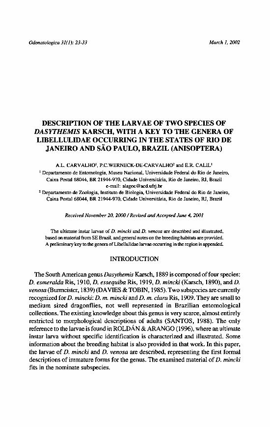
March I, 2002 Odonatologica 31(1): 23-33 Description of the larvae of two species of Dasythemis Karsch, with a key to the genera of Libellulidae occurring in the states of Rio de Janeiro and São Paulo, Brazil (Anisoptera) A.L. Carvalho ¹, P.C. Werneck-De-Carvalho ² and E .R. Calil ¹ Received November 20, 2000 / Revised and Accepted June 4, 2001 INTRODUCTION The South American genus Dasythemis Karsch, 1889 is composed of four species: D. esmeralda Ris, 1910, D. essequiba Ris, 1919, D. mincki (Karsch, 1890), and D. venosa (Burmeister, 1839) (DAVIES & TOBIN, 1985). Two subspecies are currently recognized for D. mincki: D. m. mincki and D. m. clara Ris, 1909. They are small to medium sized dragonflies, not well represented in Brazilian entomological collections. The existing knowledge aboutthis genus is very scarce, almost entirely restricted to morphological descriptions of adults (SANTOS, 1988). The only reference to the larvae is found in ROLDÂN & ARANGO ( 1996), wherean ultimate instar larva without specific identification is characterized and illustrated. Some information about the breeding habitat is also provided in that work. In this paper, the larvae of D. mincki and D. venosa are described, representing the first formal descriptions of immatureforms for the genus. The examined material of D. mincki fits in the nominate subspecies. 1 Departamento de Entomologia, Museu Nacional, Universidade Federal do Rio de Janeiro, Caixa Postal 68044, BR 21944-970, Cidade Universitdria, Rio de Janeiro, RJ, Brazil e-mail: [email protected] 2 Departamento de Zoologia, Institute de Biologia, Universidade Federal do Rio de Janeiro, Caixa Postal 68044, BR 21944-970, Cidade Universiuiria, Rio de Janeiro, RJ, Brazil The ultimate instar larvae of D. mincki and D. venosa are described and illustrated, based on material from SE Brazil, and general notes on the breeding habitats are provided. A preliminarykey to the genera of Libellulidae larvae occurring in the region is appended.
-
Upload
khangminh22 -
Category
Documents
-
view
2 -
download
0
Transcript of Janeiro and São Paulo, Brazil (Anisoptera) - Natuurtijdschriften
March I, 2002Odonatologica 31(1): 23-33
Description of the larvae of two species of
Dasythemis Karsch, with a key to the generaof
Libellulidaeoccurring in the states of Rio de
Janeiro and São Paulo, Brazil (Anisoptera)
A.L. Carvalho¹,P.C. Werneck-De-Carvalho²and E.R. Calil¹
Received November 20, 2000/ Revised and Accepted June 4, 2001
INTRODUCTION
The South American genusDasythemis Karsch, 1889is composed of four species:
D. esmeralda Ris, 1910, D. essequiba Ris, 1919, D. mincki (Karsch, 1890), and D.
venosa (Burmeister, 1839) (DAVIES & TOBIN, 1985). Two subspecies are currently
recognized for D. mincki: D. m. mincki and D. m. clara Ris, 1909. They are small to
medium sized dragonflies, not well represented in Brazilian entomological
collections. The existing knowledge aboutthis genus is very scarce, almost entirelyrestricted to morphological descriptions of adults (SANTOS, 1988). The only
reference to the larvae is foundin ROLDÂN & ARANGO ( 1996),wherean ultimate
instar larva without specific identification is characterized and illustrated. Some
informationabout the breeding habitat is also provided in that work. In this paper,
the larvae of D. mincki and D. venosa are described, representing the first formal
descriptions of immatureforms for the genus. Theexamined materialofD. mincki
fits in the nominate subspecies.
1 Departamento de Entomologia, Museu Nacional, Universidade Federal do Rio de Janeiro,
Caixa Postal 68044, BR 21944-970,Cidade Universitdria, Rio de Janeiro, RJ, Brazil
e-mail: [email protected] Departamento de Zoologia, Institute de Biologia, Universidade Federal do Rio de Janeiro,
Caixa Postal 68044, BR 21944-970,Cidade Universiuiria, Rio de Janeiro, RJ, Brazil
The ultimate instar larvae ofD. mincki and D. venosa are described and illustrated,
based on material from SEBrazil, and generalnotes onthe breedinghabitats are provided.
A preliminarykey to thegenera ofLibellulidae larvae occurring in the regionis appended.

24 A.L. Carvalho, P.C. Wemeck-de-Carvalho & E.R. Calil
METHODS
Part of the collected material was fixed in 80% ethanol, which was also used as a preservative. Some
last instar larvae were bred in laboratory in styrofoam boxes, following procedures described in
CARVALHO (1992). The emerged adults were identified using RIS (1910). The descriptions and
illustrations were made with the aid ofa stereoscopic microscope, equippedwith a camera lucidaand a
micrometric ocular. Labium and mandibles were described following CORBET (1953) and WATSON
(1956), respectively. The exuviae cited in the material have correspondingemergedadults. All specimens
studied are deposited at the Departamentode Zoologia, Instituto de Biologia, UFRJ, Rio de Janeiro,
Brazil.
DESCRIPTIONS OF THE LARVAE
DASYTHEMIS VENOSA (BURMEISTER, 1839)
Figures 1, 3-7
Material.- BRAZIL, Sao Paulo, Araras, 28-1-1993, A.L. Carvalho leg., 5 larvae: 2 9 (F), 2 ?
(F-l), 1 ? (F-2) (collected with a lost exuviae and teneral adult).
General shape of body typical of Libellulidae. Size small, general colour
ochraceous. Head, thorax and legs with many setae; legs positioned very near to
body in living larvae. Specimens collected in field entirely encrusted with mud,
except for upperpart of eyes.
Head.- General shape elipsoid, wider than long in dorsal view; occiput wider
than anteriorregion of head, with posterior angles rounded; lateral portion ofeyes
— (2) D. mincki.Figs 1-2. Ultimate instar larva, dorsal views (1) Dasythemis venosa;

25Description of two Dasythemis larvae
pronounced laterally and dorsally; anterior region of frons between and below
antennae with brush like group of setae directed forward. Antenna seven-
-segmented, 1st and 2nd segments wider than others; 1st, 2nd and 4th segments
similar and shorter than remaining ones; 3rd and 7th segments similar and longer
than remaining ones; setae occurring on all segments, some of them longer than
corresponding segment. Mandibularformula: L 1234 0 abd / R 1234 y abed; teeth
“3” and “4” ofright mandible very near each other, almost fused; tooth “d” ofleft
Figs 3-7. ultimate instar larva: (3) right antenna, dorsal view; — (4) mandibles,
internal view; (a) left, (b)right; — (5) prementum, dorsal view; — (6) right labialpalp, dorsal view; —
(7). abdominal segments 8-10 and anal appendages, dorsal view.
Dasythemis venosa,
26 A.L. Carvalho, P.C. Wemeck-de-Carvalho & E.R. Calil
mandiblesmallerand less sclerotized than thatofright mandible;concavity between
incisor and molar teeth of left mandiblemore accentuated than on right mandible.
Hypopharynx bigger than inotherLibellulidae, transversally elongated, with rounded
angles, partially covering maxillae in ventral view when labiumis removed;anterior
portion covered by setae directed forward, those on borderofglabrous region longer
than remaining ones. Prementum concave, especially in its central area between
two rows of premental setae, enlarged gradually from base to apex, a little longer
than wider; when folded reaching posteriorly level of second coxae; distal margin
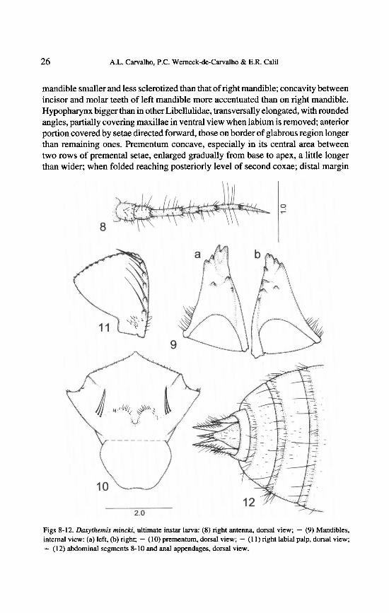
ultimateinstar larva: (8) right antenna, dorsal view; — (9) Mandibles,
internal view: (a) left, (b) right; — (10) prementum, dorsal view; — (11) right labialpalp, dorsal view;— (12) abdominal segments 8-10 and anal appendages, dorsal view.
Dasythemis mincki,Figs 8-12.

27Description of two Dasythemis larvae
of prementum (ligula) pronounced with nine to eleven regularly disposed setae;
space between setae finely serrate; external area close to palpal articulation with a
group of setae, three or four ofthem visible in dorsal view; row of premental setae
“S” shaped, separated in two distinct groups: external one formed by three long
setae very close to each other, and internal one formed by eleven to thirteen short
setae; setae of internal group increase in length toward central axis of prementum;
area between row of premental setae and palpal articulation with very short and
scattered setae in some specimens. Labial palp triangular; palp with four palpal
setae, distal one smaller and similar in length to movable hook; base of row of
palpal setae with group of fifteen to twenty short setae that may extend to level of
third palpal seta; external margin with row of very short setae partially visible in
dorsal view; distal margin composed of twelve shallow crenulations, with about
three setae on each one; internal margin with smooth serrations, with about three
setae on distal portion.
Thorax.-General shape ofpronotum semicircular, with long setae especially
numerous on lateral borders. Wing cases parallel; posterior pair reaching to
abdominalsegment6. Legs with many setae, especially long and concentratedalong
carinae; tarsi with only simple setae on ventralregion.
Abdomen. - General shape rounded, convex, with flattenedborders; apex
curvedupward; intersegmental membranesalmost invisible. Segments 8 and 9 with
shortlateral incurved spines, abouthalfmid-dorsal length ofsegment 8. Abdominal
tergites with rows of setae on lateral borders; lateral areas with some differentiated
setae, shorter andwider than others. Anal appendages conic, sharply pointed; epiproct
and paraprocts similar in length; cerci extending to halflength ofepiproct; epiproct
and lateral area of paraprocts with uniformly developed setae on dorsal subapical
regions.Measurements (in mm; n= 2). - Total length 14.88/ 15.20; median length ofhead 3.26/3,26;
maximum width ofhead 4.42 / 4.35; total length ofright antenna 2.30 / 2.24; length ofantennal segments
0.29-0.25-0.46-0.19-0.31 -0.42-0.50 / 0.26-0.25-0.45-0.21 -0.29-0.40-0.46;median length ofprementum
4.16/4.03; maximum width of prementum 3.71 / 3.78; length ofposterior right wing case 5.06 / 5.06;
length ofposterior rightfemur 4.93 /4,93; length ofposterior right tibia 5.25 / 4.93; maximum widthof
abdomen (seg. 6) 4.67 / 4.80; length of epiproct (dorsal view) 1.50 / 1.57; basal width of epiproct
(dorsal view) 0.97 / 1.00; maximum length ofright paraproct (dorsal view) 1.37 / 1.47; length of right
cerci (dorsal view) 0.63 / 0.63.
DASYTHEMISMINCKI (KARSCH, 1890)
Figures 2, 8-12
Material.- BRAZIL, Minas Gerais, Itamonte, 12-IX-1998, Lab. EntomologiaUFRJ leg., 11
larvae: 1 S (F/ exuviae), 2 <J (F),2 9 (F),3?(F-1), 1 ?(F-2), 2 ?(F-?); 02-X-1999, Lab. Entomologia
UFRJ leg., 11 larvae: 1 3 (F /exuviae), 1 9 (F/ exuviae), 2 S (F), 1 9 (F), 5 ? (F-1), 1 ? (?).
Larva very similar to that of D. venosa, described above, differing only in the
features described below.
28 A.L. Carvalho, P C. Werneck-de-Carvalho & E.R. Calil
Head.- 3rd segmentof antenna longer than 7th. Labial palp with four or five
palpal setae.
Abdomen. - General shape semi-cylindric in dorsal view; apex slightly
curved upward; intersegmental membranes visible. Lateral spines absent on all
segments. Abdominal tergites with posterior margin of each segment with row of
numerous setae irregularly developed, some ofthem very long; lateralareas without
differentiatedsetae. Epiproct and lateral area ofparaprocts with some differentiated
setae on dorsal subapical regions.Measurements (in mm, n = 5).-Total length 16.00-19.04; median length of head 2.82-3.14;
maximum width ofhead 4.48-4.67;total length ofright antenna 2.37-2.43; length ofantennal segments
0.25-0.26 / 0.30-0.35 / 0.46-0.50 / 0.25-0.26 / 0.29-0.31 / 0.37-0.42 / 0.41-0.44; median length of
prementum 3.84-4.29; maximum width of prementum 3.84-3.97; length of posterior right wing case
5.12-5.44; length ofposterior right femur 3.71-3.97; length of posterior right tibia4.67-5.12; maximum
width of abdomen (seg. 6)4.42-5.00;length ofepiproct (dorsal view) 1.27-1.37;basal width ofepiproct
(dorsal view) 0.87-0.95; maximum length of right paraproct (dorsal view) 1.20-1.37; length of right
cerci (dorsal view) 0.60-0.68,
NOTES ON BIOLOGY
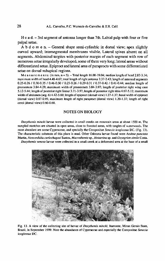
Dasythemis mincki larvae were collected in small creeks on mountain areas at about 1500 m. The
sampled stretches are situated in open areas, close to forested areas, with tangles of waterweeds. The
most abundant are some Cyperaceae, and specially the CompositaeSenecio icoglossus DC, (Fig. 13).
The characteristic substrate of this place is mud. Other Odonata larvae found were Aeshna punctata
Martin,Neocordulia carlochagasi Santos,Macrothemis sp., Hetaerina sp. and Oxyagrion simile Costa.
Dasythemis venosa larvae were collected in a small creek at a deforested area at the base ofa small
Fig. 13. A view of the collecting site of larvae of Dasythemis mincki, Itamonte, Minas Gerais State,
Brazil, in September 1999. Note the abundance of Cyperaceae and especially the Compositae Senecio
icoglossus DC.

29Description oftwo Dasythemis larvae
forested hill at about 600 m. This creek springs from a small area covered by dense vegetation. The
bottom is composed of a reddish mud covered by Cyperaceae. The surroundingareas are urbanized, or
used as sugar cane plantation. The larvae of this species were the only ones found. Deep ponds are
formed some meters down the creek, with Typha sp. on their margins, and there larvae of Aeshna
cornigeraplanalticaCalvert were found.
DISCUSSION
The most important diagnostic feature of Dasythemis larvae is the presence of a
groupofthree long setae clearly more developed than the others, very close to each
other, located externally on the rows of premental setae. The larva described by
ROLDÀN & ARANGO (1996) as Dasythemis sp. has the same condition, thus
agreeing with the generic identification.
D. minckilarvae can be easily distinguished from thoseof D. venosa by the absence
oflateral spines on abdominalsegments 8 and 9. Although very similarto D. venosa,
the larva described by ROLDÂN & ARANGO (1996) differs by the number of
palpal setae, four in D. venosa and five inDasythemis sp, and by the apical direction
of the paraprocts, straight to the back in D. venosa and curved to the sides in
Dasythemis sp.
Dasythemis larvae fit in the Libellulinaeby having the eyes dorsally projected,
just like Libellula herculea Karsch (De MARMELS, 1982). This conditionis very
pronounced in the species of Orthemis (e.g. ROLDAN & ARANGO, 1996).
The breeding habitats ofthe two Dasythemis larvae studied here are very similar,
i.e. brooklets with little water flow, in open areas, close to their springs (crenon).
Thebottom is predominantly muddy, where aquatic plants are abundant. Information
concerning the breeding environment of the larva described by ROLDAN &
ARANGO (1996) corroborates these data.
A PRELIMINARY KEY TO THE GENERA OF LIBELLULIDAE LARVAE
OCCURRING IN THE STATES OFRIO DE JANEIRO AND SÃO PAULO, BRAZIL
The difficulty in identifying libellulidlarvae occurring in Brazil is due to the lack
of proper literature. The lack of adequate keys lead to the use ofthose constructed
for the fauna of the United States of America (e.g. NEEDHAM & WESTFALL,
1955;GLOYD&WRIGHT, 1959; WESTFALL&TENNESSEN,1996), or adaptedfor thefaunaofsome othercountriesofthe neotropical region, which do not include
many endemic groups (e.g. RODRIGUEZ-CAPITULO, 1992, for Argentina).
Furthermore, the comparison of material with the descriptions and illustrations
scattered in hundreds of articles is a very arduous procedure and not always
successful.
Theknowledge about the larvaeof the family occurring in Brazil is concentrated
in the representatives of the states ofRio de Janeiroand Sao Paulo, where the group
has been regularly studied in the past fifty years (CARVALHO& NESSIMIAN,

30 A.L. Carvalho, P.C. Werneck-De-Carvalho & E.R, Calil
1998; COSTA et al„ 2000). Thus, a preliminary key to the Libellulidaegenera
occurring in these states is perfectly workable and represents the first step to a
future key for the entire country. Reliable keys to the genera of other Anisoptera
families have already been published (e.g. CARVALHO, 1989,for the Aeshnidae;
BELLE, 1992, for the Gomphidae). Of the 56 anisopteran generarecorded for these
states, 30 are Libellulidae(CARVALHO & NESSIMIAN, 1998; COSTA et al„
2000).
For the elaboration of the key presented below, in addition to the literature, we
used material deposited in the entomological collectionof the Departamento de
Zoologia, Institute de Biologia, UFRJ.
Among the genera included in the key, those asterisked are based on literature
data only. Taxa based on supposition are marked with a question mark. In order to
cover the genera containing unknown larvae of the species occurring in the states
of Rio de Janeiro and Sao Paulo, representatives from other regions are considered.
Edonis,Ipirangathemis, Oligoclada
,Tholymis and Uracis are not included, since
no larvae are described or known in collections.
This key presents an original structure, that tries to avoid problems caused bydifferences between representatives of the same genus from different regions.
Whenever possible, easily visualized external morphology features are used.
Characters’ conditions that commonly vary interspecifically are avoided.
1 Mid-dorsal spines (hooks orknobs) present on some abdominal segments 2
— No mid-dorsal spines on any abdominal segment 16
2 Eyes with conic region expanded laterally; distal margin of labial palps with digitiform crenula-
tions 3
— Eyes without conic region expanded laterally; distal margin of labial palps without digitiform
crenulations 4
3 Distal margin of labial palps without digitiform crenulations on 1/3 adjacent to internal margin;
length of lateral spines on abdominal segment 9 as long as or longer than four times the median-
dorsal length of this segment Diastatops (obscura, intensa)— Distal margin of labial palps with digitiform crenulations full length; length of lateral spines on
abdominal segment 9 shorter than four times the median-dorsal length of this segment
Zenithoptera (?)
4 No mid-dorsal spine on abdominal segment 9 5
— Mid-dorsal spine on abdominal segment 9 8
5 Distal margin oflabial palps almost smooth, without distinct crenulations; lateral spines present on
abdominal segments 6 and 7 Elasmothemis (cannacrioides , constricta)— Distal marginof labial palps with distinct crenulations;no lateral spines on abdominal segments 6
and 7 6
6 Eyes slightly protuberant dorsally, restricted in dorsal view to anterior 2/3 of head; abdominal
segment 9 with lateral spines distinctly shorter than paraprocts Libellula ( herculea )
— Eyes not protuberant dorsally. commonly enlarged laterally, reaching in dorsal view to posterior1/3 of head; abdominal segment 9 with lateral spines as long as paraprocts 7
7 Palpal setae eight or nine; epiproct in dorsal view shorter than half length of paraprocts
Tauriphila( argo )— Palpal setae six or seven; epiproct in dorsal view as long as 2/3 length of paraprocts

31Description of two Dasythemis larvae
.................................................Miathyria (marcella, simplex)
8 Epiproct in dorsal view longer than twice width of its base 9
— Epiproct in dorsal view shorter than twice width of its base 10
9 Palpal setae five or six; small mid-doisal spineon abdominal segment 10 Idiataphe(cubensis9 Palpal setae five or six; small mid-dorsal spineon abdominal segment 10 Idiataphe(cubensis ?)— Palpal setae nine to eleven; no mid-dorsal spine on abdominal segment 10
Brachymesia furcata, herbida)
10 Third segment of antenna shorter than second; prementum enlarged abruptly in its distal half, its
lateral margins in ventral view strongly concave Elga* (leptostyla)— Third segment of antenna distinctly longer than second; prementum enlarged gradually from base
to apex, its lateral margins in ventral view straight or slightly concave 11
11 Distal margin of labial palps with just one seta on each crenulation; lateral spines of abdominal
segment 9 as long as median-dorsal length of this segment Planiplax (erythropyga)
— Distal marginoflabial palps with twoormore setae on each crenulation; lateral spines of abdominal
segment 9 shorter than median-dorsal length of this segment 12
12 Anterior region ofocciput dark, forming stripe extended from eyes to centre of head, resembling a
kind of “mask” Perithemis (mooma, electra, icteroptera)— Anterior region of occiput notespecially dark 13
13 Integument of body granulöse dorsally; cerci in lateral view generally as long as or shorter than
half length ofepiproct Brechmorhoga (nubecula, travassosi, Brechmorhogahalf length ofepiproct Brechmorhoga (nubecula, travassosi, Brechmorhoga spp.)
- Integumentofbody smooth, setose ornot distinctly granulöse dorsally;cerci in lateral view distinctly
longer than half length ofepiproct 14
14 Crenulations on distal marginof labial palps not obsolete
Macrothemis ( declivata, hemichlora, musiva, tesselata)— Crenulations on distal margin oflabial palps obsolete 15
15 Anterior margin of prementurn and internal margin of labial palps armed with setae with apex
flattened; occipital margin of head with claviform setae Gynothemis (venipuctata)
— Anterior margin ofprementum and internal marginof labial palps armed with pointed setae; occipital
margin of head with cylindric setae Dythemis (multipunctata)
16 Eyes extended well above level of top of the head, directed more upward than laterally
Orthemis ( discolor, cultriformis)
— Eyes not extended well above level of top of the head, directed upward and laterally 17
17 Eyes slightly protuberant dorsally; external group ofpremental setae composed ofonly three long
setae very close to each other Dasythemis mincki, venosa)
— Eyes not protuberant dorsally; external group of premental setae composed of four or more long
setae 18
18 Lateral spines of abdominal segment 9 distinctly longer than median dorsal length of this seg-
ment 19
— Lateral spines of abdominal segment 9 distinctly shorter than median dorsal length of this seg-
ment 21
19 Distal margin oflabial palps almost smooth, without distinct crenulations; abdominal segment 8
without lateral spines Rhodopygia ( cardinalis)
— Distal marginof labial palps with distinct crenulations;abdominal segment 8 with lateral spines 20
20 Colour of anterior, median and hind tarsi similar, generally ochraceous; lateral spines ofabdominal
segments 8 and 9 similar in length Tramea ( abdominalis , binotata , cophysa)
— Colour of median and hind tarsi distinctly darker than that ofanteriorone; lateral spines ofabdominal
segment 8 distinctly shorter than those of 9 Pantala (flavescens, hymenaea)
21 Body truncate, compressed laterally; dorsal surface of head slanted forward; apex ofparaprocts in
lateral view distinctly curved downward
Erythemis (attala, credula, mithroides,plebeja, peruviana, vesiculosa )
— Body not truncate, somewhat depressed dorsoventrally; dorsal surface of head not slanted forward;
apex of paraprocts in lateral view straight or little curved downward 22

32 A.L. Carvalho, P.C. Werneck-de-Carvalho & E.R. Calil
22 Paraprocts in lateral view longer than twice lengthof cerci, distinctlylonger than epiproct 23
— Paraprocts in lateral view shorter than twice length of cerci, nearly as long as or slightly longer
than epiproct 24
23 Colour pattern of abdomen poorly defined dorsally, almost uniform; apical portion of femora with
two to four conspicuous dark setae dorsally Nephepeltia (phryne)
— Colour pattern of abdomen generally well defined dorsally, composed ofdark and clear spots and scars;
apical portionof femora without conspicuous dark setae dorsally Micrathyria (artemis, atra, borgmeieri,
didyma, hesperis, hypodidyma, mengeri, ocellata ,pirassunungae, stawiarskii)
24 Colour pattern of abdomen generally well defined dorsally, composed ofdark and clear spots and
scars; anal appendages in lateral view straight Anatya (januaria)
— Colour pattern of abdomen poorly defined dorsally, almost uniform; anal appendages in lateral
view somewhat curved downward
Erythrodiplax (anomala,fusca, hyalina,juliana,ochracea, paraguayensis, umbrata)
ACKNOWLEDGEMENTS
Thanks to the colleagues of the Laboratorio de Entomologia,Instituto de Biologia, UFRJ, for their
great help during the identification of the material, especially to Professors JULIANA ASSIS and Dr
JORGE NESSIMIAN. We are specially grateful to Professor Dr GABRIEL MEJDALANI (Museu
Nacional, UFRJ), for critical reading the manuscript; to Professor ROBERTO L. ESTEVES (UERJ)
for the identification of the Compositae, and to the supporting agencies CNPq-PIBIC-UFRJ, FAPERJ,
and FUJB.
REFERENCES
BELLE, J., 1992, Studies on ultimate instar larvae of neotropical Gomphidae, with the description of
Tibiagomphusgen, nov. (Anisoptera). Odonatologica21(1): 1-24.
CARVALHO, A.L., 1989. Description of the larva of Neuraeschna costalis (Burmeister), with notes on
its biology, and a key to the genera of Brazilian Aeshnidae larvae (Anisoptera). Odonatologica
18(4): 325-332.
CARVALHO, A.L., 1992. Aspectos da biologiede Coryphaeschnaperrensi (McLachlan, 1887) (Odonata,
Aeshnidae), com ênfase no periodo larval. Revta bras. Ent. 36(4): 791-802.
CARVALHO, A.L. & J.L. NESSIMIAN, 1998. Odonata do Estado do Rio de Janeiro, Brasil; Habitats
e hâbitos das larvas. In: Nessimian, J.L. & A.L. Carvalho, [Eds], Ecologia de insetos aqudticos
(Oecologia Brasiliensis 5), Rio de Janeiro: PPGE-UFRJ, pp, 3-28.
CORBET, P.S., 1953. A terminologyfor the labium of larval Odonata. Entomologist86: 191-196.
COSTA, J.M., A.B.M. MACHADO, F.A.A. LENCIONI & T.C. SANTOS, 2000. Diversidade e
distribuiçâodos Odonata(Insecta) no Estado de Sâo Paulo, Brasil, 1 : Lista de espécies e registres
bibliogrâftcos. Publqoes avals. Mus. nac. 80: 1-27.
DAVIES, D.A.L. & P. TOBIN, 1985. The dragonflies of the world: a systematic list of the extant
species ofOdonata, Vol. 2: Anisoptera. Soc. Int. Odonatol. rapid Comm. (Suppl.) 5: 1-151.
DE MARMELS, J., 1982. Cuatro nâyades nuevasde la Camilla Libellulidae (Odonata; Anisoptera).
Boln Ent. venez. (N.S.) 2(11): 94-101.
GLOYD, L.K. & M. WRIGHT, 1959. Odonata. In: Edmundson, W.T.,[Ed.], Fresh-water biology, pp.
917-940. John Wiley, New York.
NEEDHAM, J.G.& M.J. WESTFALL, 1955. A manual of the dragonfliesofNorth America (Anisoptera)
including the Greater Antilles and the provinces of the Mexican border. Univ. California,
Berkeley.
RIS, F., 1910. Libellulinen monographisch bearbeitet. Colins zool. de Selys Longchamps 11: 245-384,
33Description oftwo Dasythemis larvae
pis 1-3 excl.
RODRIGUEZ-CAPITULO, A., 1992. Los Odonata de la Republica Argentina (Insecta). Fauna Agua
dulce Argent. 34(1): 1-91.
ROLDÀN,P.G. & M.C. ARANGO, 1996. Orden Odonata. In: Roldân, P.G., [Ed.], Gui'a para el estudio
de los macroinvertebrados acudticos del Departamenlo de Antioquia, pp. 39-77. Fondo Fen
Colombia, Colciencias, Universidad de Antioquia, Medellin.
SANTOS, N.D., 1988. Catâlogo bibliogrâfico de ninfas de odonatos neotropicais. Acta amazon. 18
(1/2): 265-350.
WATSON, M.C., 1956. The utilization of mandibular armature in taxonomie studies of anisopterous
nymphs. Trans. Am. enl. Soc. 81: 155-202.
WESTFALL, M.J. & K.J. TENNESSEN, 1996. Odonata. In: R.W. Merritt & K.W. Cummins, [Eds],
An introduction to the aquatic insects ofNorth America [3rd ed.], pp. 164-211, Kendall &
Hunt, Dubuque,