Prediction of pKa Values for Neutral and Basic Drugs based ...
Upload
independentCategory
view
1download
0


where the microtubules converged at both sides of the nucleus inprophase, and right in the middle of the asters in prometaphase(Fig. 2). Using a centrosomal staining (g-tubulin) we confirmedthat this specific PKA localization corresponds to the centro-somes, and is independent of PKA activity inhibition (data notshown).
So prior to the increase of PKA activity highlighted frommetaphase to cytokinesis, PKA is highly concentrated in the cen-trosomal regions.
PKA activity is high near the chromosomes duringmetaphase and anaphase
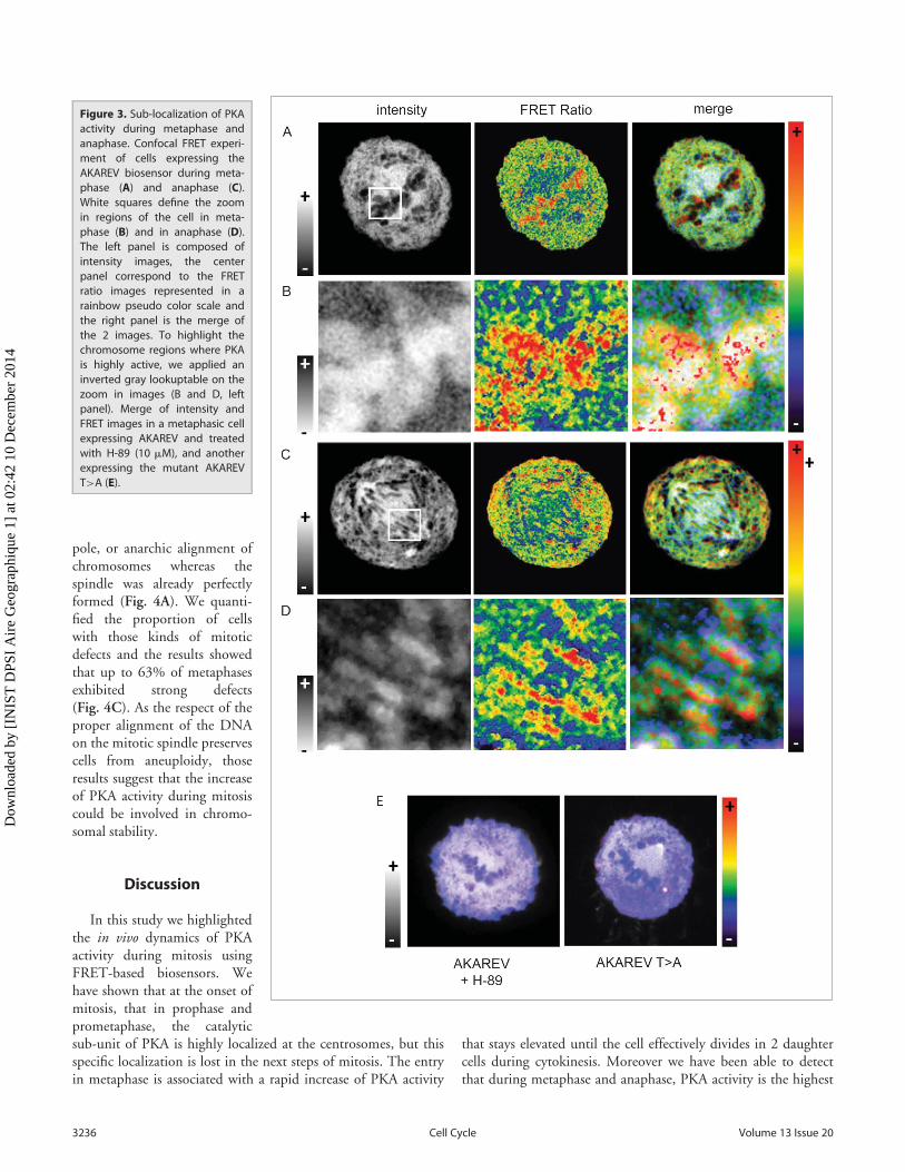
In order to observe PKA activity sub-localization, we per-formed FRET experiments increasing the spatial resolution aswell as adding optical sectioning using confocal microscopy.HeLa cells expressing the biosensor were synchronized using thy-midine block and were fixed 9h after their release in freshmedium. In Figure 3 are represented intensity images in order todefine the different structures inside the mitotic cells (Fig. 3, leftpanel) and we applied a pseudo color scale on FRET images thatreveals in red the highest activity regions within the cell (Fig. 3,center and right panel). We also chose to merge the intensity andratio channels in order to keep to sub-cellular structure informa-tion associated with PKA activity (Fig. 3, right panel). On thezoomed region (Fig. 3B and 3C) we applied an inverted gray
scale, so that on the merge thestructure of the condensed DNAwas highlighted.
Those experiments show thatduring metaphase PKA activity isparticularly high in the closevicinity of the condensed chro-mosomes (Fig. 3A and 3B) inboth metaphase and anaphase. Inthe same way a strong PKA activ-ity is detected near the chromatinduring anaphase, while this activ-ity is lower between the 2 bundlesof chromosomes (Fig. 3C and3D). Moreover cells expressingthe mutant biosensor, or treatedwith a PKA inhibitor result in theabolition of any sub localizationof PKA activity confirming theprevious results (Fig. 3E).
Inhibition of PKA activityinduces mitotic defects thatdo not delay the progressionof mitosis
Consistent with our data,according to which PKA plays arole at the centrosome and at thelevel of the condensed chromo-somes during mitosis, several
clues in literature also converge in the idea that PKA activity dur-ing mitosis is involved in chromosomes positioning.
It was also previously reported that an artificial increase of PKAactivity using a cAMP analog during metaphase accelerate the exit ofmitosis.9 Now that we define that, at least in our cell line, mitosiswas characterized by an increase of PKA activity from metaphase tocytokinesis, we wanted to observe if preventing experimentally thisincrease using the PKA activity inhibitor H-89 dihydrochloride,could lead to opposite effects. We thus synchronize the cells in lateG2 using a thymidine block followed by a nocodazole block. Cellswere then release in presence or absence of 10 mM of H-89 andfixed 1h and 2 h later.
Using a combination of DNA and spindle staining in confocalmicroscopy we image hundreds of cells in each conditions andclassified them in the different step of mitosis. The repartition ofcells through the different steps did not reveal any delay or accel-eration in the progression of mitosis compared to the controlexperiment. As an example, 2h after nocodazole release, in con-trol conditions 64.6 § 10.8% of cells exhibited the phenotype ofa G1 cell, characterized by the presence of a cytoplasmic bridgebetween 2 daughter cells. Upon treatment with the inhibitor, G1cells represented 57.7 § 5.3% of the population, revealing anabsence of delay of mitosis when PKA is inhibited (Fig. 4B).Nevertheless it appeared that cells that have been treated with thePKA inhibitor exhibited improper phenotype in metaphase, suchas a chromosome that have already migrated to one of the spindle
Figure 2. PKA localization during mitosis. HeLa cells at different mitotic stages from prophase to cytokinesisexhibiting a DNA staining (Hoechst, in blue, upper panel), a spindle staining (a-tubulin, in green, secondpanel) and a PKA catalytic sub-unit staining (in red, third panel). The lower panel represents the merge of thedifferent channels. White arrows highlight the centrosomal regions where PKA catalytic sub-unit is moreconcentrated.
www.landesbioscience.com 3235Cell Cycle
Dow
nloa
ded
by [
INIS
T D
PSI
Air
e G
eogr
aphi
que
1] a
t 02:
42 1
0 D
ecem
ber
2014
pole, or anarchic alignment ofchromosomes whereas thespindle was already perfectlyformed (Fig. 4A). We quanti-fied the proportion of cellswith those kinds of mitoticdefects and the results showedthat up to 63% of metaphasesexhibited strong defects(Fig. 4C). As the respect of theproper alignment of the DNAon the mitotic spindle preservescells from aneuploidy, thoseresults suggest that the increaseof PKA activity during mitosiscould be involved in chromo-somal stability.
Discussion
In this study we highlightedthe in vivo dynamics of PKAactivity during mitosis usingFRET-based biosensors. Wehave shown that at the onset ofmitosis, that in prophase andprometaphase, the catalyticsub-unit of PKA is highly localized at the centrosomes, but thisspecific localization is lost in the next steps of mitosis. The entryin metaphase is associated with a rapid increase of PKA activity
that stays elevated until the cell effectively divides in 2 daughtercells during cytokinesis. Moreover we have been able to detectthat during metaphase and anaphase, PKA activity is the highest
Figure 3. Sub-localization of PKAactivity during metaphase andanaphase. Confocal FRET experi-ment of cells expressing theAKAREV biosensor during meta-phase (A) and anaphase (C).White squares define the zoomin regions of the cell in meta-phase (B) and in anaphase (D).The left panel is composed ofintensity images, the centerpanel correspond to the FRETratio images represented in arainbow pseudo color scale andthe right panel is the merge ofthe 2 images. To highlight thechromosome regions where PKAis highly active, we applied aninverted gray lookuptable on thezoom in images (B and D, leftpanel). Merge of intensity andFRET images in a metaphasic cellexpressing AKAREV and treatedwith H-89 (10 mM), and anotherexpressing the mutant AKAREVT>A (E).
3236 Volume 13 Issue 20Cell Cycle
Dow
nloa
ded
by [
INIS
T D
PSI
Air
e G
eogr
aphi
que
1] a
t 02:
42 1
0 D
ecem
ber
2014

in the vicinity of the duplicated chromosomes. If the FRET-based biosensor was able to reveal PKA activity during mitosis,one shall not neglect the impact of phosphatase during this pro-cess. Indeed, intensities measured reflect the changes of a kinase/phosphatase activity balance. Finally we demonstrate that theinhibition of PKA activity prior to mitosis leads to abnormalmitotic phenotypes thus suggesting that the upkeep of PKA activ-ity during mitosis is involved in the maintenance of the chromo-somal stability.
Previous works already pointed out a link between PKA activ-ity and chromosomal stability.25 For example in mouse
hepatoma cells BW1J, the type I regulatory sub-units (RI) as wellas the catalytic subunits of PKA colocalize with the tubulin at themitotic spindle. Moreover overexpression of the a sub-type(RIa) as well as expression of a RIa mutant that acts as a domi-nant negative of PKA activity lead to an increase of the propor-tion of cells possessing a spindle that exhibits more than2 poles.26 Furthermore, PKA is able to phosphorylate oncopro-tein 18 (also called stathmin), a microtubules destabilizing pro-tein whose activity is negatively regulated by phosphorylation atthe onset of mitosis in order to allow the formation of the mitoticspindle.27,28
Figure 4. Effects of PKA inhibition using H-89 during mitosis. Immunostaining of metaphasic HeLa cells synchronized using thymidine-nocodazoleblocks. DNA was stained using Hoechst (upper panel, in red), spindle was stained using an a-tubulin antibody (second panel, in green). The lower panelrepresent the merge of the 2 channels. First column shows a metaphase in control conditions, the next columns show different kind of mitotic pheno-types observed upon inhibition of PKA activity using H-89 (10 mM). White arrows highlight chromosomes or pieces of DNA that have migrated preco-ciously (A). Percentage of cells in each phase of mitosis defined according to phenotype, at 1 hour and 2 hour after release from a thymidine-nocdazolesynchrony, in control and h-89 treated conditions (total number of cell measured on 4 independent experiments: n D 663, n D 632, n D 754 and n D1195 for respectively control condition after 1h and 2h, H-89 condition after 1h and after 2h) (B). Percentage of mitotic defects observed in control andH-89 treated cells. Stars represent p-values obtained on a student t-test performed on the results of the 4 experiments with * < 0.05 and ** < 0.01 (C).
www.landesbioscience.com 3237Cell Cycle
Dow
nloa
ded
by [
INIS
T D
PSI
Air
e G
eogr
aphi
que
1] a
t 02:
42 1
0 D
ecem
ber
2014

Centrosomes are essential to ensure the fidelity of chromo-somes segregation as they guide microtubules within the organi-zation of a bipolar spindle. Consistently with our data exhibitinga high concentration of PKA at the centrosomes (Fig. 2), it hasbeen shown that several AKAPs are able to target PKA to thoseorganelles such as AKAP350, AKAP450, CG-NAP and also peri-centrin.29 As a necessary component of the centrosomal matrix,pericentrin is involved in the organization of the mitotic spindleby interacting with dynein and g-tubulin. Very interestingly, thisprotein, that co-immuno precipitate with both PKA catalytic andtype II regulatory sub-units (RII), is highly phosphorylated uponan elevation of PKA activity.30,31 Moreover, removal of PKAanchoring in the cells leads to spindles defects.29 It has also beenshown that the cAMP level at the centrosome increases duringmitosis and come with an increase of PKA activity in this zone.32
PDE4D3 is a phosphodiesterase that degrades cAMP specificallylocalized at the centrosome. Using an inactive mutant ofPDE4D3, it was shown that disrupting the centrosomal micro-domain of the low cAMP level reported in interphase drives alocal increases of the level of cAMP and accumulation of the cellsin prophase.32 As previously mentionned, several differentAKAPs target PKA to this compartment, which reinforces theidea of multiple roles and/or targets of PKA at the centrosomeand thus its requirement.
In this study we observed that PKA activity is increased nearthe condensed DNA, and that inhibition of the global activity ofPKA inside the cell also leads to mitotic defects (Fig. 3 andFig. 4). Nevertheless we did not observe spindle defects such asmulti-polar spindles. Our data mostly included misalignment ofchromosomes on the metaphase plates when PKA activity isswitched off. We then speculated that PKA could affect theattachment of the DNA on the spindle and thus the kinetochoresfunctions or the segregation of the sister chromatids. Indeed, inyeast, dosage suppression experiments have revealed a geneticlink between PKA signaling and kinetochores. In this modeloverexpression of PKA negative regulators is able to rescue theviability of kinetochore genes mutants.33 Moreover the deletionof BCY1, a negative regulator of PKA, leads to a strong increaseof the chromosome loss rate, involving the attenuation of PKAsignaling as a contribution to kinetochore functions.34 While ourexperiments also demonstrate that the inhibition of PKA leads tomitotic defects (Fig. 4), we hypothesized that PKA activityshould be comprised in a permissive interval (not too high butnot too low) in order to favor a proper cell division.
Moreover PKA is also involved in the regulation of the Ana-phase Promoting Complex that plays an important role in sisterchromatids segregation. PKA must be inhibited during mitosis inorder to promote the activation of this complex.35 We canhypothesize that PKA inhibition allows the cells to by-pass themetaphase/anaphase checkpoint. Further experiment about thehow PKA activity could also affect this checkpoint activation insuch context could bring essential information to understand theinvolvement of this kinase in chromosomal stability.
In conclusion, as a member of the family of signaling mole-cules, PKA is able to phosphorylate multiples targets includingseveral crucial proteins required for mitosis and maintenance of
genetic integrity by preserving or acting on centrosomes andkinetochores functions as well as sister chromatids segregation orDNA condensation. All the elements, previously mentioned areconverging to the idea of a precise spatial but also temporal regu-lation of PKA activity within the cell during mitosis. We providehere a precise overview of PKA spatio-temporal dynamics, inHeLa cells, during mitosis, that appears essential to understandthe way the different specific effects of PKA are associated in theregulation of cell division.
Materials and Methods
Chemical reagents and antibodiesThymidine (http://www.sigmaaldrich.com/catalog/product/
sigma/t1895), Nocodazole (http://www.sigmaaldrich.com/catalog/product/sigma/m1404) and the PKA inhibitor H-89dihydrochloride (http://www.sigmaaldrich.com/catalog/product/sigma/b1427) were from Sigma and used respectively at2.5 mM, 100 nM and 10 mM. Forskolin was purchased fromTocris Bioscience and used at 12.5 mM (http://www.tocris.com/dispprod.php?ItemId D 2337). Anti-a-Tubulin antibodyproduced in mouse was purchased from Sigma (http://www.sigmaaldrich.com/catalog/product/sigma/t6199), Anti-cAMPProtein Kinase Catalytic subunit antibody was from Abcam(http://www.abcam.com/camp-protein-kinase-catalytic-subunit-antibody-ab26322.html). Alexa conjugated secondary antibodiesproduced in donkey were purchased from Invitrogen and weused specifically an anti-mouse IgG-alexa488 (https://www.lifetechnologies.com/order/catalog/product/A21202?CID Dsearch-a21202) and an anti-rabbit IgG-alexa594 (http://www.lifetechnologies.com/order/catalog/product/A21207?ICID Dsearch-a21207). Hoechst 33242 was purchased from MolecularProbes, Life Technologies.
PlasmidsThe plasmid containing the AKAREV biosensor sequence has
been provided by Dr Jun-ichi Miyzaki.23 The mutated biosensorAKAREV T>A was constructed by site directed mutagenesisaccording to the procedure described by Sawano and Miyawaki 36
using the reverse primer 50 GCCGTCAACCAGCGCCGCGCGCCTCAATC 30.
Cell culture, transfection and synchronyHeLa cells were grown in Dulbecco’s modified Eagle’s
medium supplemented with 10% foetal calf serum and antibiot-ics (100 units/mL of penicillin and 100 mg/mL of streptomycin)at 37�C with 5% CO2. Cells were seeded on 10 mm or 32 mmcoverslips in 6-well plates and grown to reach 30–50% of conflu-ency. For FRET experiment, cells were then transfected with1.5 mg of DNA per well using FuGENE HD (Roche) transfec-tion reagent. Cells were synchronized using 2.5mM of thymidineduring 20h, and were then released into fresh media to progressfrom S phase to mitosis (9h). Depending on the experiment, anadditional step of synchrony, prior to the mitosis, have been per-formed using a 100 nM nocodazole incubation during 4h.
3238 Volume 13 Issue 20Cell Cycle
Dow
nloa
ded
by [
INIS
T D
PSI
Air
e G
eogr
aphi
que
1] a
t 02:
42 1
0 D
ecem
ber
2014
Immuno-fluorescence stainingCells on coverslips were fixed with paraformaldehyde 4% for
30 min, then washed and permeabilised using a 0.5% Triton-100X for 5 min. Then cells were incubated 1h in a blocking solu-tion containing 2% Foetal calf serum, 5% donkey serum, and2% Bovine serum albumin (PKAc and a-tubulin antibodies) or2% Bovine serum albumin (g-Tubulin antibody). Cells werethen incubated for 2h with primary antibodies diluted respec-tively at 1/500, 1/700 and 1/2000 for PKAc (rabbit), a-tubulin(mouse) and g-Tubulin (rabbit). After washing, cells were incu-bated with the appropriate secondary antibodies: IgG anti-mouseconjugated to alexa 488 and IgG anti-rabbit conjugated to alexa594. Cells were then washed and incubated with Hoechst (2uM,10min). Finally coverslips were mounted on slides using Mowiol488 mounting medium and sealed with clear nail polish.
Time lapse FRET imagingTime lapse experiment were performed on cells in supple-
mented DMEM without red phenol for long term experimentsor in Leibowitz medium (L-15) for short term experiments, on athermostatted (37�C) inverted Leica AF6000 videomicroscope(Leica Microsystem) with 63 £ 1.3 NA glycerin-immersionobjective. Fluorescence excitation was performed with a 427 §10nm bandpass filter through a double band dichroic mirror(440/520 nm). To avoid displacements between donor andacceptor’s fluorescence measurements, we used fast detection fil-ter wheel (respectively with 472 § 30 nm and 542 § 27 nmbandpass filters)
Confocal microscopyImages were acquired on a Nikon A1R confocal microscope
using a 60 £ 1.4 NA oil-immersion objective. For immuno-staining experiments, fluorescence excitation was performed
sequentially at 404, 488 and 561 nm and signal were respectivelydetected at 450 § 50, 525 § 50 and 595 § 50. For FRET exper-iment, excitation was performed at 404 nm and signals weredetected simultaneously at 450 § 50 and 525 § 50 nm.
FRET analysisFRET ratios from widefield microscopy experiment were cal-
culated with an ImageJ and a Matlab routine developed accord-ing to the procedure described previously by Kardash andcoworkers.37 For more details about the system implementationand application, see.22 The same strategy has been used for con-focal FRET images except concerning the background subtrac-tion that was realized manually.
Disclosure of Potential Conflicts of Interest
No potential conflicts of interest were disclosed.
Acknowledgments
We thank the personal of the BICeL-Lille1-HB Facility foraccess to the microscopy systems and technical advices. We thankJun-ichi Miyazaki as source for the material. We are also gratefulto Alain Martoriati for discussion and comments, and we thankArlette Lescuyer and Franck Riquet for their technical assistance.
Funding
Pauline Vandame is granted by the University of Lille 1 andthe Region Nord-Pas-de-Calais. The research was performed inthe context of SIRIC ONCOLille (grant INCa-DGOS-Inserm6041).
References
1. Pearce LR, Komander D, Alessi DR. The nuts andbolts of AGC protein kinases. Nat Rev Mol Cell Biol2010; 11:9-22; PMID:20027184; http://dx.doi.org/10.1038/nrm2822
2. Lefkimmiatis K, Zaccolo M. cAMP signaling in sub-cellular compartments. Pharmacol Ther 2014;143:295-304; PMID:24704321; http://dx.doi.org/10.1016/j.pharmthera.2014.03.008
3. McConnachie G, Langeberg LK, Scott JD. AKAPsignaling complexes: getting to the heart of the mat-ter. Trends Mol Med 2006; 12:317-23;PMID:16809066; http://dx.doi.org/10.1016/j.molmed.2006.05.008
4. Ferrari S. Protein kinases controlling the onsetof mitosis. Cell Mol Life Sc 2006; 63:781-95;PMID:16465440; http://dx.doi.org/10.1007/s00018-005-5515-3
5. Collas P, Le Guellec K, Tasken K. The A-kinase-anchoring protein AKAP95 is a multivalent pro-tein with a key role in chromatin condensationat mitosis. J Cell Biol 1999; 147:1167-80;PMID:10601332; http://dx.doi.org/10.1083/jcb.147.6.1167
6. Chu CS, Hsu PH, Lo PW, Scheer E, Tora L, TsaiHJ, Tsai MD, Juan LJ. Protein kinase A-mediatedserine 35 phosphorylation dissociates histone H1.4from mitotic chromosome. J Biol Chem 2011;286:35843-51; PMID:21852232; http://dx.doi.org/10.1074/jbc.M111.228064
7. Yamashita YM, Nakaseko Y, Samejima I, Kumada K,Yamada H, Michaelson D, Yanagida M. 20S cyclo-some complex formation and proteolytic activityinhibited by the cAMP/PKA pathway. Nature 1996;384:276-9; PMID:8918880; http://dx.doi.org/10.1038/384276a0
8. Kotani S, Tugendreich S, Fujii M, Jorgensen PM,Watanabe N, Hoog C, Hieter P, Todokoro K. PKAand MPF-activated polo-like kinase regulate ana-phase-promoting complex activity and mitosis pro-gression. Mol Cell 1998; 1:371-80; PMID:9660921;http://dx.doi.org/10.1016/S1097-2765(00)80037-4
9. Zeilig CE, Johnson RA, Sutherland EW, FriedmanDL. Adenosine 3’:5’-monophosphate content andactions in the division cycle of synchronized HeLacells. J Cell Biol 1976; 71:515-34; PMID:186461;http://dx.doi.org/10.1083/jcb.71.2.515
10. Grieco D, Avvedimento EV, Gottesman ME. A rolefor cAMP-dependent protein kinase in early embry-onic divisions. Proc Natl Acad Sci U S A 1994;91:9896-900; PMID:7937913; http://dx.doi.org/10.1073/pnas.91.21.9896
11. Wang J, Cao WL, Liu XJ. Protein kinase A(PKA)-restrictive and PKA-permissive phases of oocytematuration. Cell Cycle 2006; 5:213-7;PMID:16397412; http://dx.doi.org/10.4161/cc.5.2.2365
12. Smith ME, Dickinson JR, Wheals AE. Intracellularand extracellular levels of cyclic AMP during the cellcycle of Saccharomyces cerevisiae. Yeast 1990; 6:53-
60; PMID:2156391; http://dx.doi.org/10.1002/yea.320060106
13. Sheppard CL, Lee LC, Hill EV, Henderson DJ,Anthony DF, Houslay DM, Yalla KC, Cairns LS,Dunlop AJ, Baillie GS, et al. Mitotic activation ofthe DISC1-inducible cyclic AMP phosphodiesterase-4D9 (PDE4D9), through multi-site phosphoryla-tion, influences cell cycle progression. Cell Signal2014; 26:1958-74; PMID:24815749; http://dx.doi.org/10.1016/j.cellsig.2014.04.023
14. Costa M, Gerner EW, Russell DH. Cell cycle-specificactivity of type I and type II cyclic adenosine 3’:5’-monophosphate-dependent protein kinases in Chi-nese hamster ovary cells. J Biol Chem 1976;251:3313-9; PMID:179994
15. Stork PJ, Schmitt JM. Crosstalk between cAMP andMAP kinase signaling in the regulation of cell prolif-eration. Trends Cell Biol 2002; 12:258-66;PMID:12074885; http://dx.doi.org/10.1016/S0962-8924(02)02294-8
16. Chen J, Iyengar R. Suppression of Ras-induced trans-formation of NIH 3T3 cells by activated G alpha s.Science 1994; 263:1278-81; PMID:8122111; http://dx.doi.org/10.1126/science.8122111
17. Deeble PD, Murphy DJ, Parsons SJ, Cox ME. Inter-leukin-6- and cyclic AMP-mediated signaling poten-tiates neuroendocrine differentiation of LNCaPprostate tumor cells. Mol Cell Biol 2001; 21:8471-82; PMID:11713282; http://dx.doi.org/10.1128/MCB.21.24.8471-8482.2001
www.landesbioscience.com 3239Cell Cycle
Dow
nloa
ded
by [
INIS
T D
PSI
Air
e G
eogr
aphi
que
1] a
t 02:
42 1
0 D
ecem
ber
2014

18. Caretta A, Mucignat-Caretta C. Protein kinase a incancer. Cancers (Basel) 2011; 3:913-26;PMID:24212646; http://dx.doi.org/10.3390/cancers3010913
19. Sample V, Mehta S, Zhang J. Genetically encodedmolecular probes to visualize and perturb signalingdynamics in living biological systems. J Cell Sci2014; 127:1151-60; PMID:24634506; http://dx.doi.org/10.1242/jcs.099994
20. Zhang J, Allen MD. FRET-based biosensors for pro-tein kinases: illuminating the kinome. Mol Biosyst2007; 3:759-65; PMID:17940658; http://dx.doi.org/10.1039/b706628g
21. Sipieter F, Vandame P, Spriet C, Leray A, Vincent P,Trinel D, Bodart JF, Riquet FB, Heliot L. FromFRET imaging to practical methodology for kinaseactivity sensing in living cells. Prog Mol Biol TranslSci 2013; 113:145-216; PMID:23244791; http://dx.doi.org/10.1016/B978-0-12-386932-6.00005-3
22. Vandame P, Spriet C, Riquet F, Trinel D, Cailliau-Maggio K, Bodart JF. Optimization of ERK activitybiosensors for both ratiometric and lifetime FRETmeasurements. Sensors (Basel) 2013; 14:1140-54;PMID:24434874; http://dx.doi.org/10.3390/s140101140
23. Komatsu N, Aoki K, Yamada M, Yukinaga H, FujitaY, Kamioka Y, Matsuda M. Development of an opti-mized backbone of FRET biosensors for kinases andGTPases. Mol Biol Cell 2011; 22:4647-56;PMID:21976697; http://dx.doi.org/10.1091/mbc.E11-01-0072
24. Depry C, Allen MD, Zhang J. Visualization of PKAactivity in plasma membrane microdomains. MolBiosyst 2011; 7:52-8; PMID:20838685; http://dx.doi.org/10.1039/c0mb00079e
25. Matyakhina L, Lenherr SM, Stratakis CA. Proteinkinase A and chromosomal stability. Ann N Y AcadSci 2002; 968:148-57; PMID:12119274; http://dx.doi.org/10.1111/j.1749-6632.2002.tb04333.x
26. Imaizumi-Scherrer T, Faust DM, Barradeau S, HellioR, Weiss MC. Type I protein kinase a is localized tointerphase microtubules and strongly associated withthe mitotic spindle. Exp Cell Res 2001; 264:250-65;PMID:11262182; http://dx.doi.org/10.1006/excr.2001.5164
27. Larsson N, Marklund U, Gradin HM, Brattsand G,Gullberg M. Control of microtubule dynamics byoncoprotein 18: dissection of the regulatory role ofmultisite phosphorylation during mitosis. Mol CellBiol 1997; 17:5530-9; PMID:9271428
28. Rubin CI, Atweh GF. The role of stathmin in theregulation of the cell cycle. J Cell Biochem 2004;93:242-50; PMID:15368352; http://dx.doi.org/10.1002/jcb.20187
29. Diviani D, Scott JD. AKAP signaling complexes atthe cytoskeleton. Journal of Cell Science 2001;114:1431-7; PMID:11282019
30. Diviani D, Langeberg LK, Doxsey SJ, Scott JD. Peri-centrin anchors protein kinase A at the centrosomethrough a newly identified RII-binding domain. CurrBiol 2000; 10:417-20; PMID:10753751; http://dx.doi.org/10.1016/S0960-9822(00)00422-X
31. Lutz W, Lingle WL, McCormick D, GreenwoodTM, Salisbury JL. Phosphorylation of centrin duringthe cell cycle and its role in centriole separation pre-ceding centrosome duplication. J Biol Chem 2001;276:20774-80; PMID:11279195; http://dx.doi.org/10.1074/jbc.M101324200
32. Terrin A, Monterisi S, Stangherlin A, Zoccarato A,Koschinski A, Surdo NC, Mongillo M, Sawa A,
Jordanides NE, Mountford JC, et al. PKA andPDE4D3 anchoring to AKAP9 provides distinct reg-ulation of cAMP signals at the centrosome. J CellBiol 2012; 198:607-21; PMID:22908311; http://dx.doi.org/10.1083/jcb.201201059
33. Ma L, Ho K, Piggott N, Luo Z, Measday V. Interac-tions between the kinetochore complex and the pro-tein kinase A pathway in Saccharomyces cerevisiae.G3 (Bethesda) 2012; 2:831-41; PMID:22870406;http://dx.doi.org/full_text
34. Magtanong L, Ho CH, Barker SL, Jiao W, Baryshni-kova A, Bahr S, Smith AM, Heisler LE, Choy JS,Kuzmin E, et al. Dosage suppression genetic interac-tion networks enhance functional wiring diagramsof the cell. Nat Biotechnol 2011; 29:505-11;PMID:21572441; http://dx.doi.org/10.1038/nbt.1855
35. Yanagida M, Yamashita YM, Tatebe H, Ishii K,Kumada K, Nakaseko Y. Control of metaphase-ana-phase progression by proteolysis: cyclosome functionregulated by the protein kinase A pathway, ubiquiti-nation and localization. Philos Trans R Soc Lond BBiol Sci 1999; 354:1559-69; discussion 69-70;PMID:10582241; http://dx.doi.org/10.1098/rstb.1999.0499
36. Sawano A, Miyawaki A. Directed evolution of greenfluorescent protein by a new versatile PCR strategyfor site-directed and semi-random mutagenesis.Nucleic Acids Res 2000; 28:E78; PMID:10931937;http://dx.doi.org/10.1093/nar/28.16.e78
37. Kardash E, Bandemer J, Raz E. Imaging proteinactivity in live embryos using fluorescence resonanceenergy transfer biosensors. Nat Protoc 2011; 6:1835-46; PMID:22051797; http://dx.doi.org/10.1038/nprot.2011.395
3240 Volume 13 Issue 20Cell Cycle
Dow
nloa
ded
by [
INIS
T D
PSI
Air
e G
eogr
aphi
que
1] a
t 02:
42 1
0 D
ecem
ber
2014
Copyright © 2022 FDOKUMEN