Electrophysiological correlates of the disrupted processing of anger in alcoholism
Upload
washingtonCategory
view
2download
0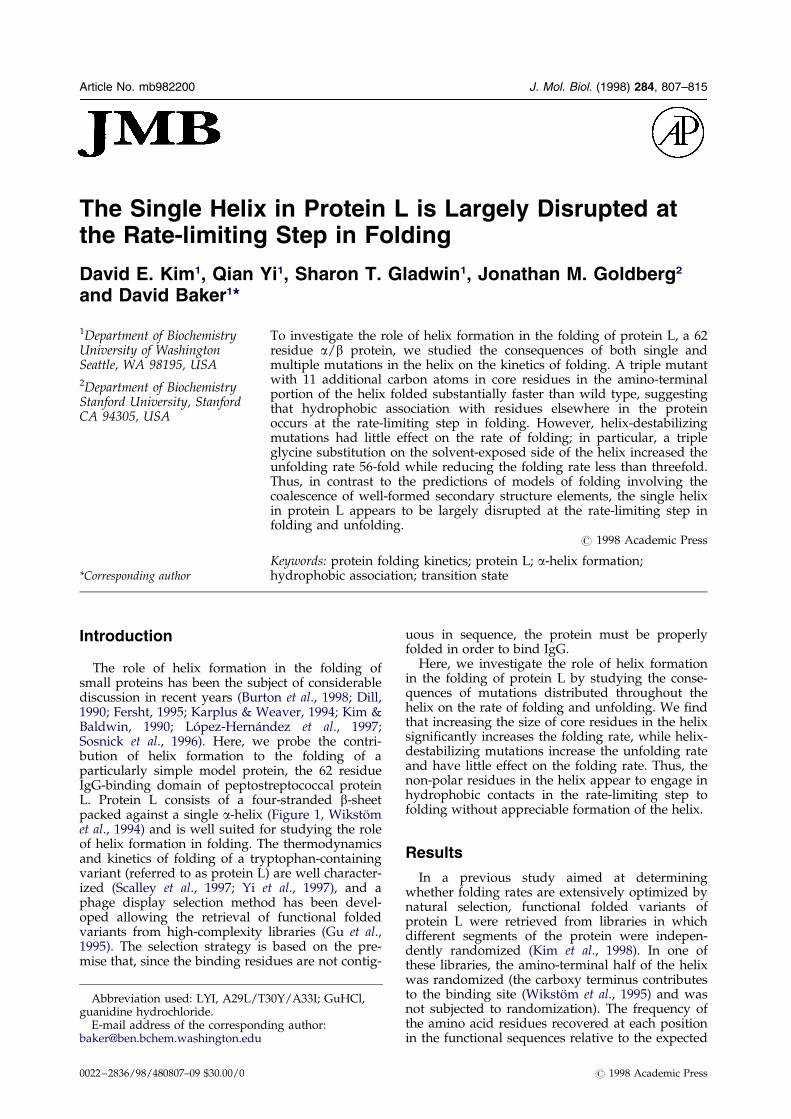
Article No. mb982200 J. Mol. Biol. (1998) 284, 807±815
The Single Helix in Protein L is Largely Disrupted atthe Rate-limiting Step in Folding
David E. Kim1, Qian Yi1, Sharon T. Gladwin1, Jonathan M. Goldberg2
and David Baker1*
1Department of BiochemistryUniversity of WashingtonSeattle, WA 98195, USA2Department of BiochemistryStanford University, StanfordCA 94305, USA
E-mail address of the [email protected]
Abbreviation used: LYI, A29L/guanidine hydrochloride.
0022±2836/98/480807±09 $30.00/0
To investigate the role of helix formation in the folding of protein L, a 62residue a/b protein, we studied the consequences of both single andmultiple mutations in the helix on the kinetics of folding. A triple mutantwith 11 additional carbon atoms in core residues in the amino-terminalportion of the helix folded substantially faster than wild type, suggestingthat hydrophobic association with residues elsewhere in the proteinoccurs at the rate-limiting step in folding. However, helix-destabilizingmutations had little effect on the rate of folding; in particular, a tripleglycine substitution on the solvent-exposed side of the helix increased theunfolding rate 56-fold while reducing the folding rate less than threefold.Thus, in contrast to the predictions of models of folding involving thecoalescence of well-formed secondary structure elements, the single helixin protein L appears to be largely disrupted at the rate-limiting step infolding and unfolding.
# 1998 Academic Press
Keywords: protein folding kinetics; protein L; a-helix formation;hydrophobic association; transition state
*Corresponding authorIntroduction
The role of helix formation in the folding ofsmall proteins has been the subject of considerablediscussion in recent years (Burton et al., 1998; Dill,1990; Fersht, 1995; Karplus & Weaver, 1994; Kim &Baldwin, 1990; LoÂpez-HernaÂndez et al., 1997;Sosnick et al., 1996). Here, we probe the contri-bution of helix formation to the folding of aparticularly simple model protein, the 62 residueIgG-binding domain of peptostreptococcal proteinL. Protein L consists of a four-stranded b-sheetpacked against a single a-helix (Figure 1, WikstoÈmet al., 1994) and is well suited for studying the roleof helix formation in folding. The thermodynamicsand kinetics of folding of a tryptophan-containingvariant (referred to as protein L) are well character-ized (Scalley et al., 1997; Yi et al., 1997), and aphage display selection method has been devel-oped allowing the retrieval of functional foldedvariants from high-complexity libraries (Gu et al.,1995). The selection strategy is based on the pre-mise that, since the binding residues are not contig-
ding author:
T30Y/A33I; GuHCl,
uous in sequence, the protein must be properlyfolded in order to bind IgG.
Here, we investigate the role of helix formationin the folding of protein L by studying the conse-quences of mutations distributed throughout thehelix on the rate of folding and unfolding. We ®ndthat increasing the size of core residues in the helixsigni®cantly increases the folding rate, while helix-destabilizing mutations increase the unfolding rateand have little effect on the folding rate. Thus, thenon-polar residues in the helix appear to engage inhydrophobic contacts in the rate-limiting step tofolding without appreciable formation of the helix.
Results
In a previous study aimed at determiningwhether folding rates are extensively optimized bynatural selection, functional folded variants ofprotein L were retrieved from libraries in whichdifferent segments of the protein were indepen-dently randomized (Kim et al., 1998). In one ofthese libraries, the amino-terminal half of the helixwas randomized (the carboxy terminus contributesto the binding site (WikstoÈm et al., 1995) and wasnot subjected to randomization). The frequency ofthe amino acid residues recovered at each positionin the functional sequences relative to the expected
# 1998 Academic Press
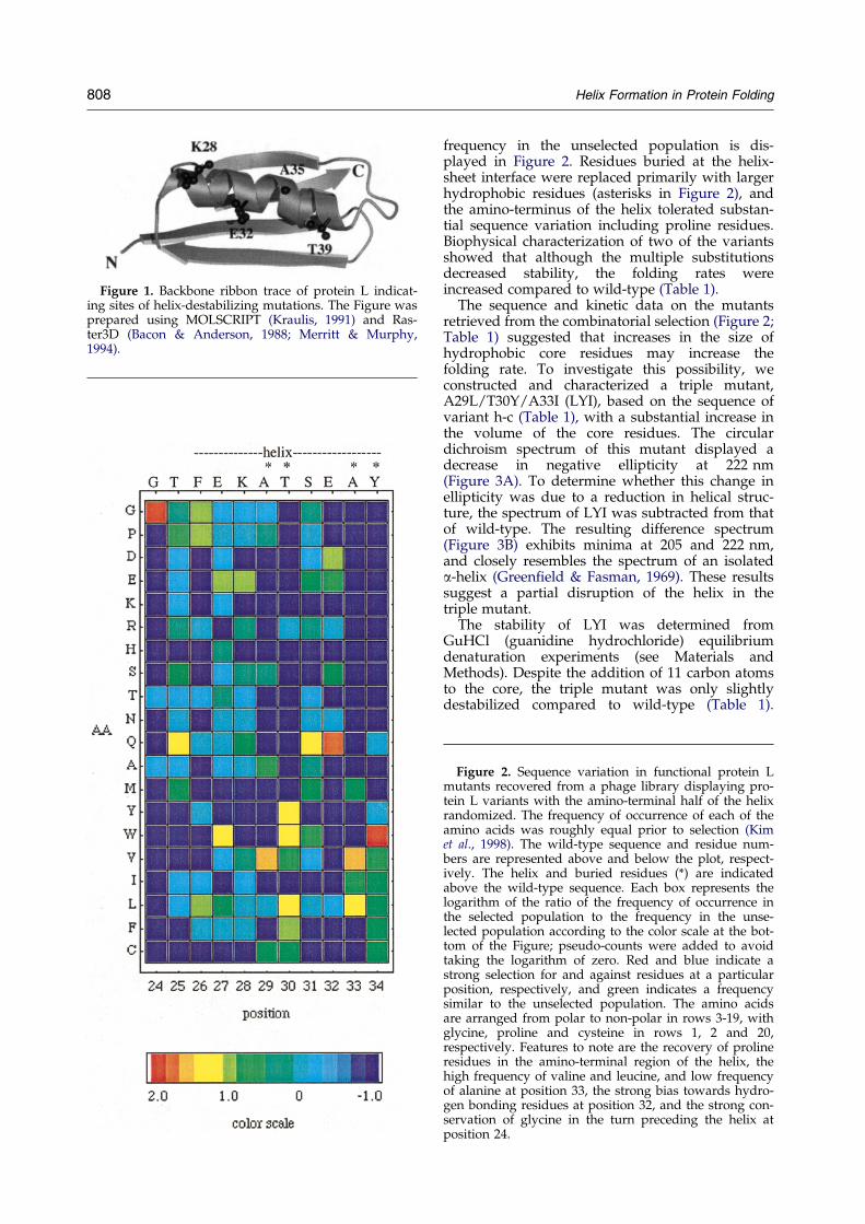
Figure 1. Backbone ribbon trace of protein L indicat-ing sites of helix-destabilizing mutations. The Figure wasprepared using MOLSCRIPT (Kraulis, 1991) and Ras-ter3D (Bacon & Anderson, 1988; Merritt & Murphy,1994).
808 Helix Formation in Protein Folding
frequency in the unselected population is dis-played in Figure 2. Residues buried at the helix-sheet interface were replaced primarily with largerhydrophobic residues (asterisks in Figure 2), andthe amino-terminus of the helix tolerated substan-tial sequence variation including proline residues.Biophysical characterization of two of the variantsshowed that although the multiple substitutionsdecreased stability, the folding rates wereincreased compared to wild-type (Table 1).
The sequence and kinetic data on the mutantsretrieved from the combinatorial selection (Figure 2;Table 1) suggested that increases in the size ofhydrophobic core residues may increase thefolding rate. To investigate this possibility, weconstructed and characterized a triple mutant,A29L/T30Y/A33I (LYI), based on the sequence ofvariant h-c (Table 1), with a substantial increase inthe volume of the core residues. The circulardichroism spectrum of this mutant displayed adecrease in negative ellipticity at 222 nm(Figure 3A). To determine whether this change inellipticity was due to a reduction in helical struc-ture, the spectrum of LYI was subtracted from thatof wild-type. The resulting difference spectrum(Figure 3B) exhibits minima at 205 and 222 nm,and closely resembles the spectrum of an isolateda-helix (Green®eld & Fasman, 1969). These resultssuggest a partial disruption of the helix in thetriple mutant.
The stability of LYI was determined fromGuHCl (guanidine hydrochloride) equilibriumdenaturation experiments (see Materials andMethods). Despite the addition of 11 carbon atomsto the core, the triple mutant was only slightlydestabilized compared to wild-type (Table 1).
Figure 2. Sequence variation in functional protein Lmutants recovered from a phage library displaying pro-tein L variants with the amino-terminal half of the helixrandomized. The frequency of occurrence of each of theamino acids was roughly equal prior to selection (Kimet al., 1998). The wild-type sequence and residue num-bers are represented above and below the plot, respect-ively. The helix and buried residues (*) are indicatedabove the wild-type sequence. Each box represents thelogarithm of the ratio of the frequency of occurrence inthe selected population to the frequency in the unse-lected population according to the color scale at the bot-tom of the Figure; pseudo-counts were added to avoidtaking the logarithm of zero. Red and blue indicate astrong selection for and against residues at a particularposition, respectively, and green indicates a frequencysimilar to the unselected population. The amino acidsare arranged from polar to non-polar in rows 3-19, withglycine, proline and cysteine in rows 1, 2 and 20,respectively. Features to note are the recovery of prolineresidues in the amino-terminal region of the helix, thehigh frequency of valine and leucine, and low frequencyof alanine at position 33, the strong bias towards hydro-gen bonding residues at position 32, and the strong con-servation of glycine in the turn preceding the helix atposition 24.
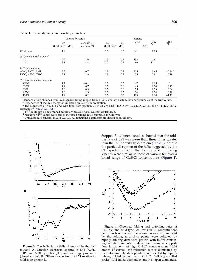
Table 1. Thermodynamic and kinetic parameters
Thermodynamic Kinetic
ma ��GU ÿ F2M ÿmf mu kH2O
f k2Mu
e �H2OF
(kcal molÿ1 Mÿ1) (kcal molÿ1) (kcal molÿ1 Mÿ1) (sÿ1)
Wild type 1.9 1.5 0.5 61 0.05
A. Combinatorial mutantsb
h-c 2.2 1.6 1.5 0.7 158 1.6h-d 3.1 4.4 2.2 0.3 90 12
B. Triple mutantsA29L, T30Y, A33I 2.2 0.7 1.3 0.7 205 0.65 ÿ0.90d
E32G, A35G, T39G 2.1 2.9 1.8 0.7 25 2.8 0.19
C. Helix destabilized mutantsK28G 1.7 ÿ0.1 1.3 0.5 47 0.05 c
E32G 1.9 0.7 1.5 0.6 48 0.20 0.12E32I 2.0 0.9 1.5 0.6 55 0.23 0.06A35G 2.0 1.3 1.5 0.5 34 0.24 0.29T39G 1.9 0.2 1.5 0.6 109 0.10 ÿ1.7d
Standard errors obtained from least squares ®tting ranged from 2±20%, and are likely to be underestimates of the true values.a Dependence of the free energy of unfolding on GuHCl concentration.b The sequences of h-c, h-d and wild-type from position 24 to 34 are GEYHTLYQEIW, GSGLEALGSVL, and GTFEKATSEAY,
respectively (Kim et al., 1998).c �F
H2
O could not be determined accurately because K28G was not destabilized.d Negative �F
H2
O values were due to increased folding rates compared to wild-type.e Unfolding rate constant in 2 M GuHCl. All remaining parameters are described in the text.
Figure 3. The helix is partially disrupted in the LYImutant. A, Circular dichroism spectra of LYI (A29L,T30Y, and A33I; open triangles) and wild-type protein L(closed circles). B, Difference spectrum of LYI relative towild-type protein L.
Helix Formation in Protein Folding 809
Stopped-¯ow kinetic studies showed that the fold-ing rate of LYI was more than three times greaterthan that of the wild-type protein (Table 1), despitethe partial disruption of the helix suggested by theCD spectrum. Both the folding and unfoldingkinetics were similar to those of variant h-c over abroad range of GuHCl concentrations (Figure 4),
Figure 4. Observed folding and unfolding rates ofLYI, h-c, and wild-type. At low GuHCl concentrations(left branch of curves), the relaxation rate is dominatedby the folding rate; data points were collected byrapidly diluting denatured protein with buffer contain-ing variable amounts of denaturant using a stopped-¯ow instrument. At high GuHCl concentrations (rightbranch of curves), the relaxation rate is dominated bythe unfolding rate; data points were collected by rapidlymixing folded protein with GuHCl. Wild-type (®lledcircles); LYI (®lled diamonds); and h-c (open diamonds).

Figure 5. Folding and unfolding rates of helix-destabi-lizing mutants. Wild-type (®lled circles), K28G (opensquares) E32I (®lled diamonds), E32G (open triangles),A35G (®lled squares), T39G (open circles), and tripleglycine mutant, E32G, A35G and T39G (®lled triangles).The helix-destabilizing mutants increase the rate ofunfolding but have relatively little effect on the foldingrate. The denaturant dependence of the folding andunfolding rates of the G15A substitution in the ®rst betaturn (Gu et al., 1997), which substantially decreasesthe folding rate, is also shown for comparison (opendiamonds).
810 Helix Formation in Protein Folding
suggesting that the increase in folding ratesobserved for variants h-c and h-d (Table 1) werelikely to result from the increases in the size ofhydrophobic core residues in the helix. Since LYI isnot signi®cantly destabilized, the increase infolding and unfolding rates suggests that theincrease in size of the core residues stabilizes thefolding transition state relative to the folded andunfolded states. Taken together, the CD data andkinetic data suggest that the rate-limiting step infolding of protein L does not require complete for-mation of the helix, but does involve hydrophobicassociation. A more complete interpretation ofthese results will require higher-resolution structur-al characterization of the LYI mutant.
To more directly probe the extent of helix for-mation in the folding transition state, we studiedthe effects of helix-destabilizing substitutions atsolvent-exposed positions (K28G, E32I, E32G,A35G and T39G) on the kinetics of folding andunfolding (Figure 1). Since these mutations affectonly local interactions, they probe the conse-quences of reductions in the population of mol-ecules with fully formed helix on the rate offolding without potentially complicating changesin tertiary interactions. A similar approach wasused to study the role of helix formation in thefully helical coiled-coil region of GCN4 (Sosnicket al., 1996).
The stabilities of the mutants were determinedusing equilibrium GuHCl denaturation exper-iments. K28G and T39G, at the amino and carboxytermini of the helix (Figure 1), respectively, do not
signi®cantly reduce the stability of the protein(Table 1). This may be because partial fraying atthe ends of the helix is compensated by theadditional degrees of freedom in the adjoiningturns (mutations at helix termini in isolated helicalpeptides have relatively little effect on helix content(Chakrabartty et al., 1991)). The small change instability could also re¯ect the elimination of anunfavorable charge-helix dipole interaction (K28G)and the low helix propensity of threonine. How-ever, mutations at solvent-exposed positions in themiddle of the helix (E32G, E32I, and A35G) desta-bilize the native state by 0.7-1.3 kcal/mol. The E32Imutant was constructed, in part, because very fewhydrophobic residues were recovered at this pos-ition in the phage selection (Figure 2). Since thepoint mutation is no more destabilized than manyof the variants recovered in the selection (Table 1),the conservation of residues capable of hydrogenbonding probably re¯ects a role for the glutamatein IgG binding (the IgG-binding site de®ned bychemical shift perturbation studies does notinclude this residue, but does include nearby resi-dues in the carboxy-terminal region of the helix;WikstoÈm et al., 1995)).
The characterization of the kinetics of foldingand unfolding revealed that the helix-destabilizingmutations increase the rate of unfolding signi®-cantly more than they decrease the rate of folding.The modest destabilization of the T39G mutantand the larger destabilization of the E32G, E32Iand A35G mutants are mostly accounted for byincreases in the unfolding rates (Table 1 andFigure 5), suggesting that the mutations destabilizethe native state considerably more than the tran-sition state. Interestingly, the T39G mutationincreased the rate of folding, suggesting that thetransition state ensemble includes conformationswith chain reversals or bends at the C terminus ofthe helix. The simplest explanation for the muchlarger effect of the mutations on the native statethan on the transition state is that the helix is notcompletely formed in the transition state.
A possible requirement for formation of only aportion of the helix in the folding transition statecould potentially have been missed in the singlemutant studies, since only a portion of the helixwas destabilized in each experiment. To destabilizehelix formation along the entire length of the helix,a triple mutant was designed in which the solvent-exposed residues E32, A35 and T39 (Figure 1) weresimultaneously changed to glycine residues. Theaddition of three glycine residues in the helixdramatically decreases the helix propensity(Chakrabartty et al., 1994) and thus shoulddecrease the population of helix in the unfoldedstate. The mutant is signi®cantly destabilized���G2M
UÿF � 2.9 kcal/mol), and the loss in stabilityis almost entirely due to an increase in the unfold-ing rate; the unfolding rate increased 56-fold whilethe folding rate decreased less than threefold(Table 1, Figure 5). These results, taken togetherwith the helix-destabilizing point mutations
Helix Formation in Protein Folding 811
described above, strongly suggest that the helix isdisrupted at the rate-limiting step in folding.
The kinetic results described above may beconveniently summarized using the �F valueparameter (��G{ÿU/��GFÿU), which re¯ects theextent to which interactions removed by mutationare present in the transition state in a simple two-state model; a value of 1 indicates that the site ofmutation is as structured in the transition state asin the native state, whereas a value of zero re¯ectsthe absence of such structure in the transition state(Matouschek et al., 1989). An important require-ment for this analysis is that the nature of the tran-sition state, inferred by the estimated amount ofhydrophobic surface exposed in the transition staterelative to the folded state (mu/m), is not perturbedupon mutation. The mu and m values for all of thehelix-destabilizing mutants are similar to those ofwild-type (Table 1). The �F values for the helix-destabilizing mutants are all less than 0.30(Table 1). In particular, the �F values for the twoglycine substitutions in the center of the helix were0.12 and 0.29, and the triple glycine mutant had alow �F value of 0.19, suggesting that the helix isnot appreciably structured along its entire lengthin the folding transition state ensemble.
Discussion
The role of the helix in folding
The results described here suggest that the helixin protein L is largely disrupted at the rate-limitingstep in folding and unfolding. The helix-destabiliz-ing mutations have much larger effects on the rateof unfolding than on the rate of folding; the tripleglycine substitution in particular increases theunfolding rate 56-fold, while decreasing the foldingrate less than three-fold. The increase in foldingrate accompanying the introduction of 11additional carbon atoms to the core of the LYImutant suggests that hydrophobic interactionsinvolving residues in the helix partially stabilizethe transition state ensemble despite the absence ofwell-formed helix. In the reverse direction, thelarge effects of the helix-destabilizing mutations onthe unfolding rate suggests that disruption of thehelix is a key step in disassembly of the hydro-phobic core in unfolding.
The � values for the helix-destabilizingmutations are greater than zero (0.06±0.29). Howare these partial � values best interpreted? Forhydrophobic core mutations, partial � valueswould result if a residue made some but not all ofits interactions in the transition state (Fersht et al.,1992). For solvent-exposed residues, this interpret-ation seems less plausible, since such residuesmake relatively few interactions even in the nativestate. Perhaps the simplest interpretation is that thelocal structural element containing the solvent-exposed residue is partially ordered in the tran-
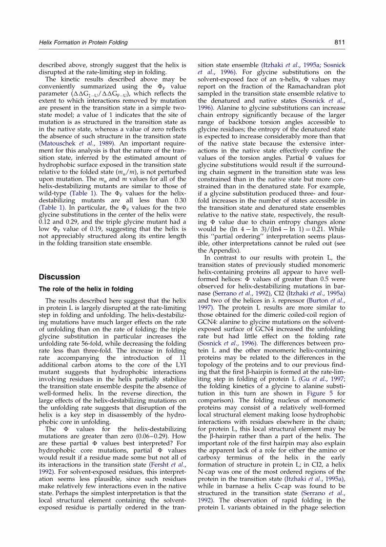
sition state ensemble (Itzhaki et al., 1995a; Sosnicket al., 1996). For glycine substitutions on thesolvent-exposed face of an a-helix, � values mayreport on the fraction of the Ramachandran plotsampled in the transition state ensemble relative tothe denatured and native states (Sosnick et al.,1996). Alanine to glycine substitutions can increasechain entropy signi®cantly because of the largerrange of backbone torsion angles accessible toglycine residues; the entropy of the denatured stateis expected to increase considerably more than thatof the native state because the extensive inter-actions in the native state effectively con®ne thevalues of the torsion angles. Partial � values forglycine substitutions would result if the surround-ing chain segment in the transition state was lessconstrained than in the native state but more con-strained than in the denatured state. For example,if a glycine substitution produced three- and four-fold increases in the number of states accessible inthe transition state and denatured state ensemblesrelative to the native state, respectively, the result-ing � value due to chain entropy changes alonewould be (ln 4 ÿ ln 3)/(ln4 ÿ ln 1) � 0.21. Whilethis ``partial ordering'' interpretation seems plaus-ible, other interpretations cannot be ruled out (seethe Appendix).
In contrast to our results with protein L, thetransition states of previously studied monomerichelix-containing proteins all appear to have well-formed helices: � values of greater than 0.5 wereobserved for helix-destabilizing mutations in bar-nase (Serrano et al., 1992), CI2 (Itzhaki et al., 1995a)and two of the helices in l repressor (Burton et al.,1997). The protein L results are more similar tothose obtained for the dimeric coiled-coil region ofGCN4: alanine to glycine mutations on the solvent-exposed surface of GCN4 increased the unfoldingrate but had little effect on the folding rate(Sosnick et al., 1996). The differences between pro-tein L and the other monomeric helix-containingproteins may be related to the differences in thetopology of the proteins and to our previous ®nd-ing that the ®rst b-hairpin is formed at the rate-lim-iting step in folding of protein L (Gu et al., 1997;the folding kinetics of a glycine to alanine substi-tution in this turn are shown in Figure 5 forcomparison). The folding nucleus of monomericproteins may consist of a relatively well-formedlocal structural element making loose hydrophobicinteractions with residues elsewhere in the chain;for protein L, this local structural element may bethe b-hairpin rather than a part of the helix. Theimportant role of the ®rst hairpin may also explainthe apparent lack of a role for either the amino orcarboxy terminus of the helix in the earlyformation of structure in protein L; in CI2, a helixN-cap was one of the most ordered regions of theprotein in the transition state (Itzhaki et al., 1995a),while in barnase a helix C-cap was found to bestructured in the transition state (Serrano et al.,1992). The observation of rapid folding in theprotein L variants obtained in the phage selection
determined for each point mutant using the followingequations:
�H2OF ���GH2O
zÿU=��GFÿU �2�
��GH2OzÿU �ÿ RT ln�kH2O
f;wt =kH2Of;mut� �3�
��GFÿU �hmi��GuHCl�50%mutÿ �GuHCl�50%;wt� �4�where kH2O
f;wt and kH2Of;mut are the folding rate constants in
the absence of denaturant for wild-type and mutant,respectively, ��GF ÿ U is the change in free energy offolding between wild-type and mutant in the absence ofdenaturant, hmi is the average m value calculated fromthe mutants and wild-type (hmi � 2.0 kcal molÿ1 Mÿ1),and [GuHCl]50%,mut and [GuHCl]50%,wt are the concen-trations of GuHCl where 50% of the protein is folded formutant and wild-type, respectively.
812 Helix Formation in Protein Folding
is perhaps the most dramatic illustration of thelack of importance of regular structure at the helixamino terminus; the sequences often contain pro-line and generally have low helix propensity(Chakrabartty et al., 1994; Figure 2), and the CDspectra of the variants are consistent with partialdisruption of the helix (data not shown).
Perhaps the two most characteristic features ofglobular proteins are the presence of a hydro-phobic core and the large amounts of regular sec-ondary structure (sheets and helices) and thusthere has been considerable debate over the pastdecade over the relative timing of hydrophobic col-lapse and secondary structure formation in folding(Dill, 1990; Fersht, 1995; Karplus & Weaver, 1994;Kim & Baldwin, 1990). Our results indicate that forprotein L, hydrophobic interactions of non-polarresidues in the helix with residues elsewhere in theprotein are more critical to the rate-limiting step infolding than is the formation of the helix. It isinteresting to note that almost all protein tertiarystructure prediction methods (Bowie & Eisenberg,1994; Monge et al., 1994; Simons et al., 1997;Skolnick et al., 1997; Srinivasan & Rose, 1995)assemble tertiary structures hierarchically frompredicted local structural elements, typically helicesand strands; the results reported here suggest thatthis may not be the way many proteins fold.
Materials and Methods
All reagents, solutions, and enzymes used for molecu-lar biology procedures were as described by Gu et al.(1995) and Scalley et al. (1997). Point mutations weremade with the QuickChange site-directed mutagenesiskit (Strategene). Expression and puri®cation were carriedout as described by Gu et al. (1995). All mutants wereveri®ed by DNA sequencing and mass spectrometry.Equilibrium CD and ¯uorescence measurements,stopped-¯ow experiments, and data analysis were car-ried out as described by Gu et al. (1997) and Scalley et al.(1997).
The free energy of folding of protein L appears to be anon-linear function of denaturant and, as a result, stan-dard linear extrapolation estimates of the free energy ofunfolding in the absence of denaturant ��GH2O
UÿF� mayinvolve considerable errors (Yi et al., 1997). To avoid theextrapolation, the change in free energy of unfoldingbetween mutant and wild-type ���G2M
UÿF� was calculatedat 2 M GuHCl.
The refolding and unfolding data were ®t by a two-state model using the following equation:
ln kobs�GuHCl� � ln�kH2Of exp�ÿmf�GuHCl��
� kH2Ou exp�mu�GuHCl��� �1�
where kobs [GuHCl] is the observed relaxation rate at agiven concentration of GuHCl, kH2O
f and kH2Ou are the
refolding and unfolding rate constants in the absence ofdenaturant, respectively, and mf and mu describe thedependence of the logarithm of the rate constant onGuHCl concentration. �H2O
F (Itzhaki et al., 1995a) was
Acknowledgments
We thank Carol Rohl and members of the Baker lab-oratory for helpful comments on the manuscript, andTobin Sosnick for helpful discussions. This work wassupported by a grant from the NIH (GM5188) and byyoung investigator awards to D.B. from the NSF and thePackard Foundation.
References
Bacon, D. J. & Anderson, W. F. (1988). A fast algorithmfor rendering space-®lling molecule pictures. J. Mol.Graph. 6, 219±220.
Bowie, J. U. & Eisenberg, D. (1994). An evolutionaryapproach to folding small a-helical proteins thatuses sequence information and an empirical guiding®tness function. Proc. Natl Acad. Sci. USA, 91, 4436±4440.
Burton, R. E., Huang, G. S., Daugherty, M. A.,Calderone, T. L. & Oas, T. G. (1997). The energylandscape of a fast-folding protein mapped byAla! Gly substitutions. Nature Struct. Biol. 4, 305±310.
Burton, R. E., Myers, J. K. & Oas, T. G. (1998). Proteinfolding dynamics: quantitative comparison betweentheory and experiment. Biochemistry, 37, 5337±5343.
Chakrabartty, A., Schellman, J. A. & Baldwin, R. L.(1991). Large differences in the helix propensities ofalanine and glycine. Nature, 351, 586±588.
Chakrabartty, A., Kortemme, T. & Baldwin, R. L. (1994).Helix propensities of the amino acids measured inalanine-based peptides without helix-stabilizingside-chain interactions. Protein Sci. 3, 843±852.
Cleland, W. W. (1975). Partition analysis and the con-cept of net rate constants as tools in enzyme kin-etics. Biochemistry, 14, 3220±3224.
Dill, K. A. (1990). Dominant forces in protein folding.Biochemistry, 29, 7133±7155.
Fersht, A. R. (1995). Optimization of rates of proteinfolding: the nucleation-condensation mechanismand its implications. Proc. Natl Acad. Sci. USA, 92,10869±10873.
Fersht, A. R., Matouschek, A. & Serrano, L. (1992). Thefolding of an enzyme. I. Theory of protein engineer-ing analysis of stability and pathway of proteinfolding. J. Mol. Biol. 224, 771±782.
Helix Formation in Protein Folding 813
Fersht, A. R., Itzhaki, L. S., El Masry, N. F., Matthews,J. M. & Otzen, D. E. (1994). Single versus parallelpathways of protein folding and fractional for-mation of structure in the transition state. Proc. NatlAcad. Sci. USA, 91, 10426±10429.
Gilmanshin, R., Williams, S., Callender, R. H.,Woodruff, W. H. & Dyer, R. B. (1997). Fast eventsin protein folding: relaxation dynamics of secondaryand tertiary structure in native apomyoglobin. Proc.Natl Acad. Sci. USA, 94, 3709±3713.
Green®eld, N. & Fasman, G. D. (1969). Computed circu-lar dichroism spectra for the evaluation of proteinconformation. Biochemistry, 8, 4108±4116.
Gu, H., Yi, Q., Bray, S. T., Riddle, D. S., Shiau, A. K. &Baker, D. (1995). A phage display system for study-ing the sequence determinants of protein folding.Protein Sci. 4, 1108±1117.
Gu, H., Kim, D. & Baker, D. (1997). Contrasting rolesfor symmetrically disposed b-turns in the folding ofa small protein. J. Mol. Biol. 274, 588±596.
Itzhaki, L. S., Otzen, D. E. & Fersht, A. R. (1995a). Thestructure of the transition state for folding of chy-motrypsin inhibitor 2 analyzed by protein engineer-ing methods: evidence for a nucleation-condensation mechanism for protein folding. J. Mol.Biol. 254, 260±288.
Itzhaki, L. S., Neira, J. L., Ruiz-Sanz, J., de Prat, Gay G.& Fersht, A. R. (1995b). Search for nucleation sitesin smaller fragments of chymotrypsin inhibitor 2.J. Mol. Biol. 254, 289±304.
Karplus, M. & Weaver, D. L. (1994). Protein foldingdynamics: the diffusion-collision model and exper-imental data. Protein Sci. 3, 650±668.
Kim, D. E., Gu, H. & Baker, D. (1998). The sequences ofsmall proteins are not extensively optimized forrapid folding by natural selection. Proc. Natl Acad.Sci. USA, 95, 4982±4986.
Kim, P. S. & Baldwin, R. L. (1990). Intermediates in thefolding reactions of small proteins. Annu. Rev. Bio-chem. 59, 631±660.
Kraulis, P. J. (1991). MOLSCRIPT: a program to produceboth detailed and schematic plots of protein struc-tures. J. Appl. Crystallog. 24, 946±950.
LoÂpez-HernaÂndez, , Cronet, P., Serrano, L. & MunÄ oz, V.(1997). Folding kinetics of Che Y mutants withenhanced native a-helix propensities. J. Mol. Biol.266, 610±620.
Matouschek, A., Kellis, J. T., Serrano, L. & Fersht, A. R.(1989). Mapping the transition state and pathway ofprotein folding by protein engineering. Nature, 340,122±126.
Merritt, E. A. & Murphy, M. E. P. (1994). Raster3D ver-sion 2.0. A program for photorealistic moleculargraphics. Acta. Crystallog. sect. D, 50, 869±873.
Monge, A., Friesner, R. A. & Honig, B. (1994). An algor-ithm to generate low-resolution protein tertiarystructures form knowledge of secondary structure.Proc. Natl Acad. Sci. USA, 91, 5027±5029.
Scalley, M. L., Yi, Q., Gu, H., McCormack, A., Yates,J. R., III & Baker, D. (1997). Kinetics of folding ofthe IgG binding domain of peptostreptococcal pro-tein L. Biochemistry, 36, 3373±3382.
Serrano, L., Matouschek, A. & Fersht, A. R. (1992). Thefolding of an enzyme. III. Structure of the transitionstate for unfolding of barnase analyzed by a proteinengineering procedure. J. Mol. Biol. 224, 805±818.
Simons, K. T., Kooperberg, C., Huang, E. & Baker, D.(1997). Assembly of protein tertiary structures fromfragments with similar local sequences using simu-
lated annealing and Bayesian scoring functions.J. Mol. Biol. 268, 209±225.
Skolnick, J., Kolinski, A. & Ortiz, A. R. (1997). MONS-STER: a method for folding globular proteins witha small number of distance restraints. J. Mol. Biol.265, 217±241.
Sosnick, T. R., Jackson, S., Wilk, R. R., Englander, S. W.& DeGrado, W. F. (1996). The role of helix for-mation in the folding of a fully a-helical coiled coil.Proteins: Struct. Funct. Genet. 24, 427±432.
Srinivasan, R. & Rose, G. D. (1995). LINUS: A hierarchicprocedure to predict the fold of a protein. Proteins:Struct. Funct. Genet. 22, 81±99.
Thompson, P. A., Eaton, W. A. & Hofrichter, J. (1997).Laser temperature jump study of the helix$ coilkinetics of an alanine peptide interpreted with a`kinetic zipper' model. Biochemistry, 36, 9200±9210.
WikstoÈm, M., Drakenberg, T., ForseÂn, S., SjoÈbring, U. &BjoÈrck, L. (1994). Three-dimensional solution struc-ture of an immunoglobulin light chain-bindingdomain of protein L. Comparison with the IgG-binding domains of protein G. Biochemistry, 33,14011±14017.
WikstoÈm, M., SjoÈbring, U., Drakenberg, T., ForseÂn, S. &BjoÈrck, L. (1995). Mapping of the immunoglobulinlight chain-binding site of protein L. J. Mol. Biol.250, 128±133.
Williams, S., Causgrove, T. P., Gilmanshin, R., Fang,K. S., Callender, R. H., Woodruff, W. H. & Dyer,R. B. (1996). Fast events in protein folding: helixmelting and formation in a small peptide. Biochemis-try, 35, 691±697.
Yi, Q., Scalley, M. L., Simons, K. T., Gladwin, S. T. &Baker, D. (1997). Characterization of the free energyspectrum of peptostreptococcal protein L. Fold. Des.2, 271±280.
Appendix: Origins of Partial ��� Valuesfor Mutations at Solvent-exposed Sites
As discussed in the main text and by previousauthors (Fersht et al., 1994; Itzhaki et al., 1995a;Sosnick et al., 1996), partial � values at solventexposed sites could re¯ect partial ordering of thelocal structural element containing the mutatedresidue in the transition state ensemble. Partial �values could arise if only one of two free energybarriers (either in parallel (Fersht et al., 1994) or inseries) of roughly equal height are affected by themutation. For example, letting H represent a localstructural element such as a b-turn or a-helix, Crepresent the unfolded form of the structuralelement, and P the rest of the (unfolded) protein,consider a simple kinetic scheme in which bothforms of the local structural element can associateproductively with the remainder of the protein, butwith different rates:
H� P!k1N; C� P!k2
N; C$Keq
H
(The use of an equilibrium constant to character-ize the C$ H transition is clearly a crude approxi-mation.) Then:
kf � �Keq=�1� Keq��k1 � �1=�1� Keq��k2 �A1�
Figure A1. Fit of parallel pathway model to exper-imental data. Equation (A1) was ®t to the refoldingrate constants and free energies of unfolding [ ÿ RT ln(ku/kf)] for the CI2 mutants expected to affect the stab-ility of the helix but not packing interactions on theinterior of the protein (S12G, S12A, E14A/E15A, S12G/E14A/E15A, S12A/E14A/E15A, K17A, K17G, K18A,K18G, Q22A and Q22G). Keq for the helix in wild-typeCI2 was estimated from experimental data on the iso-lated 12-24 segment of CI2 (9.2% helical; Itzhaki et al.,1995b); the changes in free energy were assumed toresult entirely from changes in Keq. The broken curveis the ®t to equation (A1) with k1 � 600 sÿ1 andk2 � 14 sÿ1; the broken straight line, the ®t to the par-tial ordering (BroÈnsted) model. Both models are con-sistent with these experimental data, but the partialordering model has one less free parameter and ismuch more consistent with the nucleation-condensationmechanism deduced from the extensive characterizationof the kinetic consequences of mutations throughoutthe entire protein (Fersht et al., 1994; Itzhaki et al.,1995a).
814 Helix Formation in Protein Folding
and, provided that the helix is not signi®cantlypopulated in the denatured state (Keq < 1), the �value for a mutation at a solvent-exposed site thatchanges the free energy of folding exclusively bychanging Keq is:
� ���GzÿU=��GFÿU
� �d ln kf�=�d ln Keq�� Keq=�k2=k1 � Keq� ÿ Keq=�1� Keq� �A2�
For k1 > k2, the � value is positive andapproaches 1 for k14k2/Keq; for k1 � k2, � � 0 (thefolding rate is independent of the helix popu-lation), and for k1 < k2 the � value is negative (theprotein folds faster with the helix not formed, asperhaps for the T39G mutation). This model differsfrom the parallel pathway model described byFersht et al. (1994), in that the folding rate dependson the fraction of helix in the unfolded state ratherthan directly on the equilibrium constant for helixformation; the two models are identical for Keq51,but equation (A1) predicts smaller � values whenKeq approaches 1.
Distinguishing this parallel pathway model fromthe partial ordering model requires mutations thatproduce a wide range of stabilities (Fersht et al.,1994). Even for the large number of mutationscharacterized at solvent-exposed sites in the helixin CI2, it is dif®cult to distinguish between the twomodels (Figure A1); it appears that a range ofmore than 4 kcal is required for the parallel path-way model to produce enough curvature to clearlydistinguish it from the partial ordering model (incontrast, the BroÈnsted behavior observed over amore than 4 kcal range for the CI2 minicore clearlyrules out parallel pathway models (Fersht et al.,1994)). A range of >4 kcal/mol is not easily achiev-able for protein L since mutations producing suchdestabilizations would also greatly destabilize thenative state (the free energy of unfolding is �5±6 kcal/mol). However, very large destabilizationshave been obtained for S peptide binding to S pro-tein (J.M.G. and R.L.B., unpublished results), inthis case the � values remain constant, supportingthe partial ordering mechanism. It should be notedthat the actual folding transition state ensemble fora protein is likely to lie somewhere between thesetwo extremes; the simple kinetic models are usefulfor illustrating the consequences of differences inthe breadth of the transition state ensemble onexperimentally measurable quantities.
There has recently been considerable progress indetermining the rate of formation of isolatedhelices, and it is instructive to consider models inwhich the helix formation rate enters directly. Forexample, consider a simple sequential kineticmechanism in which the ®rst step corresponds tothe formation of conformations with a critical sub-set of the low resolution topological features of thenative state, and the second step, to helix for-mation in the context of this overall topological
order. Helix formation in this context is assumedto lead to very rapid completion of folding. Theexpression for the rate constant of a sequentialreaction of this type is given by Cleland (1975);reasonable rate constants for refolding are obtainedwhen the free energy of the critical subset is7 kcal/mol more than that of unfolded protein,and the rate constants for helix formation andbreakdown are �107 sÿ1 (Gilmanshin et al., 1997;Thompson et al., 1997; Williams et al., 1996) atKeq � 1. In this scenario, the rate of foldingdepends on the rate of helix formation, not theequilibrium population of helix, and hence verydifferent � values will be obtained for mutationsthat produce the same net destabilization of helixbut affect the helix formation and disruption ratesdifferently: mutations that exclusively slow the rateof helix formation, increase the rate of helix disrup-tion, or slow the formation rate and increase thedisruption rate by the same factor will have �values of 1.0, 0.0 and 0.5, respectively. Interpret-ation of partial � values in this scenario wouldclearly be facilitated by data on the effects of
Helix Formation in Protein Folding 815
mutations on the rate of formation and disruptionof isolated helices.
References
Cleland, W. W. 1975 Partition analysis and the conceptof net rate constants as tools in enzyme kinetics Bio-chemistry 14 3220±3224
Fersht, A. R., Itzhaki, L. S., El Masry, N. F., Matthews, J.M. & Otzen, D. E. 1994 Single versus parallel path-ways of protein folding and fractional formation ofstructure in the transition state Proc. Natl Acad. Sci.USA 91 10426±10429
Gilmanshin, R., Williams, S., Callender, R. H., Woodruff,W. H. & Dyer, R. B. 1997 Fast events in proteinfolding: relaxation dynamics of secondary and ter-
tiary structure in native apomyoglobin Proc. NatlAcad. Sci. USA 94 3709±3713
Itzhaki, L. S., Neira, J. L., Ruiz-Sanz, J., de Prat Gay, G.& Fersht, A. R. 1995b Search for nucleation sites insmaller fragments of chymotrypsin inhibitor 2 J.Mol. Biol. 254 289±304
Thompson, P. A., Eaton, W. A. & Hofrichter, J. 1997Laser temperature jump study of the helix () coilkinetics of an alanine peptide interpreted with a`kinetic zipper' model Biochemistry 36 9200±9210
Williams, S, Causgrove, T. P., Gilmanshin, R., Fang, K.S., Callender, R. H., Woodruff, W. H. & Dyer, R. B.1996 Fast events in protein folding: helix meltingand formation in a small peptide Biochemistry 35691±697
Edited by P. E. Wright
(Received 30 April 1998; received in revised form 31 August 1998; accepted 1 September 1998)
Copyright © 2022 FDOKUMEN