Patent Foramen Ovale in Recreational and Professional Divers: An Important and Largely Unrecognized...
26
Accepted Manuscript Patent Foramen Ovale in Recreational and Professional Divers: An Important and Largely Unrecognized Problem Jakub Honěk, MD, Luděk Šefc, PhD, Tomáš Honěk, MD, PhD, Martin Šrámek, MD, Martin Horváth, MD, Josef Veselka, MD, PhD PII: S0828-282X(15)00199-3 DOI: 10.1016/j.cjca.2015.03.010 Reference: CJCA 1622 To appear in: Canadian Journal of Cardiology Received Date: 25 January 2015 Revised Date: 10 March 2015 Accepted Date: 10 March 2015 Please cite this article as: Honěk J, Šefc L, Honěk T, Šrámek M, Horváth M, Veselka J, Patent Foramen Ovale in Recreational and Professional Divers: An Important and Largely Unrecognized Problem, Canadian Journal of Cardiology (2015), doi: 10.1016/j.cjca.2015.03.010. This is a PDF file of an unedited manuscript that has been accepted for publication. As a service to our customers we are providing this early version of the manuscript. The manuscript will undergo copyediting, typesetting, and review of the resulting proof before it is published in its final form. Please note that during the production process errors may be discovered which could affect the content, and all legal disclaimers that apply to the journal pertain.
Transcript of Patent Foramen Ovale in Recreational and Professional Divers: An Important and Largely Unrecognized...

Accepted Manuscript
Patent Foramen Ovale in Recreational and Professional Divers: An Important andLargely Unrecognized Problem
Jakub Honěk, MD, Luděk Šefc, PhD, Tomáš Honěk, MD, PhD, Martin Šrámek, MD,Martin Horváth, MD, Josef Veselka, MD, PhD
PII: S0828-282X(15)00199-3
DOI: 10.1016/j.cjca.2015.03.010
Reference: CJCA 1622
To appear in: Canadian Journal of Cardiology
Received Date: 25 January 2015
Revised Date: 10 March 2015
Accepted Date: 10 March 2015
Please cite this article as: Honěk J, Šefc L, Honěk T, Šrámek M, Horváth M, Veselka J, Patent ForamenOvale in Recreational and Professional Divers: An Important and Largely Unrecognized Problem,Canadian Journal of Cardiology (2015), doi: 10.1016/j.cjca.2015.03.010.
This is a PDF file of an unedited manuscript that has been accepted for publication. As a service toour customers we are providing this early version of the manuscript. The manuscript will undergocopyediting, typesetting, and review of the resulting proof before it is published in its final form. Pleasenote that during the production process errors may be discovered which could affect the content, and alllegal disclaimers that apply to the journal pertain.
MANUSCRIP
T
ACCEPTED
ACCEPTED MANUSCRIPT1
Patent Foramen Ovale in Recreational and Professional Divers: An Important and
Largely Unrecognized Problem
Jakub Honěk, MD1,2; Luděk Šefc, PhD2; Tomáš Honěk, MD, PhD1; Martin Šrámek,
MD3,2; Martin Horváth, MD1; Josef Veselka, MD, PhD1
1 Department of Cardiology, Charles University in Prague, 2nd Faculty of Medicine and
Motol University Hospital, Prague, Czech Republic; 2 Institute of Pathological
Physiology, Charles University in Prague, First Faculty of Medicine, Prague, Czech
Republic; 3 Department of Neurology, Charles University in Prague, 2nd Faculty of
Medicine and Motol University Hospital, Prague, Czech Republic
PFO in Divers
Word count: 4771
Correspondence: Josef Veselka, MD, PhD, Department of Cardiology, University
Hospital Motol, V Úvalu 84, 150 06, Praha 5, Czech Republic.
Phone: +420224434901, Fax: +420224434920, e-mail: [email protected]

MANUSCRIP
T
ACCEPTED
ACCEPTED MANUSCRIPT2
Brief summary
Patent foramen ovale (PFO) is associated with increased risk of decompression
sickness (DCS) in divers due to paradoxical embolization of nitrogen bubbles. Despite
the high number of scuba divers and the high prevalence of PFO, many important
questions including screening, risk stratification and management strategy remain to
be answered. Recently published data suggest the possible effectiveness of both
transcatheter PFO closure and conservative diving measures. This review aims to
summarize the current literature on PFO and diving.
MANUSCRIP
T
ACCEPTED
ACCEPTED MANUSCRIPT3
Abstract
Patent foramen ovale (PFO) is associated with an increased risk of
decompression sickness (DCS) in divers due to paradoxical embolization of nitrogen
bubbles. The number of scuba divers worldwide is estimated in millions and the
prevalence of PFO is 25-30% in adults. It is interesting that, despite these numbers,
many important questions regarding optimal screening, risk stratification and
management strategy still remain to be answered. Recently published data suggest
the possible effectiveness of both PFO closure and conservative diving measures in
preventing arterial gas embolization. This review aims to introduce the basic principles
of physiology and the pathophysiology of bubble formation and DCS, summarize the
current literature on PFO and diving and review the possibilities of diagnostic work-up
and management.
MANUSCRIP
T
ACCEPTED
ACCEPTED MANUSCRIPT4
Scuba (self-contained underwater breathing apparatus) diving is a popular sport
that attracts millions of participants worldwide.1 The exposure to the hyperbaric
environment is associated with unique effects on human physiology and with specific
disorders. Much attention has been paid to the risks related to patent foramen ovale
(PFO).2 In divers, PFO is associated with the increased risk of decompression
sickness (DCS).3 Despite the high prevalence of PFO (25-30% in adults)4, many
questions including optimal screening, risk stratification and management strategy
remain to be answered. This review aims to introduce the basic principles of
physiology and the pathophysiology of bubble formation and DCS, summarize current
literature on PFO and diving and review the possibilities of diagnostic work-up and
management.
Historical Introduction
With the development of professional and recreational diving in the 20th century, the
knowledge of DCS progressed. Decompression sickness was first described in an
animal model in 1670 by Robert Boyle.5 The first clinical cases of divers suffering from
DCS were reported much later, in 1841.6 The description of 110 cases (of whom 14
died) during the construction of the Brooklyn Bridge in 1873 is more well-known. Five
years later, in his classical work La Pression Barometrique, French zoologist and
physiologist Paul Bert postulated that DCS is caused by nitrogen gas bubbles and
showed the advantages of breathing oxygen after developing DCS.7 In 1908, following
a series of animal decompression experiments, John Scott Haldane developed the first
dive tables that advised staged decompression for the British Admiralty.8 This marked

MANUSCRIP
T
ACCEPTED
ACCEPTED MANUSCRIPT5
the development of further decompression models, that are nowadays routinely used
by recreational and professional divers to prevent DCS.
Decompression Sickness – Physiology and Pathophysiology
The diver is exposed to a hyperbaric environment during submersion. In scuba
diving, air (or other breathing mixture of oxygen and inert gases) is breathed at
ambient pressure. According to Henry’s law, the amount of gases dissolved in tissues
is proportional to their partial pressures. Thus, at depth, the concentration of gases in
tissues increases over time. The rate of gas saturation is dependent on the chemical
composition and density of capillaries in a particular tissue.9 As the diver ascends to
the surface, a pressure gradient drives the dissolved gases back from peripheral
tissues to venous blood and ultimately to the alveolar space from where it is expired
out of the body. If the pressure drops too quickly, the tissues become supersaturated
with gases not utilized by the body (nitrogen, inert gases) and a gas phase forms.10
The process of bubble formation has attracted the interest of researchers for
more than a century, but many aspects remain unclear. Although still being
controversial, it is generally agreed that an a priori presence of some form of
micronuclei is required for bubbles to form in divers.10 It is of importance that the rate
of saturation and desaturation differs among tissues. Mathematical models
incorporating several tissue compartments are used to characterize whole body gas
kinetics.11 To prevent DCS, divers routinely use specialized dive computers or tables
that are based on these models.
Decompression sickness is caused by the formation and growth of gas bubbles
in supersaturated blood or tissues during the diver's ascent (fig. 1). These bubbles
MANUSCRIP
T
ACCEPTED
ACCEPTED MANUSCRIPT6
cause either local tissue damage or embolize through venous blood.12 Small quantities
of venous gas emboli (VGE) were confirmed by Doppler studies in 80-91% of scuba
divers.13,14 Most divers with VGE, however, remain asymptomatic, as these are
effectively filtered by pulmonary circulation. Symptoms may occur either with high
bubble load (i.e. pulmonary gas embolism in case of violation of the decompression
regimen) or due to paradoxical embolization (arterialization of bubbles) in a diver with
a permanent or transient right-to-left shunt. In divers with a PFO, if paradoxical
embolization occurs, arterialized bubbles lodge in peripheral capillaries. Furthermore,
excess gas from supersaturated tissues promotes further growth of these bubbles.
The resulting obstruction of capillaries causes local ischemia.12
The clinical picture of DCS is heterogeneous and reflects the amount of bubbles
and the sites of their formation and embolization. Based on symptomatology,
cutaneous, musculoskeletal, neurological and pulmonary forms of DCS are
recognized. The musculoskeletal form, manifesting as severe joint pain, is thought to
be caused by local bubble formation in the avascular joint cartilage.15 On the other
side of the spectrum are diverse and potentially severe neurological manifestations in
which it seems that bubble embolization through a PFO might play an important role.
The Role of Patent Foramen Ovale
The connection between PFO and DCS was first described in the 1980s.16,17
Since then, a high prevalence of PFO has been repeatedly reported in divers with the
neurological or cutaneous form of DCS (see Table 1). In an important study Torti et
al.18 reported an incidence of major DCS per 10,000 dives of 1.5 with no PFO, less
than 1 with a grade 1 PFO, 3 with a grade 2 PFO and 9 with a grade 3 PFO. The
MANUSCRIP
T
ACCEPTED
ACCEPTED MANUSCRIPT7
associated odds would be 1 for a grade 1, 2 for a grade 2 and 6 for a grade 3 PFO
compared to no PFO. However, this study had important limitations including its
retrospective nature and possible selection bias.23 In another study, the incidence of
PFO was 77% among 61 divers who had suffered the cutaneous form of DCS,
compared with 28% in controls. Additionally, besides the higher incidence of acute
DCS, it has been suggested that repeated exposure to asymptomatic arterial
embolisms could lead to chronic sequelae. Knauth et al.24 reported an association of
PFO with multiple brain lesions in a follow-up study using magnetic resonance
imaging. There is, however, an ongoing debate regarding whether this finding has a
pathophysiological link to PFO or any clinical significance.25
Bearing in mind the high prevalence of PFO,4 these reports raise concern
among divers and involved medical professionals. Moreover, in divers with a PFO, a
paradoxical embolization to the systemic circulation may cause various, mostly
neurological or cutaneous DCS symptoms, even after a dive with an appropriate
decompression regimen.20 This unpredictable event has been coined “unprovoked
DCS”.
Paradoxical embolization results from increased right atrial pressure due to
hemodynamic changes that occur in divers. After submersion, blood redistributes from
the periphery to the thorax, which results in an increased right atrial pressure.26
Moreover, divers may perform a Valsalva maneuver during or after the dive (to
equalize pressure in the middle ear or while lifting heavy diving equipment), which
further contributes to the increased right atrial pressure and might lead to transient
right-to-left shunting through the PFO. On the other hand, it has been suggested that
the transpulmonary passage might also play an important role in the occurrence of
post-dive arterial gas emboli.27 However, the estimated prevalence of large pulmonary

MANUSCRIP
T
ACCEPTED
ACCEPTED MANUSCRIPT8
arteriovenous malformations is low28 and the clinical significance of small functional
shunts is doubtful.29,30 Also, the numerous aforementioned clinical studies18-22 support
the fact that PFO might be the major route of paradoxical embolization in divers. It is
important to note, that a small shunt probably does not impart risk, while a large shunt
should be considered to increase risk of decompression sickness. The prevalence of
large PFOs is estimated to be 6-10% in general population31 and the prevalence of
PFO was reported to decrease with age in a large autopsy study of normal hearts.4 On
the other hand, there is some evidence for increasing patency of the foramen ovale in
divers over years.32
Diagnostic Imaging
Three ultrasonographic techniques are available for imaging of PFO or
detection of right-to-left intracardiac shunts: transthoracic echocardiography (TTE),
transesophageal echocardiography (TEE) and transcranial color-coded sonography
(TCCS). These methods may be used for screening, to plan and assist device closure
and to monitor the presence of post-dive venous and arterial bubbles.
Transesophageal echocardiography has traditionally been considered the gold
standard of PFO diagnostics.33 The proximity of the probe to atrial septum ensures
optimal resolution and enables quality 2D and 3D imaging of PFO and surrounding
structures. In the diagnostic work-up of cryptogenic stroke, TEE importantly enables
the visualization of other potential sources of embolism, e.g. a thrombus in the left
atrial appendage or atherosclerotic lesions in the proximal aorta. On the other hand, in
divers, there are several disadvantages to take into account. Especially in the context
of PFO screening, both the semi-invasiveness and the cost of the procedure need to
be considered. Furthermore, the patient positioning and sedation make it difficult to
MANUSCRIP
T
ACCEPTED
ACCEPTED MANUSCRIPT9
perform a sufficient Valsalva maneuver to visualize a shunt with the use of the contrast
agent. On the other hand, if PFO closure is considered, TEE is an optimal tool to
confirm the intracardiac localization of a right-to-left shunt and to reveal the anatomy.
Transesophageal echocardiography is standardly used to assist trans-catheter PFO
closure, although intracardiac echocardiography may be used as an alternative.34
In several studies, contrast-enhanced transthoracic echocardiography was
shown to have similar sensitivity and specificity when compared to contrast-enhanced
TEE.35,36,37 On the other hand, in a study by Ha and colleagues,38 the sensitivity and
specificity of TTE was found to be 63% and 100%, respectively, when compared to
TEE as a gold standard. This would suggest that TTE could generate a significant
proportion of false negative results. Also, the spatial resolution is inferior to TEE.
However, the negative results from TTE may be due to reduced sensitivity in detecting
small shunts, which are not considered to be a risk. On the other hand, a potential
advantage is that it is easier for the patient to perform a Valsalva maneuver. Thus, it
remains to be determined whether TTE could be used as a screening tool. Besides
PFO detection, TTE may be used to monitor post-dive venous bubbles. In this setting,
bubbles may be visualized in an apical four-chamber view (fig. 2) and quantified either
on still images or by using pulse-wave Doppler in the right ventricular outflow tract.39,40
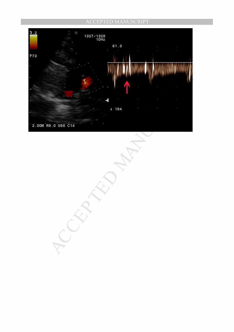
Transcranial color-coded sonography visualizes blood flow in the middle
cerebral artery (MCA) through a temporal window in the skull. A pulse wave Doppler
study is used to detect gas bubbles (either post-dive nitrogen bubbles or microbubbles
of ultrasonographic contrast) as high-intensity transient signals (HITS) (Fig. 3). The
presence of HITS confirms right-to-left shunting. The localization of the shunt may be
intracardiac or transpulmonary. The transpulmonary passage is longer and the bubbles
usually appear after >15 cardiac cycles following the administration of
MANUSCRIP
T
ACCEPTED
ACCEPTED MANUSCRIPT10
ultrasonographic contrast.30 When using standardized protocols, a sensitivity of 94-
100% and specificity of 75-100% compared to TEE has been reported.30,41 This makes
TCCS a valuable screening tool. A possible concern is that the temporal window may
be inadequate to visualize reliably the MCA in 10-12% of patients.42 However, this is
dependent on the examiner, the sonographic equipment and the age of the patients.43
Therefore, this might not be a limitation in young healthy subjects, such as most
recreational and professional divers.39 For screening, agitated saline or hydroxyethyl
starch solutions or a dedicated contrast agent may be used.44 The monitoring for HITS
should be performed according to a standardized protocol at rest and after a Valsalva
maneuver.45 The shunt is graded as follows: 0 - no HITS, 1 - <10 HITS, 2 - >10 HITS
but no curtain (uncountable number of bubbles), and 3 - curtain.45 Post-dive arterial
gas bubbles may be assessed in the same manner. However, to date, there is no
standardized protocol for this application. We suggest monitoring the MCA flow for 60
seconds during native breathing and subsequently three times for 40 seconds after a
Valsalva maneuvre.39
Therapeutic Options
There is still a large knowledge gap with regards to the optimal risk stratification
and management strategy in divers with a PFO. Routine screening for PFO in divers is
currently not recommended in most countries.46,47 Suggested recommendations for
divers with diagnosed PFO and a history of DCS include the cessation of diving, a
conservative approach to diving, and PFO closure. The evidence for both conservative
dive profiles (CDP) and catheter-based PFO closure is still sparse.
It has been suggested by several authors that a catheter-based PFO closure in
MANUSCRIP
T
ACCEPTED
ACCEPTED MANUSCRIPT11
divers might eliminate the arterialization of bubbles and prevent unprovoked
DCS.48,49,50 So far, only one study has provided data on the effect of PFO closure on
elimination of post-dive arterial gas emboli (AGE).39 In this study VGE and AGE were
assessed by means of ultrasound in 47 divers after surfacing from a simulated dive in
hyperbaric chamber. All divers had a large PFO (grade 3 according to the International
Consensus Criteria)45 and previously suffered from DCS; in 20, the PFO was occluded
with a catheter-based device (closure group), the other 27 divers did not undergo any
closure procedure (PFO group). The Amplatzer septal occluder (AGA Medical
Corporation, Golden Valley, MN, USA) and the Occlutech Figulla PFO Occluder N
(Occlutech GmbH, Jena, Germany) were used. In this study, no divers in the closure
group had post-dive AGE. Also, none of these divers had DCS symptoms. However,
the reduction in DCS incidence did not reach statistical significance. The lack of
predefined clinical endpoints, the small scale of the study and the experimental setting
are important limitations that must be considered. Clearly, more clinical data are
needed to obtain a definitive answer regarding DCS and PFO closure. Also, we must
bear in mind that this is an invasive procedure with potential major complications,
although the occurrence is generally low (<1%).51 The success rate of the procedure is
high, but a moderate residual shunt may occur in about 10% of cases.52 Furthermore,
it is important to note that PFO closure might have the potential to decrease the risk of
DCS to the level of non-PFO divers, but not to zero.
It is often recommended to cease diving to symptomatic divers diagnosed with
PFO. This solution is mostly not accepted and alternatives are sought. Conservative
dive profiles (CDP) are measures aiming to lower the probability of nitrogen bubble
formation in order to decrease the risk of DCS. The probability of tissue
supersaturation and subsequent bubble formation can theoretically be lowered by both
MANUSCRIP
T
ACCEPTED
ACCEPTED MANUSCRIPT12
minimizing tissue saturation (i.e. limiting nitrogen exposure) and allowing more time for
the desaturation of tissues. To lower nitrogen exposure, various CDP
recommendations limit maximum depth, dive time, number of dives per day or advise
the use of mixtures with lower nitrogen content (enriched air nitrox).22,51 Similarly, to
allow more time for desaturation, a slower ascent rate and performing longer safety
stops is recommended.53 There is also some evidence that pre-dive hydration and pre-
dive exercise reduce the risk of DCS.54 Few data are available regarding the safety of
these measures in divers with PFO. However, recently published study suggested a
significant decrease in arterial bubble occurrence that was achieved among divers with
large PFOs by limiting the exposure time and reducing the ascent rate.55
Conclusion
It seems likely that the presence of a PFO is associated with an increased risk
of DCS in recreational and professional divers, due to paradoxical embolism of
nitrogen bubbles. It is interesting that, despite the high number of divers and high
prevalence of PFO, a large knowledge gap exists regarding optimal screening, risk
stratification and management strategy. It seems that catheter-based PFO closure
might play a role in secondary DCS prevention in highly symptomatic divers in the
future. Currently, however, there is lack of clinical evidence to justify this approach. We
assume that clinical studies will bring important pathophysiological and clinical insights
in years to come.
Acknowledgments: The authors would like to acknowledge Lenka Hoňková, MD for
the preparation of illustrations.
MANUSCRIP
T
ACCEPTED
ACCEPTED MANUSCRIPT13
Funding sources: Supported by MH CZ – DRO, University Hospital Motol, Prague,
Czech Republic 00064203; SVV-2014-260033 from the Charles University in Prague
and PRVOUK-P24/LF1/3 of the Charles University in Prague — First Faculty of
Medicine.
Disclosures: none
MANUSCRIP
T
ACCEPTED
ACCEPTED MANUSCRIPT14
References
1. Vann RD, Freiberger JJ, Caruso JL. Divers Alert Network report on decompression
illness, diving fatalities and project dive exploration: 2005 edition (based on 2003
data). DAN technical Report, 2005. Available at:
http://www.diversalertnetwork.org/medical/report/2005DCIReport.pdf. Accessed
September 24, 2014.
2. Landzberg MJ, Khairy P. Patent foramen ovale: when is intervention warranted?
Can J Cardiol 2013;29:890-2.
3. Bove AA. The PFO gets blamed again…perhaps this time it is real. J Am Coll
Cardiol Intv 2014;7:409-10.
4. Hagen PT, Scholz DG, Edwards WD. Incidence and size of patent foramen ovale
during the first 10 decades of life: an autopsy study of 965 normal hearts. Mayo
Clin Proc 1984;59:17-20.
5. Boyle R. New pneumatical experiments about respiration. Philos Trans
1670;5:2011-58.
6. Triger M. Letter to Monsieur Arago. Comptes Rendus de l’Academie des Sciences
1845;20:445-9.
7. Bert P. Barometric pressure: researches in experimental physiology. Columbus:
College Book Company, 1943.
8. Acott C. A brief history of diving and decompression illness. South Pacific
Underwater Med Soc J 1999;29:98-109.
9. Doolette DJ, Mitchell SJ. The physiological kinetics of nitrogen and the prevention
of decompression sickness. Clin Pharmacokinet 2001;40:1-14.

MANUSCRIP
T
ACCEPTED
ACCEPTED MANUSCRIPT15
10. Papadopoulou V, Eckersley RJ, Balestra C, Karapantsios TD, Tang MX. A critical
review of physiological bubble formation in hyperbaric decompression. Adv Colloid
Interface Sci 2013;191-192:22-30.
11. Bove AA. Diving medicine. Am J Respir Crit Care Med 2014;189:1479-86.
12. Vann RD, Butler FK, Mitchell SJ, Moon RE. Decompression Illness. Lancet
2010;377:153-64.
13. Dunford RG, Vann RD, Gerth WA, et al. The incidence of venous gas emboli in
recreational diving. Undersea Hyperb Med 2002;29:247-59.
14. Ljubkovic M, Dujic Z, Møllerløkken A, et al. Venous and arterial bubbles at rest after
no-decompression air dives. Med Sci Sports Exerc 2011;43:990-5.
15. Gempp E, Blatteau JE, Simon O, Stephant E. Musculoskeletal decompression
sickness and risk of dysbaric osteonecrosis in recreational divers. Diving Hyperb
Med 2009;39:200-4.
16. Wilmhurst PT, Ellis PT, Jenkins BS. Paradoxical gas embolism in a scuba diver with
an atrial septal defect. British Med J 1986;293:1277.
17. Moon RE, Camporesi EM, Kisslo JA. Patent foramen ovale and decompression
sickness in divers. Lancet 1989;1:513-4.
18. Torti SR, Billinger M, Schwerzmann M, et al. Risk of decompression illness among
230 divers in relation to the presence and size of patent foramen ovale. Eur Heart J
2004;25:1014-20.
19. Wilmhurst PT, Pearson MJ, Walsh KP, Morrison WL, Bryson P. Relationship
between right-to-left shunts and cutaneous decompression illness. Clinical Science
2001;100:539-42.
20. Germonpré P, Dendale P, Unger P, Balestra C. Patent foramen ovale and
decompression sickness in sports divers. J Appl Physiol 1998;84:1622-6.
MANUSCRIP
T
ACCEPTED
ACCEPTED MANUSCRIPT16
21. Cantais E, Louge P, Suppini A, Foster PP, Palmier B. Right-to-left shunt and risk of
decompression illness with cochleovestibular and cerebral symptoms in divers:
case control study in 101 consecutive dive accidents. Crit Care Med 2003;31:84-8.
22. Gempp E, Louge P, Blatteau JE, Hugon M. Risk factors for recurrent neurological
decompression sickness in recreational divers: a case-control study. J Sports Med
Phys Fitness 2012;52:530-6.
23. Germonpre P, Balestra C. Risk of decompression illness among 230 divers in
relation to the presence and size of patent foramen ovale. Eur Heart J
2004;25:2173-4.
24. Knauth M, Ries S, Pohimann S, et al. Cohort study of multiple brain lesions in sport
divers: role of a patent foramen ovale. British Med J 1997;314:701-5.
25. Balestra C, Marroni A, Farkas B, et al. The Fractal Approach as a tool to
understand asymptomatic Brain Hyperintense MRI Signals. Fractals 2004;12:67-
72.
26. Marabotti C, Scalzini A, Menicucci D, Passera M, Bedini R, L'abbate A.
Cardiovascular changes during SCUBA diving: an underwater Doppler
echocardiographic study. Acta Physiol (Oxf) 2013 May 2 [Epub ahead of print], doi:
10.1111/apha.12112.
27. Ljubkovic M, Zanchi J, Breskovic T, Marinovic J, Lojpur M, Dujic Z. Determinants of
arterial gas embolism after scuba diving. J Appl Physiol 2012;112:91-5.
28. Cartin-Ceba R, Swanson KL, Krowka MJ. Pulmonary arteriovenous malformations.
Chest 2013; 144:1033-44.
29. Lovering AT, Elliott JE, Beasley KM, Laurie SS. Pulmonary pathways and
mechanisms regulating transpulmonary shunting into the general circulation: an
update. Injury 2010;41:S16-23.

MANUSCRIP
T
ACCEPTED
ACCEPTED MANUSCRIPT17
30. Sastry S, MacNab A, Daly K, Ray S, McCollum C. Transcranial Doppler detection
of venous-to-arterial circulation shunts: criteria for patent foramen ovale. J Clin
Ultrasound 2009;37:276-80.
31. Kerut EK, Norfleet WT, Plotnick GD, Giles TD. Patent foramen ovale: a review of
associated conditions and the impact of physiological size. J Am Coll Cardiol
2001;38:613-23.
32. Germonpre P, Hastir F, Dendale P, Marroni A, Nguyen AF, Balestra C. Evidence for
increasing patency of the foramen ovale in divers. Am J Cardiol 2005;95:912-5.
33. Pinto FJ. When and how to diagnose patent foramen ovale. Heart 2005;91:438-40.
34. Bartel T, Müller S. Device closure of inter-atrial communications: peri-interventional
echocardiographic assessment. Eur Heart J Cardiovasc Imaging 2013;14:618-24.
35. Van Camp G, Franken P, Melis P, Cosyns B, Schoors D, Vanoverschelde JL.
Comparison of transthoracic echocardiography with second harmonic imaging with
transesophageal echocardiography in the detection of right to left shunts. Am J
Cardiol 2000;86:1284-7.
36. Thanigaraj S, Valika A, Zajarias A, Lasala JM, Perez JE. Comparison of
transthoracic versus transesophageal echocardiography for detection of right-to-left
atrial shunting using agitated saline contrast. Am J Cardiol 2005;96:1007-10.
37. Clarke NR, Timperley J, Kelion AD, Banning AP. Transthoracic echocardiography
using second harmonic imaging with Valsalva manoeuvre for the detection of right
to left shunts. Eur J Echocardiogr 2004;5:176-81.
38. Ha JW, Shin MS, Kang S, et al. Enhanced detection of right-to-left shunt through
patent foramen ovale by transthoracic contrast echocardiography using harmonic
imaging. Am J Cardiol 2001;87:669-71.
MANUSCRIP
T
ACCEPTED
ACCEPTED MANUSCRIPT18
39. Honěk J, Šrámek M, Šefc L, et al. Effect of catheter-based patent foramen ovale
closure on the occurrence of arterial bubbles in scuba divers. J Am Coll Cardiol Intv
2014;7:403-8.
40. Blogg SL, Gennser M, Møllerløkken A, Brubakk AO. Ultrasound detection of
vascular decompression bubbles: the influence of new technology and
considerations on bubble load. Diving Hyperb Med 2014;44:35-44.
41. Droste DW, Schmidt-Rimpler C, Wichter T, et al. Right-to-left-shunts detected by
transesophageal echocardiography and transcranial Doppler sonography.
Cerebrovasc Dis 2004;17:191-6.
42. Postert T, Federlein J, Przuntek H, Büttner T. Insufficient and absent acoustic
temporal bone window: potential and limitations of transcranial contrast-enhanced
color-coded sonography and contrast-enhanced power-based sonography.
Ultrasound Med Biol 1997;23:857-62.
43. Spacek M, Sorrell VL, Veselka J. Transcranial Doppler Ultrasound in the Current
Era of Carotid Artery Stenting. Ultraschall Med. 2014 Jun 25. [Epub ahead of print]
DOI: 10.1055/s-0034-1366677
44. Droste DW, Lakemeier S, Wichter T, et al. Optimizing the technique of contrast
transcranial Doppler ultrasound in the detection of right-to-left shunts. Stroke
2002;33:2211-6.
45. Jauss M, Zanette E. Detection of right-to-left shunt with ultrasound contrast agent
and transcranial Doppler sonography. Cerebrovasc Dis 2000;10:490-6.
46. Undersea and Hyperbaric Medical Society. UHMS best practice guidelines:
prevention and treatment of decompression sickness and arterial gas embolism.
Available at: https://www.uhms.org/images/DCS-AGE-
Committee/dcsandage_prevandmgt_uhms-fi.pdf Accessed Dec 17, 2014.

MANUSCRIP
T
ACCEPTED
ACCEPTED MANUSCRIPT19
47. Torti SR, Kraus M, Vőllm EB. SUHMS Guidelines for diving with a patent foramen
ovale. Available at: http://www.tauchschuleluzern.ch/pdf/suhms_pfo.pdf. Accessed
Dec 17, 2014.
48. Billinger M, Zbinden R, Mordasini R, et al. Patent foramen ovale closure in
recreational divers: effect on decompression illness and ischaemic brain lesions
during long-term follow-up. Heart 2011;97:1932-7.
49. Walsh KP, Wilmhurst PT, Morrison WL. Transcatheter closure of patent foramen
ovale using the Amplatzer septal occluder to prevent recurrence of neurological
decompression illness in divers. Heart 1999;81:257-61.
50. Lairez O, Cournot M, Minville V, et al. Risk of neurological decompression sickness
in the diver with right-to-left shunt: literature review and meta-analysis. Clin J Sport
Med 2009;19:231-5.
51. Meier B. Closure of patent foramen ovale: technique, pitfalls, complications, and
follow up. Heart 2005;91:444-8.
52. Matsumura K, Gevorgyan R, Mangels D, Masoomi R, Mojadidi MK, Tobis J.
Comparison of residual shunt rates in five devices used to treat patent foramen
ovale. Catheter Cardiovasc Interv 2014;84:455-63.
53. Klingmann C, Rathmann N, Hausmann D, Bruckner T, Kern R. Lower risk of
decompression sickness after recommendation of conservative decompression
practices in divers with and without vascular right-to-left shunt. Diving Hyperb Med
2012;42:146-50.
54. Gempp E, Blatteau JE. Preconditioning methods and mechanisms for preventing
the risk of decompression sickness in scuba divers: a review. Res Sports Med
2010;18:205-18.

MANUSCRIP
T
ACCEPTED
ACCEPTED MANUSCRIPT20
55. Honěk J, Šrámek M, Šefc L, et al. Effect of conservative dive profiles on the
occurrence of venous and arterial bubbles in divers with a patent foramen ovale: a
pilot study. Int J Cardiol 2014;176:1001-2.
MANUSCRIP
T
ACCEPTED
ACCEPTED MANUSCRIPT21
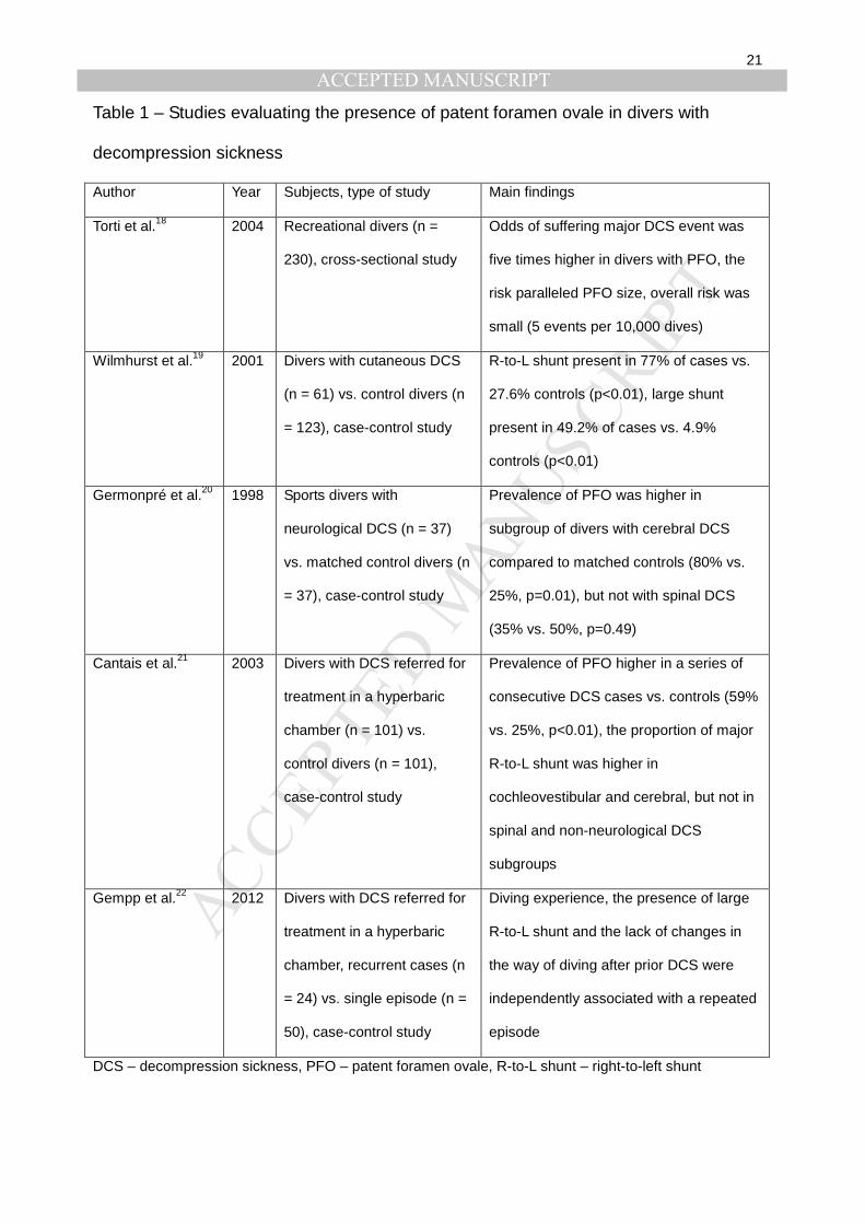
Table 1 – Studies evaluating the presence of patent foramen ovale in divers with
decompression sickness
Author Year Subjects, type of study Main findings
Torti et al.18 2004 Recreational divers (n =
230), cross-sectional study
Odds of suffering major DCS event was
five times higher in divers with PFO, the
risk paralleled PFO size, overall risk was
small (5 events per 10,000 dives)
Wilmhurst et al.19 2001 Divers with cutaneous DCS
(n = 61) vs. control divers (n
= 123), case-control study
R-to-L shunt present in 77% of cases vs.
27.6% controls (p<0.01), large shunt
present in 49.2% of cases vs. 4.9%
controls (p<0.01)
Germonpré et al.20 1998 Sports divers with
neurological DCS (n = 37)
vs. matched control divers (n
= 37), case-control study
Prevalence of PFO was higher in
subgroup of divers with cerebral DCS
compared to matched controls (80% vs.
25%, p=0.01), but not with spinal DCS
(35% vs. 50%, p=0.49)
Cantais et al.21 2003 Divers with DCS referred for
treatment in a hyperbaric
chamber (n = 101) vs.
control divers (n = 101),
case-control study
Prevalence of PFO higher in a series of
consecutive DCS cases vs. controls (59%
vs. 25%, p<0.01), the proportion of major
R-to-L shunt was higher in
cochleovestibular and cerebral, but not in
spinal and non-neurological DCS
subgroups
Gempp et al.22 2012 Divers with DCS referred for
treatment in a hyperbaric
chamber, recurrent cases (n
= 24) vs. single episode (n =
50), case-control study
Diving experience, the presence of large
R-to-L shunt and the lack of changes in
the way of diving after prior DCS were
independently associated with a repeated
episode
DCS – decompression sickness, PFO – patent foramen ovale, R-to-L shunt – right-to-left shunt

MANUSCRIP
T
ACCEPTED
ACCEPTED MANUSCRIPT22
Figure legends
Fig. 1 Pathophysiology of bubble formation and embolization in decompression
sickness
A dive profile of 18 m maximum depth and bottom time (time to ascent) of 80 min is
depicted to demonstrate pathophysiology of bubble formation and embolization in
divers. During descent, the diver breathes air at elevated ambient pressure, and
excess nitrogen dissolves in tissues (A). During ascent, the ambient pressure drops
and a pressure gradient drives nitrogen from tissues to venous blood (B). If the
pressure drops too quickly, the tissues become supersaturated and nitrogen bubbles
form and embolize through venous blood. In a diver with a PFO, a paradoxical right-to-
left embolization of bubbles may occur and the bubbles lodge into peripheral
capillaries (C). The resulting ischemia may manifest as decompression sickness. pN2
– partial pressure of nitrogen.
Fig. 2 Echocardiographic appearance of post-dive venous bubbles
Transthoracic echocardiography apical four-chamber view: post-dive nitrogen bubbles
(arrow) are apparent in right-sided, but not left-sided heart chambers in a diver with a
patent foramen ovale and no right-to-left shunt during native breathing.
Fig. 3 Arterial gas emboli visualized by transcranial color-coded sonography
Transcranial color-coded sonography: post-dive arterial gas emboli apparent as high-
intensity transient signals (arrow) in the Doppler spectrum in the middle cerebral artery
in a diver with a patent foramen ovale.

MANUSCRIP
T
ACCEPTED
ACCEPTED MANUSCRIPT

MANUSCRIP
T
ACCEPTED
ACCEPTED MANUSCRIPT
MANUSCRIP
T
ACCEPTED
ACCEPTED MANUSCRIPT