The roles of BRCA2 in chromosome integrity
151
The roles of BRCA2 in chromosome integrity by Florian Joseph Groelly A thesis submitted for the degree of Doctor of Philosophy St Cross College, University of Oxford Hilary Term 2021
-
Upload
khangminh22 -
Category
Documents
-
view
1 -
download
0
Transcript of The roles of BRCA2 in chromosome integrity

The roles of BRCA2 in chromosome integrity
by
Florian Joseph Groelly
A thesis submitted for the degree of
Doctor of Philosophy
St Cross College, University of Oxford
Hilary Term 2021

II
The roles of BRCA2 in chromosome integrity
A thesis submitted to the University of Oxford
for the degree of Doctor of Philosophy
Florian Joseph Groelly
St Cross College, Hilary term 2021
High frequency of fork stalling and fork degradation are associated with DNA
damage accumulation and represent key replication pathologies in cells lacking
BRCA2. As a consequence, these cells enter mitosis with under-replicated DNA,
which leads to interlinks between sister chromatids in anaphase. Failure to
resolve these interlinks causes chromosome mis-segregation, aneuploidy and
micronuclei. Here we show that BRCA2 loss leads to micronuclei formation,
which triggers an innate immune response signalling via activation of the cGAS-
STING pathway. However, cleavage of these interlinks by the MUS81
endonuclease activates mitotic DNA synthesis (MiDAS), thereby completing
genome replication and ensuring correct chromosome segregation. Which
genomic loci require BRCA2 for their replication and therefore become unstable
upon BRCA2 inactivation remains unknown. Here we label mitotic nascent DNA
and perform EdU-seq to monitor at high-resolution sites where MiDAS occurs in
absence of BRCA2. Our findings reveal that loss of BRCA2 triggers MiDAS at
150 genomic loci, which map within regions replicating in early S phase and are
therefore distinct from aphidicolin-induced common fragile sites. Our results
suggest that BRCA2 inactivation causes replication failure at early-replicating
genomic regions, a subset of which completes replication during mitosis.

III
Acknowledgements
I would like to thank Madalena Tarsounas for welcoming and advising me
throughout my DPhil. Not only did I benefit from her scientific guidance, but I was
also able to gain precious communication, responsibility, leadership and
teamworking experience.
I also would like to thank Thanos Halazonetis for hosting me in his
laboratory. Many thanks to Frank, Jeltje, Pauline and Elena for organising my
secondment at Merck KGaA. Whilst the work performed during these 2 months
is not directly described in this thesis, I had an exceptional first touch of the
contribution of Medical Affairs.
Thank you to my colleagues, Rebecca, Giuliana, Angelina, Timo, Hugo,
Emilia, Yanti, Pia, Yuri, Sophie, Anastasiya and Irene, for the great moments
shared inside and outside of the lab. In particular, thank you to Rebecca for
teaching me a plethora of molecular and cell biology techniques, and to Timo for
the fruitful teamwork as well as for commenting on this thesis.
To my dearest housemates, Ashwin, Charlotte, Luana, Sergio, Nico, and
Gabriele: what great times we had. Thank you for your support, for picking (not
only) the best movies and for experimenting my cuisine.
Thank you to all my dearest friends and all the people who made my
Oxford experience exceptional: Timo, Hugo, Ashwin, Luana, Asmita, Maria,
Viviana, Giovanna, Angela, Andrea, Prashanti, Gerardo, Simone, Sophie, Egon,
Manon, Niloufar, Jakob, Javier, Sam, Shudong, John, Tiffany, Christie, Giacomo,
Tzveta, Salomé, Shahd, Jack, James, Xiaoning, Hongbin, Lottie, Tobias and
many others.

IV
I would like to thank all members of the SYNTRAIN network for the great
collaborations, training and outcomes. In particular thank you to all the fellows for
the great atmosphere: Giacomo, Giorgos, Christos, Nibal, Ana, Mariana, Luka,
Karolin, Nanda, Bishoy, Aleks, Anna, Jemina, and Timo.
Thank you who were not here but supported nonetheless: Charlotte,
Eileen, Pierre-Louis, Cédric, Émilie, Stéphanie, Aurélia, Maxime, Dario, Michael,
Hugo, Johanna, and many others. A special note also goes to my parents, Patrick
and Christine, as well as to my siblings, Nicolas and Marie.

V
Declaration of authorship
I confirm that this thesis I am submitting is wholly my own work except where
otherwise indicated. Part of this thesis was published in peer-reviewed journals
(Reisländer et al., 2019; Reisländer, Groelly and Tarsounas, 2020).
The cells for RNA-sequencing were grown by Timo Reisländer and
differential gene expression was analysed by Emilia Puig Lombardi. Quantitative
reverse transcription PCR (RT-qPCR) was performed by Timo Reisländer, using
sample prepared together. Analysis of the EdU-sequencing data was done with
help from Emilia Puig Lombardi.
Funding
The work presented here was performed at the Department of Oncology,
University of Oxford and has received funding from the European Union’s Horizon
2020 research and innovation programme under the Marie Skłodowska-Curie
grant agreement number 722729.

VI
Table of contents
The roles of BRCA2 in chromosome integrity ................................................. II
Acknowledgements ............................................................................................ III
Declaration of authorship ................................................................................... V
Funding ................................................................................................................. V
Table of contents ................................................................................................ VI
List of tables ......................................................................................................... X
List of figures ...................................................................................................... XI
List of abbreviations ........................................................................................ XIII
Chapter 1 Introduction ...................................................................................... 16
1.1 DNA damage, DNA repair pathways and genome stability...................... 17
1.1.1 DNA as the molecular basis of genetic information ....................... 17
1.1.2 Damage of nucleic bases and repair .............................................. 18
1.1.3 Mechanisms of SSB repair ............................................................. 20
1.1.4 Mechanisms of DSB repair ............................................................. 21
1.2 Stalled replication forks as a source of genomic instability ...................... 25
1.2.1 Functions of BRCA2 at stalled replication forks ............................. 25
1.2.2 Restart of stalled replication forks in BRCA2-deficient cells .......... 30
1.3 Chromosome fragility ................................................................................. 32
1.3.1 Common fragile sites ...................................................................... 32
1.3.2 Other fragile sites ............................................................................ 34

VII
1.3.3 Mitotic DNA synthesis (MiDAS) ...................................................... 34
1.3.4 BRCA2 inactivation in chromosome fragility and instability ........... 38
1.4 Research aims ........................................................................................... 38
Chapter 2 Material and methods ..................................................................... 40
2.1 Cell lines and cell culture ........................................................................... 41
2.2 RNA-sequencing (RNA-seq) ..................................................................... 42
2.3 RNA-seq data processing ......................................................................... 42
2.4 Differential gene expression analysis ....................................................... 43
2.5 Quantitative RT-PCR ................................................................................. 43
2.6 Western blotting ......................................................................................... 44
2.7 Resazurin-based viability assay ................................................................ 45
2.8 Flow cytometry-based assays ................................................................... 46
2.8.1 Detection of EdU and Histone H3 (S10) ......................................... 46
2.8.2 Cell cycle analysis ........................................................................... 46
2.8.3 S phase entry .................................................................................. 47
2.8.4 S-to-M progression .......................................................................... 47
2.8.5 DNA content analysis ...................................................................... 47
2.9 Drug treatment ........................................................................................... 47
2.10 Cell synchronization for mitotic EdU-seq ................................................ 48
2.11 EdU-seq ................................................................................................... 48
2.12 EdU-seq data processing ........................................................................ 50
2.13 Analysis of MiDAS peaks ........................................................................ 50
2.14 Analysis of the replication timing ............................................................. 51
2.15 Assignment of genic and intergenic regions ........................................... 51
2.16 Statistical analyses .................................................................................. 51

VIII
Chapter 3 BRCA2 abrogation activates the cGAS-STING-IRF3 and JAK-
STAT pathways to promote expression of interferon-stimulated genes ... 52
3.1 Introduction ................................................................................................ 53
3.2 Characterisation of the inducible models for BRCA2 inactivation ............ 54
3.3 Long-term BRCA2 abrogation triggers innate immune response signalling
.......................................................................................................................... 57
3.4 Long-term BRCA2 abrogation activates the cGAS-STING-IRF3 and the
JAK-STAT pathways ........................................................................................ 59
3.5 PARP inhibitors enhance the type I innate immune response in BRCA2-
deficient cells.................................................................................................... 65
3.6 DNA damaging agents targeting BRCA2-deficiency trigger a type I IFN
response........................................................................................................... 69
3.7 Functional up-regulation of ISG15 and its ISGylation activity .................. 71
3.8 Discussion .................................................................................................. 73
Chapter 4 Protocols for high-resolution detection of mitotic DNA synthesis
(MiDAS) using mitotic EdU-seq. ...................................................................... 79
4.1 Introduction ................................................................................................ 80
4.2 Synchronous S phase progression ........................................................... 80
4.3 Cell synchronisation in G2/M..................................................................... 83
4.4 Protocol for detection of MiDAS under untreated conditions using mitotic
EdU-seq ........................................................................................................... 83
4.5 Low dose aphidicolin delays S phase progression ................................... 85
4.6 Protocol for detection of MiDAS events induced by low dose aphidicolin 88
4.7 Discussion .................................................................................................. 90

IX
Chapter 5 High-resolution detection of MiDAS upon BRCA2 abrogation . 93
5.1 Introduction ................................................................................................ 94
5.2 Detection of aphidicolin-induced MiDAS at common fragile sites ............ 95
5.3 Detection of MiDAS in BRCA2-deficient cells ........................................... 99
5.4 BRCA2 abrogation does not activate MiDAS at common fragile sites .. 101
5.5 BRCA2 abrogation activates MiDAS within genomic regions that replicate
during early S phase ...................................................................................... 101
5.6 Resemblance between the aphidicolin-induced MiDAS pattern in BRCA2-
proficient and BRCA2-deficient cells ............................................................. 105
5.7 Discussion ................................................................................................ 107
Chapter 6 Discussion and conclusion.......................................................... 111
6.1 Micronuclei formation and innate immune response signalling ............. 112
6.2 Replication defects and mitotic DNA synthesis (MiDAS) ....................... 114
6.3 The role of BRCA2 for genome and chromosome stability .................... 116
References ....................................................................................................... 117
Appendix........................................................................................................... 150

X
List of tables
Table 2.1. Primer pairs used for RT-qPCR. .................................................... 43
Table 2.2. Primary antibodies used for immunoblotting. ................................ 45
Table 2.3. Secondary antibodies used for immunoblotting. ........................... 45

XI
List of figures
Fig. 1.1. Repair pathway for SSBs. ................................................................. 21
Fig. 1.2. Repair pathways for DSBs. ............................................................... 29
Fig. 1.3. Mechanisms of stabilisation and restart of stalled replication forks. 32
Fig. 1.4. Model of MiDAS at under-replicated loci. ......................................... 37
Fig. 2.1. Strategy for high-resolution detection of nascent DNA using EdU-seq
.......................................................................................................................... 49
Fig. 3.1. Time-dependent activation of the cGAS-STING-IRF3 and JAK-STAT
pathways upon BRCA2 depletion. .................................................................. 56
Fig. 3.2. RNA-seq analysis reveals upregulation of immune response signalling
upon long-term BRCA2 depletion. .................................................................. 58
Fig. 3.3. Time-dependent expression of interferon-stimulated genes (ISGs)
upon BRCA2 depletion. ................................................................................... 61
Fig. 3.4. Time-dependent activation of the cGAS-STING-IRF3 and JAK-STAT
pathways upon BRCA2 depletion. .................................................................. 64
Fig. 3.5. Olaparib-treatment affects growth and survival of BRCA2-deficient
H1299+shBRCA2DOX cells. .............................................................................. 67
Fig. 3.6. Olaparib-treatment activates the cGAS-STING-IRF3 and JAK-STAT
pathways in BRCA2-deficient H1299+shBRCA2DOX cells. ............................. 68
Fig. 3.7. DNA damaging agents activate the cGAS-STING-IRF3 and JAK-
STAT pathways in BRCA2-deficient H1299+shBRCA2DOX cells. .................. 70
Fig. 3.8. Functional expression of ISG15 upon BRCA2 depletion and Olaparib
treatment. ......................................................................................................... 72
Fig. 3.9. Interplay between the DNA damage response and the innate immune
response for cancer (immuno-)therapy. .......................................................... 78

XII
Fig. 4.1. Cell cycle progression after thymidine block and release. ............... 82
Fig. 4.2. RO-3306-induced G2 block does not stop S phase progression. ... 85
Fig. 4.3. Mitotic EdU-seq protocol for H1299+shBRCA2DOX cells. ................. 86
Fig. 4.4. S phase progression upon aphidicolin treatment. ............................ 87
Fig. 4.5. Mitotic EdU-seq protocol for aphidicolin-treated H1299+shBRCA2DOX
cells. ................................................................................................................. 90
Fig. 5.1. Detection of MiDAS in BRCA2-proficient H1299+shBRCA2DOX treated
or not with aphidicolin. ..................................................................................... 96
Fig. 5.2. MiDAS upon aphidicolin treatment takes place at known common
fragile sites. ...................................................................................................... 98
Fig. 5.3. Detection of MiDAS in BRCA2-deficient H1299+shBRCA2DOX cells.
........................................................................................................................ 100
Fig. 5.4. BRCA2 inactivation and aphidicolin-treatment trigger MiDAS at distinct
genomic regions............................................................................................. 102
Fig. 5.5. BRCA2 inactivation triggers MiDAS at early S phase replicating
domains. ......................................................................................................... 104
Fig. 5.6. Comparison of aphidicolin-induced MiDAS in BRCA2-proficient and
BRCA2-deficient cells. ................................................................................... 106

XIII
List of abbreviations
% Percent
ºC Celsius degree
DNA Deoxyribonucleic acid
A Adenosine
BER Base-excision repair
BIR Break-induced repair
C Cytosine
cDNA Complementary DNA
D-loop Displacement loop
Da Dalton(s)
DDR DNA damage response
DMEM Dulbecco’s Modified Eagle’s Medium
DMSO Dimethyl sulfoxide
DOX Doxycycline
DSB Double-strand break
DSBR Double-strand break repair
DTT Dithiothreitol
EDTA Ethylenediaminetetraacetic acid
EdU 5-Ethynyl-2´-deoxyuridine
EdU-seq EdU-sequencing
ERFS Early replicating fragile sites
FBS Foetal bovine serum
FDR False discovery rate
Fig. Figure
XIV
FISH Fluorescence in situ hybridization
G Guanine
GO Gene Ontology
h Hour(s)
HR Homologous recombination
HRP Horseradish peroxidase
HU Hydroxyurea
IFN Interferon
IR Ionising radiation
ISG Interferon-stimulated gene
ISRE IFN-stimulated response elements
M Molar
MiDAS Mitotic DNA synthesis
min Minute(s)
MMR Mismatch repair
mtDNA Mitochondrial DNA
NER Nucleotide excision repair
NHEJ Non-homologous end joining
PAR Poly [ADP-ribose]
PARG PAR glycohydrolase
PARP PAR polymerase
PBS Phosphate saline buffer
PBST 0.05% Tween 20 in PBS
PCR Polymerase chain reaction
QIBC Quantitative image-based cytometry

XV
RNA Ribonucleic acid
RNA-seq RNA-sequencing
ROS Reactive oxygen species
RT-qPCR Quantitative reverse transcription PCR
SDS Dodecyl sulfate-polyacrylamide
SDSA Synthesis-dependent strand annealing
shRNA Short hairpin RNA
siRNA Small interfering RNA
SSA Single-strand annealing
SSB Single-strand break
ssDNA Single-stranded DNA
T Thymidine
UV Ultra-violet
V Volt

16
Chapter 1
Introduction

17
1.1 DNA damage, DNA repair pathways and genome stability
1.1.1 DNA as the molecular basis of genetic information
The human genome consists of linear arrays of nucleotides forming double-
stranded DNA (deoxyribonucleic acid) molecules assembled into 23 pairs of
chromosomes within the nucleus (Gartler, 2006). A fraction of the genetic
information is also stored on circular DNA within mitochondria (Taanman, 1999),
known as mitochondrial DNA (mtDNA). The human genome is highly conserved
among the population, with variations randomly distributed across the genome
(Genomes Project et al., 2010; Genomes Project et al., 2015). Because DNA is
duplicated in a semi-conserved manner (Meselson and Stahl, 1958), all of an
individual’s nucleated cells should, in theory, contain the same genetic
information. There are, however, specific cellular contexts that can lead to
programmed changes in the genetic information. Prominent examples are
lymphocytes undergoing V(D)J recombination (Roth and Craig, 1998; Schatz and
Ji, 2011) and gametes undergoing meiosis (Marston and Amon, 2004).
Importantly, spontaneous changes in the nucleotide sequence can occur as a
consequence of inaccurate DNA replication (e.g., nucleotide mismatch) or of
inaccurate repair of DNA damage (Abbotts and Wilson, 2017; Tubbs and
Nussenzweig, 2017).
DNA damage can arise spontaneously, as a result of cellular metabolism,
or can be caused by exposure to exogenous genotoxic agents. DNA damage can
interfere with DNA replication and transcription and can trigger cellular
senescence or apoptosis. If incorrectly repaired, DNA damage can lead to
mutations, such as point mutations, insertion and deletions (indels), chromosome
rearrangements and chromosome fusions (Abbotts and Wilson, 2017; Tubbs and

18
Nussenzweig, 2017). Of note, a DNA alteration, which is highly relevant in the
context of cancer, is DNA methylation (Ehrlich, 2002; Das and Singal, 2004). It
however does not alter nucleotide sequences, but impacts on DNA transcription:
promoter hypermethylation can result in loss of gene expression. DNA
methylation is, however, part of the cell’s epigenome, which is beyond the scope
of this thesis.
1.1.2 Damage of nucleic bases and repair
Spontaneous DNA damage often affects nucleic bases, through deamination of
cytosine, guanine or adenine that can negatively impact on genome stability
(Abbotts and Wilson, 2017). Deamination of cytosine or adenine does not directly
impact on the Watson-Crick base pairing. However, deamination of methylated
cytosine (5-methylcytosine) produces thymidine, which can result in a C-to-T
mutation. This DNA lesion is repaired by the base-excision repair (BER) pathway
(Abbotts and Wilson, 2017; Wu and Zhang, 2017), which requires the G/T
mismatch-specific thymine DNA glycosylase and produces an abasic sites. Such
sites can also result from the spontaneous cleavage of the covalent bound
between the deoxyribose and an adenine or guanine base. Repair of abasic sites
requires the recruitment of the apurinic/apyrimidinic endonucleases APE1 or
APE2, which leads to the formation of a DNA single-strand break (SSB; see
below).
Replication of damaged nucleic bases can also lead to mismatched
Watson-Crick bases. DNA polymerases are error-prone enzymes with an error
rate ranging from 10-3 to 10-7. The high-fidelity polymerases, including DNA
polymerase δ or ε, replicate most of the human genome and exhibit a
proofreading exonuclease activity (Kunkel, 2004; Kunkel and Erie, 2015; Liu,

19
Keijzers and Rasmussen, 2017). In contrast, low fidelity DNA polymerases
including polymerase tetha (POLQ), are usually required for translesion synthesis
and lack the proof-reading mechanism, inserting errors at a rate of 10-3 (Arana et
al., 2008). Whilst mismatches do not represent DNA damage per se, they can be
mutagenic and alter gene expression and therefore their detection by the DNA
mismatch repair (MMR) pathway is critical for genome stability. MMR activation
leads to the excision of several hundred nucleotides surrounding the mismatch,
followed by pol δ/ε-dependant DNA re-synthesis and ligation (Kunkel and Erie,
2015; Hsieh and Zhang, 2017).
Nucleic bases can also be damaged by endogenous and exogenous
agents, including reactive oxygen species (ROS) or ultra-violet (UV) light,
respectively. For example, exposure to UV light catalyses the formation of
pyrimidine dimers between two consecutive thymidine or cytosine bases (Rastogi
et al., 2010; Abbotts and Wilson, 2017). Failure to repair pyrimidine dimers
causes increased frequency of C-to-T substitutions and DNA deletions (Sale,
2013). Studies into the genetic and molecular defects underlying the Cockayne
syndrome or xeroderma pigmentosum disorders, led to the discovery of specific
repair mechanisms for bulky DNA lesions, such as UV-induced pyrimidine dimers
(Cleaver, Lam and Revet, 2009; Vaisman and Woodgate, 2017). Indeed,
Cockayne syndrome patients suffer from photosensitivity and neurological
disorders caused by deleterious mutations in the ERCC6 (sometimes referred to
as CSB) or ERCC8 (or CSA) genes, whose products are essential for nucleotide
excision repair (NER) (Cleaver, Lam and Revet, 2009). The NER pathway
detects bulky DNA lesions and promotes the excision of 22-32 nucleotides
around the lesion, which results in a single-stranded DNA gap. Subsequently,

20
DNA polymerases and ligases are required to re-synthesise DNA and complete
the repair.
1.1.3 Mechanisms of SSB repair
SSBs are obligate intermediates in several repair pathways, including BER, NER
and MMR. Oxidative attack of the deoxyribose constitutive of the DNA backbone
by ROS represent a well-characterised source of SSBs. Importantly, direct sugar
disintegration leads to lesions of the 5’-phosphate or 3’-hydroxyl groups, which
are particularly cytotoxic (Kathe, Shen and Wallace, 2004). SSBs are deleterious
lesions because they can cause replication fork stalling (see below) or can be
converted to DNA double-strand breaks (DSBs).
SSB repair pathways are essential for the repair of SSBs arising during
BER or NER and resembles the repair of direct SSBs (Caldecott, 2008; Abbotts
and Wilson, 2017). Consequently, these different repair mechanisms are
considered to be sub-pathways of SSB repair pathway (Caldecott, 2008). Four
steps define the SSB repair pathway: 1) SSB detection and signalling; 2) 5’- and
3’-termini processing; 3) DNA gap filling; 4) DNA ligation. For example, ROS-
induced SSBs are detected by members of the poly [ADP-ribose] (PAR)
polymerase (PARP) family, which conjugate PAR units to proteins present at the
break, including itself, in a process known as PARylation. Conversely,
dePARylation is mediated by the PAR glycohydrolase (PARG). PARylation and
dePARylation events facilitate the recruitment of the SSB repair protein XRCC1,
which acts as a scaffold for the recruitment and assembly of further SSB repair
factors. Amongst those, DNA end processing enzymes, such as DNA polymerase
β, FEN-1 or APE1, are required to remove damaged 5’- or 3’-flaps, thus allowing
access of DNA polymerases, for instance the previously mentioned Pol δ/ε, to fill

21
the DNA gap, and of DNA ligases to covalently attach the newly synthesis DNA
to the original DNA strand.
Fig. 1.1. Repair pathway for SSBs.
SSBs are detected by PARP1, which PARylation activity is counteracted
by PARG. These events trigger the recruitment of downstream repair
factors, including the XRCC1 scaffold protein. Next, damaged DNA ends
are processed by Polβ, FEN-1 or APE1 in preparation for DNA gap filling
and ligation. Upon short-patch repair. Polβ and LIG3 are responsible for
the synthesis of the missing nucleotide and its ligation, respectively. Upon
long-patch repair, Pol δ/ε synthesise 2–12 nucleotides and LIG1
catalyses DNA ligation.
1.1.4 Mechanisms of DSB repair
DSBs may arise from unrepaired SSB, from exposure to DNA damaging agents,
such as the TOP1 poison etoposide, ionising radiation (IR) or crosslinking agents
including platinum drugs, or from the collapse of stalled replication forks (Chang
et al., 2017; Scully et al., 2019). DSBs are less frequent, yet more cytotoxic than
SSBs (Abbotts and Wilson, 2017; Chang et al., 2017). Moreover, these lesions

22
are highly mutagenic as illustrated by increased tumorigenesis onset after
exposure to IR (Gilbert, 2009), a source of DSBs routinely used in the clinic for
cancer treatment (Baskar et al., 2012). DSB detection by the heterotrimeric
complex MRN, formed by MRE11, RAD50 and NBS1, stimulates ATM, which
initiates the cellular DNA damage response (DDR,(Abbotts and Wilson, 2017;
Blackford and Jackson, 2017; Chang et al., 2017)). ATM kinase activity is
required to orchestrate DSB repair and/or to phosphorylate p53, which arrests
cell cycle progression (Banin et al., 1998; Canman et al., 1998).
ATM activation triggers two main DNA DSB repair pathways: non-
homologous end joining (NHEJ) and homologous recombination (HR).
Alternative end-joining (sometimes referred to as microhomology-mediated end
joining) and single-strand annealing (SSA) rely on the annealing of short
homologous sequences and are considered to be backup repair pathways
(Ceccaldi, Rondinelli and D'Andrea, 2016; Chang et al., 2017; Scully et al., 2019).
Whilst NHEJ is active both in interphase, HR repair is restricted to S and G2
phases, where a sister chromatid is available as a template for repair (Hustedt
and Durocher, 2016). Because it uses an intact DNA molecule as template, HR
is considered the most accurate DSB repair pathway. Whether NHEJ is indeed
mutagenic is still unclear, as increasing evidence suggests that either failed
NHEJ or its illegitimate activation can lead to toxic DSB repair (Zong et al., 2015;
Balmus et al., 2019).
The choice of DNA repair pathway at a given DSB is not only regulated by
the cell cycle stage, but also by the type of DNA ends. Blunt or short single-
stranded DNA ends are preferentially repaired by NHEJ, whilst long single-
stranded DNA ends favour HR (Chang et al., 2017; Scully et al., 2019).

23
Consistently, 53BP1 (Bunting et al., 2010), RIF1 (Chapman et al., 2013;
Di Virgilio et al., 2013; Escribano-Diaz et al., 2013; Zimmermann et al., 2013) and
REV7 (also known as MAD2L2; (Boersma et al., 2015; Xu et al., 2015)) act
together to counteract DNA end resection at DSBs and promote NHEJ. Recent
studies showed that SHLD3 binds to RIF1 to recruit REV7 to DSBs. REV7 and
SHLD3 interact with SHLD1-SHLD2 to form the Shieldin complex (Dev et al.,
2018; Ghezraoui et al., 2018; Gupta et al., 2018; Mirman et al., 2018;
Noordermeer et al., 2018). Upon its recruitment, SHLD2 is believed to bind to
ssDNA at the DSB and protect DNA ends from resection. Whether ssDNA is
required for the assembly of the Shieldin complex and which factors promote the
formation of ssDNA at Shieldin-protected DSBs remain unknown (Noordermeer
and van Attikum, 2019). The heterotrimeric CST complex, formed by CTC1,
STN1 and TEN1, also impact on DSB repair pathway choice by counteracting
resection. Indeed, the CST complex interacts with the Shieldin complex and is
recruited to DSBs (Mirman et al., 2018), where it promotes Pol α-mediated fill-in
DNA synthesis of resected ends (Barazas et al., 2018; Mirman et al., 2018).
Consistent with their functions at DSBs, loss of 53BP1, RIF1 or of members of
the Shieldin or CST complexes restores HR and causes resistance to PARP
inhibitors in BRCA1-deleted cells (Noordermeer and van Attikum, 2019).
The recruitment of the heterodimeric Ku70-Ku80 complex to each DNA
end constitutes the first step of DSB repair by NHEJ. Next, two DNA-PKcs
molecules interact with the Ku heterodimers to form the DNA-PK complex.
Bridging of the broken ends through the DNA-PK complex creates a “long-range”
synaptic complex (Scully et al., 2019). The recruitment of XRCC4, LIG4, XLF and
PAXX (Tadi et al., 2016) permits the alignment of broken DNA ends and the

24
formation of a “short-range” synaptic complex (Scully et al., 2019). ATM-mediated
DNA-PKcs phosphorylation is required to recruit Artemis and specialised DNA
polymerases to process DNA ends. Finally, DNA-PKcs autophosphorylation
promotes DNA end ligation (Jiang et al., 2015).
DSB repair by HR requires formation of long single-strand DNA
overhangs at the DSB site (Chang et al., 2017; Scully et al., 2019). Whilst the
importance of BRCA1 in promoting DNA end resection is yet unclear (Tarsounas
and Sung, 2020), the nucleolytic activity of the MRN complex and the CtIP protein
are required to initiate resection (Sartori et al., 2007). Other factors further
promote long-range DNA resection, including the EXO1 and DNA2 nucleases, as
well as the BLM helicase (Scully et al., 2019). The newly generated single-
stranded DNA is rapidly coated by the heterotrimeric RPA complex, which
prevents its degradation or annealing to another single-stranded DNA. Next, the
BRCA1-BARD1 complex (Tarsounas and Sung, 2020) cooperates with PALB2
(Xia et al., 2006; Zhang et al., 2009; Zong et al., 2019; Belotserkovskaya et al.,
2020) and the BRCA2-DSS1 complex (Pellegrini et al., 2002; Yang et al., 2002)
to promote the exchange of RPA with RAD51 on single-stranded DNA (Ogawa
et al., 1993; Sung, 1994). BRCA1-BARD1 (Zhao et al., 2017) and RAD54
(Petukhova, Stratton and Sung, 1998; Mazina and Mazin, 2004) stimulate
RAD51-mediated invasion the sister chromatid and homology search
(Petukhova, Stratton and Sung, 1998) (Bugreev and Mazin, 2004; Xu et al.,
2017b). Annealing of the RAD51-nucleoprotein filament to a complementary
sequence within the homologous sister chromatid strand displaces the non-
complementary strand, to form a displacement loop (D-loop). The second RAD51
nucleofilament anneals to the displaced strand and generates a four-way

25
branched DNA structure called double Holliday junction. Alternatively, synthesis-
dependent strand annealing (SDSA) promotes the re-annealing of the invading
strand with the non-invading, second DSB DNA end. The invaded strand serves
as a template for pol δ-dependant DNA synthesis. Subsequent gap filling and
ligation complete DNA repair (Scully et al., 2019).
Double-strand break repair (DSBR) promote the formation of double
Holliday junction, where both DSB DNA ends anneal to their complementary
sister chromatid strand. Two distinct pathways may promote resolution of the
double Holliday junction, which is required to complete of HR repair (Figure 2 (Li
and Heyer, 2008; Tarsounas and Sung, 2020)). Resolution of double Holliday
junctions by the scaffold protein complex SLX1-SLX4 and nucleases such as
MUS81 or GEN1 can lead to chromosomal crossover. In contrast, non-crossover
dissolution of double Holliday junctions requires the interaction of BLM, TOPIIIα,
RMI1 and RMI2. It is noteworthy that a break-induced replication (BIR), which
has been described as a third pathway for D-loop resolution in yeast, is involved
in the repair of collapsed replication forks in human cells (Costantino et al., 2014;
Sotiriou et al., 2016). In this pathway, the invading strand uses the sister
chromatid to replicate the entire distal part of the chromosome.
1.2 Stalled replication forks as a source of genomic instability
1.2.1 Functions of BRCA2 at stalled replication forks
Replication fork barriers, such as DNA crosslinks, DNA-protein crosslinks, 3’
adducts or abasic sites, impede the progression of the replisome, leading to
replication fork stalling. This is caused by uncoupling of the helicase and
polymerase activities and leads to accumulation of single stranded DNA (Byun et

26
al., 2005; Saldivar, Cortez and Cimprich, 2017; Berti, Cortez and Lopes, 2020).
Several pathways have been shown to act at stalled replication forks to promote
their restart and stabilisation, and to prevent DNA damage (Saldivar, Cortez and
Cimprich, 2017; Berti, Cortez and Lopes, 2020). For example, transcription-
replication conflicts have been shown to cause replication fork stalling (Hamperl
et al., 2017; Macheret and Halazonetis, 2018; Chappidi et al., 2020) and underlie
oncogene-induced replication stress. Similarly, spontaneous or chemically-
induced stabilisation of the topoisomerase-1 or topoisomerase-2 complexes
prevents replication fork progression. Whilst BRCA2 protects stalled replication
forks (see below), the SPRTN or FAM111A proteases orchestrate the resolution
of the DNA-protein crosslink (Ruggiano and Ramadan, 2021). BRCA1 has also
been proposed to act with MRE11 as an alternative pathway for the resolution of
topoisomerase-2-DNA adducts (Aparicio et al., 2016; Sasanuma et al., 2018).
BRCA2 acts at stalled replication forks, thereby preventing DNA damage
accumulation (Lomonosov et al., 2003). BRCA2 also interacts with RAD51
nucleoprotein filament assembled at stalled forks to prevent the nucleolytic
degradation of nascent DNA (Schlacher et al., 2011; Schlacher, Wu and Jasin,
2012). It was later shown that BRCA2 acts downstream of fork remodelling,
where the classic three-branch fork is converted into a four-branch structure,
called reversed fork (sometimes referred as “chicken foot” structure). This
process requires RAD51 (Zellweger et al., 2015; Bhat and Cortez, 2018) but here
RAD51 loading is independent of BRCA2. Instead, the DNA translocases HLTF
(Kile et al., 2015; Bai et al., 2020), SMARCAL (Couch et al., 2013), ZRANB3
(Ciccia et al., 2012; Weston, Peeters and Ahel, 2012; Vujanovic et al., 2017) or
the DNA helicase FBH1 (Fugger et al., 2015) have been shown to promote

27
reversal of stalled replication forks. BRCA2, however, stabilises the RAD51
nucleofilament on reversed forks to prevents their nucleolytic degradation
(Kolinjivadi et al., 2017; Mijic et al., 2017; Taglialatela et al., 2017). Fork reversal
therefore generates a structure vulnerable to fork degradation in BRCA2-deficient
cells, and suppression of fork remodelling restores fork stability, thus leading to
therapy resistance (Kolinjivadi et al., 2017; Mijic et al., 2017; Schlacher, 2017;
Taglialatela et al., 2017; Noordermeer and van Attikum, 2019).
RECQ1 restores the classical three-branch structure in a process call branch
migration (Berti, Cortez and Lopes, 2020) and permits to restart reversed forks
following the repair of the replication fork barrier. BRCA2 may also promote the
restart of stalled replication forks via template switch, which resembles the strand
invasion step of HR repair reactions (Fig. 1.2). Indeed, Brh2, a yeast (Ustilago
maydis) ortholog of human BRCA2, has been shown to promote template switch
both in vivo (Mazloum and Holloman, 2009) and in vitro (Giannattasio et al.,
2014). Other studies showed that RAD51 promotes the restart of stalled
replication forks in human cells (Lambert et al., 2010; Petermann et al., 2010;
Zimmer et al., 2016). Whilst fork restart results in double Holliday junctions, it is
unclear whether template switch is initiated at intact stalled replication forks or
after MUS81-dependent cleavage of the stalled fork (Hanada et al., 2007;
Petermann et al., 2010). Moreover, recent evidence suggest that template switch
follows re-priming-mediated lesion by-pass (Fig. 1.3 ; (Berti, Cortez and Lopes,
2020)).

28

29
Fig. 1.2. Repair pathways for DSBs.
DSBs are detected by the MRN complex which recruits ATM to
orchestrate DSB repair (not shown). Top: Non-homologous end-joining
(NHEJ) is initiated by the binding of Ku70/80 to DNA ends, which
promotes the recruitment of DNA-PKcs to form a “long-range” synaptic
complex. The recruitment of XRCC4-LIG4 and XLF-PAXX brings DNA
ends together to form a “short-range” synaptic complex. The nuclease
Artemis acts with Pol λ and Pol μ to process DNA ends. Finally, DNA-
PKcs autophosphorylation promotes end-joining. Bottom: Schematic
representation of the homologous recombination (HR) repair pathway.
The endonuclease activity of MRE11 initiates DNA resection and its
exonuclease activity promote short-range (3’-5’) resection. EXO1 and
DNA2 promote long-range (5’-3’) resection. BRCA1, PALB2 and BRCA2
target RAD51 to ssDNA, where it displaces RPA (not shown). Invasion of
the sister chromatid by the RAD51 nucleofilament results in the formation
of a D-loop (left). Capture of the displaced strand generate a double
Holliday junction (right). Disassembly of the D-loop leads is a non-
crossover pathway. “Dissolution” or “resolution” of double Holliday
junctions are non-crossover or crossover pathways, respectively. Sister
chromatids are shown in yellow. Newly synthesised DNA is shown as
dashed lines. SDSA: synthesis-dependent strand annealing; DSBR:
double-strand break repair.
An alternative fork restart pathway requires PrimPol-mediated re-priming
downstream of the DNA lesions (Mouron et al., 2013). Because re-priming does
not require fork remodelling (Bai et al., 2020; Quinet et al., 2020), PrimPol has
been associated with increased fork stability and therapy resistance in BRCA2-
deficient cells (Quinet et al., 2020). PrimPol-mediated fork restart is, however,
associated with ssDNA gaps (Piberger et al., 2020; Quinet et al., 2020), which
undergo post-replicative repair (Berti, Cortez and Lopes, 2020). Whilst the exact
function of BRCA2 at these sites is not clearly established, a recent study

30
reported a DSB-independent role of HR in ssDNA gap-filling (Piberger et al.,
2020). This suggests that BRCA2 may not only act at stalled replication forks, but
is also required to promote genome stability downstream, following BRCA2-
independent fork restart.
1.2.2 Restart of stalled replication forks in BRCA2-deficient
cells
In addition to PrimPol-mediated DNA re-priming, translesion DNA synthesis
represents another tolerance mechanism, which requires specific polymerases,
such a DNA polymerase eta (Pol η). These low-fidelity polymerases are
specialised in the replication of damaged nucleic bases (Sale, 2013; Vaisman
and Woodgate, 2017).
Additionally, MUS81 is known to promote fork restart by creating a
substrate to initiate HR in a BRCA2-dependent manner (Hanada et al., 2007;
Petermann et al., 2010). However, recent reports proposed a role of MUS81 in
BRCA2-independent fork restart (Lemacon et al., 2017; Rondinelli et al., 2017).
Cleavage of stalled replication forks by MUS81 rescues nascent DNA
degradation and activates a break-induced repair (BIR)-like repair, which requires
POLD3 (Lemacon et al., 2017). It is noteworthy that MUS81 is involved in mitotic
BIR mechanisms following replication stress. Indeed, MUS81 activates mitotic
DNA synthesis (MiDAS) at under-replicated loci (Minocherhomji et al., 2015),
including in the context of BRCA2 deficiency (see below; (Feng and Jasin, 2017;
Lai et al., 2017).

31

32
Fig. 1.3. Mechanisms of stabilisation and restart of stalled
replication forks.
Replication fork barriers cause helicase-polymerase uncoupling and
accumulation of RPA-coated ssDNA. (A) Schematic representation of
fork reversal and protection. RAD51 monomers compete with RPA for
binding at ssDNA and form transient, unstable filaments. The recruitment
of DNA translocases promotes fork reversal, whilst RECQ5 counteracts
it. BRCA2 stabilises RAD51 nucleofilaments on ssDNA, which protects
reversed forks from nucleolytic degradation. (B) Schematic
representation of fork restart. During template switch (right), RAD51
loading onto the ssDNA promotes the annealing of the parental strands
and allows the replication to resume using the nascent strand (orange
strand) as a template for DNA synthesis. Alternatively, PrimPol-mediated
re-priming (left) promotes DNA synthesis downstream of the replication
fork barrier. The ssDNA gap can be refilled by translesion DNA synthesis
(not shown) or template switch reactions. Newly synthesised DNA is
shown as dashed lines.
1.3 Chromosome fragility
1.3.1 Common fragile sites
Cytogeneticists identified gaps or breaks within condensed mitotic chromosome
(Glover et al., 1984; Sutherland, 2003). In particular, treatment with aphidicolin
revealed the presence of chromosome gaps at non-random loci, termed common
fragile sites (Glover et al., 1984). To date, 86 common fragile sites have been
identified by cytogenetics studies (Wilson et al., 2015). These sites correlate with
chromosome rearrangements in cancers and thus, common fragile sites have
been associated with tumorigenesis (Glover, Wilson and Arlt, 2017; Debatisse
and Rosselli, 2019).
Common fragile sites map to late replicating regions (Le Beau et al., 1998;
Debatisse and Rosselli, 2019). Given that premature mitotic entry recapitulated
33
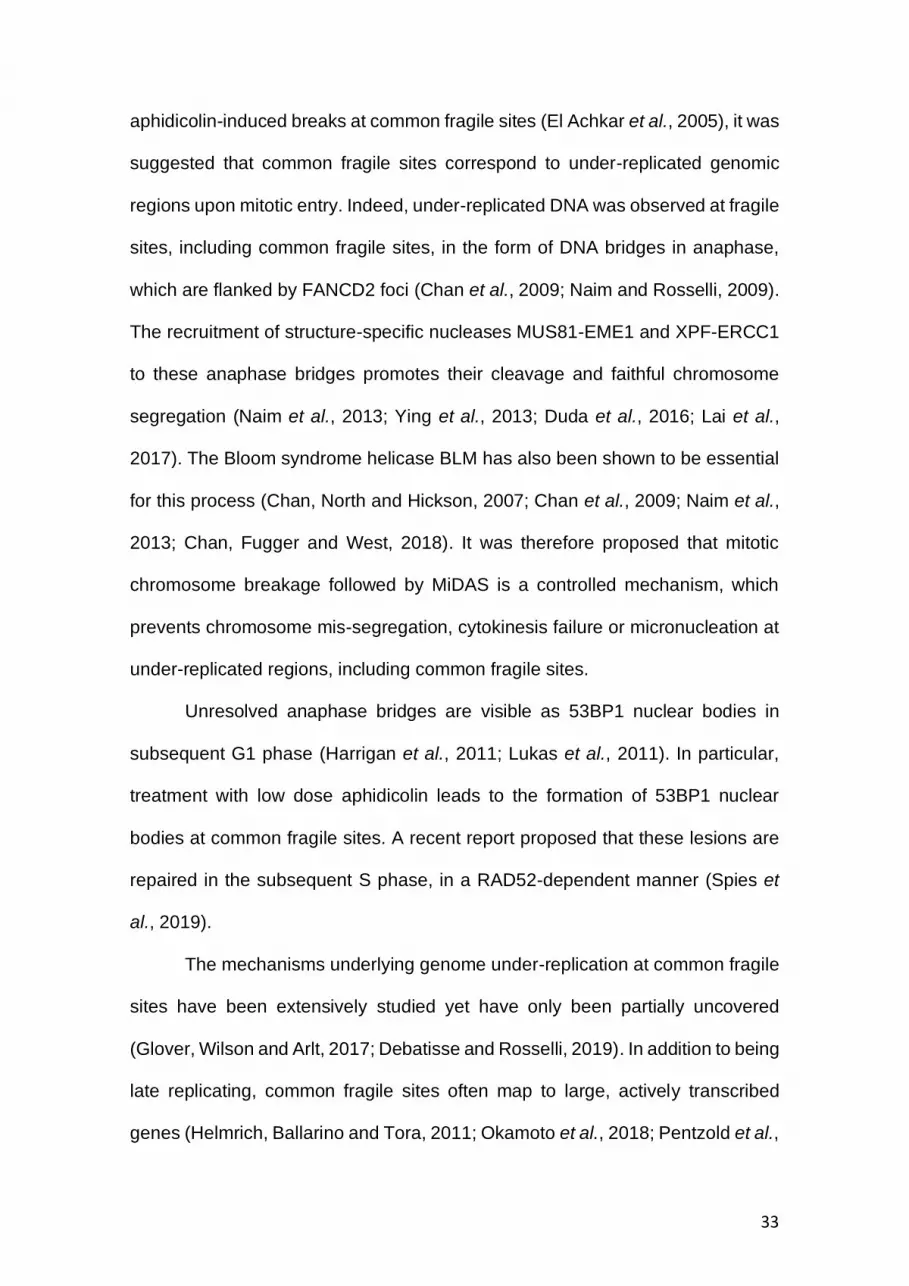
aphidicolin-induced breaks at common fragile sites (El Achkar et al., 2005), it was
suggested that common fragile sites correspond to under-replicated genomic
regions upon mitotic entry. Indeed, under-replicated DNA was observed at fragile
sites, including common fragile sites, in the form of DNA bridges in anaphase,
which are flanked by FANCD2 foci (Chan et al., 2009; Naim and Rosselli, 2009).
The recruitment of structure-specific nucleases MUS81-EME1 and XPF-ERCC1
to these anaphase bridges promotes their cleavage and faithful chromosome
segregation (Naim et al., 2013; Ying et al., 2013; Duda et al., 2016; Lai et al.,
2017). The Bloom syndrome helicase BLM has also been shown to be essential
for this process (Chan, North and Hickson, 2007; Chan et al., 2009; Naim et al.,
2013; Chan, Fugger and West, 2018). It was therefore proposed that mitotic
chromosome breakage followed by MiDAS is a controlled mechanism, which
prevents chromosome mis-segregation, cytokinesis failure or micronucleation at
under-replicated regions, including common fragile sites.
Unresolved anaphase bridges are visible as 53BP1 nuclear bodies in
subsequent G1 phase (Harrigan et al., 2011; Lukas et al., 2011). In particular,
treatment with low dose aphidicolin leads to the formation of 53BP1 nuclear
bodies at common fragile sites. A recent report proposed that these lesions are
repaired in the subsequent S phase, in a RAD52-dependent manner (Spies et
al., 2019).
The mechanisms underlying genome under-replication at common fragile
sites have been extensively studied yet have only been partially uncovered
(Glover, Wilson and Arlt, 2017; Debatisse and Rosselli, 2019). In addition to being
late replicating, common fragile sites often map to large, actively transcribed
genes (Helmrich, Ballarino and Tora, 2011; Okamoto et al., 2018; Pentzold et al.,

34
2018; Macheret et al., 2020). The paucity of replication origins also governs
common fragile site expression (Le Tallec et al., 2011; Letessier et al., 2011).
Finally, several reports linked chromosome fragility, including at common fragile
sites, to replication fork stalling (Ozeri-Galai et al., 2011; Tubbs et al., 2018).
1.3.2 Other fragile sites
Aphidicolin-induced common fragile sites have been extensively studied, yet
chromosome fragility at other sites have also been described. For example,
folate-sensitive regions are fragile sites and were describe even before common
fragile sites (Sutherland, 2003). These sites lie within micro- or mini-satellites
repeats acquiring unusual structures, such as hairpins (Zlotorynski et al., 2003).
More recently, early replicating fragile sites (ERFSs) have been identified
upon hydroxyurea (HU)-induced replication in murine B lymphocytes (Barlow et
al., 2013). The presence of repetitive deoxyadenosine and deoxythymidine
(poly(dA:dT)) tracts may promote replication fork stalling and obstruct their restart
(Tubbs et al., 2018). Similar to common fragile sites, ERFSs also correlate with
transcription and are sensitive to ATR inhibition (Barlow et al., 2013).
1.3.3 Mitotic DNA synthesis (MiDAS)
EdU foci were detected in mitosis in asynchronous U2OS cells treated with low-
dose aphidicolin and pulse-labelled with EdU. Because the duration of the EdU
pulse was relatively short compared to the rate of mitotic progression following
its onset, it was suggested that the presence of EdU foci in anaphase is unlikely
to result from DNA synthesis in interphase. This shed light on MiDAS as
compensatory mechanism activated at under-replicated loci in cells exposed to
replication stress (Minocherhomji et al., 2015).

35
Following release from CDK1 inhibitor (RO-3306)-mediated G2 arrest,
mitotic EdU foci were detected at FANCD2-labelled common fragile sites. Indeed,
MiDAS occurred at apparent chromosome gaps and at known common fragile
sites, as shown by fluorescence in situ hybridization (FISH) experiments
(Minocherhomji et al., 2015). The authors showed that activation of MiDAS
depended on the SLX4 scaffold protein-mediated recruitment of MUS81-EME1
(Minocherhomji et al., 2015) and RAD52 (Bhowmick, Minocherhomji and
Hickson, 2016). MUS81 and RAD52 were also required for the recruitment of
POLD1 and POLD3, the catalytic and non-catalytic subunits of Pol δ,
respectively. Importantly, POLD3 co-localised with EdU foci and its depletion
abrogated MiDAS (Minocherhomji et al., 2015). A recent study in Caenorhabditis
elegans showed that the recruitment of FANCD2 and of MiDAS-promoting factors
at under-replicated loci follows TRAIP-mediated replisome disassembly
(Sonneville et al., 2019).
Because studies on MiDAS often uses CDK1 inhibition as a mean to
reversibly block cells in G2, it remains unclear whether high CDK1 activity alone
or whether mitotic entry elicits the detection of under-replicated loci remains
unclear. Indeed, CDK1 activity promotes the phosphorylation and activation of
MUS81 and EME1 (Duda et al., 2016; Palma et al., 2018). Moreover, mitotic
kinases are required for the TRAIP-catalysed ubiquitination of MCM7 by and its
subsequent disassembly (Deng et al., 2019; Priego Moreno et al., 2019;
Sonneville et al., 2019). However, Minocherhomji and colleagues showed that
depletion of SMC2, and thus abrogation of chromosome condensation, prevented
MiDAS in aphidicolin-treated cells released from G2 block. Whether common

36
fragile expression caused by premature chromosome condensation (El Achkar et
al., 2005) is associated with activation of MiDAS has yet not been tested.
Because RAD52 and POLD3 are involved in BIR of stalled replication forks
(Costantino et al., 2014; Sotiriou et al., 2016), it has been hypothesised that
MiDAS acts as a BIR pathway occurs via conservative DNA synthesis
(Minocherhomji et al., 2015; Bhowmick, Minocherhomji and Hickson, 2016;
Macheret et al., 2020).
Inactivation of MiDAS caused by MUS81, RAD52 or POLD3 depletion, or
by chemical inhibition of RAD52 or of DNA synthesis with high-dose aphidicolin
lead to increase 53BP1 nuclear bodies in the subsequent G1, micronuclei
formation and reduced survival (Minocherhomji et al., 2015; Bhowmick,
Minocherhomji and Hickson, 2016). Moreover, error in the repair of under-
replicated loci via activation of MiDAS has been proposed to promote genomic
duplications at common fragile sites (Macheret et al., 2020). The exact
consequences of MiDAS on chromosome fragility, replication stress tolerance
and genomic instability are yet not fully understood.

37
Fig. 1.4. Model of MiDAS at under-replicated loci.
Upon mitotic entry, TRAIP-mediated ubiquitination of the CMG (Cdc45-
MCM-GINS) helicase triggers its disassembly whilst SMC2 promotes
chromosome condensation. This exposes replication forks at under-
replicated loci which are marked by FANCD2 and the SLX4 scaffold
protein. SLX4 recruits RAD52 and MUS81-EME1 leading to fork

38
cleavage, D-loop formation and POLD3-mediated conservative DNA
synthesis. Newly synthesised DNA is shown as dashed lines.
1.3.4 BRCA2 inactivation in chromosome fragility and
instability
BRCA2-deficient cells were shown to exhibit mitotic abnormalities comparable to
those caused by aphidicolin-treatment, suggesting that BRCA2 functions in
preventing chromosome fragility. In particular, BRCA2 abrogation leads to
anaphase bridges, chromosome mis-segregation and 53BP1 nuclear bodies
(Feng and Jasin, 2017; Lai et al., 2017). BRCA2 abrogation also triggers
spontaneous MiDAS at FANCD2-labelled sites (Feng and Jasin, 2017; Lai et al.,
2017; Graber-Feesl et al., 2019), suggesting that MiDAS plays a key role in
maintaining chromosome integrity in the absence of BRCA2.
BRCA2 has also been proposed to activate the spindle assembly
checkpoint by facilitating the acetylation of BUBR1 (Ehlen et al., 2021). More
recently, BRCA2 has been shown to promote proper chromosome alignment in
metaphase and, thus preventing chromosome mis-segregation (Ehlen et al.,
2020). Indeed, phosphorylated BRCA2 interacts with PLK1, BUBR1 and the
PP2A phosphatase, which activity counteracts Aurora B-mediated destabilisation
of kinetochore-microtubule attachment. This suggests that BRCA2 function in
chromosome integrity is not only dependant on its role in interphase but also to
its DDR-independent mitotic activity (Ehlen et al., 2021).
1.4 Research aims
The research presented here provides a better understanding of the causes and
consequences of the chromosome instability cause by BRCA2 abrogation. In a

39
first part, we aim to elucidate the transcriptional adaptation caused by BRCA2
abrogation and their implication on cellular signalling (Chapter 3). In a second
part, we provide new insights into the mechanisms underlying mitotic
abnormalities, in particular MiDAS activation, caused by BRCA2 abrogation.

40
Chapter 2
Material and methods

41
2.1 Cell lines and cell culture
Cell lines were cultivated at 37˚C in 5% CO2. Human non-small-cell lung
carcinoma H1299 and invasive ductal breast cancer MDA-MB-231 cells carrying
a doxycycline (DOX)-inducible shRNA against BRCA2 (H1299+shBRCA2DOX and
MDA-MB-231+shBRCA2DOX) cells were grown as monolayer in Dulbecco’s
Modified Eagle’s Medium (DMEM) supplemented with 10% foetal bovine serum
(FBS), certified tetracycline-free. Expression of shRNA against BRCA2 was
induced by adding 2 µg/mL DOX in the growth medium. The shRNA-expressing
cell lines were previously established (Zimmer et al 2016). In brief, the shRNA
sequence (GAG AAU GUC UCA CAA AUA A) was cloned into pLKO-Tet-On and
introduced into cells using lentiviral infection.
H1299 cells harbour TP53 deletion and heterozygous NRAS (NRAS
Q61K) mutation, which increased signalling activates the MAPK pathway to
enhance cell proliferation and motility (Song, Liu and Zhang, 2017). MDA-MB-
231 cells harbour homozygous gain-of-function mutation in TP53 (p53 R280K
(Ghosh et al., 2021)), MAPK pathway activation via KRAS (G13D) and BRAF
(G464V) mutations (Hollestelle et al., 2010; Bracht et al., 2019; McFall et al.,
2020) and CDKN2A deletion (Hollestelle et al., 2010).
For the 28-day time course, 500’000 cells were seeded in a T75 in order
to establish a mass cell culture for each condition (H1299+shBRCA2DOX and
MDA-MB-231+shBRCA2DOX, ±DOX). Cells were passaged every fourth day
using trypsin, and 500’000 cells were seeded in the same flask, to maintain the
mass cell culture. 1’300’000 and 500’000 cells were seeded in a T75 and were
collected for Western Blotting two (e.g., at day 2, 6, 10, etc. of the 28-day period)
or four days later (e.g., at day 4, 8, 12, etc. of the 28-day period), respectively.

42
160’000 and 60’000 cells were seeded in a 6-well and were collected for
quantitative RT-PCR two or four days later, respectively. Passaged cells were
grown in fresh medium and DOX was added where appropriate.
2.2 RNA-sequencing (RNA-seq)
RNA was extracted using the RNeasy Mini Kit (Qiagen) according to the
manufacturer’s instructions and quantified using RiboGreen (Invitrogen) on the
FLUOstar OPTIMA plate reader (BMG Labtech). Input material was normalized
to 1 µg prior to library preparation. The TruSeq Stranded mRNA kit (Illumina) was
used according to the manufacturer’s instructions for poly(A) transcript
enrichment and strand specific library preparation. Libraries were amplified on a
Tetrad (Bio-Rad) and individual libraries were normalized using Qubit. Individual
libraries were pooled together. Each library aliquot was denatured and further
diluted prior to loading on the sequencer. Paired-end sequencing was performed
using a HiSeq4000 75 base pair platform (Illumina), generating a raw read count
of > 22 million reads per sample.
2.3 RNA-seq data processing
RNA-seq reads were aligned to the human reference genome (GRCh37) using
HISAT245 and duplicate reads removed using the Picard MarkDuplicates tool
(Broad Institute). Reads mapping uniquely to Ensembl-annotated genes were
summarised using featureCounts. The R/BioConductor environment for analysis
of the raw count gene matrix. Genes with less than 10 reads in more than three
samples were removed and sets of 14,000–15,000 genes remained for
differential gene expression analysis.

43
2.4 Differential gene expression analysis
The analyses were carried out using the R package DESeq2. Differentially
expressed genes were identified based on their false discovery rate (FDR) using
Benjamini-Hochberg adjusted p-values <0.05 and absolute value of Log2(Fold
change). Absolute value of Log2(Fold change) > 0.5 was used to determine
differentially expressed genes expression between H1299+shBRCA2DOX treated
with DOX or not (+DOX vs -DOX) or MDA-MB-231+shBRCA2DOX treated with
DOX or not (+DOX vs -DOX). Absolute value of Log2(Fold change) > 0.3 was
used to determine differentially expressed genes common to both cell lines.
2.5 Quantitative RT-PCR
Cells were grown in the presence or absence of DOX for a 28-day period and
were passaged every four days in order to be 70-90% confluent two or four days
later. RNA was extracted using TRIzol reagent and the SYBR Green cells-to-CT
kit (Thermo Fisher Scientific) was used according to the manufacturer’s
instruction to generate complementary DNA (cDNA). The SensiMix SYBR No-
ROX kit (Bioline) was used to prepare 10 µL reactions using 0.2 µL of cDNA and
10 µM forward and reverse primers. Quantitative PCR was performed in 384-well
plates using the QuantStudio 5 Real-Time PCR System (Thermo Fisher
Scientific). Gene expression was normalised to housekeeping genes ACTB and
expressed relative to day 2 of treatment using the Livak 2-ΔΔCT method.
Table 2.1. Primer pairs used for RT-qPCR.
Target Forward primer Reverse primer
ACTB 5’- ATTGGCAATGAGCGGTTC 5’- GGATGCCACAGGACTCCAT
IFI6 5’- TCGCTGATGAGCTGGTCTGC 5’- ATTACCTATGACGACGCTGC
IFIT1 5’- TACCTGGACAAGGTGGAGAA 5’- GTGAGGACATGTTGGCTAGA
IFIT2 5’- TGTGCAACCTACTGGCCTAT 5’- TTGCCAGTCCAGAGGTGAAT

44
ISG15 5’- GCGAACTCATCTTTGCCAGTA 5’- CCAGCATCTTCACCGTCAG
OAS1 5’- CGCCTAGTCAAGCACTGG TA 5’- CAGGAGCTCAGGGCATA
OAS2 5’- TCCAGGGAGTGGCCATAG 5’- TCTGATCCTGGAATTGTTTTAAGTC
2.6 Western blotting
Cells were lysed using loading buffer (0.16 M Tris pH 8, 4% sodium dodecyl
sulfate-polyacrylamide (SDS), 20% glycerol, 0.01% bromophenol blue)
supplemented with 100 mM dithiothreitol (DTT), and protease and phosphatase
inhibitor cocktails (Roche). Samples were sonicated for 3 sec on ice, heated at
70ºC for 10 min and centrifuged at >20’000 g for 7 min. The protein concentration
was quantified using a NanoDrop-1000 spectrophotometer. Equal amounts of
protein were loaded on 4-12% Bis-Tris, 10% Bis-Tris or 3-8% Tris-acetate gels
(Thermo Fisher Scientific). Bis-Tris gels were run in MOPS buffer and Tris-
acetate gels in Tris-acetate buffer (Thermo Fisher Scientific) at 100-180 V until
the desired separation was achieved. PageRuler prestained protein ladder
(Thermo Fisher Scientific) and HiMark prestained protein standard (Thermo
Fisher Scientific) were used as molecular weight markers. Protein transfer onto
a nitrocellulose membrane was run in transfer buffer supplemented with 10%
methanol (Thermo Fisher Scientific) at 30 V for 100 min. The membranes were
subsequently blocked in 5% skimmed milk dissolved in 0.05% Tween 20 (also
known as polysorbate 20) in PBS (hereafter PBST). Membranes were incubated
with primary antibodies diluted in 2% bovine serum albumin and 0.05% azide in
PBST over-night at 4ºC and with horseradish peroxidase (HRP)-conjugated
secondary antibodies diluted in 5% skimmed milk in PBST for 1h at room-
temperature. Detection was achieved by enhanced chemiluminescence detected
on X-ray films.

45
Table 2.2. Primary antibodies used for immunoblotting.
Specificity Host Provider Catalogue
Number Dilution
BRCA2 Mouse Calbiochem OP95 1:1’000
Cyclin B Mouse Cell Signaling Technology 4135 1:1’000
GAPDH Mouse Novus Biologicals 6C5 1:30’000
HERC5 Rabbit Thermo Fisher Scientific 703675 1:1’000
Histone H3 Mouse Thermo Fisher Scientific 865R2 1:1’000
Histone H3 (S10) Rabbit Abcam AB5176 1:1’000
IRF3 Rabbit Abcam AB76409 1:1’000
IRF3 (S386) Rabbit Abcam AB76493 1:1’000
ISG15 Rabbit Cell Signaling Technology 2743
KAP1 Rabbit Bethyl Laboratories A300-274A 1:5’000
KAP1 (S824) Rabbit Bethyl Laboratories A300-767A 1:1’000
SMC1 Rabbit Bethyl Laboratories A300-055A 1:5’000
STAT1 Rabbit Cell Signaling Technology 9175 1:1’000
STAT1 (Y701) Rabbit Cell Signaling Technology 9167 1:1’000
STING Rabbit Cell Signaling Technology 13647 1:1’000
USP18 Rabbit Cell Signaling Technology 4813 1:1’000
Table 2.3. Secondary antibodies used for immunoblotting.
Specificity Host Provider Catalogue
Number Dilution
Mouse Goat Dako P0447 1:5’000
Rabbit Goat Dako P0448 1:5’000
2.7 Resazurin-based viability assay
Where indicated, DOX was added to the grow medium four days before the start
of the experiment. A range of 250-750 H1299+shBRCA2DOX cells or 375-1000
MDA-MB-231+shBRCA2DOX cells were seeded in 96-well plates in order for the
untreated cells to reach 80-90% confluency at the end of the assay The following
day, cells were treated with olaparib or talazoparib at indicated concentration. Six
days later, cells were grown in medium supplemented with 10 μg/mL resazurin
for 2 h. Cell viability was determined by fluorescence (590 nm) using a plate

46
reader (POLARstar, Omega). Cell viability was expressed relative to cells mock-
treated cells.
2.8 Flow cytometry-based assays
2.8.1 Detection of EdU and Histone H3 (S10)
Cells were collected using trypsin and fixed in 90% ice-cold methanol overnight.
Cells were then processed using the Click-iT EdU Alexa Fluor 647 flow cytometry
assay kit (Thermo Fisher Scientific) according to manufacturer’s instructions.
They were permeabilised using a saponin-based buffer. For EdU detection, a
Click-iT reaction was performed according to the manufacturer’s instructions. For
detection of phosphorylated Histone H3 (S10), cells were washed with 2% FBS
in PBS and incubated with mouse anti-Histone H3 (S10) antibody (Cell Signaling
Technology #9701) diluted in 2% FBS in PBS (1:50) for 90 min at room
temperature. They were subsequently washed with 2% FBS in PBS and
incubated with goat anti-mouse AlexaFluor488 (Life Technologies A-10667)
diluted in 2% FBS in PBS (1:200) for 1 h at room temperature. Finally, cells were
washed in 2% FBS in PBS and resuspended in PBS containing 20 μg/mL
propidium iodide and 400 μg/ml RNaseA. A total of 5’000 to 10’000 events per
condition were recorded using a FACS Calibur flow cytometer. Flow cytometry
data were analysed with the FlowJo software.
2.8.2 Cell cycle analysis
Asynchronous cells were pulse-labelled with 25 µM EdU for 30 min and
processed as described above.

47
2.8.3 S phase entry
Cells were blocked in G2 with 9 μM RO-3306 for 16 h and subsequently released
in a medium containing 100 ng/ml nocodazole for 2h. Mitotic cells were harvested
by mitotic shake-off and released in fresh medium supplemented with 25 µM
EdU. The percentage of EdU-labelled cells was determined as described above.
2.8.4 S-to-M progression
Asynchronous cells were pulse-labelled with 10 µM EdU for 1 h and subsequently
allowed to grow for 7 h. Cells were then processed as mentioned above. The
percentage of Histone H3 (S10)-positive cells amongst the EdU-positive cells is
reported.
2.8.5 DNA content analysis
Cells were incubated in 1.5 mM thymidine for 16 h and released in fresh medium.
At indicated timepoints after the release, cells were collected using trypsin and
fixed in 90% ice-cold methanol overnight. Fixed cells were washed with PBS,
resuspended in PBS containing 20 μg/mL propidium iodide and 400 μg/mL
RNaseA and processed as described above.
2.9 Drug treatment
Cells were grown in the presence or absence of DOX for 3 days and seeded in
order to reach 70-90% confluency at the end of the experiment (160’000 cells in
6-cm dish, or 60’000 cells in 6-well respectively). The following day cells were
treated as indicated. Mock-treated cells were treated with equal volume of vehicle
(DMSO for Olaparib, H2O for pyridostatin, and ethanol for chlorambucil).

48
2.10 Cell synchronization for mitotic EdU-seq
Cells were seeded in order to reach 70-80% confluence at the end of the
experiment (1’800’000 cells per T175 flask, 10-25 flasks per condition) and DOX
was added where indicated. After 12 h, cells were treated with 1.5 mM thymidine
for 16 h. To detect MiDAS under untreated conditions, cells were released in
fresh medium containing 6 µM RO-3306 for 10.5 h. To detect aphidicolin-induced
MiDAS, cells were released in fresh medium containing 6 µM RO-3306 and 0.2
µM aphidicolin. For both protocols, cells were then washed three times with warm
medium and released in medium containing 100 ng/ml nocodazole and 10 µM
EdU. 2 mM HU was added during the final 3 h of treatment.
2.11 EdU-seq
Mitotic cells were harvested by mitotic shake-off and fixed with 90% ice-cold
methanol. Cells were washed with PBS and permeabilised with 0.2% Triton-X in
PBS. A cleavable biotin linker (Azide-PEG(3+3)-S-S-biotin, Jena Biosciences)
was attached to the EdU using the Click-It kit described above. The DNA was
isolated by phenol-chloroform extraction and precipitated in ethanol. Isolated
DNA was resuspended in TE buffer and sonicated to 100-500 nucleotide-long
fragments. Dynabeads MyOne streptavidin C1 (Invitrogen) were used to purify
Edu-labelled DNA fragments. The beads were washed three times, resuspended
in 5 mM Tris-HCl, pH 7.5, 0.5 mM EDTA, 1 M NaCl, 0.5% Tween 20 (buffer A)
containing the sonicated DNA and incubated on a rotating wheel for 15 min at
room temperature. The beads were washed three times with buffer A and once
with Tris-EDTA buffer. The EdU-labelled DNA fragments were eluted with 2% β-
mercaptoethanol in Tris-EDTA buffer for 1h at room temperature. Purified EdU-
labelled DNA was used for libraries preparation (TruSeq ChIP Library Preparation

49
Kit, Illumina) and high-throughput 100-base-pair single-end sequencing was
performed on Illumina Hi-Seq 4000 sequencer.
Fig. 2.1. Strategy for high-resolution detection of nascent DNA using
EdU-seq

50
2.12 EdU-seq data processing
Sequencing reads were aligned on the masked human genome assembly
(GRCh37/hg19) using the Burrows-Wheeler Aligner software and reads with
quality score below 60 were removed. Custom scripts were used to assign the
aligned reads to 10 kb genomic bins. The normalised number of reads per
genomic bin was calculated by dividing the number of EdU-seq reads per
genomic bin by the number of reference reads for the same genomic bin. Here
the reference reads were obtained from high-coverage sequencing of genomic
DNA. Finally, sigma values were calculated as the normalised number of reads
per bin divided by its standard deviation (Macheret and Halazonetis, 2018).
2.13 Analysis of MiDAS peaks
For plots showing individual genomic regions, the EdU-seq data were plotted
using sigma values, which corrects for systemic biases in high throughput
sequencing data. Custom scripts were used to identify sites (peaks) with sigma
values significantly higher than background signal (local maxima) within a 2 Mb-
long region. MiDAS peaks were plotted, validated manually and subsequently
classified as single- or multiple-peaks manually. The maximal sigma value of
each individual MiDAS site is indicated in the figure legend. The “was used to
generate BigWig files using BAM files aligned as described above. were used to
generate average plots and heatmaps across multiple regions. Genome-wide
averages of all MiDAS sites were plotted using the “computeMatrix” and
“plotHeatmap” functions of deepTools (Ramirez et al., 2016) using BigWig files
generated with the “bamCoverage” function.

51
2.14 Analysis of the replication timing
REPLI-seq data were previously generate in asynchronous U2OS cells
(Macheret and Halazonetis, 2018).
2.15 Assignment of genic and intergenic regions
Ensembl gene annotations (v99 for GRCh37/hg19) were used to list the genomic
position of all human genes. Genomic bins that mapped entirely within genes
were defined as genic. Genomic bins that mapped entirely within intergenic
regions were defined as intergenic. Genomic bins that contained both genic and
intergenic regions were classified as mixed genic/intergenic.
2.16 Statistical analyses
Statistical analyses were performed using GraphPad Prism. For comparison of
means, unpaired two-tailed Student’s t-test was applied. The interpretation of the
p-value is described in the figure legends.

52
Chapter 3
BRCA2 abrogation activates the cGAS-
STING-IRF3 and JAK-STAT pathways to
promote expression of interferon-stimulated
genes

53
3.1 Introduction
BRCA2 plays major roles in the maintenance of genome stability. It facilitates
DNA replication by protecting stalled replication forks from nucleolytic
degradation and promoting their restart via template switch reactions (Saldivar,
Cortez and Cimprich, 2017; Berti, Cortez and Lopes, 2020). BRCA2 functions are
essential for homologous recombination (HR) repair of DNA double-strand
breaks (DSBs) (Chang et al., 2017; Scully et al., 2019). Therefore, loss of BRCA2
leads to the accumulation of mutations and fuels tumorigenesis (Wooster et al.,
1994; Wooster et al., 1995). Several studies have contributed to a better
understanding of the consequences of BRCA2 abrogation. For example, DDR-
deficiency caused by BRCA2 abrogation sensitizes cancer cells to DNA
damaging therapies, such as IR (Sharan et al., 1997). Moreover, PARP inhibition
is synthetic lethal with BRCA2-deficiency (Bryant et al., 2005; Farmer et al., 2005;
Lord and Ashworth, 2017). However, how cells respond to BRCA2 abrogation is
yet not fully understood. In particular, why germline BRCA2 mutations predispose
to breast, ovarian or prostate cancers remains unknown . We hypothesised that
the accumulation of stalled replication forks and DNA damage in BRCA2-
depleted cells may impact on gene expression and therefore contribute to their
tumorigenicity. We therefore sought to investigate deregulated gene expression
following loss of BRCA2 function.
Based on RNA-sequencing (RNA-seq) analyses performed in two distinct
cellular models for BRCA2 inactivation, we identified differentially expressed
genes at early and late timepoints after BRCA2 abrogation, which brought new
insights into the signalling triggered by BRCA2 abrogation.

54
3.2 Characterisation of the inducible models for BRCA2
inactivation
To investigate the mechanisms of adaptation following BRCA2 abrogation, we
analysed changes in gene expression by RNA-seq. For this purpose, we used
two inducible models of BRCA2 abrogation: human non-small cell lung carcinoma
H1299 cells and invasive ductal breast cancer MDA-MB-231 cells expressing a
doxycycline (DOX)-inducible short hairpin RNA (shRNA) against BRCA2
(H1299+shBRCA2DOX and MDA-MB-231+shBRCA2DOX). DOX addition to the
growth medium effectively inhibited BRCA2 expression in both
H1299+shBRCA2DOX and MDA-MB-231+shBRCA2DOX cells (Fig. 3.1A, B).
We then compared gene expression upon short-term (acute, 4 days) or
long-term (chronic, 28 days) BRCA2 abrogation. We grew cells in the presence
of doxycycline for 4 or 28 days to trigger acute or chronic BRCA2 abrogation,
respectively and performed RNA-seq analyses in triplicate to detect
transcriptional changes at each timepoint relative to BRCA2-expressing control
cells.
Importantly, loss of BRCA2 expression resulted in a mild proliferation
defect in both H1299+shBRCA2DOX and MDA-MB-231+shBRCA2DOX, as
observed by resazurin-based population doublings assay. However, short-term
and long-term BRCA2-depleted cells exhibited the same proliferation rate (Fig.
3.1B, C). Although we did not observe obvious signs of increased cell death over
the course of the experiment, we measured apoptosis by Annexin V staining on
cells grown in the absence or presence of DOX for 4 or 28 days. We observed a
small, yet significant increase in the Annexin V-positive population at day 4 of
DOX treatment in H1299+shBRCA2DOX cells (Fig. 3.1E, F). A small increase was

55

56
Fig. 3.1. Time-dependent activation of the cGAS-STING-IRF3 and
JAK-STAT pathways upon BRCA2 depletion.
H1299+shBRCA2DOX cells or MDA-MB-231+shBRCA2DOX cells were
grown with or without DOX for 29 days. (A, B) Whole-cell extracts were
immunoblotted as indicated at day 2 of DOX treatment. SMC1 was used
as a loading control. (C, D) Populations doublings were measured using
a resazurin-based assay starting at day 4 or day 28 of DOX treatment. (E,
F) Percentage of Annexin V-positive cells as determined by flow
cytometry at day 4 or day 28 of DOX treatment. (G, H) Flow cytometry-
based cell cycle analysis at day 4 or day 28 of DOX treatment. (I)
Percentage of EdU-positive H1299+shBRCA2DOX cells grown with or
without DOX for 2 days and at indicated timepoint after release from
nocodazole arrest. Graphs represent mean values and SD of n = 2
independent experiments.
also observed at day 28 of DOX treatment in both cell line studied, yet without
statistical significance. Moreover, cell cycle analysis revealed that BRCA2
abrogation altered cell cycle distribution, independently of the duration of DOX
treatment. Indeed, BRCA2-depleted cells accumulated in the G1-phase (Fig.
3.1G, H), (Lai et al., 2017)). Flow cytometry analysis following nocodazole-
induced mitotic arrest and release revealed that BRCA2-depleted
H1299+shBRCA2DOX exhibit slow rate S phase entry compared to control cells
(Fig. 3.1I). Altogether, this shows that BRCA2 abrogation in our models leads to
a mild proliferation defect, which is characterised by slow cell cycle progression
rather than increased cell death. Notably, this phenotype is not enhanced nor
diminished upon long-term BRCA2 abrogation.
In brief, our RNA-seq analyses revealed limited alteration to gene
expression in MDA-MB-231+shBRCA2DOX cells upon acute BRCA2 abrogation.
In contrast, acute BRCA2 abrogation in H1299+shBRCA2DOX cells was

57
characterised by the downregulation of genes involved in processes such as cell
cycle, chromosome segregation, DNA replication and DNA repair (data not
shown; Reisländer et al. (2019)). Chronic BRCA2 abrogation led to deregulation
of a high number of genes in both cell lines studied. In H1299+shBRCA2DOX cells,
194 genes were significantly downregulated (Log2(Fold Change) < -0.5; FDR <
0.05) and 213 genes were significantly upregulated (Log2(Fold Change) > 0.5;
FDR < 0.05) (Fig. 3.1A) whilst 75 and 96 genes were significantly down- and
upregulated, respectively, in MDA-MB-231+shBRCA2DOX cells (Fig. 3.1B). The
work presented here focuses on transcriptional changes observed upon chronic
BRCA2 abrogation.
3.3 Long-term BRCA2 abrogation triggers innate immune
response signalling
Gene set enrichment analysis of significantly upregulated genes, using
annotations from the Gene Ontology (GO) for biological processes, showed that
in H1299+shBRCA2DOX chronic BRCA2 abrogation upregulated genes involved
in the immune response, innate immune response, cytokine signalling, immune
effector process, and type I interferon (IFN) response (Fig. 3.1C). GO analysis of
significantly upregulated genes upon chronic BRCA2 abrogation in MDA-MB-
231+shBRCA2DOX revealed that these genes belong to extracellular matrix
organisation, IFN signalling, degradation of the extracellular matrix, cytokine
signalling in immune system, haemostasis, and fatty acid metabolism processes
(Fig. 3.1D).
Immune response genes were upregulated in both cell lines (Fig. 3.1C,D).
Therefore, we next compared upregulated genes in H1299+shBRCA2DOX and

58
Fig. 3.2. RNA-seq analysis reveals upregulation of immune response
signalling upon long-term BRCA2 depletion.
(A) Volcano plot shows changes in gene expression in
H1299+shBRCA2DOX after 28 days of doxycycline (DOX) treatment.
Genes with Log2(Fold Change) > 0.5 (red dashed lines) and a false
discovery rate (FDR) < 0.05 were considered significantly deregulated.
(B) Volcano plot shows changes in genes expression in MDA-MB-

59
231+shBRCA2DOX after 28 days of DOX treatment. (C) Gene Ontology
(GO) pathway analysis of significantly upregulated genes identified in (A)
for H1299+shBRCA2DOX. (D) GO pathway analysis of significantly
upregulated genes identified in (B) for MDA-MB-231+shBRCA2DOX. (E)
Venn diagram shows upregulated genes (Log2(Fold change) >0.3, FDR
< 0.05) in H1299+shBRCA2DOX and MDA-MB-231+shBRCA2DOX after 28
days of DOX treatment. (F) GO pathway analysis of upregulated genes
common to H1299+shBRCA2DOX and MDA-MB-231+shBRCA2DOX
identified in (E).
MDA-MB-231+shBRCA2DOX upon chronic BRCA2 abrogation, in order to
determine genes in common between the data sets. To increase the relevance
of our results and to account for differences in expression due to cell line
specificity, we considered upregulated genes with a Log2(Fold Change) > 0.3 and
FDR < 0.05 for this analysis. Our analysis revealed 28 genes common to both
datasets (Fig. 3.1E). GO annotations showed that these commonly upregulated
genes belong to cytokine signalling, type I IFN response, response to cytokine,
immune response and immune effector process pathways (Fig. 3.1F). As an
example, IFIT2 and IFIT3 are coding for proteins involved in type I IFN response
and were significantly upregulated in both H1299+shBRCA2DOX and MDA-MB-
231+shBRCA2DOX cells. Hence, we concluded that chronic BRCA abrogation
leads to immune response signalling.
3.4 Long-term BRCA2 abrogation activates the cGAS-STING-
IRF3 and the JAK-STAT pathways
To understand the differences between chronic and acute BRCA2 inactivation on
immune response signalling, we then monitored gene expression over a 28-day
period using quantitative reverse transcription PCR (RT-qPCR). Selected genes

60
included IFIT1, IFIT2, IFI6, OAS1, OAS2 and ISG15. Whilst the expression of
these genes remained stable over the 28-day period in BRCA2-proficient
H1299+shBRCA2DOX cells, we observed a constant increase with time in their
expression upon BRCA2 abrogation (Fig. 3.3A). Strikingly, OAS2 mRNA levels
were 40-fold increased at day 28 compared to day 2 of doxycycline treatment
(Fig. 3.3A). Similar trends were observed in most tested ISGs following BRCA2
abrogation in MDA-MB-231+shBRCA2DOX cells, but to a smaller extent. Indeed,

61
Fig. 3.3. Time-dependent expression of interferon-stimulated genes
(ISGs) upon BRCA2 depletion.
(A) H1299+shBRCA2DOX cells were grown with or without DOX for 28
days. At indicated days, gene expression was analysed by RT-qPCR
using primers specific for a subset of ISGs. mRNA levels are expressed
relative to ACTB mRNA (encoding β-actin) and normalised to day 2 of
DOX treatment. Dots represent the value obtained from an individual
experiment. Lines represent the rolling average of n = 3 independent
repeats. (B) MDA-MB-231+shBRCA2DOX were grown with or without DOX
for 28 days. Gene expression was analysed and is expressed as in (A).

62
IFIT1, IFIT2 and OAS2 mRNA levels increased over the time course of the
experiments (Fig. 3.3B). Altogether, these data suggest that whilst upregulation
of the immune response pathways were only detected upon chronic BRCA2
abrogation, the underlying signalling is activated at an earlier stage of BRCA2
abrogation.
The upregulated genes are known to mediate a type I IFN response. In
particular, they belong to a category of genes called interferon-stimulated genes
(ISGs), whose expression is activated in the presence of extracellular IFNα or
IFNβ. Indeed, the interaction between the IFNα or IFNβ ligands and the IFNAR1-
IFNAR2 heterodimeric receptor (Schindler et al., 1992; Velazquez et al., 1992)
triggers activation of a signalling cascade ultimately leading to JAK-mediated
phosphorylation of STAT1 and STAT2 (Shuai et al., 1993; Silvennoinen et al.,
1993; Schneider, Chevillotte and Rice, 2014). These proteins, together with IRF9,
form the ISGF3 complex and bind to IFN-stimulated response elements (ISRE)
in ISG promoters to initiate transcription (Fu et al., 1990; Improta et al., 1994;
Schneider, Chevillotte and Rice, 2014). Therefore, we decided to test whether
activation of the JAK-STAT pathway could explain the expression of ISGs upon
chronic BRCA2 abrogation.
We monitored the phosphorylation of STAT1 at tyrosine 701 (Y701; (Shuai
et al., 1993)) over the 28-day period in both H1299+shBRCA2DOX and MDA-MB-
231+shBRCA2DOX by immunoblotting (Fig. 3.4A,B). We observed a strong
phosphorylation of STAT1 in BRCA2-deficient H1299+shBRCA2DOX cells
compared to their BRCA2-proficient counterparts. Moreover, phosphorylated
STAT1 was detected early after BRCA2 abrogation (Fig. 3.4A, day 4 of
doxycycline treatment) and further increased upon long-term BRCA2 abrogation

63
(Fig. 3.4A, days 24 and 28 of doxycycline treatment). We found the same trend
in MDA-MB-231+shBRCA2DOX (Fig. 3.4B), although the increase in STAT1
phosphorylation upon BRCA2 abrogation was less pronounced than in
H1299+shBRCA2DOX cells (Fig. 3.4A). This is consistent with the weaker
upregulation of ISGs observed by RT-qPCR in this cell line (Fig. 3.3B). Taken
together, these data demonstrate that BRCA2 abrogation leads to activation of
the JAK-STAT pathway, which is responsible for the upregulation of ISGs
observed in our RNA-seq experiments.
Next, we sought to elucidate the mechanisms underlying the activation of
the JAK-STAT pathway. Whilst secreted IFNs are known to activate this pathway,
the IFNA and IFNB genes (which encode IFNα and IFNβ, respectively), did not
appear significantly upregulated in our RNA-seq analysis upon chronic BRCA2
abrogation (Fig. 3.1A,B). We assessed the activation of the cGAS-STING-IRF3
pathway, which is an upstream regulator of IFNα and IFNβ expression (Ishikawa,
Ma and Barber, 2009; Burdette et al., 2011; Ablasser et al., 2013; Gao et al.,
2013; Sun et al., 2013; Wu et al., 2013). The cGAS-STING-IRF3 pathway was
recently found to be activated by defects in DNA damage repair pathways
(Reisländer, Groelly and Tarsounas, 2020). Moreover, IR was shown to induce
micronuclei formation, followed by detection by cGAS upon rupture of the
micronuclear membrane. Thus, we monitored the phosphorylation of the
transcription factor IRF3 at serine 386 (S386; (Tanaka and Chen, 2012)), which
controls the transcription of IFN genes. We detected an increase in
phosphorylated IRF3 upon BRCA2 abrogation from day 4 of doxycycline
treatment onward, in both H1299+shBRCA2DOX and MDA-MB-231+shBRCA2DOX
(Fig. 3.4A,B).

64
Fig. 3.4. Time-dependent activation of the cGAS-STING-IRF3 and
JAK-STAT pathways upon BRCA2 depletion.
(A) H1299+shBRCA2DOX cells or (B) MDA-MB-231+shBRCA2DOX were
grown with or without DOX for 28 days. At indicated days, whole-cell
extracts were immunoblotted as indicated. Phosphorylation sites are
indicated in red. GAPDH was used as a loading control.
BRCA1 or BRCA2 deficiencies correlate with increased micronuclei
formation (Ban et al., 2001). Therefore, we tested whether micronuclei formation
and cGAS surveillance underlies the activation of the type I IFN response upon
BRCA2 abrogation. We performed immunofluorescence to monitor cGAS
association with micronuclei in BRCA2-proficient and BRCA2-deficient

65
H1299+shBRCA2DOX cells at day 4 and 28 of DOX treatment. We observed an
increase in cGAS-positive micronuclei over time upon BRCA2 abrogation in
H1299+shBRCA2DOX (data not shown; (Reisländer et al., 2019)). Moreover, small
interfering RNA (siRNA)-mediated knock-down of STING inhibited the
phosphorylation of IRF3 and STAT1 as well as the expression of ISGs in the
same cell line (data not shown; (Reisländer et al., 2019)).
Following up on our initial RNA-seq results (Fig. 3.1), we demonstrated
that BRCA2 abrogation results in micronuclei formation that accumulate cGAS
and in activation of the cGAS-STING-IRF3 pathway (Fig. 3.4). This, in turn, leads
to the activation of the JAK-STAT pathway (Fig. 3.4) and activation of a type I
IFN response, which is characterised by the expression of ISGs (Fig. 3.1A,B; Fig.
3.3).
3.5 PARP inhibitors enhance the type I innate immune response
in BRCA2-deficient cells
We next tested the clinical significance of these findings and addressed whether
the synthetic lethal interaction between BRCA2 deficiency and PARP inhibition
impacts on the immune response signalling. Indeed, PARP inhibition leads to the
accumulation of DNA damage and mitotic abnormalities (Schoonen et al., 2017).
Moreover, identifying ways to turn ‘‘cold’’ tumours into ‘‘hot’’ tumours which
respond to immunotherapy is a particularly pressing clinical challenge (Sharma
and Allison, 2015).
We used two PARP inhibitors, olaparib and talazoparib, both of which
have recently been approved for treatment of cancers with loss of HR repair or
BRCA1/2 genes (Lord and Ashworth, 2017). We first evaluated the effect of
PARP inhibition in our models of BRCA2-deficiency. Consistent with previous

66
data (Tacconi et al., 2017), olaparib was found to be specifically toxic to BRCA2-
deficient H1299+shBRCA2DOX cells, as measured by a resazurin-based viability
assay (Fig. 3.5A). Similar results were obtained upon treatment with talazoparib
(Fig. 3.5B). To further characterise our model’s response to PARP inhibition, we
performed cell cycle analysis of H1299+shBRCA2DOX treated with olaparib.
Whilst olaparib treatment did not further enhance the G1 arrest observed in
BRCA2-deficient cells, it decreased the fraction of S phase cells and increased
the fraction of G2/M-phase cells (Fig. 3.5B,C). For instance, S phase gated cells
dropped from 31% in mock-treated cells to 19% upon treatment with 10 µM
olaparib. This further confirms that PARP inhibition negatively impacts on the
proliferation of BRCA2-deficient cells.
We next used phosphorylated IRF3 and phosphorylated STAT1 as
markers of the activation of type I IFN response. Treatment with olaparib induced
a dose-dependent phosphorylation of both IRF3 and STAT1 in BRCA2-deficient
H1299+shBRCA2DOX cells (Fig. 3.6). Importantly, this correlated with a dose-
dependent increase in DNA damage, as shown by KAP1 phosphorylation at
serine 824 (S824; (White et al., 2006)). Treatment with 10 µM olaparib but not
with 1 µM olaparib caused phosphorylation of IRF3 or STAT1 in BRCA2-proficient
H1299+shBRCA2DOX cells, to a lesser extent than in their BRCA2-deficient
counterparts (Fig. 3.6). Again, activation of the immune response signalling was
associated with KAP1 phosphorylation, which is consistent with the fact that DNA
damage results in micronuclei formation and cGAS-STING activation in
replicating cells (Reisländer, Groelly and Tarsounas, 2020).

67
Fig. 3.5. Olaparib-treatment affects growth and survival of BRCA2-
deficient H1299+shBRCA2DOX cells.
Dose-dependent viability assays of BRCA2-proficient (-DOX) or -deficient
(+DOX) H1299+shBRCA2DOX cells treated with (A) olaparib or (B)
talazoparib at the indicated concentrations for six days. Graphs represent
mean values and SEM of n = 3 independent experiments, each performed
in technical triplicate. (C) Flow cytometry-based cell cycle analysis of
BRCA2-proficient (-DOX) or -deficient (+DOX) H1299+shBRCA2DOX cells
treated with olaparib at the indicated concentrations for three days. (D) S
phase fraction of cells shown in (C). Graphs represent mean values and
SEM of n = 3 independent experiments. p-values were calculated using
an unpaired two-tailed t-test. p ≤ 0.05, * ; p > 0.5, NS.

68
Fig. 3.6. Olaparib-treatment activates the cGAS-STING-IRF3 and
JAK-STAT pathways in BRCA2-deficient H1299+shBRCA2DOX cells.
H1299+shBRCA2DOX were grown in the presence or absence of DOX for
four days and subsequently treated with olaparib at the indicated
concentrations for three days. Whole-cell extracts were immunoblotted as
indicated. Phosphorylation sites are indicated in red. GAPDH and SMC1
were used as loading controls.
Activation of a type I IFN response upon PARP inhibition was recapitulated in
BRCA1-deficient and BRCA2-deficient xenograft tumour models. Indeed, RT-
qPCR analysis showed that treatment with talazoparib upregulates the
expression of ISGs in BRCA1-mutated MDA-MB-436 xenografts (data not shown;
(Reisländer et al., 2019)). Moreover, treatment talazoparib specifically increased
the expression of ISGs in BRCA2-/- HCT116 xenograft models compared to their
BRCA2+/+ counterparts.
Altogether, this demonstrates that the PARP inhibitors are not only specifically
toxic to BRCA1- and BRCA2-deficient cells and tumours by inflicting DNA lesions,
but they also trigger a type I IFN response in these cells and tumours.

69
3.6 DNA damaging agents targeting BRCA2-deficiency trigger a
type I IFN response
Endogenous and exogenous DNA damage is associated with immune response
signalling (Reisländer, Groelly and Tarsounas, 2020). As we subsequently found
olaparib-induced immune response signalling to be associated with DSB
signalling, we tested whether other DNA damaging agents could trigger a type I
IFN response. In particular, we focused on agents which have been reported to
specifically induce DNA damage in HR repair-deficient cells.
G-quadruplex stabilizers, such as pyridostatin, are known to be specifically
toxic to BRCA1- or BRCA2-deficient cells (Zimmer et al., 2016; Xu et al., 2017a;
De Magis et al., 2019). Whilst it has been suggested that pyridostatin’s cytotoxic
activity also involves R-loop formation (De Magis et al., 2019; Miglietta, Russo
and Capranico, 2020) or topoisomerase II poisoning (Olivieri et al., 2020), it was
shown to bind to G-quadruplex forming sequences and cause DNA damage
(Rodriguez et al., 2008; Rodriguez et al., 2012). Pyridostatin treatment induced
DNA damage in a dose-dependent manner, and we observed a greater effect in
BRCA2-deficient than in BRCA2-proficient cells (Fig. 3.7A). Moreover, we
observed an increase in IRF3 phosphorylation upon treatment with 10 µM
pyridostatin, primarily in cells in which BRCA2 has been abrogated (Fig. 3.7A).
Consistent with this finding, pyridostatin treatment caused micronuclei formation
in U2OS transfected with an siRNA targeting BRCA2 (De Magis et al., 2019).
We then evaluated the effect of the alkylating agent chlorambucil on the
immune response signalling. Whilst widely used to treat chronic lymphocytic
leukaemia, chlorambucil was also proposed as a potential clinical candidate to
target BRCA2-mutated tumours (Tacconi et al., 2019). Indeed, chlorambucil-

70
induced DNA crosslinks lead to DNA damage accumulation and have been
shown to be particularly toxic to BRCA2-deficient cells and tumours, including
those with acquired PARP inhibitor resistance.
Fig. 3.7. DNA damaging agents activate the cGAS-STING-IRF3 and
JAK-STAT pathways in BRCA2-deficient H1299+shBRCA2DOX cells.
H1299+shBRCA2DOX were grown in the presence or absence of DOX for
four days and subsequently treated with (A) pyridostatin (PDS) or (B)
chlorambucil (CHL) at the indicated concentrations for three days. Whole-
cell extracts were immunoblotted as indicated. Phosphorylation sites are
indicated in red. GAPDH and SMC1 were used as loading controls.
Treatment of BRCA2-deficient H1299+shBRCA2DOX cells with 1 or 10 µM
chlorambucil caused activation of the cGAS-STING-IRF3 and JAK-STAT
pathways, as indicated by the phosphorylation of IRF3 and STAT1, respectively
(Fig. 3.7B). Thus, chlorambucil triggers a type I IFN response in cells lacking
functional BRCA2.
This suggests that DNA damage, and in particular DSBs, is associated
with immune response signalling. We demonstrate that DNA damaging agents
with specific activity against BRCA2-deficient cells, such as PARP inhibitors, G-

71
quadruplex stabilisers, or alkylating agents, activate a type I IFN response in
these cells.
3.7 Functional up-regulation of ISG15 and its ISGylation activity
We next tested whether the increase in ISGs mRNA levels after BRCA2
abrogation can lead to an increase in protein levels. We focused on ISG15, which
encodes the eponym ubiquitin-like protein (Skaug and Chen, 2010; Zuo et al.,
2016). ISG15 can covalently bind to target proteins, in a process called
ISGylation (Fig. 3.8A). Moreover, we included in our analysis two other ISGs,
whose products play key roles in the ISGylation pathway: the E3 ligase HERC5,
and the hydrolase USP18, which catalyses the deISGylation reaction (Fig. 3.8A;
(Skaug and Chen, 2010; Zuo et al., 2016)). ISG15 has been linked to DNA
replication and to the DNA damage response (Park et al., 2014; Raso et al.,
2020). In particular, PCNA ISGylation has been shown to facilitate its de-
ubiquitination following UV-induced DNA damage, suggesting that ISG15
negatively regulates translesion synthesis and prevents error-prone DNA
replication (Park et al., 2014). Moreover, USP18 knock-out radio-sensitised
HAP1 cells (Pinto-Fernandez et al., 2020).
Endogenous ISG15 was detected in H1299+shBRCA2DOX cells
independently of the BRCA2 status (Fig. 3.8B). As we were interested in ISG15
functions in response to DNA damage, we used olaparib to stimulate ISG15
expression. Treatment with 10 µM olaparib did not only activate the JAK-STAT
pathway (Fig. 3.6) but also increased ISG15 levels in BRCA2-deficient cells (Fig.
3.8B). Moreover, HERC5 and USP18 levels were higher in BRCA2-deficient
H1299+shBRCA2DOX cells than in their BRCA2-proficient counterparts (Fig.
3.8B).

72
Fig. 3.8. Functional expression of ISG15 upon BRCA2 depletion and
Olaparib treatment.
(A) Schematic representation of the ISGylation and deISGylation
mechanism. Covalent binding of ISG15 to target proteins is catalysed by
its sequential interaction with E1, E2 and E3 ligases, whilst conjugated-
ISG15 is cleaved from target protein by the hydrolase USP18. (B)
H1299+shBRCA2DOX were grown in the presence or absence of DOX for
four days and subsequently treated with olaparib at the indicated
concentrations for three days. Whole-cell extracts were immunoblotted
as indicated. Arrows indicate ISGylated HERC5. GAPDH and SMC1
were used as loading controls.

73
Olaparib treatment increased ISG15, HERC5 and USP18 levels in both
BRCA2-proficient and BRCA2-deficient cells, albeit a stronger effect was
observed in the latter. This shows that that ISG induction observed in BRCA2-
deficient cells and further enhanced by PARP inhibition leads to an increase in
the levels of the encoded proteins and therefore may have a biological function.
Finally, we did not find evidence of ISGylation when detecting ISG15 by
immunoblotting. Indeed, ISG15 appeared as a single band at its expected
molecular weight of 18 kDa. In contrast, HERC5 appeared as three distinct bands
in olaparib-treated BRCA2-deficient cells. Whilst one band is common to all
tested samples, the two additional bands, less than 54 kDa above HERC5’s
expected molecular weight, have been reported to be ISGylated HERC5 (Fig.
3.8B; (Pinto-Fernandez et al., 2020)). Hence, this suggest that the activation of a
type I IFN response observed in BRCA2-deficient cells treated with olaparib,
leads to a functional expression of ISG15 and increased ISGylation. Whether this
impacts on the DNA damage response in the absence of BRCA2 remains to be
elucidated.
3.8 Discussion
In this study we use RNA-seq analyses to identify genes which expression was
altered upon long-term (chronic), but not short-term (acute) BRCA2 abrogation.
Indeed, we found that genes mediating immune response signalling are
upregulated upon chronic BRCA2 abrogation. Whilst this phenotype was more
pronounced in H1299+shBRCA2DOX than in MDA-MB-231+shBRCA2DOX cells,
our analysis revealed 28 genes to be commonly upregulated in both cell lines.
Because these genes are ISGs, we concluded that BRCA2 abrogation triggers a
type I IFN response. We showed that BRCA2 abrogation leads to micronuclei

74
formation, which activates the cGAS-STING-IRF3 and JAK-STAT pathways.
Similar findings were reported in a recent study (Heijink et al., 2019). Using stable
isotope labelling by amino acids in cell culture (SILAC) coupled to mass
spectrometry, the authors observed an upregulation of immune response
proteins upon shRNA-mediated BRCA2 abrogation in BT-549 and HCC38 cells
(Heijink et al., 2019). Unlike what we observed in H1299 and MDA-MB-231 cells,
the authors found that loss of BRCA2 expression resulted in a strong decrease
in cell viability. In particular, p53-negative KBM-7 cells died within a week of
BRCA2 abrogation. Strikingly, BRCA2 abrogation led to the production of pro-
inflammatory cytokines, including TNFα, and activation of a pro-apoptotic
signalling cascade via its receptor, TNFR1. In contrast, we found the pro-survival
receptor TNFR2 (TNFRSF1B, Fig. 3.2) as a highly upregulated hit after 28 days
of DOX treatment in H1299 cells. Whilst we have not tested whether TNFR2-
mediated signalling underlies the long-term survival of our BRCA2-deficient
models, these findings elegantly demonstrate that inactivation of TP53 is not
sufficient to promote the survival of BRCA1/2-mutated cells. Whether the tissue-
specific tumorigenesis observed in patient carrying deleterious BRCA1/2
mutations relies on such transcriptional changes remains to be elucidated
(Elledge and Amon, 2002).
Other recent studies have associated the loss of other DNA repair genes
with micronuclei formation and immune response signalling (Reisländer, Groelly
and Tarsounas, 2020). For instance, loss of RNASE2H or SAMHD1, which are
associated with the autoinflammatory disorder Aicardi-Goutières syndrome,
leads to micronuclei formation or release of genomic DNA into the cytosol,

75
respectively, which activates the cGAS-STING axis (Mackenzie et al., 2016;
Mackenzie et al., 2017; Coquel et al., 2018).
We also demonstrated that treatment with DNA damaging agents further
enhance the innate immune response. In particular, we used drugs specifically
targeting BRCA2-deficient cells and tumours, such as PARP inhibitors, G-
quadruplex stabilisers and DNA crosslinking molecules. We showed that these
agents preferentially trigger an immune response in BRCA2-deficient cells, which
correlated with accumulation of DSBs and activation of the cGAS-STING-IRF3
axis. Similar results were obtained in BRCA1-deficient tumour models treated
with PARP inhibitors (Ding et al., 2018; Pantelidou et al., 2019; Reisländer et al.,
2019). These findings are in line with reports associating IR-induced DNA
damage with micronuclei formation or release of genomic DNA into the cytosol,
and subsequent activation of an immune response signalling (Mackenzie et al.,
2016; Erdal et al., 2017).
Finally, we showed that the activation of a type I IFN response leads to
functional upregulation of proteins catalysing ISGylation reactions, especially in
BRCA2-deficient cells treated with PARP inhibitors. We detected an increase in
ISG15, USP18 and HERC5 protein levels, as well as in ISGylated HERC5. How
increased ISGylation affects the survival and drug response of BRCA2-deficient
cells is as yet unknown. However, given previously described functions of ISG15
in DNA replication and repair (Park et al., 2014; Pinto-Fernandez et al., 2020;
Raso et al., 2020), this post-translational modification could be of clinical
significance for the treatment of BRCA2-mutated cancers. Because loss of
USP18 radio-sensitises cells to IR (Pinto-Fernandez et al., 2020), ISGyation
reactions may play role in therapy resistance. It will therefore be crucial to assess

76
whether the upregulation of ISGs observed upon long-term BRCA2 inactivation
impacts on the cell’s intrinsic response to therapies. Moreover, establishing the
“ISGylome” (Pinto-Fernandez et al., 2020) of BRCA2-deficient cells may bring
new insights into the role of the ISGs. Finally, with the increased use of
CRISPR/Cas9 screens to identify mechanisms of resistance or sensitivity to anti-
tumoural compounds (Olivieri et al., 2020), our findings indicate that context-
specific transcriptional changes must be taken into account. For instance, a
dropout screen with PARP inhibitors may show different outcome depending on
the level of expressed ISGs in the cell line studied, and may therefore identify
novel synthetic lethal interactions.
Whilst we did not investigate whether immune response signalling impacts
on the host’s immune system, other studies brought insights into this interplay.
Increased CD4+ and CD8+ T cell infiltration was observed in DDR-deficient,
including BRCA1 and BRCA2-mutated, breast cancer tumours (Parkes et al.,
2017). Moreover, PARP inhibitors’ in vivo anti-tumour activity relies, in part, on
STING activation and the consequent lymphocyte infiltration within the tumour
micro-environment (Ding et al., 2018; Pantelidou et al., 2019). Accordingly,
synergistic anti-tumour effects were observed in mice harbouring BRCA1-
deficient tumours and treated with PARP inhibitors in combination with anti-PD-1
antibody (Ding et al., 2018; Wang et al., 2019) or anti-CTLA-4 antibody (Higuchi
et al., 2015). Such combinations are currently being extensively explored in
clinical contexts (Reisländer, Groelly and Tarsounas, 2020). Finally, the fact that
other DNA damaging agents (e.g. alkylating agents) trigger an immune response
signalling in BRCA2-deficient cells suggests that they may also have synergistic

77
anti-tumour activity when combined to immune checkpoint inhibitors (Reisländer,
Groelly and Tarsounas, 2020).

78
Fig. 3.9. Interplay between the DNA damage response and the
innate immune response for cancer (immuno-)therapy.
Top: Endogenous (e.g., BRCA2 abrogation) or exogenous (e.g. PARP
inhibition, IR) DNA damaging factors can lead to micronuclei formation
and activation of the cGAS-STING pathway. Alternatively, DNA
damage can lead to accumulation of mutations in protein coding
sequences and expression of neoantigens. The cGAS-STING axis
trigger the nuclear translocation of the dimeric IRF3 or NF-kB
transcription factors which triggers secretion of IFNα and IFNβ. In
cancer cells, autocrine IFN signalling activates the JAK/STAT pathway
and causes expression of interferon-stimulated genes (ISGs), which is
known as a type I IFN response. Bottom: Type I IFN signalling
facilitates the recruitment of immune effector cells by promoting tumour
antigen/neo-antigen presentation on the major histocompatibility
complex (MHC) of dendritic cells. T-cell activation depends on the
balance between the MHC:T cell receptor (TCR) stimulatory signal
(green), B7:CD28 stimulatory signals (green) and B7:CTLA-4 inhibitory
signals (red). T cells tumour infiltration is stimulated by chemokines
(e.g., CXCL10). The immune effectors’ anti-tumour activity depends on
the balance between the MHC:TCR stimulatory signal (green), and the
PD-1:PD-L1 inhibitory signal (red). Inhibition of immunosuppressive
signals (red) by anti-CTLA-4, anti-PD-1, or anti-PD-L1 antibodies
(green) potentiates the anti-tumorigenic immune responses. (Figure
adapted from Reisländer, Groelly and Tarsounas, 2020).

79
Chapter 4
Protocols for high-resolution detection of
mitotic DNA synthesis (MiDAS) using mitotic
EdU-seq.

80
4.1 Introduction
Chromosome fragility is often detected as the breakage of condensed
chromosomes in mitosis, observed by microscopy. Similarly, MiDAS is detected
using fluorescence microscopy of EdU incorporation during mitosis. In order to
facilitate the analysis and increase the number of detected events, mitotic cells
are enriched by drug-induced mitotic arrest, often preceded by drug-induced G2-
arrest and release into mitosis. However, there are limitations to these strategies,
the most important being that whilst chromosome breaks can be detected
throughout mitosis, MiDAS occurs early after mitotic entry. Indeed, addition of a
thymidine analogue at early but not late M-phase stages allowed detection of
aphidicolin-induced MiDAS in U2OS (Minocherhomji et al., 2015). Hence, the
timing of nascent DNA labelling needs to be tightly controlled and MiDAS
detection would preferentially take place upon synchronised mitotic entry.
To ensure that the MiDAS events detected in each single experiment
originate from a homogenous cell population (e.g., is not affected by the position
within the cell cycle of individual cells at the start of the experiment), we used the
double synchronisation strategy described below.
4.2 Synchronous S phase progression
Cells were blocked at the G1/S transition by a single thymidine block. Thymidine
concentration and treatment duration were optimised to obtain the best
synchronisation upon release into S phase, for both BRCA2-proficient and
BRCA2-deficient H1299+shBRCA2DOX cells, as measured by propidium iodide-
based flow cytometry. Treatment with 1.5 mM thymidine for 16 h efficiently
blocked cells at the G1/S transition, independently of the BRCA2 status (Fig.

81
4.1A). Flow cytometry analysis showed that cells entered S phase and initiated
DNA replication 2-7 h after release from the thymidine block (Fig. 4.1A).
Moreover, a large fraction of cells appeared to have duplicated their genome 8 h
after release from the thymidine block (Fig. 4.1A). Finally, passage through
mitosis and cytokinesis took place 9 h to 11 h after S phase entry (Fig. 4.1A).
Importantly, S phase progression occurred in a synchronous manner in both
BRCA2-proficient and BRCA2-deficient H1299+shBRCA2DOX cells with no
detectable difference between the two conditions (Fig. 4.1A).
To confirm these results, we next monitored Cyclin B levels and
phosphorylation of Histone H3 at serine 10 (S10), markers of S and M phases
progression, respectively. Cyclin B levels are known to increase in late S and G2
phases to promote mitotic entry and its degradation in anaphase promotes mitotic
exit (Jin, Hardy and Morgan, 1998; Saldivar et al., 2018). Consistently with DNA
content analysis, we observed a time-dependant increase in Cyclin B levels 7 to
9 h after release from thymidine block in both BRCA2-proficient and BRCA2-
deficient H1299+shBRCA2DOX (Fig. 4.1B). Phosphorylated Histone H3 levels
increased throughout the analysed time period with higher levels detected 9 h
and 10 h after release from thymidine block (Fig. 4.1B). Finally, we observed a
drop in Cyclin B levels 10 h after release from thymidine block (Fig. 4.1B), which
is consistent with the large fraction of cell undergoing cytokinesis observed by
flow cytometry at the same timepoint (Fig. 4.1A).
Altogether, these data demonstrate that a single thymidine block is
sufficient to ensure synchronous S phase progression in H1299+shBRCA2DOX.
Moreover, S phase progression and mitotic entry are not affected by the BRCA2
status under these conditions.

82
Fig. 4.1. Cell cycle progression after thymidine block and release.
BRCA2-proficient (-DOX) or -deficient (+DOX) H1299+shBRCA2DOX cells
were blocked at the G1/S transition using 1.5 mM thymidine for 16 h and
released in fresh medium. (A) At indicated timepoints, cells were subject
to flow cytometry analysis and their total DNA content was determined
using propidium iodide staining. (B) At indicated timepoints, whole-cell
extracts were immunoblotted as shown. Phosphorylation sites are
indicated in red. GAPDH and Histone H3 were used as loading controls.

83
4.3 Cell synchronisation in G2/M
To ensure that cells enter mitosis synchronously, we synchronised the cells in
G2/M using the CDK1 inhibitor RO-3306. We performed flow cytometry and
detected mitotic cells based on Histone H3 phosphorylation at serine 10 and
propidium iodide staining. Whilst 59% of the BRCA2-proficient cells and 43% of
the BRCA2-deficient cells were in mitosis 12 h after release from thymidine block,
incubation with 6 or 9 µM RO-3306 abrogated mitotic entry (Fig. 4.2A).
Next, we tested if addition of RO-3306 impacted on S phase progression.
DNA content analysis by flow cytometry confirmed that treatment with 6 µM RO-
3306 does not impair S phase progression (Fig. 4.2B).
4.4 Protocol for detection of MiDAS under untreated conditions
using mitotic EdU-seq
To detect MiDAS by mitotic EdU-seq, we synchronised cells using a combination
of thymidine and RO-3306 blocks, optimised as above. As an initial starting point,
we allowed synchronised cells to progress through S phase in the presence of 6
µM RO-3306 for 10.5 h after release from the thymidine block. Cells were then
released from the G2 arrest and allowed to enter mitosis for 90 min. This
corresponded to the time required for about half of the cells (40% to 60%) to
reach mitosis (Fig. 4.2A; Fig. 4.3A). Nascent DNA was labelled with EdU in order
to detect MiDAS. Moreover, we used the microtubule poison nocodazole to
prevent mitotic exit. Of note, MiDAS takes place in early M-phase and hence, can
still be captured in nocodazole arrested cells (Minocherhomji et al., 2015). Finally,
mitotic cells were collected by mitotic shake-off (Fig. 4.3B) and EdU-labelled DNA
was purified as described in the material and methods section (chapter 2).

84

85
Importantly, we added 2 mM hydroxyurea (HU) to the growth medium for
3 h prior to the mitotic shake-off. This prevented contamination of the EdU-
labelled mitotic DNA with EdU-labelled S phase DNA. Whilst HU depletes the
dNTP pool and stalls DNA replication in S phase, it does not prevent MiDAS
((Macheret et al., 2020); Chapter 5). Moreover, the presence or absence of HU
prior to the mitotic shake-off did not impact on aphidicolin-induced MiDAS foci in
U2OS (Macheret et al., 2020).
An overview of the protocol used to detect MiDAS under untreated
conditions is shown in Fig. 4.3B.
4.5 Low dose aphidicolin delays S phase progression
Low dose aphidicolin partially inhibits DNA polymerases Pol α, δ, and ϵ, as well
as Pol ζ (Baranovskiy et al., 2014), and therefore slows down replication rates
(Glover et al., 1984). Hence, treatment with low dose aphidicolin induces
common fragile sites expression and activates MiDAS at these sites (Glover et
al., 1984; Minocherhomji et al., 2015; Macheret et al., 2020). To map common
fragile sites in H1299+shBRCA2DOX cells, we used a variation of the previously
described protocol where we treated S phase cell with aphidicolin.
Fig. 4.2. RO-3306-induced G2 block does not stop S phase
progression.
BRCA2-proficient (-DOX) or -deficient (+DOX) H1299+shBRCA2DOX cells
were blocked at the G1/S transition using 1.5 µM thymidine for 16 h and
released in fresh medium containing 6 or 9 µM RO-3306 for 12h. (A) Cells
were subject to flow cytometry analysis and mitotic cells were detected
using propidium iodide and phosphorylated Histone H3 (S10) staining.
(B) At indicated timepoints, cells were subject to flow cytometry analysis
and their total DNA content was determined using propidium iodide
staining.

86
Fig. 4.3. Mitotic EdU-seq protocol for H1299+shBRCA2DOX cells.
(A) BRCA2-proficient (-DOX) or -deficient (+DOX) H1299+shBRCA2DOX
cells were blocked at the G1/S transition using 1.5 µM thymidine for 16 h,
subsequently released in medium containing 6 µM RO-3306 for 10.5 h
and released in medium containing 100 ng/ml nocodazole for 90 min.
Cells were subject to flow cytometry analysis and mitotic cells were
detected using propidium iodide and phosphorylated Histone H3 staining.
(B) Overview of the protocol for detection of MiDAS under untreated
conditions by mitotic EdU-seq in BRCA2-proficient (-DOX) or -deficient
(+DOX) H1299+shBRCA2DOX cells.

87
Fig. 4.4. S phase progression upon aphidicolin treatment.
BRCA2-proficient (-DOX) or -deficient (+DOX) H1299+shBRCA2DOX cells
were blocked at the G1/S transition using 1.5 µM thymidine for 16 h and
released in medium containing (A) 0.2 µM, (B) 0.3 µM or (C) 0.4 µM
aphidicolin (APH). At indicated timepoints, cells were subject to flow
cytometry analysis and their total DNA content was determined using
propidium iodide staining.

88
We assessed the effect of a range of aphidicolin concentrations on S
phase progression following release from thymidine block and in the presence of
RO-3306. Unlike untreated cells which had completed DNA duplication (Fig.
4.1A), aphidicolin-treated cells were mainly in S phase 8 h after release from the
thymidine block (Fig. 4.4A-C). Indeed, cells treated with 0.2 µM or 0.3 µM
aphidicolin required 20 or 24 h, respectively, to duplicate their genome (Fig.
4.4A,B). Cells treated with 0.4 µM aphidicolin only reached mid- to late S phase
20 h after release from thymidine block (Fig. 4.4C). We did not observe obvious
disparities in S phase progression between BRCA2-proficient and BRCA2-
deficient H1299+shBRCA2DOX treated with aphidicolin. Since 0.2 µM aphidicolin
was sufficient to double the time needed for genome duplication, we decided to
continue using these conditions.
4.6 Protocol for detection of MiDAS events induced by low dose
aphidicolin
We next assessed mitotic entry of aphidicolin-treated cells. After an initial
thymidine block, cells were allowed to progress through S phase in the presence
of 0.2 µM aphidicolin and 6 µM RO-3306 for 17.5 or 20.5 h, and subsequently
released into mitosis for 90 min. Surprisingly, we detected more mitotic cells at
the earliest timepoint (e.g., cells collected after 19 h compared to cells collected
after 22 h, Fig. 4.5A). Moreover, floating cells we observed in the growth medium
20 h after release in aphidicolin- and RO-3306-containing medium, suggesting
that prolonged treatment is toxic to the cells. We could not see any rounded cells
characteristic of mitosis when the duration of the aphidicolin and RO-3306
treatment was reduced to 15.5 or 16.5 h (data not shown). We decided to collect
cells for mitotic EdU-seq at 19 h post-thymidine.

89
An overview of the protocol used to detect aphidicolin-induced MiDAS is
shown in Fig. 4.5B.

90
Fig. 4.5. Mitotic EdU-seq protocol for aphidicolin-treated
H1299+shBRCA2DOX cells.
BRCA2-proficient (-DOX) or -deficient (+DOX) H1299+shBRCA2DOX cells
were blocked at the G1/S transition using 1.5 µM thymidine for 16 h,
subsequently released in medium containing 6 µM RO-3306 and 0.2 µM
aphidicolin (APH) for (A) 17.5 or (B) 20.5 h and released in medium
containing 100 ng/ml nocodazole for 90 min. Cells were subject to flow
cytometry analysis and mitotic cells were detected using propidium iodide
and phosphorylated Histone H3 staining. (C) Overview of the protocol for
detection of aphidicolin-induced MiDAS by mitotic EdU-seq in BRCA2-
proficient (-DOX) or -deficient (+DOX) H1299+shBRCA2DOX cells.
4.7 Discussion
In this chapter we describe optimisation of a protocol for MiDAS high-resolution
detection using mitotic EdU-seq. Two distinct versions of the protocol permit
detection of MiDAS induced by aphidicolin treatment or by BRCA2 inactivation.
MiDAS can be detected upon shRNA-mediated knock-down of BRCA2, as well
as in control, BRCA2-expressing cells in the absence of exogenous replication
stress. In contrast, MiDAS occurring at common fragile sites can be identified in
cells grown in the presence of the DNA polymerase inhibitor, aphidicolin
((Macheret et al., 2020); see Chapter 5). Both protocols were developed around
two main requirements: a) synchronous S phase progression and b) synchronous
mitotic entry in the presence of EdU.
To ensure synchronous S phase progression, we arrested cells at the
G1/S transition using a single thymidine block. Optimal conditions (1.5 mM
thymidine for 16 h) were identified experimentally, although alternative
synchronisation approaches, such as serum-starvation or lovastatin-induced G1
arrest, may also be considered. We then confirmed that S phase entry and

91
progression were synchronous, and measured the time required for genome
duplication for both BRCA2-proficient and BRCA2-deficient cells, treated or not
with aphidicolin. Whilst S phase progression was delayed by aphidicolin
treatment in a dose-dependent manner, it was largely unaffected by BRCA2
abrogation relative to BRCA2-expressing cells.
To ensure synchronous mitotic entry, S phase progressing cells were
treated with the CDK1 inhibitor RO-3306 and subsequently released into mitosis.
The release from G2 block as performed by RO-3306 removal was timed to
obtain the maximum number of mitotic cells as possible, without causing
extensive G2 arrest. Of note, only one tenth of the aphidicolin-treated cells
entered mitosis under these conditions. Whilst RO-3306 inhibits CDK1-mediated
mitotic entry without stopping S phase progression (Fig.4.2 and 4.4), the
consequences of CDK1 inhibition on DNA replication, origin firing, or DNA
damage repair are not fully understood (Johnson et al., 2011; Moiseeva et al.,
2019; Brison et al., 2020). In the future, it will be important to ensure that the
described chromosome fragility and MiDAS phenotypes are independent of the
methods used for cell synchronisation, namely thymidine-induced G1/S arrest
and RO-3306-mediated G2 block.
Finally, mitotic cells were collected and subjected to mitotic EdU-seq.
Importantly, HU was added 3 h prior to the mitotic shake-off to avoid
contamination from S phase DNA synthesis, without affecting MiDAS (Macheret
et al., 2020).
Of note, a fraction of the untreated cell population was arrested in G1 (Fig.
4.2 A, B) and a larger part of the aphidicolin-treated cell population was in G1 and
S phases (Fig. 4.5A) at the end of the final timepoint. Because S phase DNA

92
replication was inhibited by addition of HU, and because mitotic cells were
selectively collected by mitotic shake-off, the presence of G1 and S phases cells
is, however, unlikely to affect the sequencing results. This is in line with the site-
specific detection of MiDAS at common fragile sites in aphidicolin-treated cells
(see Chapter 5) rather than detection of EdU incorporation throughout the
genome. Importantly, this protocol may not allow to directly distinguish between
DNA synthesis in late G2 or early mitosis. Indeed, we cannot exclude that DNA
synthesis in G2 cells and their progression to mitosis during the 90-minute EdU
pulse accounts for part of the captured signal. Detection of EdU incorporation in
G2 cells, released or not from RO-3306-induced G2 block, will allow to confirm
whether DNA synthesis in late G2 may account for part of the signal detected
using the protocol developed here. Moreover, depletion of SMC2, which is
required for chromosome condensation and subsequent activation of MiDAS,
would permit to confirm whether this protocol specifically captures mitotic EdU
incorporation.

93
Chapter 5
High-resolution detection of MiDAS upon
BRCA2 abrogation

94
5.1 Introduction
BRCA2 promotes HR repair by loading the RAD51 recombinase at sites of DSBs
(Chang et al., 2017; Scully et al., 2019). Moreover, BRCA2 protects stalled
replication forks from nucleolytic degradation and promotes their restart in
RAD51-dependent manner (Saldivar, Cortez and Cimprich, 2017; Berti, Cortez
and Lopes, 2020). Although these functions of BRCA2 are confined to the S- and
G2-phases of the cell cycle, when DNA replication occurs (Hustedt and Durocher,
2016), the DNA replication and repair defects caused by BRCA2 abrogation have
consequences beyond the S and G2-phases of cell cycle. BRCA2 abrogation
also leads to genome under-replication, which causes mitotic abnormalities such
as DNA bridges, chromosome mis-segregation or cytokinesis failure (Feng and
Jasin, 2017; Lai et al., 2017). These lesions can be transmitted to daughter cells,
in the form of aneuploidy, multinucleation, or 53BP1 nuclear bodies(Feng and
Jasin, 2017; Lai et al., 2017). MiDAS is activated in early mitosis at under-
replicated loci and suppresses these DNA lesions, enabling correct segregation
of BRCA2-deficient chromosomes (Feng and Jasin, 2017; Lai et al., 2017).
Several studies have identified difficult to replicate genomic regions, such
as common fragile sites, ribosomal DNA, telomeres or centromeres (Gadaleta
and Noguchi, 2017; Ozer and Hickson, 2018; Lezaja and Altmeyer, 2020). Failure
to complete DNA replication at common fragile sites triggers MiDAS or causes
chromosome breaks (Minocherhomji et al., 2015; Bhowmick, Minocherhomji and
Hickson, 2016). DNA replication and repair defects caused by BRCA2 abrogation
result in similar mitotic abnormalities (Feng and Jasin, 2017; Lai et al., 2017).
Hence, we proposed that BRCA2 is required for the replication of certain genomic
regions, which are intrinsically difficult-to-replicate. Our hypothesis is that these

95
genomic regions will remain under-replicated upon BRCA2 abrogation, and
hence, will be sites where MiDAS takes place. We therefore set out to map
MiDAS sites by mitotic EdU-seq in order to identify genomic regions which require
BRCA2 for their replication.
5.2 Detection of aphidicolin-induced MiDAS at common fragile
sites
We first analysed untreated and aphidicolin-treated BRCA2-proficient
H1299+shBRCA2DOX cells for MiDAS activation using our protocols of EdU
incorporation in mitosis (Chapter 4), followed by sequencing, sequence mapping
to human genome and peak-finding algorithms (Macheret et al., 2020). MiDAS is
a rare event under normal, unperturbed growth conditions, whilst aphidicolin-
treatment triggers MiDAS at common fragile sites (Minocherhomji et al., 2015;
Macheret et al., 2020). Using this approach, we found MiDAS to take place at 17
loci in untreated cells (Fig. 5.1A, +BRCA2) and 347 loci in aphidicolin-treated
cells (Fig. 5.1B, +BRCA2 +APH). The maximal EdU-seq peak height (σ-value)
was 5-fold lower and the median peak height was 2-fold lower in the absence of
aphidicolin. This is consistent with MiDAS occurring at a low frequency in these
cells.
We classified MiDAS sites depending on whether the EdU-seq signal was
detected as a single-peak or as a double-peak (Fig. 5.1A,B). Mitotic EdU-seq was
used to demonstrate that MiDAS at common fragile sites is initiated at the border
of the under-replicated regions (Macheret et al., 2020). Therefore, MiDAS at
larger under-replicated regions appears as double-peaks, whilst it appears as
single-peaks in shorter under-replicated regions. In the same report, the authors

96
Fig. 5.1. Detection of MiDAS in BRCA2-proficient
H1299+shBRCA2DOX treated or not with aphidicolin.
Top: Genome-wide average sigma value of (A) all MiDAS peaks for
H1299+shBRCA2DOX or (B) of all double- or single MiDAS peaks for
H1299+shBRCA2DOX treated with 0.2 µM aphidicolin (+APH). The height
of the peaks is adjusted so that each MiDAS region contributes equally to
the plot. Bottom: Heatmaps showing MiDAS signal, each line
corresponding to one MiDAS region. Span of genomic regions is 0.7 Mb.
showed that increased aphidicolin concentration in S phase-progressing cells
leads to larger under-replicated regions upon mitotic entry and thus, an increase
in the number of MiDAS regions classifying as double-peaks. Consistent with this,

97
we detected double-peak MiDAS sites in H1299+shBRCA2DOX cells treated with
aphidicolin, albeit less frequently than single-peaks (Fig. 5.1B), but not in control
untreated cells (Fig. 5.1A).
We next compared our results with previously identified MiDAS sites in
aphidicolin-treated U2OS cells (Macheret et al., 2020). In this study, the authors
used mitotic EdU-seq to detect MiDAS at 373 distinct sites across three cell lines,
which encompassed all 73 known common fragile sites mapped to large genes
(Wilson et al., 2015). MiDAS sites exhibited common fragile site-specific features.
In particular, these regions were found to replicate in mid- or late S phase, to be
actively transcribed, to lack replication origins and to correlate with chromosome
rearrangements present in cancers. Hence, mitotic EdU-seq can be used as a
high-resolution surrogate to map common fragile sites. We confirmed that the
MiDAS sites identified in aphidicolin-treated H1299+shBRCA2DOX cells
correspond to known common fragile sites. As an example, MiDAS was detected
in both H1299+shBRCA2DOX and U2OS cells within the FHIT and the WWOX
genes (Fig. 5.2A), which encompass the FRA3B and the FRA16D common
fragile sites, respectively (Ohta et al., 1996; Bednarek et al., 2000). This was also
confirmed by genome-wide analysis of all MiDAS sites in H1299+shBRCA2DOX
or U2OS cells treated with aphidicolin. Indeed, amongst the 293 MiDAS sites
identified in aphidicolin-treated U2OS cells, 162 (55%) were also found in
aphidicolin-treated H1299+shBRCA2DOX cells (Fig. 5.2B). The partial overlap
between the two cells lines is consistent with the heterogeneity of common fragile
sites observed in different cell types by both cytogenetic and DNA sequencing
methods (Le Tallec et al., 2013; Macheret et al., 2020). Finally, double-peaks

98
Fig. 5.2. MiDAS upon aphidicolin treatment takes place at known
common fragile sites.
(A) MiDAS signal (sigma value) detected at representative common
fragile sites in BRCA2-proficient U2OS treated with 0.4 µM aphidicolin
(+APH) (Macheret et al., Cell Research 2020) and H1299+shBRCA2DOX
treated with 0.2 µM aphidicolin. Gene names and genomic coordinates
are indicated below. Genic regions coloured in green or red indicate
forward or reverse direction of transcription, respectively. Maximal peak
height detected in H1299+shBRCA2DOX +APH cells (in sigma units):

99
PARD3B: 131.4; FHIT: 237.9; WWOX:63.3; ELAVL2: 24.6; LSAMP: 6.4.
Bin resolution: 10 kb. (B) Venn diagram shows common MiDAS regions
between BRCA2-proficient U2OS treated with aphidicolin (+APH)
(Macheret et al., 2020) and H1299+shBRCA2DOX treated with aphidicolin.
(C) Genome-wide classification of MiDAS as single or double peaks in
BRCA2-proficient U2OS treated with aphidicolin (Macheret et al., 2020)
and H1299+shBRCA2DOX treated with aphidicolin.
were observed less frequently than single-peaks in both cells line (Fig. 5.2C) and
corresponded to larger, known common fragile sites (Fig. 5.2A; (Macheret et al.,
2020). We therefore concluded that aphidicolin-induced MiDAS maps to common
fragile sites in H1299+shBRCA2DOX.
Of note, we did not include EdU-unlabelled samples as negative control.
These samples would allow to confirm the specificity of the pull-down method for
EdU-labelled DNA. However, the quasi-absence of EdU incorporation in BRCA2-
proficient cells and the detection of MiDAS at common fragile site in aphidicolin-
treated cells are in good agreement with previously published data and suggest
that the EdU-seq assay specifically detects nascent DNA.
5.3 Detection of MiDAS in BRCA2-deficient cells
Next, we performed mitotic EdU-seq to detect MiDAS in BRCA2-deficient
H1299+shBRCA2DOX cells using the previously described protocols (Chapter 4).
We detected MiDAS at 150 loci (Fig.5.3, -BRCA2), which we classified depending
on their EdU-seq signal. Whilst 108 (72%) sites were classified as single-peaks,
42 sites exhibited multiple EdU-seq peaks. The EdU-seq signal detected at the
latter did not classify as double-peak such as those observed in aphidicolin-
treated cells (Fig. 5.1B) and consequently were labelled as multiple peaks

100
Fig. 5.3. Detection of MiDAS in BRCA2-deficient
H1299+shBRCA2DOX cells.
Top: Genome-wide average sigma value of all multiple- or single MiDAS
peaks for BRCA2-deficient H1299+shBRCA2DOX. The height of the
peaks is adjusted so that each MiDAS region contributes equally to the
plot. Bottom: Heatmaps showing MiDAS signal, each line corresponding
to one MiDAS region. Span of genomic regions is 0.7 Mb.
(Fig.5.3). The median EdU-seq peak height (σ-value) was 2-fold lower in BRCA2-
deficient cells (Fig.5.3, -BRCA2) than in aphidicolin-treated BRCA2-proficient
cells (Fig. 5.1B, +BRCA2 +APH). The maximal peak height in BRCA2-deficent

101
5.4 BRCA2 abrogation does not activate MiDAS at common
fragile sites
We then compared MiDAS sites triggered by BRCA2 abrogation with common
fragile sites induced by aphidicolin treatment identified by mitotic EdU-seq in the
same cell line. Surprisingly, we did not detect MiDAS at common fragile sites in
BRCA2-deficient H1299+shBRCA2DOX cells (-BRCA2), as illustrated at the
FRA1E or 3p12 common fragile sites, found within the DPYD (Hormozian et al.,
2007) or CADM2 genes (Le Tallec et al., 2011), respectively (Fig. 5.4A).
Conversely, we did not detect aphidicolin-induced MiDAS at the sites identified
upon BRCA2 abrogation (Fig. 5.4A). Genome-wide comparison of MiDAS events
in BRCA2-deficient cells (Fig.5.3, -BRCA2) and in aphidicolin-treated BRCA2-
proficient cells (Fig. 5.1B, +BRCA2 +APH) confirmed this observation. Indeed,
amongst the 487 unique MiDAS sites identified, only 6 were common to both
datasets (Fig. 5.4B). We concluded that BRCA2 abrogation triggers MiDAS at
genomic loci that are distinct from common fragile sites.
5.5 BRCA2 abrogation activates MiDAS within genomic regions
that replicate during early S phase
To investigate the differences between BRCA2i-induced MiDAS and
cells (Fig.5.3, -BRCA2) was 2-fold lower than in aphidicolin-treated cells (Fig.
5.1B, +BRCA2 +APH), yet 2-fold higher than in untreated BRCA2-proficient cells
(Fig. 5.1A, +BRCA2). Altogether this shows that BRCA2-inactivation (BRCA2i)-
induced MiDAS can be detected by mitotic EdU-seq in the absence of exogenous
stress.

102
Fig. 5.4. BRCA2 inactivation and aphidicolin-treatment trigger
MiDAS at distinct genomic regions.
(A) MiDAS signal (sigma value) detected at representative sites in
BRCA2-proficient H1299+shBRCA2DOX cells treated with aphidicolin
(+BRCA2 +APH) and BRCA2-deficient H1299+shBRCA2DOX cells (-
BRCA2). The plot showing overlay of the two conditions is scale 1:1.
Gene names and genomic coordinates are indicated below. Maximal
peak height detected in H1299+shBRCA2DOX +BRCA2 +APH cells (in
sigma units): DPYD: 102.8; CADM2: 38.1. Maximal peak height detected
in H1299+shBRCA2DOX -BRCA2 cells (in sigma units): NCOR2: 20.8;

103
aphidicolin-induced MiDAS, we decided to characterise MiDAS sites relative to
their replication timing in S phase. We used publicly available replication timing
data from asynchronous U2OS cells (Macheret and Halazonetis, 2018)
generated by REPLI-seq (Hansen et al., 2010).
Common fragile sites replicate in late S phase (Le Beau et al., 1998; Debatisse
and Rosselli, 2019; Macheret et al., 2020). Indeed, aphidicolin-treatment
triggered MiDAS within the late S phase replicating CADM2 gene (Fig. 5.5A).
Genome-wide analysis of all common fragile sites identified by mitotic EdU-seq
in aphidicolin-treated H1299+shBRCA2DOX indicated that 54% (188) and 41%
(142) of the MiDAS sites mapped to mid- and late S phase replicating domains,
respectively (Fig. 5.5B, +BRCA2 +APH). Compared to
early- and late- S replicating domains, mid-S replicating domains are over-
represented in REPLI-seq data (Fig. 5.5C). To test if aphidicolin-induced MiDAS
was preferentially activated at regions harbouring a particular replication timing,
we normalised the number of MiDAS events observed within each replication
timing group against its genomic size. This confirmed that aphidicolin-induced
MiDAS sites occurred with higher frequency in mid- or late-S phase replicating
domains (Fig. 5.5D. +BRCA2 +APH).
We then applied the same method to analyse the replication timing of the
MiDAS sites detected upon BRCA2 abrogation. We found that MiDAS sites were
not randomly distributed across the genome in BRCA2-deficient cells but
preferentially map to early S phase replicating domains (70%; Fig. 5.5D, -BRCA2)
Multiple genes: 13.5. Bin resolution: 10 kb. (B) Venn diagram shows
common MiDAS regions between BRCA2-proficient
H1299+shBRCA2DOX treated with aphidicolin (+BRCA2 +APH) and
BRCA2-deficient H1299+shBRCA2DOX (-BRCA2).

104
whilst only one third (29%) mapped to mid-S phase replicating domains (Fig.
5.5B, -BRCA2). As an example, MiDAS was triggered within the early replicating
NCOR2 gene (Fig. 5.5A). This demonstrates that aphidicolin-treatment or BRCA2
abrogation do not only trigger MiDAS at distinct sets of genomic loci, but that
these loci differ in their replication timing.
Fig. 5.5. BRCA2 inactivation triggers MiDAS at early S phase
replicating domains.
(A) MiDAS signal (sigma value) detected at representative sites in
BRCA2-proficient H1299+shBRCA2DOX treated with aphidicolin (+BRCA2

105
+APH) and BRCA2-deficient H1299+shBRCA2DOX cells (-BRCA2).
Replication timing are data from U2OS cells (Macheret and Halazonetis,
2018). Blue, green or yellow colours indicate early, mid or late S phase,
respectively. (B) Number of MiDAS regions within early, mid or late S
phase replicating domains for in BRCA2-proficient H1299+shBRCA2DOX
cells treated with aphidicolin (+BRCA2 +APH) and BRCA2-deficient
H1299+shBRCA2DOX cells (-BRCA2). (C) Fraction of the genome
classified as early, mid or late S phase replicating domain. (D) Frequency
of MiDAS regions shown in (B) corrected for the replicating domain size
shown in (C).
5.6 Resemblance between the aphidicolin-induced MiDAS
pattern in BRCA2-proficient and BRCA2-deficient cells
We then compared the effect of BRCA2 abrogation in the context of aphidicolin-
induced replication stress. Under these conditions, we detected MiDAS at 290
loci, which we classified as double- or single-peaks (Fig. 5.6A, C). Next, we
examined the replication timing of the genomic regions where MiDAS was
detected. Consistent with aphidicolin causing chromosome fragility in late-S
phase replicating regions of the genome, we found that aphidicolin-induced
MiDAS in BRCA2-deficient H1299+shBRCA2DOX cells mapped to mid- or late-S
phase replicating domains (Fig. 5.6B). In contrast, only few regions mapped to
early-S phase replicating domain. Indeed, 94% of the aphidicolin-induced sites
detected upon BRCA2 abrogation overlapped with aphidicolin-induced MiDAS
sites found in control cells (Fig. 5.6D). Yet, we have not tested whether the 17
remaining MiDAS sites were also detected in non-treated BRCA2-deficient cells.
We therefore concluded that, under these conditions, aphidicolin-treatment in
BRCA2-deficient H1299+shBRCA2DOX cells triggers MiDAS at common fragile
sites and not at the previously identified BRCA2i-induced MiDAS sites (Fig. 5.4).

106
Why BRCA2i-induced MiDAS is not detected when upon combination of
aphidicolin-treatment and BRCA2 abrogation is yet unclear. More careful analysis
Fig. 5.6. Comparison of aphidicolin-induced MiDAS in BRCA2-
proficient and BRCA2-deficient cells.
(A) Top: Genome-wide average sigma value of all double- or single
MiDAS peaks for BRCA2-deficient H1299+shBRCA2DOX cells treated with
0.2 µM aphidicolin (-BRCA2 +APH). The height of the peaks is adjusted
so that each MiDAS region contributes equally to the plot. Bottom:
Heatmaps showing MiDAS signal, each line corresponding to one MiDAS
region. Span of genomic regions is 0.7 Mb. (B) Number of MiDAS regions
within early, mid or late S phase replicating domains for in BRCA2-

107
deficient H1299+shBRCA2DOX cells treated with aphidicolin (-BRCA2
+APH). (C) Genome-wide classification of MiDAS as single or double
peaks in BRCA2-deficient H1299+shBRCA2DOX treated with aphidicolin (-
BRCA2 +APH). (D) Venn diagram shows common MiDAS regions
between BRCA2-proficient H1299+shBRCA2DOX cells treated with
aphidicolin (+BRCA2 +APH) and BRCA2-deficient H1299+shBRCA2DOX
treated with aphidicolin (-BRCA2 +APH).
will be required to ensure that high levels of EdU incorporation at common fragile
sites does not mask the BRCA2i-induced MiDAS signal within early-S phase
replicating domains. However, the slower mitotic entry in aphidicolin-treated cells
compared to untreated cells (17.5 and 10.5 hours after release from thymidine,
respectively) could permit the repair of early-S phase under-replicated regions.
Alternatively, one could hypothesis that reduced fork speed may prevent the
accumulation under-replicated DNA during early-S phase in the absence of
BRCA2.
5.7 Discussion
In this thesis chapter we used mitotic EdU-seq (Chapter 4; (Macheret et al.,
2020)) to detect MiDAS events. Using this approach, we detected MiDAS at 347
loci in aphidicolin-treated, BRCA2-proficient H1299+shBRCA2DOX cells, including
within the FRA3B, FRA16D, FRA1E, FRA3p12 and FRA3q13.3 common fragile
sites (Fig.5.2A and Fig. 5.4A). In contrast, we detected very low levels of MiDAS
activation in untreated control cells (Fig. 5.1). Consistent with the concept that
low doses of aphidicolin decrease replication rates and obstruct common fragile
sites replication in mid- and late S phase, we found that 95% of the aphidicolin-
induced MiDAS sites mapped to mid or late S phase replicating domains. We
concluded that these 347 MiDAS sites represent the common fragile sites of

108
H1299+shBRCA2DOX cells. These common fragile sites partially overlapped with
MiDAS sites identified in aphidicolin-treated U2OS cells (Macheret et al., 2020).
We applied the same analysis to BRCA2-deficient H1299+shBRCA2DOX cells and
detected MiDAS at 150 loci (Fig.5.3). Importantly, these loci did not correspond
to common fragile sites identified in these cells (Fig. 5.4B) and instead mapped
to regions replicating during early S phase (Fig. 5.5A-C). Our EdUseq-HU
experiments showed that BRCA2 abrogation does not alter early replication origin
firing (Dagg et al., under revision). It is therefore unlikely that under-replication of
early S phase replicating regions is due to changes in the replication timing
program upon BRCA2 abrogation. Moreover, BRCA2 abrogation results in a mild
decrease in replication fork speed (Dagg et al., under revision), yet not to the
same extent as aphidicolin treatment. Our flow cytometry analysis confirmed that
the time required for DNA duplication following release from thymidine block was
not affected by the BRCA2 status (Chapter 4). Taken together, this suggests that
BRCA2i-induced genome under-replication and MiDAS do not originate from
reduced replication speed or premature mitotic entry, but from yet unknown
mechanisms.
DNA replication is tightly controlled to ensure that the genome is replicated
once (and only once) per cell cycle (Petropoulos et al., 2019). Whilst loading of
the MCM machinery is restricted to G1 phase, deregulation of origin licensing
factors (e.g., loss of geminin) cause de novo origin firing in G2 phase and
therefore lead to re-replication (Wohlschlegel et al., 2000; Yanow, Lygerou and
Nurse, 2001). Because BRCA2 has not been reported to regulate origin licensing
and BRCA2 abrogation has not been associated with over-replicated genome

109
before mitotic entry, the EdU-signal detected in BRCA2-deficient cells is unlikely
to result from de novo origin firing in late G2.
In contrast, several lines of evidence suggest a link between DNA
synthesis in mitosis and stalled replication forks in S phase. First, BRCA2-
deficient cells accumulate mitotic abnormalities, such as FANCD2 foci or DNA
bridges, the latter requiring MUS81 activity for their resolution (Feng and Jasin,
2017; Lai et al., 2017). Second, these cells accumulated G1 abnormalities
following mitotic progression, such as multinucleation and 53BP1 nuclear bodies
(Feng and Jasin, 2017; Lai et al., 2017), which have been shown to arise at
under-replicated loci (Lukas et al., 2011). Third, BRCA2i-induced MiDAS regions
replicated in early S phase, a feature shared with ERFSs (Barlow et al., 2013).
Indeed, HU-induced fork stalling caused fragile site expression at these genomic
loci.
To test whether replication fork stalling underlies the activation of MiDAS
in BRCA2-deficient cells, one could identify sites of stalled replication forks using
chromatin immunoprecipitation followed by sequencing (ChIP-seq). In the
absence of BRCA2, ssDNA is rapidly coated by RPA, which is subsequently
phosphorylated at serine 33 (S33) by ATR (Zeman and Cimprich, 2014). Hence,
ChIP-seq of RPA or RPA (S33) may be used as a marker of stalled replication
forks in BRCA2-deficient cells. The exact experimental conditions will have to be
determined. For example, ChIP of RPA (S33) can be done in asynchronous or
synchronous cells or following HU treatment. To determine the optimal
conditions, quantitative image-based cytometry (QIBC) could permit to monitor
the accumulation of ssDNA (e.g., RPA or RPA (S33) foci) at the different stages
of S phase.

110
Microscopy-based detection of EdU foci in mitosis could bring mechanistic
insights into the possible functions of BRCA2 at stalled replication forks. Indeed,
modulation of the fork stability (e.g., depletion or inhibition of MRE11), plasticity
(e.g., depletion of HLTF or SMARCAL) or restart (e.g. depletion of PrimPol or
POLQ) could inform on how BRCA2 promotes chromosome integrity.
Finally, why early replicating regions require BRCA2 for their replication
remains unknown. A possible explanation is the increase in replication-
transcription conflicts in early S phase (Barlow et al., 2013; Macheret and
Halazonetis, 2018). For example, aberrant origin firing within transcribed regions
leads to fork stalling and collapse (Macheret and Halazonetis, 2018). To test this
hypothesis, one could assess the proximity of BRCA2i-induced MiDAS sites to
constitutive or to oncogene-induced origins. Moreover, mapping of newly
synthesised RNA using EU-seq in early S phase could whether transcription in S
phase impacts on DNA synthesis in mitosis.

111
Chapter 6
Discussion and conclusion

112
6.1 Micronuclei formation and innate immune response
signalling
We performed RNA-seq to identify transcriptional changes upon BRCA2
abrogation, using shRNA-mediated BRCA2 silencing in H1299+shBRCA2DOX
and MDA-MB-231+shBRCA2DOXcells. We compared both the immediate, short-
term, and the long-term response between BRCA2-proficient and -deficient cells.
Our analysis revealed a two-stage adaptation to BRCA2 abrogation.
Upon short-term BRCA2 abrogation, the expression of genes participating
in cell cycle, organelle fission, chromosome segregation, DNA replication and
DNA repair pathways was significantly down-regulated (Reisländer et al., 2019).
Consistent with previous reports (Lai et al., 2017; Tacconi et al., 2017), these
cells also exhibited slower proliferation rates (Reisländer et al., 2019),
characterised by an extended G1 phase. Therefore, the DNA replication and
repair defects associated with BRCA2 abrogation cause growth defects even in
p53-deficient cells. This suggests that TP53 mutation is not sufficient to promote
cell survival upon BRCA2 loss (Hakem et al., 1997; Heijink et al., 2019).
Long-term BRCA2 abrogation was associated with a type I interferon
signature, where ISGs were significantly up-regulated. We performed RT-qPCR
and found that the expression of these genes increased over time in BRCA2-
deficient cells. Detection of ruptured micronuclei by cGAS was shown to activate
the innate immune response signalling. We found that the number of cGAS-
infiltrated micronuclei increased in a time-dependant manner after BRCA2
abrogation (Reisländer et al., 2019), which was in line with other studies (Ban et
al., 2001; Heijink et al., 2019). Moreover, cGAS surveillance resulted in

113
phosphorylation of IRF3 and STAT1, two effectors of the type I IFN response.
This demonstrates that BRCA2 loss is immunogenic.
Whether the immune system only acts as a barrier against tumorigenesis
or whether inflammation promotes cancer onset is still unclear (Zitvogel et al.,
2015; Gonzalez, Hagerling and Werb, 2018). Also unknown is whether tissue-
specific cancer predisposition is regulated by immune response signalling.
Additionally, the innate and adaptive immunity could be modulated to reduce the
risk of developing cancer. It would therefore be particularly interesting to assess
the potential role of immune response signalling in pre-cancerous cells in the
context germline heterozygous BRCA2 mutations. A recent study demonstrated
that aldehyde-induced BRCA2 haploinsufficiency is associated with chromosome
instability (Tan et al., 2017). Whilst this may, in part, explain how monoallelic
BRCA2 mutations promotes genomic instability, it also suggests that induced
haploinsufficiency may trigger an immune response signalling. It will therefore be
important to assess whether this haploinsufficiency is immunogenic and whether
this promotes tumorigenesis or can, alternatively, be targeted therapeutically to
prevent cancer onset in heterozygous carriers of BRCA2 mutations.
BRCA2-deficient cells are sensitive to DNA damaging drugs, prominently
PARP inhibitors (Bryant et al., 2005; Farmer et al., 2005; Lord and Ashworth,
2017), and exposure to DNA damaging agents was shown to trigger an immune
response signalling (Reisländer, Groelly and Tarsounas, 2020). We found that
treatment with the PARP inhibitor olaparib, the G-quadruplex stabilizer
pyridostatin or the alkylating agent chlorambucil induced DSBs and increased the
type I IFN signalling in BRCA2-deficient cells. Similarly, olaparib-induced
eradication of BRCA1-deficient tumours relied, in part, on activation of the hosts’

114
immune system (Ding et al., 2018; Pantelidou et al., 2019) and PARP inhibitors
synergised with immune checkpoint inhibitors (Higuchi et al., 2015; Ding et al.,
2018; Pantelidou et al., 2019; Sen et al., 2019; Wang et al., 2019). Loss of other
DNA damage response proteins, such as ATM (Hartlova et al., 2015), BLM
(Gratia et al., 2019) or ERCC1 (Chabanon et al., 2019) have been associated
with activation of the innate immunity and this may inform novel therapy
combinations in the clinic (Reisländer, Groelly and Tarsounas, 2020). In contrast,
loss of MMR factors does not trigger a type I IFN system but is associated with
neoantigen presentation (Germano et al., 2017) and response to immunotherapy
(Le et al., 2015; Schumacher, Scheper and Kvistborg, 2019). Future studies will
be necessary to unveil the role of ISGs, for example those mediating ISGylation
reactions, and determine whether their expression can be therapeutically
exploited.
6.2 Replication defects and mitotic DNA synthesis (MiDAS)
Loss of BRCA2 results in mitotic abnormalities characterised by chromosome
bridges, chromosome mis-segregation and cytokinesis failure (Feng and Jasin,
2017; Lai et al., 2017). MiDAS is activated in BRCA2-deficient cells and facilitates
chromosome segregation. This prompted us to identify genomic regions which
remain under-replicated upon mitotic entry in the absence of BRCA2. Hence, we
performed mitotic EdU-seq (Macheret et al., 2020) in BRCA2-proficient and
BRCA2-deficient H1299+shBRCA2DOX cells treated or not with low-dose
aphidicolin and identified loci where MiDAS occurs.
Treatment with aphidicolin induced MiDAS at common fragile sites,
independently of the BRCA2 status. Most of the regions presented a single
MiDAS peak, and double-peaks corresponded to cytogenetically-defined

115
common fragile sites, including within the FHIT and WWOX genes. The numbers
of MiDAS sites observed is likely to reflect the higher resolution of EdU-seq
methods. Indeed, chromosome gaps may only be observed at large under-
replicated loci which were not fully repaired by MiDAS (Macheret et al., 2020).
BRCA2 abrogation triggered MiDAS at 150 sites, which were distinct from
aphidicolin-induced common fragile sites. These loci corresponded to early S
phase replicating regions, hence shared similarities with ERFSs (Barlow et al.,
2013). Further analysis will be required to determine the mechanisms underlying
the instability of ERFSs and BRCA2i-induced fragile sites. ERFSs are sensitive
to HU-induced fork stalling (Barlow et al., 2013) and are contain sequences prone
to harbour unusual structures (Tubbs et al., 2018). Moreover, ATR orchestrates
stalled fork protection (Saldivar, Cortez and Cimprich, 2017) and its inhibition
caused ERFSs and common fragile sites expression (Barlow et al., 2013). In
contrast, HR-deficient but not fork protection-deficient BRCA2 mutants caused
mitotic abnormalities (Feng and Jasin, 2017). However, BRCA2-mediated fork
restart relies on the formation of stable RAD51 nucleofilament and template
switch reactions. Accordingly, failure to inactive intragenic origins and
subsequent fork collapse may underlie chromosome fragility in early S phase
replicating regions (Debatisse and Rosselli, 2019; Macheret et al., 2020). Indeed,
oncogene-induced origin firing results in lethal DNA replication defects in BRCA2-
deficient cells (Dagg et al., under revision).
We did not detect MiDAS within regions replicating in early S phase in
BRCA2-deficient cells treated with aphidicolin. Whilst this may be a consequence
of longer S phase in these cells, it may also result from reduced fork speed.
Indeed, deregulated replication fork speed causes DNA damage (Maya-Mendoza

116
et al., 2018). Accordingly, slower rate of replication after initial replication defects
may prevent fragility of regions replicating in late S phase.
6.3 The role of BRCA2 for genome and chromosome stability
BRCA2 mediates HR, which prevents mutagenic DSB repairs (Chang et al.,
2017; Scully et al., 2019). Moreover, BRCA2 acts at stalled replication fork, where
it protects nascent DNA from nucleolytic degradation (Saldivar, Cortez and
Cimprich, 2017; Berti, Cortez and Lopes, 2020). The tumour suppressor also
restarts stalled replication forks via template switch, which promotes continuous
replication and is an alternative to error-prone DNA translesion synthesis (Quinet
et al., 2021). Altogether, this shows that BRCA2 promotes genome integrity.
BRCA2 ensures chromosome integrity, possibly by facilitating replication
of certain genomic regions which replicate in early S phase. In the absence of
BRCA2, these loci remain under-replicated upon mitotic onset and require
MUS81 activity to trigger MiDAS and ensure chromosome segregation. Why
early-replicating regions require BRCA2 for their replication is yet unknown. In
mitosis, BRCA2 ensures chromosome alignment prior to their segregation (Ehlen
et al., 2020). Hence, BRCA2 abrogation is associated with micronuclei formation,
although it is likely that a subset of the micronuclei observed in these cells
originates from DNA repair or replications defects. Micronuclei formation links
BRCA2 abrogation to activation of the innate immunity. Therefore, whilst loss of
BRCA2 causes loss of chromosome integrity, which is tumorigenic, it also triggers
immune responses required for tumour elimination.

117
References
Abbotts, R., and Wilson, D.M., 3rd (2017). Coordination of DNA single strand
break repair. Free Radic Biol Med 107, 228-244.
Ablasser, A., Goldeck, M., Cavlar, T., Deimling, T., Witte, G., Rohl, I., Hopfner,
K.P., Ludwig, J., and Hornung, V. (2013). cGAS produces a 2'-5'-linked cyclic
dinucleotide second messenger that activates STING. Nature 498, 380-384.
Aparicio, T., Baer, R., Gottesman, M., and Gautier, J. (2016). MRN, CtIP, and
BRCA1 mediate repair of topoisomerase II-DNA adducts. J Cell Biol 212, 399-
408.
Arana, M.E., Seki, M., Wood, R.D., Rogozin, I.B., and Kunkel, T.A. (2008). Low-
fidelity DNA synthesis by human DNA polymerase theta. Nucleic Acids Res 36,
3847-3856.
Bai, G., Kermi, C., Stoy, H., Schiltz, C.J., Bacal, J., Zaino, A.M., Hadden, M.K.,
Eichman, B.F., Lopes, M., and Cimprich, K.A. (2020). HLTF Promotes Fork
Reversal, Limiting Replication Stress Resistance and Preventing Multiple
Mechanisms of Unrestrained DNA Synthesis. Mol Cell 78, 1237-1251 e1237.
Balmus, G., Pilger, D., Coates, J., Demir, M., Sczaniecka-Clift, M., Barros, A.C.,
Woods, M., Fu, B., Yang, F., Chen, E., Ostermaier, M., Stankovic, T., Ponstingl,
H., Herzog, M., Yusa, K., Martinez, F.M., Durant, S.T., Galanty, Y., Beli, P.,
Adams, D.J., Bradley, A., Metzakopian, E., Forment, J.V., and Jackson, S.P.
(2019). ATM orchestrates the DNA-damage response to counter toxic non-
homologous end-joining at broken replication forks. Nat Commun 10, 87.
Ban, S., Shinohara, T., Hirai, Y., Moritaku, Y., Cologne, J.B., and MacPhee, D.G.
(2001). Chromosomal instability in BRCA1- or BRCA2-defective human cancer

118
cells detected by spontaneous micronucleus assay. Mutation
Research/Fundamental and Molecular Mechanisms of Mutagenesis 474, 15-23.
Banin, S., Moyal, L., Shieh, S., Taya, Y., Anderson, C.W., Chessa, L.,
Smorodinsky, N.I., Prives, C., Reiss, Y., Shiloh, Y., and Ziv, Y. (1998). Enhanced
phosphorylation of p53 by ATM in response to DNA damage. Science 281, 1674-
1677.
Baranovskiy, A.G., Babayeva, N.D., Suwa, Y., Gu, J., Pavlov, Y.I., and Tahirov,
T.H. (2014). Structural basis for inhibition of DNA replication by aphidicolin.
Nucleic Acids Res 42, 14013-14021.
Barazas, M., Annunziato, S., Pettitt, S.J., de Krijger, I., Ghezraoui, H., Roobol,
S.J., Lutz, C., Frankum, J., Song, F.F., Brough, R., Evers, B., Gogola, E., Bhin,
J., van de Ven, M., van Gent, D.C., Jacobs, J.J.L., Chapman, R., Lord, C.J.,
Jonkers, J., and Rottenberg, S. (2018). The CST Complex Mediates End
Protection at Double-Strand Breaks and Promotes PARP Inhibitor Sensitivity in
BRCA1-Deficient Cells. Cell Rep 23, 2107-2118.
Barlow, J.H., Faryabi, R.B., Callen, E., Wong, N., Malhowski, A., Chen, H.T.,
Gutierrez-Cruz, G., Sun, H.W., McKinnon, P., Wright, G., Casellas, R., Robbiani,
D.F., Staudt, L., Fernandez-Capetillo, O., and Nussenzweig, A. (2013).
Identification of early replicating fragile sites that contribute to genome instability.
Cell 152, 620-632.
Baskar, R., Lee, K.A., Yeo, R., and Yeoh, K.W. (2012). Cancer and radiation
therapy: current advances and future directions. Int J Med Sci 9, 193-199.
Bednarek, A.K., Laflin, K.J., Daniel, R.L., Liao, Q., Hawkins, K.A., and Aldaz, C.M.
(2000). WWOX, a Novel WW Domain-containing Protein Mapping to Human
Chromosome 16q23.3–24.1, a Region Frequently Affected in Breast Cancer.
Cancer Research 60, 2140-2145.

119
Belotserkovskaya, R., Raga Gil, E., Lawrence, N., Butler, R., Clifford, G., Wilson,
M.D., and Jackson, S.P. (2020). PALB2 chromatin recruitment restores
homologous recombination in BRCA1-deficient cells depleted of 53BP1. Nat
Commun 11, 819.
Berti, M., Cortez, D., and Lopes, M. (2020). The plasticity of DNA replication forks
in response to clinically relevant genotoxic stress. Nat Rev Mol Cell Biol 21, 633-
651.
Bhat, K.P., and Cortez, D. (2018). RPA and RAD51: fork reversal, fork protection,
and genome stability. Nat Struct Mol Biol 25, 446-453.
Bhowmick, R., Minocherhomji, S., and Hickson, I.D. (2016). RAD52 Facilitates
Mitotic DNA Synthesis Following Replication Stress. Mol Cell 64, 1117-1126.
Blackford, A.N., and Jackson, S.P. (2017). ATM, ATR, and DNA-PK: The Trinity
at the Heart of the DNA Damage Response. Mol Cell 66, 801-817.
Boersma, V., Moatti, N., Segura-Bayona, S., Peuscher, M.H., van der Torre, J.,
Wevers, B.A., Orthwein, A., Durocher, D., and Jacobs, J.J.L. (2015). MAD2L2
controls DNA repair at telomeres and DNA breaks by inhibiting 5' end resection.
Nature 521, 537-540.
Bracht, J.W.P., Karachaliou, N., Bivona, T., Lanman, R.B., Faull, I., Nagy, R.J.,
Drozdowskyj, A., Berenguer, J., Fernandez-Bruno, M., Molina-Vila, M.A., and
Rosell, R. (2019). BRAF Mutations Classes I, II, and III in NSCLC Patients
Included in the SLLIP Trial: The Need for a New Pre-Clinical Treatment Rationale.
Cancers (Basel) 11.
Brison, O., Gnan, S., Azar, D., Schmidt, M., Koundrioukoff, S., El-Hilali, S.,
Jaszczyszyn, Y., Lachages, A.-M., Thermes, C., Chen, C.-L., and Debatisse, M.

120
(2020). Unscheduled origin building in S-phase upon tight CDK1 inhibition
suppresses CFS instability. bioRxiv, 2020.2011.2019.390054.
Bryant, H.E., Schultz, N., Thomas, H.D., Parker, K.M., Flower, D., Lopez, E.,
Kyle, S., Meuth, M., Curtin, N.J., and Helleday, T. (2005). Specific killing of
BRCA2-deficient tumours with inhibitors of poly(ADP-ribose) polymerase. Nature
434, 913-917.
Bugreev, D.V., and Mazin, A.V. (2004). Ca2+ activates human homologous
recombination protein Rad51 by modulating its ATPase activity. Proc Natl Acad
Sci U S A 101, 9988-9993.
Bunting, S.F., Callen, E., Wong, N., Chen, H.T., Polato, F., Gunn, A., Bothmer,
A., Feldhahn, N., Fernandez-Capetillo, O., Cao, L., Xu, X., Deng, C.X., Finkel, T.,
Nussenzweig, M., Stark, J.M., and Nussenzweig, A. (2010). 53BP1 inhibits
homologous recombination in Brca1-deficient cells by blocking resection of DNA
breaks. Cell 141, 243-254.
Burdette, D.L., Monroe, K.M., Sotelo-Troha, K., Iwig, J.S., Eckert, B., Hyodo, M.,
Hayakawa, Y., and Vance, R.E. (2011). STING is a direct innate immune sensor
of cyclic di-GMP. Nature 478, 515-518.
Byun, T.S., Pacek, M., Yee, M.C., Walter, J.C., and Cimprich, K.A. (2005).
Functional uncoupling of MCM helicase and DNA polymerase activities activates
the ATR-dependent checkpoint. Genes Dev 19, 1040-1052.
Caldecott, K.W. (2008). Single-strand break repair and genetic disease. Nat Rev
Genet 9, 619-631.
Canman, C.E., Lim, D.S., Cimprich, K.A., Taya, Y., Tamai, K., Sakaguchi, K.,
Appella, E., Kastan, M.B., and Siliciano, J.D. (1998). Activation of the ATM kinase
by ionizing radiation and phosphorylation of p53. Science 281, 1677-1679.

121
Ceccaldi, R., Rondinelli, B., and D'Andrea, A.D. (2016). Repair Pathway Choices
and Consequences at the Double-Strand Break. Trends Cell Biol 26, 52-64.
Chabanon, R.M., Muirhead, G., Krastev, D.B., Adam, J., Morel, D., Garrido, M.,
Lamb, A., Henon, C., Dorvault, N., Rouanne, M., Marlow, R., Bajrami, I.,
Cardenosa, M.L., Konde, A., Besse, B., Ashworth, A., Pettitt, S.J., Haider, S.,
Marabelle, A., Tutt, A.N., Soria, J.C., Lord, C.J., and Postel-Vinay, S. (2019).
PARP inhibition enhances tumor cell-intrinsic immunity in ERCC1-deficient non-
small cell lung cancer. J Clin Invest 129, 1211-1228.
Chan, K.L., North, P.S., and Hickson, I.D. (2007). BLM is required for faithful
chromosome segregation and its localization defines a class of ultrafine
anaphase bridges. EMBO J 26, 3397-3409.
Chan, K.L., Palmai-Pallag, T., Ying, S., and Hickson, I.D. (2009). Replication
stress induces sister-chromatid bridging at fragile site loci in mitosis. Nat Cell Biol
11, 753-760.
Chan, Y.W., Fugger, K., and West, S.C. (2018). Unresolved recombination
intermediates lead to ultra-fine anaphase bridges, chromosome breaks and
aberrations. Nat Cell Biol 20, 92-103.
Chang, H.H.Y., Pannunzio, N.R., Adachi, N., and Lieber, M.R. (2017). Non-
homologous DNA end joining and alternative pathways to double-strand break
repair. Nat Rev Mol Cell Biol 18, 495-506.
Chapman, J.R., Barral, P., Vannier, J.B., Borel, V., Steger, M., Tomas-Loba, A.,
Sartori, A.A., Adams, I.R., Batista, F.D., and Boulton, S.J. (2013). RIF1 is
essential for 53BP1-dependent nonhomologous end joining and suppression of
DNA double-strand break resection. Mol Cell 49, 858-871.

122
Chappidi, N., Nascakova, Z., Boleslavska, B., Zellweger, R., Isik, E., Andrs, M.,
Menon, S., Dobrovolna, J., Balbo Pogliano, C., Matos, J., Porro, A., Lopes, M.,
and Janscak, P. (2020). Fork Cleavage-Religation Cycle and Active Transcription
Mediate Replication Restart after Fork Stalling at Co-transcriptional R-Loops. Mol
Cell 77, 528-541 e528.
Ciccia, A., Nimonkar, A.V., Hu, Y., Hajdu, I., Achar, Y.J., Izhar, L., Petit, S.A.,
Adamson, B., Yoon, J.C., Kowalczykowski, S.C., Livingston, D.M., Haracska, L.,
and Elledge, S.J. (2012). Polyubiquitinated PCNA recruits the ZRANB3
translocase to maintain genomic integrity after replication stress. Mol Cell 47,
396-409.
Cleaver, J.E., Lam, E.T., and Revet, I. (2009). Disorders of nucleotide excision
repair: the genetic and molecular basis of heterogeneity. Nat Rev Genet 10, 756-
768.
Coquel, F., Silva, M.J., Techer, H., Zadorozhny, K., Sharma, S., Nieminuszczy,
J., Mettling, C., Dardillac, E., Barthe, A., Schmitz, A.L., Promonet, A., Cribier, A.,
Sarrazin, A., Niedzwiedz, W., Lopez, B., Costanzo, V., Krejci, L., Chabes, A.,
Benkirane, M., Lin, Y.L., and Pasero, P. (2018). SAMHD1 acts at stalled
replication forks to prevent interferon induction. Nature 557, 57-61.
Costantino, L., Sotiriou, S.K., Rantala, J.K., Magin, S., Mladenov, E., Helleday,
T., Haber, J.E., Iliakis, G., Kallioniemi, O.P., and Halazonetis, T.D. (2014). Break-
induced replication repair of damaged forks induces genomic duplications in
human cells. Science 343, 88-91.
Couch, F.B., Bansbach, C.E., Driscoll, R., Luzwick, J.W., Glick, G.G., Betous, R.,
Carroll, C.M., Jung, S.Y., Qin, J., Cimprich, K.A., and Cortez, D. (2013). ATR
phosphorylates SMARCAL1 to prevent replication fork collapse. Genes Dev 27,
1610-1623.

123
Dagg, R.A., Zonderland, G., Puig Lombardi, L., Rossetti, G.G., Groelly, F.J.,
Barroso, S., Tacconi, E.M.C., Wright, B., Lockstone, H., Aguilera, A., Halazonetis,
T.D., and Tarsounas, M. (under revision). A transcription-based mechanism for
oncogene-induced lethality in BRCA1/2-deficient cells.
Das, P.M., and Singal, R. (2004). DNA methylation and cancer. J Clin Oncol 22,
4632-4642.
De Magis, A., Manzo, S.G., Russo, M., Marinello, J., Morigi, R., Sordet, O., and
Capranico, G. (2019). DNA damage and genome instability by G-quadruplex
ligands are mediated by R loops in human cancer cells. Proc Natl Acad Sci U S
A 116, 816-825.
Debatisse, M., and Rosselli, F. (2019). A journey with common fragile sites: From
S phase to telophase. Genes Chromosomes Cancer 58, 305-316.
Deng, L., Wu, R.A., Sonneville, R., Kochenova, O.V., Labib, K., Pellman, D., and
Walter, J.C. (2019). Mitotic CDK Promotes Replisome Disassembly, Fork
Breakage, and Complex DNA Rearrangements. Mol Cell 73, 915-929 e916.
Dev, H., Chiang, T.W., Lescale, C., de Krijger, I., Martin, A.G., Pilger, D., Coates,
J., Sczaniecka-Clift, M., Wei, W., Ostermaier, M., Herzog, M., Lam, J., Shea, A.,
Demir, M., Wu, Q., Yang, F., Fu, B., Lai, Z., Balmus, G., Belotserkovskaya, R.,
Serra, V., O'Connor, M.J., Bruna, A., Beli, P., Pellegrini, L., Caldas, C., Deriano,
L., Jacobs, J.J.L., Galanty, Y., and Jackson, S.P. (2018). Shieldin complex
promotes DNA end-joining and counters homologous recombination in BRCA1-
null cells. Nat Cell Biol 20, 954-965.
Di Virgilio, M., Callen, E., Yamane, A., Zhang, W., Jankovic, M., Gitlin, A.D.,
Feldhahn, N., Resch, W., Oliveira, T.Y., Chait, B.T., Nussenzweig, A., Casellas,
R., Robbiani, D.F., and Nussenzweig, M.C. (2013). Rif1 prevents resection of
DNA breaks and promotes immunoglobulin class switching. Science 339, 711-
715.

124
Ding, L., Kim, H.J., Wang, Q., Kearns, M., Jiang, T., Ohlson, C.E., Li, B.B., Xie,
S., Liu, J.F., Stover, E.H., Howitt, B.E., Bronson, R.T., Lazo, S., Roberts, T.M.,
Freeman, G.J., Konstantinopoulos, P.A., Matulonis, U.A., and Zhao, J.J. (2018).
PARP Inhibition Elicits STING-Dependent Antitumor Immunity in Brca1-Deficient
Ovarian Cancer. Cell Rep 25, 2972-2980 e2975.
Duda, H., Arter, M., Gloggnitzer, J., Teloni, F., Wild, P., Blanco, M.G., Altmeyer,
M., and Matos, J. (2016). A Mechanism for Controlled Breakage of Under-
replicated Chromosomes during Mitosis. Dev Cell 39, 740-755.
Ehlen, A., Martin, C., Miron, S., Julien, M., Theillet, F.X., Ropars, V., Sessa, G.,
Beaurepere, R., Boucherit, V., Duchambon, P., El Marjou, A., Zinn-Justin, S., and
Carreira, A. (2020). Proper chromosome alignment depends on BRCA2
phosphorylation by PLK1. Nat Commun 11, 1819.
Ehlen, A., Sessa, G., Zinn-Justin, S., and Carreira, A. (2021). The phospho-
dependent role of BRCA2 on the maintenance of chromosome integrity. Cell
Cycle 20, 731-741.
Ehrlich, M. (2002). DNA methylation in cancer: too much, but also too little.
Oncogene 21, 5400-5413.
El Achkar, E., Gerbault-Seureau, M., Muleris, M., Dutrillaux, B., and Debatisse,
M. (2005). Premature condensation induces breaks at the interface of early and
late replicating chromosome bands bearing common fragile sites. Proc Natl Acad
Sci U S A 102, 18069-18074.
Elledge, S.J., and Amon, A. (2002). The BRCA1 suppressor hypothesis: an
explanation for the tissue-specific tumor development in BRCA1 patients. Cancer
Cell 1, 129-132.

125
Erdal, E., Haider, S., Rehwinkel, J., Harris, A.L., and McHugh, P.J. (2017). A
prosurvival DNA damage-induced cytoplasmic interferon response is mediated
by end resection factors and is limited by Trex1. Genes Dev 31, 353-369.
Escribano-Diaz, C., Orthwein, A., Fradet-Turcotte, A., Xing, M., Young, J.T.,
Tkac, J., Cook, M.A., Rosebrock, A.P., Munro, M., Canny, M.D., Xu, D., and
Durocher, D. (2013). A cell cycle-dependent regulatory circuit composed of
53BP1-RIF1 and BRCA1-CtIP controls DNA repair pathway choice. Mol Cell 49,
872-883.
Farmer, H., McCabe, N., Lord, C.J., Tutt, A.N., Johnson, D.A., Richardson, T.B.,
Santarosa, M., Dillon, K.J., Hickson, I., Knights, C., Martin, N.M., Jackson, S.P.,
Smith, G.C., and Ashworth, A. (2005). Targeting the DNA repair defect in BRCA
mutant cells as a therapeutic strategy. Nature 434, 917-921.
Feng, W., and Jasin, M. (2017). BRCA2 suppresses replication stress-induced
mitotic and G1 abnormalities through homologous recombination. Nat Commun
8, 525.
Fu, X.Y., Kessler, D.S., Veals, S.A., Levy, D.E., and Darnell, J.E., Jr. (1990).
ISGF3, the transcriptional activator induced by interferon alpha, consists of
multiple interacting polypeptide chains. Proc Natl Acad Sci U S A 87, 8555-8559.
Fugger, K., Mistrik, M., Neelsen, K.J., Yao, Q., Zellweger, R., Kousholt, A.N.,
Haahr, P., Chu, W.K., Bartek, J., Lopes, M., Hickson, I.D., and Sorensen, C.S.
(2015). FBH1 Catalyzes Regression of Stalled Replication Forks. Cell Rep 10,
1749-1757.
Gadaleta, M.C., and Noguchi, E. (2017). Regulation of DNA Replication through
Natural Impediments in the Eukaryotic Genome. Genes (Basel) 8.

126
Gao, P., Ascano, M., Wu, Y., Barchet, W., Gaffney, B.L., Zillinger, T., Serganov,
A.A., Liu, Y., Jones, R.A., Hartmann, G., Tuschl, T., and Patel, D.J. (2013). Cyclic
[G(2',5')pA(3',5')p] is the metazoan second messenger produced by DNA-
activated cyclic GMP-AMP synthase. Cell 153, 1094-1107.
Gartler, S.M. (2006). The chromosome number in humans: a brief history. Nat
Rev Genet 7, 655-660.
Genomes Project, C., Abecasis, G.R., Altshuler, D., Auton, A., Brooks, L.D.,
Durbin, R.M., Gibbs, R.A., Hurles, M.E., and McVean, G.A. (2010). A map of
human genome variation from population-scale sequencing. Nature 467, 1061-
1073.
Genomes Project, C., Auton, A., Brooks, L.D., Durbin, R.M., Garrison, E.P.,
Kang, H.M., Korbel, J.O., Marchini, J.L., McCarthy, S., McVean, G.A., and
Abecasis, G.R. (2015). A global reference for human genetic variation. Nature
526, 68-74.
Germano, G., Lamba, S., Rospo, G., Barault, L., Magri, A., Maione, F., Russo,
M., Crisafulli, G., Bartolini, A., Lerda, G., Siravegna, G., Mussolin, B., Frapolli, R.,
Montone, M., Morano, F., de Braud, F., Amirouchene-Angelozzi, N., Marsoni, S.,
D'Incalci, M., Orlandi, A., Giraudo, E., Sartore-Bianchi, A., Siena, S., Pietrantonio,
F., Di Nicolantonio, F., and Bardelli, A. (2017). Inactivation of DNA repair triggers
neoantigen generation and impairs tumour growth. Nature 552, 116-120.
Ghezraoui, H., Oliveira, C., Becker, J.R., Bilham, K., Moralli, D., Anzilotti, C.,
Fischer, R., Deobagkar-Lele, M., Sanchiz-Calvo, M., Fueyo-Marcos, E., Bonham,
S., Kessler, B.M., Rottenberg, S., Cornall, R.J., Green, C.M., and Chapman, J.R.
(2018). 53BP1 cooperation with the REV7-shieldin complex underpins DNA
structure-specific NHEJ. Nature 560, 122-127.

127
Ghosh, M., Saha, S., Bettke, J., Nagar, R., Parrales, A., Iwakuma, T., van der
Velden, A.W.M., and Martinez, L.A. (2021). Mutant p53 suppresses innate
immune signaling to promote tumorigenesis. Cancer Cell 39, 494-508 e495.
Giannattasio, M., Zwicky, K., Follonier, C., Foiani, M., Lopes, M., and Branzei, D.
(2014). Visualization of recombination-mediated damage bypass by template
switching. Nat Struct Mol Biol 21, 884-892.
Gilbert, E.S. (2009). Ionising radiation and cancer risks: what have we learned
from epidemiology? Int J Radiat Biol 85, 467-482.
Glover, T.W., Berger, C., Coyle, J., and Echo, B. (1984). DNA polymerase alpha
inhibition by aphidicolin induces gaps and breaks at common fragile sites in
human chromosomes. Hum Genet 67, 136-142.
Glover, T.W., Wilson, T.E., and Arlt, M.F. (2017). Fragile sites in cancer: more
than meets the eye. Nat Rev Cancer 17, 489-501.
Gonzalez, H., Hagerling, C., and Werb, Z. (2018). Roles of the immune system
in cancer: from tumor initiation to metastatic progression. Genes Dev 32, 1267-
1284.
Graber-Feesl, C.L., Pederson, K.D., Aney, K.J., and Shima, N. (2019). Mitotic
DNA Synthesis Is Differentially Regulated between Cancer and Noncancerous
Cells. Mol Cancer Res 17, 1687-1698.
Gratia, M., Rodero, M.P., Conrad, C., Bou Samra, E., Maurin, M., Rice, G.I.,
Duffy, D., Revy, P., Petit, F., Dale, R.C., Crow, Y.J., Amor-Gueret, M., and Manel,
N. (2019). Bloom syndrome protein restrains innate immune sensing of
micronuclei by cGAS. J Exp Med 216, 1199-1213.

128
Gupta, R., Somyajit, K., Narita, T., Maskey, E., Stanlie, A., Kremer, M., Typas,
D., Lammers, M., Mailand, N., Nussenzweig, A., Lukas, J., and Choudhary, C.
(2018). DNA Repair Network Analysis Reveals Shieldin as a Key Regulator of
NHEJ and PARP Inhibitor Sensitivity. Cell 173, 972-988 e923.
Hakem, R., de la Pompa, J.L., Elia, A., Potter, J., and Mak, T.W. (1997). Partial
rescue of Brca1 (5-6) early embryonic lethality by p53 or p21 null mutation. Nat
Genet 16, 298-302.
Hamperl, S., Bocek, M.J., Saldivar, J.C., Swigut, T., and Cimprich, K.A. (2017).
Transcription-Replication Conflict Orientation Modulates R-Loop Levels and
Activates Distinct DNA Damage Responses. Cell 170, 774-786 e719.
Hanada, K., Budzowska, M., Davies, S.L., van Drunen, E., Onizawa, H.,
Beverloo, H.B., Maas, A., Essers, J., Hickson, I.D., and Kanaar, R. (2007). The
structure-specific endonuclease Mus81 contributes to replication restart by
generating double-strand DNA breaks. Nat Struct Mol Biol 14, 1096-1104.
Hansen, R.S., Thomas, S., Sandstrom, R., Canfield, T.K., Thurman, R.E.,
Weaver, M., Dorschner, M.O., Gartler, S.M., and Stamatoyannopoulos, J.A.
(2010). Sequencing newly replicated DNA reveals widespread plasticity in human
replication timing. Proc Natl Acad Sci U S A 107, 139-144.
Harrigan, J.A., Belotserkovskaya, R., Coates, J., Dimitrova, D.S., Polo, S.E.,
Bradshaw, C.R., Fraser, P., and Jackson, S.P. (2011). Replication stress induces
53BP1-containing OPT domains in G1 cells. J Cell Biol 193, 97-108.
Hartlova, A., Erttmann, S.F., Raffi, F.A., Schmalz, A.M., Resch, U., Anugula, S.,
Lienenklaus, S., Nilsson, L.M., Kroger, A., Nilsson, J.A., Ek, T., Weiss, S., and
Gekara, N.O. (2015). DNA damage primes the type I interferon system via the
cytosolic DNA sensor STING to promote anti-microbial innate immunity. Immunity
42, 332-343.

129
Heijink, A.M., Talens, F., Jae, L.T., van Gijn, S.E., Fehrmann, R.S.N.,
Brummelkamp, T.R., and van Vugt, M. (2019). BRCA2 deficiency instigates
cGAS-mediated inflammatory signaling and confers sensitivity to tumor necrosis
factor-alpha-mediated cytotoxicity. Nat Commun 10, 100.
Helmrich, A., Ballarino, M., and Tora, L. (2011). Collisions between replication
and transcription complexes cause common fragile site instability at the longest
human genes. Mol Cell 44, 966-977.
Higuchi, T., Flies, D.B., Marjon, N.A., Mantia-Smaldone, G., Ronner, L., Gimotty,
P.A., and Adams, S.F. (2015). CTLA-4 Blockade Synergizes Therapeutically with
PARP Inhibition in BRCA1-Deficient Ovarian Cancer. Cancer Immunol Res 3,
1257-1268.
Hollestelle, A., Nagel, J.H., Smid, M., Lam, S., Elstrodt, F., Wasielewski, M., Ng,
S.S., French, P.J., Peeters, J.K., Rozendaal, M.J., Riaz, M., Koopman, D.G., Ten
Hagen, T.L., de Leeuw, B.H., Zwarthoff, E.C., Teunisse, A., van der Spek, P.J.,
Klijn, J.G., Dinjens, W.N., Ethier, S.P., Clevers, H., Jochemsen, A.G., den
Bakker, M.A., Foekens, J.A., Martens, J.W., and Schutte, M. (2010). Distinct
gene mutation profiles among luminal-type and basal-type breast cancer cell
lines. Breast Cancer Res Treat 121, 53-64.
Hormozian, F., Schmitt, J.G., Sagulenko, E., Schwab, M., and Savelyeva, L.
(2007). FRA1E common fragile site breaks map within a 370kilobase pair region
and disrupt the dihydropyrimidine dehydrogenase gene (DPYD). Cancer Lett
246, 82-91.
Hsieh, P., and Zhang, Y. (2017). The Devil is in the details for DNA mismatch
repair. Proc Natl Acad Sci U S A 114, 3552-3554.
Hustedt, N., and Durocher, D. (2016). The control of DNA repair by the cell cycle.
Nat Cell Biol 19, 1-9.

130
Improta, T., Schindler, C., Horvath, C.M., Kerr, I.M., Stark, G.R., and Darnell,
J.E., Jr. (1994). Transcription factor ISGF-3 formation requires phosphorylated
Stat91 protein, but Stat113 protein is phosphorylated independently of Stat91
protein. Proc Natl Acad Sci U S A 91, 4776-4780.
Ishikawa, H., Ma, Z., and Barber, G.N. (2009). STING regulates intracellular
DNA-mediated, type I interferon-dependent innate immunity. Nature 461, 788-
792.
Jiang, W., Crowe, J.L., Liu, X., Nakajima, S., Wang, Y., Li, C., Lee, B.J., Dubois,
R.L., Liu, C., Yu, X., Lan, L., and Zha, S. (2015). Differential phosphorylation of
DNA-PKcs regulates the interplay between end-processing and end-ligation
during nonhomologous end-joining. Mol Cell 58, 172-185.
Jin, P., Hardy, S., and Morgan, D.O. (1998). Nuclear localization of cyclin B1
controls mitotic entry after DNA damage. J Cell Biol 141, 875-885.
Johnson, N., Li, Y.C., Walton, Z.E., Cheng, K.A., Li, D., Rodig, S.J., Moreau, L.A.,
Unitt, C., Bronson, R.T., Thomas, H.D., Newell, D.R., D'Andrea, A.D., Curtin,
N.J., Wong, K.K., and Shapiro, G.I. (2011). Compromised CDK1 activity
sensitizes BRCA-proficient cancers to PARP inhibition. Nat Med 17, 875-882.
Kathe, S.D., Shen, G.P., and Wallace, S.S. (2004). Single-stranded breaks in
DNA but not oxidative DNA base damages block transcriptional elongation by
RNA polymerase II in HeLa cell nuclear extracts. J Biol Chem 279, 18511-18520.
Kile, A.C., Chavez, D.A., Bacal, J., Eldirany, S., Korzhnev, D.M., Bezsonova, I.,
Eichman, B.F., and Cimprich, K.A. (2015). HLTF's Ancient HIRAN Domain Binds
3' DNA Ends to Drive Replication Fork Reversal. Mol Cell 58, 1090-1100.
Kolinjivadi, A.M., Sannino, V., De Antoni, A., Zadorozhny, K., Kilkenny, M.,
Techer, H., Baldi, G., Shen, R., Ciccia, A., Pellegrini, L., Krejci, L., and Costanzo,
131
V. (2017). Smarcal1-Mediated Fork Reversal Triggers Mre11-Dependent
Degradation of Nascent DNA in the Absence of Brca2 and Stable Rad51
Nucleofilaments. Mol Cell 67, 867-881 e867.
Kunkel, T.A. (2004). DNA replication fidelity. J Biol Chem 279, 16895-16898.
Kunkel, T.A., and Erie, D.A. (2015). Eukaryotic Mismatch Repair in Relation to
DNA Replication. Annu Rev Genet 49, 291-313.
Lai, X., Broderick, R., Bergoglio, V., Zimmer, J., Badie, S., Niedzwiedz, W.,
Hoffmann, J.S., and Tarsounas, M. (2017). MUS81 nuclease activity is essential
for replication stress tolerance and chromosome segregation in BRCA2-deficient
cells. Nat Commun 8, 15983.
Lambert, S., Mizuno, K., Blaisonneau, J., Martineau, S., Chanet, R., Freon, K.,
Murray, J.M., Carr, A.M., and Baldacci, G. (2010). Homologous recombination
restarts blocked replication forks at the expense of genome rearrangements by
template exchange. Mol Cell 39, 346-359.
Le Beau, M.M., Rassool, F.V., Neilly, M.E., Espinosa, R., 3rd, Glover, T.W.,
Smith, D.I., and McKeithan, T.W. (1998). Replication of a common fragile site,
FRA3B, occurs late in S phase and is delayed further upon induction: implications
for the mechanism of fragile site induction. Hum Mol Genet 7, 755-761.
Le, D.T., Uram, J.N., Wang, H., Bartlett, B.R., Kemberling, H., Eyring, A.D.,
Skora, A.D., Luber, B.S., Azad, N.S., Laheru, D., Biedrzycki, B., Donehower,
R.C., Zaheer, A., Fisher, G.A., Crocenzi, T.S., Lee, J.J., Duffy, S.M., Goldberg,
R.M., de la Chapelle, A., Koshiji, M., Bhaijee, F., Huebner, T., Hruban, R.H.,
Wood, L.D., Cuka, N., Pardoll, D.M., Papadopoulos, N., Kinzler, K.W., Zhou, S.,
Cornish, T.C., Taube, J.M., Anders, R.A., Eshleman, J.R., Vogelstein, B., and
Diaz, L.A., Jr. (2015). PD-1 Blockade in Tumors with Mismatch-Repair
Deficiency. N Engl J Med 372, 2509-2520.

132
Le Tallec, B., Dutrillaux, B., Lachages, A.M., Millot, G.A., Brison, O., and
Debatisse, M. (2011). Molecular profiling of common fragile sites in human
fibroblasts. Nat Struct Mol Biol 18, 1421-1423.
Le Tallec, B., Millot, G.A., Blin, M.E., Brison, O., Dutrillaux, B., and Debatisse, M.
(2013). Common fragile site profiling in epithelial and erythroid cells reveals that
most recurrent cancer deletions lie in fragile sites hosting large genes. Cell Rep
4, 420-428.
Lemacon, D., Jackson, J., Quinet, A., Brickner, J.R., Li, S., Yazinski, S., You, Z.,
Ira, G., Zou, L., Mosammaparast, N., and Vindigni, A. (2017). MRE11 and EXO1
nucleases degrade reversed forks and elicit MUS81-dependent fork rescue in
BRCA2-deficient cells. Nat Commun 8, 860.
Letessier, A., Millot, G.A., Koundrioukoff, S., Lachages, A.M., Vogt, N., Hansen,
R.S., Malfoy, B., Brison, O., and Debatisse, M. (2011). Cell-type-specific
replication initiation programs set fragility of the FRA3B fragile site. Nature 470,
120-123.
Lezaja, A., and Altmeyer, M. (2020). Dealing with DNA lesions: When one cell
cycle is not enough. Curr Opin Cell Biol 70, 27-36.
Li, X., and Heyer, W.D. (2008). Homologous recombination in DNA repair and
DNA damage tolerance. Cell Res 18, 99-113.
Liu, D., Keijzers, G., and Rasmussen, L.J. (2017). DNA mismatch repair and its
many roles in eukaryotic cells. Mutat Res 773, 174-187.
Lomonosov, M., Anand, S., Sangrithi, M., Davies, R., and Venkitaraman, A.R.
(2003). Stabilization of stalled DNA replication forks by the BRCA2 breast cancer
susceptibility protein. Genes Dev 17, 3017-3022.

133
Lord, C.J., and Ashworth, A. (2017). PARP inhibitors: Synthetic lethality in the
clinic. Science 355, 1152-1158.
Lukas, C., Savic, V., Bekker-Jensen, S., Doil, C., Neumann, B., Pedersen, R.S.,
Grofte, M., Chan, K.L., Hickson, I.D., Bartek, J., and Lukas, J. (2011). 53BP1
nuclear bodies form around DNA lesions generated by mitotic transmission of
chromosomes under replication stress. Nat Cell Biol 13, 243-253.
Macheret, M., Bhowmick, R., Sobkowiak, K., Padayachy, L., Mailler, J., Hickson,
I.D., and Halazonetis, T.D. (2020). High-resolution mapping of mitotic DNA
synthesis regions and common fragile sites in the human genome through direct
sequencing. Cell Res 30, 997-1008.
Macheret, M., and Halazonetis, T.D. (2018). Intragenic origins due to short G1
phases underlie oncogene-induced DNA replication stress. Nature 555, 112-116.
Mackenzie, K.J., Carroll, P., Lettice, L., Tarnauskaite, Z., Reddy, K., Dix, F.,
Revuelta, A., Abbondati, E., Rigby, R.E., Rabe, B., Kilanowski, F., Grimes, G.,
Fluteau, A., Devenney, P.S., Hill, R.E., Reijns, M.A., and Jackson, A.P. (2016).
Ribonuclease H2 mutations induce a cGAS/STING-dependent innate immune
response. EMBO J 35, 831-844.
Mackenzie, K.J., Carroll, P., Martin, C.A., Murina, O., Fluteau, A., Simpson, D.J.,
Olova, N., Sutcliffe, H., Rainger, J.K., Leitch, A., Osborn, R.T., Wheeler, A.P.,
Nowotny, M., Gilbert, N., Chandra, T., Reijns, M.A.M., and Jackson, A.P. (2017).
cGAS surveillance of micronuclei links genome instability to innate immunity.
Nature 548, 461-465.
Marston, A.L., and Amon, A. (2004). Meiosis: cell-cycle controls shuffle and deal.
Nat Rev Mol Cell Biol 5, 983-997.

134
Maya-Mendoza, A., Moudry, P., Merchut-Maya, J.M., Lee, M., Strauss, R., and
Bartek, J. (2018). High speed of fork progression induces DNA replication stress
and genomic instability. Nature 559, 279-284.
Mazina, O.M., and Mazin, A.V. (2004). Human Rad54 protein stimulates DNA
strand exchange activity of hRad51 protein in the presence of Ca2+. J Biol Chem
279, 52042-52051.
Mazloum, N., and Holloman, W.K. (2009). Brh2 promotes a template-switching
reaction enabling recombinational bypass of lesions during DNA synthesis. Mol
Cell 36, 620-630.
McFall, T., Schomburg, N.K., Rossman, K.L., and Stites, E.C. (2020).
Discernment between candidate mechanisms for KRAS G13D colorectal cancer
sensitivity to EGFR inhibitors. Cell Commun Signal 18, 179.
Meselson, M., and Stahl, F.W. (1958). The Replication of DNA in Escherichia
Coli. Proc Natl Acad Sci U S A 44, 671-682.
Miglietta, G., Russo, M., and Capranico, G. (2020). G-quadruplex-R-loop
interactions and the mechanism of anticancer G-quadruplex binders. Nucleic
Acids Res 48, 11942-11957.
Mijic, S., Zellweger, R., Chappidi, N., Berti, M., Jacobs, K., Mutreja, K., Ursich,
S., Ray Chaudhuri, A., Nussenzweig, A., Janscak, P., and Lopes, M. (2017).
Replication fork reversal triggers fork degradation in BRCA2-defective cells. Nat
Commun 8, 859.
Minocherhomji, S., Ying, S., Bjerregaard, V.A., Bursomanno, S., Aleliunaite, A.,
Wu, W., Mankouri, H.W., Shen, H., Liu, Y., and Hickson, I.D. (2015). Replication
stress activates DNA repair synthesis in mitosis. Nature 528, 286-290.

135
Mirman, Z., Lottersberger, F., Takai, H., Kibe, T., Gong, Y., Takai, K., Bianchi, A.,
Zimmermann, M., Durocher, D., and de Lange, T. (2018). 53BP1-RIF1-shieldin
counteracts DSB resection through CST- and Polalpha-dependent fill-in. Nature
560, 112-116.
Moiseeva, T.N., Qian, C., Sugitani, N., Osmanbeyoglu, H.U., and Bakkenist, C.J.
(2019). WEE1 kinase inhibitor AZD1775 induces CDK1 kinase-dependent origin
firing in unperturbed G1- and S-phase cells. Proc Natl Acad Sci U S A 116,
23891-23893.
Mouron, S., Rodriguez-Acebes, S., Martinez-Jimenez, M.I., Garcia-Gomez, S.,
Chocron, S., Blanco, L., and Mendez, J. (2013). Repriming of DNA synthesis at
stalled replication forks by human PrimPol. Nat Struct Mol Biol 20, 1383-1389.
Naim, V., and Rosselli, F. (2009). The FANC pathway and BLM collaborate during
mitosis to prevent micro-nucleation and chromosome abnormalities. Nat Cell Biol
11, 761-768.
Naim, V., Wilhelm, T., Debatisse, M., and Rosselli, F. (2013). ERCC1 and
MUS81-EME1 promote sister chromatid separation by processing late replication
intermediates at common fragile sites during mitosis. Nat Cell Biol 15, 1008-1015.
Noordermeer, S.M., Adam, S., Setiaputra, D., Barazas, M., Pettitt, S.J., Ling,
A.K., Olivieri, M., Alvarez-Quilon, A., Moatti, N., Zimmermann, M., Annunziato,
S., Krastev, D.B., Song, F., Brandsma, I., Frankum, J., Brough, R., Sherker, A.,
Landry, S., Szilard, R.K., Munro, M.M., McEwan, A., Goullet de Rugy, T., Lin,
Z.Y., Hart, T., Moffat, J., Gingras, A.C., Martin, A., van Attikum, H., Jonkers, J.,
Lord, C.J., Rottenberg, S., and Durocher, D. (2018). The shieldin complex
mediates 53BP1-dependent DNA repair. Nature 560, 117-121.
Noordermeer, S.M., and van Attikum, H. (2019). PARP Inhibitor Resistance: A
Tug-of-War in BRCA-Mutated Cells. Trends Cell Biol 29, 820-834.

136
Ogawa, T., Yu, X., Shinohara, A., and Egelman, E.H. (1993). Similarity of the
yeast RAD51 filament to the bacterial RecA filament. Science 259, 1896-1899.
Ohta, M., Inoue, H., Cotticelli, M.G., Kastury, K., Baffa, R., Palazzo, J., Siprashvili,
Z., Mori, M., McCue, P., Druck, T., Croce, C.M., and Huebner, K. (1996). The
FHIT Gene, Spanning the Chromosome 3p14.2 Fragile Site and Renal
Carcinoma–Associated t(3;8) Breakpoint, Is Abnormal in Digestive Tract
Cancers. Cell 84, 587-597.
Okamoto, Y., Iwasaki, W.M., Kugou, K., Takahashi, K.K., Oda, A., Sato, K.,
Kobayashi, W., Kawai, H., Sakasai, R., Takaori-Kondo, A., Yamamoto, T.,
Kanemaki, M.T., Taoka, M., Isobe, T., Kurumizaka, H., Innan, H., Ohta, K., Ishiai,
M., and Takata, M. (2018). Replication stress induces accumulation of FANCD2
at central region of large fragile genes. Nucleic Acids Res 46, 2932-2944.
Olivieri, M., Cho, T., Alvarez-Quilon, A., Li, K., Schellenberg, M.J., Zimmermann,
M., Hustedt, N., Rossi, S.E., Adam, S., Melo, H., Heijink, A.M., Sastre-Moreno,
G., Moatti, N., Szilard, R.K., McEwan, A., Ling, A.K., Serrano-Benitez, A., Ubhi,
T., Feng, S., Pawling, J., Delgado-Sainz, I., Ferguson, M.W., Dennis, J.W.,
Brown, G.W., Cortes-Ledesma, F., Williams, R.S., Martin, A., Xu, D., and
Durocher, D. (2020). A Genetic Map of the Response to DNA Damage in Human
Cells. Cell 182, 481-496 e421.
Ozer, O., and Hickson, I.D. (2018). Pathways for maintenance of telomeres and
common fragile sites during DNA replication stress. Open Biol 8.
Ozeri-Galai, E., Lebofsky, R., Rahat, A., Bester, A.C., Bensimon, A., and Kerem,
B. (2011). Failure of origin activation in response to fork stalling leads to
chromosomal instability at fragile sites. Mol Cell 43, 122-131.
Palma, A., Pugliese, G.M., Murfuni, I., Marabitti, V., Malacaria, E., Rinalducci, S.,
Minoprio, A., Sanchez, M., Mazzei, F., Zolla, L., Franchitto, A., and Pichierri, P.

137
(2018). Phosphorylation by CK2 regulates MUS81/EME1 in mitosis and after
replication stress. Nucleic Acids Res 46, 5109-5124.
Pantelidou, C., Sonzogni, O., De Oliveria Taveira, M., Mehta, A.K., Kothari, A.,
Wang, D., Visal, T., Li, M.K., Pinto, J., Castrillon, J.A., Cheney, E.M., Bouwman,
P., Jonkers, J., Rottenberg, S., Guerriero, J.L., Wulf, G.M., and Shapiro, G.I.
(2019). PARP Inhibitor Efficacy Depends on CD8(+) T-cell Recruitment via
Intratumoral STING Pathway Activation in BRCA-Deficient Models of Triple-
Negative Breast Cancer. Cancer Discov 9, 722-737.
Park, J.M., Yang, S.W., Yu, K.R., Ka, S.H., Lee, S.W., Seol, J.H., Jeon, Y.J., and
Chung, C.H. (2014). Modification of PCNA by ISG15 plays a crucial role in
termination of error-prone translesion DNA synthesis. Mol Cell 54, 626-638.
Parkes, E.E., Walker, S.M., Taggart, L.E., McCabe, N., Knight, L.A., Wilkinson,
R., McCloskey, K.D., Buckley, N.E., Savage, K.I., Salto-Tellez, M., McQuaid, S.,
Harte, M.T., Mullan, P.B., Harkin, D.P., and Kennedy, R.D. (2017). Activation of
STING-Dependent Innate Immune Signaling By S-Phase-Specific DNA Damage
in Breast Cancer. J Natl Cancer Inst 109.
Pellegrini, L., Yu, D.S., Lo, T., Anand, S., Lee, M., Blundell, T.L., and
Venkitaraman, A.R. (2002). Insights into DNA recombination from the structure
of a RAD51-BRCA2 complex. Nature 420, 287-293.
Pentzold, C., Shah, S.A., Hansen, N.R., Le Tallec, B., Seguin-Orlando, A.,
Debatisse, M., Lisby, M., and Oestergaard, V.H. (2018). FANCD2 binding
identifies conserved fragile sites at large transcribed genes in avian cells. Nucleic
Acids Res 46, 1280-1294.
Petermann, E., Orta, M.L., Issaeva, N., Schultz, N., and Helleday, T. (2010).
Hydroxyurea-stalled replication forks become progressively inactivated and
require two different RAD51-mediated pathways for restart and repair. Mol Cell
37, 492-502.

138
Petropoulos, M., Champeris Tsaniras, S., Taraviras, S., and Lygerou, Z. (2019).
Replication Licensing Aberrations, Replication Stress, and Genomic Instability.
Trends Biochem Sci 44, 752-764.
Petukhova, G., Stratton, S., and Sung, P. (1998). Catalysis of homologous DNA
pairing by yeast Rad51 and Rad54 proteins. Nature 393, 91-94.
Piberger, A.L., Bowry, A., Kelly, R.D.W., Walker, A.K., Gonzalez-Acosta, D.,
Bailey, L.J., Doherty, A.J., Mendez, J., Morris, J.R., Bryant, H.E., and Petermann,
E. (2020). PrimPol-dependent single-stranded gap formation mediates
homologous recombination at bulky DNA adducts. Nat Commun 11, 5863.
Pinto-Fernandez, A., Salio, M., Partridge, T., Chen, J., Vere, G., Greenwood, H.,
Olie, C.S., Damianou, A., Scott, H.C., Pegg, H.J., Chiarenza, A., Diaz-Saez, L.,
Smith, P., Gonzalez-Lopez, C., Patel, B., Anderton, E., Jones, N., Hammonds,
T.R., Huber, K., Muschel, R., Borrow, P., Cerundolo, V., and Kessler, B.M.
(2020). Deletion of the deISGylating enzyme USP18 enhances tumour cell
antigenicity and radiosensitivity. Br J Cancer.
Priego Moreno, S., Jones, R.M., Poovathumkadavil, D., Scaramuzza, S., and
Gambus, A. (2019). Mitotic replisome disassembly depends on TRAIP ubiquitin
ligase activity. Life Sci Alliance 2.
Quinet, A., Tirman, S., Cybulla, E., Meroni, A., and Vindigni, A. (2021). To skip or
not to skip: choosing repriming to tolerate DNA damage. Mol Cell.
Quinet, A., Tirman, S., Jackson, J., Svikovic, S., Lemacon, D., Carvajal-
Maldonado, D., Gonzalez-Acosta, D., Vessoni, A.T., Cybulla, E., Wood, M.,
Tavis, S., Batista, L.F.Z., Mendez, J., Sale, J.E., and Vindigni, A. (2020).
PRIMPOL-Mediated Adaptive Response Suppresses Replication Fork Reversal
in BRCA-Deficient Cells. Mol Cell 77, 461-474 e469.

139
Ramirez, F., Ryan, D.P., Gruning, B., Bhardwaj, V., Kilpert, F., Richter, A.S.,
Heyne, S., Dundar, F., and Manke, T. (2016). deepTools2: a next generation web
server for deep-sequencing data analysis. Nucleic Acids Res 44, W160-165.
Raso, M.C., Djoric, N., Walser, F., Hess, S., Schmid, F.M., Burger, S., Knobeloch,
K.P., and Penengo, L. (2020). Interferon-stimulated gene 15 accelerates
replication fork progression inducing chromosomal breakage. J Cell Biol 219.
Rastogi, R.P., Richa, Kumar, A., Tyagi, M.B., and Sinha, R.P. (2010). Molecular
mechanisms of ultraviolet radiation-induced DNA damage and repair. J Nucleic
Acids 2010, 592980.
Reisländer, T., Groelly, F.J., and Tarsounas, M. (2020). DNA Damage and
Cancer Immunotherapy: A STING in the Tale. Mol Cell 80, 21-28.
Reisländer, T., Lombardi, E.P., Groelly, F.J., Miar, A., Porru, M., Di Vito, S.,
Wright, B., Lockstone, H., Biroccio, A., Harris, A., Londono-Vallejo, A., and
Tarsounas, M. (2019). BRCA2 abrogation triggers innate immune responses
potentiated by treatment with PARP inhibitors. Nat Commun 10, 3143.
Rodriguez, R., Miller, K.M., Forment, J.V., Bradshaw, C.R., Nikan, M., Britton, S.,
Oelschlaegel, T., Xhemalce, B., Balasubramanian, S., and Jackson, S.P. (2012).
Small-molecule-induced DNA damage identifies alternative DNA structures in
human genes. Nat Chem Biol 8, 301-310.
Rodriguez, R., Muller, S., Yeoman, J.A., Trentesaux, C., Riou, J.F., and
Balasubramanian, S. (2008). A novel small molecule that alters shelterin integrity
and triggers a DNA-damage response at telomeres. J Am Chem Soc 130, 15758-
15759.
Rondinelli, B., Gogola, E., Yucel, H., Duarte, A.A., van de Ven, M., van der Sluijs,
R., Konstantinopoulos, P.A., Jonkers, J., Ceccaldi, R., Rottenberg, S., and

140
D'Andrea, A.D. (2017). EZH2 promotes degradation of stalled replication forks by
recruiting MUS81 through histone H3 trimethylation. Nat Cell Biol 19, 1371-1378.
Roth, D.B., and Craig, N.L. (1998). VDJ recombination: a transposase goes to
work. Cell 94, 411-414.
Ruggiano, A., and Ramadan, K. (2021). The Trinity of SPRTN Protease
Regulation. Trends Biochem Sci 46, 2-4.
Saldivar, J.C., Cortez, D., and Cimprich, K.A. (2017). The essential kinase ATR:
ensuring faithful duplication of a challenging genome. Nat Rev Mol Cell Biol 18,
622-636.
Saldivar, J.C., Hamperl, S., Bocek, M.J., Chung, M., Bass, T.E., Cisneros-
Soberanis, F., Samejima, K., Xie, L., Paulson, J.R., Earnshaw, W.C., Cortez, D.,
Meyer, T., and Cimprich, K.A. (2018). An intrinsic S/G2 checkpoint enforced by
ATR. Science 361, 806-810.
Sale, J.E. (2013). Translesion DNA synthesis and mutagenesis in eukaryotes.
Cold Spring Harb Perspect Biol 5, a012708.
Sartori, A.A., Lukas, C., Coates, J., Mistrik, M., Fu, S., Bartek, J., Baer, R., Lukas,
J., and Jackson, S.P. (2007). Human CtIP promotes DNA end resection. Nature
450, 509-514.
Sasanuma, H., Tsuda, M., Morimoto, S., Saha, L.K., Rahman, M.M., Kiyooka, Y.,
Fujiike, H., Cherniack, A.D., Itou, J., Callen Moreu, E., Toi, M., Nakada, S.,
Tanaka, H., Tsutsui, K., Yamada, S., Nussenzweig, A., and Takeda, S. (2018).
BRCA1 ensures genome integrity by eliminating estrogen-induced pathological
topoisomerase II-DNA complexes. Proc Natl Acad Sci U S A 115, E10642-
E10651.

141
Schatz, D.G., and Ji, Y. (2011). Recombination centres and the orchestration of
V(D)J recombination. Nat Rev Immunol 11, 251-263.
Schindler, C., Shuai, K., Prezioso, V.R., and Darnell, J.E., Jr. (1992). Interferon-
dependent tyrosine phosphorylation of a latent cytoplasmic transcription factor.
Science 257, 809-813.
Schlacher, K. (2017). PARPi focus the spotlight on replication fork protection in
cancer. Nat Cell Biol 19, 1309-1310.
Schlacher, K., Christ, N., Siaud, N., Egashira, A., Wu, H., and Jasin, M. (2011).
Double-strand break repair-independent role for BRCA2 in blocking stalled
replication fork degradation by MRE11. Cell 145, 529-542.
Schlacher, K., Wu, H., and Jasin, M. (2012). A distinct replication fork protection
pathway connects Fanconi anemia tumor suppressors to RAD51-BRCA1/2.
Cancer Cell 22, 106-116.
Schneider, W.M., Chevillotte, M.D., and Rice, C.M. (2014). Interferon-stimulated
genes: a complex web of host defenses. Annu Rev Immunol 32, 513-545.
Schoonen, P.M., Talens, F., Stok, C., Gogola, E., Heijink, A.M., Bouwman, P.,
Foijer, F., Tarsounas, M., Blatter, S., Jonkers, J., Rottenberg, S., and van Vugt,
M. (2017). Progression through mitosis promotes PARP inhibitor-induced
cytotoxicity in homologous recombination-deficient cancer cells. Nat Commun 8,
15981.
Schumacher, T.N., Scheper, W., and Kvistborg, P. (2019). Cancer Neoantigens.
Annu Rev Immunol 37, 173-200.

142
Scully, R., Panday, A., Elango, R., and Willis, N.A. (2019). DNA double-strand
break repair-pathway choice in somatic mammalian cells. Nat Rev Mol Cell Biol
20, 698-714.
Sen, T., Rodriguez, B.L., Chen, L., Corte, C.M.D., Morikawa, N., Fujimoto, J.,
Cristea, S., Nguyen, T., Diao, L., Li, L., Fan, Y., Yang, Y., Wang, J., Glisson, B.S.,
Wistuba, II, Sage, J., Heymach, J.V., Gibbons, D.L., and Byers, L.A. (2019).
Targeting DNA Damage Response Promotes Antitumor Immunity through
STING-Mediated T-cell Activation in Small Cell Lung Cancer. Cancer Discov 9,
646-661.
Sharan, S.K., Morimatsu, M., Albrecht, U., Lim, D.S., Regel, E., Dinh, C., Sands,
A., Eichele, G., Hasty, P., and Bradley, A. (1997). Embryonic lethality and
radiation hypersensitivity mediated by Rad51 in mice lacking Brca2. Nature 386,
804-810.
Sharma, P., and Allison, J.P. (2015). The future of immune checkpoint therapy.
Science 348, 56-61.
Shuai, K., Ziemiecki, A., Wilks, A.F., Harpur, A.G., Sadowski, H.B., Gilman, M.Z.,
and Darnell, J.E. (1993). Polypeptide signalling to the nucleus through tyrosine
phosphorylation of Jak and Stat proteins. Nature 366, 580-583.
Silvennoinen, O., Ihle, J.N., Schlessinger, J., and Levy, D.E. (1993). Interferon-
induced nuclear signalling by Jak protein tyrosine kinases. Nature 366, 583-585.
Skaug, B., and Chen, Z.J. (2010). Emerging role of ISG15 in antiviral immunity.
Cell 143, 187-190.
Song, Z., Liu, F., and Zhang, J. (2017). Targeting NRAS(Q61K) mutant delays
tumor growth and angiogenesis in non-small cell lung cancer. Am J Cancer Res
7, 831-844.

143
Sonneville, R., Bhowmick, R., Hoffmann, S., Mailand, N., Hickson, I.D., and
Labib, K. (2019). TRAIP drives replisome disassembly and mitotic DNA repair
synthesis at sites of incomplete DNA replication. Elife 8.
Sotiriou, S.K., Kamileri, I., Lugli, N., Evangelou, K., Da-Re, C., Huber, F.,
Padayachy, L., Tardy, S., Nicati, N.L., Barriot, S., Ochs, F., Lukas, C., Lukas, J.,
Gorgoulis, V.G., Scapozza, L., and Halazonetis, T.D. (2016). Mammalian RAD52
Functions in Break-Induced Replication Repair of Collapsed DNA Replication
Forks. Mol Cell 64, 1127-1134.
Spies, J., Lukas, C., Somyajit, K., Rask, M.B., Lukas, J., and Neelsen, K.J.
(2019). 53BP1 nuclear bodies enforce replication timing at under-replicated DNA
to limit heritable DNA damage. Nat Cell Biol 21, 487-497.
Sun, L., Wu, J., Du, F., Chen, X., and Chen, Z.J. (2013). Cyclic GMP-AMP
synthase is a cytosolic DNA sensor that activates the type I interferon pathway.
Science 339, 786-791.
Sung, P. (1994). Catalysis of ATP-dependent homologous DNA pairing and
strand exchange by yeast RAD51 protein. Science 265, 1241-1243.
Sutherland, G.R. (2003). Rare fragile sites. Cytogenet Genome Res 100, 77-84.
Taanman, J.-W. (1999). The mitochondrial genome: structure, transcription,
translation and replication. Biochimica et Biophysica Acta (BBA) - Bioenergetics
1410, 103-123.
Tacconi, E.M., Badie, S., De Gregoriis, G., Reisländer, T., Lai, X., Porru, M.,
Folio, C., Moore, J., Kopp, A., Baguna Torres, J., Sneddon, D., Green, M., Dedic,
S., Lee, J.W., Batra, A.S., Rueda, O.M., Bruna, A., Leonetti, C., Caldas, C.,
Cornelissen, B., Brino, L., Ryan, A., Biroccio, A., and Tarsounas, M. (2019).

144
Chlorambucil targets BRCA1/2-deficient tumours and counteracts PARP inhibitor
resistance. EMBO Mol Med 11, e9982.
Tacconi, E.M., Lai, X., Folio, C., Porru, M., Zonderland, G., Badie, S., Michl, J.,
Sechi, I., Rogier, M., Matia Garcia, V., Batra, A.S., Rueda, O.M., Bouwman, P.,
Jonkers, J., Ryan, A., Reina-San-Martin, B., Hui, J., Tang, N., Bruna, A., Biroccio,
A., and Tarsounas, M. (2017). BRCA1 and BRCA2 tumor suppressors protect
against endogenous acetaldehyde toxicity. EMBO Mol Med 9, 1398-1414.
Tadi, S.K., Tellier-Lebegue, C., Nemoz, C., Drevet, P., Audebert, S., Roy, S.,
Meek, K., Charbonnier, J.B., and Modesti, M. (2016). PAXX Is an Accessory c-
NHEJ Factor that Associates with Ku70 and Has Overlapping Functions with
XLF. Cell Rep 17, 541-555.
Taglialatela, A., Alvarez, S., Leuzzi, G., Sannino, V., Ranjha, L., Huang, J.W.,
Madubata, C., Anand, R., Levy, B., Rabadan, R., Cejka, P., Costanzo, V., and
Ciccia, A. (2017). Restoration of Replication Fork Stability in BRCA1- and
BRCA2-Deficient Cells by Inactivation of SNF2-Family Fork Remodelers. Mol
Cell 68, 414-430 e418.
Tan, S.L.W., Chadha, S., Liu, Y., Gabasova, E., Perera, D., Ahmed, K.,
Constantinou, S., Renaudin, X., Lee, M., Aebersold, R., and Venkitaraman, A.R.
(2017). A Class of Environmental and Endogenous Toxins Induces BRCA2
Haploinsufficiency and Genome Instability. Cell 169, 1105-1118 e1115.
Tanaka, Y., and Chen, Z.J. (2012). STING specifies IRF3 phosphorylation by
TBK1 in the cytosolic DNA signaling pathway. Sci Signal 5, ra20.
Tarsounas, M., and Sung, P. (2020). The antitumorigenic roles of BRCA1-BARD1
in DNA repair and replication. Nat Rev Mol Cell Biol 21, 284-299.

145
Tubbs, A., and Nussenzweig, A. (2017). Endogenous DNA Damage as a Source
of Genomic Instability in Cancer. Cell 168, 644-656.
Tubbs, A., Sridharan, S., van Wietmarschen, N., Maman, Y., Callen, E., Stanlie,
A., Wu, W., Wu, X., Day, A., Wong, N., Yin, M., Canela, A., Fu, H., Redon, C.,
Pruitt, S.C., Jaszczyszyn, Y., Aladjem, M.I., Aplan, P.D., Hyrien, O., and
Nussenzweig, A. (2018). Dual Roles of Poly(dA:dT) Tracts in Replication Initiation
and Fork Collapse. Cell 174, 1127-1142 e1119.
Vaisman, A., and Woodgate, R. (2017). Translesion DNA polymerases in
eukaryotes: what makes them tick? Crit Rev Biochem Mol Biol 52, 274-303.
Velazquez, L., Fellous, M., Stark, G.R., and Pellegrini, S. (1992). A protein
tyrosine kinase in the interferon αβ signaling pathway. Cell 70, 313-322.
Vujanovic, M., Krietsch, J., Raso, M.C., Terraneo, N., Zellweger, R., Schmid, J.A.,
Taglialatela, A., Huang, J.W., Holland, C.L., Zwicky, K., Herrador, R., Jacobs, H.,
Cortez, D., Ciccia, A., Penengo, L., and Lopes, M. (2017). Replication Fork
Slowing and Reversal upon DNA Damage Require PCNA Polyubiquitination and
ZRANB3 DNA Translocase Activity. Mol Cell 67, 882-890 e885.
Wang, Z., Sun, K., Xiao, Y., Feng, B., Mikule, K., Ma, X., Feng, N., Vellano, C.P.,
Federico, L., Marszalek, J.R., Mills, G.B., Hanke, J., Ramaswamy, S., and Wang,
J. (2019). Niraparib activates interferon signaling and potentiates anti-PD-1
antibody efficacy in tumor models. Sci Rep 9, 1853.
Weston, R., Peeters, H., and Ahel, D. (2012). ZRANB3 is a structure-specific
ATP-dependent endonuclease involved in replication stress response. Genes
Dev 26, 1558-1572.
White, D.E., Negorev, D., Peng, H., Ivanov, A.V., Maul, G.G., and Rauscher, F.J.,
3rd (2006). KAP1, a novel substrate for PIKK family members, colocalizes with

146
numerous damage response factors at DNA lesions. Cancer Res 66, 11594-
11599.
Wilson, T.E., Arlt, M.F., Park, S.H., Rajendran, S., Paulsen, M., Ljungman, M.,
and Glover, T.W. (2015). Large transcription units unify copy number variants
and common fragile sites arising under replication stress. Genome Res 25, 189-
200.
Wohlschlegel, J.A., Dwyer, B.T., Dhar, S.K., Cvetic, C., Walter, J.C., and Dutta,
A. (2000). Inhibition of eukaryotic DNA replication by geminin binding to Cdt1.
Science 290, 2309-2312.
Wooster, R., Bignell, G., Lancaster, J., Swift, S., Seal, S., Mangion, J., Collins,
N., Gregory, S., Gumbs, C., and Micklem, G. (1995). Identification of the breast
cancer susceptibility gene BRCA2. Nature 378, 789-792.
Wooster, R., Neuhausen, S.L., Mangion, J., Quirk, Y., Ford, D., Collins, N.,
Nguyen, K., Seal, S., Tran, T., Averill, D., and et al. (1994). Localization of a
breast cancer susceptibility gene, BRCA2, to chromosome 13q12-13. Science
265, 2088-2090.
Wu, J., Sun, L., Chen, X., Du, F., Shi, H., Chen, C., and Chen, Z.J. (2013). Cyclic
GMP-AMP is an endogenous second messenger in innate immune signaling by
cytosolic DNA. Science 339, 826-830.
Wu, X., and Zhang, Y. (2017). TET-mediated active DNA demethylation:
mechanism, function and beyond. Nat Rev Genet 18, 517-534.
Xia, B., Sheng, Q., Nakanishi, K., Ohashi, A., Wu, J., Christ, N., Liu, X., Jasin,
M., Couch, F.J., and Livingston, D.M. (2006). Control of BRCA2 cellular and
clinical functions by a nuclear partner, PALB2. Mol Cell 22, 719-729.

147
Xu, G., Chapman, J.R., Brandsma, I., Yuan, J., Mistrik, M., Bouwman, P.,
Bartkova, J., Gogola, E., Warmerdam, D., Barazas, M., Jaspers, J.E., Watanabe,
K., Pieterse, M., Kersbergen, A., Sol, W., Celie, P.H.N., Schouten, P.C., van den
Broek, B., Salman, A., Nieuwland, M., de Rink, I., de Ronde, J., Jalink, K.,
Boulton, S.J., Chen, J., van Gent, D.C., Bartek, J., Jonkers, J., Borst, P., and
Rottenberg, S. (2015). REV7 counteracts DNA double-strand break resection
and affects PARP inhibition. Nature 521, 541-544.
Xu, H., Di Antonio, M., McKinney, S., Mathew, V., Ho, B., O'Neil, N.J., Santos,
N.D., Silvester, J., Wei, V., Garcia, J., Kabeer, F., Lai, D., Soriano, P., Banath, J.,
Chiu, D.S., Yap, D., Le, D.D., Ye, F.B., Zhang, A., Thu, K., Soong, J., Lin, S.C.,
Tsai, A.H., Osako, T., Algara, T., Saunders, D.N., Wong, J., Xian, J., Bally, M.B.,
Brenton, J.D., Brown, G.W., Shah, S.P., Cescon, D., Mak, T.W., Caldas, C.,
Stirling, P.C., Hieter, P., Balasubramanian, S., and Aparicio, S. (2017a). CX-5461
is a DNA G-quadruplex stabilizer with selective lethality in BRCA1/2 deficient
tumours. Nat Commun 8, 14432.
Xu, J., Zhao, L., Xu, Y., Zhao, W., Sung, P., and Wang, H.W. (2017b). Cryo-EM
structures of human RAD51 recombinase filaments during catalysis of DNA-
strand exchange. Nat Struct Mol Biol 24, 40-46.
Yang, H., Jeffrey, P.D., Miller, J., Kinnucan, E., Sun, Y., Thoma, N.H., Zheng, N.,
Chen, P.L., Lee, W.H., and Pavletich, N.P. (2002). BRCA2 function in DNA
binding and recombination from a BRCA2-DSS1-ssDNA structure. Science 297,
1837-1848.
Yanow, S.K., Lygerou, Z., and Nurse, P. (2001). Expression of Cdc18/Cdc6 and
Cdt1 during G2 phase induces initiation of DNA replication. EMBO J 20, 4648-
4656.
Ying, S., Minocherhomji, S., Chan, K.L., Palmai-Pallag, T., Chu, W.K., Wass, T.,
Mankouri, H.W., Liu, Y., and Hickson, I.D. (2013). MUS81 promotes common
fragile site expression. Nat Cell Biol 15, 1001-1007.

148
Zellweger, R., Dalcher, D., Mutreja, K., Berti, M., Schmid, J.A., Herrador, R.,
Vindigni, A., and Lopes, M. (2015). Rad51-mediated replication fork reversal is a
global response to genotoxic treatments in human cells. J Cell Biol 208, 563-579.
Zeman, M.K., and Cimprich, K.A. (2014). Causes and consequences of
replication stress. Nat Cell Biol 16, 2-9.
Zhang, F., Fan, Q., Ren, K., and Andreassen, P.R. (2009). PALB2 functionally
connects the breast cancer susceptibility proteins BRCA1 and BRCA2. Mol
Cancer Res 7, 1110-1118.
Zhao, W., Steinfeld, J.B., Liang, F., Chen, X., Maranon, D.G., Jian Ma, C., Kwon,
Y., Rao, T., Wang, W., Sheng, C., Song, X., Deng, Y., Jimenez-Sainz, J., Lu, L.,
Jensen, R.B., Xiong, Y., Kupfer, G.M., Wiese, C., Greene, E.C., and Sung, P.
(2017). BRCA1-BARD1 promotes RAD51-mediated homologous DNA pairing.
Nature 550, 360-365.
Zimmer, J., Tacconi, E.M.C., Folio, C., Badie, S., Porru, M., Klare, K., Tumiati,
M., Markkanen, E., Halder, S., Ryan, A., Jackson, S.P., Ramadan, K., Kuznetsov,
S.G., Biroccio, A., Sale, J.E., and Tarsounas, M. (2016). Targeting BRCA1 and
BRCA2 Deficiencies with G-Quadruplex-Interacting Compounds. Mol Cell 61,
449-460.
Zimmermann, M., Lottersberger, F., Buonomo, S.B., Sfeir, A., and de Lange, T.
(2013). 53BP1 regulates DSB repair using Rif1 to control 5' end resection.
Science 339, 700-704.
Zitvogel, L., Galluzzi, L., Kepp, O., Smyth, M.J., and Kroemer, G. (2015). Type I
interferons in anticancer immunity. Nat Rev Immunol 15, 405-414.

149
Zlotorynski, E., Rahat, A., Skaug, J., Ben-Porat, N., Ozeri, E., Hershberg, R.,
Levi, A., Scherer, S.W., Margalit, H., and Kerem, B. (2003). Molecular basis for
expression of common and rare fragile sites. Mol Cell Biol 23, 7143-7151.
Zong, D., Adam, S., Wang, Y., Sasanuma, H., Callen, E., Murga, M., Day, A.,
Kruhlak, M.J., Wong, N., Munro, M., Ray Chaudhuri, A., Karim, B., Xia, B.,
Takeda, S., Johnson, N., Durocher, D., and Nussenzweig, A. (2019). BRCA1
Haploinsufficiency Is Masked by RNF168-Mediated Chromatin Ubiquitylation.
Mol Cell 73, 1267-1281 e1267.
Zong, D., Callen, E., Pegoraro, G., Lukas, C., Lukas, J., and Nussenzweig, A.
(2015). Ectopic expression of RNF168 and 53BP1 increases mutagenic but not
physiological non-homologous end joining. Nucleic Acids Res 43, 4950-4961.
Zuo, C., Sheng, X., Ma, M., Xia, M., and Ouyang, L. (2016). ISG15 in the
tumorigenesis and treatment of cancer: An emerging role in malignancies of the
digestive system. Oncotarget 7, 74393-74409.

150
Appendix

151
Supplementary Fig. 1
(A) Asynchronous BRCA2-proficient (-DOX) or BRCA2-deficient (+DOX)
H1299+shBRCA2DOX cells were treated as indicated and subject to flow
cytometry analysis. The gating strategy to determine the S-to-M
progression is shown. (B) Percentage of phosphorylated Histone H3-
positive cells amongst EdU-positive cells using the strategy shown in (B).
Bars represent mean values and SEM of n = 3 independent experiments.
Dots represent value obtained from each experiment.