The Role of Nutrition in ADHD, Psychiatric, and Mental ... - MDPI
154
Edited by The Role of Nutrition in ADHD, Psychiatric, and Mental Disorders Treatment Roser Granero and Diego Redolar-Ripoll Printed Edition of the Special Issue Published in Nutrients www.mdpi.com/journal/nutrients
-
Upload
khangminh22 -
Category
Documents
-
view
7 -
download
0
Transcript of The Role of Nutrition in ADHD, Psychiatric, and Mental ... - MDPI

Edited by
The Role of Nutrition in ADHD, Psychiatric, and Mental Disorders Treatment
Roser Granero and Diego Redolar-Ripoll
Printed Edition of the Special Issue Published in Nutrients
www.mdpi.com/journal/nutrients

The Role of Nutrition in ADHD,Psychiatric, and Mental DisordersTreatment


The Role of Nutrition in ADHD,Psychiatric, and Mental DisordersTreatment
Editors
Roser Granero
Diego Redolar-Ripoll
MDPI • Basel • Beijing • Wuhan • Barcelona • Belgrade • Manchester • Tokyo • Cluj • Tianjin

Editors
Roser Granero
Autonomous University
of Barcelona
Spain
Diego Redolar-Ripoll
UOC Universitat Oberta
de Catalunya
Spain
Editorial Office
MDPI
St. Alban-Anlage 66
4052 Basel, Switzerland
This is a reprint of articles from the Special Issue published online in the open access journal
Nutrients (ISSN 2072-6643) (available at: https://www.mdpi.com/journal/nutrients/special issues/
Nutrition ADHD).
For citation purposes, cite each article independently as indicated on the article page online and as
indicated below:
LastName, A.A.; LastName, B.B.; LastName, C.C. Article Title. Journal Name Year, Volume Number,
Page Range.
ISBN 978-3-0365-3397-1 (Hbk)
ISBN 978-3-0365-3398-8 (PDF)
© 2022 by the authors. Articles in this book are Open Access and distributed under the Creative
Commons Attribution (CC BY) license, which allows users to download, copy and build upon
published articles, as long as the author and publisher are properly credited, which ensures maximum
dissemination and a wider impact of our publications.
The book as a whole is distributed by MDPI under the terms and conditions of the Creative Commons
license CC BY-NC-ND.

Contents
About the Editors . . . . . . . . . . . . . . . . . . . . . . . . . . . . . . . . . . . . . . . . . . . . . . vii
Roser Granero
Role of Nutrition and Diet on Healthy Mental StateReprinted from: Nutrients 2022, 14, 750, doi:10.3390/nu14040750 . . . . . . . . . . . . . . . . . . . 1
Elisa Berthelot, Damien Etchecopar-Etchart, Dimitri Thellier, Christophe Lancon, Laurent
Boyer and Guillaume Fond
Fasting Interventions for Stress, Anxiety and Depressive Symptoms: A Systematic Reviewand Meta-AnalysisReprinted from: Nutrients 2021, 13, 3947, doi:10.3390/nu13113947 . . . . . . . . . . . . . . . . . . 9
Julio Plaza-Diaz, Katherine Flores-Rojas, Marıa Jose de la Torre-Aguilar, Antonio Rafael
Gomez-Fernandez, Pilar Martın-Borreguero, Juan Luis Perez-Navero, Angel Gil and
Mercedes Gil-Campos
Dietary Patterns, Eating Behavior, and Nutrient Intakes of Spanish Preschool Children withAutism Spectrum DisordersReprinted from: Nutrients 2021, 13, 3551, doi:10.3390/nu13103551 . . . . . . . . . . . . . . . . . . 23
Elena Yorgidis, Lisa Beiner, Nicola Blazynski, Katja Schneider-Momm, Hans-Willi Clement,
Reinhold Rauh, Eberhard Schulz, Christina Clement and Christian Fleischhaker
Individual Behavioral Reactions in the Context of Food Sensitivities in Children withAttention-Deficit/Hyperactivity Disorder before and after an Oligoantigenic DietReprinted from: Nutrients 2021, 13, 2598, doi:10.3390/nu13082598 . . . . . . . . . . . . . . . . . . 41
Javier C. Vazquez, Ona Martin de la Torre, Judit Lopez Palome and Diego Redolar-Ripoll
Effects of Caffeine Consumption on Attention Deficit Hyperactivity Disorder (ADHD)Treatment: A Systematic Review of Animal StudiesReprinted from: Nutrients 2022, 14, 739, doi:10.3390/nu14040739 . . . . . . . . . . . . . . . . . . . 59
Monique Aucoin, Laura LaChance, Umadevi Naidoo, Daniella Remy, Tanisha Shekdar,
Negin Sayar, Valentina Cardozo, Tara Rawana, Irina Chan and Kieran Cooley
Diet and Anxiety: A Scoping ReviewReprinted from: Nutrients 2021, 13, 4418, doi:10.3390/nu13124418 . . . . . . . . . . . . . . . . . . 83
Roser Granero, Alfred Pardo-Garrido, Ivonne Lorena Carpio-Toro, Andres Alexis
Ramırez-Coronel, Pedro Carlos Martınez-Suarez and Geovanny Genaro Reivan-Ortiz
The Role of Iron and Zinc in the Treatment of ADHD among Children and Adolescents: ASystematic Review of Randomized Clinical TrialsReprinted from: Nutrients 2021, 13, 4059, doi:10.3390/nu13114059 . . . . . . . . . . . . . . . . . . 107
Mina Nicole Handel, Jeanett Friis Rohde, Marie Louise Rimestad, Elisabeth Bandak, Kirsten
Birkefoss, Britta Tendal, Sanne Lemcke and Henriette Edemann Callesen
Efficacy and Safety of Polyunsaturated Fatty Acids Supplementation in the Treatment ofAttention Deficit Hyperactivity Disorder (ADHD) in Children and Adolescents: A SystematicReview and Meta-Analysis of Clinical TrialsReprinted from: Nutrients 2021, 13, 1226, doi:10.3390/nu13041226 . . . . . . . . . . . . . . . . . . 127
v


About the Editors
Roser Granero has a PhD in Psychology (Autonomous University of Barcelona, AUB, since
1998), University Diploma in Statistics since 1997, Master in Design and Statistics in Behavioral
Sciences since 2000, and Master in Child and Adolescent Psychopathology since 1997. Professor
at the Psychobiology and Methodology Department of the AUB since 2001. Researcher at the
CIBERobn Group (Center for Biomedical Research in Network for Obesity and Nutrition, Instituto
Carlos III, Spain) and at the Department of Psychiatry of the University Hospital of Bellvitge
(Barcelona, Spain). With more than 270 published research studies. Research area: design, statistics,
and methodology for scientific analysis in health sciences, eating disorders and behavioral addictions.
Diego Redolar-Ripoll is an Associate Professor of Neuroscience and Psychobiology and
Vice-Dean of Research in the Department of Psychology and Educational Sciences at the Universitat
Oberta de Catalunya (UOC). His research focuses on the study of cognitive control using non-invasive
brain stimulation techniques. Specifically, his interest focuses on the neural dissociation of two brain
networks that integrate different portions of the prefrontal cortex, namely, a dorsal network that
includes the dorsolateral part and a ventral network that includes the ventrolateral part.
vii


Citation: Granero, R. Role of
Nutrition and Diet on Healthy
Mental State. Nutrients 2022, 14, 750.
https://doi.org/10.3390/
nu14040750
Received: 9 January 2022
Accepted: 7 February 2022
Published: 10 February 2022
Publisher’s Note: MDPI stays neutral
with regard to jurisdictional claims in
published maps and institutional affil-
iations.
Copyright: © 2022 by the author.
Licensee MDPI, Basel, Switzerland.
This article is an open access article
distributed under the terms and
conditions of the Creative Commons
Attribution (CC BY) license (https://
creativecommons.org/licenses/by/
4.0/).
nutrients
Editorial
Role of Nutrition and Diet on Healthy Mental State
Roser Granero 1,2,3
1 Department of Psychobiology and Methodology, Autonomous University of Barcelona,08193 Barcelona, Spain; [email protected]
2 Ciber Fisiopatología Obesidad y Nutrición (CIBERobn), Instituto Salud Carlos III, 28015 Madrid, Spain3 Psychoneurobiology of Eating and Addictive Behaviors Group, Neurosciences Programme, Bellvitge
Biomedical Research Institute (IDIBELL), 08908 Barcelona, Spain
1. Introduction
A large number of scientists and health professionals recognize that balanced nutritionis fundamental for a good state of physical health. The World Health Organization workinggroup focused on nutrition as a key component of disease prevention, indicating that “abalanced and varied diet, composed of a wide range of nutritious and tasty foods, adds years to lifeand life to years” [1]. In their report, this group also warns that a high percentage of commondiseases in industrialized countries (such as obesity, diabetes, hypertension, coronaryheart disease and even certain cancers) are directly or indirectly related to inefficientnutrition, especially with the elevated intake of processed foods high in trans-fatty acidsand low consumption of essential nutrients (mainly vitamins, minerals and proteins).The 13th General Programme of Work approved by the Health Assembly during May2018 (GPW13) [2], was developed to guide the work team of WHO during 2019–2023,providing priority actions to promote wellbeing during the lifetime (the key element beingthe reduction of salt/sodium intake and industrialy produced trans-fat) and encouragingthe support of the Member States with a roadmap for countries [3].
However, a greater controversy exists in the scientific community regarding the role ofnutrition on the onset and progression of mental diseases and behavioral problems, and itis unclear how diet may contribute to therapeutic efficiency regarding patients with diversepsychopathological states. Unfortunately, strategies based on making diet changes andsticking with them are often overlooked in treatments for mental health conditions.
In the following sections, we review current studies that analyze the role of specificdiet components in the interventions addressed for common mental disorders in developedcountries among children and adolescents. Since the psychopathologies considered in thisreview have elevated risk of comorbid health hazards, the evidence-based interventionsfor psychiatric patients covering proper nutrition could promote large-scale physical andmental wellbeing. Furthermore, the results of these intervention programs could providethe basis for developing targeted disease prevention programs aimed to reduce modifiablerisk factors.
2. Diet Intervention on ADHD
Attention-deficit/hyperactivity disorder (ADHD) is a chronic neurodevelopmentaldisorder whose etiology is the result of complex interactions between multiple factors,including genetic, biological and environmental influences. The disorder is usually diag-nosed when a child is of school age, with a worldwide prevalence estimate of 6% duringchildhood [4], and persistent into adulthood with a mean rate of 43% (estimates betweenadults is around 3% within population-based samples) [5]. Several harmful consequencesare associated with ADHD, including deficient academic/work performance, social iso-lation, aggressive behavior (including delinquency and illegal acts) and even prematuredeath from unnatural causes (such as accidents) [6].
Nutrients 2022, 14, 750. https://doi.org/10.3390/nu14040750 https://www.mdpi.com/journal/nutrients
1

Nutrients 2022, 14, 750
The standard intervention for ADHD combines pharmacological treatment (largelypsychostimulants) with psychological therapy. The beneficial short-term efficacy of suchtreatments in reducing acute core symptoms is largely verified, but the long-term effectsare not clearly evidenced: most patients may still show ADHD symptoms and may notattain normalized behavior even with combined medication and behavioral therapy, whichresults in frequent medication nonadherence (around 50% within months 12 to 36 of thefollow-up). Current studies on alternative interventions for ADHD aim at the preventionof ADHD progression and targeting the underlying triggers (such as stress, poor sleep,overstimulation, technology or dietary plans). On the basis that making adequate lifestylechanges to minimize these triggers could contribute to better control of ADHD symptoms,studies addressing the efficacy of nutrition on the developmental course of ADHD haveobserved that deficiencies in certain types of foods can worsen the symptoms of attentiondeficit, hyperactivity and impulsivity, while adequate dietary plans could optimize brainfunctions. Most of these treatment studies are focused on exploring the role of vitamins,minerals and polyunsaturated fatty acids [7], with controversial results depending ondiverse factors (i.e., sample composition, measurement tools or diet components).
Results obtained by a current systematic review reinforce the effectiveness of ADHDtreatments with complementary diet interventions, although the benefits could be differentfor subgroups of patients with different profiles. Concretely, the study of Granero and col-leagues [8] focused on the contribution of iron and zinc supplementation in the progressionof ADHD among children and adolescents, observed in randomized trials published duringthe last two decades. The conclusion was that at baseline (before the treatment), low zincand iron levels were associated with higher symptom levels (particularly with attentioncapacity and hyperactivity behavior), suggesting a pathway mediated by the dopaminergicsystem. Additionally, regarding the contribution to the treatment of ADHD, it was observedthat zinc supplementation consistently improved and compensated baseline borderlinezinc nutrition, contributing to improvements in most ADHD cases. However, the role ofiron supplementation was more inconsistent, and it seemed that its benefits are centeredspecifically in children with low ferritin/iron stores at baseline, as restoring adequate levelscontributes to optimizing the response to psychostimulants used as medical intervention.
The systematic review of Händel et al. was centered around assessing the contributionof using polyunsaturated fatty acids (PUFAs, concretely omega 3 and omega 6) in thetreatment plans of ADHD children and adolescents, obtained from randomized clinicaltrials [9]. Based on a substantial body of evidence, this review concluded that the benefitsof PUFAs were not clear on core symptoms of ADHD reported by parents and teachers,neither were they clear on other behavioral measures nor on quality of life measures.However, the authors’ advise that these global results should be considered with cautiondue to discrepancies regarding their methodologies and assessment tools.
An open trial conducted by Yorgidis and colleagues explored the role of oligoantigenicdiet (OD) as a therapeutic tool within ADHD children [10] and observed that the combina-tion of this diet and the subsequent food challenge was efficient to identify individual foodsensitivity in connection with ADHD. OD is an individualized type of elimination dietaimed at identifying specific foods that worsen psychopathological symptoms, followedby reintroducing other alternative foods that improve neuropsychological performance.The results observed in the study of Yorgidis are relevant considering that some treatmentguidelines on ADHD simply recommend restricted/elimination diets among ADHD chil-dren with a history of adverse reactions to specific (groups of) foods without providingalternative dietary plans. The basis of the OD is the identification of high food sensitivityin ADHD children, with the objective of including individualized dietary plans withinmultimodal therapies. Other current studies have also observed that this is a promisingapproach for decreasing ADHD symptoms [11,12].
On the other hand, some studies have also assessed the role of caffeine exposurewithin the ADHD profile and as a therapeutic tool. Since it was observed that both chronicand acute exposure to caffeine impacts the central nervous system modulating neuronal
2

Nutrients 2022, 14, 750
pathways, the possible existence of a link between the modulation of these neurotransmit-ters and the development/attenuation of neuropsychiatric outcomes is supposed. Animalresearch has observed that non-toxic doses of caffeine elicit neuro-pharmacological actionsby blocking adenosine A receptors in the brain, which leads to the blockade of adeno-sine kinase and a decrease in the release of adenosine [13]. Since adenosine A2 receptoris a G-protein coupled receptor that regulates several functions in the central nervoussystem (for example, it is related to dopaminergic functions), it was hypothesized thatcaffeine could impact the specific symptoms of neurological disorders comprising this neu-rotransmitter [14]. It could also represent a target for the development of therapeutic plansaddressing diverse neuropsychiatric conditions (including ADHD). Within this researcharea, the study of Vázquet et al. addressed a systematic review of scientific studies focusedon the underlying effects of caffeine consumption on treating ADHD-like symptoms inanimal studies [15]. The combined results of the 13 selected works suggested the concretebenefit of caffeine treatment, increasing attention levels and improving learning capacity,memory ability and olfactory discrimination without altering blood pressure and bodyweight. The authors concluded that the cumulate evidence of this review, supported at theneuronal level in animal models, strengthens the hypothesis that the cognitive effects ofcaffeine could be positive for intervention within ADHD, particularly during adolescence.However, they also observed that some of the reviewed studies provided inconsistentresults, particularly concerning data referring to caffeine effects on locomotor activity andimpulsivity. Exploring clues to explain the effects of caffeine within ADHD treatment plansis a growing area that warrants further research.
3. Diet Intervention on Anxiety and Depression
Studies have identified a strong link between persistent stress and several adverseeffects on the body’s immune, neuroendocrine, cardiovascular and central nervous sys-tems [16]. Untreated chronic stress could also lead to other serious mental disorders suchas anxiety and depression [17], recognized as the most common psychopathological stateswith high prevalence in contemporary societies. With an impact rate of around 332 millionpeople for depression and 264 million for anxiety, depression and anxiety are the mainreasons for disability worldwide [18]. Unfortunately, their rising prevalence, publishedglobally during the last two years and characterized by COVID-19 [19], has increasedawareness surrounding these disorders, recognizing them as major contributors to theglobal burden of disease.
Medical intervention (benzodiazepines and antidepressants) plus cognitive behavioraltherapy is considered the gold standard intervention for depression and anxiety states.However, in some cases, these treatments are not effective, induce severe side effects (mostlymedications) or are not recommended due to the patients’ characteristics (for example,very young or older age, or high risk of interaction with other medications). This scenarioguided the search for new interventions for assisting in the prevention and managementof anxiety-depressive illnesses, with less risk of side effects. Since the prerequisite to de-veloping adequate evidence-based clinical guidelines is to assimilate dietary patterns andidentify the biological mechanisms of action of critical nutrients [20], studies within thenutritional psychiatry research area have established similar pathophysiology systemsrelated to the onset and development of depression and anxiety. Both mental illnessesincrease oxidative stress, heighten inflammatory markers and over-activate stress andneuroplasticity pathways, with the result of altered neurotransmission and other brainstructural changes [21,22]. The microbiome–gut–brain axis was identified as a key mediat-ing pathway for the biological processes (including hypothalamic–pituitary–adrenal axis,immune function, modulation of BDNF and serotonin neurotransmission) [23]. Compellingevidence has encouraged research into the complementary use of nutrition supplementsplus dietary plans with the aim to impact the pathways implied in psychiatric disorders.Studies have also reported common dietary factors in the underlying processes of anxietyand depression; particularly diets or supplements that are high in antioxidants and anti-
3

Nutrients 2022, 14, 750
inflammatories (such as Vitamin C, Vitamin A, Polyphenol and beta-carotene) have shownan inverse relationship with depression and anxiety severity levels [24,25].
Dietary changes and nutritional supplementation may also be beneficial in improvingtreatment response and quality of life among patients with depression and anxiety. The com-prehensive scoping review conducted by Aucoin and colleagues [26], with a full-text reviewof 1541 articles selected (comprising animal research, human observational/experimentalstudies and meta-analyses of randomized trials), observed that decreased anxiety levelswere related to healthy nutrition practices. Concretely, the least severe anxiety states re-ported dietary patterns characterized by caloric restriction, breakfast consumption, a broadspectrum of micronutrients–macronutrients supplementation (such as minerals, trace ele-ments, vitamins, essential fatty acids), use of probiotics and intake of a range of fruits andvegetables. On the other extreme of the anxiety continuum, the most impairing states wereassociated with a high fat/high sugar/high cholesterol/high trans-fat diet, unbalancedtryptophan and protein and a high intake of carbohydrates. Regarding the prospectivestudies assessing treatment efficacy for dietary interventions, this scoping study concludedthat the elimination of inflammatory foods, nutritional supplements and increases in theintakes of fruits and vegetables could contribute to better therapy responses, but only incombination with exercise and the other psychiatric and psychological interventions.
The meta-analysis of Berthelot and colleagues selecting randomized controlled trialsconcluded that fasting interventions, compared to control groups, achieved lower anxietyand depression levels, and it was also useful to decrease body mass index levels [27]. Thebenefit on weight was also interpreted as relevant since overweight and obesity are commonamong patients with depressive and anxiety states [28], and this comorbid condition isrelated to poor therapy response. [29]. The treatments meta-analyzed by Berthelot alsoshowed to be safe in patients with other comorbid physical conditions, including type-2diabetes. However, results did not allow recommending the fasting intervention typeaccording to the baseline profiles (diagnostic subtype and severity).
This set of results are consistent with established evidence regarding healthy eatingpatterns and improvements in anxiety and depression states and the benefit of comple-menting the classic treatments with dietary interventions according to the specific needs ofeach patient. The low cost and the high effectiveness of these complementary plans mayalso confer additional benefits to physical aspects of health [30].
4. Diet Intervention on the Autism Spectrum
It is known that some mental health conditions, such as those included within theautism spectrum, could severely impact the appetite and food choices [31]. Autism orautistic spectrum disorder (ASD) constitutes a complex wide spectrum of neurodevelop-mental conditions typically identified during the first years of life, which affects brainfunctions and particularly areas of communication skills and social interaction. Typicalsymptoms are poor eye contact, refusing to be held or cuddled, impaired talking capac-ities, repeated–compulsive behaviors, restricted interests, lack of enthusiasm in playingwith other children and lack of imaginative play. These impairments affect eating habits,encompassing some unhealthy concerns [32]: (a) eating an inadequate amount of food(some children within the ASD spectrum may show difficulties focusing on one concretetaste for an extended time or for eating a complete meal); (b) high sensitivity to the taste,smell, color or texture of foods, companied with avoiding specific foods or whole foodgroups (ASD individuals may express strong food dislikes and limited food selection);(c) potential medication interactions, which can lower appetite and/or even affect theabsorption of certain elements (such as minerals and vitamins); and (d) the tendency ofsome ASD individuals to decrease their food intake as the children move from infancyto toddlerhood and childhood. Other frequent behavioral problems related to eating aregorging/hoarding food in the mouth, gagging/vomiting due to unliked food, not wantingto sit for meals or refusing social eating habits, sniffing/inspecting their own and others’foods and taking food from others’ plates.
4

Nutrients 2022, 14, 750
A current empirical study aimed to explore the dietary patterns and nutrient intakesof ASD children compared to a healthy control group [33] observed that the autism groupwas characterized by higher consumption of cereals and pasta and a smaller intake oflean meat and eggs compared to reference dietary guidelines. The consumption of fruit,vegetables and fish was similar between the groups (lower than expected for a balanceddiet), while the amounts of fatty meat and derivates, snacks, sweets and baked goodsconfectionery was higher than expected in an adequate diet in both ASD patients andcontrols. This research also observed that less than one-half of the ASD children toleratedsolid foods and that compared to the control group, nutrient intakes were higher for energy,fat and saturated fat and lower for fiber, iron, iodine and some vitamins. These resultsare consistent with other studies identifying inadequate micronutrient intakes for someminerals among individuals within the autistic spectrum [34]. These individuals couldbenefit from adequate monitoring of nutritional presence and, if necessary, introducingsupplements into their diet [35].
However, most interventions used to treat dysfunctional eating behaviors within theASD spectrum (such as escape extinction, fading techniques and positive reinforcement)are aimed to increase the volume of food consumed, and few consider increasing thevariety of foods and addressing protein–energy–micronutrient deficiencies [36,37]. Otherdietary restrictions imposed by parents/caretakers as a therapeutic tool aimed to improvebehavior and gastrointestinal symptoms (such as gluten-free or casein-free diets) furtherintensify the dietary vulnerability of individuals with ASD and in turn, could represent a bigbarrier to balanced eating [38,39]. Because nutritional status is the consequence of complexmechanisms and interactions, and since micronutrient deficiencies can seriously interactwith development (such as scurvy due to low vitamin intake or reduced bone growth dueto low levels of calcium), it is essential to explore the effectiveness of multi-componentinterventions aimed to provide more balanced eating within ASD [40].
5. Conclusions
Problems related to nutrition are often overlooked in patients with common mentalhealth disorders (such as depression, anxiety, ADHD and ASD) towards interventionsfocused on medication complemented by behavior/psychotherapy treatments. Currentresearch within the nutritional psychiatry area provides evidence regarding the role ofnutrition and diet on these psychiatric conditions and offers a basis for developing newevidence-based intervention plans from a multidisciplinary perspective. Given the mul-tifaceted and complex nature of mental and neurodevelopmental problems, the onset atearly ages of the child (particularly for ADHD and ASD) and its persistent presentationacross development stages (from early childhood to older age), the findings of these workscould also contribute to the elaboration of guidelines/recommendations for improving thecaring capacity of healthcare practitioners and family caregivers. In the end, improving thenutritional status of the patient will contribute to the individuals’ wellbeing and facilitatebetter progression of medical conditions.
However, the design of effective dietary plans is based on the existence of reliable andvalid assessment tools. Unfortunately, the current evidence does not propose nutritional as-sessment instruments specifically developed for individuals with different mental disorders(such as depression, anxiety, ADHD and ASD). Nutritional psychiatric research warrantsadditional attention and effort, combining varied methodologies and analyzing largergroups (clinical and population-based samples). Therefore, based on the existing research,dietary markers (foods variety, nutrients intake, sensory issues, preferences/restrictions anddietary intakes), biochemical indexes (vitamins, proteins, minerals, carbohydrates, lipidsand other essential nutrients) and anthropometric evaluation (length-for-age, weight-for-age, weight-for-length, head circumference and other age-related-developmental indexes)should be key components of these measurement tools.
As a final thought, the study of how nutrition and mental health are linked is a growingresearch area, and the results obtained so far are highly promising. The ultimate objective
5

Nutrients 2022, 14, 750
is to facilitate new strategies for improving the quality of life and health of people withmental illness and to prevent the onset, aggravation and negative impacts of diseases.
Funding: This research received no external funding.
Data Availability Statement: No original data has been analyzed for this manuscript.
Acknowledgments: This manuscript was supported by the Catalan Institution for Research andAdvanced Studies (ICREA-Academia, 2021-Programme). This funding institution had no role in thewriting of the manuscript, or the decision to submit the paper for publication.
Conflicts of Interest: The authors declare no conflict of interest.
References
1. World Health Organization. Nutrition, Overweight and Obesity; World Health Organization: Geneva, Switzerland, 2021. Availableonline: https://www.euro.who.int/en/health-topics/disease-prevention/nutrition/publications/2021/fact-sheet-on-the-sdgs-nutrition,-overweight-and-obesity-2021 (accessed on 24 December 2021).
2. World Health Organization. Thirteenth General Programme of Work, 2019–2023; World Health Organization: Geneva, Switzer-land, 2018. Available online: https://www.who.int/about/what-we-do/thirteenth-general-programme-of-work-2019---2023(accessed on 24 December 2021).
3. World Health Organization. Healthy Diet; World Health Organization: Geneva, Switzerland, 2020. Available online: https://www.who.int/news-room/fact-sheets/detail/healthy-diet (accessed on 24 December 2021).
4. Polanczyk, G.V.; Willcutt, E.G.; Salum, G.A.; Kieling, C.; Rohde, L.A. ADHD prevalence estimates across three decades: Anupdated systematic review and meta-regression analysis. Int. J. Epidemiol. 2014, 43, 434–442. [CrossRef] [PubMed]
5. Di Lorenzo, R.; Balducci, J.; Poppi, C.; Arcolin, E.; Cutino, A.; Ferri, P.; D’Amico, R.; Filippini, T. Children and adolescents with ADHDfollowed up to adulthood: A systematic review of long-term outcomes. Acta Neuropsychiatr. 2021, 33, 283–298. [CrossRef] [PubMed]
6. Dalsgaard, S.; Østergaard, S.D.; Leckman, J.F.; Mortensen, P.B.; Pedersen, M.G. Mortality in children, adolescents, and adults withattention deficit hyperactivity disorder: A nationwide cohort study. Lancet 2015, 385, 2190–2196. [CrossRef]
7. Pelsser, L.M.; Frankena, K.; Toorman, J.; Rodrigues Pereira, R. Diet and ADHD, Reviewing the Evidence: A Systematic Reviewof Meta-Analyses of Double-Blind Placebo-Controlled Trials Evaluating the Efficacy of Diet Interventions on the Behavior ofChildren with ADHD. PLoS ONE 2017, 12, e0169277. [CrossRef] [PubMed]
8. Granero, R.; Pardo-Garrido, A.; Carpio-Toro, I.L.; Ramírez- Coronel, A.A.; Martínez-Suárez, P.C.; Reivan-Ortiz, G.G. The Role ofIron and Zinc in the Treatment of ADHD among Children and Adolescents: A Systematic Review of Randomized Clinical Trials.Nutrients 2021, 13, 4059. [CrossRef] [PubMed]
9. Händel, M.N.; Rohde, J.F.; Rimestad, M.L.; Bandak, E.; Birkefoss, K.; Tendal, B.; Lemcke, S.; Callesen, H.E. Efficacy and Safety ofPolyunsaturated Fatty Acids Supplementation in the Treatment of Attention Deficit Hyperactivity Disorder (ADHD) in Childrenand Adolescents: A Systematic Reviewand Meta-Analysis of Clinical Trials. Nutrients 2021, 13, 1226. [CrossRef] [PubMed]
10. Yorgidis, E.; Beiner, L.; Blazynski, N.; Schneider-Momm, K.; Clement, H.-W.; Rauh, R.; Schulz, E.; Clement, C.; Fleischhaker, C.Individual Behavioral Reactions in the Context of Food Sensitivities in Children with Attention-Deficit/ Hyperactivity Disorderbefore and after an Oligoantigenic Diet. Nutrients 2021, 13, 2598. [CrossRef]
11. Dölp, A.; Schneider-Momm, K.; Heiser, P.; Clement, C.; Rauh, R.; Clement, H.-W.; Schulz, E.; Fleischhaker, C. Oligoantigenic DietImproves Children’s ADHD Rating Scale Scores Reliably in Added Video-Rating. Front. Psychiatry 2020, 11, e730. [CrossRef]
12. Ly, V.; Bottelier, M.; Hoekstra, P.J.; Arias Vasquez, A.; Buitelaar, J.K.; Rommelse, N.N. Elimination diets’ efficacy andmechanisms in attention deficit hyperactivity disorder and autism spectrum disorder. Eur. Child Adolesc. Psychiatry 2017,26, 1067–1079. [CrossRef] [PubMed]
13. Alasmari, F. Caffeine induces neurobehavioral effects through modulating neurotransmitters. Saudi Pharm. J. 2020,28, 445–451. [CrossRef]
14. Domenici, M.R.; Ferrante, A.; Martire, A.; Chiodi, V.; Pepponi, R.; Tebano, M.T.; Popoli, P. Adenosine A2A receptor as potentialtherapeutic target in neuropsychiatric disorders. Pharmacol. Res. 2019, 147, e104338. [CrossRef] [PubMed]
15. Vázquez, J.C.; de la Torre, O.M.; López-Palomé, J.; Redolar-Ripoll, D. Effects of caffeine consumption on Attention DeficitHyperactivity Disorder (ADHD) treatment: A systematic review of animal studies. Nutrients 2022, in press.
16. Noushad, S.; Ahmed, S.; Ansari, B.; Mustafa, U.H.; Saleem, Y.; Hazrat, H. Physiological biomarkers of chronic stress: A systematicreview. Int. J. Health Sci. 2021, 15, 46–59.
17. Khan, S.; Khan, R.A. Chronic Stress Leads to Anxiety and Depression. Ann. Psychiatry Ment. Health 2017, 5, e1091. Availableonline: https://www.jscimedcentral.com/Psychiatry/psychiatry-5-1091.pdf (accessed on 24 December 2021).
18. World Health Organization. Depression and Other Common Mental Disorders; World Health Organization: Geneva, Switzerland, 2017.Available online: https://www.who.int/publications/i/item/depression-global-health-estimates (accessed on 24 December2021).
6

Nutrients 2022, 14, 750
19. Dragioti, E.; Li, H.; Tsitsas, G.; Lee, K.H.; Choi, J.; Kim, J.; Choi, Y.J.; Tsamakis, K.; Estradé, A.; Agorastos, A.; et al. A large scalemeta-analytic atlas of mental health problems prevalence during the COVID-19 early pandemic. J. Med. Virol. 2021, e27549.[CrossRef] [PubMed]
20. Felger, J.C. Imaging the role of inflammation in mood and anxiety-related disorders. Curr. Neuropharmacol. 2018, 16, 533–558.[CrossRef] [PubMed]
21. Kris-Etherton, P.M.; Petersen, K.S.; Hibbeln, J.R.; Hurley, D.; Kolick, V.; Peoples, S.; Rodriguez, N.; Woodward-Lopez, G. Nutritionand behavioral health disorders: Depression and anxiety. Nutr. Rev. 2021, 79, 247–260. [CrossRef]
22. Lee, C.-H.; Giuliani, F. The role of inflammation in depression and fatigue. Front. Immunol. 2019, 10, e1696. [CrossRef] [PubMed]23. Pereira, G.A.; da Silva, A.; Hermsdorff, H.; Moreira, A.; de Aguiar, A.S. Association of dietary total antioxidant capacity with depression,
anxiety, and sleep disorders: A systematic review of observational studies. J. Clin. Transl. Res. 2021, 7, 631–640. [PubMed]24. Marx, W.; Moseley, G.; Berk, M.; Jacka, F. Nutritional psychiatry: The present state of the evidence. Proc. Nutr. Soc. 2017, 76,
427–436. [CrossRef] [PubMed]25. Lin, K.; Li, Y.; Toit, E.D.; Wendt, L.; Sun, J. Effects of Polyphenol Supplementations on Improving Depression, Anxiety, and
Quality of Life in Patients With Depression. Front. Psychiatry 2021, 12, e765485. [CrossRef] [PubMed]26. Aucoin, M.; LaChance, L.; Naidoo, U.; Remy, D.; Shekdar, T.; Sayar, N.; Cardozo, V.; Rawana, T.; Chan, I.; Cooley, K. Diet and
Anxiety: A Scoping Review. Nutrients 2021, 13, 4418. [CrossRef] [PubMed]27. Berthelot, E.; Etchecopar-Etchart, D.; Thellier, D.; Lancon, C.; Boyer, L.; Fond, G. Fasting Interventions for Stress, Anxiety and
Depressive Symptoms: A Systematic Review and Meta-Analysis. Nutrients 2021, 13, 3947. [CrossRef] [PubMed]28. Silva, D.A.; Coutinho, E.D.S.F.; Ferriani, L.O.; Viana, M.C. Depression subtypes and obesity in adults: A systematic review and
meta-analysis. Obes. Rev. 2020, 21, e12966. [CrossRef] [PubMed]29. Grigolon, R.B.; Trevizol, A.P.; Gerchman, F.; Bambokian, A.D.; Magee, T.; McIntyre, R.S.; Gomes, F.A.; Brietzke, E.; Mansur, R.B. Is
Obesity A Determinant Of Success With Pharmacological Treatment For Depression? A Systematic Review, Meta-Analysis AndMeta-Regression. J. Affect. Disord. 2021, 287, 54–68. [CrossRef]
30. Chatterton, M.L.; Mihalopoulos, C.; O’Neil, A.; Itsiopoulos, C.; Opie, R.; Castle, D.; Dash, S.; Brazionis, L.; Berk, M.; Jacka, F.Economic evaluation of a dietary intervention for adults with major depression (the “SMILES” trial). BMC Public Health 2018, 18,e599. [CrossRef] [PubMed]
31. Chawner, L.R.; Blundell-Birtill, P.; Hetherington, M.M. Interventions for Increasing Acceptance of New Foods Among Childrenand Adults with Developmental Disorders: A Systematic Review. J. Autism Dev. Disord. 2019, 49, 3504–3525. [CrossRef] [PubMed]
32. Margari, L.; Marzulli, L.; Gabellone, A.; de Giambattista, C. Eating and Mealtime Behaviors in Patients with Autism SpectrumDisorder: Current Perspectives. Neuropsychiatr. Dis. Treat. 2020, 16, 2083–2102. [CrossRef] [PubMed]
33. Plaza-Diaz, J.; Flores-Rojas, K.; Torre-Aguilar, M.J.d.l.; Gomez-Fernández, A.R.; Martín-Borreguero, P.; Perez-Navero, J.L.; Gil, A.;Gil-Campos, M. Dietary Patterns, Eating Behavior, and Nutrient Intakes of Spanish Preschool Children with Autism SpectrumDisorders. Nutrients 2021, 13, 3551. [CrossRef]
34. Esteban-Figuerola, P.; Canals, J.; Fernandez-Cao, J.C.; Arija Val, V. Differences in food consumption and nutritional intakebetween children with autism spectrum disorders and typically developing children: A meta-analysis. Autism 2019, 23, 1079–1095.[CrossRef] [PubMed]
35. Brzoska, A.; Kazek, B.; Koziol, K.; Kapinos-Gorczyca, A.; Ferlewicz, M.; Babraj, A.; Makosz-Raczek, A.; Likus, W.; Paprocka, J.;Matusik, P. Eating Behaviors of Children with Autism-Pilot Study. Nutrients 2021, 13, 2687. [CrossRef] [PubMed]
36. Ledford, J.R.; Whiteside, E.; Severini, K.E. A systematic review of interventions for feeding-related behaviors for individuals withautism spectrum disorders. Res. Autism Spectr. Disord. 2018, 52, 69–80. [CrossRef]
37. Tachibana, Y.; Miyazaki, C.; Ota, E.; Mori, R.; Hwang, Y.; Kobayashi, E.; Terasaka, A.; Tang, J.; Kamio, Y. A systematic review andmeta-analysis of comprehensive interventions for pre-school children with autism spectrum disorder (ASD). PLoS ONE 2017, 12,e0186502. [CrossRef] [PubMed]
38. Sathe, N.; Andrews, J.C.; McPheeters, M.L.; Warren, Z.E. Nutritional and Dietary Interventions for Autism Spectrum Disorder: ASystematic Review. Pediatrics 2017, 139, e20170346. [CrossRef] [PubMed]
39. Alamri, E.S. Efficacy of gluten-and casein-free diets on autism spectrum disorders in children. Saudi Med. J. 2020, 41, 1041–1046.[CrossRef] [PubMed]
40. Babinska, K.; Celusakova, H.; Belica, I.; Szapuova, Z.; Waczulikova, I.; Nemcsicsova, D.; Tomova, A.; Ostatnikova, D. Gas-trointestinal Symptoms and Feeding Problems and Their Associations with Dietary Interventions, Food Supplement Use, andBehavioral Characteristics in a Sample of Children and Adolescents with Autism Spectrum Disorders. Int. J. Environ. Res. PublicHealth 2020, 17, 6372. [CrossRef]
7


nutrients
Article
Fasting Interventions for Stress, Anxiety and DepressiveSymptoms: A Systematic Review and Meta-Analysis
Elisa Berthelot 1,2, Damien Etchecopar-Etchart 1,2, Dimitri Thellier 3, Christophe Lancon 1,2, Laurent Boyer 1,2
and Guillaume Fond 1,2,*
Citation: Berthelot, E.;
Etchecopar-Etchart, D.; Thellier, D.;
Lancon, C.; Boyer, L.; Fond, G. Fasting
Interventions for Stress, Anxiety and
Depressive Symptoms: A Systematic
Review and Meta-Analysis. Nutrients
2021, 13, 3947. https://doi.org/
10.3390/nu13113947
Academic Editors: Roser Granero and
Diego Redolar Ripoll
Received: 2 October 2021
Accepted: 2 November 2021
Published: 5 November 2021
Publisher’s Note: MDPI stays neutral
with regard to jurisdictional claims in
published maps and institutional affil-
iations.
Copyright: © 2021 by the authors.
Licensee MDPI, Basel, Switzerland.
This article is an open access article
distributed under the terms and
conditions of the Creative Commons
Attribution (CC BY) license (https://
creativecommons.org/licenses/by/
4.0/).
1 Assistance Publique-Hôpitaux de Marseille, Aix-Marseille University, Faculté de Médecine—Secteur Timone,EA 3279: CEReSS—Centre d’Etude et de Recherche sur les Services de Santé et la Qualité de vie,27 Boulevard Jean Moulin, 13005 Marseille, France; [email protected] (E.B.);[email protected] (D.E.-E.); [email protected] (C.L.); [email protected] (L.B.)
2 Fondation Fonda Mental, 94000 Créteil, France3 Institut de Neuro-Épidémiologie Tropicale, Université de Limoges, 27 Boulevard Jean Moulin,
13005 Marseille, France; [email protected]* Correspondence: [email protected]
Abstract: Background. Fasting interventions have shown effectiveness in alleviating stress, anxietyand depressive symptoms. However, no quantitative analysis has been carried out thus far. Theobjective was to determine the effectiveness of fasting interventions on stress, anxiety and depressionand if these interventions were associated with increased or decreased fatigue/energy. Methods.Overall, 11 studies and 1436 participants were included in the quantitative analyses. Results. Afterlimiting analyses to randomized controlled trials with low risk of bias, we found that fasting groupshad lower anxiety (b = −0.508, p = 0.038), depression levels (b= −0.281, p = 0.012) and body mass indexcompared to controls without increased fatigue. There was no publication bias and no heterogeneityfor these results. These interventions were safe, even in patients with type 2 diabetes. Conclusions.These results should be taken with a caveat. These results are preliminary and encouraging andfasting appears to be a safe intervention. Data are not sufficient to recommend one fasting interventionmore than the others. No study was carried out in psychiatric populations and further trials shouldbe carried out in these populations that may be good candidates for fasting interventions.
Keywords: public health; mental health; fasting; antidepressant; depression; anxiety; schizophrenia;physical health; obesity
1. Introduction
Depressive and anxiety disorders are leading worldwide causes of disability and lossof quality-adjusted life year in people aged < 40 years [1]. Antidepressants are the goldstandard treatments for these disorders but are effective in only approximately half of thepatients and induce frequent side effects. Identifying new pathophysiological pathwaysto develop personalized treatments for these disorders and improve the benefit/risk ratiois a major challenge of current research. Among these new pathways, the gut–brain axishas generated a lot of interest with the recent discoveries concerning the microbiota andits role in anxiety and depression [2,3]. The field of psychonutrition has developed inparallel with the discovery of the protective role of a healthy/anti-inflammatory diet ondepression onset [4,5] and the effectiveness, among other nutrients, of omega 3 fatty acidsin the treatment of anxiety and depression [6].
In the 1990s and 2000s, some trials explored the effect of therapeutic fasting (or verylow-caloric fasting) on depression and anxiety with inconsistent results and without non-fasting group control [7–9]. Fasting interventions are becoming in parallel more and morepopular in the general population. Individuals experiencing these fasts mostly report asubjective psychological improvement.
Nutrients 2021, 13, 3947. https://doi.org/10.3390/nu13113947 https://www.mdpi.com/journal/nutrients
9

Nutrients 2021, 13, 3947
Intermittent fasting is defined by reducing the daily duration of diet intake. Inter-mittent fasting can take different forms, from fasting one or two days a week to fasting12 to 18 h a day. The potential effectiveness of intermittent fasting on mood has raisedgrowing interest. Overweight/obesity is associated with increased depression and fastingmay be effective in improving depressive symptoms by favoring weight loss [10–13]. Inaddition to weight loss, rodent and human studies have shown that daily intermittentfasting may switch glucose metabolism to ketone metabolism, inducing anti-inflammatory,anti-oxidative and stress resistance effects [14]. Fasting may improve microbiota distur-bances and intestinal inflammation through decreased inflammatory foods intake anddecreased blood flow dedicated to digestion [15]. The safety and acceptability of intermit-tent fasting may be limitations to the development of fasting interventions. Among them, itis unclear if fasting interventions may decrease energy/increase fatigue. This question is ofimportance as fatigue is a common depressive symptom [16]. Another remaining questionis that intermittent fasting is often combined with caloric restriction, and there is a debate toknow which is the true effective intervention to improve anxiety and depressive symptoms.To address this question, a randomized controlled trial has been carried out comparingcaloric restriction with or without 14 h of restricted feeding in type 2 diabetes patients [17].The authors concluded that both regimens were associated with improved depression,suggesting that caloric restriction should also be studied among fasting interventions.
The primary objective of this systematic review and meta-analysis was to determinethe effectiveness of fasting interventions on stress, anxiety and depression. The secondaryobjective was to confirm that these interventions were also effective in reducing bodymass index and if these interventions were associated with increased or decreased fa-tigue/energy.
2. Materials and Methods
2.1. Literature Search Strategy
This meta-analysis was conducted in accordance with the Preferred Reporting Itemsfor Systematic reviews and Meta-Analysis guidelines. Systematic bibliographic searcheswere carried out according to the Cochrane methodology. This project was registered inPROSPERO (reference number CRD42020197359) (https://www.crd.york.ac.uk/prospero/accessed on 18 August 2020). The search paradigm was based on the PubMed interface(Medline database) and adapted for 2 databases: ScienceDirect and Google Scholar. Therewere no restrictions for languages and dates. The search paradigm was based on thefollowing combination of MeSH terms “fasting” AND each MeSH terms: “anxiety” OR“stress” OR “mood disorder” OR “depression” OR “depressive”. In the case of missingdata, authors were contacted by email if possible. The reference lists and bibliographies ofrelevant reviews and articles retrieved from the database searches were manually searchedfor additional eligible articles. The last search was carried out on 30 August 2021.
2.2. Eligibility
The inclusion criteria were: (1) any language and date of publication; (2) originalresearch papers; (3) fasting intervention; (4) evaluation of stress and/or anxiety symptomsand/or depression after one fasting intervention with a validated scale; (5) observationalstudies or controlled trials. The exclusion criterion was diet interventions not directly tar-geting fasting or caloric restriction. The titles and abstracts were screened by 2 researchers(E.B. and G.F). The full texts of the manuscripts were then reviewed to determine whethera study would be included (E.B. and G.F). In the case of non-consensus, a third author(L.B.) had the final decision for inclusion.
2.3. Data Extraction
Two researchers (E.B. and G.F.) extracted data from the included studies in a systematicmanner using a predesigned extraction form. Each discrepancy in data extraction wasexamined by three authors (E.B, D.E.E and G.F) to reach consensus.
10

Nutrients 2021, 13, 3947
The variables were extracted as follows: study ID and design, author, year, type ofstudy, sample size, fasting intervention description, fasting duration, religious fasting (y/n),fasting intervention including caloric restriction (y/n), socio-demographic data (country,mean age, percentage of men, study including clinical population vs. healthy volunteers,baseline and post-intervention body mass index (mean/standard deviation (SD)), baselineand post-intervention stress, anxiety and depressive symptoms scores in fasting and controlgroups (mean (SD)), delay between the end of fasting intervention and first evaluation(weeks), and number and type of potential adverse event.
2.4. Study Quality
The study quality was assessed by DEE and GF with the study quality assessmenttool for observational cohort or cross-sectional studies for Ramadan studies and for qualityassessment tools for controlled studies for fasting intervention controlled trials [18]. Incases of non-consensus, a third author (L.B.) made the final decision for study quality.
2.5. Statistical Analyses
As Ramadan studies were all observational except one [19], we calculated a pre/postRamadan effect. For controlled trials, we calculated mean standardized estimate betweengroups receiving fasting intervention vs. controls at the endpoint following the end offasting intervention (i.e., differential changes from baseline to post-fast in the fasting versuscontrol groups). Heterogeneity between studies was measured by Cochrane’s Q test.Publication bias was assessed using Egger’s test funnel plot. We used comprehensivemeta-analysis software (v3.0), Biostat, Englewood, NJ 07631, USA.
2.6. Role of the Funding Source
This work received no funding. No drug manufacturing company was involved inthe study design, the data collection, the data analysis, the data interpretation, the writingof the report, or the decision to submit the report for publication.
3. Results
3.1. Characteristics of Studies
Overall, 11 studies were included [17,19–28] (flow chart, Figure 1). Among the1436 participants, 1009 subjects experienced Ramadan fasting, 239 received other fast-ing interventions (2 days/week (N = 28) or one day/week fasting (N = 22), 14 h restrictedfasting with caloric restriction (N = 27) or caloric restriction without intermittent fasting(25% caloric restriction or 800 cal/day) (N = 162)). One study was included in both Ra-madan and fasting controlled studies [19]. Three very low-calorie diet interventions couldnot be included because both groups received fasting interventions [7–9].
3.2. Ramadan Studies
Ramadan study (Figure 2) characteristics are presented in Table 1 and study qualityin Supplementary Table S1. Overall, five studies (1009 participants) [19,22,23,26,28] wereincluded in the Ramadan studies. Two studies were carried out in Iran [22,23], one inGermany [19], one in Turkey [26], and one in Kuwait [28]. One study [22] was carried outin hospital nurses, one in a type II diabetes mellitus population [28], and the others inhealthy volunteers [23].
11

Nutrients 2021, 13, 3947
Figure 1. Flow chart.
12

Nutrients 2021, 13, 3947
Stress
Anxiety
Depression
Figure 2. Forests plots of Ramadan studies for stress, anxiety and depression.
Overall, Ramadan was associated with improved stress (b = −0.222 [−0.323;−0.121],p < 0.0001, I2 = 0), improved anxiety (b = −0.387 [−0.689;−0.084], p = 0.012, I2 = 87.79) andimproved depression (b = −0.618 [−0.977;−0.258], p = 0.001, I2 = 95.32).
Four studies were classified with moderate risk of bias [19,23,26,28] and one withhigh risk of bias [22] (Supplementary Table S1). Removing this study did not change ourresults. The moderate risk of bias was due to studies using self-reported questionnairesand participants being aware of the exposure, as for all nutritional intervention studies.
Funnel plots for Ramadan studies are presented in Supplementary Figure S1. Wefound no publication bias (Egger’s tests > 0.05 for anxiety and depression).
The observational Ramadan studies did not report fasting’s adverse events [22,23,26,28].Fatigue was reported only in the controlled study [19]. Ramadan was associated withincreased fatigue during the first week but decreased fatigue during week 2 to 4 anddecreased sleepiness during the whole of Ramadan. No study reported dropout due toinability to follow Ramadan.
3.3. Fasting Controlled Trials
Fasting controlled trials (Figure 3) characteristics are presented in Table 1 and studyquality in Supplementary Table S2. Seven studies (452 participants, 264 receiving fasting in-tervention, 188 being controls) were included (five randomized controlled trials [17,20,21,24,25]and two controlled trials [19,27]). The two studies carried out in Malaysia assessed the effec-tiveness of a 12-week, 300–500 kCal daily caloric restriction associated with 2 days a weekof Sunnah Muslim fasting [20,21]. One study carried out in the Czech Republic studied theeffects of 12 weeks of caloric restriction with or without 14 h/day intermittent fasting in adiabetes population [17]. One US study measured the effects of 104 weeks of 25% caloricrestriction [24]. Among the three studies carried out in Germany, one studied the effects ofRamadan [19], one the effects of 12 weeks of an 800 Cal/day low calorie diet [25] and onethe effects of 8 weeks of one day per week fasting, totaling 24 h a week [27].
13

Nutrients 2021, 13, 3947
Ta
ble
1.
Stud
ych
arac
teri
stic
s.
Stu
dy
Co
un
try
NN F
N CN
(%)
Me
nD
esi
gn
Po
pu
lati
on
Fa
stin
gIn
terv
en
tio
nC
on
tro
lsE
nd
po
int
*S
cale
s**
Au
tho
rs’
Co
ncl
usi
on
Ad
ve
rse
Ev
en
ts
ND
rop
ou
tF
ast
ing
ND
rop
ou
tC
on
tro
ls
Kou
shal
i(2
013)
[22]
Iran
313
313
NA
177(
56.5
%)
OBS
Hos
pita
lnu
rses
Ram
adan
NA
1to
2
Anx
iety
:D
ASS
21D
epre
ssio
n:D
ASS
21
Dep
ress
ion
and
stre
ssw
ere
sign
ifica
ntly
redu
ced
(p<
0.05
)but
nota
nxie
ty.
md
NA
NA
Mou
savi
(201
4)[2
3]Ir
an11
011
0N
A13
(11.
8%)
OBS
Res
iden
tsof
Ker
man
-sh
ahci
ty
Ram
adan
NA
MD
Anx
iety
:GH
Qsu
bsco
reD
epre
ssio
n:G
HQ
subs
core
Stre
ss:G
HQ
Sign
ifica
ntre
duct
ion
inan
xiet
y(p
=0.
011)
butn
osi
gnifi
cant
redu
ctio
nin
depr
essi
on(p
>0.
05)a
fter
Ram
adan
.
md
NA
NA
Erde
m(2
018)
[26]
Turk
ey73
73N
A63
(86.
3%)
OBS
Mus
limhe
alth
yvo
lunt
eers
Ram
adan
NA
0
Stre
ss:D
ASS
-42
Anx
iety
:DA
SSan
xiet
yD
epre
ssio
n:D
ASS
Sign
ifica
ntre
duct
ion
inde
pres
sion
(p=
0.00
1),
anxi
ety
(p=
0.01
)and
stre
ss(p
=0.
002)
scor
esaf
ter
Ram
adan
.
md
NA
NA
Al-
Oza
iri
(201
9)[2
8]K
uwai
t46
346
3N
A25
1(54
.2%
)O
BS
Type
2di
abet
esM
uslim
pati
ents≥2
1ye
arsR
amad
anN
A4–
6D
epre
ssio
n:PH
Q-9
Sign
ifica
ntre
duct
ion
inde
pres
sive
sym
ptom
saf
ter
Ram
adan
(p<
0.05
).m
dN
AN
A
Nug
raha
(201
7)[1
9]G
erm
any5
025
2550
(100
%)
CT
Hea
lthy
mal
evo
lunt
eers
≥18
year
s(m
ostl
yst
uden
ts)
Ram
adan
No
fast
ing
and
noot
her
inte
rven
-ti
on
1A
nxie
ty:H
AD
SD
epre
ssio
n:BD
I-II
Sign
ifica
ntre
duct
ion
inde
pres
sive
sym
ptom
saf
ter
Ram
adan
(p<
0.05
).
Incr
ease
dfa
tigu
edu
ring
first
wee
kof
Ram
adan
,th
ende
crea
sed
fati
gue
duri
ngw
eek
2to
4bu
tde
crea
sed
slee
pine
ssdu
ring
who
leR
amad
an.
3/28
(10.
7%)
(2ti
me
sche
dule
,1
othe
rre
ason
)
2/28
(7.6
%)
(oth
erre
ason
)
Teng
(201
1)[2
0]M
alay
sia2
512
1325
(100
%)
CT
Hea
lthy
men
aged
50to
70ye
ars,
BMI2
3.0
to29
.9kg
/m2
Red
ucti
onin
300
to50
0kc
al/d
ayfr
omth
eiha
bitu
alen
ergy
inta
ke+
two
days
ofM
uslim
sunn
ah*
fast
ing
per
wee
k12
wee
ks
No
fast
ing
and
noot
her
inte
rven
-ti
on
0D
epre
ssio
n:BD
I-II
Non
-sig
nific
antr
educ
tion
inde
pres
sive
sym
ptom
saf
ter
fast
ing
inte
rven
tion
(p>
0.05
).
Adv
erse
even
tsw
ere
not
repo
rted
but2
part
icip
ants
wer
eun
able
tofo
llow
the
fast
ing
inte
rven
tion
2/14
(14.
2%)
(una
ble
tofo
llow
the
fast
ing
inte
rven
-ti
on)
1/14
(7.1
%)
(per
sona
lre
ason
s)
14

Nutrients 2021, 13, 3947
Ta
ble
1.
Con
t.
Stu
dy
Co
un
try
NN F
N CN
(%)
Me
nD
esi
gn
Po
pu
lati
on
Fa
stin
gIn
terv
en
tio
nC
on
tro
lsE
nd
po
int
*S
cale
s**
Au
tho
rs’
Co
ncl
usi
on
Ad
ve
rse
Ev
en
ts
ND
rop
ou
tF
ast
ing
ND
rop
ou
tC
on
tro
ls
Hus
sin
(201
3)[2
1]M
alay
sia3
216
1632
(100
%)
RC
T
Hea
lthy
men
aged
50to
70ye
ars,
BMI
23.0
to29
.9kg
/m2
Red
ucti
onof
300
to50
0kc
al/d
ayfr
omth
eiha
bitu
alen
ergy
inta
ke+
two
days
ofM
uslim
sunn
ah*
fast
ing
per
wee
k12
wee
ks
No
fast
ing
and
noot
her
inte
rven
-ti
on
0D
epre
ssio
n:BD
I-II
.Fa
tigu
e:PO
MS
Non
-sig
nific
antr
educ
tion
inde
pres
sive
sym
ptom
saf
ter
fast
ing
inte
rven
tion
(p>
0.05
).
No
repo
rted
adve
rse
even
ts.
0(0%
)1/
16(6
.2%
)
Kah
leov
a(2
015)
[17]
Cze
chR
e-bu
blic
5427
2729
(54
%)
RC
T
Pati
entw
ith
type
2di
abet
es,
mea
nag
e59
.4ye
ars,
mea
nBM
I32
.6kg
/m2
Tim
eR
estr
icte
dfe
edin
g(1
4h
fast
ing/
day)
+ca
lori
cre
stri
ctio
n12
wee
ks
6m
eals
/day
(3m
eals
+3
snac
ks)
0D
epre
ssio
n:BD
I-II
Sign
ifica
ntre
duct
ion
inde
pres
sion
scor
ew
asde
crea
sed
inth
efa
stin
ggr
oup
(p<
0.05
),an
dfe
elin
gsof
hung
erw
ere
grea
ter
than
inth
eco
ntro
lgr
oup.
Qua
lity
oflif
ein
crea
sed
(p<
0.01
)co
mpa
rabl
yun
der
both
regi
men
s.
No
repo
rted
adve
rse
even
ts.
3/27
(11.
1%)
(1pe
rson
alre
ason
s,2
lack
ofm
otiv
a-ti
on)
4/27
(14.
8%)
(2pe
rson
alre
ason
s,2
lack
ofm
otiv
a-ti
on)
Mar
tin(
2016
)[2
4]U
SA21
814
375
66(3
0%)
RC
T
Hea
lthy
men
aged
20to
50ye
ars
and
wom
enag
ed20
to47
year
s,w
ith
aBM
Ibe
twee
n22
.0an
d28
.0
25%
Cal
oric
Res
tric
tion
104
wee
ks
No
fast
ing
and
noot
her
inte
rven
-ti
on
0D
epre
ssio
n:BD
I-II
Fati
gue:
POM
S
Sign
ifica
ntim
prov
emen
tin
the
depr
essi
onsc
ore
(p<
0.05
),te
nsio
n(p
<0.
01),
and
Gen
eral
heal
th(p
<0.
001)
.
No
repo
rted
adve
rse
even
tsbu
t3/
117(
2.6%
)pa
rtic
ipan
tsof
the
fast
ing
grou
pw
ere
rem
oved
for
safe
tyre
ason
s(n
otde
taile
d).
26/1
43(1
8.2%
)(8
wit
hdre
wco
nsen
t,6
mov
edaw
ayfr
omst
udy
site
,6
for
pers
onal
and
othe
rre
ason
s,3
wom
enbe
cam
epr
egna
nt,
3w
ith-
draw
nfo
rsa
fety
)
5/75
(6.7
%)
(3w
omen
beca
me
preg
nant
,1
wit
hdre
wco
nsen
t)
Preh
n(2
017)
[25]
Ger
man
y37
1918
0(0%
)R
CT
Old
erob
ese
wom
en,
mea
nag
e61
year
s,m
ean
BMI3
5
Low
calo
rie
diet
(800
kcal
/J)
12w
eeks
No
fast
ing
and
noot
her
inte
rven
-ti
on
0A
nxie
ty:S
TAI
Dep
ress
ion:
BDI-
II
Red
ucti
onin
Beck
’sde
pres
sion
scor
e(p
<0.
001)
and
anxi
ety
scor
e(p
<0.
004)
inth
efa
stin
ggr
oup.
No
repo
rted
adve
rse
even
tsbu
t6su
bjec
tsw
ere
excl
uded
for
inst
ruct
ion
failu
rew
itho
utde
tails
.
5/23
(21.
7%)
(per
sona
lre
ason
s)5/
24(2
0.8%
)
15

Nutrients 2021, 13, 3947
Ta
ble
1.
Con
t.
Stu
dy
Co
un
try
NN F
N CN
(%)
Me
nD
esi
gn
Po
pu
lati
on
Fa
stin
gIn
terv
en
tio
nC
on
tro
lsE
nd
po
int
*S
cale
s**
Au
tho
rs’
Co
ncl
usi
on
Ad
ve
rse
Ev
en
ts
ND
rop
ou
tF
ast
ing
ND
rop
ou
tC
on
tro
ls
Kes
sler
(201
8)[2
7]G
erm
any3
622
1414
(39)
%C
TH
ealt
hyvo
lunt
eers
Fixe
dfa
stin
gda
ype
rw
eek
for
8w
eeks
,afix
edw
eek
day
8w
eeks
2gr
oups
coun
sel-
ing
sess
ions
for
heal
thy
diet
+w
aiti
nglis
tfor
fast
ing
inte
rven
-ti
on
0
Anx
iety
:H
AD
S-A
Dep
ress
ion:
HA
DS-
DFa
tigu
e:PO
MS
Sign
ifica
ntw
ithi
n-gr
oup
diff
eren
ces
inth
efa
stin
ggr
oup
wer
eob
serv
edaf
ter
6m
onth
sfo
rth
eH
AD
Sto
tals
core
,and
the
HA
DS
depr
essi
onan
dan
xiet
ysu
bsca
les,
the
POM
Sto
tal
scor
e(i
nclu
ding
subs
cale
sfo
rpo
siti
vem
ood
and
vigo
r).
Adv
erse
even
ts:
head
ache
,m
igra
ine,
naus
ea,
rave
nous
-ne
ss,
circ
ulat
ory
dist
urba
nce,
hung
er,
gene
ral
feel
ing
ofw
eakn
ess,
tire
dnes
s,st
omac
hac
he,
met
eori
sm,
hear
tbur
n,an
dco
ldse
nsat
ions
inth
ebo
dy.
N=
4/22
(9.1
%)(
2de
clin
edto
furt
her
part
ici-
pate
,2lo
stof
follo
w-
up)
N=
2/14
(14.
2%)
*in
wee
ksaf
ter
end
offa
stin
gin
terv
enti
on.*
*al
lsca
les
wer
ese
lf-r
epor
ted
ques
tion
nair
es.B
DI-
II,B
eck
Dep
ress
ion
Inve
ntor
y.D
ASS
,Dep
ress
ion
Anx
iety
Stre
ssSc
ale
(DA
SS-4
2).G
HQ
,G
ener
alH
ealt
hQ
uest
ionn
aire
.GH
Q-2
8,G
ener
alH
ealt
hQ
uest
ionn
aire
-28.
HA
DS,
Hos
pita
lizat
ion
Anx
iety
and
Dep
ress
ion
scal
e.PO
MS,
Profi
leof
Moo
dst
ates
.PH
Q,P
atie
ntH
ealt
hQ
uest
ionn
aire
.STA
I,St
ate-
Trai
tAnx
iety
Inve
ntor
y.F,
Fast
ing.
C,C
ontr
ols.
MD
,mis
sing
data
.OBS
,obs
erva
tiona
l.R
CT,
rand
omiz
edco
ntro
lled
tria
l.SD
,sta
ndar
dize
dde
viat
ion.
NA
:not
adap
ted.
16

Nutrients 2021, 13, 3947
Anxiety
Depression
All studies
Body mass index
Sensitivity analysis: Randomized controlled studies only
Figure 3. Forest plots of fasting intervention controlled studies for anxiety, depression and body mass index.
Overall, the fasting groups were not found to have lower anxiety or depression levelscompared to control groups at the end of fasting (p > 0.05, Figure 3), but they were foundto have lower body mass index (b = −1.446[−2.677;−0.274], p = 0.021).
After removing the two non-randomized controlled trials [19,27], fasting groups werefound to have lower anxiety and depression levels (respectively, b = −0.508[−0.988;−0.028],p = 0.038, I2 = 0 and b = −0.281[−0.502;−0.061], p = 0.012, I2 = 0).
Four randomized controlled trials were evaluated to have low risk of bias [17,20,21,24],one with intermediate risk of bias [25] and the two controlled trials were evaluated to havea high risk of bias [19,27].
There was no publication bias for anxiety and depression (Egger’s tests > 0.05) but apublication bias for body mass index (Egger’s test = 0.01). Funnel plots are presented inSupplementary Figure S2.
Fatigue was measured in four studies [19,21,24,27]. The fasting groups were not foundto have lower or increased fatigue levels at the end of fasting compared to control groups(p > 0.05, data not shown). Limiting the analysis to randomized controlled trials [21,24] didnot change our results.
Overall, 42 (15.4%) dropouts were reported in fasting groups and 20 (10.2%) in controlgroups (p > 0.05). Among dropouts of fasting interventions, two diabetic patients reporteda lack of motivation for 2 meals a day [17], two participants were unable to follow thetwo days/week fasting combined with caloric restriction for 12 weeks [20] and threeparticipants were removed for safety reasons for 104 weeks of 25% caloric restriction [24].The other dropouts were for reasons not related to fasting or for unknown reasons.
Only one study reported detailed adverse events for 1 day/week fasting [27]. Theseadverse events were: headache, migraine, nausea, ravenousness, circulatory disturbance,hunger, general feeling of weakness, tiredness, stomach ache, meteorism, heartburn, andcold sensations in the body.
4. Discussion
In our meta-analysis including 11 studies and 1436 participants, we found that post-Ramadan scores for stress, anxiety and depression were lower compared to those beforeRamadan. In fasting controlled trials, we found no significant effect of fasting on anxietyand depression when analyzing all studies. However, we found that fasting groups had
17

Nutrients 2021, 13, 3947
lower anxiety and depression levels compared to control groups when limiting the analysesto randomized controlled trials. Fasting was associated with decreased body mass indexin all studies without increased fatigue in fasting groups compared to controls. Adverseevents were only reported for 1 day/week fasting.
First, we found a positive effect of Ramadan on stress, anxiety and depression. Ra-madan is a religious fasting, i.e., including a spiritual and social dimension that may bemissing in other forms of fasting. One may hypothesize that depression improvementmay not be only due to fasting but also to other lifestyle modifications. For example,Ramadan fasting includes tobacco abstinence, and tobacco abstinence has been associatedwith improved depressive symptoms [29]. As Ramadan is a dry fasting between sunriseand sunset, the Ramadan fasters may wake up earlier in the morning to feed before sunriseand may therefore reduce their sleep duration. Sleep reduction has been associated withdepression improvement [30] and may also play a role in the observed results. Despiteits significance, we found heterogeneous results across Ramadan studies for anxiety anddepression. We have identified the following factors that may explain this heterogeneity:country/cultural context, clinical (diabetes) vs. non clinical populations, various delaybetween end of Ramadan and first endpoint evaluation (from 0 to 6 weeks), various scalesto assess stress, anxiety and depressive symptoms, various ages and sex ratios. Thesevariables could not be tested due to the small number of studies. Other uncaptured data,such as socioeconomic environment, addictive behaviors, sleep, diet, physical activity andphysical comorbidities, including overweight/obesity, may also contribute to heterogeneity.For example, the German study included only healthy male students [19] and the effectof Ramadan on anxiety was much higher in this study compared to the others. However,the results were still significant after removing this study. It should be underlined that allscales to evaluate stress, anxiety or depression were self-reported, and that clinician-ratedscales could be useful to confirm these results.
The second major finding is the result of fasting controlled trials. Our overall resultswere not significant (with a trend toward significance for anxiety, p = 0.07). However, afterremoving two studies with a high risk of bias, the results became significant with lowheterogeneity. These results are encouraging to pursue research on fasting interventioneffects on mental health, especially in psychiatric samples that have been untested thusfar. It should be underlined that the mean stress/anxiety/depression scores were mostlyunder the pathological rank at baseline, suggesting that fasting interventions are effectivefor moving from a “healthy” to “even healthier” mood. Despite this fact, the effect sizesindicated a mild effect for depression and moderate effect for anxiety when limited torandomized controlled trials. As most interventions are most effective in patients withmore severe baseline symptoms, we believe that fasting interventions should therefore beeffective in psychiatric samples. Moreover, we found that patients receiving these inter-ventions benefited from body mass index reduction. Obesity is common in patients withmajor depression and may influence psychiatric trajectory [31,32]. Intentional weight lossimproves the symptoms of depression [10]. However, no study explored if fasting inter-ventions were more effective in overweight participants and it remains to be determined ifbody mass index is correlated with depressive symptoms. It remains also to be determinedif overeating is conversely associated with impaired mood and the direction of the causalrelationship [33]. Animal studies also suggest that fasting improves oxidative stress [34].Further studies should explore if the improvement of oxidative stress parameters wouldbe associated with improved anxiety/depression in humans.
Adverse events were poorly reported. The only study reporting adverse events wasthose exploring the effects of 24 h/week fasting. Daily intermittent fasting may be effectivein limiting these adverse events. The absence of significant dropout rate differences betweenfasting and control groups is encouraging for the acceptability of fasting interventions. Itshould be underlined that two meals/day associated with caloric restriction appeared as asafe intervention in patients with diabetes. No hypoglycemia was reported in this study.Metabolic disorders are frequent in psychiatric populations and should therefore not be a
18

Nutrients 2021, 13, 3947
limitation to test fasting interventions in psychiatry. Additional data are needed to confirmthese preliminary results.
Strengths. We used the most recent meta-analysis standards to carry out the presentmeta-analysis. A comprehensive search following PRISMA criteria has been carried out,and we used leave-1-out analyses and quality evaluations to determine the risk of bias.The present work therefore adds important knowledge in the field.
Limitations. Our results must be interpreted with caution. Only 11 studies withrelatively small sample sizes were included. Four Ramadan studies were observationaland two fasting controlled trials were not randomized. The small number of studies didnot enable us to carry out sensitivity analyses. An important limitation and direction forfuture research is that our results were insufficient to determine if caloric restriction isthe true effective intervention to improve anxiety or depression, or if intermittent fasting(time-restricted feeding) may have a specific effect. Lastly, although visual inspection offunnel plots did not suggest publication bias, definitive confidence in excluding publicationbias was limited by the small number of studies included in our funnel plots. Very low-calorie interventions could not be included in the quantitative analyses. However, most ofthem reported mood improvement after these interventions. Further trials are thereforewarranted to explore their effectiveness.
5. Conclusions
Preliminary evidence suggests that fasting interventions may have a positive effect onanxiety, depression and body mass index reduction without increasing fatigue. It remainsunknown if caloric restriction is the true effective component of fasting or if intermittentfasting may increase its effectiveness on stress, anxiety and depressive symptoms. A 12-week intermittent fasting associated with caloric restriction appears as a safe and acceptableintervention, even in patients with diabetes. Further randomized controlled trials arewarranted to strengthen these results, especially in psychiatric populations that have notbeen tested thus far.
Supplementary Materials: The following are available online at https://www.mdpi.com/article/10.3390/nu13113947/s1, Figure S1: Funnel plots of Ramadan studies; Figure S2: Funnel plots of fastingcontrolled trials; Table S1: Study quality for Ramadan studies, Table S2: Study quality for fastingcontrolled trials.
Author Contributions: Conceptualization, G.F. and L.B.; methodology, L.B.; software, L.B.; valida-tion, L.B.; formal analysis, L.B.; investigation, E.B. and G.F.; resources, E.B. and G.F.; data curation,G.F.; writing—original draft preparation, E.B. and G.F.; writing—review and editing, D.E.-E., D.T.,C.L., G.F. and L.B.; supervision, G.F. and L.B. All authors have read and agreed to the publishedversion of the manuscript.
Funding: This work received no funding. No drug manufacturing company was involved in thestudy design, the data collection, the data analysis, the data interpretation, the writing of the report,or the decision to submit the report for publication.
Institutional Review Board Statement: Not applicable.
Informed Consent Statement: Not applicable.
Conflicts of Interest: The authors declare no conflict of interest.
References
1. Vos, T.; Lim, S.S.; Abbafati, C.; Abbas, K.M.; Abbasi, M.; Abbasifard, M.; Abbasi-Kangevari, M.; Abbastabar, H.; Abd-Allah,F.; Abdelalim, A.; et al. Global Burden of 369 Diseases and Injuries in 204 Countries and Territories, 1990–2019: A SystematicAnalysis for the Global Burden of Disease Study 2019. Lancet 2020, 396, 1204–1222. [CrossRef]
2. Chevalier, G.; Siopi, E.; Guenin-Macé, L.; Pascal, M.; Laval, T.; Rifflet, A.; Boneca, I.G.; Demangel, C.; Colsch, B.; Pruvost, A.; et al.Effect of gut microbiota on depressive-like behaviors in mice is mediated by the endocannabinoid system. Nat. Commun. 2020,11, 1–15. [CrossRef] [PubMed]
3. Fond, G.B.; Lagier, J.-C.; Honore, S.; Lancon, C.; Korchia, T.; De Verville, P.-L.S.; Llorca, P.-M.; Auquier, P.; Guedj, E.; Boyer, L.Microbiota-Orientated Treatments for Major Depression and Schizophrenia. Nutrients 2020, 12, 1024. [CrossRef]
19

Nutrients 2021, 13, 3947
4. Lassale, C.; Batty, G.; Baghdadli, A.; Jacka, F.; Villegas, A.S.; Kivimäki, M.; Akbaraly, T. Healthy dietary indices and riskof depressive outcomes: A systematic review and meta-analysis of observational studies. Mol. Psychiatry 2019, 24, 965–986.[CrossRef]
5. Matison, A.P.; Mather, K.A.; Flood, V.M.; Reppermund, S. Associations between nutrition and the incidence of depression inmiddle-aged and older adults: A systematic review and meta-analysis of prospective observational population-based studies.Ageing Res. Rev. 2021, 70, 101403. [CrossRef] [PubMed]
6. Liao, Y.; Xie, B.; Zhang, H.; He, Q.; Guo, L.; Subramanieapillai, M.; Fan, B.; Lu, C.; McIntyre, R.S. Efficacy of omega-3 PUFAs indepression: A meta-analysis. Transl. Psychiatry 2019, 9, 1–9. [CrossRef] [PubMed]
7. Wadden, T.A.; Mason, G.; Foster, G.D.; Stunkard, A.J.; Prange, A.J. Effects of a very low calorie diet on weight, thyroid hormonesand mood. Int. J. Obes. 1990, 14, 249–258.
8. Wing, R.R.; Marcus, M.D.; Blair, E.H.; Burton, L.R. Psychological Responses of Obese Type II Diabetic Subjects to Very-Low-CalorieDiet. Diabetes Care 1991, 14, 596–599. [CrossRef] [PubMed]
9. Wadden, T.A.; Stunkard, A.J.; Brownell, K.D.; Day, S.C. A comparison of two very-low-calorie diets: Protein-sparing-modifiedfast versus protein-formula-liquid diet. Am. J. Clin. Nutr. 1985, 41, 533–539. [CrossRef] [PubMed]
10. Fabricatore, A.N.; Wadden, T.A.; Higginbotham, A.J.; Faulconbridge, L.; Nguyen, A.M.; Heymsfield, S.B.; Faith, M.S. Intentionalweight loss and changes in symptoms of depression: A systematic review and meta-analysis. Int. J. Obes. 2011, 35, 1363–1376.[CrossRef]
11. Sadeghirad, B.; Motaghipisheh, S.; Kolahdooz, F.; Zahedi, M.J.; Haghdoost, A. Islamic fasting and weight loss: A systematicreview and meta-analysis. Public Health Nutr. 2014, 17, 396–406. [CrossRef] [PubMed]
12. Harris, L.; Hamilton, S.; Azevedo, L.B.; Olajide, J.; De Brún, C.; Waller, G.; Whittaker, V.; Sharp, T.; Lean, M.; Hankey, C.; et al.Intermittent fasting interventions for treatment of overweight and obesity in adults. JBI Database Syst. Rev. Implement. Rep. 2018,16, 507–547. [CrossRef] [PubMed]
13. Guerrero, A.E.; Martín, I.S.M.; Vilar, E.G.; Martín, M.A.C. Effectiveness of an intermittent fasting diet versus continuous energyrestriction on anthropometric measurements, body composition and lipid profile in overweight and obese adults: A meta-analysis.Eur. J. Clin. Nutr. 2021, 75, 1024–1039. [CrossRef] [PubMed]
14. De Cabo, R.; Mattson, M.P. Effects of intermittent fasting on health, aging, and disease. N. Engl. J. Med. 2019, 381, 2541–2551.[CrossRef] [PubMed]
15. Fond, G.; Macgregor, A.; Leboyer, M.; Michalsen, A. Fasting in mood disorders: Neurobiology and effectiveness. A review of theliterature. Psychiatry Res. 2013, 209, 253–258. [CrossRef] [PubMed]
16. Gergelyfi, M.; Sanz-Arigita, E.J.; Solopchuk, O.; Dricot, L.; Jacob, B.; Zénon, A. Mental fatigue correlates with depression oftask-related network and augmented DMN activity but spares the reward circuit. NeuroImage 2021, 243, 118532. [CrossRef]
17. Kahleova, H.; Belinova, L.; Hill, M.; Pelikanova, T. Do patients with type 2 diabetes still need to eat snacks? Eur. J. Clin. Nutr.2015, 69, 755–756. [CrossRef]
18. NIH Study Quality Assessment Tools|NHLBI, NIH 2021. Available online: https://www.nhlbi.nih.gov/health-topics/study-quality-assessment-tools (accessed on 1 September 2021).
19. Nugraha, B.; Ghashang, S.K.; Hamdan, I.; Gutenbrunner, C. Effect of Ramadan fasting on fatigue, mood, sleepiness, andhealth-related quality of life of healthy young men in summer time in Germany: A prospective controlled study. Appetite 2017,111, 38–45. [CrossRef]
20. Teng, N.I.M.F.; Shahar, S.; Manaf, Z.A.; Das, S.K.; Taha, C.S.C.; Ngah, W.Z.W. Efficacy of fasting calorie restriction on quality oflife among aging men. Physiol. Behav. 2011, 104, 1059–1064. [CrossRef] [PubMed]
21. Hussin, N.M.; Shahar, S.; Teng, N.I.M.F.; Ngah, W.Z.W.; Das, S.K. Efficacy of Fasting and Calorie Restriction (FCR) on mood anddepression among ageing men. J. Nutr. Health Aging 2013, 17, 674–680. [CrossRef] [PubMed]
22. Koushali, A.N.; Hajiamini, Z.; Ebadi, A.; Bayat, N.; Khamseh, F. Effect of Ramadan fasting on emotional reactions in nurses. Iran.J. Nurs. Midwifery Res. 2013, 18, 232–236.
23. Mousavi, S.A.; Rezaei, M.; Baghni, S.A.; Seifi, M. Effect of fasting on mental health in the general population of Kermanshah, Iran.J. Nutr. Health 2014, 2, 65–70.
24. Martin, C.K.; Bhapkar, M.; Pittas, A.G.; Pieper, C.F.; Das, S.K.; Williamson, D.A.; Scott, T.; Redman, L.M.; Stein, R.; Gilhooly, C.H.;et al. Effect of Calorie Restriction on Mood, Quality of Life, Sleep, and Sexual Function in Healthy Nonobese Adults. JAMAIntern. Med. 2016, 176, 743–752. [CrossRef] [PubMed]
25. Prehn, K.; Von Schwartzenberg, R.J.; Mai, K.; Zeitz, U.; Witte, A.V.; Hampel, D.; Szela, A.-M.; Fabian, S.; Grittner, U.; Spranger,J.; et al. Caloric Restriction in Older Adults—Differential Effects of Weight Loss and Reduced Weight on Brain Structure andFunction. Cereb. Cortex 2016, 27, 1765–1778. [CrossRef] [PubMed]
26. Erdem, O. The investigation of the effects of Ramadan fasting on the mood state of healthy volunteer persons. Fam. Pract. Palliat.CARE 2018, 3, 1–6. [CrossRef]
27. Kessler, C.S.; Stange, R.; Schlenkermann, M.; Jeitler, M.; Michalsen, A.; Selle, A.; Raucci, F.; Steckhan, N. A nonrandomizedcontrolled clinical pilot trial on 8 wk of intermittent fasting (24 h/wk). Nutrition 2018, 46, 143–152.e2. [CrossRef] [PubMed]
28. Al-Ozairi, E.; Alawadhi, M.M.; Al-Ozairi, A.; Taghadom, E.; Ismail, K.; Isamil, K. A prospective study of the effect of fastingduring the month of Ramadan on depression and diabetes distress in people with type 2 diabetes. Diabetes Res. Clin. pract. 2019,153, 145–149. [CrossRef] [PubMed]
20

Nutrients 2021, 13, 3947
29. Secades-Villa, R.; González-Roz, A.; García-Pérez, Á.; Becoña, E. Psychological, pharmacological, and combined smoking cessationinterventions for smokers with current depression: A systematic review and meta-analysis. PLoS ONE 2017, 12, e0188849.[CrossRef]
30. Ioannou, M.; Wartenberg, C.; Greenbrook, J.T.V.; Larson, T.; Magnusson, K.; Schmitz, L.; Sjögren, P.; Stadig, I.; Szabó, Z.;Steingrimsson, S. Sleep deprivation as treatment for depression: Systematic review and meta-analysis. Acta Psychiatr. Scand. 2021,143, 22–35. [CrossRef]
31. Silva, D.A.; Coutinho, E.D.S.F.; Ferriani, L.O.; Viana, M.C. Depression subtypes and obesity in adults: A systematic review andmeta-analysis. Obes. Rev. 2019, 21, e12966. [CrossRef] [PubMed]
32. Grigolon, R.B.; Trevizol, A.P.; Gerchman, F.; Bambokian, A.D.; Magee, T.; McIntyre, R.S.; Gomes, F.A.; Brietzke, E.; Mansur, R.B. IsObesity A Determinant Of Success With Pharmacological Treatment For Depression? A Systematic Review, Meta-Analysis AndMeta-Regression. J. Affect. Disord. 2021, 287, 54–68. [CrossRef] [PubMed]
33. Garcia, G.D.; Pompeo, D.A.; Eid, L.P.; Cesarino, C.B.; Pinto, M.H.; Gonçalves, L.W.P. Relationship between anxiety, depressivesymptoms and compulsive overeating disorder in patients with cardiovascular diseases. Rev. Lat.-Am. Enferm. 2018, 26, 3040.[CrossRef] [PubMed]
34. Ensminger, D.C.; Salvador-Pascual, A.; Arango, B.G.; Allen, K.N.; Vázquez-Medina, J.P. Fasting ameliorates oxidative stress: Areview of physiological strategies across life history events in wild vertebrates. Comp. Biochem. Physiol. Part A Mol. Integr. Physiol.2021, 256, 110929. [CrossRef] [PubMed]
21


nutrients
Article
Dietary Patterns, Eating Behavior, and Nutrient Intakes ofSpanish Preschool Children with Autism Spectrum Disorders
Julio Plaza-Diaz 1,2,3,†, Katherine Flores-Rojas 4,5,†, María José de la Torre-Aguilar 4,*,
Antonio Rafael Gomez-Fernández 4, Pilar Martín-Borreguero 6, Juan Luis Perez-Navero 4,7, Angel Gil 1,5,8
and Mercedes Gil-Campos 4,5
Citation: Plaza-Diaz, J.; Flores-Rojas,
K.; Torre-Aguilar, M.J.d.l.;
Gomez-Fernández, A.R.;
Martín-Borreguero, P.; Perez-Navero,
J.L.; Gil, A.; Gil-Campos, M. Dietary
Patterns, Eating Behavior, and
Nutrient Intakes of Spanish Preschool
Children with Autism Spectrum
Disorders. Nutrients 2021, 13, 3551.
https://doi.org/10.3390/nu13103551
Academic Editors: Roser Granero and
Diego Redolar Ripoll
Received: 8 September 2021
Accepted: 8 October 2021
Published: 10 October 2021
Publisher’s Note: MDPI stays neutral
with regard to jurisdictional claims in
published maps and institutional affil-
iations.
Copyright: © 2021 by the authors.
Licensee MDPI, Basel, Switzerland.
This article is an open access article
distributed under the terms and
conditions of the Creative Commons
Attribution (CC BY) license (https://
creativecommons.org/licenses/by/
4.0/).
1 Department of Biochemistry and Molecular Biology II, School of Pharmacy, University of Granada,18071 Granada, Spain; [email protected] (J.P.-D.); [email protected] (A.G.)
2 Instituto de Investigación Biosanitaria IBS.GRANADA, Complejo Hospitalario Universitario de Granada,18014 Granada, Spain
3 Children’s Hospital of Eastern Ontario Research Institute, Ottawa, ON K1H 8L1, Canada4 Pediatric Research and Metabolism Unit, Maimónides Institute for Biomedical Research of Córdoba (IMIBIC),
Reina Sofia University Hospital, University of Córdoba, Av. Menéndez Pidal, s/n, 14010 Córdoba, Spain;[email protected] (K.F.-R.); [email protected] (A.R.G.-F.);[email protected] (J.L.P.-N.); [email protected] (M.G.-C.)
5 CIBEROBN (Physiopathology of Obesity and Nutrition), Instituto de Salud Carlos III (ISCIII),28029 Madrid, Spain
6 Department of Child and Adolescent Clinical Psychiatry and Psychology, Maimónides Institute forBiomedical Research of Córdoba (IMIBIC), Reina Sofia University Hospital, Av. Menéndez Pidal, s/n,14010 Córdoba, Spain; [email protected]
7 Centre for Biomedical Research on Rare Diseases (CIBERER), ISCIII, 28029 Madrid, Spain8 Biomedical Research Center, Institute of Nutrition and Food Technology “José Mataix”, University of
Granada, Parque Tecnológico de la Salud, Avenida del Conocimiento, s/n, 18016 Granada, Spain* Correspondence: [email protected]; Tel.: +34-957736467† These authors contributed equally to this work.
Abstract: Eating behavior problems are characteristic of children with autism spectrum disorders(ASD) with a highly restricted range of food choices, which may pose an associated risk of nutritionalproblems. Hence, detailed knowledge of the dietary patterns (DPs) and nutrient intakes of ASDpatients is necessary to carry out intervention strategies if required. The present study aimed todetermine the DPs and macro-and micronutrient intakes in a sample of Spanish preschool childrenwith ASD compared to typically developing control children. Fifty-four children with ASD (two tosix years of age) diagnosed with ASD according to the Diagnostic Manual-5 criteria), and a controlgroup of 57 typically developing children of similar ages were recruited. A validated food frequencyquestionnaire was used, and the intake of energy and nutrients was estimated through three non-consecutive 24-h dietary registrations. DPs were assessed using principal component analysis andhierarchical clustering analysis. Children with ASD exhibited a DP characterized by high energyand fat intakes and a low intake of vegetables and fruits. Likewise, meat intake of any type, bothlean and fatty, was associated with higher consumption of fish and dietary fat. Furthermore, theincreased consumption of dairy products was associated with increased consumption of cereals andpasta. In addition, they had frequent consumption of manufactured products with poor nutritionalquality, e.g., beverages, sweets, snacks and bakery products. The percentages of children with ASDcomplying with the adequacy of nutrient intakes were higher for energy, saturated fat, calcium, andvitamin C, and lower for iron, iodine, and vitamins of group B when compared with control children.In conclusion, this study emphasizes the need to assess the DPs and nutrient intakes of children withASD to correct their alterations and discard some potential nutritional diseases.
Keywords: autism spectrum disorders; disabled children; food and nutrition; feeding behavior
Nutrients 2021, 13, 3551. https://doi.org/10.3390/nu13103551 https://www.mdpi.com/journal/nutrients
23

Nutrients 2021, 13, 3551
1. Introduction
Autism Spectrum Disorders (ASD) are neurodevelopmental disorders characterizedby disturbances in communication and social interaction and by the presence of restricted,repetitive patterns of behaviors, activities, and interests. These alterations are presentfrom early childhood, although some difficulties may not manifest until the environment’sdemands exceed the child’s capacity [1]. Most studies coincide with a significant increasein the prevalence of ASD, which does not seem to be explained by improving the detectionsystems. For 2016, ASD prevalence in the USA was 18.5 per 1000 (one in 54) children agedeight years, and ASD was 4.3 times as prevalent among boys as among girls [2]. It musttherefore be considered a serious public health problem.
Children with ASD frequently have significant eating difficulties with a highly re-stricted range of food choices [3], and there is consensus that children with ASD haveselective dietary patterns (DPs), food neophobia and sensory issues [4]. Indeed, the Diag-nostic Manual-5 (DSM-5) now includes sensory symptoms in the diagnostic criteria forASD, such as food selectivity [1]. Eating behavior problems are characteristics of ASD [5–7].The dietary complications are more common among children with ASD than among thepopulation during the first year of life from the time of introducing the complementaryfoods [8]. The rejection of solid foods is very frequent, and the introduction of foods withnew textures, consistencies, and flavors tends to be difficult, so they preferentially consumethe same foods in a repetitive manner [9,10]. Those behaviours do not seem to influencetheir growth long-term [4]. Still, they generate a great deal of family anxiety, becoming oneof the main concerns of caregivers and family members [11,12]. Although these behavioursusually improve with time [13], the possible nutritional repercussions and the importantsocial limitations are relevant and sensory goals should be included in treatment objectivesfor children with ASD [11].
The origin of eating behavior alterations is not entirely clear. Among some of the theo-ries that can explain this phenomenon, the following stand out: (1) cognitive/behavioralalterations [14–16]; (2) sensory alterations [4,16,17]; and (3) gastrointestinal disturbances [18,19].Eating problems in children and adolescents with ASD represent a concern for parents andcaregivers and a potential cause of health issues. However, studies are diverse, present-ing differences in the methodological steps, so their outcomes are hard to compare [20].Some investigations have reviewed the consumption of children with ASD compared tothose with typical development and determined some nutritional preferences of childrenwith ASD. In general, they found stronger preferences for energy-dense foods like snacks,sweets, sugary beverages and juice in children with ASD. In contrast, children with ASDtend to eat less from food groups like vegetables, fruit and dairy products than childrenwith typical development [21].
Without curative treatments and with these eating behavior alterations [22], the use ofnutritional supplements and alternative medicine, which is widely used among familiesof ASD patients, has been greatly encouraged [23,24] when seeking clinical improvement.However, the use of nutritional supplements e.g., those containing omega-3 fatty acids [25],multivitamins, as well as a gluten-free casein-free diet [26], is not supported by the currentscientific evidence, and it should not be generally recommended.
It has been suggested that the identification and development of nutritional assessmentindicators that serve as early warning signs during routine practice are of interest [4].Indeed, the present study aimed to determine the feeding behavior, DPs, and macro-andmicronutrient intakes in a sample of Spanish preschool children with ASD compared totypically developing control children of the same age.
2. Material and Methods
2.1. Study Design and Subjects
The present work is part of an observational and case-control study described else-where [27]. ASD patients from two to six years old in a specialized Unit for ASD in a thirdlevel hospital (Reina Sofia University Hospital, Cordoba, Spain) and diagnosed according
24

Nutrients 2021, 13, 3551
to DSM-5 criteria and validated by the Autism Diagnostic Observation Schedule (ADOS)score were recruited. A group of healthy children of similar age to that of the ASD groupthat came to the hospital for minimal surgery interventions with a normal detailed clinicalhistory, general examination, anthropometric assessment, and control analysis to rule outany pathology was used as a control group. Both groups lived in the same urban area ofCórdoba (Spain), with families in similar economic and sociocultural situations and closeenvironmental exposure. Exclusion criteria were receiving medication for ASD comorbidi-ties and any other pathology, be receiving or have received any nutritional supplementin the last 12 months; undertaking specific diets for therapeutic purposes in the previous12 months e.g., celiac disease; and having a percentile higher than p97 or lower than p3 inthe recommended tables of anthropometry of the Spanish child population [28,29].
The diagnosis of ASD was assessed following the clinical criteria of the DSM-5 andconfirmed with the completion of the ADOS. An initial sample of 55 children with ASD(46 males and 9 females) between two and six years of age was recruited, following theinclusion criteria. During the recruitment phase, a male was excluded because he wasdiagnosed with celiac disease (n = 54). Fifty-seven healthy children, after normal results asnoted above, were also included in the study as a control group.
The present study was approved by the Clinical Research and Bioethics Committeeof the Hospital Reina Sofía, Cordoba, Spain. It was conducted in full compliance with thefundamental principles established in the Declaration of Helsinki. The data relating to theASD patients were collected at the time they were recruited into the study. The recruitedsubjects were incorporated into the study after all the criteria for inclusion were fulfilled,and informed written consent was obtained from the children’s legal guardians.
2.2. Variables and Data Collection
In the recruited patients diagnosed with ASD and in the control subjects, a detailedhistory and general physical examination was performed.
A previously modified, adapted and validated food frequency questionnaire (FFQ)with the portion sizes and food groups usually consumed by the Spanish child populationwas used [30]. The adequacy of food consumption was evaluated using the NutritionalObjectives of the Consensus Document of the Spanish Community Nutrition Society; thesequestionnaires included one item about the dietary type and amount of supplementa-tion [31].
The intake of energy and nutrients was estimated from the data obtained through threenon-consecutive 24-h dietary registrations (24-h-DR), registered by parents, including twoweekdays and one weekend day, following the Guidance on the EU Menu Methodology ofthe European Food Safety Agency (EFSA) [32]. Mean daily intakes of energy and nutrientswere calculated by using the computer software of the Center of Endocrinology and ClinicalResearch, University of Valladolid, Institute of Studies on Health Sciences of Castilla andLeon, Spain (IENVA) (https://calcdieta.ienva.org/, accessed on 1 September 2021).
2.3. Statistical Analysis2.3.1. General Analyses
The sample size for the study was calculated assuming a 30% difference in the meanfor one of the main study variables (plasma level of docosahexaenoic acid) between childrenwith ASD and healthy children, an α error = 0.05, a power of 0.90 (β error of 0.1) and 5%of dropout during the follow-up of the study. For these calculations, data from recentstudies were used, considering that the percentages expected would be similar to severalstudies carried out in populations with similar characteristics to those of children withASD. Data were expressed as mean ± standard deviation or median plus interquartilerange, depending on whether each variable’s values followed a normal distribution or not.The Shapiro-Wilk normality test was used to determine the normality of variables. Thecomparison between the groups was carried out using the Student’s t-test for continuousvariables when the distributions were normal and the Mann-Whitney U test when the
25

Nutrients 2021, 13, 3551
distributions did not follow the normality. The differences between the frequencies of thesexes were studied by means of the Chi-square test. For the statistical analysis of the data,the computer program IBM SPSS 25.0 (IBM Corp., Armonk, NY, USA) was used.
2.3.2. Dietary Patterns
A principal component analysis (PCA) was accomplished to identify underlying DPsusing each individual’s serving average from nine food groups as input variables [31].These food groups considered in this study were as follows: “milk and dairy products(e.g., milk, yogurt, cheese, and milkshakes)”, “cereals and pasta (e.g., bread, cereal bars,and rice)”, “fatty meat and derivates”, “fats”, “snacks, sweets, bakery and pastry (e.g.,chocolate, cookies, ice cream, candies, and bakery products)”, “fruits and vegetables”,“beverages”, “fish and shellfish (e.g., white fish, bluefish, and shellfish”, and “lean meatand eggs”. This mathematical model calculates new variables (principal components) thataccount for the variability in the food group’s data and enables the study of covariances orcorrelations between variables. We interpreted only components with eigenvalues overone and factor loadings with an absolute value higher than 0.4 (which explains around 16%of variance) as the significance of factor loading depends on the sample size. A high factorscore for a given pattern indicated a high consumption of the foods constituting that foodfactor, and a low score indicated a low intake of those foods.
The Kaiser–Meyer–Olkin (KMO) and Bartlett test of sphericity were applied to as-sess the sampling adequacy. KMO values >0.50 were considered. Communalities wereestimated using the squared multiple correlations of each variable with all others. Weretained variables with communalities higher than 0.5. Factors were orthogonally rotated(the Varimax option) to maximize the dispersion of loading within factors, facilitatinginterpretability. Radar maps were used to display data in the form of a two-dimensionalmap of nine food groups represented on axes starting from the same point.
2.3.3. Dietary Patterns with Clustering Analysis
A two-step cluster analysis procedure was conducted for the automatic selection ofthe best number of clusters that would otherwise not be apparent on the FFQ variables.Three clusters were assumed to be the optimum number. Later, unsupervised hierarchicalclustering analysis was applied to construct clusters of subjects with similar characteristicsusing the “pheatmap” R software package. The distance matrix was defined by Euclideandistances, and Ward’s method was used as linkage criteria to group the clusters. Theagglomerative coefficient, calculated by the Agnes function, was always higher than 0.85.
Heat maps were used to visualize hierarchical clustering, which allowed us to simul-taneously picture clusters of subjects and features. Hierarchical clustering was done ofboth the rows and the columns of the data matrix, which were re-ordered according to thehierarchical clustering result, putting similar observations close to each other. The blocksof ‘high’ and ‘low’ values are adjacent in the data matrix. A red-blue color scheme wasapplied for the visualization to help to find the variables that appear to be characteristic foreach subject cluster.
3. Results
3.1. Subjects Characteristics
Table 1 shows the control group’s demographic characteristics, comprising 57 healthychildren and the ASD group, including 54 patients. The proportion of boys with ASD washigher than that of girls. There were no significant differences in weight, height, and bodymass index (BMI) between the two groups.
26

Nutrients 2021, 13, 3551
Table 1. Demographic and anthropometric characteristics of the group of children with autismspectrum disorders and the control healthy group.
VariablesControl(n = 57)
ASD(n = 54)
p-Value
Age (months) 51.5 (33–60) 42 (33–51) 0.07Sex (male) 43 (75%) 45 (83.33%) 0.28
Weight (kg) 17.3 ± 2.5 16.7 ± 3.5 0.3Height (cm) 106.3 ± 8.3 103.4 ± 9.7 0.6
BMI (kg/m2) 16.1 ± 1.7 15.9 ± 1.9 0.4ASD: Autism Spectrum Disorders; BMI: Body Mass Index. The data have been expressed as mean ± standarddeviation and absolute frequencies (%).
3.2. Food Group Consumption According to Food Frequency Questionnaires and Adequacy to theSpanish Society of Community Nutrition (SENC) Dietary Guidelines
Children with ASD consumed more cereals and pasta, and milk and dairy productsthan the control group; in contrast, ASD children consumed fewer lean meat, eggs, andbeverages. The questionnaires highlighted the consumption, in shredded form, preferablyof chicken or beef with vegetables, and to a lesser extent fatty meats and fish (Table 2).
Table 2. Food group consumption according to food frequency questionnaires in Spanish preschoolchildren with autism spectrum disorders (ASD).
Food Groups Control (n = 57) ASD (n = 54)
Cereals and pasta (s/d) 2.6 ± 1.0 4.6 ± 1.5 *Fruits and vegetables (s/d) 2.0 ± 1.2 2.2 ± 0.9
Milk and dairy products (s/d) 3.2 ± 1.3 4.3 ± 1.6 *Fish and shellfish (s/wk) 2.6 ± 1.1 2.5 ± 1.2
Lean meat and eggs (s/wk) 4.6 ± 1.4 2.0 ± 0.5 *Fatty meats and derivatives (s/wk) 3.6 ± 2.3 2.8 ± 1.5
Fats (s/d) 2.8 ± 1.8 2.3 ± 0.6Beverages (s/wk) 6.2 ± 3.1 4.1 ± 1.0 *
Snacks, sweets, bakery and pastry (s/wk) 5.5 ± 3.2 4.6 ± 2.8Food servings are shown in mean ± standard deviation in servings per day (s/d) or servings per week (s/wk).Statistical differences were calculated using the U-Mann Whitney test between ASD and the control groups(* p < 0.05).
Table 3 shows the food group consumption according to food frequency questionnairesand adherence to the Spanish Society of Community Nutrition (SENC) dietary guidelinesfor preschool children with ASD compared with the control group. A higher percentageof ASD children consumed four to six servings per day of cereals and pasta comparedwith the control group. A significantly higher percentage of ASD children consumed morethan six servings of milk and dairy products compared with the control children. A highpercentage of ASD children consumed less than three servings/wk of lean meats and eggscompared to the control children.
In contrast, both groups of children consumed an excess of fatty meats and theirfoodstuffs. The majority of ASD children consumed 2–3 servings of fat daily, whereas22.8% of the control children consumed more than 3 servings/d. Finally, more than 85%of both ASD and control children consumed more servings than those recommended forbeverages, snacks, sweets, bakery, and pastry.
27

Nutrients 2021, 13, 3551
Table 3. Percentage of food group consumption (in servings per day or week) according to food frequency questionnairesand adequacy to the Spanish Society of Community Nutrition (SENC) dietary guidelines of preschool children with ASDcompared with a healthy control group.
Food Groups (SENC Guidelines) Control (n = 57) ASD (n = 54) p-Value
Cereals and pasta (4–6 s/d) <4 4–6 >6 <4 4–6 >6<0.00182.5 17.5 0 25.9 63 11.1
Fruits and vegetables (≥5 s/d) <3 >3 <3 >30.09887.7 12.3 96.3 3.7
Milk and dairy products (2–4 s/d) <2 2–4 >4 <2 2–4 >40.04110.5 71.9 17.5 3.7 59.3 37
Fish and shellfish (3–4 s/wk) >3 3–4 >4 >3 3–4 >40.2249.1 45.6 5.3 63 29.6 7.4
Lean meat and eggs (3–5 s/wk) <3 3–5 >5 <3 3–5 >5<0.0017.0 61.4 31.6 94.4 5.6 0
Fatty meats and derivatives (≤1 s/wk) 0 1 >2 0 1 >20.6371.8 10.5 87.7 1.9 16.7 81.5
Fats (2–3 s/d) * <2 2–3 >3 <2 2–3 >3<0.00115.8 61.4 22.8 3.7 94.4 1.9
Beverages (≤1 s/wk) 0 1 >2 0 1 >20.3280 1.8 98.2 0 0 100
Snacks, sweets, bakery and pastry (≤1 s/wk) 0 1 >2 0 1 >20.5117 3.5 89.5 3.7 7.4 88.9
SENC recommended food servings are shown in servings per day (s/d) or servings per week (s/wk), and the frequency of food consumptiondata are expressed as percentages (below). Statistical differences were calculated using a Chi-square test * Includes mostly olive oil for mealpreparation.
3.3. Dietary Patterns
Figure 1 depicts the DPs of ASD and control children evaluated through PCA’s bi-dimensional plots related to the major food groups consumed. The two dimensions, Dim1(X-axis) and Dim2 (Y-axis) explained 22.5% and 17.5% of the total variance, respectively.Most ASD subjects separated well from the control subjects because of the increasedconsumption of cereals and pasta and milk and dairy products and the relatively lowerconsumption of lean meat and egg, fatty meats and derivatives, fish and shellfish, beverages,snacks, sweets, and bakery and pastry, compared with the control children.
A three-dimensional PCA was used to maximize the diet’s predominant food groups’information. The combination of food group variables with the greatest amount of variabil-ity is the first principal component. The following components (second and third principalcomponents) describe the maximum amount of remaining variability.
Figure 2 shows the DPs extracted from the three-dimensional PCA of nine majorfood groups for the control group (Panel A) and children with ASD (Panel B). In thecontrol children, the first component was associated with a healthy pattern that includedfruit and vegetables, fish, and lean meats and eggs. The second component was relatedto the consumption of milk and dairy, cereals and pasta to snacks, sweets, bakery, andpastry and beverages. The third component associated a high consumption of fat withlow consumption of fatty meats and their derivatives. In contrast, the ASD group’s firstcomponent showed an unhealthy pattern characterized by a high relationship betweenlow consumption of fruits and vegetables and increased consumption of snacks, sweets,bakery, and pastry, and beverages. The second component was described by the associatedconsumption of different meats, fish, and fat types. The third component included therelated consumption of milk and dairy products and cereals and pasta.
Clusters of subjects classified based on their dietary characteristics are shown in aheat-map (Figure 3). There were two main clusters of children (columns) and three maindietary variables (rows) identified by the clustering algorithm. All ASD children, exceptingone subject, and all healthy children, were classified into two distinct clusters (blue forthe ASD and red for the control population in Figure 3. Within the first food cluster, ASDchildren exhibited low consumption of lean meat and eggs, snacks, sweets, bakery, andpastry, fats, and beverages compared with the control population’s children. In contrast,ASD children showed higher milk and dairy consumption and cereals and pasta for the
28

Nutrients 2021, 13, 3551
second food cluster. In the third food cluster, the consumption of fatty meats and theirderivatives, fruits and vegetables, and fish varied for different subjects in the ASD andcontrol children.
Figure 1. Principal component analysis (PCA) plot in the control and the children with autismspectrum disorders (ASD) according to the intake of major food groups determined by a standardizedfood frequency questionnaire.
Figure 2. Cont.
29

Nutrients 2021, 13, 3551
Figure 2. Dietary patterns extracted from the three-dimension principal component analysis of9 major food groups. (A) Control group (n = 57), (B) Children with ASD (n = 54).
Figure 3. Clusters of subjects and dietary and lifestyle variables were identified via hierarchical clustering in control andASD groups. The clusters are visually separated by longitudinal marks on vertical and horizontal faces (clusters of subjectsand dietary variables, respectively). The vertical and horizontal dendrograms denote the relationship between the clusters,i.e., similar observations. The color bar refers to levels above (red) or below (blue) the mean intake of the dietary variable ormeans scores of food groups. Increased color intensities indicate larger differences around the mean. For the ASD controlvariable, ASD children are represented by the color blue and children of the control group by the color red.
30

Nutrients 2021, 13, 3551
3.4. Eating Behavior
Of the ASD 54 patients, 42% tolerated solid foods, whereas 58% tolerated only pureedfoods. When comparing the number of servings for the different food groups within theASD patient groups (solid vs. pureed), significant differences were obtained in vegetables,fruit, fish, and fatty meats (p < 0.05) (Figure 4). All control children tolerated solid foods.
Figure 4. The average consumption of solids and pureed foods in Spanish preschool childrenwith autism spectrum disorders for selected food groups (vegetables and fruit are expressed inservings/day, and fish and meat in servings/week). * p < 0.05.
3.5. Adequacy of Nutrient Intakes to the European Food Safety Authority (EFSA)Recommendations
To assess nutrient adequacy, individual usual intake was compared to the currentrecommendations of adequate intake (AI) and reference intake range (RI) defined byEFSA [30]. Table 4 shows the percentages of children meeting and not meeting the EFSArecommendations for energy, macronutrients, minerals, and vitamins.
The majority of ASD children had an adequate energy intake, while about 23% of thecontrol group did not reach the EFSA recommendations. Both ASD and the control grouphad a higher intake of protein than recommended by EFSA. The intake of carbohydrateswas similar in the two groups of children, with more than 70% following the recommenda-tions. Nevertheless, the intake of sugars in both groups was higher than recommendedin the majority of children. In contrast, the recommended intake of fiber was not reachedby about 51% of ASD children compared with only 23.4% in the healthy control group,although only a statistical trend was found (p = 0.085). Besides, the percentage of ASDchildren who had an intake of saturated fat above the guidelines was higher comparedwith that of the control children. The intake of total polyunsaturated fatty acids was similarin both groups of children. Even the intakes of essential fatty acids [linoleic acid (LA)and α-linolenic acid (LNA)] were similar; the average energy intake was lower than thatrecommended by the EFSA (LA: 1.25% and 1.13%; LNA 0.13% and 0.13%, respectively).The average intake of EPA + DHA was about 0.3 g/d for the control and 0.15 g/d for ASDchildren, which is close to the EFSA guidelines.
About 79% of ASD children had an adequate calcium intake compared with onlyabout 50% in the control group. Adequacies of phosphorus, magnesium, and potassiumintake were similar in both groups. However, most of the subjects in both groups did notreach the AI for magnesium. Similarly, the iron, zinc, copper, and manganese intakes werelower than the corresponding AI in most ASD and in the control group. The adequacy ofintake was lower in ASD children than in the control group; in fact, 100.0% of ASD childrendid not meet the AI for iodine (Table 4).
31

Nutrients 2021, 13, 3551
Table 4. Adequacy of nutrient intake to the European Food Safe Authority (EFSA) recommendations of Spanish preschoolchildren with autism spectrum disorders (ASD) compared with a healthy control group.
Variables
Control Group (n = 57) Children ASD (n = 47)
p-ValuePercentagebelow AI or
RI
Percentagewithin RI
Percentageover AI or RI
Percentagebelow AI or
RI
Percentagewithin RI
Percentageover AI or RI
Energy 22.8 77.2 2.1 97.9 0.002Proteins 0 100 0 100 1
Carbohydrates 14.3 76.8 8.9 21.3 76.6 2.1 0.254Sugars 15.8 84.2 6.4 93.6 0.135Fiber 50.9 49.1 34.0 66.6 0.085Fats 50.9 28.1 21.1 23.4 38.3 38.3 0.014
Polyunsaturated fats 68.4 31.6 72.3 27.7 0.664Saturated fats 47.4 52.6 23.4 76.6 0.012
Potassium 31.6 68.4 34.0 66.6 0.79Calcium 49.1 50.1 21.3 78.7 0.003
Phosphorus 14.0 86 19.1 80.9 0.483Magnesium 98.2 1.8 100 0 0.362
Iron 80.7 19.3 97.9 2.1 0.006Zinc 70.2 29.8 80.9 19.1 0.211
Copper 96.5 3.5 100 0 0.195Selenium 15.8 84.2 29.8 70.2 0.087
Manganese 100 0 100 0 1Iodine 63.2 36.8 100 0 <0.001
Vitamin A 61.4 38.6 57.4 42.6 0.682Vitamin D 91.2 8.8 95.7 4.3 0.36Vitamin E 94.7 5.3 85.1 14.9 0.097Vitamin K 57.9 42.1 44.7 55.3 0.179Vitamin B1 56.1 43.9 91.5 8.5 <0.001Vitamin B2 24.6 75.4 68.1 31.9 <0.001Vitamin B3 96.5 3.5 100 0 0.195Vitamin B6 33.3 66.7 53.2 46.8 0.041Vitamin B9 91.2 8.8 91.5 8.5 0.962
Vitamin B12 24.6 75.4 48.9 51.1 0.01Vitamin C 42.1 57.9 21.3 78.7 0.024
Nutrient adequacy was assessed by comparison of estimated usual individual intakes with the recommendations of adequate intake(AI) and reference intake range (RI) defined by EFSA [30]. Data are expressed as percentages of children below, within and over therecommended EFSA intake range for each nutrient. Percentage differences between ASD and control children were calculated using theChi-square test.
Regarding the vitamins, the usual individual intakes of vitamins D, E and B9 werebelow the recommended AI in most of ASD and control children. The intakes of vitamin Aand K were similar in both groups, with about one half following the recommendations.Finally, we observed significantly higher percentages of ASD children that did not meet theEFSA guidelines for vitamin B1, B2, B6, and B12 when compared with the control children.
4. Discussion
The major findings of the present study were: (1) children with ASD showed a DPcharacterized by relatively high consumption of cereals, pasta and dairy products, and asmall intake of lean meat and eggs compared with the SENC guidelines [31]; (2) all children(control and ASD) consumed little fruit, vegetables, and fish. Instead, they ingested highamounts of fatty meat and its derivatives, as well as drinks, snacks, sweets, and bakedgoods confectionery. In particular, in children with ASD, the high intake of snacks, sweets,and bakery was associated with increased consumption of beverages and fat and lowerconsumption of fruits and vegetables; (3) the intake of meat of any type, both lean andfatty, was associated with higher consumption of fish and dietary fat. Also, the increasedconsumption of dairy products was associated with high consumption of cereals and pasta;(4) ASD children were grouped in a well-differentiated cluster from that of their controlpeers; (5) only about one-half of the children with ASD tolerated solid foods; (6) comparedwith control children, the percentages of ASD children complying with the adequacy of
32

Nutrients 2021, 13, 3551
nutrient intakes were higher for energy, fat, saturated fat, calcium and vitamin C, andlower for fiber, iron, iodine, and vitamins of group B.
Families have related excessive consumption of cereals and dairy to an increasedworsening of symptoms of ASD. However, systematic reviews on the gluten- and casein-free diet (GCFD) indicate that the evidence is insufficient to support or refute it [33–36].Therefore, today GCFD for children with ASD cannot be recommended unless they areappropriately diagnosed with an allergy or intolerance to a certain compound or allergen.Instead, improvement of the DPs should be promoted by increasing consumption of fruitsand vegetables, lean meat and fish, and decreasing consumption of enriched sugar andfatty food products, avoiding restricted diets unless there is a medical indication after aclear diagnosis of allergy or intolerance. In our study, even though having carried out arestriction diet for the treatment of ASD in the last 12 months was considered an exclusioncriterion, none of the families interviewed reported using such diets.
Common DPs in children with ASD include a strong preference for processed foods,snacks, and starches coinciding with a bias against fruits and vegetables [37–39].
Some studies have shown that a DP with a low content of fibers (from legumes, nuts,and others) is associated with a reduced feeling of satiety during meals [40]. In addition, ahigher intake of high energy density food, such as sweet cereals, sweets, and sugary drinks,constitutes an eating DP of high energy intake in all children. However, it is significantto note that this high-risk DP is more prominent among children with ASD, since theirspecificities in the consumption of some food groups lead them to a DP that diverges evenfurther from the dietary guidelines (e.g., SENC guidelines) [21]. In the present work, exceptfor dairy products, a relatively high proportion of children with ASD complied with therecommended food consumption guidelines of the SENC for pediatrics. In terms of foodintake in ASD patients, there are some difficulties to consider which are not only relatedto their low tolerance of solid food but also with regard to their cognitive or behavioralrigidity, which determines a tendency in these patients to remain in a stable environment,and therefore makes the introduction of new foods difficult [16]. They also have difficultyin accepting certain textures, with alterations in palatability and also problems chewing andswallowing [41]. The intake of fruits and vegetables was lower than that recommended,mimicking what is usual in the whole Spanish population [42–45]. In agreement with ourresults, a study involving a sample of 70 children with ASD with severe food selectivity,i.e., complete omission of one or more food groups (e.g., fruit, vegetable, protein, grain,dairy) or consuming a narrow range of items weekly (e.g., five or fewer total food items)found that67% of the sample omitted vegetables and 27% omitted fruits [46].
One aspect of interest is the higher consumption of snacks, sweets, and baked goods inASD patients compared with the SENC recommendations. However, the families reportedthat snacks were used as a positive reinforcement in psychoeducational therapies thatthese patients usually carry out. Besides that, these foods are sometimes used to introducepatients to solid foods in those with low tolerance. Even though the ingestion of snacks isnot recommended except occasionally, we are aware of the importance of psychoeduca-tional treatments based on selectivity towards some foods, including snacks [47]. Hence,although a flexible attitude about the ingestion of this type of food in ASD children isunderstandable, the high consumption of snacks, sweets, and baked goods in all Spanishchildren has been repeatedly reported [42–45], and should be modified.
Other authors have also reported alterations in eating behavior patterns in ASD. Com-pared to children with typical development, preschoolers with ASD consumed fewervegetables, fish, and eggs, while primary school children consumed fewer legumes,cheese/yogurt, olive oil, citrus fruits, and more meat [21]. In terms of food tolerance,more than half of our children with ASD consumed a high percentage of pureed foods,particularly fruits, vegetables, fish, and fatty meats, despite being over two years old, whichis when the bulk of the foods and modes of consumption of an adult have already beenincorporated [48]. Indeed, the families mentioned they usually had jars of comminutedfood, preferably chicken or beef with vegetables, and to a lesser extent fatty meats and fish.
33

Nutrients 2021, 13, 3551
A recent study demonstrates that food selectivity and mealtime problems are commonissues in preschoolers, school-age children, and adolescents with ASD, and they are associ-ated with a higher frequency of gastrointestinal (GI) symptoms [49,50]. In our sample, atleast in part, the increased consumption of shredded foods as well as the use of soft foods,e.g., porridges made by cereals with milk, and homogenized foods, might be due to thepresence of minor GI symptoms.
Regarding nutrient intake, results in different studies are affected by many environ-mental and cultural factors. Sharp et al. (2013) [15] and Esteban-Figuerola et al. (2019) [51]have reported a lower intake of calcium and protein in autistic children compared with acontrol group, while here the intake of protein was higher than the recommendations andsimilar to the general population [15,51]. Likewise, the intake of fat for a majority of theASD children complied with the recommendations, and even the intake of saturated fatwas higher than in their control peers.
Essential fatty acids, LA and LNA, and their long-chain PUFA derivatives, mainlyarachidonic acid (AA) and docosahexaenoic acid (DHA), play essential roles in growth andneurodevelopment, as well as in the prevention of diseases. Furthermore, low DHA levelshave been associated with impaired language and motor skills in infants and children [52].It has been suggested that children with ASD may be deficient in n-3 PUFA; in that sense,some studies have shown altered phospholipid–fatty acid compositions in plasma and redblood cells from children with ASD [53,54] and that dietary supplementation with EPA andDHA may contribute to improving the symptomatology [55–59]. However, the availabledata in this regard are scarce and often contradictory [34,59–62]. In contrast to otherstudies, in our sample the average daily intake of EPA + DHA in ASD children was about0.125 g/d, close to the recommendations of the EFSA and other international entities e.g.,FAO-OMS [63], and that value did not differ significantly from that observed in the controlchildren. So these data do not seem to justify the use of omega 3 as a treatment in ASD, atleast in Spain, where the familiar consumption of fish is traditionally higher compared toother countries [64]. In cases with low intake of fish, it could be recommended to plan atherapeutic test with supplementations to analyze blood levels fatty acids beforehand
Several studies have shown that children with ASD have inadequate micronutrientintakes [65,66], and they are at particular risk for specific inadequacies of vitamins andminerals such as calcium, magnesium, vitamin D, and vitamin E [39,46,51]. Children withASD have also demonstrated low levels of folate, vitamins B6, and B12, which have beenassociated with simultaneous B6, B9, and B12 deficiencies leading to the accumulation ofhomocysteine [59,67]. In relation to the intake of calcium, our study shows different resultscompared with that of the Esteban-Figuerola et al. meta analysis (2019) [51]. In the presentstudy, patients with ASD had a higher calcium intake than typically developing controls.This difference seems to be due to the increased consumption of dairy products reported bymost of the families. This result can be contrasted with the tendency to use restriction dietsin these patients, mainly GFCD, as described earlier, which implies the withdrawal of milkfrom the diet [33]. On the other hand, the limited intake of some micronutrients involvedin the metabolism of 1-carbon fragments, such as folate, vitamin B12, and vitamin B6, maybe critical because they contribute to the processes of DNA and histone methylation, whichin turn influence the expression of numerous genes involved in neurodevelopment [68].Moreover, these compounds prevent the accumulation of homocysteine in the brain, inaddition to many other organs that are involved in increasing oxidative stress [69]. Theetiology of ASD could be a relevant genetic component [70]. Nonetheless, environmentalfactors can contribute significantly to the disease’s evolution [71].
Strengths and Limitations
Accurate quantification of dietary intake in free-living populations is a major challengein nutritional epidemiology. Moreover, there is a strong debate over the validity of memory-based dietary assessment methods utilized in epidemiological research related to foodgroup consumption and major events of disease [72]. In the present study, to evaluate the
34

Nutrients 2021, 13, 3551
food consumption and estimate the nutrient intake, both a FFQ and a 24-hDR were usedin a homogeneous sample of Spanish children from the same area and sharing the samesocio-cultural habits. Because of its standardized format and the way these questionnairesare administered, they are methods with high performance in terms of cost-effectiveness,which has contributed to their widespread use in large epidemiological cohort studies andalso with other designs. However, they have the disadvantage of incorporating systematicerrors and biases, which is why procedures are currently being sought to improve thequality of the data collected [73].
This highlights that nutrient intakes variance is usually augmented due to day-to-dayvariation in individual intake, resulting in misleading estimates of low or high intakes [74].To avoid the intra-individual variability of the data and obtain an estimate of the popula-tion’s usual intake distribution, repeated 24-h-DR must be used.
The use of validated tools for the assessment of DPs is limited. It has been suggestedthat the assessment of DPs, including their consistency and construct validity, shouldbe evaluated over multiple administrations of the same dietary source, different dietarysources, or across various studies [75,76]. In the present study, we used both PCA andclustering analysis for the estimation of DPs. Based on the available evidence, mostidentified DPs showed good reproducibility, fair relative validity, and good constructvalidity across different statistical solutions [75].
One of the major limitations of our study is that we focused on a specific populationof ASD children living in the southern of Spain. Therefore, our results should be cross-validated in other regions of Spain and other countries. Likewise, the sample of ASDchildren was very homogenous with regard to cultural and socio-economic aspects; thus,we cannot establish whether different eating patterns would affect nutrient intake adequacy.Finally, we did not attempt to evaluate gastrointestinal symptoms that could affect DPSand nutrient intakes.
5. Conclusions
In the present study, we reported differential DPs between children with ASD andchildren from the control using both PCA and hierarchical clustering analysis. This high-lights high energy and fat consumption and frequently manufactured products with poornutritional quality and a low intake of vegetables and fruits in ASD children. Likewise,this work adds further support to previous studies identifying inadequate micronutrientintakes for minerals like iron, iodine, and vitamins of group B. Adequate monitoring ofthe nutritional presence for these nutrients should be assessed and, if necessary, the use ofsupplements should be introduced into the diet. Hence, it seems relevant to assess the DPsand nutrient intakes in children with ASD to correct eating behavior disorders and rule outnutritional diseases.
Author Contributions: A.R.G.-F., M.G.-C. and J.L.P.-N., contributed to the study conception andstudy design. M.G.-C. and J.L.P.-N. coordinated all the study. A.R.G.-F., K.F.-R., P.M.-B. and M.J.d.l.T.-A. collected all the data, acquired the behavioral data, and assisted with regulatory responsibilitiesand carried out the analysis. J.P.-D. and M.J.d.l.T.-A. realized statistical analysis. J.P.-D., M.G.-C.and A.G. were responsible for the interpretation of the data, as well as drafting the manuscript. Allauthors have read and agreed to the published version of the manuscript.
Funding: This study was supported by the FUNDACIÓ AGRUPACIÓ Àmbit de la Infància, 404Research Grant INVEST from the Spanish Society of Pediatrics and Red de Salud Materno Infantil(RED SAMID). Á.G. was co-financed by the Research Plan of the Vice-Rectorate of Research andTransfer of the University of Granada, Spain. The funding bodies did not have any role in the design,collection, analyses, or interpretation of data or in writing the manuscript.
Institutional Review Board Statement: The present work is framed within a broader researchproject, developed by the Pediatric Research Unit of the Pediatric Service of the Reina Sofia UniversityHospital of Cordoba, entitled “Monitoring of biomarkers of inflammation, oxidative stress, intestinalmicrobiota, and heavy metals in Autism Spectrum Disorder in childhood.” This study was approvedby the Ethics Committee of the Reina Sofia University Hospital of Cordoba, Spain, and follows the
35

Nutrients 2021, 13, 3551
rules of Law 14/2007 on Biomedical Research and the Organic Law 15/1999, RD 1720/2007 on theprotection of personal data as well as international rules for research using samples from humanbeings. Part of this project’s results has been published elsewhere [52,77–79].
Informed Consent Statement: The participants’ parents or legal guardians accepted their inclusionin the study, signing the approved protocol. The confidentiality of the data obtained and any personaldata used in this study have been kept and respected.
Data Availability Statement: The datasets generated during and/or analyzed during the currentstudy are available from the corresponding author upon reasonable request.
Acknowledgments: The authors would like to thank the children and their parents for their partici-pation in the study. Julio Plaza-Diaz and Angel Gil are part of the UGR Plan Propio de Investigación2016 Excellence actions: Unit of Excellence on Exercise and Health (UCEES), University of Granada.Julio Plaza-Díaz is supported by a grant to postdoctoral researchers at foreign universities andresearch centers from the “Fundación Ramón Areces”, Madrid, Spain.
Conflicts of Interest: The authors declare no conflict of interest.
References
1. American Psychology Association. Paraphilic Disorders. Diagnostic and Statistical Manual of Mental Disorders; American PsychologyAssociation: Washington, DC, USA, 2013; Volume 5, pp. 685–686.
2. Maenner, M.J.; Shaw, K.A.; Baio, J. Prevalence of autism spectrum disorder among children aged 8 years—Autism and develop-mental disabilities monitoring network, 11 sites, United States, 2016. MMWR Surveill. Summ. 2020, 69, 1. [CrossRef]
3. Williams, P.G.; Dalrymple, N.; Neal, J. Eating habits of children with autism. Pediatr. Nurs. 2000, 26, 259.4. Ranjan, S.; Nasser, J.A. Nutritional status of individuals with autism spectrum disorders: Do we know enough? Adv. Nutr. 2015,
6, 397–407. [CrossRef] [PubMed]5. Lázaro, C.P.; Pondé, M.P. Narratives of mothers of children with autism spectrum disorders: Focus on eating behavior. Trends
Psychiatry Psychother. 2017, 39, 4–11. [CrossRef] [PubMed]6. Ledford, J.R.; Gast, D.L. Feeding problems in children with autism spectrum disorders: A review. Focus Autism Other Dev. Disabil.
2006, 21, 153–166. [CrossRef]7. Råstam, M. Eating disturbances in autism spectrum disorders with focus on adolescent and adult years. Clin. Neuropsychiatry J.
Treat. Eval. 2008, 5, 31–42.8. Brzoska, A.; Kazek, B.; Koziol, K.; Kapinos-Gorczyca, A.; Ferlewicz, M.; Babraj, A.; Makosz-Raczek, A.; Likus, W.; Paprocka, J.;
Matusik, P.; et al. Eating Behaviors of Children with Autism-Pilot Study. Nutrients 2021, 13, 2687. [CrossRef] [PubMed]9. Schreck, K.A.; Williams, K.; Smith, A.F. A comparison of eating behaviors between children with and without autism. J. Autism
Dev. Disord. 2004, 34, 433–438. [CrossRef] [PubMed]10. Hubbard, K.L.; Anderson, S.E.; Curtin, C.; Must, A.; Bandini, L.G. A comparison of food refusal related to characteristics of food
in children with autism spectrum disorder and typically developing children. J. Acad. Nutr. Diet. 2014, 114, 1981–1987. [CrossRef][PubMed]
11. Hyman, S.L.; Levy, S.E.; Myers, S.M. Identification, evaluation, and management of children with autism spectrum disorder.Pediatrics 2020, 145, e20193447. [CrossRef] [PubMed]
12. Wigham, S.; Rodgers, J.; South, M.; McConachie, H.; Freeston, M. The interplay between sensory processing abnormalities,intolerance of uncertainty, anxiety and restricted and repetitive behaviours in autism spectrum disorder. J. Autism Dev. Disord.2015, 45, 943–952. [CrossRef] [PubMed]
13. Geraghty, M.E.; Depasquale, G.M.; Lane, A.E. Nutritional intake and therapies in autism: A spectrum of what we know: Part 1.ICAN Infant Child Adolesc. Nutr. 2010, 2, 62–69. [CrossRef]
14. Lukens, C.T.; Linscheid, T.R. Development and validation of an inventory to assess mealtime behavior problems in children withautism. J. Autism Dev. Disord. 2008, 38, 342–352. [CrossRef] [PubMed]
15. Sharp, W.G.; Berry, R.C.; McCracken, C.; Nuhu, N.N.; Marvel, E.; Saulnier, C.A.; Klin, A.; Jones, W.; Jaquess, D.L. Feedingproblems and nutrient intake in children with autism spectrum disorders: A meta-analysis and comprehensive review of theliterature. J. Autism Dev. Disord. 2013, 43, 2159–2173. [CrossRef] [PubMed]
16. Baumer, N.; Spence, S.J. Evaluation and management of the child with autism spectrum disorder. Contin. Lifelong Learn. Neurol.2018, 24, 248–275. [CrossRef]
17. Bennetto, L.; Kuschner, E.S.; Hyman, S.L. Olfaction and taste processing in autism. Biol. Psychiatry 2007, 62, 1015–1021. [CrossRef]18. Bresnahan, M.; Hornig, M.; Schultz, A.F.; Gunnes, N.; Hirtz, D.; Lie, K.K.; Magnus, P.; Reichborn-Kjennerud, T.; Roth, C.;
Schjølberg, S. Association of maternal report of infant and toddler gastrointestinal symptoms with autism: Evidence from aprospective birth cohort. JAMA Psychiatry 2015, 72, 466–474. [CrossRef]
19. Samsam, M.; Ahangari, R.; Naser, S.A. Pathophysiology of autism spectrum disorders: Revisiting gastrointestinal involvementand immune imbalance. World J. Gastroenterol. WJG 2014, 20, 9942. [CrossRef]
36

Nutrients 2021, 13, 3551
20. Margari, L.; Marzulli, L.; Gabellone, A.; de Giambattista, C. Eating and Mealtime Behaviors in Patients with Autism SpectrumDisorder: Current Perspectives. Neuropsychiatr Dis. Treat. 2020, 16, 2083–2102. [CrossRef]
21. Canals-Sans, J.; Esteban-Figuerola, P.; Morales-Hidalgo, P.; Arija, V. Do Children with Autism Spectrum Disorders Eat Differentlyand Less Adequately than Those with Subclinical ASD and Typical Development? EPINED Epidemiological Study. J. Autism Dev.Disord. 2021. [CrossRef]
22. Stepanova, E.; Dowling, S.; Phelps, M.; Findling, R.L. Pharmacotherapy of emotional and behavioral symptoms associated withautism spectrum disorder in children and adolescents. Dialogues Clin. Neurosci. 2017, 19, 395.
23. DeFilippis, M. The use of complementary alternative medicine in children and adolescents with autism Spectrum disorder.Psychopharmacol. Bull. 2018, 48, 40. [PubMed]
24. Nath, D. Complementary and alternative medicine in the school-age child with autism. J. Pediatr. Health Care 2017, 31, 393–397.[CrossRef] [PubMed]
25. Posar, A.; Visconti, P. Complementary and alternative medicine in autism: The question of omega-3. Pediatr. Ann. 2016, 45,e103–e107. [CrossRef]
26. Hopf, K.P.; Madren, E.; Santianni, K.A. Use and perceived effectiveness of complementary and alternative medicine to treat andmanage the symptoms of autism in children: A survey of parents in a community population. J. Altern. Complement. Med. 2016,22, 25–32. [CrossRef] [PubMed]
27. Gil-Hernandez, F.; Gomez-Fernandez, A.R.; la Torre-Aguilar, M.J.; Perez-Navero, J.L.; Flores-Rojas, K.; Martin-Borreguero, P.;Gil-Campos, M. Neurotoxicity by mercury is not associated with autism spectrum disorders in Spanish children. Ital. J. Pediatr.2020, 46, 19. [CrossRef]
28. Hernandez, M.; Castellet, J.; Narvaiza, J.; Rincón, J.; Ruiz, E.; Sánchez, E.; Sobradillo, B.; Zurimendi, A. Curvas y Tablas deCrecimiento; Instituto de Investigación Sobre Crecimiento y Desarrollo; Fundación Faustino Orbegozo: Bilbao, Spain, 2002.
29. Kim, S.H.; Thurm, A.; Shumway, S.; Lord, C. Multisite study of new autism diagnostic interview-revised (ADI-R) algorithms fortoddlers and young preschoolers. J. Autism Dev. Disord. 2013, 43, 1527–1538. [CrossRef]
30. Fernández-Ballart, J.D.; Piñol, J.L.; Zazpe, I.; Corella, D.; Carrasco, P.; Toledo, E.; Perez-Bauer, M.; Martínez-González, M.Á.;Salas-Salvadó, J.; Martín-Moreno, J.M. Relative validity of a semi-quantitative food-frequency questionnaire in an elderlyMediterranean population of Spain. Br. J. Nutr. 2010, 103, 1808–1816. [CrossRef] [PubMed]
31. Aranceta-Bartrina, J.; Partearroyo, T.; López-Sobaler, A.M.; Ortega, R.M.; Varela-Moreiras, G.; Serra-Majem, L.; Pérez-Rodrigo, C.Updating the Food-Based Dietary Guidelines for the Spanish Population: The Spanish Society of Community Nutrition (SENC)Proposal. Nutrients 2019, 11, 2675. [CrossRef]
32. EFSA Panel on Dietetic Products, Nutrition and Allergies (NDA). Scientific Opinion on nutrient requirements and dietary intakesof infants and young children in the European Union. EFSA J. 2013, 11, 3408.
33. Alamri, E.S. Efficacy of gluten-and casein-free diets on autism spectrum disorders in children. Saudi Med. J. 2020, 41, 1041–1046.[CrossRef]
34. Horvath, A.; Łukasik, J.; Szajewska, H. ω-3 fatty acid supplementation does not affect autism spectrum disorder in children: Asystematic review and meta-analysis. J. Nutr. 2017, 147, 367–376. [CrossRef]
35. Mari-Bauset, S.; Zazpe, I.; Mari-Sanchis, A.; Llopis-Gonzalez, A.; Morales-Suarez-Varela, M. Evidence of the gluten-free andcasein-free diet in autism spectrum disorders: A systematic review. J. Child Neurol. 2014, 29, 1718–1727. [CrossRef]
36. Reissmann, A.; Hauser, J.; Makulska-Gertruda, E.; Tomsa, L.; Lange, K.W. Gluten-free and casein-free diets in the treatment ofautism. Funct. Foods Health Dis. 2014, 4, 349–361. [CrossRef]
37. Emond, A.; Emmett, P.; Steer, C.; Golding, J. Feeding symptoms, dietary patterns, and growth in young children with autismspectrum disorders. Pediatrics 2010, 126, e337–e342. [CrossRef] [PubMed]
38. Martins, Y.; Young, R.L.; Robson, D.C. Feeding and eating behaviors in children with autism and typically developing children. J.Autism Dev. Disord. 2008, 38, 1878–1887. [CrossRef] [PubMed]
39. Schmitt, L.; Heiss, C.J.; Campbell, E.E. A comparison of nutrient intake and eating behaviors of boys with and without autism.Top. Clin. Nutr. 2008, 23, 23–31. [CrossRef]
40. Clark, M.J.; Slavin, J.L. The effect of fiber on satiety and food intake: A systematic review. J. Am. Coll. Nutr. 2013, 32, 200–211.[CrossRef] [PubMed]
41. Sahan, A.K.; Ozturk, N.; Demir, N.; Karaduman, A.A.; Serel Arslan, S. A Comparative Analysis of Chewing Function and FeedingBehaviors in Children with Autism. Dysphagia 2021. [CrossRef]
42. Madrigal, C.; Soto-Mendez, M.J.; Hernandez-Ruiz, A.; Valero, T.; Avila, J.M.; Ruiz, E.; Villoslada, F.L.; Leis, R.; Martinez deVictoria, E.; Moreno, J.M.; et al. Energy Intake, Macronutrient Profile and Food Sources of Spanish Children Aged One to <10Years-Results from the EsNuPI Study. Nutrients 2020, 12, 893. [CrossRef]
43. Partearroyo, T.; Samaniego-Vaesken, M.L.; Ruiz, E.; Aranceta-Bartrina, J.; Gil, A.; Gonzalez-Gross, M.; Ortega, R.M.; Serra-Majem,L.; Varela-Moreiras, G. Current Food Consumption amongst the Spanish ANIBES Study Population. Nutrients 2019, 11, 2663.[CrossRef]
44. Plaza-Diaz, J.; Molina-Montes, E.; Soto-Mendez, M.J.; Madrigal, C.; Hernandez-Ruiz, A.; Valero, T.; Lara Villoslada, F.; Leis, R.;Martinez de Victoria, E.; Moreno, J.M.; et al. Clustering of Dietary Patterns and Lifestyles Among Spanish Children in the EsNuPIStudy (dagger). Nutrients 2020, 12, 2536. [CrossRef]
37

Nutrients 2021, 13, 3551
45. Samaniego-Vaesken, M.L.; Partearroyo, T.; Valero, T.; Rodriguez, P.; Soto-Mendez, M.J.; Hernandez-Ruiz, A.; Villoslada, F.L.; Leis,R.; Martinez de Victoria, E.; Moreno, J.M.; et al. Carbohydrates, Starch, Total Sugar, Fiber Intakes and Food Sources in SpanishChildren Aged One to <10 Years-Results from the EsNuPI Study. Nutrients 2020, 12, 3171. [CrossRef] [PubMed]
46. Sharp, W.G.; Postorino, V.; McCracken, C.E.; Berry, R.C.; Criado, K.K.; Burrell, T.L.; Scahill, L. Dietary intake, nutrient status, andgrowth parameters in children with autism spectrum disorder and severe food selectivity: An electronic medical record review. J.Acad. Nutr. Diet. 2018, 118, 1943–1950. [CrossRef]
47. Tachibana, Y.; Miyazaki, C.; Ota, E.; Mori, R.; Hwang, Y.; Kobayashi, E.; Terasaka, A.; Tang, J.; Kamio, Y. A systematic review andmeta-analysis of comprehensive interventions for pre-school children with autism spectrum disorder (ASD). PLoS ONE 2017, 12,e0186502. [CrossRef] [PubMed]
48. Busdiecker, S.; Castillo, C.; Salas, I. Cambios en los hábitos de alimentación durante la infancia: Una visión antropológica. Rev.Chil. Pediatr. 2000, 71, 5–11. [CrossRef]
49. Babinska, K.; Celusakova, H.; Belica, I.; Szapuova, Z.; Waczulikova, I.; Nemcsicsova, D.; Tomova, A.; Ostatnikova, D. Gas-trointestinal Symptoms and Feeding Problems and Their Associations with Dietary Interventions, Food Supplement Use, andBehavioral Characteristics in a Sample of Children and Adolescents with Autism Spectrum Disorders. Int. J. Environ. Res. PublicHealth 2020, 17, 6372. [CrossRef]
50. Kohane, I.S.; McMurry, A.; Weber, G.; MacFadden, D.; Rappaport, L.; Kunkel, L.; Bickel, J.; Wattanasin, N.; Spence, S.; Murphy, S.The co-morbidity burden of children and young adults with autism spectrum disorders. PLoS ONE 2012, 7, e33224. [CrossRef]
51. Esteban-Figuerola, P.; Canals, J.; Fernandez-Cao, J.C.; Arija Val, V. Differences in food consumption and nutritional intakebetween children with autism spectrum disorders and typically developing children: A meta-analysis. Autism 2019, 23, 1079–1095.[CrossRef] [PubMed]
52. Rangel-Huerta, O.D.; Gomez-Fernandez, A.; de la Torre-Aguilar, M.J.; Gil, A.; Perez-Navero, J.L.; Flores-Rojas, K.; Martin-Borreguero, P.; Gil-Campos, M. Metabolic profiling in children with autism spectrum disorder with and without mentalregression: Preliminary results from a cross-sectional case-control study. Metabolomics 2019, 15, 99. [CrossRef]
53. Jory, J. Abnormal fatty acids in Canadian children with autism. Nutrition 2016, 32, 474–477. [CrossRef]54. Vancassel, S.; Durand, G.; Barthelemy, C.; Lejeune, B.; Martineau, J.; Guilloteau, D.; Andres, C.; Chalon, S. Plasma fatty acid levels
in autistic children. Prostaglandins Leukot. Essent. Fat. Acids (PLEFA) 2001, 65, 1–7. [CrossRef]55. Amminger, G.P.; Berger, G.E.; Schafer, M.R.; Klier, C.; Friedrich, M.H.; Feucht, M. Omega-3 fatty acids supplementation in
children with autism: A double-blind randomized, placebo-controlled pilot study. Biol. Psychiatry 2007, 61, 551–553. [CrossRef]56. Bell, J.G.; Sargent, J.R.; Tocher, D.R.; Dick, J.R. Red blood cell fatty acid compositions in a patient with autistic spectrum disorder:
A characteristic abnormality in neurodevelopmental disorders? Prostaglandins Leukot. Essent. Fat. Acids (PLEFA) 2000, 63, 21–25.[CrossRef]
57. Bozzatello, P.; Brignolo, E.; De Grandi, E.; Bellino, S. Supplementation with Omega-3 Fatty Acids in Psychiatric Disorders: AReview of Literature Data. J. Clin. Med. 2016, 5, 67. [CrossRef] [PubMed]
58. Das, U. Long-chain polyunsaturated fatty acids in the growth and development of the brain and memory. Nutrition 2003, 19, 62.[CrossRef]
59. Karhu, E.; Zukerman, R.; Eshraghi, R.S.; Mittal, J.; Deth, R.C.; Castejon, A.M.; Trivedi, M.; Mittal, R.; Eshraghi, A.A. Nutritionalinterventions for autism spectrum disorder. Nutr. Rev. 2020, 78, 515–531. [CrossRef] [PubMed]
60. Agostoni, C.; Nobile, M.; Ciappolino, V.; Delvecchio, G.; Tesei, A.; Turolo, S.; Crippa, A.; Mazzocchi, A.; Altamura, C.A.; Brambilla,P. The role of omega-3 fatty acids in developmental psychopathology: A systematic review on early psychosis, autism, andADHD. Int. J. Mol. Sci. 2017, 18, 2608. [CrossRef] [PubMed]
61. Cheng, Y.-S.; Tseng, P.-T.; Chen, Y.-W.; Stubbs, B.; Yang, W.-C.; Chen, T.-Y.; Wu, C.-K.; Lin, P.-Y. Supplementation of omega 3fatty acids may improve hyperactivity, lethargy, and stereotypy in children with autism spectrum disorders: A meta-analysis ofrandomized controlled trials. Neuropsychiatr. Dis. Treat. 2017, 13, 2531. [CrossRef] [PubMed]
62. Mazahery, H.; Stonehouse, W.; Delshad, M.; Kruger, M.C.; Conlon, C.A.; Beck, K.L.; Von Hurst, P.R. Relationship between longchain n-3 polyunsaturated fatty acids and autism spectrum disorder: Systematic review and meta-analysis of case-control andrandomised controlled trials. Nutrients 2017, 9, 155. [CrossRef]
63. FAO. Grasas y Ácidos Grasos en Nutrición Humana Consulta de Expertos; Organización de las Naciones Unidas para la Alimentacióny la Agricultura [FAO] y la Fundación Iberoamericana de Nutrición [FINUT]: Ginebra, Switzerland, 2008; pp. 1–175.
64. Ministry of Environment and Rural and Marine Affairs (MARM) (2008–2021). La Alimentación en España. Ministerio deAgricultura, Pesca y Alimentación, Madrid, España: Madrid. Available online: https://www.mapa.gob.es/es/estadistica/temas/estadisticas-pesqueras/default.aspx (accessed on 1 September 2021).
65. Hyman, S.L.; Stewart, P.A.; Schmidt, B.; Lemcke, N.; Foley, J.T.; Peck, R.; Clemons, T.; Reynolds, A.; Johnson, C.; Handen, B.Nutrient intake from food in children with autism. Pediatrics 2012, 130, S145–S153. [CrossRef]
66. Malhi, P.; Venkatesh, L.; Bharti, B.; Singhi, P. Feeding problems and nutrient intake in children with and without autism: Acomparative study. Indian J. Pediatr. 2017, 84, 283–288. [CrossRef]
67. Adams, J.B.; Audhya, T.; McDonough-Means, S.; Rubin, R.A.; Quig, D.; Geis, E.; Gehn, E.; Loresto, M.; Mitchell, J.; Atwood, S.Effect of a vitamin/mineral supplement on children and adults with autism. BMC Pediatr. 2011, 11, 1–30. [CrossRef] [PubMed]
68. Amberg, N.; Laukoter, S.; Hippenmeyer, S. Epigenetic cues modulating the generation of cell-type diversity in the cerebral cortex.J. Neurochem. 2019, 149, 12–26. [CrossRef] [PubMed]
38

Nutrients 2021, 13, 3551
69. James, S.J.; Melnyk, S.; Jernigan, S.; Cleves, M.A.; Halsted, C.H.; Wong, D.H.; Cutler, P.; Bock, K.; Boris, M.; Bradstreet, J.J.Metabolic endophenotype and related genotypes are associated with oxidative stress in children with autism. Am. J. Med. Genet.Part B Neuropsychiatr. Genet. 2006, 141, 947–956. [CrossRef] [PubMed]
70. Abrahams, B.S.; Geschwind, D.H. Connecting genes to brain in the autism spectrum disorders. Arch. Neurol. 2010, 67, 395–399.[CrossRef] [PubMed]
71. Modabbernia, A.; Velthorst, E.; Reichenberg, A. Environmental risk factors for autism: An evidence-based review of systematicreviews and meta-analyses. Mol. Autism 2017, 8, 1–16. [CrossRef] [PubMed]
72. Archer, E.; Marlow, M.L.; Lavie, C.J. Controversy and debate: Memory-Based Methods Paper 1: The fatal flaws of food frequencyquestionnaires and other memory-based dietary assessment methods. J. Clin. Epidemiol. 2018, 104, 113–124. [CrossRef] [PubMed]
73. Golley, R.; Bell, L.; Hendrie, G.A.; Rangan, A.M.; Spence, A.; McNaughton, S.; Carpenter, L.; Allman-Farinelli, M.; de Silva, A.;Gill, T. Validity of short food questionnaire items to measure intake in children and adolescents: A systematic review. J. Hum.Nutr. Diet. 2017, 30, 36–50. [CrossRef] [PubMed]
74. Mackerras, D.; Rutishauser, I. 24-Hour national dietary survey data: How do we interpret them most effectively? Public HealthNutr. 2005, 8, 657–665. [CrossRef]
75. Edefonti, V.; De Vito, R.; Dalmartello, M.; Patel, L.; Salvatori, A.; Ferraroni, M. Reproducibility and validity of a posteriori dietarypatterns: A systematic review. Adv. Nutr. 2020, 11, 293–326. [CrossRef]
76. Moeller, S.M.; Reedy, J.; Millen, A.E.; Dixon, L.B.; Newby, P.; Tucker, K.L.; Krebs-Smith, S.M.; Guenther, P.M. Dietary patterns:Challenges and opportunities in dietary patterns research: An Experimental Biology workshop, April 1, 2006. J. Am. Diet. Assoc.2007, 107, 1233–1239. [CrossRef]
77. Gomez-Fernandez, A.; de la Torre-Aguilar, M.J.; Gil-Campos, M.; Flores-Rojas, K.; Cruz-Rico, M.D.; Martin-Borreguero, P.;Perez-Navero, J.L. Children With Autism Spectrum Disorder With Regression Exhibit a Different Profile in Plasma Cytokines andAdhesion Molecules Compared to Children Without Such Regression. Front. Pediatr. 2018, 6, 264. [CrossRef]
78. Plaza-Diaz, J.; Gomez-Fernandez, A.; Chueca, N.; Torre-Aguilar, M.J.; Gil, A.; Perez-Navero, J.L.; Flores-Rojas, K.; Martin-Borreguero, P.; Solis-Urra, P.; Ruiz-Ojeda, F.J.; et al. Autism Spectrum Disorder (ASD) with and without Mental Regression isAssociated with Changes in the Fecal Microbiota. Nutrients 2019, 11, 337. [CrossRef]
79. Martin-Borreguero, P.; Gomez-Fernandez, A.R.; De La Torre-Aguilar, M.J.; Gil-Campos, M.; Flores-Rojas, K.; Perez-Navero, J.L.Children With Autism Spectrum Disorder and Neurodevelopmental Regression Present a Severe Pattern After a Follow-Up at 24Months. Front. Psychiatry 2021, 12, 644324. [CrossRef] [PubMed]
39


nutrients
Article
Individual Behavioral Reactions in the Context of FoodSensitivities in Children with Attention-Deficit/HyperactivityDisorder before and after an Oligoantigenic Diet
Elena Yorgidis †, Lisa Beiner †, Nicola Blazynski †, Katja Schneider-Momm, Hans-Willi Clement, Reinhold Rauh ,
Eberhard Schulz, Christina Clement *,‡ and Christian Fleischhaker *,‡
Citation: Yorgidis, E.; Beiner, L.;
Blazynski, N.; Schneider-Momm, K.;
Clement, H.-W.; Rauh, R.; Schulz, E.;
Clement, C.; Fleischhaker, C.
Individual Behavioral Reactions in
the Context of Food Sensitivities in
Children with Attention-Deficit/
Hyperactivity Disorder before and
after an Oligoantigenic Diet. Nutrients
2021, 13, 2598. https://doi.org/
10.3390/nu13082598
Academic Editor: Roser Granero
Received: 23 June 2021
Accepted: 26 July 2021
Published: 28 July 2021
Publisher’s Note: MDPI stays neutral
with regard to jurisdictional claims in
published maps and institutional affil-
iations.
Copyright: © 2021 by the authors.
Licensee MDPI, Basel, Switzerland.
This article is an open access article
distributed under the terms and
conditions of the Creative Commons
Attribution (CC BY) license (https://
creativecommons.org/licenses/by/
4.0/).
Department of Child and Adolescent Psychiatry, Psychotherapy and Psychosomatics, Faculty of Medicine,Medical Center—University of Freiburg, D-79104 Freiburg, Germany; [email protected] (E.Y.);[email protected] (L.B.); [email protected] (N.B.);[email protected] (K.S.-M.); [email protected] (H.-W.C.);[email protected] (R.R.); [email protected] (E.S.)* Correspondence: [email protected] (C.C.);
[email protected] (C.F.); Tel.: +49-761-270-68620 (C.F.); Fax: +49-761-270-69490 (C.F.)† The authors contributed equally to this work.‡ These authors share senior authorship.
Abstract: The influence of food intake on behavior problems of children with Attention-Deficit/Hyperactivity Disorder (ADHD) was already described in the early 20th century. Eliminating foodcomponents by using the Oligoantigenic Diet (OD) leads to reduction of ADHD symptoms for morethan two-thirds of patients. The aim of our study was to reveal how to identify foods having an impacton ADHD symptomatology. Therefore, 28 children with ADHD participating in this uncontrolled,open trial were examined before and after a restricted elimination diet. They kept a daily 24-h recallnutrition and behavior journal and filled out the abbreviated Conners’ scale (ACS) to identify foodswhich increased ADHD symptoms. The study was completed by 16 children (13 m/3 f). After fourweeks of elimination diet the individual food sensitivities were identified in a reintroduction phase.A repetitive increase of ADHD symptoms by at least two points in ACS after food introduction hintsat food sensitivity. Twenty-seven food sensitivity reactions were identified. Most of the participantswere sensitive to more than one food. Food intolerances could not be identified without precedingOD. The combination of OD and subsequent food challenge appears as a valid method to identifyindividual food sensitivity in ADHD.
Keywords: adolescents; attention-deficit/hyperactivity disorder; behavior; child; diet; elimination;food intolerance; nutrition; Oligoantigenic Diet
1. Introduction
With a worldwide prevalence of 5.3% among children and adolescents, Attention-Deficit/Hyperactivity Disorder (ADHD) is the most common behavioral disorder [1,2].ADHD occurs across cultures in about 5% of children and about 2.5% of adults. In childrenthere is a gender related ratio of 2:1 (male: female), [3]. The mechanisms triggering ADHDhave not yet been fully identified. Genetic predisposition and pre-, peri- and postnatal envi-ronmental influences play a decisive role as do multiple interacting factors [4–6]. Nutritionalso plays a role in the development of ADHD [7]. Previous study results have supportedthe theory that ADHD is an expression of a genetically determined neurodevelopmentaldisorder. Depending on the degree of severity, the guidelines recommend different treat-ment options, including parent training, behavioral therapy, pharmacotherapy, and dietaryinterventions [8–10].
As early as 1922, Shannon noted an increase in restlessness and sleep disorder inchildren in association with food allergies. After eliminating foods such as tomatoes, eggs
Nutrients 2021, 13, 2598. https://doi.org/10.3390/nu13082598 https://www.mdpi.com/journal/nutrients
41

Nutrients 2021, 13, 2598
and grains, there was reduction or even disappearance of ADHD symptoms [10]. In 1983,Egger et al. were the first to carry out the Oligoantigenic Diet (OD)—as a dietary diagnosticmethod to identify food allergies in the field of allergology—in the context of ADHD.Foodstuffs during diet were consciously reduced to very few hypoallergenic foods. Thechoice of food was initially kept to a minimum. Approved foods were hypoallergenic,mainly including foods which rarely caused adverse reactions. Throughout the OD theADHD patients showed significant improvements: of 76 participants in this study, 62 chil-dren reduced their symptoms. Furthermore, 21 patients no longer met diagnostic criteriafor ADHD. Most children responded to two to seven different foods. Re-exposure to thefoods caused reappearance or intensified symptoms of ADHD [11]. A study by Pelsseret al. (2009) also showed a reduction in ADHD symptoms after an OD. Here 60% of theparticipants showed a reduction in symptoms of at least 50% measured with the ADHDRating Scale [12].
Since then, further studies have shown that nutrition is a strong mediator and/ormoderator of ADHD symptoms [8–22]. Dietary interventions in ADHD including elim-ination diets have shown highly significant effects [13–15,17–22] with effect sizes up toCohen’s d = 5.0 in unblinded studies [7].
Severe dietary interventions such as restricted elimination diets do have a clear impacton daily life and therefore cannot be kept under blinded conditions. In order to minimizethe various biases which influence the assessment of ADHD symptoms, Dölp et al. [22]used blinded video ratings to evaluate their primary outcome diagnostic tool, ARS, in thecontext of dietary intervention. The results showed hardly any difference between blindedand unblinded ratings. Dölp et al. found that OD can lead to symptom reductions in foodsensitive children and adolescents with ADHD. After 4 weeks of diet, approximately 60%of the patients showed significant improvement in their condition in ARS [22].
The objective of the present study is to answer the following questions: is it possibleto identify foods that intensify typical ADHD symptoms in children by applying OD?Do the participants show different reactions to the same food? Can individual foodsensitivities already be identified in a pre-diet phase? Can strong leads to later diagnosedfood sensitivities be seen already in the anamnesis?
2. Materials and Methods
The study was approved by the Ethics Committee of Freiburg (application num-ber 111/14) in accordance with the World Medical Association’s Declaration of Helsinki.Patients and parents gave written informed consent before participating in the study.
2.1. Participants
The study took place at the Department of Child and Adolescent Psychiatry, Psy-chotherapy and Psychosomatics of the Medical Center, University of Freiburg. Psychother-apists and general practitioners informed their patients about the study. They were thenrecruited by study staff. Some participants also became aware of the study via the lo-cal press or information on the Internet. Families did not receive any reward for theircollaboration in the study.
Interested participants were instructed in detail on the procedure of the study, eitherin group meetings or individually. ADHD diagnoses were confirmed with Kiddie-SADS-Present and Lifetime Version (K-SADS-PL) [23]. Table 1 shows the characteristics ofthe participants.
Four of the participants (25%) had a disease at the beginning of the study (rhinitis(n = 2), influenza (n = 1), gastroenteritis (n = 1)).
42

Nutrients 2021, 13, 2598
Table 1. Participants’ characteristics.
Included (n) 16 out of 28Age (means ± SD (range)) 9.25 ± 1.73 (7–13)
Gender (m/f) 13/3Subtypes c/hi/i (n = 28)
Responder (n = 16)16/10/29//6/1
Comorbidity
Dyslexia (F81.0, n = 6)Dyscalculia (F81.2, n = 2)
Oppositional Defiant Disorder (F91.3, F91.8, n = 2)Autism (F84.0, n = 2) diagnosed in the course of the study
Encopresis (F98.1, n = 1)
Subtypes: c: combined type; hi: predominantly hyperactive/impulsive; i: predominantly inattentive.
2.2. Inclusion and Exclusion Criteria
Children and adolescents between the age of 7 and 18 attending at least 2nd grade of ageneral education school with a confirmed ADHD diagnosis according to the criteria of ICD-10 were included in the study. Children and their parents had to sign for informed consent.
Exclusion criteria were severe concomitant disease, neurological or organic comor-bidities which cannot be subjected to dietary intervention. Patients could not participateif there was a lack of compliance either of the parents or children, or a lack of reading orwriting skills. Concurrent drug therapy of ADHD or participation in other studies at thesame time was not allowed. Children were not to be included when following a specialdiet (e.g., vegetarian, vegan).
2.3. Measures
The primary outcome was measured using the ADHD rating scale IV (ARS) that isfrequently used in ADHD trials [24–34].
2.4. Anamnesis Concerning Food Sensitivity and Allergies in the Beginning
Participants were asked about existing food intolerances, such as allergic reactions tofoodstuff or diagnosed food intolerances such as malabsorption or enzyme deficiency.
In the special dietary anamnesis, the children were asked about their preferences andaversions to certain foods.
2.5. Conners’ Rating Scale
To assess daily changes of behavior more precisely, the abbreviated Conners´ scale(ACS) was used on a daily basis [15,35,36]. In addition to diagnostics, the Conners´ scalesare important for treatment planning, monitoring, and therapy evaluation. The ACS usedhere is derived from the long version (105 items), the Conners 3®. This is composed ofthe items that achieved the highest scores in patients with ADHD and which react verysensitively to therapy effects. Therefore, they are suitable as a short-term follow-up andtherapy evaluation [35]. It consists of ten selected items. The ten items are: (1) restless andoveractive; (2) excitable impulsive; (3) disturbs other children; (4) fails to finish things—short attention span; (5) constantly fidgeting; (6) inattentive, easily distracted; (7) demandsmust be met immediately—easily frustrated, (8) cries often and easily; (9) mood changesquickly and drastically (10) temper outbursts, explosive and unpredictable behavior.
Each item was assessed on a 4-point rating scale which resulted in a total score rangingfrom 0 to 3. With a time requirement of about five minutes, the ACS is a suitable instrumentfor daily progress monitoring or therapy evaluation. [36].
2.6. Nutrition and Behavior Diary
The nutrition and behavior diary, developed for this study, is based on the allergydiary described by Körner and Schareina [35]. It was kept daily as a 24-h recall protocolby the parents and/or children during the entire study in order to be able to track a
43

Nutrients 2021, 13, 2598
temporal and causal relationship between the foods consumed and the occurring ADHDsymptoms [34].
Leisure activities that may also affect ACS are also reported e.g., birthday parties,physical training, circus events, and school hiking days.
2.7. Procedure
Initial examination, verifying the ADHD diagnosis, and an assessment of medicalhealth status was followed by a one-week retrospective ADHD rating scale IV [24]. T0 wasconsidered baseline. Figure 1 shows the timeline of the study.
Figure 1. Timescales and measures for each appointment. Yellow bar: pre-diet phase, no change in everyday food intake;Orange bar: diet phase OD; Red bars: food challenge phase, testing the different main food groups. T0: physical examination,Baseline ARS; T1: physical examination, ARS; T2: physical examination, ARS; T3: physical examination, ARS; T4: physicalexamination, ARS, individual dietary recommendations.
Between T0 and T4, parent ACS and 24-h nutrition and behavior protocols were keptdaily. Participants were asked to keep their daily eating habits.
From T1–T2 the children and their families are in a 4-week period of OD. During thediet phase, only a limited selection of hypoallergenic food was allowed to be eaten. Thestructure of this diet was based on the protocol of Egger et al. and Pelsser et al. [11,14].Supplementation of vitamins and minerals was advised.
At all time-points, physical examination was kept by a medical professional. At T0,the physical condition and basic neurological findings were additionally recorded.
Throughout the study the families were advised by a nutritionist in order to avoid therisk of malnutrition and to facilitate the implementation of the diet.
All children with an improvement of at least 40% in the ARS total between T1 and T2were considered to be responders [14].
During T2–T4 the reintroduction of usually consumed foods was proceeded at a timeinterval of 3–4 days testing each food. ACS questionnaire was completed daily and thenutrition and behavior diary was maintained.
Food sensitivities were defined for foods that showed a repetitive increase in symp-toms of at least 2 points on the ACS scale after ingestion compared to the three days beforebaseline. After identifying the intolerant food in the reintroduction phase, it was assessedwhether the intolerant food was consumed before the diet.
2.8. Statistics
The data of 16 participants (out of 28) that completed the whole study was includedin statistical analysis.
44

Nutrients 2021, 13, 2598
In cases in which subsamples exceeded five observations, t-tests were performed tocompare ACS values on the day of reintroduction (dE0), and one, two or three days afterreintroduction (dE + 1, dE + 2, dE + 3, etc.) of a particular food to the values one daybefore reintroduction (dE − 1). All subsamples were too small to apply more complexstatistical analyses such as ANOVA with repeated measurements. All statistical analyseswere performed with SPSS version 23.0 (IBM Corporation, Armonk, NY, USA).
3. Results
3.1. Participants
16 out of 28 children and adolescents completed the study. The proportion by gendercorresponds to the general prevalence of ADHD worldwide. We had 81% male and 19%female participants in our study.
During the diet phase 12 participants either dropped out (n = 2) of the study at theirown request or were considered non-responders (n = 10). None of the participants hadmedical treatment for ADHD during the whole study (Table 2).
Table 2. Characteristics of dropped out participants.
Dropped out (n) 12 out of 28Age (means ± SD (range)) 10.5 ± 1.86 (8–14)
Gender (m/f) 9/3
3.2. ADHD Symptoms According to ARS
The results of this study show that, under careful supervision, children can maintaina 4 weeks OD as documented in the nutrition and health diary. Reductions in ADHDsymptoms of 40% or more were seen in 17 participants.
At T0, the 28 ARS parent ratings yielded M = 30.36 and SD = 8.87. Figure 2 shows theindividual ARS total score trajectories from T0 to T2.
After 2 weeks (T1) of continuing usual nutrition behavior, parental ARS was notsignificantly different to T0 (T0: M = 30.36, SD = 8.87; T1: M = 29.54, SD = 9.64; F(1, 27) < 1).
From the 28 participants starting the diet, 26 (91.6%) completed. As shown inFigure 2A, after 4 weeks of OD (T2) we observed a significant reduction of ARS totalscore (T2: M = 15.62, SD = 8.05, F(1, 25) = 112.34, p < 0.0001). The percentage of improve-ment observed after the diet, according to the change in ARS total score, was 47.4% onaverage, ranging from 3.3% to 81.8%. Nine children showed at least 40% symptom reduc-tions in both ARS subscales of inattention and hyperactivity/impulsivity. Thirteen childrenshowed at least a 40% reduction in one subscale. Only three of the participants did notrespond in either subscale.
According to Storebø et al. [27] a change of 6.6 points on the ARS is considered asthe minimum for a clinically relevant difference. 22 of the 26 participants (84%) showedimprovements of between 9 and 27 points between T1 and T2. Three of the 26 participants(11%) showed improvements of between 9 and 13 points between T0 and T1.
45

Nutrients 2021, 13, 2598
Figure 2. (A) Average changes in all participants (n = 28) of the ARS scores at each time point (means± SD), p < 0.001 *** (ANOVA with repeated measurements). (B) ADHD Rating Scale total scale forall participants in the study. n = 28, T0: Baseline, T1: Start of the OD, T2: End of the OD.
3.3. Identified Food Sensitivity
The statistical analysis of the reintroduction phase showed 27 different types of foodsensitivity (see Figure 3).
46

Nutrients 2021, 13, 2598
Figure 3. Identified food sensitivities during the study: Percentages of children (n = 16) reacting tothe respective food with an increase in ADHD symptoms.
3.4. Reactions to Intolerant Food after the OD
Each responder to the OD showed individual food sensitivity, reacting to between 1–10different foods in the food challenge phase. The three most common detected sensitivitiesare listed below as examples.
3.4.1. Example 1: Milk Products
The group of dairy consisted of milk, yoghurt, curd cheese, cream, cheese and buttermade from cow’s milk. Lactose-free products and products made from sheep’s or goat’smilk were not included.
A total of 68.8% (n = 11) of respondents showed a change in ACS after taking dairyproducts. Among the foods tested, milk products are the ones that most often lead to anincrease in ACS levels.
On average, the analysis shows a significant increase in ACS on days dE + 1, dE + 2and dE + 3 with a sustained effect observed for three days.
Evaluating the milk products, we found two differently reacting groups as shown inFigure 4. Group 1 (n = 5) initially responded to the intake of milk products with a decreasein ACS followed by a significant increase in the ACS rating after 1 day. In contrast, group 2(n = 6) showed a significant increase in the ACS rating on the day of consumption of dairy.
47

Nutrients 2021, 13, 2598
Figure 4. Individual reactions of participants to dairy products, measured by ACS. Group 1 (participants with a decrease ofvalue in ACS at day of reintroduction) vs. Group 2 (participants with an increase of value in ACS at the day of reintroduction)p < 0.05 *, p < 0.01 **, p < 0.001 *** (one sided t-test). dE − 1: day before reintroduction, dE: day of reintroduction, dE + 1:day 1 after reintroduction etc.
Figure 4, Tables 3 and 4 show the daily courses of the mean values as well as thestandard deviations of all children in the respective groups. In addition, the individualtime courses of each child are displayed.
Table 3. Means and standard deviations of behavioral reactions to dairy products of Group 1 (n = 5).
Day M SD t(4) p
dE −3.40 0.89 −8.50 0.001dE + 1 1.40 2.30 1.36 0.246dE + 2 3.60 4.83 1.67 0.171dE + 3 2.00 6.16 0.73 0.508
48

Nutrients 2021, 13, 2598
Table 4. Means and standard deviations of intolerance reactions to dairy products of Group 2 (n = 6).
Day M SD t(5) p
dE 8.17 4.36 4.59 0.006dE + 1 5.83 2.32 6.17 0.002dE + 2 4.17 5.74 1.78 0.136dE + 3 7.67 7.34 2.56 0.051
3.4.2. Example 2: Corn
Figure 5, Tables 5 and 6 show the behavioral reactions to corn.In total, seven children (43.8%) responded to the intake of products containing corn,
as shown in Figure 5.Group 1 shows a decrease in the ACS value on the day of corn introduction, the largest
increase (11 points) on day dE + 1 and a value of 0 on day dE + 3.Group 2 shows an ACS value increase directly on the day the corn is taken. This is
significant on day dE + 1 and dE + 2.
Figure 5. Individual reactions of participants to corn, measured by ACS. Group 1 (participants with a decrease of value inACS on the day of reintroduction), vs. Group 2 (participants with an increase of value in ACS at the day of reintroduction)p < 0.05 *, p < 0.001 *** (one-sided t-test). dE − 1: day before reintroduction, dE: day of reintroduction, dE + 1: day 1 afterreintroduction etc.
49

Nutrients 2021, 13, 2598
Table 5. Means and standard deviations of intolerance reactions to corn of Group 1 (n = 2).
Day M SD
dE −2.0 0dE + 1 11.0 5.66dE + 2 5.5 6.36dE + 3 0 2.83
Table 6. Means and standard deviations of behavioral reactions to corn of Group 2 (n = 5).
Day M SD t(4) p
dE 4.40 4.83 2.04 0.111dE + 1 7.80 3.42 5.10 0.007dE + 2 6.80 5.40 2.81 0.048dE + 3 7.40 8.68 1.91 0.129
3.4.3. Example 3: Grain
Figure 6, and Table 7 show the intolerance reaction to grain.
Figure 6. Individual reactions of participants to grain (including oats, wheat and other grain containing gluten) measuredby ACS, p < 0.01 **, p < 0.001 *** (one-sided t-test). dE − 1: day before reintroduction, dE: day of reintroduction, dE + 1: day1 after reintroduction etc.
50

Nutrients 2021, 13, 2598
Table 7. Means and standard deviations of behavioral reactions to grain (n = 6).
Day M SD t(5) p
dE 4.83 4.67 2.54 0.052dE + 1 3.17 2.14 3.63 0.015dE + 2 2.33 1.63 3.50 0.017dE + 3 1.00 2.10 1.17 0.296
For a total of six children (37.5%) the symptoms worsen when eating grain. Theseinclude oats, wheat and other grains containing gluten. Corn was excluded.
3.5. Behavioral Reactions to Intolerant Foods before Starting the OD
In the pre-diet phase, 13 out of 16 children consumed several of the observed sensitivefoods at the same time. Because of the overlap with the incompatible foods, a reliableprognosis is difficult. In three subjects incompatibilities were observed individually andwithout overlapping with one another. This was due to the fact that they only reacted toone incompatibility each.
One child showed an increase and decrease in ACS according to the course of theweek, so that ACS increased at the weekend without a change in diet (Figure 7A). Overall,none of the children’s behavior disturbances can be attributed to a clear food sensitivereaction just on the basis of the data from the pre-diet phase. Two illustrative examples areshown in Figure 7A,B. Figure 7A shows the food sensitivity of one subject before startingthe diet. Food sensitivity was safely detected during the reintroduction phase: to milk,cocoa, peanut and corn. Out of four intolerant foods, three were consumed at differenttimepoints in the pre-diet phase as shown below.
Looking at the individual foods, milk shows an average decrease in ACS within thefirst 24 h after consuming by −1.67. In the following days there is an increase in ACScompared to the value before the intake (<24 h = −0.67; 24–48 h = +0.67; 48–72 h= +1).
When cocoa is consumed, an immediate increase in ACS is only notable on day 10(+2). On day 13 there is a delayed increase the next day (+1). The remaining days (day 3,day 6–8) are inconspicuous, with an average increase of 0.5 in the first 24 h (<24 h = 0.5;24–48 h = −0.5; 48–72 h= −0.5).
Corn was consumed only in small amounts. On day 4, most corn was consumed inthe form of popcorn, with a delayed increase of ACS the next day (+3). (<24 h = −0.67;24–48 h = +1; 48–72 h= −2).
Due to the weak fluctuations, no clear statement is possible. Overall, possible changesin ADHD symptoms due to intolerant foods overlap, which makes a reliable prognosis ofindividual food sensitivity almost impossible.
Figure 7B shows the reactions of another participant. Food sensitivities safely detectedduring the reintroduction phase were to bell pepper and wheat. Wheat was supplieddaily in the form of baked goods (bread). On average, there was a mean decrease inACS in the days after ingestion compared to the initial value (<24 h = −0.75; 24–48 h = 0;48–72 h = −0.75).
Bell peppers were eaten as spice powder (day 3) or raw (day 12). There is a smallincrease in ACS (+2) on the respective days, but day 12, when raw paprika was eaten,is particularly noteworthy with an increase from +3 to 15 (<24 h = +2; 24–48 h = 0; 48–72 h = +0.5).
It is also noticeable that on days with physical activity (soccer training, swimmingtraining) ACS is relatively low (day 1, 2, 5) or decreased compared to the previous day(day 8). One reason for an increase in ACS could be that subject in 7B had a friend visitinghim on day 6 and a birthday party on day 7.
The overlay of food makes a reliable prognosis difficult. In addition, there are fluc-tuations that may be related to special events in everyday life, which makes it difficult toidentify possibly ADHD-promoting foods.
51

Nutrients 2021, 13, 2598
Figure 7. (A) Food sensitivity of participant 1. Reactions to different foods in the pre-diet phase. Food sensitivities safelydetected during the reintroduction phase: milk, cocoa, peanut and corn. Peanuts are not mentioned because they werenot eaten in the pre-diet phase. (B) Food sensitivity of participant 2. Reactions to different foods in the pre-diet phase.Food sensitivities safely detected during the reintroduction phase: bell peppers and wheat. ACS value declared by parents,displayed in red bars on the left axis. Amount of intolerable food consumed in grams * shown in lines on the right axis.* estimated amount.
3.6. Anamnestic Intolerance Prior to OD and Observed Sensitivity during Food Reintroduction
In an initial anamnesis, the children were asked about their eating habits. In additionto “likes” and “dislikes,” they were also asked about suspected sensitivity as a possibletrigger of ADHD. Of the 16 subjects, eleven (68.8%) did not match the information in theanamnesis questionnaire with the sensitivities found later. Of the 16 subjects, there is amatch between a favorite food (eggs) and a sensitivity only in one participant (6.25%). One
52

Nutrients 2021, 13, 2598
presented (6.25%) a correspondence of a dislike (fish) with an incompatible food. In threeof the 16 subjects (18.75%) the suspicion was confirmed. The questionable foods are milk (2)and soy (1). In all three participants, physical symptoms in addition to ADHD symptomsoccurred after consuming these foods.
4. Discussion
A main aspect of the study is to show that OD is a useful method to find out if thereare food related changes of ADHD symptoms in children. Furthermore, our study is thefirst to investigate the individual behavioral responses on different foods related to ADHDsymptoms.
Pelsser recommends that a change in diet should be considered in all children withADHD, as Pelsser’s study showed a significant effect in children with ADHD and ODDof the elimination diet. This requires medical supervision and parental cooperation whenfollowing this restrictive elimination diet [14]. In a previous study we could also show thepositive effects of OD in children with ADHD [22].
Before starting the diet, it is not obvious if children react to food, and when they do so,what kind of foodstuff result in an increase of ADHD symptoms. Children included in thisstudy did react after 4 weeks of diet. The participants showed individual food sensitivityconcerning type of food, intensity or pattern of reactivity. In all patients, ADHD symptomswere intensified by various foods during the food challenge after OD.
After detecting food related ADHD symptoms by OD, it is important to find out,by reintroduction, to what kind of food the children are sensitive. In our small samplewe could demonstrate 27 different foods which increased the symptoms of ADHD inour participants.
Comparing this study with previous studies on the OD by Egger et al., a concordanceof frequency of intolerance more often regarding milk products can be shown. Cow’s milksensitivity occurred in 64% of the cases in Egger and Carter, in 60% of the cases in Hiedl,and in our current study in 68% of the cases. From this, one can conclude that the mostcommon food intolerance that leads to an increase in ADHD symptoms is cow’s milk.However, not all of the food sensitive children did react to milk, so there should neverbe a general recommendation to avoid milk in the context of ADHD. In all four studies,wheat and grains in general could be detected as common provoking foods. Egg wasalso more often on the list of intolerant foods in all studies. [11,13,14,16,37,38], but onlyindividual dietary recommendations should be given in the context of the individuallydetected food sensitivity.
The study also shows that almost every participant experienced an increase of farmore than two points in ACS after the food challenge. The amount and type of reactionwere individually different. The strongest reaction was seen in one subject after takingpaprika. There was an immediate increase in ADHD symptoms by 25 points as measuredby ACS.
It could not be shown that a connection between certain foods and behavior canalready be established during the pre-diet phase by evaluating the food and behavior diary.The pre-diet phase should, however, continue to be at the forefront when carrying out anOD to observe usual eating habits and to train the protocoling. It is also useful to have apre-diet control protocol in order to test whether the change in daily life when focusing onnutrition and behavior of the child does influence ADHD symptoms or not.
An OD with subsequent reintroduction can be the most useful diagnostic tool toidentify individual food sensitivity in connection with ADHD. Because every participantreacted very individually to different foods, there must be an individual dietary recom-mendations for each individual child.
Several possible different pathomechanisms are noted to cause an increase in symp-toms. Most children (70%) reacted to milk products. Of these, 73% did not react to dairyproducts which were free of lactose. This leads to a hypothesis that the pathomechanismof lactose intolerance could be connected to ADHD symptoms. In a previous study from
53

Nutrients 2021, 13, 2598
Edreffy et al. [39], the differences in oligosaccharide metabolism between ADHD andhealthy controls were described. This study supports the hypothesis that carbohydratemetabolism differs in ADHD subjects compared to control. Alabaf et al. [40] investigatedphysical health in children with a neurodevelopmental disorder on the basis of the databaseof The Children and Adolescent Twin Study in Sweden (CATSS). Their results showed thatchildren 9–12 years of age diagnosed with ADHD suffered more than twice as often fromlactose intolerance as the age-matched total population. The occurrence of celiac diseasewas also described to be higher in patients with ADHD. The prevalence of celiac diseaseincreased significantly in patients with ADHD and comorbidity such as autism spectrumdisorder and learning disorder.
We found ADHD related symptoms worsening in children after the consumptionof different grains. This could indicate probable digestive problems in the context ofdifferent grains. Niederhofer et al. [41] reported an overrepresentation of celiac disease,identified by measuring the celiac specific antibodies anti-gliadine and anti-endomysium inpatients with ADHD. Ten out of 67 patients with ADHD were diagnosed with celiac disease.A significant improvement of ADHD symptoms under gluten-free diet was observed bypatients and parents. In our study, we also found remission of ADHD symptoms in grainsensitive children after three days of grain-elimination during the food reintroduction.
“Brain-gut axis” and “microbiome” have also been shown to be related to mentaldisorders [42,43]. Though the microbiome in humans shows a high interpersonal variation,its composition is influenced by geography, culture, and diet [44,45]. The type of nutritionplays a decisive role in the composition of the microbiome. Despite the evidence thatabnormal development of the intestinal microbiome has long-term implications on hosthealth, the causal contributions of abnormal intestinal microbiome variations to diseasestates have yet to be elucidated [45].
One can therefore consider whether there is a complicated mechanism behind ADHDsymptoms in relation to food intake. The microbiome can be regarded as a trigger oramplifier of ADHD symptoms [43]. Kumperscak et al. found improvements in children´sbehavior after three months probiotic treatment with “Lactobacillus rhamnosus” [46]. A ran-domized controlled trial published in 2016 showed a significant improvement in ADHDsymptoms in children with autism after supplementation of “Lactobacillus PlantarumPS128” [47]. These results reinforce the hypothesis of microbial influence on ADHD symp-toms.
Pelsser investigated the immunological response comparing allergic and non-allergicreactions in food intolerances and food allergies in children with ADHD. IgE is implicatedin typical food allergies. If reactions to foods are not mediated by IgE, the assessment ofIgG levels might be useful, when considering the aim of establishing a relation betweenfoods and ADHD. According to this theory, eating foods that induce high IgG levels wouldlead to a substantial behavioral relapse whereas eating foodstuff that induces low IgGlevels would not. The results did not support this hypothesis [14].
This suggests that food sensitivities in ADHD are not necessarily allergic reactions.However, a cell-mediated allergic reaction has not been investigated and therefore cannotbe excluded. A pilot study from Dieterich et al. [48] could identify inflammatory processesin the gut reflected in inflammation-related intolerance reactions to foods, without showinga systemic inflammation in blood parameters. Food intolerances relate to high interferonIFN-γ concentrations in different gut regions. Gut mediated reactions to food intolerancesshowed differences to inflammation parameters in food allergy. In different gut regions,they found an increase of IFN-γ which might result from an unspecific immune response toan intestinal dysbiosis in the intestine and to a release of micro-biotic peptides as describedby Farin et al. [49]. This might explain our results concerning the individual reactions tointolerant foods in food reintroduction. We found different intensities of reactions andindividual time-courses to recovery. This might be related to a highly individual gutmicrobiome and subsequently its metabolites, which can stimulate local inflammatoryprocesses in the gut.
54

Nutrients 2021, 13, 2598
This supports the theory that the composition of gut microbiome plays an importantrole in this context.
Whatever mechanisms a dietary intervention relies on, Stevenson et al. [50] pointedout in their research review form 2014 that a restricted elimination diet might be beneficialfor ADHD symptoms in children and adults.
Limitations and Future Directions
The evaluation of the daily behavior could, besides dietary intervention, also beimpacted by various factors such as social interactions or physical health.
For the assessment of ADHD symptoms, they must occur in at least two contexts(usually at home and at school). Unfortunately, teacher´s ratings could not be collected intotal. This is a clear limitation of the study.
The study was open, non-randomized, without a control group and without blindingthe diet. Focusing on behavior and eating habits while implementing serious changesin daily life might lead to a remarkable bias in parent’s ratings. To corroborate thesepreliminary findings, an extension of the study with a larger number of subjects would beimportant to confirm the effects already observed.
Children had to control their eating behavior by themselves. Whether children alwaysprovided their information on food consumption truthfully is not guaranteed. Althoughthe importance of correct information for the diary was clearly explained, there certainlywere undocumented dietary violations.
5. Conclusions
The European treatment guidelines on ADHD recommend that restricted eliminationdiets may be beneficial for children with ADHD and with a history of adverse reactions tofood. However, our data indicate that there are many ADHD children without a history ofadverse reaction to food that may profit from OD and subsequent identification of highlyindividual food sensitivity.
In summary of the available results, Oligoantigenic Diet seems to be a useful tool toidentify food sensitive ADHD patients. Subsequently, detected individual food sensitivitiesleading to individualized dietary recommendations are useful as an additional option tothe existing multimodal therapy concept.
Author Contributions: E.Y., L.B., N.B., C.C., H.-W.C., C.F. contributed to the literature search, E.Y.,L.B., N.B., K.S.-M., H.-W.C., R.R., E.S., C.C., C.F. to the methodology, E.Y., L.B., N.B., H.-W.C., R.R.,E.S., C.C., C.F. the interpretation of the data and E.Y., L.B., N.B., H.-W.C., R.R., E.S., C.C., C.F. to thewriting of the manuscript. All authors had full access to the data. All authors have read and agreedto the published version of the manuscript.
Funding: This research received no external funding.
Institutional Review Board Statement: The study was approved by the local ethics committee (appli-cation number 111/14) in accordance with the World Medical Association’s Declaration of Helsinki.
Informed Consent Statement: Informed consent and assent were obtained for all participantsinvolved in the study.
Data Availability Statement: The data are not publicly available according to description of confiden-tiality and data sharing procedures described in the study’s informed consent and assent documents.
Conflicts of Interest: The authors declare no conflict of interest.
References
1. Polanczyk, G.; de Lima, M.S.; Horta, B.L.; Biederman, J.; Rohde, L.A. The Worldwide Prevalence of ADHD: A Systematic Re-viewand Metaregression Analysis. Am. J. Psychiatry 2007, 164, 942–948. [CrossRef]
2. Sayal, K.; Prasad, V.; Daley, D.; Ford, T.; Coghill, D. ADHD in Children and Young People: Prevalence, Care Pathways, andService Provision. Lancet Psychiatry 2018, 5, 175–186. [CrossRef]
55

Nutrients 2021, 13, 2598
3. Taylor, E.; Sergeant, J.; Asherson, P.; Banaschewski, T.; Buitelaar, J.; Coghill, D.; Danckaerts, M.; Rothenberger, A.; Sonuga-Barke,E.; Steinhausen, H.-C.; et al. European Clinical Guidelines for Hyperkinetic Disorder ? First Upgrade. Eur. Child Adolesc.Psychiatry 2004, 13, i7–i30. [CrossRef]
4. Faraone, S.V.; Perlis, R.H.; Doyle, A.E.; Smoller, J.W.; Goralnick, J.J.; Holmgren, M.A.; Sklar, P. Molecular Genetics of Attention-Deficit/Hyperactivity Disorder. Biol. Psychiatry 2005, 57, 1313–1323. [CrossRef] [PubMed]
5. Khan, S.A.; Faraone, S.V. The Genetics of ADHD: A Literature Review of 2005. Curr. Psychiatry Rep. 2006, 8, 393–397.[CrossRef] [PubMed]
6. Gillis, J.J.; Gilger, J.W.; Pennington, B.F.; DeFries, J.C. Attention Deficit Disorder in Reading-Disabled Twins: Evidence for Ge-neticEtiology. J. Abnorm. Child Psychol. 1992, 20, 303–315. [CrossRef]
7. Sonuga-Barke, E.J.; Brandeis, D.; Cortese, S.; Daley, D.; Ferrin, M.; Holtmann, M.; Stevenson, J.; Danckaerts, M.; Van Der Oord,S.; Döpfner, M.; et al. Nonpharmacological Interventions for ADHD: Systematic Review and Meta-Analyses of RandomizedControlled Trials of Dietary and Psychological Treatments. Am. J. Psychiatry 2013, 170, 275–289. [CrossRef]
8. American Academy of Pediatrics. Wolraich ML, Hagan JF, Allan C, et al; Subcommittee on Children and Adolescents withAttention-Deficit/Hyperactive Disorder. Clinical Practice Guideline for the Diagnosis, Evaluation, and Treatment of Attention-Deficit/Hyperactivity Disorder in Children and Adolescents. Pediatrics 2020, 145, e20193997. [CrossRef]
9. Sharma, A.; Couture, J. A Review of the Pathophysiology, Etiology, and Treatment of Attention-Deficit Hyperactivity Disorder(ADHD). Ann. Pharmacother. 2014, 48, 209–225. [CrossRef] [PubMed]
10. Shannon, W.R. Neuropathic Manifestations in Infants and Children as a Result of Anaphylactic Reaction to Foods Contained inTheir Dietary. Arch. Pediatr. Adolesc. Med. 1922, 24, 89–94. [CrossRef]
11. Egger, J.; Graham, P.; Carter, C.; Gumley, D.; Soothill, J. Controlled Trial of Oligoantigenic Treatment in the HYPERKINETICSYNDROME. Lancet 1985, 325, 540–545. [CrossRef]
12. Pelsser, L.; Frankena, K.; Toorman, J.; Savelkoul, H.F.J.; Pereira, R.R.; Buitelaar, J.K. A Randomised Controlled Trial into the Effectsof Food on ADHD. Eur. Child Adolesc. Psychiatry 2008, 18, 12–19. [CrossRef] [PubMed]
13. Pelsser, L.M.; Frankena, K.; Buitelaar, J.K.; Rommelse, N.N. Effects of Food on Physical and Sleep Complaints in Children withADHD: A Randomised Controlled Pilot Study. Eur. J. Nucl. Med. Mol. Imaging 2010, 169, 1129–1138. [CrossRef] [PubMed]
14. Pelsser, L.; Frankena, K.; Toorman, J.; Savelkoul, H.F.; Dubois, E.A.; Pereira, R.R.; Haagen, A.T.; Rommelse, N.N.; Buitelaar, J.K.Effects of a Restricted Elimination Diet on the Behaviour of Children with Attention-Deficit Hyperactivity Disorder (INCA study):A Randomised Controlled Trial. Lancet 2011, 377, 494–503. [CrossRef]
15. Kaplan, B.J.; McNicol, J.; Conte, A.R.; Moghadam, H.K. Dietary replacement in preschool-aged hyperactive boys. Pediatrics 1989,83, 7–17. [PubMed]
16. Carter, C.M.; Urbanowicz, M.; Hemsley, R.; Mantilla, L.; Strobel, S.; Graham, P.J.; Taylor, E. Effects of a Few Food Diet in AttentionDeficit Disorder. Arch. Dis. Child. 1993, 69, 564–568. [CrossRef]
17. McCann, D.; Barrett, A.; Cooper, A.; Crumpler, D.; Dalen, L.; Grimshaw, K.; Kitchin, E.; Lok, K.Y.-W.; Porteous, L.; Prince, E.;et al. Food Additives and Hyperactive Behaviour in 3-Year-Old and 8/9-Year-Old Children in the Community: A Randomised,Double-Blinded, Placebo-Controlled Trial. Lancet 2007, 370, 1560–1567. [CrossRef]
18. Boris, M.; Mandel, F.S. Foods and Additives are Common Causes of the Attention Deficit Hyperactive Disorder in Children. Ann.Allergy 1994, 72, 462–468.
19. Rytter, M.J.H.; Andersen, L.B.B.; Houmann, T.; Bilenberg, N.; Hvolby, A.; Mølgaard, C.; Michaelsen, K.F.; Lauritzen, L. Diet in theTreatment of ADHD in Children—A Systematic Review of the Literature. Nord. J. Psychiatry 2014, 69, 1–18. [CrossRef]
20. Hill, P. Current Topic: An Auditable Protocol for Treating Attention Deficit/Hyperactivity Disorder. Arch. Dis. Child. 2001, 84,404–409. [CrossRef]
21. Pelsser, L.; Frankena, K.; Toorman, J.; Pereira, R.R. Diet and ADHD, Reviewing the Evidence: A Systematic Review of Meta-Analyses of Double-Blind Placebo-Controlled Trials Evaluating the Efficacy of Diet Interventions on the Behavior of Childrenwith ADHD. PLoS ONE 2017, 12, e0169277. [CrossRef] [PubMed]
22. Dölp, A.; Schneider-Momm, K.; Heiser, P.; Clement, C.; Rauh, R.; Clement, H.-W.; Schulz, E.; Fleischhaker, C. Oligoantigenic DietImproves Children’s ADHD Rating Scale Scores Reliably in Added Video-Rating. Front. Psychiatry 2020, 11, 730. [CrossRef]
23. Chambers, W.J.; Puig-Antich, J.; Hirsch, M.; Paez, P.; Ambrosini, P.J.; Tabrizi, M.A.; Davies, M. The Assessment of AffectiveDis-Orders in Children and Adolescents by Semistructured Interview. Test-Retest Reliability of the Schedule for AffectiveDisorders and Schizophrenia for School-Age Children, Present Episode Version. Arch. Gen. Psychiatry 1985, 42, 696–702.[CrossRef] [PubMed]
24. Dupaul, G.J. Parent and Teacher Ratings of ADHD Symptoms: Psychometric Properties in a Community-Based Sample. J. Clin.Child Psychol. 1991, 20, 245–253. [CrossRef]
25. Dupaul, G.J.; Reid, R.; Anastopoulos, A.D.; Lambert, M.C.; Watkins, M.W.; Power, T.J. Parent and Teacher Ratings of Attention-Deficit/Hyperactivity Disorder Symptoms: Factor Structure and Normative Data. Psychol. Assess. 2016, 28, 214–225. [CrossRef]
56

Nutrients 2021, 13, 2598
26. Anastopoulos, A.D.; Beal, K.K.; Reid, R.J.; Reid, R.; Power, T.J.; Dupaul, G.J. Impact of Child and Informant Gender on Parent andTeacher Ratings of Attention-Deficit/Hyperactivity Disorder. Psychol. Assess. 2018, 30, 1390–1394. [CrossRef]
27. Storebø, O.J.; Ramstad, E.; Krogh, H.B.; Nilausen, T.D.; Skoog, M.; Holmskov, M.; Rosendal, S.; Groth, C.; Magnusson, F.L.;Moreira-Maia, C.R.; et al. Methylphenidate for Children and Adolescents with Attention Deficit Hyperactivity Disorder (ADHD).Cochrane Database Syst. Rev. 2015, CD009885. [CrossRef] [PubMed]
28. Mercier, C.; Roche, S.; Gaillard, S.; Kassai, B.; Arzimanoglou, A.; Herbillon, V.; Roy, P.; Rheims, S. Partial Validation of a FrenchVersion of the ADHD-Rating Scale IV on a French Population of Children with ADHD and Epilepsy. Factorial Structure, Reliability,and Responsiveness. Epilepsy Behav. 2016, 58, 1–6. [CrossRef]
29. Zhang, S.; Faries, D.; Vowles, M.; Michelson, D. ADHD Rating Scale IV: Psychometric Properties from a Multinational Study asClinician-Administered Instrument. Int. J. Methods Psychiatr. Res. 2005, 14, 186–201. [CrossRef]
30. Alexandre, J.L.; Lange, A.-M.; Bilenberg, N.; Gorrissen, A.M.; Søbye, N.; Lambek, R. The ADHD Rating Scale-IV PreschoolVersion: Factor Structure, Reliability, Validity, and Standardisation in a Danish Community Sample. Res. Dev. Disabil. 2018, 78,125–135. [CrossRef]
31. Richarte, V.; Corrales, M.; Pozuelo, M.; Serra-Pla, J.; Ibáñez, P.; Calvo, E.; Corominas, M.; Bosch, R.; Casas, M.; Ramos-Quiroga,J.A. Spanish Validation of the Adult Attention Deficit/Hyperactivity Disorder Rating Scale (ADHD-RS): Relevance of clinicalsubtypes. Rev. Psiquiatr. Salud Ment. 2017, 10, 185–191. [CrossRef]
32. Wittchen, H.-U.; Zaudig, M.; Fydrich, T. SKID Strukturiertes Klinisches Interview für DSM-IV. Achse I und II. Göttingen: Hogrefe.Zeitschrift für klinische Psychologie und Psychotherapie. 1999. Available online: https://econtent.hogrefe.com/doi/10.1026//0084-5345.28.1.68 (accessed on 27 July 2021).
33. DuPaul, G.J.; Power, T.J.; Anastopoulos, A.D.; Reid, R. ADHD Rating Scale—IV: Checklists, Norms, and Clinical Interpretation;Guilford Press: New York, NY, USA, 1998; pp. 1–80.
34. Conners, C.K.; Goyette, C.H.; Southwick, D.A.; Lees, J.M.; Andrulonis, P.A. Food additives and hyperkinesis: A controlleddouble-blind experiment. Pediatrics 1976, 58, 154–166.
35. Koerner, U.; Schareina, A. Nahrungsmittelallergien und -Unverträglichkeiten. In Diagnostik, Therapie und Beratung; Haug:Stuttgart, Germany, 2010.
36. Lidzba, K.; Christiansen, H.; Drechsler, R. Conners 3: Conners Skalen zur Aufmerksamkeit und Verhalten-3: Deutschsprachige Adaptationder Conners 3rd Edition (Conners 3) von C. Keith Conners: Manual (K. Conners, Hrsg.); Hans Huber: Bern, Switzerland, 2013.
37. Pelsser, L.M.; Van Steijn, D.J.; Frankena, K.; Toorman, J.; Buitelaar, J.K.; Rommelse, N.N. A Randomized Controlled Pilot Studyinto the Effects of a Restricted Elimination Diet on Family Structure in Families with ADHD and ODD. Child. Adolesc. Ment.Health 2012, 18, 39–45. [CrossRef] [PubMed]
38. Hiedl, S. Duodenale VIP-Rezeptoren in der Dünndarmmukosa bei Kindern mit Nahrungsmittelinduziertem Hyperkinetischen Syndrom.Zur Erlangung des Medizinischen Doktorgrades; Ludwig-Maximillians-Universität München: München, Germany, 2004.
39. Endreffy, I.; Bjørklund, G.; Urbina, M.A.; Chirumbolo, S.; Dosa, M.D.; Dicso, F. High Levels of Glycosaminoglycans in the Urinesof Children with Attention-Deficit/Hyperactivity Disorder (ADHD). J. Mol. Neurosci. 2020, 70, 1018–1025. [CrossRef] [PubMed]
40. Alabaf, S.; Gillberg, C.; Lundström, S.; Lichtenstein, P.; Kerekes, N.; Rastam, M.; Anckarsäter, H. Physical Health in Children withNeurodevelopmental Disorders. J. Autism Dev. Disord. 2019, 49, 83–95. [CrossRef]
41. Niederhofer, H. Association of Attention-Deficit/Hyperactivity Disorder and Celiac Disease: A Brief Report. Prim. Care CompanionCNS Disord. 2011, 13, e1–e3. [CrossRef] [PubMed]
42. Aarts, E.; Ederveen, T.; Naaijen, J.; Zwiers, M.P.; Boekhorst, J.; Timmerman, H.M.; Smeekens, S.P.; Netea, M.G.; Buite-laar, J.K.; Franke, B.; et al. Gut Microbiome in ADHD and Its Relation to Neural Reward Anticipation. PLoS ONE 2017,12, e0183509. [CrossRef]
43. Hiergeist, A.; Gessner, J.; Gessner, A. Current Limitations for the Assessment of the Role of the Gut Microbiome for AttentionDeficit Hyperactivitiy Disorder (ADHD). Front. Psychiatry 2020, 11, 623. [CrossRef]
44. Schink, M.; Konturek, P.C.; Tietz, E.; Dieterich, W.; Pinzer, T.C.; Wirtz, S.; Neurath, M.F.; Zopf, Y. Microbial Patterns in Patientswith Histamine Intolerance. J. Physiol. Pharmacol. 2018, 69, 579–593.
45. Barko, P.; McMichael, M.; Swanson, K.; Williams, D. The Gastrointestinal Microbiome: A Review. J. Vet. Intern. Med. 2017, 32,9–25. [CrossRef] [PubMed]
46. Kumperscak, H.G.; Gricar, A.; Ülen, I.; Micetic-Turk, D. A Pilot Randomized Control Trial with the Probiotic Strain Lactobacillusrhamnosus GG (LGG) in ADHD: Children and Adolescents Report Better Health-Related Quality of Life. Front. Psychiatry 2020,11, 181. [CrossRef] [PubMed]
47. Liu, Y.-W.; Liong, M.T.; Chung, Y.-C.E.; Huang, H.-Y.; Peng, W.-S.; Cheng, Y.-F.; Lin, Y.-S.; Wu, Y.-Y.; Tsai, Y.-C. Effects ofLactobacillus Plantarum PS128 on Children with Autism Spectrum Disorder in Taiwan: A Randomized, Double-Blind, Placebo-Controlled Trial. Nutrients 2019, 11, 820. [CrossRef] [PubMed]
48. Dieterich, W.; Tietz, E.; Kohl, M.; Konturek, P.C.; Rath, T.; Neurath, M.F.; Zopf, Y. Food Intolerance of Unknown Origin: Causedby Mucosal Inflammation? A Pilot Study. Clin. Transl. Gastroenterol. 2021, 12, e00312. [CrossRef]
57

Nutrients 2021, 13, 2598
49. Farin, H.F.; Karthaus, W.R.; Kujala, P.; Rakhshandehroo, M.; Schwank, G.; Vries, R.G.; Kalkhoven, E.; Nieuwenhuis, E.E.; Clevers,H. Paneth Cell Extrusion and Release of Antimicrobial Products is Directly Controlled by Immune Cell–Derived IFN-γ. J. Exp.Med. 2014, 211, 1393–1405. [CrossRef] [PubMed]
50. Stevenson, J.; Buitelaar, J.; Cortese, S.; Ferrin, M.; Konofal, Éric; Lecendreux, M.; Simonoff, E.; Wong, I.C.K.; Sonuga-Barke, E.; TheEuropean ADHD Guidelines Group. Research Review: The Role of Diet in the Treatment of Attention-Deficit/HyperactivityDisorder-An Appraisal of the Evidence on Efficacy and Recommendations on the Design of Future Studies. J. Child. Psychol.Psychiatry 2014, 55, 416–427. [CrossRef] [PubMed]
58

Citation: Vázquez, J.C.; Martin de la
Torre, O.; López Palomé, J.;
Redolar-Ripoll, D. Effects of Caffeine
Consumption on Attention Deficit
Hyperactivity Disorder (ADHD)
Treatment: A Systematic Review of
Animal Studies. Nutrients 2022, 14,
739. https://doi.org/10.3390/
nu14040739
Academic Editors: Edward D. Barker
and Marilyn Cornelis
Received: 23 December 2021
Accepted: 8 February 2022
Published: 10 February 2022
Publisher’s Note: MDPI stays neutral
with regard to jurisdictional claims in
published maps and institutional affil-
iations.
Copyright: © 2022 by the authors.
Licensee MDPI, Basel, Switzerland.
This article is an open access article
distributed under the terms and
conditions of the Creative Commons
Attribution (CC BY) license (https://
creativecommons.org/licenses/by/
4.0/).
nutrients
Review
Effects of Caffeine Consumption on Attention DeficitHyperactivity Disorder (ADHD) Treatment: A SystematicReview of Animal Studies
Javier C. Vázquez 1,2,*, Ona Martin de la Torre 1,2, Júdit López Palomé 3 and Diego Redolar-Ripoll 1,2
1 Faculty of Psychology and Educational Sciences, Cognitive NeuroLab, Universitat Oberta de Catalunya,08018 Barcelona, Spain; [email protected] (O.M.d.l.T.); [email protected] (D.R.-R.)
2 Neuromodulation Unit, Institut Brain 360, 08022 Barcelona, Spain3 Consorci d’Educació de Barcelona, Centre de Màxima Complexitat Elisenda de Montcada, Generalitat de
Catalunya, 08010 Barcelona, Spain; [email protected]* Correspondence: [email protected]
Abstract: Attention deficit hyperactivity disorder (ADHD) is a neurodevelopmental disorder charac-terized by a persistent pattern of inattention and/or hyperactivity-impulsivity. ADHD impairmentsarise from irregularities primarily in dopamine (DA) and norepinephrine (NE) circuits within theprefrontal cortex. Due to ADHD medication’s controversial side effects and high rates of diagnosis,alternative/complementary pharmacological therapeutic approaches for ADHD are needed. Al-though the number of publications that study the potential effects of caffeine consumption on ADHDtreatment have been accumulating over the last years, and caffeine has recently been used in ADHDresearch in the context of animal models, an updated evidence-based systematic review on the effectsof caffeine on ADHD-like symptoms in animal studies is lacking. To provide insight and value at thepreclinical level, a systematic review based on PRISMA guidelines was performed for all publicationsavailable up to 1 September 2021. Caffeine treatment increases attention and improves learning,memory, and olfactory discrimination without altering blood pressure and body weight. Theseresults are supported at the neuronal/molecular level. Nonetheless, the role of caffeine in modulatingADHD-like symptoms of hyperactivity and impulsivity is contradictory, raising discrepancies thatrequire further clarification. Our results strengthen the hypothesis that the cognitive effects of caffeinefound in animal models could be translated to human ADHD, particularly during adolescence.
Keywords: caffeine; attention deficit hyperactivity disorder; impulsivity; ADHD; animal models
1. Introduction
Attention Deficit Hyperactivity Disorder (ADHD) is the most commonly diagnosedand treated mental disorder during childhood [1] and it is increasingly diagnosed andtreated in during adulthood [2]. ADHD is a neurodevelopmental disorder characterizedby a pattern of inattention and/or hyperactivity-impulsivity, persisting no less than sixmonths, that is inconsistent with developmental level and has negative impact in at leasttwo settings (academic, occupational or social) [3]. Inattention refers to important difficul-ties in sustaining attention to tasks that do not deliver a high level of stimulation or regularrewards, distractibility, and difficulties with organisation. Hyperactivity refers to dispro-portionate motor activity and difficulties with remaining still, most manifest in structuredsituations that involve behavioral self-control. Finally, impulsivity is a propensity to behavein response to immediate stimuli, without consideration of the risks and consequences [4].Specific manifestations vary across individuals, and may change over the course of devel-opment. Depending on the symptoms presented, three different types of ADHD can bediagnosed: predominantly inattentive presentation, predominantly hyperactive-impulsivepresentation, or combined presentation [3,4]. Although ADHD onset occurs during child-hood and it often persists into adulthood, there is an important knowledge gap concerning
Nutrients 2022, 14, 739. https://doi.org/10.3390/nu14040739 https://www.mdpi.com/journal/nutrients
59

Nutrients 2022, 14, 739
ADHD lifespan aspects [5]. Population surveys suggest that ADHD occurs in most culturesin about 5% of children and about 2.5% of adults [6] and, as of 2019, it was estimated to affect84.7 million people worldwide [7]. ADHD management recommendations depend on thecountry [8–10] and usually include psychotherapy (essentially Cognitive Behavior Therapy,CBT), lifestyle changes and medications [11]. ADHD medication treatment, however, hasbeen historically considered controversial [12], particularly due to its side effects [13–15].In the face of these controversies and high rates of diagnosis, alternative/complementarypharmacological therapeutic approaches for ADHD are needed.
Although larger ADHD models containing supplementary pathways have been sug-gested [16,17], it is widely accepted that ADHD impairments, including selective andsustained attention, impulsivity, and motor activity, arise from abnormalities in differ-ent circuits involving the prefrontal cortex [18]: sustained attention is modulated by acortico-striato-thalamocortical (CSTC) loop that comprises the dorsolateral prefrontal cor-tex (DLPFC) projecting to the striatal complex. Selective attention is modulated by acortico-striato-thalamo-cortical (CSTC) loop ascending from the dorsal anterior cingulatecortex (dACC) and projecting to the striatal complex, followed by the thalamus, and backto the dACC. Impulsivity is related to a cortico-striato-thalamocortical (CSTC) loop thatcontains the orbitofrontal cortex (OFC), the striatal complex, and the thalamus. Finally,motor activity, including hyperactivity and psychomotor agitation or retardation, can bemodulated by a cortico-striato-thalamo-cortical (CSTC) loop arising from the prefrontalmotor cortex to the lateral striatum to the thalamus and back to the prefrontal motorcortex. ADHD patients cannot activate prefrontal cortex areas in an appropriate mannerwhen responding to cognitive tasks requiring attention and executive control, and show adysfunction in reward and motivation, hindering cognitive control of behaviour [19,20].Children diagnosed with ADHD, in this regard, need stronger incentives to adapt theirbehaviour [21], showing impaired responses to partial schedules of reinforcement anddifficulties in delaying gratification [22,23].
In ADHD, inefficient information processing and arousal-related behaviours are hy-pothetically caused by imbalances mainly in the dopamine (DA) and norepinephrine(NE) circuits [24,25] and the serotonin (5-HT), glutamate (GLU), and acetylcholine (ACh)pathways within these areas of the brain [26–28].
Different genes are associated with the disorder, including the serotonin transporter(SERT), the synaptosomal-associated protein (SNAP-25), and the brain-derived neurotrophicfactor (BDNF) [29,30], while some genes directly affect DA neurotransmission, includingthe DA transporter (DAT) or the DA receptor 4 (DRD4) [31,32]. In this respect, the ventraltegmental area (VTA) and locus coeruleus (LC) neurons have different targets, althoughtheir efferent fibers converge into the PFC: DA is released into the nucleus accumbens(NAcc), facilitating reward; NE is released in different posterior cortical areas, optimizingthe organism reaction to significant stimuli; and both organic compounds are released intothe PFC, enhancing working memory and attention in the face of significant stimuli [33].
Animal studies have provided insights into the pathological and neurochemical basisof ADHD through different types of animal model (see Figure 1) [34]. Among these, thespontaneously hypertensive rat (SHR) is considered an excellent and validated hyperactivemodel to study ADHD. Concerning its behavioral profile, SHR presents anomalies in DAneurotransmission [35] and, importantly, in adenosine neurotransmission [36].
Figure 1. Animal models of Attention Deficit Hyperactivity Disorder. Key for abbreviations used:SHR: spontaneously hypertensive rat, low-density lipoprotein receptor, SI: social isolated, 6-OHDA:6-hydroxy-dopamine, ADHD: Attention Deficit Hyperactivity Disorder.
60
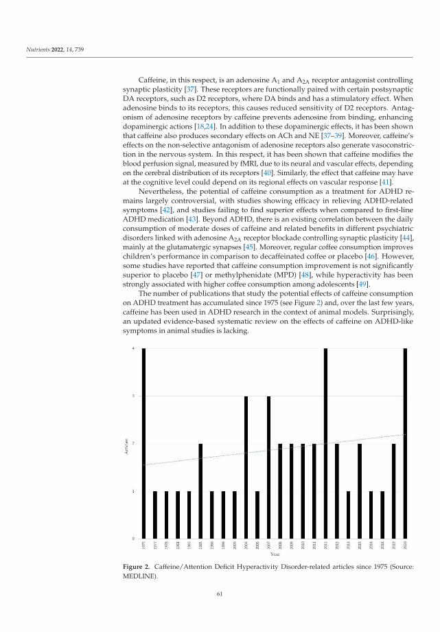
Nutrients 2022, 14, 739
Caffeine, in this respect, is an adenosine A1 and A2A receptor antagonist controllingsynaptic plasticity [37]. These receptors are functionally paired with certain postsynapticDA receptors, such as D2 receptors, where DA binds and has a stimulatory effect. Whenadenosine binds to its receptors, this causes reduced sensitivity of D2 receptors. Antag-onism of adenosine receptors by caffeine prevents adenosine from binding, enhancingdopaminergic actions [18,24]. In addition to these dopaminergic effects, it has been shownthat caffeine also produces secondary effects on ACh and NE [37–39]. Moreover, caffeine’seffects on the non-selective antagonism of adenosine receptors also generate vasoconstric-tion in the nervous system. In this respect, it has been shown that caffeine modifies theblood perfusion signal, measured by fMRI, due to its neural and vascular effects, dependingon the cerebral distribution of its receptors [40]. Similarly, the effect that caffeine may haveat the cognitive level could depend on its regional effects on vascular response [41].
Nevertheless, the potential of caffeine consumption as a treatment for ADHD re-mains largely controversial, with studies showing efficacy in relieving ADHD-relatedsymptoms [42], and studies failing to find superior effects when compared to first-lineADHD medication [43]. Beyond ADHD, there is an existing correlation between the dailyconsumption of moderate doses of caffeine and related benefits in different psychiatricdisorders linked with adenosine A2A receptor blockade controlling synaptic plasticity [44],mainly at the glutamatergic synapses [45]. Moreover, regular coffee consumption improveschildren’s performance in comparison to decaffeinated coffee or placebo [46]. However,some studies have reported that caffeine consumption improvement is not significantlysuperior to placebo [47] or methylphenidate (MPD) [48], while hyperactivity has beenstrongly associated with higher coffee consumption among adolescents [49].
The number of publications that study the potential effects of caffeine consumptionon ADHD treatment has accumulated since 1975 (see Figure 2) and, over the last few years,caffeine has been used in ADHD research in the context of animal models. Surprisingly,an updated evidence-based systematic review on the effects of caffeine on ADHD-likesymptoms in animal studies is lacking.
Figure 2. Caffeine/Attention Deficit Hyperactivity Disorder-related articles since 1975 (Source:MEDLINE).
61

Nutrients 2022, 14, 739
Consequently, to provide insight and value at the preclinical level, we sought toproduce a comprehensive compilation and systematically review all the relevant scientificpublications that make reference to the underlying effects of caffeine intake on treatingADHD-like symptoms in animal studies.
2. Materials and Methods
We conducted a systematic review of ADHD research in the context of animal modelsto assess the association between caffeine and ADHD-dependent variables includingattention, locomotor activity, impulsive behavior, learning, and memory.
2.1. Search Strategy
Figure 3 depicts the search strategy. We followed the guidelines and recommendationscontained in the Preferred Reporting Items for Systematic Reviews and Meta-Analyses(PRISMA) statement [50], in order to reliably structure the gathered information in this sys-tematic review. Academic articles were located using two electronic databases: MEDLINEand Web of Science. Only the results from these two databases were reported, since resultsfrom other sources (Scopus, Google Scholar) did not provide any relevant new results. Norestrictions regarding publication date were applied. The literature search was conductedon 5 September 2021.
According to our proposal, the MEDLINE search strategy was established on thefollowing key search terms: “caffeine” [Mesh] AND “Attention Deficit Disorder withHyperactivity” [Mesh]. MeSH (Medical Subject Headings) terms were therefore used in thedevelopment of this search. The Web of Science search strategy was based on the followingkey search terms: (“attention” OR “hyperactivity” OR “ADHD”) AND “caffeine”.
2.2. Study Selection Criteria
The search was limited to preclinical and original experiments on non-human animals.The inclusion criteria were: (1) English-written, indexed studies; (2) non-human animalpreclinical/experimental studies; (3) the mention of the relationship between caffeinetreatment and ADHD-like symptoms; and (4) controlled studies with separately treatedgroups. The exclusion criteria were: (1) clinical/experimental/qualitative studies onhumans; (2) not mentioning caffeine treatment and ADHD-like symptoms at all; (3) reviews,posters, conference abstracts, oral speeches, commentaries, theoretical papers, unpublishedrelevant studies, and other studies relevant to the topic but not published in peer-reviewedjournals; and (4) case and cross-over studies.
2.3. Study Selection
Duplicates of all the databases were removed. Titles and abstracts were independentlyscreened by two authors (J.C.V. and O.M.d.l.T.) according to the inclusion and exclusioncriteria. Articles interpreted as compatible were selected for a full-text analysis to determinewhether they were or were not within the inclusion criteria. Furthermore, the references ofselected studies were screened in search of additional articles that met the inclusion criteria.Whenever a divergence of opinions emerged, a third author (D.R.-R.) was consulted todiscuss and reach an agreement between the authors.
2.4. Data Extraction and Analyses
Following selection of the studies, data were extracted and prearranged into a table.The subsequent information was collected: (1) author/s and year; (2) species: strain, sex,and sample (n); (3) animal model; (4) age; (5) independent variables; (6) caffeine treatment;(7) behavioral tests/types of stress; (8) dependent variables; and (9) main results.
62

Nutrients 2022, 14, 739
Figure 3. Flow diagram of study’s selection based on PRISMA guidelines [50].
3. Results
Due to the large number of results acquired by the search terms, strict inclusion/exclusioncriteria were applied to limit the final selection of studies. Figure 2 shows the studies in-cluded in quantitative synthesis.
3.1. Study Selection
A total of 121 unique citations was initially retrieved through the combined search,after which 108 citations were excluded after full-text screening because they did not meetthe inclusion criteria. Therefore, 13 studies (Pandolfo et al., 2013 [51]; Ouichi et al., 2013 [52];Caballero et al., 2011 [53]; Ruiz-Oliveira et al., 2019 [54]; Higgins et al., 2007 [55]; Françaet al., 2020 [56]; Nunes et al., 2018 [57]; Szczepanik et al., 2016 [58]; Pires et al., 2010 [59];Prediger et al., 2005 [60]; Leffa et al., 2019 [61]; Pires et al., 2009 [62]; Alves et al., 2020 [63])on animal models were finally considered. Based on their methodology, the studies inthis review could be classified as experimental (n = 10; 76.9%), randomly assigning thesubjects sample to the experimental groups, and quasi-experimental (n = 3; 23.1%), wherethe groups were usually constructed according to the subject’s characteristics. The firststudies relevant to the topic were from 2005, while the most recent studies included in thisreview were published in 2020. Table 1 describes each article individually.
3.1.1. Species, Animal Model, Sex, and Treatment
Most of the animal studies were performed on rodents. Ten studies were conductedwith rats and two with mice. Only one of the studies used zebrafish as an animal model.Four studies used only males, five used both males and females, and four used only females.Different caffeine treatments and routes of administration were used, along with differentdurations (Table 1). Chronic treatments were mainly performed by dilating caffeine powder
63

Nutrients 2022, 14, 739
in the system water, whereas acute treatments were mainly administered intraperitoneally(i.p.).
3.1.2. Animal Models of ADHD
Overall, nine studies used genetic animal models of ADHD: eight studies used SHR,and one study used the low-density lipoprotein receptor (LDLr) mouse. Finally, two studiesused physical trauma to provide an epigenetic animal model of ADHD: one study caused6-hydroxy-dopamine (6-OHDA) lesions in rats, while one study used social isolation (SI)as an intensely stressful environment in mice (Table 1).
3.1.3. Behavioral Tests
Five studies used the object recognition task; four studies used the Y maze test; threestudies used the open field test; two studies used the water maze test; and two studies usedthe novel object recognition test. In addition, other tests were performed, including thewater-finding test, the five-choice serial reaction time task (5-CSRTT), the locomotor activitytest, the discrimination task, the Olton maze behavioral assay, the attention set-shiftingtask, the fear-conditioning test, the tolerance to delay of reward task, and the olfactorydiscrimination test. Finally, one study induced the animals to a certain type of stress bymeans of social isolation and aggressivity (Table 1).
3.2. Study Outcomes
The results are summarized in Table 1. Considering the amount of data providedin the reviewed articles, we decided to categorize all the information based on caffeine’seffects on each relevant ADHD-like evaluated parameter, as follows.
3.2.1. AttentionAttention and Behavioral Flexibility
Pandolfo et al. [51] examined the impact of chronic caffeine treatment during ado-lescence on SHR and Wistar Kyoto (WKY) rats’ performance in an attention set-shiftingtask, placing emphasis on response to conflict. The task was divided into different phases:familiarization, response discrimination, and visual cue discrimination. During the re-sponse discrimination phase, statistical analysis showed that vehicle-treated SHR needed asuperior amount of trials to reach the benchmark of 10 consecutive correct choices, com-pared with WKY rats. Importantly, treatment with caffeine (2 mg/kg, i.p.) improved SHRdiscriminative learning in a selective manner, as indicated by a reduction in the number oftrials needed to reach the benchmark, while treatment with caffeine had no effect on WKYrats. During the visual cue discrimination phase, SHR required more trials to master thetask, compared with WKY rats. Once more, caffeine treatment (2 mg/kg, i.p.) diminishedthe number of trials needed to reach the benchmark. Finally, statistical analysis showedthat vehicle-treated SHR made significantly more regressive and never-reinforced errorsthan vehicle-treated WKY rats. Remarkably, while treatment with caffeine (2 mg/kg, i.p.)diminished the number of these errors in SHR, it had no effect on WKY rats.
64

Nutrients 2022, 14, 739
Ta
ble
1.
Sum
mar
yof
incl
uded
stud
ies.
Au
tho
r/s
&Y
ea
r
Sp
eci
es,
Str
ain
,S
ex
&S
am
ple
(n)
An
ima
lM
od
el
Ag
eIn
de
pe
nd
en
tV
ari
ab
les
Ca
ffe
ine
Tre
atm
en
tB
eh
av
iora
lT
est
s/T
yp
eo
fS
tre
ssD
ep
en
de
nt
Va
ria
ble
sM
ain
Re
sult
s
Szcz
epan
iket
al.,
2016
Mic
eC
57Bl
/6w
ild-t
ype
(8)
LDLr
(8)
Fem
ale
Gen
etic
(LD
Lr)
3 mon
ths
8 mon
ths
Trea
tmen
t(c
affe
ine
or vehi
cle)
Stra
in(C
57Bl
/6w
ildty
peor
LDLr
)
10m
g/kg
oral
rout
eC
hron
ictr
eatm
ent
(21
days
)
Ope
n-fie
ldar
ena
Spon
tane
ous
loco
mot
orac
tivi
ty(t
otal
dist
ance
trav
elle
d)A
nxie
ty(t
ime
inth
ece
nter
)Ex
plor
ator
ybe
havi
or(v
isua
lins
pect
ion
ofth
eoc
cupa
tion
plot
)
-LD
Lrm
ice
trav
elle
dgr
eate
rdi
stan
ces
than
the
C57
BI/6
wild
type
mic
edu
ring
the
5m
inpe
riod
ofan
alys
is.
-Caf
fein
etr
eatm
enti
nduc
eda
reno
rmal
izat
ion
effe
ctin
8m
onth
-old
mou
selo
com
otio
n.-C
affe
ine
trea
tmen
twas
unab
leto
mod
ify
the
hype
rloc
omot
ion
obse
rved
in3
mon
th-o
ldLD
Lrm
ice.
-All
anim
algr
oups
spen
tasi
mila
ram
ount
ofti
me
inth
ece
nter
ofan
open
field
.-S
imila
rex
plor
ator
ybe
havi
orbe
twee
ngr
oups
.
Hig
gins
etal
.,20
07
Rat
sLE
(15–
16)
CD
(12–
16)
Mal
e
Not
used
Not
spec
ified
Trea
tmen
t(c
affe
ine,
SCH
4123
48,
KW
-600
2,D
PCPX
,C
GS-
2168
0,am
phet
amin
e)St
rain
(LE
orC
D)
1m
L/kg
i.p.r
oute
One
dose
,pr
ior
test
ing
Five
-cho
ice
seri
alre
acti
onti
me
task
Loco
mot
orac
tivi
tyte
st
Sele
ctiv
eat
tent
ion
(Cor
rect
/inc
orre
cttr
ials
,om
issi
ons,
prem
atur
ean
dpe
rsev
erat
ive
resp
onse
s,ch
oice
accu
racy
,co
rrec
t/in
corr
ect,
and
mag
azin
ela
tenc
y)H
ypol
ocom
otio
n(d
ista
nce
trav
elle
d)
-Caf
fein
e,SC
H41
2348
and
KW
-600
2au
gmen
ted
time
reac
tion
inLE
and
CD
,wit
hout
effe
cton
accu
racy
.-E
ffec
tsof
SCH
4123
48w
ere
atdo
ses
that
wer
eno
tov
ertl
yps
ycho
stim
ulat
ory.
-CG
S-21
680
redu
ced
spee
dre
acti
onan
dau
gmen
ted
omis
sion
s.A
CG
S-21
680
low
erdo
sere
duce
dth
ein
crea
sed
prem
atur
ere
spon
seca
used
byam
phet
amin
e.-C
affe
ine’
sat
tent
iona
l-en
hanc
ing
effe
cts
wer
efa
cilit
ated
thro
ugh
A2A
rece
ptor
bloc
kade
.Sel
ecti
veA
2Are
cept
oran
tago
nist
sco
uld
bein
clud
edas
apo
tent
ialt
hera
pyfo
rA
DH
D.
Rui
z-O
livei
raet
al.,
2019
Zeb
rafis
hw
ild-t
ype
(40)
Mal
eFe
mal
e
Not
used
4 mon
ths
Trea
tmen
t(c
affe
ine
or vehi
cle)
10m
g/L
50m
g/L
drin
king
wat
erC
hron
ictr
eatm
ent
(14
days
)
Dis
crim
inat
ion
task
Con
diti
oned
lear
ning
abili
ty(a
vera
gesw
imm
ing
spee
d,in
terg
roup
free
zing
,m
axim
umsp
eed,
tim
esp
enti
nea
char
ea,
late
ncy
toen
ter
each
area
)
-0an
d10
mg/
Lca
ffei
negr
oups
spen
tmos
toft
heti
me
clos
eto
the
targ
et.
-10
mg
caff
eine
grou
pha
dth
esh
orte
stla
tenc
yto
reac
hth
eta
rget
.-0
and
10m
g/L
caff
eine
grou
psin
crea
sed
the
aver
age
spee
dan
ddi
stan
cetr
avel
led.
-Caf
fein
eex
posu
reat
low
dose
sse
ems
toen
hanc
evi
sual
cue
disc
rim
inat
ion
and
zebr
afish
perf
orm
ance
.
Pred
iger
etal
.,20
05
Rat
sW
KY
(7–8
)SH
R(7
–8)
Fem
ale
Gen
etic
(SH
R)
3 mon
ths
Trea
tmen
t(c
affe
ine
or vehi
cle)
Stra
in(W
KY
orSH
R)
1.3
mg/
kg10
mg/
kgi.p
.rou
teO
nedo
se,
prio
rte
stin
g
Wat
erm
aze
task
Spat
iall
earn
ing
(esc
ape
late
ncy,
dist
ance
trav
elle
d,sw
imm
ing
spee
d)M
ean
arte
rial
pres
sure
-SH
Rne
eded
ala
rger
amou
ntof
tria
lsdu
ring
the
trai
ning
sess
ion
tole
arn
the
spat
iali
nfor
mat
ion,
alth
ough
asi
mila
rpr
ofile
toth
atof
WK
Yra
tsdu
ring
the
test
sess
ion,
show
ing
ase
lect
ive
spat
iall
earn
ing
defic
it.
-Caf
fein
e’s
pre-
trai
ning
adm
inis
trat
ion
enha
nced
SHR
s’sp
atia
llea
rnin
gde
ficit
.-C
affe
ine’
spo
st-t
rain
ing
adm
inis
trat
ion
did
not
enha
nce
SHR
s’te
stpe
rfor
man
ce,a
ltho
ugh
itim
prov
edW
KY
rats
’mem
ory
rete
ntio
n.-M
ean
bloo
dpr
essu
rew
asno
talt
ered
byca
ffei
ne.
65

Nutrients 2022, 14, 739
Ta
ble
1.
Con
t.
Au
tho
r/s
&Y
ea
r
Sp
eci
es,
Str
ain
,S
ex
&S
am
ple
(n)
An
ima
lM
od
el
Ag
eIn
de
pe
nd
en
tV
ari
ab
les
Ca
ffe
ine
Tre
atm
en
tB
eh
av
iora
lT
est
s/T
yp
eo
fS
tre
ssD
ep
en
de
nt
Va
ria
ble
sM
ain
Re
sult
s
Pire
set
al.,
2009
Rat
sW
KY
(15)
SHR
(18)
Mal
eFe
mal
e
Gen
etic
(SH
R)
3 mon
ths
Trea
tmen
t(M
PD,
DPC
PX,
caff
eine
,Z
M24
1385
orve
hicl
e)St
rain
(WK
Yor
SHR
)
1m
g/kg
3m
g/kg
10m
g/kg
i.p.r
oute
One
dose
,pr
ior
test
ing
Obj
ect
reco
gnit
ion
task
Obj
ectr
ecog
niti
on(i
nves
tiga
tion
tim
e,di
scri
min
atio
nti
me)
Mea
nar
teri
alpr
essu
re
-SH
Ron
lydi
scri
min
ated
betw
een
the
mos
tst
ruct
ural
lydi
stin
ctpa
irs
ofob
ject
s.-P
re-t
rain
ing
adm
inis
trat
ion
ofM
PD,c
affe
ine,
the
sele
ctiv
ead
enos
ine
rece
ptor
anta
goni
sts
DPC
PXan
dZ
M24
1385
,or
the
asso
ciat
ion
ofin
effe
ctiv
edo
ses
ofD
PCPX
and
ZM
2413
85,i
mpr
oved
the
perf
orm
ance
ofSH
Rin
the
obje
ct-r
ecog
niti
onta
sk.
-The
adm
inis
trat
ion
ofth
esa
me
dose
sof
MPD
and
caff
eine
did
nots
igni
fican
tly
alte
rth
em
ean
arte
rial
pres
sure
ofei
ther
WK
Ys
orSH
Rs.
Pire
set
al.,
2010
Rat
sW
KY
(37)
SHR
(38)
Fem
ale
Gen
etic
(SH
R)
25/3
8da
ys
Trea
tmen
t(c
affe
ine,
MPD
orve
hicl
e)St
rain
(WK
Yor
SHR
)
3m
g/kg
i.p.R
oute
.C
hron
ictr
eatm
ent
(14
days
)
Obj
ect
reco
gnit
ion
task
Obj
ectr
ecog
niti
on(i
nves
tiga
tion
tim
e,di
scri
min
atio
nti
me)
Spon
tane
ous
loco
mot
orac
tivi
tyM
ean
arte
rial
pres
sure
Body
wei
ght
-WK
Yra
tsdi
stin
guis
hed
allt
heit
ems.
SHR
sw
ere
unab
leto
dist
ingu
ish
pair
sof
item
sw
ith
slig
htst
ruct
ural
alte
rati
ons.
-Caf
fein
eor
MPD
chro
nic
trea
tmen
tenh
ance
dSH
Rit
em-r
ecog
niti
onde
ficit
s.T
hesa
me
trea
tmen
tsim
pair
edth
ead
ultW
KY
rats
’sho
rt-t
erm
obje
ctre
cogn
itio
nab
ility
.-E
ffec
tsw
ere
inde
pend
ento
fvar
iati
ons
inlo
com
otio
n,ar
teri
albl
ood
pres
sure
,and
body
wei
ght.
Cab
alle
roet
al.,
2011
Rat
s6-
OH
DA
lesi
oned
(9)
Salin
e-le
sion
ed(9
)M
ale
Fem
ale
Phys
ical
trau
ma
(6-O
HD
Ale
sion
ed)
25da
ysTr
eatm
ent
(caf
fein
eor ve
hicl
e)
1m
g/m
Ldr
inki
ngw
ater
Chr
onic
trea
tmen
t(1
4da
ys)
Olt
onm
aze
beha
vior
alas
say
Mot
orbe
havi
or(n
umbe
rof
arm
scr
osse
d)A
tten
tion
beha
vior
(tot
alnu
mbe
rof
arm
sw
alke
d,an
dto
tal
num
ber
ofar
ms
wal
ked
unti
lon
ew
asre
peat
ed)
-Caf
fein
etr
eatm
ents
igni
fican
tly
impr
oved
6-O
HD
Ale
sion
edra
ts’a
tten
tion
defic
it.
-Aft
erca
ffei
neco
nsum
ptio
n,no
chan
ges
wer
efo
und
inm
otor
acti
vity
.
66

Nutrients 2022, 14, 739
Ta
ble
1.
Con
t.
Au
tho
r/s
&Y
ea
r
Sp
eci
es,
Str
ain
,S
ex
&S
am
ple
(n)
An
ima
lM
od
el
Ag
eIn
de
pe
nd
en
tV
ari
ab
les
Ca
ffe
ine
Tre
atm
en
tB
eh
av
iora
lT
est
s/T
yp
eo
fS
tre
ssD
ep
en
de
nt
Va
ria
ble
sM
ain
Re
sult
s
Pand
olfo
etal
.,20
13
Rat
sW
KY
(16)
SHR
(16)
Mal
e
Gen
etic
(SH
R)
24da
ys
Trea
tmen
t(c
affe
ine
or vehi
cle
(sal
ine)
)St
rain
(WK
Yor
SHR
)
2m
g/kg
i.p.r
oute
Chr
onic
trea
tmen
t(t
wic
eda
ilyfo
r21
days
)
Att
enti
onal
-set
shif
ting
;an
xiet
y-re
late
dbe
havi
or;Y
maz
e;lo
com
otio
n-r
elat
edbe
havi
or
Att
enti
on(r
egre
ssiv
ean
dne
ver-
rein
forc
eder
rors
,pe
rsev
erat
ive
erro
rs,t
otal
num
ber
oftr
ials
requ
ired
befo
rere
achi
ng10
corr
ectc
onse
cuti
vech
oice
s)Lo
com
otio
nan
dan
xiet
y(n
umbe
rof
peri
pher
alsq
uare
scr
osse
d,nu
mbe
rof
cent
ral
squa
res
cros
sed,
perc
enta
geof
cent
rall
ocom
otio
n)Sp
atia
lrec
ogni
tion
(num
ber
ofen
trie
s;ti
me
spen
tpe
rar
m;r
ando
mex
plor
atio
n)
-SH
Rs
wer
ehy
pera
ctiv
ean
dsh
owed
poor
erpe
rfor
man
cein
the
atte
ntio
nals
et-s
hift
ing
and
Y-m
aze
para
digm
s,di
spla
yed
incr
ease
ddo
pam
ine
tran
spor
ter
dens
ity,
and
incr
ease
ddo
pam
ine
upta
kein
fron
toco
rtic
alan
dst
riat
alte
rmin
als.
-Chr
onic
caff
eine
trea
tmen
tim
prov
edm
emor
yan
dat
tent
ion
defic
its,
and
norm
aliz
eddo
pam
iner
gic
func
tion
inSH
R.
-Fir
stin
dica
tion
ofad
enos
ine
A2A
rece
ptor
s(A
2AR
)in
nerv
ete
rmin
als
infr
onta
lcor
tex.
-Fir
stev
iden
ceth
atA
2AR
dens
ity
isim
prov
edin
SHR
.
Oui
chi
etal
.,20
13
Mic
eIC
R(9
)M
ale
Fem
ale
Phys
ical
trau
ma
(SI)
4w
eeks
Trea
tmen
t(M
PDan
dca
ffei
ne)
SI
0.5–
1m
g/kg
i.p.r
oute
One
dose
,pr
ior
test
ing
Wat
er-fi
ndin
gte
st;a
ggre
ssio
n;m
odifi
edY-
maz
ete
st;n
ovel
obje
ctre
cogn
itio
nte
st;
fear
-con
diti
onin
gte
st
Spat
iala
tten
tion
(ent
erin
g&
drin
king
late
ncy)
Agg
ress
ion
(dur
atio
nof
wre
stlin
g)Sp
atia
lrec
ogni
tion
(tim
esp
enti
nth
ene
war
m;
tota
ltim
eex
plor
ing
obje
cts)
Fear
cond
itio
ning
(fre
ezin
gbe
havi
or)
-SIr
ats
show
edde
ficit
sin
spat
iala
tten
tion
onth
ew
ater
-find
ing
test
.Re-
soci
aliz
eddi
dno
tred
uce
defic
itin
spat
iala
tten
tion
.SIe
ffec
ton
spat
ial
atte
ntio
nre
veal
edno
diff
eren
cein
gend
eror
corr
elat
ion
wit
hag
gres
sive
beha
viou
r.-S
Iim
pair
edco
nditi
onal
and
cont
extu
alfe
arm
emor
y.-M
PDan
dca
ffei
neen
hanc
edde
ficit
sin
SI-i
nduc
edla
tent
lear
ning
ina
man
ner
that
was
reve
rsib
lew
ith
chol
iner
gic
butn
otdo
pam
iner
gic
anta
goni
sts.
67

Nutrients 2022, 14, 739
Ta
ble
1.
Con
t.
Au
tho
r/s
&Y
ea
r
Sp
eci
es,
Str
ain
,S
ex
&S
am
ple
(n)
An
ima
lM
od
el
Ag
eIn
de
pe
nd
en
tV
ari
ab
les
Ca
ffe
ine
Tre
atm
en
tB
eh
av
iora
lT
est
s/T
yp
eo
fS
tre
ssD
ep
en
de
nt
Va
ria
ble
sM
ain
Re
sult
s
Nun
eset
al.,
2018
Rat
sW
KY
(5–1
5)SH
R(5
–15)
Mal
eFe
mal
e
Gen
etic
(SH
R)
15da
ys28
days
50da
ys
Trea
tmen
t(c
affe
ine/
wat
er,
caf-
fein
e/ca
ffei
neor
wat
er)
Stra
in(W
KY
orSH
R)
0.3
g/L
drin
king
wat
erU
ntil
PND
28
Ope
n-fie
ldte
st;
Nov
elob
ject
reco
gnit
ion;
Ym
aze
task
Ope
nfie
ldte
st(t
rave
ldis
tanc
epe
riph
ery)
Hab
itua
tion
(tot
altr
avel
led
dist
ance
inth
eop
enfie
ld)
Spat
ialr
ecog
niti
on-Y
maz
ean
dob
ject
reco
gnit
ion
(exp
lora
tion
,di
scri
min
atio
nra
tio,
num
ber
ofen
trie
s,ti
me
spen
tin
nove
lar
m,t
otal
num
ber
ofen
trie
sin
thre
ear
ms)
-Ado
lesc
entS
HR
from
both
sexe
sdi
spla
yed
hype
rloc
omot
ion,
reco
gnit
ion,
and
spat
ialm
emor
ydi
stur
banc
es.F
emal
esdi
spla
yed
ala
ckof
habi
tuat
ion
and
dete
rior
ated
spat
ialm
emor
y.-C
affe
ine
was
effe
ctiv
eat
impr
ovin
gre
cogn
itio
nm
emor
yda
mag
ein
both
sexe
s.-S
pati
alm
emor
yw
asim
prov
edon
lyin
fem
ale
SHR
s.-F
emal
eSH
Rs
disp
laye
dim
pair
edhy
perl
ocom
otio
nfo
llow
ing
caff
eine
trea
tmen
t.-S
HR
sof
both
sexe
spr
esen
ted
incr
ease
sin
BDN
F,tr
unca
ted
and
phos
pho-
TrkB
rece
ptor
s,an
dph
osph
o-C
REB
leve
lsin
the
hipp
ocam
pus.
-Caf
fein
eno
rmal
ized
BDN
Fin
mal
esan
dtr
unca
ted
TrkB
rece
ptor
inbo
thse
xes.
Leff
aet
al.,
2019
Rat
sW
KY
(7–9
)SH
R(7
–9)
Mal
e
Gen
etic
(SH
R)
60/6
5da
ys24
days
Trea
tmen
t(W
IN,
AM
251,
caff
eine
or vehi
cle)
Stra
in(W
KY
orSH
R)
2or
5m
g/kg
i.p.r
oute
Acu
tepr
etre
atm
ent,
one
dose
Chr
onic
trea
tmen
t(2
1da
ys)
Tole
ranc
eto
dela
yof
rew
ard;
Tm
aze
Impu
lsiv
ebe
havi
or(t
oler
ance
tode
lay
ofre
war
d)
-WIN
trea
tmen
tdec
reas
edla
rge
rew
ard
choi
ces
and
AM
251
trea
tmen
tinc
reas
edla
rge
rew
ard
choi
ces
inSH
R.
-Acu
teca
ffei
nepr
etre
atm
entb
lock
edW
INef
fect
s.-C
hron
icca
ffei
netr
eatm
enti
ncre
ased
the
impu
lsiv
eph
enot
ype
and
pote
ntia
ted
the
WIN
effe
cts.
-Can
nabi
noid
and
aden
osin
ere
cept
ors
mod
ulat
eim
puls
ive
beha
vior
inSH
R.
Alv
eset
al.,
2020
Rat
s-pr
egna
ntSH
R(4
0–70
)W
KY
(40–
70)
Fem
ale
Gen
etic
(SH
R)
Invi
tro
Trea
tmen
t(c
affe
ine,
DM
SO,
LY29
4002
,ad
enos
ine
sele
ctiv
eag
onis
tan
dan
tago
nist
s)St
rain
(WK
Yor
SHR
)
Caf
fein
ein
cuba
tion
(30μ
M)
One
dose
No
beha
vior
alta
sk
Mor
phol
ogic
alal
tera
tion
s(s
ingl
ing,
neur
ite
bran
chin
g)
-SH
Rne
uron
sdi
spla
yed
less
neur
ite
bran
chin
g,sh
orte
rm
axim
alne
urit
ele
ngth
and
decr
ease
dax
onal
outg
row
th.
-Caf
fein
ere
cove
red
neur
itebr
anch
ing
and
elon
gatio
nfr
omSH
Rne
uron
svi
aPK
Aan
dPI
3Ksi
gnal
ing,
-A2A
Rag
onis
t(C
GS
2168
0)pr
omot
edm
ore
neur
ite
bran
chin
gvi
aPK
Asi
gnal
ing.
-The
sele
ctiv
eA
2AR
anta
goni
st(S
CH
5826
1)w
asef
ficie
ntat
reco
veri
ngax
onal
outg
row
thfr
omSH
Rne
uron
sth
roug
hPI
3Kan
dno
tPK
Asi
gnal
ing.
68

Nutrients 2022, 14, 739
Ta
ble
1.
Con
t.
Au
tho
r/s
&Y
ea
r
Sp
eci
es,
Str
ain
,S
ex
&S
am
ple
(n)
An
ima
lM
od
el
Ag
eIn
de
pe
nd
en
tV
ari
ab
les
Ca
ffe
ine
Tre
atm
en
tB
eh
av
iora
lT
est
s/T
yp
eo
fS
tre
ssD
ep
en
de
nt
Va
ria
ble
sM
ain
Re
sult
s
Fran
çaet
al.,
2020
Rat
sW
KY
(9)
SHR
(11)
Mal
e
Gen
etic
(SH
R)
30da
ys4–
5m
onth
s
Trea
tmen
t(c
affe
ine
orw
ater
)Ph
ysic
alex
erci
seSt
rain
(WK
Yor
SHR
)
0.3
mg/
mL,
drin
king
wat
erO
nedo
se
Olf
acto
rydi
scri
min
atio
n;O
pen
field
;O
bjec
tre
cogn
itio
n;W
ater
maz
e
Olf
acto
rydi
scri
min
atio
n(t
ime
spen
tin
com
part
men
ts,
num
bers
ofcr
ossi
ngs)
Loco
mot
orac
tivi
ty(t
otal
dist
ance
,tim
esp
enti
nth
ece
ntra
lzon
e)Sh
ort-
term
mem
ory
(tot
alti
me
spen
tex
plor
ing
the
obje
cts,
disc
rim
inat
ion
inde
x)W
orki
ngan
dpr
oced
ural
mem
orie
s(e
scap
ela
tenc
y)
-SH
Rsh
owed
olfa
ctor
yan
dsh
ort-
term
reco
gnit
ion
mem
ory
defic
ienc
ies
from
adol
esce
nce
toad
ulth
ood,
acco
mpa
nied
bylo
wer
pref
ront
alco
rtex
and
hipp
ocam
pus
SNA
P-25
leve
ls.
-Caf
fein
ean
dph
ysic
alex
erci
sedu
ring
adol
esce
nce
orad
ulth
ood
repa
ired
the
olfa
ctor
ydi
scri
min
atio
nab
ility
and
enha
nced
shor
t-te
rmre
cogn
itio
nm
emor
yin
SHR
s.-C
affe
ine
cons
umpt
ion
and
phys
ical
exer
cise
duri
ngad
oles
cenc
eau
gmen
ted
hipp
ocam
pus
and
pref
ront
alco
rtex
SNA
P-25
,syn
taxi
n,an
dse
roto
nin
leve
ls,a
sw
ella
sSH
Rs’
stri
atal
dopa
min
ele
vels
.
Key
for
abbr
evia
tions
used
:LD
Lr:l
ow-d
ensi
tylip
opro
tein
rece
ptor
,LE
rat:
Long
–Eva
nsra
t,C
Dra
t:C
esar
ean-
deri
ved
rat,
i.p.:
intr
aper
itone
ally
,WK
Yra
t:w
ista
rK
yoto
rat,
SHR
:spo
ntan
eous
lyhy
pert
ensi
vera
t,M
PD:m
ethy
lphe
nida
te,6
-OH
DA
:6-h
ydro
xy-d
opam
ine,
A2A
R:A
deno
sine
A2A
rece
ptor
s,IC
Rm
ice:
Inst
itute
ofC
ance
rR
esea
rch
mic
e,SI
:soc
iali
sola
tion
,PN
D:p
ostn
atal
day,
BDN
F:br
ain-
deri
ved
neur
otro
phic
fact
or,S
NA
P-25
:syn
apto
som
al-a
ssoc
iate
dpr
otei
n25
.
69

Nutrients 2022, 14, 739
Spatial Attention
Ouchi et al. [52] tested the effect of SI on latent learning using the water-finding test,measuring entering latency and drinking latency. The authors eventually discussed theutility of SI as an ADHD epigenetic model. They socially isolated male or female Instituteof Cancer Research (ICR) mice for one week or more. Subsequently, the animals displayedspatial attention deficit during the water-finding task. Five weeks of resocialization follow-ing one week of SI failed to improve this deficit. Drinking latency depended on how muchattention the animal paid to environmental factors, including the location of a tap waternozzle, which they were exposed to in the training trial. Therefore, a decrease in drinkinglatency correlated with the animal remembering the position/location of the nozzle. Caf-feine (0.3–1 mg/kg, i.p.) induced changes in drinking latency on the water-finding test, inthis sense, significantly ameliorating SI-induced latent learning deficits in a dose-dependentmanner, independently of gender or age.
Caballero et al. [53] examined caffeine’s therapeutic use in neonatal 6-OHDA lesionedrats, which constitute another existing ADHD animal model. At postnatal day (PND) 7,the rats were lesioned at the left striatum with 6-OHDA. At PND 25, spatial attention wasmeasured with an eight arm radial maze, the Olton maze. The animals were then placed inthe maze. The total number of arms the animals walked before completing six out of eight,or until they repeated one of them, was measured. After 14 days of treatment with caffeine,administered ad libitum into the drinking water, the authors assessed caffeine’s effects onthe attention deficit of the animals, using the same task. Interestingly, the 6-OHDA lesionedrats significantly improved their attention deficit after caffeine treatment. Consequently,the authors highlighted the properties through which caffeine managed the attentionaldeficits occurring during the prepubertal period of ADHD.
Discrimination
Ruiz-Oliveira et al. [54] evaluated the effect of caffeine on zebrafish performancein a task requiring focus and attention, the discrimination task. The task took place inthree phases: tank acclimation, training, and test. The authors used visual cues duringthe training trials and the test trials. Distractors, objects resembling the target, were usedto confuse the fish and impair conditioning. The fish were exposed to different caffeineconcentrations for 14 days: 0 mg/L (control), 10 mg/L (low), and 50 mg/L (high). Notably,low caffeine doses improved the fishes’ ability to discriminate the cues and reach thetarget; the fish spent most of the time close to the target where the reward was offered, andshowing the shortest latency to reaching the target. The higher dose impaired the fishes’ability to find the target; the fish demonstrated increased anxiety, a possible side effect ofthe substance.
Selective Attention
Higgins et al. [55] evaluated the effect of caffeine on Long–Evans (LE) and Cesarean-derived (CD) rat performance in a selective attention task, the 5-CSRTT. The effects ofcaffeine were compared to the selective A2A antagonists, SCH 412348 and KW-6002, and theA1 antagonist, DPCPX. Caffeine (3–10 mg/kg, i.p.) increased reaction time in both LE andCD rats, with no effect on accuracy, an effect replicated by SCH 412348 (0.1–1 mg/kg PO)and KW-6002 (1–3 mg/kg PO), but not DPCPX (3–30 mg/kg PO). The faster responsespeed was observed in both the CD and LE rat strains at 3 mg/kg, although increasedpremature responses were confined to the LE strain at the 10 mg/kg dose. These resultssuggest that the attention-enhancing effects of caffeine were mediated through A2A receptorblockade. Selective A2A receptor antagonists may therefore have potential as therapies forattention-related disorders, such as ADHD.
70

Nutrients 2022, 14, 739
3.2.2. Hyperactivity and ImpulsivityLocomotor Activity
França et al. [56] tested the effect of caffeine on the hyperlocomotion characteristic ofADHD by examining locomotor activity. Caffeine consumption (0.3 mg/mL in drinkingwater) and physical exercise in running wheels for 6 weeks, either during adolescence(30 days old) or adulthood (4–5 months old), did not relate to changes in spontaneouslocomotion in SHR, in an open field, during the 5 min habituation phase of the object recog-nition test. Ruiz-Oliveira et al. [54] studied the effects of caffeine on zebrafish (4 monthsold, wild type, both sexes). Low concentrations of caffeine (10 mg/L) affected locomotorparameters, increasing average speed and decreasing freezing behavior. Interestingly, thelevels of freezing and locomotor behavior were the same for the 50 mg/L caffeine groupand the control group. Nunes et al. [57] evaluated locomotor activity during the latechildhood and the end of adolescence of male and female SHR, using an open field arenaand measuring the total distance travelled in meters along the periphery during 5 min.Although caffeine (0.3 g/L) did not impact hyperlocomotion during late childhood (PND28) in either sex, continuous treatment aggravated adolescent female SHR hyperactivity(PND 50), suggesting that the consumption of caffeine during childhood may aggravate hy-peractivity in females, but only if the administration persists up to adolescence. Szczepaniket al. [58] demonstrated that young (3 months old) and middle-aged (8 months old) LDLrmice display different responses to chronic caffeine treatment in terms of motor activity.Although caffeine was unable to modify the hyperlocomotion observed in 3 months oldLDLr mice, caffeine attenuated the increased locomotor activity observed in 8 months oldLDLr mice. Pandolfo et al. [51] tested whether chronic treatment with caffeine was ableto counteract the hyperlocomotion characteristic of ADHD in SH, during the open fieldtest. Chronic treatment with caffeine did not alter central and total locomotion in SHR.Similarly, Pires et al. [59] showed that chronic treatment with caffeine did not producechanges in SHR locomotion during the object recognition task sample phase. Interestingly,Caballero et al. [53] showed that neonatal 6-OHDA lesioned rats, a different ADHD animalmodel, demonstrated a non-significant tendency to decrease their motor activity after adlibitum caffeine consumption throughout the prepubertal period during an Olton maze be-havioral assay. Higgins et al. [55] conducted two separate types of locomotor activity study.In a CGS-21680-induced hypolocomotion assay, pretreatment with caffeine (3–30 mg/kg,i.p.) produced a significant attenuation of the CGS-21680 hypolocomotion at different doses,of 10 mg/kg and 30 mg/kg, in CD rats. In a second experiment, caffeine (1–30 mg/kg, i.p.)produced a dose-related increase in locomotion in the animals habituated to the test cham-bers. Finally, Prediger et al. [60] did not find a direct increase in locomotor performancein SHR after the administration of acute doses of caffeine (1–10 mg/kg i.p.) when using aspatial version of the Morris water maze. No alteration was observed in the swimmingspeed in this regard.
Impulsive Behavior
Leffa et al. [61] focused on impulsive behavior to clarify the neurobiology of ADHD.They treated SHRs with caffeine, a non-selective adenosine receptor antagonist, to assessthe modulating effects of the adenosine systems on tolerance to the delay of a reward. Theanimals had to choose between a small, but immediate, or a large, but delayed, reward. Anacute pretreatment with caffeine (2 mg/kg or 5 mg/kg) increased number of large-rewardchoices. Conversely, chronic treatment with caffeine (2 mg/kg, for 21 days) augmented theimpulsive phenotype and decreased the number of large-reward choices.
3.2.3. Learning and MemoryNon-Associative Learning
Habituation is a form of non-associative learning in which the animal’s innate re-sponse to a stimulus decreases after prolonged or repeated presentations of this stimulus.Nunes et al. [57] analyzed habituation during late childhood and the end of adolescence in
71

Nutrients 2022, 14, 739
male and female SHRs. The authors observed a sex and age difference in habituation, withfemale SHRs showing lack of habituation from childhood onwards, and male SHRs show-ing a lack of habituation in adolescence. These difficulties observed in female habituation,however, were overturned by treatment with caffeine (0.3 g/L) during childhood.
Working Memory
The object recognition task is recognized as a working-memory task, relies on theanimal’s natural tendency for novelty, and tests the ability to discriminate between familiarand unfamiliar objects. França et al. [56] assessed working memory using an adaptedversion of the object recognition task, conducted in an open field during three differentphases: habituation, sample and discrimination. Although the study results indicated thatthe disruption of the short-term recognition memory persisted into adulthood, the associ-ation of caffeine (0.3 mg/mL) and exercise during adulthood and adolescence improvedshort-term recognition memory in the SHR strain. Nunes et al. [57] also carried out thenovel object recognition test and observed similar recognition memory disturbances in ado-lescent SHRs of both sexes. Nonetheless, caffeine intake (0.3 g/L) restricted to childhoodrestored recognition memory in adolescent SHRs of both sexes. To evaluate the potentialof caffeine in ADHD therapy, Pires et al. [59] treated female WKY rats and SHR withcaffeine (3 mg/kg, i.p.) for 14 consecutive days during the prepubertal period. The animalswere tested during the object recognition test in the course of adulthood. While WKY ratsdiscriminated between all the used objects, the SHRs were unable to differentiate betweenpairs of objects with subtle structural differences. Nonetheless, caffeine or MPD chronictreatment improved the deficits in object recognition in SHR. Pires et al. [62] showed, forthe first time, the significant impairment of SHRs’ short-term object-recognition abilityin comparison with WKY rats. They further investigated the effects of caffeine (1, 3 or10 mg/kg), 30 min before the sample phase, on the performance of WKY rats and SHRof both sexes in the object recognition task. The injection of caffeine (1, 3 or 10 mg/kg,i.p.) improved the discrimination index of female SHRs, while the highest tested dose ofcaffeine (10 mg/kg, i.p.) increased the discrimination index of male SHRs.
Spatial Learning
The water maze task is a behavioral procedure widely used with rodents to studyspatial learning or spatial memory. Prediger et al. [60] used a circular swimming pool toassess the effect of caffeine administration on spatial learning deficit in SHRs. Adult femaleWKY rats and SHRs were treated with caffeine (1–10 mg/kg i.p.) before or immediately aftertraining, or before the test session. Spatial learning deficit in SHR was improved throughthe pre-training administration of caffeine (1–10 mg/kg i.p.). SHR test performance wasnot altered by the post-training administration of caffeine (3 mg/kg i.p.), although WKYrats’ memory retention was increased. Although França et al. [25] observed proceduralmemory impairment in adolescent SHRs during a cued version of the water maze, thesenormalized in adulthood.
Spatial Short-Term Memory
Given the willingness of rodents to explore new environments, the Y-Maze Test iswidely used for testing the conditions affecting memory and learning. Pandolfo et al. [51]assessed SHRs’ spatial short-term memory, using a Y-maze paradigm. When comparedwith WKY rats, the control group SHRs displayed a spatial learning deficit. Importantly,treatment with caffeine (2 mg/kg, i.p.) during adolescence improved SHR memory im-pairment. Nunes et al. [57] evaluated spatial memory in male and female SHRs usingthe Y-maze task at PND 53. Female SHRs showed worsened spatial memory. Althoughcaffeine (0.3 g/L) showed effectiveness against recognition memory deficiency in malesand females, only female SHRs increased the number of entries in the novel arm followingcaffeine treatment, from PND 15 to 55, and showed spatial memory recovery.
72

Nutrients 2022, 14, 739
3.2.4. Olfactory Discrimination
França et al. [56] assessed the effects of caffeine consumption (0.3 mg/mL) and physicalexercise on running on wheels over 6 weeks, during either adolescence (30 days old) oradulthood (4–5 months old), by means of SHR during the olfactory discrimination test.Besides providing the first evidence of deficits in olfactory discrimination in both adolescentand adult SHRs, the authors showed how caffeine, together with physical exercise, was ableto restore olfactory discrimination ability in these animals during adolescence or adulthood.
3.2.5. Blood Pressure
França et al. [56] measured systolic blood pressure using the tail-cuff method in anon-invasive manner. For animals treated during adolescence, the systolic arterial pressurewas measured before (basal values) and 14, 28, and 42 days after beginning the treatment,before the behavioral tests. For the rats subjected to caffeine treatment and physicalexercise during adult life, two measurements were taken, one before the protocols (basalvalues) and the other after the last behavioral task. Notably, the hypertensive phenotypewas not significantly altered by caffeine (0.3 mg/mL) or exercise. When applied fromadolescence, caffeine and exercise had no effect on the development of hypertension and at42 days of treatment (72 days of age), all the SHRs were hypertensive. For adult animalsthat were already hypertensive at the beginning of the treatment, no further significantdifferences between groups were observed. To investigate whether the SHRs’ cognitivedeficits could be directly associated with hypertension, Pires et al. [59] measured theeffects of chronic caffeine administration (3 mg/kg, i.p.) during the prepubertal period onthe arterial blood pressure of adult female WKY rats and female SHRs. The SHRs werehypertensive in comparison to the WKY control rats. The chronic administration of caffeineduring the prepubertal period, at the same doses that reversed the cognitive deficits ofadult SHR (3 mg/kg, i.p.), did not cause significant changes in blood pressure valuesin adulthood SHR and WKY rats. Again, Pires et al. [62] measured blood pressure aftercaffeine treatment to investigate whether the cognitive deficits of SHR could be directlyrelated with hypertension. Accordingly, the arterial blood pressure (mmHg) of femaleWKY rats and female SHRs were measured 30 min after treatment with caffeine (1, 3, or10 mg/kg, i.p.). As expected, the SHRs were hypertensive in comparison with the WKYcontrol rats. However, the administration of the same doses of caffeine, which was able toimprove the object discrimination deficits of the SHRs, did not significantly alter the meanarterial pressure of either the WKY rats or the SHRs. In a similar vein, Prediger et al. [60]measured the arterial blood pressure (mm Hg) of adult female WKY rats and SHRs 30 minafter the injection of caffeine (1, 3 or 10 mg/kg, i.p.). Although the SHRs presented asignificantly higher mean arterial pressure compared to the WKY control rats, treatmentwith caffeine did not significantly alter the mean arterial pressure of either the WKY orSHR groups. Caffeine was consequently able to improve the spatial learning deficits of theSHRs without varying their hypertensive state, showing that cognitive impairment in SHRmight not be entirely explained by hypertension.
3.2.6. Body Weight
Pires et al. [59] measured the effects of chronic caffeine treatment during the prepu-bertal period on the body weight of juvenile and adult female WKY rats and female SHR.The body weights of the WKY rats and SHRs was were accordingly measured every 2 daysduring the treatment (14 days) with caffeine (1, 3, or 10 mg/kg, i.p.). The body weight of theadult rats also recorded during the performance of the object recognition task. Statisticalcomparisons indicated that juvenile rats from the SHR strain presented significantly lowermean body weight than the juvenile WKY rats. Notably, chronic treatment with caffeinedid not alter the body weight of the evaluated rat strains. During adulthood, similar resultsfor the body weight of the animals were found. Although significant strain differenceswere observed, chronic treatment with caffeine throughout the prepubertal period did notalter the final body weight of the animals in adulthood (regardless of strain). Likewise,
73

Nutrients 2022, 14, 739
Pandolfo et al. (2013) [51] found no weight differences among groups following caffeinetreatment (2 mg/kg, i.p.).
3.2.7. NeurobiologyBrain Levels of Synaptosomal-Associated Protein-25
França et al. [56] evaluated the effects of caffeine consumption (0.3 mg/mL in drink-ing water) and physical exercise on running wheels by measuring the brain levels ofmonoamine, using high-performance liquid chromatography, for 6 weeks. Regardingprefrontal cortex SNAP-25 levels, a statistical analysis revealed a significant increase inSNAP-25 levels in the prefrontal cortex in the group submitted to the combination ofcaffeine consumption with physical exercise. Regarding hippocampus SNAP-25 levels,the statistical analysis indicated a significant increase in hippocampal SNAP-25 levelsselectively in animals submitted to the combination of caffeine consumption with physi-cal exercise.
Brain Levels of Syntaxin
SNAP-25 is a component of the soluble N-ethylmaleimidesensitive factor attachmentprotein receptor (SNARE) complex, which is critical in regulating synaptic vesicle fusionand neurotransmitter release, along with syntaxin 1. Regarding prefrontal cortex syntaxinlevels, a statistical analysis performed by França et al. (2020) [56] revealed a significantincrease in syntaxin levels in the prefrontal cortex selectively in the group submitted tothe combination of caffeine consumption and physical exercise. Regarding hippocampussyntaxin levels, the statistical analysis indicated a main effect of treatment with a marginaleffect for treatment versus exercise interaction.
Brain Levels of Serotonin
França et al. [56] measured the effects of caffeine consumption and physical exer-cise throughout adolescence on serotonin (5-hydroxytryptamine, 5-HT) through high-performance liquid chromatography (HPLC). A statistical analysis performed by the au-thors showed that the combination of caffeine consumption and physical exercise duringadolescence increased 5-HT levels in the prefrontal cortex of SHRs. Concerning hippocam-pal 5-HT levels, statistical comparisons showed that caffeine consumption and physicalexercise, alone or in combination, significantly augmented hippocampal 5-HT levels.
Brain Levels of Dopamine
França et al. [56] evaluated dopamine levels in the prefrontal cortex, hippocampus,and striatum by using HPLC. Dopamine levels were not detectable in the hippocampus. Al-though a significant effect of treatment was observed in the prefrontal cortex, no significanteffects were observed for exercise or their interaction. Statistical comparisons indicated nosignificant differences between groups in the levels of dopamine in the prefrontal cortex.Notably, the statistical analysis revealed significant effects of treatment, exercise, and theirinteraction on striatal dopamine levels. Subsequent statistical comparisons showed thatcaffeine intake and physical exercise, alone or in combination, significantly augmentedstriatal dopamine levels.
Dopamine Transporter Density
Pandolfo et al. [51] examined if the cognitive and attentional deficits of SHR and theirattenuation by caffeine treatment were associated with alterations in the density of DATin frontocortical and striatal terminals. The number of animals analyzed was four in theWKY control group, four in the WKY caffeine-treated group, three in the SHR controlgroup, and four in the SHR caffeine-treated group. Statistical analysis showed a significanteffect of the interaction between strain and treatment in the density of DAT in striatal andfrontocortical synaptosomes. Consequently, DAT density was increased in both SHR brainareas of SHR and, significantly, caffeine treatment (2 mg/kg) during adolescence attenuated
74

Nutrients 2022, 14, 739
this enhanced DAT density in both brain areas of the SHRs, while caffeine treatment hadno effect on the WKY rats.
Dopamine Uptake
Pandolfo et al. [51] tested whether a higher frontocortical density of DAT in SHR wascomplemented by an augmented uptake of dopamine. The authors directly measureddopamine uptake by synaptosomes. The number of animals was four per group. Bothfrontocortical and striatal synaptosomes from the SHRs took up almost the double amountof (3H) dopamine during the 3 min incubation period than the synaptosomes from the WYKrats. Remarkably, chronic treatment with caffeine (2 mg/kg, i.p.) significantly reduced thedopamine uptake by synaptosomes from both brain areas in the SHRs when compared tovehicle-treated SHRs, while caffeine had no effect on the WKY rats.
AdenosineA2A Receptor Density
The effects of chronic caffeine intake are generally attributed to the antagonism ofA2AR. Consequently, Pandolfo et al. [51] compared the density of A2AR in striatal andfrontocortical terminals from SHR or WKY rats treated with caffeine or saline. The numberof animals analyzed was four in the WKY control group, three in the WKY caffeine-treatedgroup, four in the SHR control group, and four in the SHR caffeine-treated group. Statisticalanalysis indicated a significant effect of the interaction between strain and treatment onA2AR density both in the striatum and in the frontal cortex. Notably, fronto-cortical nerveterminals in the SHRs displayed more colocalization between A2AR and synaptophysinimmunoreactivities than in the WKY rats. This provided the first direct demonstration ofthe presence of A2AR in fronto-cortical nerve terminals, and the first indication that A2ARdensity is improved in SHRs.
Colocalization of Dopamine Transporter and Adenosine A2A Receptors
Chronic treatment with caffeine is proposed to operate through A2AR and was shownto affect DAT density and function. Pandolfo et al. [51] proved the colocalization of A2ARand DAT in striatal and frontocortical nerve terminals. The number of animals analyzedwas three in the WKY control group, four in the WKY caffeine-treated group, three in theSHR control group and three in the SHR caffeine-treated group. In the striatum, statisticalanalysis revealed a significant effect of strain on the colocalization of A2AR and DATimmunoreactivities, and a subsequent comparison exhibited that nerve terminals fromvehicle-treated SHR displayed a significantly lower colocalization of A2AR and DAT incomparison with vehicle-treated WKY. In the frontal cortex, a statistical analysis revealedno significant effect of strain or treatment on the colocalization between A2AR and DAT.
Brain-Derived Neurotrophic Factor
Nunes et al. [57] examined the effects of caffeine (0.3 g/L) administered from child-hood onwards in the BDNF and its related proteins in both sexes of SHR rats. BDNF andits related proteins were therefore evaluated in the hippocampus of WKYs and SHRs ofboth sexes at PND 55. A statistical analysis revealed a significant effect of strain on BDNFlevels, while the precursor form (proBDNF) remained unaltered. The TrkB receptor fulllength (TrkB-FL), phospho-TrkB, and truncated-form TrkB receptors were immunodetectedin the hippocampuses of the WKYs and SHRs of both sexes. A statistical analysis revealeda significant effect of strain on the truncated form and also on phospho-TrkB. Furthermore,the transcription factor CREB was not altered either by strain or sex, although its phospho-rylated form (phospho-CREB) was increased in the SHR hippocampus from both sexes.Finally, Nunes et al. [57] evaluated the impact of caffeine only on the BDNF levels andTrkB receptors (TrkB-FL, phospho-TrkB, and TrkB-T). Caffeine administered from PND15 up to PND 55 (caff/caff) reduced the BDNF levels in the hippocampuses of SHR malerats, whereas the BDNF levels were unaltered in the SHR female rats in both schedules oftreatment. In the male rats, caffeine in both schedules of treatment did not change either
75

Nutrients 2022, 14, 739
TrkB-FL or TrkB-T levels, whereas female SHRs showed reduced TrkB-FL and TrkB-T formsas a consequence of caffeine treatment. Neither the increased phospho-TrkB nor the CREBwere modified in the hippocampuses of the SHRs following caffeine treatment.
Neuronal Development In Vitro
Alves et al. [63] investigated caffeine’s in vitro effects at the neuronal level. At first,SHR and WKY rats’ cultured frontal cortical neurons were immunostained for MAP-2 dur-ing in vitro development. Later on, somatodendritic analyses were performed, measuringbranch point number, root number, and maximal and total neurite length. Neurons fromthe SHRs displayed fewer differentiation patterns, including neurite branching, shortermaximal neurite length, and decreased axonal outgrowth. Following a 24 h period ofcaffeine incubation (30 μM), the SHR neurons showed an inferior percentage of zero branchpoints, and a superior percentage of two branch points. A trend toward a superior per-centage of one-branch-point-neurons was observed for SHR neurons following treatmentwith caffeine. Caffeine also promoted a rise in the total and maximal neurite length inneurons from both strains. PKA or PI3K inhibitor were subsequently used to study whetherone of the transducing systems activated by adenosine receptors, and in the neuronaldifferentiation, are responsible for the effects produced by caffeine. PKA inhibitor KT5720(5 μM) did not change caffeine’s ability to augment the percentage of SHR neurons withmore branch points. Caffeine’s effect on the recovery of the total neurite length of the SHRneurons was obstructed by PKA inhibitor. Comparable results were seen for maximalneurite length, in which PKA inhibitor completely decreased caffeine’s effects. Finally,LY294002 (50 μM) was used as an inhibitor of PI3K and its presence blocked caffeine’s effecton the increase in the number of branch points in SHR neurons. Furthermore, caffeine’seffect on the prevention of reductions in the total neurite length were eliminated in thepresence of PI3K inhibitor. Similar results were found for the maximal neurite length. Thenumber of roots was also reduced by PI3K inhibitor in SHR neurons.
4. Discussion
ADHD is characterized by symptoms including attention deficits, impulsivity, andhyperactivity [3,4] that frequently persist throughout life [1,2,6]. Prefrontal cortex func-tion modulation and attentional/behavioral regulation depends on the optimal releaseof signalling molecules such as NE, DA [24,25], as well as 5-HT, GLU, or ACh [26–28].In this respect, genes, including the DAT or the DRD4 [31,32] or the SERT, the SNAP-25,and the BDNF [29,30], might play a role in causing ADHD. Therefore, agents that canlead to the optimal balance of these organic compounds are hypothetically beneficial inpatients with ADHD by mainly returning prefrontal activity to adequate functional lev-els [18,33]. In this sense, it has long been discussed whether caffeine could become aneffective pharmacological compound for the management of symptoms of ADHD [64,65].
This systematic review analyzed 13 animal studies that investigated the effects ofcaffeine on the modulation of ADHD-like symptoms. Overall, the reviewed results showthat caffeine treatment increases attention and improves learning, memory, and olfactorydiscrimination without altering blood pressure and body weight.
Regarding attention, caffeine treatment improved the attentional and behavioralflexibility of SHRs [51], the spatial attention of 6-OHDA lesioned rats [53], and SI in ICRmice [52] during adolescence. Caffeine treatment improved the reaction time of LE and CDrats [55] and focus and attention in zebrafish [54] during adulthood.
Regarding learning and memory, caffeine treatment plus physical exercise duringadulthood and adolescence improved working memory in SHRs [56]. In the same vein,caffeine treatment alone restored non-associative learning in female SHRs [57], improvedworking memory in SHRs [59], female SHRs [62], and adolescent SHRs [57]. The adminis-tration of caffeine improved spatial learning deficit in SHRs, increased memory retentionin WKY rats [60], and improved spatial short-term memory in SHRs [51] and femaleSHRs [57].
76

Nutrients 2022, 14, 739
Concerning olfactory discrimination, caffeine treatment, together with physical ex-ercise, was able to restore olfactory discrimination in SHRs during adolescence or adult-hood [56]. Concerning blood pressure, caffeine treatment did not alter the hypertensivephenotype in SHR [60,62] during adolescence or adult life [56], nor during the adult femaleSHR prepubertal period [59]. Finally, caffeine treatment did not alter body weight inSHRs [51,59].
If we are ever to acquire a truly in-depth understanding of ADHD pharmacotherapy,we need to face the following question: Does caffeine deserve a place in the battery ofpharmacological agents for ADHD treatment, particularly during adolescence? Althoughprevious meta-analyses [64] and reviews [65] were unable to provide any recommenda-tions for adolescents diagnosed with ADHD, due to a lack of data, our reviewed resultsprovide updated preclinical evidence and support the therapeutic potential of caffeineto improve attention, learning, memory, or olfactory discrimination in ADHD, especiallyduring adolescence.
Beyond its clear effects on improving performance in tasks requiring attention, learn-ing, memory and olfactory discrimination, without altering blood pressure and bodyweight, the implication of caffeine in modulating ADHD-like hyperactivity symptomsremains controversial. Indeed, caffeine treatment plus physical exercise did not affectlocomotor activity in SHRs [56]. In a similar manner, caffeine treatment alone did not alterlocomotion in SHR [51,59,60], preadolescent SHR [57], or young LDLr mice [58]. Nonethe-less, caffeine treatment did increase locomotor activity in adolescent female SHRs [57],zebrafish [54]. Furthermore, it produced an increase related to dose in locomotion in CDrats and a significant attenuation of CGS-21680-induced hypolocomotion in CD rats [55],and it attenuated locomotor activity in middle-aged LDLr mice [58] and 6-OHDA lesionedrats throughout the prepubertal period [53]. This apparent discrepancy may have resultedfrom caffeine ‘s promotion of different effects according to age and sex. In this regard,Nunes et al. [57] suggested that the intake of caffeine from the childhood period onwardsmay aggravate hyperactivity in females, if the consumption continues up to the adolescenceperiod. Szczepanik et al. [58] linked the age-dependent effect induced by caffeine with theidea that the blockade of adenosine A1/A2A receptors attempts to renormalize a potentiallymaladaptive system [66], with age an important escalating factor in mice. In a differentstudy, Ruiz-Oliveira et al. [54] proposed that caffeine-induced bursts of locomotion may becaused by a decrease in fatigue [67] rather than by an anxiogenic response. Importantly, theattenuation of motor activity by caffeine consumption was determined as a natural effectof growth rather than an effect of caffeine intake by Caballero et al. [53].
In terms of impulsivity, although acute pretreatment with caffeine increased thenumber of large-reward choices made by SHRs, chronic treatment with caffeine increasedthe impulsive phenotype and decreased choices of large rewards by SHRs [61]. Thisdiscrepancy may be explained by previous studies performed on animal models of braindiseases, showing that while acute treatment acts mainly on A1 receptors, chronic treatmentacts mainly on A2A receptors [68]. Leffa et al. [61], in this direction, underscored the abilityof the adenosine modulation system to control behavioral inhibition.
Besides reviewing animal studies deciphering the effects of caffeine in the modula-tion of ADHD-like symptoms, we reviewed for the first time animal studies examiningthe effects of caffeine and adenosine receptors on neurons isolated from SHRs, at theneuronal level.
In this respect, treatment with caffeine and physical exercise during the adolescenceperiod augmented the quantity of SNAP-25, syntaxin, and serotonin in the prefrontalcortex and the hippocampus, as well as striatal dopamine quantity, in SHRs [56]. In asimilar manner, caffeine treatment alone during the adolescence period attenuated theimprovement in DAT density in the fronto-cortical and striatal terminals of SHRs anddiminished the dopamine uptake by synaptosomes from SHRs’ fronto-cortical and striatalterminals [51]. Furthermore, Pandolfo et al. [51] demonstrated that fronto-cortical nerveterminals are provided with AdenosineA2A receptor, the target of chronic caffeine exposure,
77

Nutrients 2022, 14, 739
whose density was found to be increased in SHRs. Caffeine treatment normalized BDNFlevels in the hippocampuses of SHR males, while the same treatment normalized TrkBreceptors TrkB-FL and TrkB-T SHR in the hippocampuses of SHR females [57]. Finally,neurons from SHRs showed an inferior number of zero-branch points, and a superiornumber of two-branch-points-neurons following in vitro caffeine treatment consisting of24 h of caffeine incubation. After treatment with caffeine, an increase in the total andmaximal neurite length and a tendency toward a superior number of one-branch-pointneurons was also observed for SHR neurons. The effect of caffeine on increasing maximalneurite length, and on recuperating the entire neurite length of neurons from SHR, wasentirely blocked by PKA inhibitor. LY294002, as an inhibitor of PI3K, blocked caffeine’seffects on the increase in the amount of branch points in SHR neurons. Finally, the effectof caffeine on the prevention of reductions in the total neurite length, increasing maximalneurite length, and the number of roots was eradicated by the presence of PI3K inhibitor inSHR neurons [63].
5. Conclusions
Overall, our reviewed data suggest that caffeine is a possible adjuvant pharmacologicalstrategy for the treatment of ADHD. The compiled preclinical data support the notion thatcaffeine improves ADHD-like symptoms of inattention and its related learning and memoryimpairments without affecting blood pressure and body weight. Our results are supportedat the neuronal/molecular level, and strengthen the hypothesis that the cognitive effectsof caffeine found in animal models of ADHD could be translated to humans diagnosedwith the disorder, particularly during adolescence. Nonetheless, caution is needed whenextrapolating potential effects identified in animal studies to human patients. In thiswork, studies that explored caffeine’s effects on locomotor activity and impulsivity werecontradictory, raising discrepancies that require further clarification. Although we considerthat the reviewed results in this manuscript can potentially impact the scientific, pre-clinical,and clinical community and expand our knowledge regarding ADHD, more studies shouldbe performed to validate our present knowledge while offering prospective clues to supportcaffeine as a therapeutic approach for the treatment of ADHD.
Author Contributions: J.C.V. contributed significantly to the conception and design of the manuscript,the acquisition of data, and data analysis and interpretation, agreeing to be responsible for all aspectsof the work in ensuring that questions related to the accuracy or veracity of any part of the work areappropriately investigated and resolved. J.C.V., J.L.P., O.M.d.l.T. and D.R.-R. drafted the manuscript.D.R.-R. provided critical revision of the manuscript and final consent of the version to be published.All authors have read and agreed to the published version of the manuscript.
Funding: This research received no external funding.
Informed Consent Statement: Not applicable.
Data Availability Statement: Not applicable.
Acknowledgments: We thank William J. Giardino from Stanford University for helpful commentson this manuscript.
Conflicts of Interest: The authors declare no potential conflict of interest regarding the research,authorship, and/or publication of this article.
References
1. Ferguson, J.H. National Institutes of Health Consensus Development Conference Statement: Diagnosis and treatment of attention-deficit/hyperactivity disorder (ADHD). J. Am. Acad. Child Adolesc. Psychiatry 2000, 39, 182–193. [CrossRef] [PubMed]
2. Dopheide, J.A.; Pliszka, S.R. Attention-deficit-hyperactivity disorder: An update. Pharmacotherapy 2009, 29, 656–679. [CrossRef][PubMed]
3. American Psychiatric Association. Diagnostic and Statistical Manual of Mental Disorders: DSM-5; American Psychiatric Association:Arlington, VA, USA, 2013.
4. ICD-11: 6A05 Attention Deficit Hyperactivity Disorder. Available online: https://icd.who.int/browse11/l-m/en#/http%3a%2f%2fid.who.int%2ficd%2fentity%2f821852937 (accessed on 22 December 2021).
78

Nutrients 2022, 14, 739
5. Franke, B.; Michelini, G.; Asherson, P.; Banaschewski, T.; Bilbow, A.; Buitelaar, J.K.; Cormand, B.; Faraone, S.V.; Ginsberg,Y.; Haavik, J.; et al. Live fast, die young? A review on the developmental trajectories of ADHD across the lifespan. Eur.Neuropsychopharmacol. 2018, 28, 1059–1088. [CrossRef] [PubMed]
6. Tsuang, M.T.; Tohen, M.; Jones, P. Textbook of Psychiatric Epidemiology, 3rd ed.; John Wiley & Sons: Chichester, UK, 2011.7. Global Health Metrics: Attention-Deficit/Hyperactivity Disorder—Level 3 Cause. Available online: https://www.thelancet.com/
pb-assets/Lancet/gbd/summaries/diseases/adhd.pdf (accessed on 22 December 2021).8. NICE Guideline [NG87]—Attention Deficit Hyperactivity Disorder: Diagnosis and Management. Available online: https:
//www.nice.org.uk/guidance/ng87/ (accessed on 22 December 2021).9. Canadian ADHD Practice Guidelines. Available online: https://www.caddra.ca/cms4/pdfs/caddraGuidelines2011Introduction.
pdf (accessed on 22 December 2021).10. Centers for Disease Control and Prevention: ADHD Treatment Recommendations. Available online: https://www.cdc.gov/
ncbddd/adhd/guidelines.html (accessed on 22 December 2021).11. Wolraich, M.L.; Hagan, J.F.; Allan, C.; Chan, E.; Davison, D.; Earls, M.; Evans, S.W.; Flinn, S.K.; Froehlich, T.; Frost, J.; et al.
Clinical practice guideline for the diagnosis, evaluation, and treatment of attention-deficit/hyperactivity disorder in children andadolescents. Pediatrics 2019, 144, e20192528. [CrossRef] [PubMed]
12. Schonwald, A.; Lechner, E. Attention deficit/hyperactivity disorder: Complexities and controversies. Curr. Opin. Pediatr. 2006, 18,189–195. [CrossRef]
13. Wigal, S.B. Efficacy and safety limitations of attention-deficit hyperactivity disorder pharmacotherapy in children and adults.CNS Drugs 2009, 23, 21–31. [CrossRef]
14. Parker, J.; Wales, G.; Chalhoub, N.; Harpin, V. The long-term outcomes of interventions for the management of attention-deficithyperactivity disorder in children and adolescents: A systematic review of randomized controlled trials. Psychol. Res. Behav.Manag. 2013, 6, 87. [CrossRef]
15. Arnold, L.E.; Hodgkins, P.; Caci, H.; Kahle, J.; Young, S. Effect of treatment modality on long-term outcomes in attention-deficit/hyperactivity disorder: A systematic review. PLoS ONE 2015, 10, e0116407. [CrossRef]
16. Castellanos, F.X.; Proal, E. Large-scale brain systems in ADHD: Beyond the prefrontal–striatal model. Trends Cogn. Sci. 2012, 16,17–26. [CrossRef]
17. Cortese, S.; Kelly, C.; Chabernaud, C.; Proal, E.; Di Martino, A.; Milham, M.P.; Castellanos, F.X. Toward systems neuroscience ofADHD: A meta-analysis of 55 fMRI studies. Am. J. Psychiatry 2012, 169, 1038–1055. [CrossRef]
18. Stahl, S.M. Stahl’s Essential Psychopharmacology: Neuroscientific Basis and Practical Applications, 4th ed.; Cambridge University Press:Cambridge, UK, 2013.
19. Haenlein, M.; Caul, W.F. Attention deficit disorder with hyperactivity: A specific hypothesis of reward dysfunction. J. Am. Acad.Child Adolesc. Psychiatry 1987, 26, 356–362. [CrossRef]
20. Johansen, E.B.; Killeen, P.R.; Russell, V.A.; Tripp, G.; Wickens, J.R.; Tannock, R. Origins of altered reinforcement effects in ADHD.Behav. Brain Funct. 2009, 5, 7. [CrossRef]
21. Kollins, S.H.; Lane, S.D.; Shapiro, S.K. The experimental analysis of childhood psychopathology: A matching analysis of thebehavior of children diagnosed with Attention Deficit Hyperactivity Disorder. Psychol. Record. 1997, 47, 25–44. [CrossRef]
22. Sonuga-Barke, E.J. The dual pathway model of AD/HD: An elaboration of neuro-developmental characteristics. Neurosci.Biobehav. Rev. 2003, 27, 593–604. [CrossRef] [PubMed]
23. Tripp, G.; Wickens, J.R. Research review: Dopamine transfer deficit: A neurobiological theory of altered reinforcement mechanismsin ADHD. J. Child Psychol. Psychiatry 2008, 49, 691–704. [CrossRef]
24. Nestler, E.J.; Hyman, S.E.; Malenka, R.C.; Holtzman, D.M. Molecular Neuropharmacology: A Foundation for Clinical Neuroscience, 2nded.; McGraw-Hill Medical: New York, NY, USA, 2009.
25. Biederman, J.; Faraone, S.V. Current concepts on the neurobiology of Attention-Deficit/Hyperactivity Disorder. J. Atten. Disord.2002, 6, 7–16. [CrossRef]
26. Bidwell, L.C.; McClernon, F.J.; Kollins, S.H. Cognitive enhancers for the treatment of ADHD. Pharmacol. Biochem. Behav. 2011, 99,262–274. [CrossRef] [PubMed]
27. Cortese, S. The neurobiology and genetics of attention-deficit/hyperactivity disorder (ADHD): What every clinician should know.Eur. J. Paediatr. Neurol. 2012, 16, 422–433. [CrossRef] [PubMed]
28. Lesch, K.P.; Merker, S.; Reif, A.; Novak, M. Dances with black widow spiders: Dysregulation of glutamate signalling enters centrestage in ADHD. Eur. Neuropsychopharmacol. 2013, 23, 479–491. [CrossRef]
29. Gizer, I.R.; Ficks, C.; Waldman, I.D. Candidate gene studies of ADHD: A meta-analytic review. Hum. Genet. 2009, 126, 51–90.[CrossRef]
30. Kebir, O.; Joober, R. Neuropsychological endophenotypes in attention-deficit/hyperactivity disorder: A review of geneticassociation studies. Eur. Arch. Psychiatry Clin. Neurosci. 2011, 261, 583–594. [CrossRef] [PubMed]
31. Berry, M.D. The potential of trace amines and their receptors for treating neurological and psychiatric diseases. Rev. Recent Clin.Trials. 2007, 2, 3–19. [CrossRef]
32. Sotnikova, T.D.; Caron, M.G.; Gainetdinov, R.R. Trace amine-associated receptors as emerging therapeutic targets. Mol. Pharmacol.2009, 76, 229–235. [PubMed]
79

Nutrients 2022, 14, 739
33. Chandler, D.J.; Waterhouse, B.D.; Gao, W.J. New perspectives on catecholaminergic regulation of executive circuits: Evidence forindependent modulation of prefrontal functions by midbrain dopaminergic and noradrenergic neurons. Front. Neural Circuits2014, 8, 53. [CrossRef] [PubMed]
34. Rahi, V.; Kumar, P. Animal models of attention-deficit hyperactivity disorder (ADHD). Int. J. Dev. Neurosci. 2021, 81, 107–124.[CrossRef]
35. Russell, V.A. Hypodopaminergic and hypernoradrenergic activity in prefrontal cortex slices of an animal model for attention-deficit hyperactivity disorder—The spontaneously hypertensive rat. Behav. Brain Res. 2002, 130, 191–196. [CrossRef]
36. Matias, A.; Zimmer, F.J.; Lorenzen, A.; Keil, R.; Schwabe, U. Affinity of central adenosine A1 receptors is decreased in sponta-neously hypertensive rats. Eur. J. Pharmacol. 1993, 244, 223–230. [CrossRef]
37. Fredholm, B.B.; Bättig, K.; Holmén, J.; Nehlig, A.; Zvartau, E.E. Actions of Caffeine in the Brain with Special Reference to FactorsThat Contribute to Its Widespread Use. Pharmacol. Rev. 1999, 51, 83–133.
38. Latini, S.; Pedata, F. Adenosine in the central nervous system: Release mechanisms and extracellular concentrations. J. Neurochem.2001, 79, 463–484. [CrossRef]
39. Ferré, S. An update on the mechanisms of the psychostimulant effects of caffeine. J. Neurochem. 2008, 105, 1067–1079. [CrossRef]40. Chen, Y.; Parrish, T.B. Caffeine dose effect on activation-induced BOLD and CBF responses. Neuroimage 2009, 46, 577–583.
[CrossRef]41. Diukova, A.; Ware, J.; Smith, J.E.; Evans, C.J.; Murphy, K.; Rogers, P.J.; Wise, R.G. Separating neural and vascular effects of caffeine
using simultaneous EEG-FMRI: Differential effects of caffeine on cognitive and sensorimotor brain responses. Neuroimage 2012,62, 239–249. [CrossRef] [PubMed]
42. Garfinkel, B.D.; Webster, C.D.; Sloman, L. Responses to methylphenidate and varied doses of caffeine in children with attentiondeficit disorder. Can. J. Psychiatry 1981, 26, 395–401.
43. Firestone, P.; Davey, J.; Goodman, J.T.; Peters, S. The effects of caffeine and methylphenidate on hyperactive children. J. Am. Acad.Child Psychiatry 1978, 17, 445–456. [CrossRef]
44. Cunha, R.A.; Ferré, S.; Vaugeois, J.M.; Chen, J.F. Potential therapeutic interest of adenosine A2A receptors in psychiatric disorders.Curr. Pharm. Des. 2008, 14, 1512–1524. [CrossRef]
45. Cunha, R.A. How does adenosine control neuronal dysfunction and neurodegeneration? J. Neurochem. 2016, 139, 1019–1055.[CrossRef] [PubMed]
46. Harvey, D.H.; Marsh, R.W. The effects of de-caffeinated coffee versus whole coffee on hyperactive children. Dev. Med. ChildNeurol. 1978, 20, 81–86. [CrossRef]
47. Huestis, R.; Arnold, L.; Smeltzer, D. Caffeine versus methylphenidate and d-amphetamine in minimal brain dysfunction:A double-blind comparison. Am. J. Psychiatry 1975, 132, 868–870. [PubMed]
48. Garfinkel, B.D.; Webster, C.D.; Sloman, L. Individual responses to methylphenidate and caffeine in children with minimal braindysfunction. Can. Med. Assoc. J. 1975, 113, 729–732.
49. Marmorstein, N.R. Energy drink and coffee consumption and psychopathology symptoms among early adolescents: Crosssec-tional and longitudinal associations. J. Caffeine Res. 2016, 6, 64–72. [CrossRef]
50. Moher, D.; Liberati, A.; Tetzlaff, J.; Altman, D.G.; The PRISMA Group. Preferred Reporting Items for Systematic Reviews andMeta-Analyses: The PRISMA Statement. PLoS Med. 2009, 6, e1000097. [CrossRef] [PubMed]
51. Pandolfo, P.; Machado, N.J.; Köfalvi, A.; Takahashi, R.N.; Cunha, R.A. Caffeine regulates frontocorticostriatal dopaminetransporter density and improves attention and cognitive deficits in an animal model of attention deficit hyperactivity disorder.Eur. Neuropsychopharmacol. 2013, 23, 317–328. [CrossRef]
52. Ouchi, H.; Ono, K.; Murakami, Y.; Matsumoto, K. Social isolation induces deficit of latent learning performance in mice: A putativeanimal model of attention deficit/hyperactivity disorder. Behav. Brain. Res. 2013, 238, 146–153. [CrossRef]
53. Caballero, M.; Núñez, F.; Ahern, S.; Cuffí, M.L.; Carbonell, L.; Sánchez, S.; Fernández-Dueñas, V.; Ciruela, F. Caffeine improvesattention deficit in neonatal 6-OHDA lesioned rats, an animal model of attention deficit hyperactivity disorder (ADHD). Neurosci.Lett. 2011, 494, 44–48. [CrossRef]
54. Ruiz-Oliveira, J.; Silva, P.F.; Luchiari, A.C. Coffee time: Low caffeine dose promotes attention and focus in zebrafish. Learn. Behav.2019, 47, 227–233. [CrossRef]
55. Higgins, G.A.; Grzelak, M.E.; Pond, A.J.; Cohen-Williams, M.E.; Hodgson, R.A.; Varty, G.B. The effect of caffeine to increasereaction time in the rat during a test of attention is mediated through antagonism of adenosine A2A receptors. Behav. Brain. Res.2007, 185, 32–42. [CrossRef] [PubMed]
56. França, A.P.; Schamne, M.G.; de Souza, B.S.; da Luz Scheffer, D.; Bernardelli, A.K.; Corrêa, T.; de Souza Izídio, G.; Latini, A.;da Silva-Santos, J.E.; Canas, P.M.; et al. Caffeine consumption plus physical exercise improves behavioral impairments andstimulates neuroplasticity in Spontaneously Hypertensive Rats (SHR): An animal model of attention deficit hyperactivity disorder.Mol. Neurobiol. 2020, 57, 3902–3919. [CrossRef]
57. Nunes, F.; Pochmann, D.; Almeida, A.S.; Marques, D.M.; Porciúncula, L.O. Differential behavioral and biochemical responses tocaffeine in male and female rats from a validated model of attention deficit and hyperactivity disorder. Mol. Neurobiol. 2018, 55,8486–8498. [CrossRef] [PubMed]
80

Nutrients 2022, 14, 739
58. Szczepanik, J.C.; de Oliveira, P.A.; de Oliveira, J.; Mack, J.M.; Engel, D.F.; Rial, D.; Moreira, E.L.; de Bem, A.F.; Prediger, R.D.Caffeine Mitigates the Locomotor Hyperactivity in Middle-aged Low-density Lipoprotein Receptor (LDLr)-Knockout Mice. CNSNeurosci. Ther. 2016, 22, 420. [CrossRef]
59. Pires, V.A.; Pamplona, F.A.; Pandolfo, P.; Prediger, R.D.S.; Takahashi, R.N. Chronic caffeine treatment during prepubertal periodconfers long-term cognitive benefits in adult spontaneously hypertensive rats (SHR), an animal model of attention deficithyperactivity disorder (ADHD). Behav. Brain. Res. 2010, 215, 39–44. [CrossRef]
60. Prediger, R.D.S.; Pamplona, F.A.; Fernandes, D.; Takahashi, R.N. Caffeine improves spatial learning deficits in an animal model ofattention deficit hyperactivity disorder (ADHD)-the spontaneously hypertensive rat (SHR). Int. J. Neuropsychopharmacol. 2005, 8,583–594. [CrossRef] [PubMed]
61. Leffa, D.T.; Ferreira, S.G.; Machado, N.J.; Souza, C.M.; Rosa, F.D.; de Carvalho, C.; Kincheski, G.C.; Takahashi, R.N.; Porciúncula,L.O.; Souza, D.O. Caffeine and cannabinoid receptors modulate impulsive behavior in an animal model of attentional deficit andhyperactivity disorder. Eur. J. Neurosci. 2019, 49, 1673–1683. [CrossRef] [PubMed]
62. Pires, V.A.; Pamplona, F.A.; Pandolfo, P.; Fernandes, D.; Prediger, R.D.; Takahashi, R.N. Adenosine receptor antagonists improveshort-term object-recognition ability of spontaneously hypertensive rats: A rodent model of attention-deficit hyperactivitydisorder. Behav. Pharmacol. 2009, 20, 134–145. [CrossRef]
63. Alves, C.B.; Almeida, A.S.; Marques, D.M.; Faé, A.H.L.; Machado, A.C.L.; Oliveira, D.L.; Porciúncula, L.O. Caffeine and adenosineA2A receptors rescue neuronal development in vitro of frontal cortical neurons in a rat model of attention deficit and hyperactivitydisorder. Neuropharmacology 2020, 166, 107782. [CrossRef] [PubMed]
64. Leon, M.R. Effects of caffeine on cognitive, psychomotor, and affective performance of children with attention-deficit/hyperactivitydisorder. J. Atten. Disord. 2000, 4, 27–47. [CrossRef]
65. Ioannidis, K.; Chamberlain, S.R.; Müller, U. Ostracising caffeine from the pharmacological arsenal for attention-deficit hyperactiv-ity disorder—Was this a correct decision? A literature review. J. Psychopharmacol. 2014, 28, 830–836. [CrossRef]
66. Fredholm, B.B. Adenosine and neuroprotection. Int. Rev. Neurobiol. 1996, 40, 259–280.67. Claghorn, G.C.; Thompson, Z.; Wi, K.; Van, L.; Garland, T., Jr. Caffeine stimulates voluntary wheel running in mice without
increasing aerobic capacity. Physiol. Behav. 2017, 170, 133–140. [CrossRef]68. Cunha, R.A.; Agostinho, P.M. Chronic caffeine consumption prevents memory disturbance in different animal models of memory
decline. J. Alzheimers Dis. 2010, 20, 95–116. [CrossRef]
81


nutrients
Review
Diet and Anxiety: A Scoping Review
Monique Aucoin 1,*, Laura LaChance 2,3, Umadevi Naidoo 4,5, Daniella Remy 1,6, Tanisha Shekdar 1, Negin Sayar 1,
Valentina Cardozo 1, Tara Rawana 1, Irina Chan 1 and Kieran Cooley 1,7,8,9
Citation: Aucoin, M.; LaChance, L.;
Naidoo, U.; Remy, D.; Shekdar, T.;
Sayar, N.; Cardozo, V.; Rawana, T.;
Chan, I.; Cooley, K. Diet and Anxiety:
A Scoping Review. Nutrients 2021, 13,
4418. https://doi.org/10.3390/
nu13124418
Academic Editors: Roser Granero,
Diego Redolar Ripoll and
Ina Bergheim
Received: 22 September 2021
Accepted: 4 December 2021
Published: 10 December 2021
Publisher’s Note: MDPI stays neutral
with regard to jurisdictional claims in
published maps and institutional affil-
iations.
Copyright: © 2021 by the authors.
Licensee MDPI, Basel, Switzerland.
This article is an open access article
distributed under the terms and
conditions of the Creative Commons
Attribution (CC BY) license (https://
creativecommons.org/licenses/by/
4.0/).
1 Canadian College of Naturopathic Medicine, Toronto, ON M2K 1E2, Canada; [email protected] (D.R.);[email protected] (T.S.); [email protected] (N.S.); [email protected] (V.C.);[email protected] (T.R.); [email protected] (I.C.); [email protected] (K.C.)
2 Department of Psychiatry, McGill University, Montreal, QC H3A 0G4, Canada; [email protected] St. Mary’s Hospital Centre, Montreal, QC H3T 1M5, Canada4 Massachusetts General Hospital, Boston, MA 02114, USA; [email protected] Department of Psychiatry, Harvard Medical School, Boston, MA 02115, USA6 Anthrophi Technologies, Toronto, ON M6H1W2, Canada7 School of Public Health, Australian Research Centre in Complementary and Integrative Medicine (ARCCIM),
University of Technology Sydney, Ultimo 2007, Australia8 Pacific College of Health Sciences, San Diego, CA 92108, USA9 National Centre for Naturopathic Medicine, Southern Cross University, Lismore 2480, Australia* Correspondence: [email protected]
Abstract: Anxiety disorders are the most common group of mental disorders. There is mountingevidence demonstrating the importance of nutrition in the development and progression of mentaldisorders such as depression; however, less is known about the role of nutrition in anxiety disorders.This scoping review sought to systematically map the existing literature on anxiety disorders andnutrition in order to identify associations between dietary factors and anxiety symptoms or disorderprevalence as well as identify gaps and opportunities for further research. The review followed estab-lished methodological approaches for scoping reviews. Due to the large volume of results, an onlineprogram (Abstrackr) with artificial intelligence features was used. Studies reporting an associationbetween a dietary constituent and anxiety symptoms or disorders were counted and presented infigures. A total of 55,914 unique results were identified. After a full-text review, 1541 articles metcriteria for inclusion. Analysis revealed an association between less anxiety and more fruits andvegetables, omega-3 fatty acids, “healthy” dietary patterns, caloric restriction, breakfast consumption,ketogenic diet, broad-spectrum micronutrient supplementation, zinc, magnesium and selenium,probiotics, and a range of phytochemicals. Analysis revealed an association between higher levels ofanxiety and high-fat diet, inadequate tryptophan and dietary protein, high intake of sugar and refinedcarbohydrates, and “unhealthy” dietary patterns. Results are limited by a large percentage of animaland observational studies. Only 10% of intervention studies involved participants with anxietydisorders, limiting the applicability of the findings. High quality intervention studies involvingparticipants with anxiety disorders are warranted.
Keywords: nutrition; diet; food; anxiety; mental health; psychiatry; nutritional science; dietetics
1. Introduction
The term anxiety describes the experience of worry, apprehension, or nervousness inassociation with physical, cognitive, and behavioral symptoms. Anxiety may be experi-enced occasionally as part of normal life and may be adaptive if it increases preparednessfor novel situations. If anxiety symptoms are persistent, excessive, or interfere with func-tioning, they can become pathological [1].
Several anxiety disorders have been defined. Generalized anxiety disorder involvesexcessive worry in multiple domains and associated physical symptoms that are present forat least six months leading to clinically significant distress or impairment in functioning [1].
Nutrients 2021, 13, 4418. https://doi.org/10.3390/nu13124418 https://www.mdpi.com/journal/nutrients
83

Nutrients 2021, 13, 4418
Panic disorder is characterized by unexpected and recurrent panic attacks and at least onemonth of persistent worry about having a subsequent panic attack or significant behaviorchanges related to the attack [2]. Agoraphobia involves feelings of intense fear of situationsor spaces where escape may be difficult or help may not be available in the event or panicor other incapacitating symptoms [3]. Social anxiety disorder involves marked anxiety andfear of a social situation where an individual is exposed to possible scrutiny by others [4].Specific phobia is an excessive fear of specific object or situation [5].
Anxiety disorders exert a significant burden at both an individual and societal level.Individuals with anxiety disorders report a high degree of psychological distress, sig-nificant disability [6] and a reduction in quality of life [7]. The presence of an anxietydisorder is associated with higher use of both primary care, emergency room visits, andspecialist healthcare services [8]. These disorders are also highly prevalent. The nationalcomorbidities study established the lifetime prevalence of any anxiety disorder at 31.2%,the highest of any category of psychiatric illnesses [9].
The treatment approaches most frequently used in the management of anxiety disor-ders are psychotherapy and psychopharmacology [10]. While many patients find thesetherapies beneficial, a significant number of individuals report that these treatment optionsare not accessible, tolerable, or effective in providing adequate relief of anxiety symp-toms [11]. For these reasons, there is interest in the evaluation of adjunctive or alternativetherapeutic approaches.
Nutritional psychiatry is an emerging field of study related to the use of nutritionalinterventions in the prevention and treatment of mental health disorders. Despite increas-ing evidence of beneficial effects, nutritional recommendations are provided to psychiatricpatients infrequently in clinical practice. Recently, high quality intervention studies havedemonstrated an antidepressant effect of nutritional interventions [12,13]. However, theamount of research on anxiety disorders lags behind that of mood disorders [14,15]. Thereis a clear lack of studies delivering diet counselling, education, or food as an intervention toindividuals with diagnosed anxiety disorders as well as systematic synthesis of the existingliterature on the relationship between dietary factors and anxiety symptoms or disorders.The objective of the present review was to systematically map out the body of existingliterature on anxiety symptoms/disorders and nutrition in order to identify nutritionalfactors associated with higher or lower levels of anxiety and to identify knowledge gapsand opportunities for further research.
2. Materials and Methods
The review followed established methodological approaches for scoping reviews usingthe framework presented by Arskey and O’Malley for the conduct of scoping reviews [16].Scoping reviews aim to identify and describe the breadth of literature on a topic when it iseither highly complex, involves a broad array of study designs, or when a comprehensivereview is being completed for the first time; all of these factors apply to the present review.Scoping reviews aim to map key concepts in a field of study and the available typesof evidence. The review is completed in a way that is systematic, highly rigorous, andtransparent in order to minimize bias. The protocol used in the present study was adaptedfrom a similar project completed by the authors on the topic of diet and psychosis [17].
An extensive a priori search strategy was developed and executed with the guidance ofan experienced medical librarian. Using the Ovid platform, we searched Ovid MEDLINE®,including Epub Ahead of Print, In-Process & Other Non-Indexed Citations, and EmbaseClassic + Embase. We used controlled vocabulary (e.g., “Anxiety Disorders”, “NutritionalPhysiological Phenomena”, “Food”) and keywords (e.g., anxiety, nutrition, diet). Weadjusted vocabulary and syntax as necessary across the databases. There were no languageor date restrictions on any of the searches, but we removed opinion pieces (e.g., editorials)from the results. We performed the searches on 25 March 2020. The full search, as executed,is available in Supplemental File S1.
84

Nutrients 2021, 13, 4418
Screening of abstracts and titles was completed using the online open-source programAbstrackr [18] which allowed for concurrent and blind duplicate screening as well astagging by dietary constituent. Manual screening was completed until the program’sartificial intelligence predicted the presence of additional relevant studies as unlikely.Previous testing of this program has demonstrated that the likelihood of missing relevantstudies is very low [18–20]. As an extra precaution, once screening had reached the pointwhere Abstrackr’s prediction score reported ‘zero additional studies likely to be relevant’,and no studies were being identified as being relevant, an additional 100 articles werescreened in each section before screening was stopped. Screening of abstracts and titleswas completed in duplicate. Disagreement was resolved by consensus.
Studies were eligible for inclusion if they involved the evaluation of changes in thelevel of anxiety symptoms or the presence/absence of anxiety disorders in humans oranimal models as well as assessing or modifying a component of participant diet. Thisincluded assessment or modification of dietary patterns, individual foods, supplements, ornatural health products that provide an active constituent naturally occurring in the generalNorth American diet. Studies were ineligible if they assessed or administered herbalmedicines (apart from those used for culinary purposes in the general North Americandiet) or constituents which are not typically found in significant quantities in the humandiet (i.e., St. John’s Wort, GABA, or S-adenosylmethionine) or if they assessed levels ofendogenously produced dietary components (i.e., cholesterol, vitamin D, or non-essentialamino acids) in the absence of supplementation or measurement of intake. Eligible studydesigns included human observational and experimental studies, animal studies, and meta-analyses. Studies were excluded if they assessed the impact of maternal diet on offspringanxiety levels. Review articles, opinion papers, letters, and systematic reviews (withoutmeta-analysis) were excluded, as were non-English language papers or inaccessible papersin cases where the abstract contained insufficient information for data extraction.
Full text screening was completed concurrently with data extraction. Data extractionwas completed using piloted extraction templates developed for a similar scoping reviewconducted by the study authors and double checked by MA for accuracy [17]. Analysiswas completed by sorting the studies with common interventions and methodology typesand counting the number of studies reporting an association with increased or decreasedanxiety symptoms/disorders, or no association. These data were used to create figures thatcommunicate an overview of the evidence on each topic. Studies reporting a statisticallysignificant improvement in at least one subpopulation or measure of anxiety symptomatol-ogy were categorized as “associated with decreased anxiety”. Studies reporting an increasein anxiety symptoms or prevalence in at least one subpopulation or measure of anxietysymptomatology were categorized as “associated with increased anxiety”. Studies thatreported no significant change in anxiety symptoms or prevalence were categorized as “noassociation with anxiety”. A small number of studies which reported mixed findings suchas a combination of increased and decreased anxiety symptoms were not included in thefigures. In order to allow concise display and the comparison of all studies, the numberof studies reporting an association between improved symptoms with higher intake of anutrient were combined with studies reporting and association between worse symptomswith lower intake of the nutrient. Counts are depicted in figures. The figures are orientedso that they report the relationship between higher intake of the diet constituent withanxiety. Within each section, a narrative summary was completed to highlight trends,gaps, and areas that warrant further study. When available, narrative summaries alsoreported on proposed mechanisms and safety. Finally, we created a list of dietary factorsthat, based on the review findings, may to be associated with less anxiety and more anxietysymptoms/disorders. This process of categorization was done based on the followingcriteria related to the volume and consistency of evidence. Dietary factors were includedin these two categories when there were at least five studies reporting on the relationshipwith anxiety, and the majority of the data points (>60%) showed a consistent association.
85

Nutrients 2021, 13, 4418
These criteria were developed post hoc as the volume, and consistency of the evidence wasunknown at the time of protocol development.
3. Results
3.1. Search Results
The search identified 55,914 unique results that were screened in two phases: bytitle/abstract and by full text. The study authors manually screened 13,286 results whileAbstrackr’s artificial intelligence screened the remaining results. Following title andabstract screening, 2213 articles were included.
Seventeen articles could not be retrieved in full text. During full text screening, anadditional 655 studies were excluded (Supplemental File S2). 1541 studies were includedin the final data analysis (Figure 1, Supplemental File S3).
Figure 1. PRISMA flow diagram. AI: artificial intelligence.
3.2. Study Characteristics
More than half of the studies in our analysis were conducted using animal models(n = 859) (Figure 2). The animal studies were primarily conducted in rodents (97%) withthe remaining 3% of studies conducted in zebrafish, pigs, lemurs, monkeys, cats, horses,and tilapia.
86

Nutrients 2021, 13, 4418
Figure 2. Distribution of included studies by methodology (count, percent).
The observational studies included 14 case reports; in 11 reports, subjects had anx-iety disorders or elevated anxiety symptoms at baseline (Figure 3). In total, the reportsdescribed 44 individual cases. An additional 255 publications described cross-sectional,prospective, or retrospective observational studies. One meta-analysis of observationalstudies was conducted [21]. Of the observational studies, 88% were cross-sectional indesign and 13 studies (5%) specifically included individuals with anxiety disorders orelevated anxiety symptoms. Nutrient intake was assessed in 201 studies while nutrientlevels, in various body tissues, were measured in 40 studies. Sample size varied widely,from 14 to 296,121 participants (Mean: 6315.7, SD: 28,423.9).
Figure 3. Types of observational studies (count, percent).
Regarding experimental studies, 395 met criteria for inclusion as well as an addi-tional 18 meta-analyses of experimental studies. Of the individual studies, the number ofparticipants ranged from 3 to 2730 (Mean: 99.3, SD: 198.3). Of the 395 trials, 335 (85%) in-cluded a comparison arm, 312 (79%) utilized randomization, and 23 (61%) utilized blinding.Thirty-nine trials (10%) included participants with anxiety disorders or elevated anxiety
87

Nutrients 2021, 13, 4418
symptoms. An additional 57 trials assessed anxiety in participants with other psychi-atric illnesses while the remaining studies included participants with medical illnesses orhealthy participants (Figure 4). Excluding the studies that assessed the immediate impactof food on anxiety symptoms (n = 72), the average duration of the experimental studies was15.8 weeks (SD: 18.3 weeks). Most of the studies (n = 331) provided the dietary interventionwithout co-interventions. An exercise co-intervention was delivered in 32 studies, while20 included a psychosocial component and ten co-administered a medication. Sixty-ninepercent of experimental studies identified a primary outcome related to mental health.
Figure 4. Participant populations in human experimental studies (count, percent).
3.3. Dietary Patterns
Many studies were identified that assessed the impact of dietary patterns on anxietysymptoms severity or anxiety disorder prevalence including both animal models (n = 101)and human studies with observational (n = 102) and experimental (n = 84) designs. Dietarypattern studies evaluated the impact of combinations of foods or patterns of eating. Studieslooked at both the types of foods that were consumed (Figure 5) and the quantity andtiming of food consumption (Figure 6). Because studies may have defined specific dietarypatterns differently, it is noted that significant heterogeneity exists within each category. Ingeneral, “Healthy” diet patterns were described as diets aligned with generally acceptedprinciples of healthy eating. Many involved the calculation of a healthy eating score orindex and were defined by higher intake of vegetables, fruit, whole grains, fish, legumes,and unprocessed meat. Diets or dietary patterns defined as “Unhealthy” or “Western”generally included higher intake of processed foods, sugar and sweetened foods, softdrinks, fried foods, processed meats, “junk food”, and “fast food”. In the animal studies,the “Western” or “Cafeteria” diet included a combination of high fat and high carbohydrate,in particular, high saturated fat and high refined carbohydrates. In many cases, the dietwas designed to be highly palatable and to induce obesity [22,23].
88

Nutrients 2021, 13, 4418
Figure 5. Studies assessing the composition of dietary patterns. � Higher intake or levels associated with decreased anxiety.� No association between intake or levels and anxiety. � Higher intake or levels associated with increased anxiety.
Figure 6. Studies assessing the amount of food consumed or timing of eating. � Higher intake orlevels associated with decreased anxiety. � No association between intake or levels and anxiety.� Higher intake or levels associated with increased anxiety.
The animal studies reported a mixture of anxiogenic and anxiolytic effects follow-ing administration of the “unhealthy”, “cafeteria”, or “Western” style eating patterns.Predominantly anxiolytic effects were seen following caloric restriction and fasting.
Observational studies showed an association between lower anxiety symptom severityor disorder prevalence and “healthy” diet patterns, the Mediterranean diet, traditionaldiets, the vegetarian diet, consumption of breakfast, anti-inflammatory diet patterns, andincreased diet variety. Higher anxiety symptom severity or disorder prevalence was associ-ated with “unhealthy” diet patterns, caloric restriction, and snacking. It is noted that amongthe observational studies assessing the relationship between vegetarianism and anxietysymptoms, two of the three studies that reported an increase in anxiety were prospectivein design while all of the studies reporting an association between the diet pattern and lessanxiety were cross-sectional in design. Two case reports described improvement in anxietysymptom severity following multimodal interventions with a dietary component. Oneinvolved the elimination of “inflammatory foods” in combination with exercise and psy-chological treatment [24]. The other delivered a vegan diet in combination with fruit andvegetable juice, nutritional supplements, exercise and stress management techniques [25].
Of the experimental studies delivering an intervention that promoted healthy eat-ing behaviors, twenty reported a reduction in anxiety symptoms while sixteen did not
89

Nutrients 2021, 13, 4418
observe a significant effect. Only two of these trials recruited individuals with anxietydisorders [26,27]; the remaining studies recruited individuals with medical illnesses orhealthy participants. One randomized controlled trial enrolled participants with moderateto severe anxiety disorders and assessed the impact of dietary counselling, in combinationwith a multivitamin and a herbal remedy, compared to a psychosocial intervention, and re-ported a significant improvement in anxiety symptoms with the combination interventionas compared to the psychosocial intervention [26]. Another study randomized individualswith anxiety and/or depressive disorders to either dietician consultations or an attentioncontrol intervention; significant improvement was seen in both treatment groups [27]. Nineof thirteen studies promoting caloric restriction reported an improvement in anxiety symp-toms. While one of the studies recruited participants with elevated anxiety symptoms [28],all involved overweight and obese participants. A meta-analysis of studies deliveringcaloric restriction to adults with obesity did not find a significant reduction in anxietysymptoms [29] while a meta-analysis on the same topic that included child and adolescentpopulations did report a reduction in anxiety [30].
3.4. Carbohydrates
One trend that was observed among the carbohydrate studies was a relationshipbetween higher intake of simple or refined carbohydrates, higher glycemic index diet, orsugar intake, and higher levels of anxiety (Figure 7). This association was reported byseveral animal and observational studies. Similarly, 75% of the 12 animal studies and theonly human experimental trial assessing the impact of artificial sweeteners (aspartame,saccharin, and sorbitol) reported an increase in anxiety symptoms. One observationalstudy reported on the relationship between fiber and anxiety; at long term follow up two tothree years after completing a program aimed at increasing fiber intake, 14 irritable bowelsyndrome patients reported lower anxiety symptoms [31].
Figure 7. Studies assessing dietary carbohydrates and anxiety. � Higher intake or levels associated with decreased anxiety.� No association between intake or levels and anxiety. � Higher intake or levels associated with increased anxiety.
Very limited research has been undertaken in the form of human experimental studiesthat sought to reduce carbohydrate intake (n = 4); none included participants with anxietydisorders. Of four studies, one study involving obese or overweight participants reportedan improvement in anxiety symptoms following a low carbohydrate diet. Eight studiesevaluated the effect of consuming sweetened drinks on anxiety in humans. These studiesadministered carbohydrate-rich drinks to patients undergoing surgical procedures andmeasured the immediate effects on anxiety symptom severity, often in comparison withpre-operative fasting. The findings of these studies were mixed.
90

Nutrients 2021, 13, 4418
3.5. Protein
Limited research has investigated the effects of different levels of dietary proteinon anxiety symptom severity or disorder prevalence (Figure 8). Two thirds of the 12 ani-mal studies assessing the impact of protein malnutrition reported a worsening of anxietysymptoms. Four human experimental studies delivered a high protein diet; three studies,involving participants without anxiety disorders, reported no effect on anxiety symptomseverity. One experimental study involving three participants with elevated anxiety symp-toms reported improvement following a high protein, low carbohydrate diet [32]. A verysmall amount of research has been completed comparing plant and animal sources of pro-tein. One observational study reported a benefit with higher intake of animal protein [33],the other reported no difference [34]. The food additive monosodium glutamate (MSG), amodified amino acid, was associated with increased levels of anxiety in animal models.
Figure 8. Studies assessing protein and anxiety. � Higher intake or levels associated with decreased anxiety. � Noassociation between intake or levels and anxiety. � Higher intake or levels associated with increased anxiety.
There was an association between higher anxiety symptoms and tryptophan depletion,among human experimental studies. Supplementation of tryptophan resulted in decreasedanxiety symptoms in both animal and experimental studies. Of the studies assessing the ef-fect of tryptophan depletion, 10 involved individuals with anxiety disorders; five reportedno effect while five reported a worsening of anxiety symptoms. Of the studies adminis-tering a tryptophan supplement, two involved individuals with anxiety disorders [35,36];both studies reported improvement.
3.6. Fats
Compared with other categories of nutrients, a large number of animal studies haveinvestigated the effects of increased intake of fat on rodent models of anxiety (Figure 9).Many animal studies (n = 39) have reported an anxiogenic effect of a high fat diet. Asmaller number have reported a similar effect from diets high in cholesterol (n = 5) andtrans fats (n = 6). A large number of animal studies have reported anxiolytic effects ofomega-3 fatty acids (n = 35), Docosahexaenoic acid (n = 9), Eicosapentaenoic acid (n = 3),and alpha-linolenic acid (n = 4). Human observational studies reported an associationbetween higher intake of omega-3 fatty acids and lower levels of anxiety.
91

Nutrients 2021, 13, 4418
Figure 9. Studies assessing dietary fats and anxiety. � Higher intake or levels associated with decreased anxiety. � Noassociation between intake or levels and anxiety. � Higher intake or levels associated with increased anxiety.
Twenty-two human experimental studies have measured changes in anxiety symp-toms following supplementation with omega-3 fatty acids. Eleven studies reported animprovement in anxiety symptoms, the remaining studies were equivocal. Two of threemeta-analyses of trials delivering omega-3 fatty acid supplements reported benefit toanxiety symptoms [37–39]. While nine of the experimental studies included participantswith psychiatric disorders such as eating disorders, substance use disorders and ADHD,only one trial involved participants with anxiety disorders [40]. In this trial, researchersprovided polyunsaturated fatty acids (omega-3 and omega-6) to 126 sufferers of test anxietyand reported an improvement in symptom severity after three weeks.
3.7. Vitamins
A large number of animal studies (n = 56) have investigated the effects of vitamins B,C, D, E, choline, and folic acid and reported primarily anxiolytic effects (Figure 10). Amongthe human observational studies, the majority of studies assessing vitamin C, vitamin E,and broad-spectrum micronutrients reported an association with less anxiety symptomswhile the majority of the studies assessing levels or intake of B vitamins and folic acidreported no association with anxiety symptom severity. Additionally, five case reportsreported improvement in anxiety symptoms following broad-spectrum micronutrient sup-plementation.
Figure 10. Studies assessing vitamins and anxiety. � Higher intake or levels associated with decreased anxiety. � Noassociation between intake or levels and anxiety. � Higher intake or levels associated with increased anxiety.
Among the human experimental studies, several studies reported improved anxietysymptoms following supplementation of vitamin C (n = 3) and broad-spectrum micronu-
92

Nutrients 2021, 13, 4418
trients (n = 12); mixed findings were reported by studies assessing vitamin D, B vitamins,and folic acid. Two human experimental studies involved participants with anxiety disor-ders. These trials delivered vitamin D [41] and two different broad-spectrum vitamin andmicronutrient formulas [42], one assessed at a low and high dose. Both studies reported areduction in anxiety symptoms.
A number of studies were identified that delivered combination natural health prod-ucts to human (n = 22) or animal (n = 11) participants. The formulas included combinationsof vitamins (typically two or three) or combinations of fatty acids, vitamins, minerals,probiotics, and amino acids. Among the human studies, 16 of 22 reported a significantimprovement in anxiety symptoms. Among the animal studies, eight of 11 reported asignificant improvement. The remaining studies reported no change. Two human trialsinvolved participant with anxiety disorders or elevated anxiety symptoms. One includedindividuals with GAD [43], one included individuals with high trait anxiety levels [44].The former delivered a combination of three vitamins (A, C, and E) and reported an im-provement. The latter delivered a combination of probiotics, B vitamins and proteins anddid not find a reduction in anxiety symptoms; however, other self-reported and biologicalmarkers of stress improved.
3.8. Minerals
The minerals that were assessed for an impact on anxiety symptoms in animal modelsmost frequently are zinc, magnesium, manganese, and selenium (Figure 11). The animalstudies reported a largely consistent anti-anxiety effect from supplementing these nutrients.The observational studies assessing tissue levels of magnesium most frequently reportedno association with anxiety levels or anxiety disorder prevalence. Observational studiesassessing tissue levels of zinc reported a combination of protective effects and no associationwhile the observational studies assessing selenium primarily reported an associationbetween higher intake and lower symptom severity. A small number of observationalstudies reported an association between higher copper or sodium and increased anxietysymptoms or disorder prevalence.
Figure 11. Studies assessing minerals and anxiety. � Higher intake or levels associated with decreased anxiety. � Noassociation between intake or levels and anxiety. � Higher intake or levels associated with increased anxiety.
A limited number of intervention studies have delivered minerals in supplementalform, including selenium, zinc, magnesium, iron, copper, and calcium. All four studies thatadministered selenium supplements reported improvement in anxiety symptoms. Studiesadministering other minerals reported a mixture of anti-anxiety effects and no effect. Onlyone experimental study included participants with anxiety disorders [45]. This open labelstudy provided zinc to 38 participants for eight weeks and reported an improvement inanxiety symptoms.
93

Nutrients 2021, 13, 4418
3.9. Vegetables and Fruit
The results of the studies related to vegetables and fruit were largely positive (Figure 12).The majority (64%, n = 60) of the studies were conducted in animal models. These trialsinvolved the administration of single fruits or vegetables and 58 of 60 reported a reductionin anxiety symptom severity. The most commonly studied foods included citrus fruits(n = 13), grape (n = 7), berries (n = 6), pomegranate (n = 6), fennel (n = 4), and lettuce(n = 4) with a wide variety of vegetables and fruits used in the remaining studies. Severalobservational studies reported an association between higher fruit and vegetable intake andlower levels of anxiety symptoms or disorder prevalence. A small number of interventionstudies increased intake of individual vegetable or fruits; studies delivering cherries,tomato juice, orange juice, and fennel reported reduction of anxiety symptom severity.Three experimental studies delivered interventions aimed at increasing intake of vegetableand/or fruit servings consumed by participants [46–48]. Two reported a decrease in anxietysymptoms. None of the human experimental studies involved participants with anxietydisorders.
Figure 12. Studies assessing vegetables and/or fruit and anxiety. � Higher intake or levels associatedwith decreased anxiety. � No association between intake or levels and anxiety. � Higher intake orlevels associated with increased anxiety.
3.10. Phytochemicals
A large number of studies have explored the effects of caffeine on anxiety symptoms(Figure 13). Many of these animal (n = 37) and human experimental (n = 33) studiesadministered caffeine as a supplement or provided energy drinks to subjects. While theanimal studies reported a mixture of anxiogenic and anxiolytic effects, the observationaland human experimental studies were more likely to report worsening anxiety symptomswith higher intake of caffeine. Several animal and human experimental studies assessed theimpact of green tea and its constituents epigallocatechin-3-gallate (EGCG) and l-theanine onanxiety levels. The studies reported primarily anti-anxiety effects; however, a meta-analysisof trials delivering L-theanine and EGCG failed to detect a significant benefit [49].
Anti-anxiety effects have been reported in animal studies assessing a wide range ofplants and plant constituents that may be found in the North American diet with pri-marily anxiolytic findings reported. These included culinary herbs (rosemary, cinnamon,coriander, basil, and nigella), herbal teas (chamomile, hibiscus, and rose tea), the phytonu-trients curcumin (found in turmeric), quercetin (found in various vegetables and fruits),resveratrol (found in grapes), saffron and its constituents, soy and its constituents andother phytoestrogenic foods, nut and seed extracts, chocolate and cocoa and a varietyof flavonoids, polyphenols, and carotenoids. Additionally, human experimental studiesdelivering green tea, curcumin, saffron, chamomile, and soy also reported anti-anxietyeffects. Meta-analyses of experimental studies using chamomile [50], saffron [51], andcurcumin [52,53] found a decrease in anxiety symptoms, while the meta-analysis of studiesadministering resveratrol reporting a non-significant improvement [54]. A small numberof the human experimental studies involved participants with anxiety disorders including
94

Nutrients 2021, 13, 4418
three studies using chamomile [55–57], two using saffron [58,59], one using curcumin [60],and one using l-theanine [61]. All trials, with the exception of the l-theanine study [61],reported a reduction in anxiety symptoms severity.
Figure 13. Studies assessing phytochemicals and anxiety * Culinary Herbs: rosemary, cinnamon, coriander, basil, nigella;Herbal tea: chamomile, hibiscus, rose tea. � Higher intake or levels associated with decreased anxiety. � No associationbetween intake or levels and anxiety. � Higher intake or levels associated with increased anxiety.
3.11. Food Allergy and Intolerance
A small body of evidence, primarily observational in nature, suggests a possibleconnection between food allergy or intolerance and anxiety symptoms (Figure 14). Elevenof thirteen studies found higher levels of anxiety symptoms in participants with celiacdisease or a food allergy. Consumption of a gluten-free diet or avoidance of other foodallergens was associated with improved anxiety symptoms in observational studies and twocase reports. Seven experimental studies have assessed these diet interventions with threereporting benefit. Participants were predominantly irritable bowel syndrome sufferers;none included individuals with anxiety disorders.
Figure 14. Studies assessing food allergies or sensitivities and anxiety. � Higher intake or levelsassociated with decreased anxiety. � No association between intake or levels and anxiety. � Higherintake or levels associated with increased anxiety.
3.12. Gut Microbiome
A number of studies have assessed the impact of probiotic and prebiotic supple-mentation in animals and humans (Figure 15). Animal trials have reported significantimprovement in anxiety symptoms with supplementation of Lactobacillus strains, Bifidobac-terium strains, combinations of Lactobacillus and Bifidobacterium strains, prebiotics, andsynbiotics (combinations of prebiotics and probiotics). Interventions aimed at increasing
95

Nutrients 2021, 13, 4418
microbiome diversity or addition of fermented foods or fiber were associated with de-creased anxiety symptom severity. Administration of pathogenic organisms and antibioticswere associated with worsening anxiety symptom severity.
Figure 15. Studies assessing the microbiome and anxiety. � Higher intake or levels associated with decreased anxiety. � Noassociation between intake or levels and anxiety. � Higher intake or levels associated with increased anxiety.
Among the human studies, predominantly positive findings were reported by trialsadministering Bifidobacterium strains and multi-strain probiotics while trials administeringLactobacillus strains, combinations of Lactobacillus and Bifidobacterium strains, and prebioticsreported either positive findings or no effect. Two trials involved participants with anxietydisorders [62,63]; one reported a benefit [63]. Three meta-analyses have pooled the trialsassessing anxiety outcomes following probiotic supplementation [64–66]; one of threereported a significant improvement [65].
4. Discussion
The results of this scoping review suggest a possible association between more or lessanxiety and a range of dietary constituents and patterns. Table 1 presents a summary ofthe associations identified in this review.
Table 1. Summary of nutrients and diet patterns associated with more or less anxiety symptom severity or disorder prevalence.
Association with Less Anxiety Association with More Anxiety
• Vegetables and Fruit• Omega-3 Fatty Acids, Alpha-lipoic acid, Omega-9 Fatty acids• Nuts and seeds• “Healthy” Dietary Patterns, Mediterranean diet, Traditional
Dietary Patterns, Anti-inflammatory diet pattern• Caloric Restriction• Fasting or intermittent fasting• Breakfast Consumption• Broad-Spectrum Micronutrients• Vegan Diet• Zinc, Magnesium, Selenium• Vitamin C, Vitamin E, Choline• Ketogenic Diet• Food sources of Lactobacillus and Bifidobacterium• Culinary herbs, Turmeric, Saffron, Soy, Green tea, Herbal tea,
Quercetin, Resveratrol, other phytochemicals (flavonoids,polyphenols, carotenoids)
• High-fat diet, high cholesterol, high trans fat• Inadequate tryptophan and dietary protein• High intake of sugar and refined carbohydrates,
artificial sweeteners• “Unhealthy” Dietary Patterns, typically defined as high in
unhealthy fats and refined sugars• Snacking
96

Nutrients 2021, 13, 4418
4.1. Dietary Patterns
Overall, there is evidence that certain dietary patterns may influence the developmentand progression of anxiety disorders. The diets associated with lower anxiety include“healthy” diet patterns, the Mediterranean diet, traditional diets, the anti-inflammatorydiet, and diets with increased variety. All of these diet patterns share common elementssuch as an emphasis on vegetables, fruit, limited sugar and refined grains, and greaterconsumption of minimally processed foods. However, interpretation of dietary patternsstudies is somewhat hindered by the dissimilar definitions used for dietary patterns. Forexample, in the studies delivering “Western” style high fat/high sugar diets meant toinduce obesity, a variety of dietary fats were used. The fats used to supplement someof the high fat diets included lard [67], fish oil [68], soybean oil [69] while other studiesspecified the percentage fat in the diet but not the type of fat that was added to achievethis amount [70–72]. Because the impact of different fatty acids on health outcomes can behighly different [73], categorizing diets as low or high in fat may result in heterogenousfindings as a result of the type of fat delivered. Similarly, definitions of “healthy” dietshave changed over time [74], potentially contributing to the heterogeneity of study results.Unfortunately, many studies lacked clear definitions of “healthy” or “unhealthy” dietpatterns and interventions.
The outcomes associated with the vegetarian or vegan diet were generally positivealthough somewhat mixed and limited by being largely observational in design. The mixedfindings may be due to a variety of factors. There is documentation of the adoption ofa vegetarian or vegan diet following the development of an eating disorder [75]. Giventhe association between eating disorders and anxiety disorders [76], this may explain theassociation between vegetarianism and higher anxiety found in some observational studies.Furthermore, vegan diets, without adequate supplementation, may lack certain essentialnutrients shown to play a role in anxiety disorders such as vitamin B12 [77] and longchain omega-3 fatty acids (EPA and DHA) [78]. Bioavailability of certain nutrients suchas iron differ between plant and animal sources, possibly limiting nutrient absorption invegan diets.
There is a significant lack of human intervention studies involving participants withanxiety disorders or elevated baseline anxiety symptoms. Many studies employed dietarypatterns that were indicated for the other medical concerns of the study participants. Forexample, studies delivered the Low FODMAP diet to participants with irritable bowelsyndrome and hypocaloric diets to participants with obesity and assessed changes inanxiety as secondary outcomes. The mechanisms by which dietary patterns impact anxietysymptoms may be the result of a combination of the mechanistic factors discussed in thefollowing sections.
4.2. Carbohydrates
The findings of the carbohydrate studies suggest that high intake of sugar and refinedcarbohydrates may contribute to anxiety symptoms; however, a large proportion of trialsare cross-sectional in design, preventing conclusions about causation. There is a need forintervention studies that assess the impact of differing levels of carbohydrate intake inparticipants with anxiety disorders.
With respect to mechanism, there is evidence that healthy blood sugar regulation is animportant factor in mental wellbeing [79]. This relationship may explain the associationsseen in the present review between factors that improve blood sugar regulation and lowerlevels of anxiety symptoms. These include lower intake of sugar and refined carbohydrates,higher fiber intake, regular meals, and caloric restriction.
4.3. Protein
The evidence related to the role of protein in anxiety symptoms is preliminary. Thereis some evidence suggesting that adequate dietary protein and, in particular, adequatetryptophan, may be important in improving anxiety symptoms. Amino acids serve as the
97

Nutrients 2021, 13, 4418
building blocks for neurotransmitter synthesis, with tryptophan needed for the productionof serotonin [80]. The established role of serotonin in the pathogenesis of anxiety disor-ders [81] may explain the potential harm associated with inadequate dietary protein andtryptophan. This evidence is strengthened by the involvement of many participants withdiagnosed anxiety disorders in the intervention studies included in the present review. Thehuman experimental studies used doses of tryptophan ranging from 250 mg per day froma food source (squash seeds) to 3 g per day as a supplement. Although these doses areconsidered to be below the level associated with side-effects [82], the trial that adminis-tered 3 g per day reported side effects such as itching, nausea and urinary changes [83].Tryptophan supplements should not be used in combination with serotonergic medicationssuch as SSRI/SNRI due to the possible risk of precipitating serotonin syndrome [84]. Foodsources of tryptophan include egg, soy, seeds, fish and meat [85].
4.4. Fats
Overall, there is significant animal and human evidence that adequate or supplementalomega-3 fatty acids may have anti-anxiety effects. There is early evidence, predominantlyfrom animal studies, that diets high in total fat, cholesterol, or trans fat may have ananxiogenic effect. With respect to a possible mechanism, there is evidence that inflamma-tion plays an important role in the pathogenesis of psychiatric disorders [86], includinganxiety [87], and that dietary fats can influence levels of inflammation [88]. Through theireffects on enzyme pathways involved in the production of anti-inflammatory cytokines,omega-3 fatty acids contribute to lower levels of inflammation [89]. Conversely, omega-6fatty acids increase levels of inflammation through increased pro-inflammatory cytokineproduction. Additionally, there is evidence that omega-3 fatty acids impact oxidativestress [90], neurotransmission [91], and neuroplasticity [92], which are known or hypoth-esized mechanisms for their use in the treatment of anxiety disorders [93,94]. Dietaryomega-3 sources include fish and seafood, as well as flax seeds, chia and hemp seeds.
One somewhat inconsistent finding that has been observed in the present review is theimpact of high fat diets. A large number of animal studies (39 of 63) reported a worsening ofanxiety symptoms in response to intake of a high-fat diet. In contrast, the studies assessingthe ketogenic diet, a diet that is very low in carbohydrates and generally high in fat content,suggest a possible therapeutic benefit. While these findings may be considered conflicting,it is speculated that the type of dietary fats used may have differed, with the high-fat dietdelivered to the animals being composed of more omega-6, saturated and trans fatty acids.As such, the type of dietary fat, may be a significant factor in addition to the quantity offat consumed.
4.5. Vitamins and Minerals
There is significant animal data suggesting an anxiolytic effect of several vitamins andminerals as well as supplemental formulas which deliver a combination or broad range ofmicronutrients. Given the presence of micronutrients in whole, unprocessed foods such asvegetables, fruit, and whole grains, these findings add evidence to the importance of eatinga healthy diet containing a variety of unprocessed foods. Intake of foods that provide arich source of zinc (oysters, crustaceans, meat, organ meat, leafy and root vegetables [95]),and selenium (Brazil nuts, seafood, meat, beans, and lentils [85]) could be prioritized.
Micronutrients such as zinc and selenium are necessary as coenzymes in the synthesisand regulation of neurotransmitters and neurotrophic factors [96] which may explaintheir importance in maintaining mental wellbeing. Additionally, B vitamins and folicacid contribute to the methylation balance which is hypothesized to be relevant to thepathophysiology of psychiatric illnesses [97].
4.6. Vegetables, Fruits, and Phytochemicals
There is fairly consistent evidence that vegetables, fruit, and plant constituents mayexert anti-anxiety actions; however, the majority of the evidence comes from animal studies.
98

Nutrients 2021, 13, 4418
Caffeine on its own or added to energy drinks appears to be associated with increasedanxiety. Whole foods containing caffeine such as coffee, teas and cacao may have beneficialor equivocal impacts on anxiety, likely due to the co-occurrence of caffeine with otherbeneficial phytochemicals. Vegetables and fruit contribute to lower levels of inflammationand oxidative stress through their phytochemical and antioxidant constituents [98].
4.7. Food Allergy and Intolerance
The body of evidence related to the connection between food allergies and anxietysymptoms is limited and the majority of the evidence pertains to the presence of elevatedanxiety symptoms among individuals with celiac disease and the anti-anxiety effects ofimplementing a gluten-free diet in this population. The presence of neuropsychiatricsymptoms in celiac disease is established, with hypothesized mechanisms including mi-cronutrient deficiency due to malabsorption and hyperhomocysteinemia [99]; however, itis unclear how the findings of these studies may apply to anxious individuals unaffectedby celiac disease.
4.8. Gut Microbiome
Preliminary evidence suggests that the intake of beneficial microorganisms and prebi-otic fiber may be beneficial in the treatment of anxiety. Habitual diet strongly influences thecomposition of the gut microbiome, thus adding more rationale for the inclusion of fruits,vegetables, fiber, and fermented foods in the diet [100]. Potential mechanisms for the im-pact of the microbiome on psychiatric wellbeing include the modulation of the productionof gut peptides involved in the gut-brain axis [101] and neurotransmitter synthesis [102].
4.9. Strengths and Limitations
Strengths of the present review include an extremely rigorous search strategy intendedto capture the full range of publications presenting data on this topic. A priori inclusioncriteria and duplicate screening decreased the risk of bias. Completion of the projectby an interdisciplinary team including clinicians and researchers contributed a range ofperspectives and expertise.
The very large scope of this review was both a strength and limitation. Due to thevery large volume of articles included in the review, in-depth analysis of individual articleswas not possible. The results of the review may include over-simplification of the findingsand a lack of attention to evaluating study quality, assessing study or publication bias, orproviding contextual information (e.g., dose). In our data extraction and analysis we didnot evaluate of the methods used for assessing participant anxiety symptoms or disorders,Anxiety symptoms can be assessed through a variety of methods including clinician- orself-administered questionnaires, or interviews which may utilize a range of diagnosticcriteria. These different methods differ in their reliability as well as the exact nature of thesymptoms or disorders that they assess. As a result, the studies included in this reviewreport on relationships between food and a heterogeneous group of outcomes includingthe presence or absence of different anxiety disorders and a range of anxiety symptoms.The decision to include this heterogenous collection of research was an effort to capture abroad range of data related to this topic.
Another limitation of the present review is the unclear relevance of experimentalstudies which assessed the impact of high dose supplements of a dietary constituent. Dosesof some of the nutrients delivered in trials as dietary supplement, such as zinc and omega-3fatty acids, can be achieved through dietary modification; however, some of the nutrients,such as vitamin B6 (50 mg/day) were delivered in doses that cannot be achieved withfood alone.
Another limitation was the exclusion of studies that failed to report changes in anxietyseparately from other outcomes. Several studies that were not included in the present anal-ysis reported ‘psychological distress’ as a composite of anxiety and depression symptomsbut did not report anxiety results alone [103,104]. These studies were excluded from the
99

Nutrients 2021, 13, 4418
present analysis as the purpose of this project was to identify research reporting anxietyoutcomes specifically; however, it is noted that this resulted in the exclusion of a numberof articles (n = 22).
The ability to draw conclusions from the data is also limited by a number of factorsrelated to the methodology used in the included studies. This scoping review included alarge number of animal studies which may have unclear applicability to humans. Thereare well established tests designed to measure changes in anxiety levels in animals throughmonitoring their behavior in a variety of experimental settings [105]; however, the ap-plicability of these results to the human experience of anxiety is inherently limited. Thebenefits of animal research include the ability to manipulate dietary factors in a highlycontrolled environment, the ability to observe effects rapidly as a result of the animals’reduced lifespan and the ability to withhold potentially beneficial nutrients. There werealso a large number of observational studies, mostly cross-sectional in nature. This type ofstudy cannot draw conclusions about causality. The association between diet and mentalhealth is known to be highly complex and bidirectional. While there is robust evidencethat dietary patterns impact the likelihood of developing mental illness [14], there is alsoevidence that mental illness impacts eating behaviors [106]. This occurs through changesin motivation and appetite that can results from mental illness [107] and metabolic changes,increased appetite and cravings, and gastrointestinal distress [108] that can occur as aresults of psychiatric medications [109]. Additionally, confounding factors such as eatingdisorders may be responsible for associations that are present. Given this bidirectional rela-tionship, the findings of cross-sectional studies have limited ability to answer the questionof how food impacts anxiety. While a small number of prospective observational studieswere identified in the present review, additional prospective studies are needed in order toaccurately assess the impact of dietary patterns on the development of mental disorders,particularly the avoidance of potentially beneficial foods and increased intake of potentiallyharmful foods, which cannot be studied using an experimental design for ethical reasons.
Another important consideration when interpreting the study findings is the potentialfor difference between short- and long-term impacts of food on anxiety symptoms. Aspreviously mentioned, it is known that the relationship between mental health symptomsand diet choices is bidirectional; emotional symptoms may drive eating behavior because oftheir immediate effects on the mitigation of emotional symptoms. The term “comfort eating”has been used to describe the phenomenon where individuals consume foods, especiallythose higher in calories, sugar, and fat, in response to negative affect [110]. Evidence frommechanistic studies suggest that corticosterone, a stress hormone, positively influences ananimal’s intake of a sweet beverage [111] and that consumption of comfort food decreasesmRNA production of hormones related to the stress response in animals [110]. It has beenhypothesized that comfort eating is a behavior that decreases the stress-response duringthe experience of anxiety [110]. This phenomenon might explain some of the mixed findingof the present study. When considering the studies using “unhealthy”, “cafeteria”, orWestern diets in animal models, 17 studies reported a decrease in anxiety symptoms and21 reported an increase in anxiety symptoms. In contrast, of the 17 human observationalstudies assessing the relationship between unhealthy diet patterns and anxiety symptoms,15 reported an association with more anxiety, and two reported no association. The mixedfindings among the animal studies may be due to the duration of the experiment. Manyof these studies assessed the impact of three to four weeks of the diet exposure on animalbehavior and many of the studies reporting benefit assessed the impact of the diet onanimals experiencing stress. The reported beneficial effects may be capturing the short-termstress-reducing effect of foods high in sugar and fat. In contrast, the human observationalstudies may have been capturing the effects of chronic consumption of unhealthy diets.
Another limitation that impacts the ability to draw clear conclusions from the presentdata is the enormous complexity of studying nutritional science. When considering therole of macronutrients (carbohydrates, protein, and fat), it is necessary to consider boththe amount and type of the nutrient consumed. As highlighted previously, studies which
100

Nutrients 2021, 13, 4418
categorized dietary patterns as high or low in macronutrients such as fat or carbohydratesmay not have considered the types of fat or carbohydrates being consumed. Given thehighly different health impacts of complex and refined carbohydrates, significant atten-tion should be given to the studies differentiating these rather than those assessing totalcarbohydrates only.
Only a small number of intervention studies involved participants with anxiety dis-orders, many involved healthy participants or individuals with medical illnesses such asirritable bowel syndrome, diabetes, and cardiovascular disease. This has several impli-cations. First, many of these studies were designed to assess cardiometabolic outcomesprimarily and the studies may have not been adequately powered to detect changes inmental health symptoms. Secondly, the participants recruited to participate in these studiesrelated to physical illness may have had low baseline levels of anxiety symptoms making itdifficult to detect statistically significant changes in symptoms or becoming more suscepti-ble to other scale attenuation effects (e.g., floor and ceiling effects). Thirdly, the impact ofa nutritional intervention on a healthy or non-anxious individual may not be relevant tounderstanding how the intervention might impact individuals with clinically significantanxiety disorders. There is a clear need for intervention studies enrolling participants withanxiety disorders or elevated anxiety symptoms. Similarly, studies designed with changesin mental health symptoms as the primary outcomes are needed.
5. Conclusions
Although the results of this review reflect a field of study that is preliminary andemerging, the findings are consistent with established evidence about healthy eating pat-terns [112]. There is evidence of an association between healthy eating patterns and reducedanxiety symptoms. In the absence of a contraindication such as an allergy or specific med-ical condition, dietary interventions are considered low in risk, cost-effective [113], mayconfer secondary benefit to physical aspects of health and have at least some evidencesuggesting a beneficial effect. However, the delivery of nutritional counselling as part ofthe treatment of anxiety disorders by primary care, psychiatry, dieticians, naturopaths, orother care providers is currently limited.
Prospective observational studies are needed to more clearly establish the causal roleof diet factors in the development and progression of anxiety disorders. High qualityintervention studies involving participants with anxiety disorders are warranted in orderto evaluate the therapeutic potential of nutrition interventions in the management ofanxiety disorders.
Supplementary Materials: The following are available online at https://www.mdpi.com/article/10.3390/nu13124418/s1, File S1: Search Strategy, File S2: Excluded studies, File S3: All included studies.
Author Contributions: Conceptualization, M.A., L.L. and K.C.; methodology, M.A., L.L. and K.C.;data extraction, M.A., D.R., T.S., N.S., V.C., T.R. and I.C. formal analysis, M.A. and L.L.; writing—original draft preparation, M.A.; writing—review and editing, L.L., U.N., D.R., T.S., N.S., V.C., T.R.,I.C. and K.C. All authors have read and agreed to the published version of the manuscript.
Funding: This research received no external funding.
Institutional Review Board Statement: Not applicable.
Informed Consent Statement: Not applicable.
Data Availability Statement: Supporting data available on request.
Acknowledgments: Becky Skidmore: design of search strategy.
Conflicts of Interest: The authors declare no conflict of interest.
101

Nutrients 2021, 13, 4418
References
1. Gale, C.K.; Millichamp, J. Generalised anxiety disorder. BMJ Clin. Evid. 2011, 2011, 1002.2. Kim, Y.-K. Panic Disorder: Current Research and Management Approaches. Psychiatry Investig. 2019, 16, 1–3. [CrossRef]
[PubMed]3. Wittchen, H.-U.; Gloster, A.T.; Beesdo-Baum, K.; Fava, G.A.; Craske, M.G. Agoraphobia: A review of the diagnostic classificatory
position and criteria. Depress. Anxiety 2010, 27, 113–133. [CrossRef] [PubMed]4. Glue, P.; Canton, J.; Scott, K.M. Optimal treatment of social phobia: Systematic review and meta-analysis. Neuropsychiatr. Dis.
Treat. 2012, 8, 203–215. [CrossRef]5. Eaton, W.W.; Bienvenu, O.J.; Miloyan, B. Specific phobias. Lancet Psychiatry 2018, 5, 678–686. [CrossRef]6. Louise, P.; Siobhan, O.; Louise, M.; Jean, G.; Pelletier, L.; O’Donnell, S.; McRae, L.; Grenier, J. The burden of generalized anxiety
disorder in Canada. Health Promot. Chronic Dis. Prev. Can. 2017, 37, 54–62. [CrossRef]7. Saarni, S.I.; Suvisaari, J.; Sintonen, H.; Pirkola, S.; Koskinen, S.; Aromaa, A.; Lönnqvist, J. Impact of psychiatric disorders on
health-related quality of life: General population survey. Br. J. Psychiatry 2007, 190, 326–332. [CrossRef]8. Revicki, D.A.; Travers, K.; Wyrwich, K.W.; Svedsäter, H.; Locklear, J.; Mattera, M.S.; Sheehan, D.V.; Montgomery, S. Humanistic
and economic burden of generalized anxiety disorder in North America and Europe. J. Affect. Disord. 2012, 140, 103–112.[CrossRef]
9. Kessler, R.C.; McGonagle, K.A.; Zhao, S.; Nelson, C.B.; Hughes, M.; Eshleman, S.; Wittchen, H.-U.; Kendler, K.S. Lifetime and12-Month Prevalence of DSM-III-R Psychiatric Disorders in the United States. Arch. Gen. Psychiatry 1994, 51, 8–19. [CrossRef]
10. Gliatto, M.F. Generalized anxiety disorder. Am. Fam. Physician 2000, 62, 1591–1600.11. Collins, K.A.; Westra, H.A.; Dozois, D.J.; Burns, D.D. Gaps in accessing treatment for anxiety and depression: Challenges for the
delivery of care. Clin. Psychol. Rev. 2004, 24, 583–616. [CrossRef] [PubMed]12. Jacka, F.N.; O’Neil, A.; Opie, R.; Itsiopoulos, C.; Cotton, S.; Mohebbi, M.; Castle, D.; Dash, S.; Mihalopoulos, C.; Chatterton, M.L.;
et al. A randomised controlled trial of dietary improvement for adults with major depression (the ‘SMILES’ trial). BMC Med.2017, 15, 23. [CrossRef]
13. Parletta, N.; Zarnowiecki, D.; Cho, J.; Wilson, A.; Bogomolova, S.; Villani, A.; Itsiopoulos, C.; Niyonsenga, T.; Blunden, S.;Meyer, B.; et al. A Mediterranean-style dietary intervention supplemented with fish oil improves diet quality and mental healthin people with depression: A randomized controlled trial (HELFIMED). Nutr. Neurosci. 2019, 22, 474–487. [CrossRef]
14. Marx, W.; Moseley, G.; Berk, M.; Jacka, F. Nutritional psychiatry: The present state of the evidence. Proc. Nutr. Soc. 2017,76, 427–436. [CrossRef] [PubMed]
15. Xu, Y.; Zeng, L.; Zou, K.; Shan, S.; Wang, X.; Xiong, J.; Zhao, L.; Zhang, L.; Cheng, G. Role of dietary factors in the prevention andtreatment for depression: An umbrella review of meta-analyses of prospective studies. Transl. Psychiatry 2021, 11, 478. [CrossRef]
16. Arksey, H.; O’Malley, L. Scoping studies: Towards a methodological framework. Int. J. Soc. Res. Methodol. 2005, 8, 19–32.[CrossRef]
17. Aucoin, M.; LaChance, L.; Cooley, K.; Kidd, S. Diet and Psychosis: A Scoping Review. Neuropsychobiology 2020, 79, 20–42.[CrossRef]
18. Gates, A.; Johnson, C.; Hartling, L. Technology-assisted title and abstract screening for systematic reviews: A retrospectiveevaluation of the Abstrackr machine learning tool. Syst. Rev. 2018, 7, 45. [CrossRef]
19. Rathbone, J.; Hoffmann, T.; Glasziou, P. Faster title and abstract screening? Evaluating Abstrackr, a semi-automated onlinescreening program for systematic reviewers. Syst. Rev. 2015, 4, 80. [CrossRef] [PubMed]
20. Gates, A.; Gates, M.; Sebastianski, M.; Guitard, S.; Elliott, S.A.; Hartling, L. The semi-automation of title and abstract screening: Aretrospective exploration of ways to leverage Abstrackr’s relevance predictions in systematic and rapid reviews. BMC Med. Res.Methodol. 2020, 20, 139. [CrossRef]
21. Clappison, E.; Hadjivassiliou, M.; Zis, P. Psychiatric Manifestations of Coeliac Disease, a Systematic Review and Meta-Analysis.Nutrients 2020, 12, 142. [CrossRef]
22. Hunsche, C.; de Toda, I.M.; De la Fuente, M. Impacts of the late adulthood diet-induced obesity onset on behavior, immunefunction, redox state and life span of male and female mice. Brain Behav. Immun. 2019, 78, 65–77. [CrossRef]
23. Souza, C.; Moreira, J.D.; Siqueira, I.; Pereira, A.; Rieger, D.; Souza, D.; Souza, T.; Portela, L.; Perry, M. Highly palatable dietconsumption increases protein oxidation in rat frontal cortex and anxiety-like behavior. Life Sci. 2007, 81, 198–203. [CrossRef]
24. Clemente-Suárez, V.J. Multidisciplinary intervention in the treatment of mixed anxiety and depression disorder. Physiol. Behav.2020, 219, 112858. [CrossRef]
25. Null, G.; Pennesi, L.; Feldman, M. Nutrition and Lifestyle Intervention on Mood and Neurological Disorders. J. Evid.-Based Integr.Med. 2017, 22, 68–74. [CrossRef]
26. Cooley, K.; Szczurko, O.; Perri, D.; Mills, E.J.; Bernhardt, B.; Zhou, Q.; Seely, D. Naturopathic Care for Anxiety: A RandomizedControlled Trial ISRCTN78958974. PLoS ONE 2009, 4, e6628. [CrossRef]
27. Forsyth, A.; Deane, F.P.; Williams, P. A lifestyle intervention for primary care patients with depression and anxiety: A randomisedcontrolled trial. Psychiatry Res. 2015, 230, 537–544. [CrossRef]
28. Laporte, D.J. Treatment response in obese binge eaters: Preliminary results using a very low calorie diet (VLCD) and behaviortherapy. Addict. Behav. 1992, 17, 247–257. [CrossRef]
102

Nutrients 2021, 13, 4418
29. Ein, N.; Armstrong, B.; Vickers, K. The effect of a very low calorie diet on subjective depressive symptoms and anxiety:Meta-analysis and systematic review. Int. J. Obes. 2018, 43, 1444–1455. [CrossRef]
30. Jebeile, H.; Gow, M.L.; Baur, L.A.; Garnett, S.P.; Paxton, S.J.; Lister, N.B. Association of Pediatric Obesity Treatment, Including a DietaryComponent, with Change in Depression and Anxiety: A Systematic Review and Meta-analysis. JAMA Pediatr. 2019, 173, e192841.[CrossRef] [PubMed]
31. Hillman, L.C.; Stace, N.H.; Pomare, E.W. Irritable bowel patients and their long-term response to a high fiber diet. Am. J.Gastroenterol. 1984, 79, 1–7.
32. Christensen, L.; Krietsch, K.; White, B.; Stagner, B. Impact of a dietary change on emotional distress. J. Abnorm. Psychol. 1985,94, 565–579. [CrossRef]
33. Mohammed, H.; Ghosh, S.; Vuvor, F.; Mensah-Armah, S.; Steiner-Asiedu, M. Dietary intake and the dynamics of stress,hypertension and obesity in a peri-urban community in Accra. Ghana Med. J. 2016, 50, 16–21. [CrossRef] [PubMed]
34. Mesgarani, M.; Hosseinbor, M.; Shafiee, S.; Sarkoubi, R. The Relationship of Parental Mental Health and Dietary Pattern withAdolescent Mental Health. Int. J. High Risk Behav. Addict. 2016, 5, e26616. [CrossRef] [PubMed]
35. Hudson, C.; Hudson, S.; MacKenzie, J. Protein-source tryptophan as an efficacious treatment for social anxiety disorder: A pilotstudyThis article is one of a selection of papers published in this special issue (part 1 of 2) on the Safety and Efficacy of NaturalHealth Products. Can. J. Physiol. Pharmacol. 2007, 85, 928–932. [CrossRef] [PubMed]
36. Capello, A.E.; Markus, C.R. Effect of sub chronic tryptophan supplementation on stress-induced cortisol and appetite in subjectsdiffering in 5-HTTLPR genotype and trait neuroticism. Psychoneuroendocrinology 2014, 45, 96–107. [CrossRef] [PubMed]
37. Smith, C.A.; Shewamene, Z.; Galbally, M.; Schmied, V.; Dahlen, H. The effect of complementary medicines and therapies onmaternal anxiety and depression in pregnancy: A systematic review and meta-analysis. J. Affect. Disord. 2019, 245, 428–439.[CrossRef]
38. Deane, K.H.O.; Jimoh, O.F.; Biswas, P.; O’Brien, A.; Hanson, S.; Abdelhamid, A.S.; Fox, C.; Hooper, L. Omega-3 and polyunsatu-rated fat for prevention of depression and anxiety symptoms: Systematic review and meta-analysis of randomised trials. Br. J.Psychiatry 2021, 218, 135–142. [CrossRef]
39. Su, K.-P.; Tseng, P.-T.; Lin, P.-Y.; Okubo, R.; Chen, T.-Y.; Chen, Y.-W.; Matsuoka, Y.J. Association of Use of Omega-3 Polyunsaturated FattyAcids with Changes in Severity of Anxiety Symptoms: A Systematic Review and Meta-analysis. JAMA Netw. Open 2018, 1, e182327.[CrossRef]
40. Yehuda, S.; Rabinovitz, S.; Mostofsky, D.I. Mixture of essential fatty acids lowers test anxiety. Nutr. Neurosci. 2005, 8, 265–267.[CrossRef] [PubMed]
41. Abdul-Razzak, K.K.; Almanasrah, S.O.; Obeidat, B.A.; Khasawneh, A.G. Vitamin D is a potential antidepressant in psychiatricoutpatients. Int. J. Clin. Pharmacol. Ther. 2018, 56, 585–596. [CrossRef]
42. Rucklidge, J.J.; Andridge, R.; Gorman, B.; Blampied, N.; Gordon, H.; Boggis, A. Shaken but unstirred? Effects of micronutrientson stress and trauma after an earthquake: RCT evidence comparing formulas and doses. Hum. Psychopharmacol. Clin. Exp. 2012,27, 440–454. [CrossRef] [PubMed]
43. Gautam, M.; Agrawal, M.; Gautam, M.; Sharma, P.; Gautam, A.S.; Gautam, S. Role of antioxidants in generalised anxiety disorderand depression. Indian J. Psychiatry 2012, 54, 244–247. [CrossRef]
44. Jaatinen, N.; Korpela, R.; Poussa, T.; Turpeinen, A.; Mustonen, S.; Merilahti, J.; Peuhkuri, K. Effects of daily intake of yoghurtenriched with bioactive components on chronic stress responses: A double-blinded randomized controlled trial. Int. J. Food Sci.Nutr. 2013, 65, 507–514. [CrossRef] [PubMed]
45. Russo, A. Decreased Zinc and Increased Copper in Individuals with Anxiety. Nutr. Metab. Insights 2011, 4, NMI.S6349. [CrossRef]46. Choi, E.B.; Lee, J.E.; Hwang, J.-Y. Fruit and vegetable intakes in relation to behavioral outcomes associated with a nutrition
education intervention in preschoolers. Nutr. Res. Pract. 2018, 12, 521–526. [CrossRef] [PubMed]47. Conner, T.S.; Brookie, K.L.; Carr, A.C.; Mainvil, L.A.; Vissers, M.C.M. Let them eat fruit! The effect of fruit and vegetable
consumption on psychological well-being in young adults: A randomized controlled trial. PLoS ONE 2017, 12, e0171206.[CrossRef]
48. Smith, A.P.; Rogers, R. Positive Effects of a Healthy Snack (Fruit) Versus an Unhealthy Snack (Chocolate/Crisps) on SubjectiveReports of Mental and Physical Health: A Preliminary Intervention Study. Front. Nutr. 2014, 1, 10. [CrossRef]
49. Camfield, D.; Stough, C.; Farrimond, J.; Scholey, A.B. Acute effects of tea constituents L-theanine, caffeine, and epigallocatechingallate on cognitive function and mood: A systematic review and meta-analysis. Nutr. Rev. 2014, 72, 507–522. [CrossRef]
50. Hieu, T.H.; Dibas, M.; Dila, K.A.S.; Sherif, N.A.; Hashmi, M.U.; Mahmoud, M.; Trang, N.T.T.; Abdullah, L.; Nghia, T.L.B.;Mai Nhu, Y.; et al. Therapeutic efficacy and safety of chamomile for state anxiety, generalized anxiety disorder, insomnia, andsleep quality: A systematic review and meta-analysis of randomized trials and quasi-randomized trials. Phytotherapy Res. 2019,33, 1604–1615. [CrossRef]
51. Marx, W.; Lane, M.; Rocks, T.; Ruusunen, A.; Loughman, A.; Lopresti, A.; Marshall, S.; Berk, M.; Jacka, F.; Dean, O.M. Effectof saffron supplementation on symptoms of depression and anxiety: A systematic review and meta-analysis. Nutr. Rev. 2019,77, 557–571. [CrossRef]
52. Fusar-Poli, L.; Vozza, L.; Gabbiadini, A.; Vanella, A.; Concas, I.; Tinacci, S.; Petralia, A.; Signorelli, M.; Aguglia, E. Curcumin fordepression: A meta-analysis. Crit. Rev. Food Sci. Nutr. 2020, 60, 2643–2653. [CrossRef] [PubMed]
103

Nutrients 2021, 13, 4418
53. Ng, Q.X.; Koh, S.S.H.; Chan, H.W.; Ho, C.Y.X. Clinical Use of Curcumin in Depression: A Meta-Analysis. J. Am. Med. Dir. Assoc.2017, 18, 503–508. [CrossRef]
54. Farzaei, M.H.; Rahimi, R.; Nikfar, S.; Abdollahi, M. Effect of resveratrol on cognitive and memory performance and mood: Ameta-analysis of 225 patients. Pharmacol. Res. 2018, 128, 338–344. [CrossRef]
55. Keefe, J.R.; Guo, W.; Li, Q.S.; Amsterdam, J.D.; Mao, J.J. An exploratory study of salivary cortisol changes during chamomileextract therapy of moderate to severe generalized anxiety disorder. J. Psychiatr. Res. 2018, 96, 189–195. [CrossRef] [PubMed]
56. Amsterdam, J.D.; Li, Y.; Soeller, I.; Rockwell, K.; Mao, J.J.; Shults, J. A Randomized, Double-Blind, Placebo-Controlled Trial of OralMatricaria recutita (Chamomile) Extract Therapy for Generalized Anxiety Disorder. J. Clin. Psychopharmacol. 2009, 29, 378–382.[CrossRef]
57. Mao, J.J.; Xie, S.X.; Keefe, J.R.; Soeller, I.; Li, Q.S.; Amsterdam, J.D. Long-term chamomile (Matricaria chamomilla L.) treatment forgeneralized anxiety disorder: A randomized clinical trial. Phytomedicine 2016, 23, 1735–1742. [CrossRef]
58. Jafarnia, N.; Ghorbani, Z.; Nokhostin, M.; Manayi, A.; Nourimajd, S.; Jahromi, S.R. Effect of Saffron (Crocus satious L.) as anAdd-On Therapy to Sertraline in Mild to Moderate Generalized Anxiety Disorder: A Double Blind Randomized Controlled Trial.Arch. Neurosci. 2017, 4, e14332. [CrossRef]
59. Milajerdi, A.; Jazayeri, S.; Shirzadi, E.; Hashemzadeh, N.; Azizgol, A.; Djazayery, A.; Esmaillzadeh, A.; Akhondzadeh, S. Theeffects of alcoholic extract of saffron (Crocus satious L.) on mild to moderate comorbid depression-anxiety, sleep quality, and lifesatisfaction in type 2 diabetes mellitus: A double-blind, randomized and placebo-controlled clinical trial. Complement. Ther. Med.2018, 41, 196–202. [CrossRef]
60. Sudheeran, S.P.; Jacob, D.; Mulakal, J.N.; Nair, G.G.; Maliakel, A.; Maliakel, B.; Ramadasan, K.; Krishnakumar, I.M. Safety,Tolerance, and Enhanced Efficacy of a Bioavailable Formulation of Curcumin with Fenugreek Dietary Fiber on OccupationalStress: A Randomized, Double-Blind, Placebo-Controlled Pilot Study. J. Clin. Psychopharmacol. 2016, 36, 236–243. [CrossRef]
61. Sarris, J.; Byrne, G.J.; Cribb, L.; Oliver, G.; Murphy, J.; Macdonald, P.; Nazareth, S.; Karamacoska, D.; Galea, S.; Short, A.; et al.L-theanine in the adjunctive treatment of generalized anxiety disorder: A double-blind, randomised, placebo-controlled trial. J.Psychiatr. Res. 2019, 110, 31–37. [CrossRef]
62. Pinto-Sanchez, M.I.; Hall, G.B.; Ghajar, K.; Nardelli, A.; Bolino, C.; Lau, J.T.; Martin, F.-P.; Cominetti, O.; Welsh, C.; Rieder, A.; et al.Probiotic Bifidobacterium longum NCC3001 Reduces Depression Scores and Alters Brain Activity: A Pilot Study in Patients withIrritable Bowel Syndrome. Gastroenterology 2017, 153, 448–459.e8. [CrossRef]
63. Eskandarzadeh, S.; Effatpanah, M.; Khosravi-Darani, K.; Askari, R.; Hosseini, A.F.; Reisian, M.; Jazayeri, S. Efficacy of amultispecies probiotic as adjunctive therapy in generalized anxiety disorder: A double blind, randomized, placebo-controlledtrial. Nutr. Neurosci. 2021, 24, 102–108. [CrossRef] [PubMed]
64. Liu, B.; He, Y.; Wang, M.; Liu, J.; Ju, Y.; Zhang, Y.; Liu, T.; Li, L.; Li, Q. Efficacy of probiotics on anxiety-A meta-analysis ofrandomized controlled trials. Depress. Anxiety 2018, 35, 935–945. [CrossRef] [PubMed]
65. Liu, R.T.; Walsh, R.F.L.; Sheehan, A.E. Prebiotics and probiotics for depression and anxiety: A systematic review and meta-analysisof controlled clinical trials. Neurosci. Biobehav. Rev. 2019, 102, 13–23. [CrossRef]
66. Reis, D.J.; Ilardi, S.S.; Punt, S.E.W. The anxiolytic effect of probiotics: A systematic review and meta-analysis of the clinical andpreclinical literature. PLoS ONE 2018, 13, e0199041. [CrossRef]
67. Del Olmo, N.; Blanco-Gandia, M.C.; Mateos-Garcia, A.; Del Rio, D.; Minarro, J.; Ruiz-Gayo, M.; Rodríguez-Arias, M. DifferentialImpact of Ad Libitum or Intermittent High-Fat Diets on Bingeing Ethanol-Mediated Behaviors. Nutrients 2019, 11, 2253. [CrossRef]
68. Dornellas, A.P.S.; Boldarine, V.T.; Pedroso, A.P.; Carvalho, L.O.T.; de Andrade, I.S.; Vulcani-Freitas, T.M.; dos Santos, C.C.C.;da Penha Oller do Nascimento, C.M.; Oyama, L.M.; Ribeiro, E.B. High-Fat Feeding Improves Anxiety-Type Behavior Induced byOvariectomy in Rats. Front. Neurosci. 2018, 12, 557. [CrossRef] [PubMed]
69. Segat, H.; Barcelos, R.; Metz, V.; Rosa, H.; Roversi, K.; Antoniazzi, C.; Vey, L.; Kronbauer, M.; Veit, J.; Piccolo, J.; et al. Influenceof physical activity on addiction parameters of rats exposed to amphetamine which were previously supplemented withhydrogenated vegetable fat. Brain Res. Bull. 2017, 135, 69–76. [CrossRef] [PubMed]
70. Segev, Y.; Livne, A.; Mints, M.; Rosenblum, K. Concurrence of High Fat Diet and APOE Gene Induces Allele Specific Metabolicand Mental Stress Changes in a Mouse Model of Alzheimer’s Disease. Front. Behav. Neurosci. 2016, 10, 170. [CrossRef]
71. Otsuka, A.; Shiuchi, T.; Chikahisa, S.; Shimizu, N.; Séi, H. Sufficient intake of high-fat food attenuates stress-induced socialavoidance behavior. Life Sci. 2019, 219, 219–230. [CrossRef]
72. Haleem, D.J.; Mahmood, K. Brain serotonin in high-fat diet-induced weight gain, anxiety and spatial memory in rats. Nutr.Neurosci. 2021, 24, 226–235. [CrossRef] [PubMed]
73. Hu, F.B.; Manson, J.E.; Willett, W.C. Types of Dietary Fat and Risk of Coronary Heart Disease: A Critical Review. J. Am. Coll. Nutr.2001, 20, 5–19. [CrossRef] [PubMed]
74. Ramsden, C.E.; Zamora, D.; Majchrzak-Hong, S.; Faurot, K.; Broste, S.K.; Frantz, R.; Davis, J.M.; Ringel, A.; Suchindran, C.M.;Hibbeln, J.R. Re-evaluation of the traditional diet-heart hypothesis: Analysis of recovered data from Minnesota CoronaryExperiment (1968–73). BMJ 2016, 353, i1246. [CrossRef] [PubMed]
75. Sergentanis, T.; Chelmi, M.-E.; Liampas, A.; Yfanti, C.-M.; Panagouli, E.; Vlachopapadopoulou, E.; Michalacos, S.; Bacopoulou, F.;Psaltopoulou, T.; Tsitsika, A. Vegetarian Diets and Eating Disorders in Adolescents and Young Adults: A Systematic Review.Children 2020, 8, 12. [CrossRef]
104

Nutrients 2021, 13, 4418
76. Swinbourne, J.M.; Touyz, S. The co-morbidity of eating disorders and anxiety disorders: A review. Eur. Eat. Disord. Rev. 2007,15, 253–274. [CrossRef]
77. Woo, K.S.; Kwok, T.C.; Celermajer, D.S. Vegan Diet, Subnormal Vitamin B-12 Status and Cardiovascular Health. Nutrients 2014,6, 3259–3273. [CrossRef]
78. Lakin, V.; Haggarty, P.; Abramovich, D.; Ashton, J.; Moffat, C.; McNeill, G.; Danielian, P.; Grubb, D. Dietary intake and tissueconcentration of fatty acids in omnivore, vegetarian and diabetic pregnancy. Prostaglandins Leukot. Essent. Fat. Acids 1998,59, 209–220. [CrossRef]
79. Anderson, R.J.; Grigsby, A.B.; Freedland, K.E.; De Groot, M.; McGill, J.B.; Clouse, R.E.; Lustman, P.J. Anxiety and poor glycemiccontrol: A meta-analytic review of the literature. Int. J. Psychiatry Med. 2002, 32, 235–247. [CrossRef]
80. Moja, E.A.; Cipolla, P.; Castoldi, D.; Tofanetti, O. Dose-response decrease in plasma tryptophan and in brain tryptophan andserotonin after tryptophan-free amino acid mixtures in rats. Life Sci. 1989, 44, 971–976. [CrossRef]
81. Stein, D.J.; Stahl, S. Serotonin and anxiety: Current models. Int. Clin. Psychopharmacol. 2000, 15, S1–S6. [CrossRef] [PubMed]82. Cynober, L.; Bier, D.M.; Kadowaki, M.; Morris, J.S.M.; Elango, R.; Smriga, M. Proposals for Upper Limits of Safe Intake
for Arginine and Tryptophan in Young Adults and an Upper Limit of Safe Intake for Leucine in the Elderly. J. Nutr. 2016,146, 2652S–2654S. [CrossRef] [PubMed]
83. Seltzer, S.; Dewart, D.; Pollack, R.L.; Jackson, E. The effects of dietary tryptophan on chronic maxillofacial pain and experimentalpain tolerance. J. Psychiatr. Res. 1982, 17, 181–186. [CrossRef]
84. Alusik, S.; Kalatova, D.; Paluch, Z. Serotonin syndrome. Neuroendocrinol. Lett. 2014, 35, 265–273.85. U.S. Department of Agriculture, Agricultural Research Service. FoodData Central. 2019. Available online: Fdc.nal.usda.gov
(accessed on 22 September 2021).86. Firth, J.; Veronese, N.; Cotter, J.; Shivappa, N.; Hebert, J.R.; Ee, C.; Smith, L.; Stubbs, B.; Jackson, S.E.; Sarris, J. What Is the Role of
Dietary Inflammation in Severe Mental Illness? A Review of Observational and Experimental Findings. Front. Psychiatry 2019,10, 350. [CrossRef] [PubMed]
87. Salim, S.; Chugh, G.; Asghar, M. Inflammation in Anxiety. Adv. Protein Chem. Struct. Biol. 2012, 88, 1–25. [CrossRef]88. Calder, P.C. Polyunsaturated fatty acids and inflammation. Prostaglandins Leukot. Essent. Fat. Acids 2006, 75, 197–202. [CrossRef]89. Bäck, M.; Hansson, G.K. Omega-3 fatty acids, cardiovascular risk, and the resolution of inflammation. FASEB J. 2019, 33,
1536–1539. [CrossRef]90. Chen, J.; Wang, D.; Zong, Y.; Yang, X. DHA Protects Hepatocytes from Oxidative Injury through GPR120/ERK-Mediated
Mitophagy. Int. J. Mol. Sci. 2021, 22, 5675. [CrossRef]91. DeLion, S.; Chalon, S.; Hérault, J.; Guilloteau, D.; Besnard, J.C.; Durand, G. Chronic dietary alpha-linolenic acid deficiency alters
dopaminergic and serotoninergic neurotransmission in rats. J. Nutr. 1994, 124, 2466–2476. [CrossRef]92. Marino, A.; Cuzzocrea, S. n-3 Fatty Acids: Role in Neurogenesis and Neuroplasticity. Curr. Med. Chem. 2013, 20, 2953–2963.
[CrossRef]93. Gałecki, P.; Mossakowska-Wójcik, J.; Talarowska, M. The anti-inflammatory mechanism of antidepressants—SSRIs, SNRIs. Prog.
Neuro-Psychopharmacol. Biol. Psychiatry 2018, 80, 291–294. [CrossRef] [PubMed]94. Branchi, I.; Santarelli, S.; Capoccia, S.; Poggini, S.; D’Andrea, I.; Cirulli, F.; Alleva, E. Antidepressant Treatment Outcome Depends
on the Quality of the Living Environment: A Pre-Clinical Investigation in Mice. PLoS ONE 2013, 8, e62226. [CrossRef]95. Solomons, N.W. Dietary Sources of Zinc and Factors Affecting its Bioavailability. Food Nutr. Bull. 2001, 22, 138–154. [CrossRef]96. Rucklidge, J.J.; Kaplan, B.J. Broad-spectrum micronutrient formulas for the treatment of psychiatric symptoms: A systematic
review. Expert Rev. Neurother. 2013, 13, 49–73. [CrossRef] [PubMed]97. Abdolmaleky, H.M.; Smith, C.; Faraone, S.; Shafa, R.; Stone, W.; Glatt, S.; Tsuang, M.T. Methylomics in psychiatry: Modulation of
gene-environment interactions may be through DNA methylation. Am. J. Med. Genet. 2004, 127B, 51–59. [CrossRef] [PubMed]98. Zhu, F.; Du, B.; Xu, B. Anti-inflammatory effects of phytochemicals from fruits, vegetables, and food legumes: A review. Crit. Rev.
Food Sci. Nutr. 2018, 58, 1260–1270. [CrossRef]99. Trovato, C.M.; Raucci, U.; Valitutti, F.; Montuori, M.; Villa, M.P.; Cucchiara, S.; Parisi, P. Neuropsychiatric manifestations in celiac
disease. Epilepsy Behav. 2019, 99, 106393. [CrossRef] [PubMed]100. Wastyk, H.C.; Fragiadakis, G.K.; Perelman, D.; Dahan, D.; Merrill, B.D.; Yu, F.B.; Topf, M.; Gonzalez, C.G.; Van Treuren, W.;
Han, S.; et al. Gut-microbiota-targeted diets modulate human immune status. Cell 2021, 184, 4137–4153.e14. [CrossRef]101. Lach, G.; Schellekens, H.; Dinan, T.G.; Cryan, J.F. Anxiety, Depression, and the Microbiome: A Role for Gut Peptides. Neurothera-
peutics 2018, 15, 36–59. [CrossRef] [PubMed]102. Shafiq, J.; Khan, B.; Saleem, F.; Abbas, G.; Ahmed, A. Gut microbiome interferes with host tryptophan metabolism pathway and
regulate basal anxiety–like behavior. Int. J. Infect. Dis. 2020, 101, 5. [CrossRef]103. Arbour-Nicitopoulos, K.P.; Faulkner, G.; Irving, H.M. Multiple Health-Risk Behaviour and Psychological Distress in Adolescence.
J. Can. Acad. Child Adolesc. Psychiatry 2012, 21, 171–178. [PubMed]104. Grønning, K.; Espnes, G.A.; Nguyen, C.; Rodrigues, A.M.F.; Gregorio, M.J.; Sousa, R.; Canhão, H.; André, B. Psychological
distress in elderly people is associated with diet, wellbeing, health status, social support and physical functioning—A HUNT3study. BMC Geriatr. 2018, 18, 205. [CrossRef]
105. Steimer, T. Animal models of anxiety disorders in rats and mice: Some conceptual issues. Dialog. Clin. Neurosci. 2011, 13, 495–506.
105

Nutrients 2021, 13, 4418
106. van der Pols, J.C. Nutrition and mental health: Bidirectional associations and multidimensional measures. Public Health Nutr.2018, 21, 829–830.
107. Macht, M.; Roth, S.; Ellgring, H. Chocolate eating in healthy men during experimentally induced sadness and joy. Appetite 2002,39, 147–158. [CrossRef]
108. Willner, P.; Benton, D.; Brown, E.; Cheeta, S.; Roderique-Davies, G.; Morgan, J.; Morgan, M. “Depression” increases “craving” forsweet rewards in animal and human models of depression and craving. Psychopharmacology 1998, 136, 272–283. [CrossRef]
109. Shobo, M.; Yamada, H.; Mihara, T.; Kondo, Y.; Irie, M.; Harada, K.; Ni, K.; Matsuoka, N.; Kayama, Y. Two models for weight gainand hyperphagia as side effects of atypical antipsychotics in male rats: Validation with olanzapine and ziprasidone. Behav. BrainRes. 2011, 216, 561–568. [CrossRef]
110. Dallman, M.F.; Pecoraro, N.; Akana, S.F.; la Fleur, S.E.; Gomez, F.; Houshyar, H.; Bell, M.E.; Bhatnagar, S.; Laugero, K.D.; Manalo, S.Chronic stress and obesity: A new view of “comfort food”. Proc. Natl. Acad. Sci. USA 2003, 100, 11696–11701. [CrossRef][PubMed]
111. Bhatnagar, S.; Bell, M.E.; Liang, J.; Soriano, L.; Nagy, T.R.; Dallman, M.F. Corticosterone Facilitates Saccharin Intake in Adrenalec-tomized Rats: Does Corticosterone Increase Stimulus Salience? J. Neuroendocr. 2000, 12, 453–460. [CrossRef]
112. Health Canada, Eating Well with Canada’s Food Guide. 2007. Available online: http://www.hc-sc.gc.ca/fn-an/food-guide-aliment/index-eng.php (accessed on 9 December 2021).
113. Chatterton, M.L.; Mihalopoulos, C.; O’Neil, A.; Itsiopoulos, C.; Opie, R.; Castle, D.; Dash, S.; Brazionis, L.; Berk, M.; Jacka, F. Economicevaluation of a dietary intervention for adults with major depression (the “SMILES” trial). BMC Public Health 2018, 18, 599. [CrossRef][PubMed]
106

nutrients
Review
The Role of Iron and Zinc in the Treatment of ADHD amongChildren and Adolescents: A Systematic Review ofRandomized Clinical Trials
Roser Granero 1,2,3,*, Alfred Pardo-Garrido 1, Ivonne Lorena Carpio-Toro 4,5, Andrés Alexis Ramírez-Coronel 4,6,
Pedro Carlos Martínez-Suárez 4,6 and Geovanny Genaro Reivan-Ortiz 3,4
Citation: Granero, R.; Pardo-Garrido,
A.; Carpio-Toro, I.L.; Ramírez-
Coronel, A.A.; Martínez-Suárez, P.C.;
Reivan-Ortiz, G.G. The Role of Iron
and Zinc in the Treatment of ADHD
among Children and Adolescents: A
Systematic Review of Randomized
Clinical Trials. Nutrients 2021, 13,
4059. https://doi.org/10.3390/
nu13114059
Academic Editor:
Ann-Sofie Sandberg
Received: 17 September 2021
Accepted: 11 November 2021
Published: 13 November 2021
Publisher’s Note: MDPI stays neutral
with regard to jurisdictional claims in
published maps and institutional affil-
iations.
Copyright: © 2021 by the authors.
Licensee MDPI, Basel, Switzerland.
This article is an open access article
distributed under the terms and
conditions of the Creative Commons
Attribution (CC BY) license (https://
creativecommons.org/licenses/by/
4.0/).
1 Department of Psychobiology and Methodology, Autonomous University of Barcelona,08193 Barcelona, Spain; [email protected]
2 Ciber Fisiopatología Obesidad y Nutrición (CIBERobn), Instituto Salud Carlos III, 28029 Madrid, Spain3 Basic Psychology, Behavioral Analysis and Programmatic Development PAD-Group,
Catholic University of Cuenca, Cuenca 010107, Ecuador; [email protected] Laboratory of Basic Psychology, Behavioral Analysis and Programmatic Development PAD-Lab,
Catholic University of Cuenca, Cuenca 010107, Ecuador; [email protected] (I.L.C.-T.);[email protected] (A.A.R.-C.); [email protected] (P.C.M.-S.)
5 Laboratory of Psychometry, Comparative Psychology and Ethology, Catholic University of Cuenca,Cuenca 010107, Ecuador
6 Health and Behavior Research Group (HBR), Catholic University of Cuenca, Cuenca 010107, Ecuador* Correspondence: [email protected]
Abstract: Attention-deficit/hyperactivity disorder (ADHD) is a neurodevelopmental disorder com-mon from childhood to adulthood, affecting 5% to 12% among the general population in developedcountries. Potential etiological factors have been identified, including genetic causes, environmentalelements and epigenetic components. Nutrition is currently considered an influencing factor, andseveral studies have explored the contribution of restriction and dietary supplements in ADHDtreatments. Iron is an essential cofactor required for a number of functions, such as transport ofoxygen, immune function, cellular respiration, neurotransmitter metabolism (dopamine production),and DNA synthesis. Zinc is also an essential trace element, required for cellular functions related tothe metabolism of neurotransmitters, melatonin, and prostaglandins. Epidemiological studies havefound that iron and zinc deficiencies are common nutritional deficits worldwide, with importantroles on neurologic functions (poor memory, inattentiveness, and impulsiveness), finicky appetite,and mood changes (sadness and irritability). Altered levels of iron and zinc have been related withthe aggravation and progression of ADHD. Objective: This is a systematic review focused on thecontribution of iron and zinc in the progression of ADHD among children and adolescents, and howtherapies including these elements are tolerated along with its effectiveness (according to PRISMAguidelines). Method: The scientific literature was screened for randomized controlled trials publishedbetween January 2000 to July 2021. The databases consulted were Medline, PsycINFO, Web of Science,and Google Scholar. Two independent reviewers screened studies, extracted data, and assessedquality and risk of bias (CONSORT, NICE, and Cochrane checklists used). Conclusion: Nine studiesmet the eligibility criteria and were selected. Evidence was obtained regarding the contribution ofiron-zinc supplementation in the treatment of ADHD among young individuals. The discussion wasfocused on how the deficits of these elements contribute to affectation on multiple ADHD correlates,and potential mechanisms explaining the mediational pathways. Evidence also suggested thattreating ADHD with diet interventions might be particularly useful for specific subgroups of childrenand adolescents, but further investigations of the effects of these diet interventions are needed.
Keywords: ADHD; zinc; iron; treatment; children
Nutrients 2021, 13, 4059. https://doi.org/10.3390/nu13114059 https://www.mdpi.com/journal/nutrients
107

Nutrients 2021, 13, 4059
1. Introduction
Attention-deficit hyperactivity disorder (ADHD) is a neurodevelopmental disorder,usually beginning in early childhood and with a chronic progression to adulthood withseveral negative consequences, such as low self-esteem, difficulties in interpersonal rela-tionships, and problems in school learning [1]. Subtypes according to the Diagnostic andStatistical Manual of Mental Disorders (5th edition) are inattention (being unable to keepfocus), hyperactivity (excess movement that is not fitting to the setting) and impulsivity(hasty acts that occur without thought) [2]. Epidemiological studies indicate that ADHDis one of the most common mental disorders affecting children [3,4], with prevalence inthe childhood–adolescent community ranging between 2% and 8% globally [5,6]. Highprevalence rates persist in adult ADHD patients, into the range of 2.6% to 6.8% [7,8].
Etiological research outlines that ADHD constitutes a complex condition with mul-tiple interactive factors [9]. Specific causes of the disorder are yet to be determined,but various contributing risk factors have been identified, including genetics [10–13],epigenetics [14,15], problems during pregnancy (such as stress, substance use, and othermental and physical diseases) [16–18], premature birth [19,20], obstetric and neonatal com-plications [21–24], and infections [25,26]. Neuropsychological mechanisms have also beenidentified in the onset and course of ADHD, such as brain injuries [27], neuroanatomicalsubstrates related with genuine motor dysfunction [28] and deficient decision-making [29].Diet-related factors (such as vitamin and/or mineral deficiencies) have also been evidencedto contribute to the levels of ADHD and the progression of the disease [30–32].
Diverse treatment plans have demonstrated to improve the symptoms of ADHD,usually combining medication (stimulants and non-stimulants have proved efficacy) withbehavioral therapy [33,34]. Empirical-based pharmacological plans that have been usedfor decades include methylphenidate, amphetamine, atomoxetine, and guanfacine [35,36].However, some children using these medications only experience partial relief of symptoms,which persist even with a change in medication or adjustment in dose [37]. Additionally,a number of patients with medication report significant adverse effects, such as loss ofappetite, irritability, and sleep disorders [38,39]. Other typical ADHD correlates, such asrestless leg syndrome, also persist despite medication [40,41].
On the other hand, despite growing pharmacological discoveries, mental disorders(such as ADHD) have shown a gradual rise during the last few decades, and rates areexpected to continue increasing in the coming years [42–44]. Interestingly, it has beenobserved that worsening mental health within developed countries could be related todiet habits (more precisely, to the transition to more calorically dense ones) and to lowerlevels of physical activity [45]. Centered in the area of ADHD, various studies have shownharmful effects of the diet in the onset and progression of ADHD, including the use ofpreservatives and food additives [46,47]. Within this research area, it has also been observedthat changes in the diet style could improve treatment efficiency for ADHD, particularlythe utilization of some minerals and vitamins [48].
Dietary models have proved to be relevant to both metabolic performance and individ-uals’ behavior. Restriction and elimination diets have been tested in ADHD treatments [49],and supplements and nutritional products have also been used as complementary inter-vention plans [50,51]. Studies assessing the specific role of iron and zinc on the course ofADHD have observed the deficiency of both minerals in most cases of hyperactivity [52].Zinc is an essential mineral involved in numerous cellular metabolic processes, requiredfor the catalytic activity of a large number of enzymes, implied in the accurate functioningof the immune system and with a role in the protein synthesis, DNA synthesis, and celldivision [53]. Zinc also contributes to normal growth and development from pregnancy toadolescence, and harmonizes the performance of dopamine and melatonin [54]. Iron is acofactor mineral that systematizes the production of dopamine and norepinephrine, anessential element participating in a wide variety of metabolic processes, including oxygentransport, deoxyribonucleic acid synthesis, and electron transport [55].
108

Nutrients 2021, 13, 4059
Some studies have analyzed the contribution of zinc and iron on ADHD occurrence,but conflicting findings have been obtained [56]. As a trend, compared to healthy controlgroups, lower levels of iron and zinc have been observed in children diagnosed with ADHD,but it is not clear whether changes in nutrient levels in blood tests mediate treatmentoutcomes in children with ADHD who consume mineral supplements [57,58], or evenwhat sub-groups could particularly benefit from.
On the basis of the controversial results published regarding the role of dietary nutri-ents with zinc and iron for the treatment of ADHD [59,60], we performed this qualitativesystematic review aimed to provide an updated account of the evidence published inrandomized clinical trials assessing the efficacy of both supplements in the treatment ofADHD among children and adolescents.
2. Materials and Methods
2.1. Procedure
This systematic review was done in accordance with the eligible criteria reported in thePreferred Reporting Items for Systematic Reviews and Meta-Analysis (PRISMA) [61] (see TableS1, Supplementary Material), the Assessment of Multiple Systematic Review (AMSTAR) [62]and the Cochrane Handbook for Systematic Reviews of Interventions [63].
2.2. Inclusion and Exclusion Criteria
Medline, PsychINFO, Web of Science, and Google Scholar databases were searched.The search was further limited to the next inclusion criteria: (1) studies which examinedtreatments for ADHD; (2) which were based on randomized-controlled clinical trials;(3) which were published in English/Spanish language; (4) which were published inpeer-reviewed Journals between January 2000 and July 2021; (5) which aimed to assessthe efficacy of a nutritional intervention through iron or zinc supplementation; (6) whichassessed clinical samples of ADHD patients diagnosed according to the Diagnostic andStatistical Manual of Mental Disorders (DSM) criteria, the 4th [64] or 5th [2] versions;(7) which analyzed participants with an age range between 5 to 18 years old; and (8) whichused validated self-report instruments for measuring ADHD-related problems before andafter treatment.
Specifically excluded from this systematic review were non-pill-based treatmentmodalities, such as behavioral interventions, neurofeedback, restriction, or alternativefood exclusion diets or chiropractic interventions. No restrictions were considered for thelongitudinal follow-up and interventions for the control group.
2.3. Research and Selection of Studies
The research was conducted on 20 July 2021. The strategy research (keywords andsearch sequence) for each database was:
• Medline: search = [((zinc OR iron) AND (ADHD)) AND ((treatment or therapy))]. Thenext filters were employed: (a) type of publication: (Clinical Trial) and (RandomizedControlled Trial); and (b) Date of publication: (From 1 January 2000–31 July 2021).
• PsycINFO: search = [((zinc OR iron) AND (ADHD)) AND ((treatment or therapy))].The next filters were defined: (a) Type of publication: [Peer Reviewed Journal]; and (b)Date of publication: [1 January 2000–31 July 2021];
• Web of Science: search = [(((zinc OR iron OR ferritin) AND (ADHD)) AND ((treatmentor therapy))))]. The next filters were defined: (a) Document Types: [Clinical Trial], and(b) Publication years: [1 January 2000–31 July 2021];
• Google Scholar: search = [(zinc OR iron) AND (supplement) AND (ADHD) AND(treatment or therapy) AND (randomized or “clinical trial”) AND (children or adoles-cent) AND (“Peer Reviewed Journal”)].
Two authors of this systematic review independently analyzed the title and abstractof each record, according to the inclusion/exclusion criteria. Only studies meeting theeligibility criteria were then extracted. Data validation was discussed by the same authors,
109

Nutrients 2021, 13, 4059
and disagreements were resolved by discussion and if needed by consulting a third teammember until a consensus was reached.
The authors also extracted data of the identified screening records to be described andevaluated in the results section. The information considered for applying the eligibility cri-teria was: the date and location of publication, type of publication, sample size, participants’sex and age range, study design, type and duration of the treatment, and measures andoutcomes (changes in the ADHD status, metabolic levels, and other psychological areas).
2.4. Study Quality Assessment
The Consolidated Standards of Reporting Trials guidelines were employed (CONSORT-2010) [65]) for assessing the study quality. This checklist is used worldwide to improvereported randomized controlled clinical trials through a list of 25 items for assessing thetitle (inclusion of the design type), elaboration of the abstract (structured and completed),background and explanation of the rationale, definition of the objectives and hypothesis,description of the trial design (including important changes to methods after trial com-mencement and reasons), eligibility criteria for participants, the setting and location wherethe data were collected, intervention description (sufficient details to allow replication),completely defined outcome measures, sample size calculation (or power analysis), themethod used to generate the random allocation sequence (including type of randomization),use of blinding methods, statistical procedures used for the analyses, results description(including comparison at baseline), discussion of the results (including limitations andgeneralizability), and other information (registration, protocol, and funding).
In addition, two checklists were used. First, the guidance elaborated by the NationalInstitute for Health and Care Excellence: The NICE Methodology Checklist for RandomizedControlled Trials [66], which includes items structured in four sections: (a) assessmentof the selection bias (appropriate method of randomization used, adequate concealmentof allocation and comparability at baseline); (b) assessment of the performance bias (thecomparison groups received the same care apart from the interventions, and using double-blind method); (c) assessment of the attrition bias (the groups followed for an equal lengthof time, and comparable treatment completion); and (d) assessment of detection bias(adequate length of follow-up, use of precise definition for the outcomes, valid and reliablemethods for measuring the outcomes and triple-blind method used). Second, the CriticalAppraisal Skills Programme (CASP) Randomized Controlled Trial Standard Checklist [67],which includes a set of items organized in four sections: (a) validity of the study design(clearly focused research question, randomized assignment of the participants to groupsand complete follow-up); (b) using adequate methodology procedures (blinded methods,similarity between the groups at the start of the controlled trial, and groups receiving thesame level of care); (c) results reported comprehensively, including the estimate of theeffect sizes and carrying out a cost-effectiveness analysis cost; and 4) applicability of theresults (generalization features and value provided by the intervention).
The risk of bias was also assessed using the Cochrane Risk of Bias 2 checklist [68], basedon seven domains: (a) the randomization process, (b) deviations from intended interven-tion/s, (c) missing outcome data, (d) outcome measurement/s, (e) selection of the reportedresults, (f) incomplete reporting, and (g) power calculation (or sample size justification).
The assessment of the methodological quality was rated by authors of this systematicreview, and discrepancies were discussed and solved.
3. Results
3.1. Descriptive for the Selected Studies
The number of studies identified through database-searching was n = 123. Afterremoving duplicate articles, n = 97 studies were screened and n = 9 were finally selectedon the basis of the title-abstract and the eligibility criteria (Figure 1 contains the searchflow-chart).
110
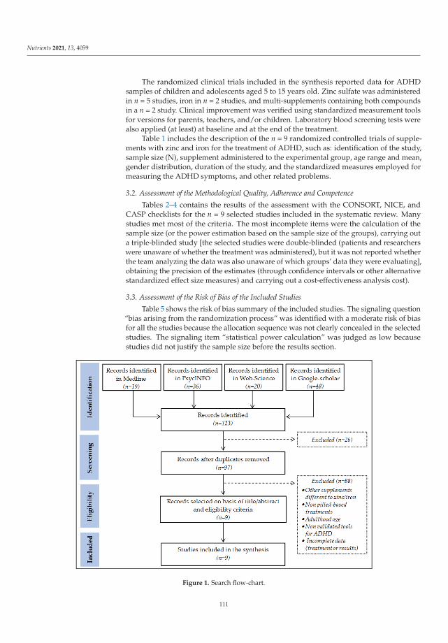
Nutrients 2021, 13, 4059
The randomized clinical trials included in the synthesis reported data for ADHDsamples of children and adolescents aged 5 to 15 years old. Zinc sulfate was administeredin n = 5 studies, iron in n = 2 studies, and multi-supplements containing both compoundsin a n = 2 study. Clinical improvement was verified using standardized measurement toolsfor versions for parents, teachers, and/or children. Laboratory blood screening tests werealso applied (at least) at baseline and at the end of the treatment.
Table 1 includes the description of the n = 9 randomized controlled trials of supple-ments with zinc and iron for the treatment of ADHD, such as: identification of the study,sample size (N), supplement administered to the experimental group, age range and mean,gender distribution, duration of the study, and the standardized measures employed formeasuring the ADHD symptoms, and other related problems.
3.2. Assessment of the Methodological Quality, Adherence and Competence
Tables 2–4 contains the results of the assessment with the CONSORT, NICE, andCASP checklists for the n = 9 selected studies included in the systematic review. Manystudies met most of the criteria. The most incomplete items were the calculation of thesample size (or the power estimation based on the sample size of the groups), carrying outa triple-blinded study [the selected studies were double-blinded (patients and researcherswere unaware of whether the treatment was administered), but it was not reported whetherthe team analyzing the data was also unaware of which groups’ data they were evaluating],obtaining the precision of the estimates (through confidence intervals or other alternativestandardized effect size measures) and carrying out a cost-effectiveness analysis cost).
3.3. Assessment of the Risk of Bias of the Included Studies
Table 5 shows the risk of bias summary of the included studies. The signaling question“bias arising from the randomization process” was identified with a moderate risk of biasfor all the studies because the allocation sequence was not clearly concealed in the selectedstudies. The signaling item “statistical power calculation” was judged as low becausestudies did not justify the sample size before the results section.
Figure 1. Search flow-chart.
111

Nutrients 2021, 13, 4059
Ta
ble
1.
Ran
dom
ized
cont
rolle
dtr
ials
ofsu
pple
men
tsw
ith
iron
and
zinc
for
AD
HD
.
Stu
dy
Re
fer.
NS
up
p.
Do
seA
ge
(Ye
ars
)S
ex
Du
rati
on
Me
asu
res
for
AD
HD
Re
sult
s
1.N
oora
zar
etal
.(20
20)
[69]
60Z
inc
10m
gzi
nc/d
ayR
ange
=7–
12M
ean
=9.
67
20%
Wom
en80
%M
en6
wee
ks•C
onne
rsPa
rent
’sQ
uest
ionn
aire
.Z
inc
rela
ted
wit
him
prov
emen
t,bu
tonl
yin
the
inat
tent
ion
fact
or
2.Z
amor
aet
al.
(201
1)[7
0]40
Zin
c10
mg
zinc
/day
Ran
ge=
7–14
Mea
n=
9.8
22.5
%W
omen
77.5
%M
en6
wee
ks•C
onne
rsR
atin
gSc
ales
-Rev
ised
.
Impr
ovem
enti
nth
eC
onne
r’s
scor
e,bu
ton
lyfo
rth
ete
ache
rve
rsio
n
3.A
rnol
det
al.
(201
1)[7
1]52
Zin
c15
mg
zinc
/day
Ran
ge=
6–14
Mea
n=
9.8
17.3
%W
omen
82.7
%M
en13
wee
ks
•Chi
lden
’sIn
terv
iew
for
Psyc
hiat
ric
Synd
rom
es(p
aren
tve
rsio
n).
•Par
enta
ndte
ache
rbe
havi
oral
rati
ngs.
•Clin
ical
Glo
balI
mpr
essi
ons
(CG
I).
•Con
ners
’Par
entR
atin
gSc
ale
•Neu
rops
ycho
logi
cal
cogn
itiv
e-m
otor
test
batt
ery.
Equi
voca
lres
ults
for
mos
tmea
sure
s.O
nly
neur
opsy
chol
ogic
alm
easu
res
mos
tly
favo
red
zinc
4.A
khon
dzad
ehet
al.(
2004
)[7
2]44
Zin
c15
mg
zinc
/day
Ran
ge=
5–11
Mea
n=
7.9
40.9
%W
omen
59.1
%M
en6
wee
ks•A
DH
Rat
ing
Scal
e(A
DH
D-R
S)Pa
rent
and
Teac
her
rati
ngsc
ale
scor
esim
prov
edw
ith
zinc
5.Bi
licie
tal.
(200
4)[7
3]40
0Z
inc
40m
gzi
nc/d
ayR
ange
=6–
14M
ean
=9.
6
18%
Wom
en82
%M
en12
wee
ks
•Att
enti
onD
efici
tHyp
erac
tivi
tyD
isor
der
Scal
e(A
DH
DS)
•Con
ners
Teac
her
Que
stio
nnai
re•D
uPau
lPar
entR
atin
gsof
AD
HD
Zin
cre
late
dw
ith
impr
ovem
ents
inhy
pera
ctiv
e,im
puls
ive
and
soci
aliz
atio
nsy
mpt
oms.
No
impa
ctof
zinc
was
obse
rved
for
the
atte
ntio
nde
ficit
leve
ls.A
mod
erat
oref
fect
with
age
and
BMI
was
obse
rved
6.K
onof
alet
al.
(200
8)[7
4]22
Iron
80m
g/da
y(f
erro
ussu
lfat
e,Ta
rdyf
eron
)
Ran
ge=
5–8
Mea
n=
5.9
21.7
%W
omen
78.3
%M
en12
wee
ks
•Con
ners
’Par
entR
atin
gSc
ale
•Con
ners
’Tea
cher
Rat
ing
Scal
e•A
tten
tion
Defi
citH
yper
acti
vity
Dis
orde
rR
atin
gSc
ale
(AD
HD
RS)
.•C
linic
alG
loba
lIm
pres
sion
-Sev
erit
y(C
GI-
S).
Iron
rela
ted
toim
prov
emen
ton
the
AD
HD
RS
scal
ean
dth
eC
GI-
Ssc
ore.
Iron
did
nota
chie
ved
impr
ovem
ents
onth
eC
onne
r’s
test
s
112

Nutrients 2021, 13, 4059
Ta
ble
1.
Con
t.
Stu
dy
Re
fer.
NS
up
p.
Do
seA
ge
(Ye
ars
)S
ex
Du
rati
on
Me
asu
res
for
AD
HD
Re
sult
s
7.Pa
naha
ndeh
etal
.(20
17)
[75]
42Ir
on5
mg/
kg/d
ay(f
erro
ussu
lfat
e)
Ran
ge=
5–15
Mea
n=
8.9
9.5%
Wom
en90
.5%
Men
8w
eeks
•Chi
ldSy
mpt
omIn
vent
ory-
4(C
SI-4
).
Iron
rela
ted
with
high
erde
crea
ses
onth
eC
SI-4
tota
land
fact
orsc
ores
8.R
uckl
idge
etal
.(20
18)
[76]
93Z
inc-
Iron
Zin
c(3
.2m
g/ca
psul
e)Ir
on(0
.9m
g/ca
psul
e).
Dos
e:st
arti
ng3
and
incr
easi
ngto
12ca
psul
es/d
ay
Ran
ge=
5–15
Mea
n=
9.7
23.7
%W
omen
76.3
%M
en10
wee
ks
•Con
ners
’Par
entR
atin
gSc
ale
(CPR
S-R
)•S
tren
gths
and
Diffi
cult
ies
Que
stio
nnai
re(S
DQ
,Par
ents
vers
ion)
•Str
engt
hsan
dD
ifficu
ltie
sQ
uest
ionn
aire
(SD
Q,T
each
ers
vers
ion)
Supp
lem
ents
rela
ted
wit
him
prov
emen
tsin
inat
tent
ive
leve
ls.N
oco
ntri
buti
onw
ere
obse
rved
onhy
pera
ctiv
e-im
puls
ive
sym
ptom
s.
9.R
uckl
idge
etal
.(20
21)
[58]
38Z
inc-
Iron
Zin
c(3
.2m
g/ca
psul
e)Ir
on(0
.9m
g/ca
psul
e).
Dos
e:st
arti
ng3
and
incr
easi
ngto
12ca
psul
es/d
ay
Ran
ge=
7–13
Mea
n=
10.1
21%
Wom
en79
%M
en10
wee
ks
•AD
HR
atin
gSc
ale
IV(A
DH
D-R
S-IV
).•C
hild
ren’
sD
epre
ssio
nR
atin
gSc
ale
(CD
RS)
•Chi
ldre
n’s
Glo
babl
Ass
essm
ent
Scal
e(C
GA
S)
Diff
eren
ces
inth
efe
rrit
inle
vels
achi
eved
anin
tera
ctio
nro
lefo
rim
prov
ing
the
AD
HD
seve
rity
leve
ls.
Not
e:N
=sa
mpl
esi
ze.S
uppl
em:s
uppl
emen
tadm
inis
tere
din
the
expe
rim
enta
lGro
up.R
efer
:cite
ofth
est
udy.
Dos
e:do
sage
for
the
supp
lem
ent(
zinc
and
iron
).R
esul
ts:r
elat
edw
ithth
esu
pple
men
t(zi
ncan
dir
on).
113

Nutrients 2021, 13, 4059
Ta
ble
2.
Ass
essm
ento
fthe
stud
ies’
qual
ity
base
don
the
CO
NSO
RT
chec
klis
t.
Title:IncludesDesignType
Abstract:Structured-Complete
Introduction:Background
Introduction:Objectives-Hypothesis
Methods:DesignDescribed
Methods:Participants
Methods:Interventions
Methods:Outcomes
Methods:Sample-Sizecalculation—Power
Methods:Randomization
Methods:Implementation
Methods:StatisticalProcedure
Results:ParticipantsFlow
Results:NumbersAnalyzed
Results:Outcomes-Estimates
Discussion:Limitations
Discussion:Generalization
Discussion:Interpretation
OtherRegistration-Protocol-Funding
1.N
oora
zar
etal
.(20
20)
[69]
(+)
(+)
(+)
(P)
(+)
(+)
(+)
(+)
(+)
(+)
(+)
(+)
(+)
(P)
(+)
(P)
(+)
(+)
2.Z
amor
aet
al.(
2011
)[7
0](+
)(+
)(+
)(P
)(+
)(+
)(+
)(+
)(+
)(+
)(+
)(+
)(+
)(P
)(?
)(P
)(+
)(+
)
3.A
rnol
det
al.(
2011
)[7
1](+
)(+
)(+
)(+
)(+
)(+
)(+
)(+
)(+
)(+
)(+
)(+
)(+
)(+
)(+
)(P
)(+
)(+
)
4.A
khon
dzad
ehet
al.(
2004
)[7
2](+
)(+
)(+
)(P
)(+
)(+
)(+
)(+
)(+
)(+
)(+
)(+
)(+
)(+
)(+
)(P
)(+
)(+
)
5.Bi
licie
tal.
(200
4)[7
3](+
)(+
)(+
)(+
)(+
)(+
)(+
)(+
)(+
)(+
)(+
)(+
)(P
)(P
)(?
)(P
)(+
)(+
)
6.K
onof
alet
al.(
2008
)[7
4](?
)(+
)(+
)(+
)(+
)(+
)(+
)(+
)(+
)(+
)(+
)(+
)(+
)(+
)(+
)(P
)(+
)(+
)
7.Pa
naha
ndeh
etal
.(20
17)
[75]
(?)
(+)
(+)
(P)
(+)
(+)
(+)
(+)
(+)
(+)
(+)
(+)
(+)
(+)
(+)
(P)
(+)
(+)
8.R
uckl
idge
etal
.(20
18)
[76]
(+)
(+)
(+)
(P)
(+)
(+)
(+)
(+)
(+)
(+)
(+)
(+)
(+)
(+)
(+)
(P)
(+)
(+)
9.R
uckl
idge
etal
.(20
21)
[58]
(?)
(+)
(+)
(P)
(+)
(+)
(+)
(+)
(+)
(+)
(+)
(+)
(+)
(+)
(+)
(P)
(+)
(+)
Not
e.(+
)Gre
enco
lor
cell:
pres
ente
d-re
port
ed.(
P)G
rey
colo
rce
ll:pa
rtia
llypr
esen
ted
orre
port
edw
ith
som
elim
itat
ions
.(?)
Whi
teco
lor
cell:
notp
rese
ntor
notr
epor
ted.
114

Nutrients 2021, 13, 4059
Table 3. Assessment of the studies’ quality based on the NICE checklist.
A1
.A
de
qu
ate
Ra
nd
om
iza
tio
nM
eth
od
A2
.A
de
qu
ate
Co
nce
alm
en
to
fA
llo
cati
on
A3
.G
rou
ps
Co
mp
ara
ble
at
Ba
seli
ne
B1
.C
om
pa
riso
nG
rou
ps
Re
ceiv
ed
Sa
me
Ca
re
B2
.P
art
icip
an
tsR
ece
ivin
gC
are
Bli
nd
toT
rea
tme
nt
B3
.In
div
idu
als
Ad
min
iste
rin
gC
are
Bli
nd
toA
llo
cati
on
C1
.A
llG
rou
ps
Fo
llo
we
dU
pfo
rE
qu
al
Le
ng
tho
fT
ime
C2
a.
Ho
wM
an
yP
art
icip
an
tsD
idN
ot
Co
mp
lete
Tre
atm
en
t
C2
b.
Gro
up
sC
om
pa
rab
lefo
rT
rea
tme
nt
Co
mp
leti
on
C3
.P
art
icip
an
tsin
Ea
chW
ere
No
Ou
tco
me
Da
taA
va
ila
ble
C3
b.
Gro
up
sC
om
pa
rab
leR
esp
ect
Av
ail
ab
ilit
yo
fO
utc
om
eD
ata
D1
.A
de
qu
ate
Le
ng
tho
fF
oll
ow
-Up
D2
.P
reci
seD
efi
nit
ion
of
Ou
tco
me
D3
.R
eli
ab
leM
eth
od
Use
dto
De
term
ine
the
Ou
tco
me
D4
.In
ve
stig
ato
rsK
ep
tB
lin
dto
Pa
rtic
ipa
nts
Ex
po
sure
D5
.In
ve
stig
ato
rsW
ere
Ke
pt
Bli
nd
toC
on
fou
nd
ing
-Pre
dic
tors
1. Noorazar et al. (2020) [69] (+) (+) (+) (+) (+) (+) (+) (+) (+) (+) (+) (+) (+) (+) (?) (?)
2. Zamora et al. (2011) [70] (+) (+) (+) (+) (+) (+) (+) (+) (+) (+) (+) (+) (+) (+) (?) (?)
3. Arnold et al. (2011) [71] (+) (+) (+) (+) (+) (+) (+) (+) (+) (+) (+) (+) (+) (+) (?) (?)
4. Akhondzadeh et al. (2004) [72] (+) (+) (+) (+) (+) (+) (+) (+) (+) (+) (+) (+) (+) (+) (?) (?)
5. Bilici et al. (2004) [73] (+) (+) (?) (+) (+) (+) (+) (+) (+) (+) (+) (+) (+) (+) (?) (?)
6. Konofal et al. (2008) [74] (+) (+) (+) (+) (+) (+) (+) (+) (+) (+) (+) (+) (+) (+) (?) (?)
7. Panahandeh et al. (2017) [75] (+) (+) (+) (+) (+) (+) (+) (+) (+) (+) (+) (+) (+) (+) (?) (?)
8. Rucklidge et al. (2018) [76] (+) (+) (+) (+) (+) (+) (+) (+) (+) (+) (+) (+) (+) (+) (?) (?)
9. Rucklidge et al. (2021) [58] (+) (+) (+) (+) (+) (+) (+) (+) (+) (+) (+) (+) (+) (+) (?) (?)
Note. (+) Green color cell: presented-reported. (?) White color cell: not present or not reported. Items A1 to A3: selection bias (systematicdifferences between the comparison groups). Items B1 to B3: performance bias (systematic differences between groups in the care provided,apart from the intervention under investigation). Items C1 to C3b: Attrition bias (systematic differences between the comparison groupswith respect to loss of participants). Items D1 to D5: Detection bias (bias in how outcomes are ascertained, diagnosed or verified).
115

Nutrients 2021, 13, 4059
Table 4. Assessment of the studies’ quality based on the CASP checklist.
A1
.Cle
arl
yF
ocu
sed
Qu
est
ion
A2
.U
seo
fR
an
do
miz
ati
on
Me
tho
d
A3
.P
art
icip
an
tsA
cco
un
ted
for
B4
.U
seo
f“
Bli
nd
ed
”M
eth
od
s
B5
.G
rou
ps
Sim
ila
ra
tth
eS
tart
of
Ra
nd
om
iza
tio
n
B6
.E
ach
Stu
dy
Gro
up
Re
ceiv
ed
the
Sa
me
Ca
re
C7
.E
ffe
cts
of
Inte
rve
nti
on
Ad
eq
ua
tely
Re
po
rte
d
C8
.P
reci
sio
nE
stim
ate
sR
ep
ort
ed
(CI
or
Oth
er
Eff
ect
Siz
es)
C9
.C
ost
-Eff
ect
ive
ne
ssA
na
lysi
sW
as
Do
ne
D1
0.
Ap
pli
cab
ilit
yo
fth
eR
esu
lts
D1
1.
Inte
rve
nti
on
Pro
vid
es
Va
lue
1. Noorazar et al. (2020) [69] (+) (+) (+) (+) (+) (+) (+) (?) (?) (+) (+)
2. Zamora et al. (2011) [70] (+) (+) (+) (+) (+) (+) (+) (?) (?) (+) (+)
3. Arnold et al. (2011) [71] (+) (+) (+) (+) (+) (+) (+) (+) (?) (+) (+)
4. Akhondzadeh et al. (2004) [72] (+) (+) (+) (+) (+) (+) (+) (?) (?) (+) (+)
5. Bilici et al. (2004) [73] (+) (+) (+) (+) (+) (?) (+) (?) (?) (+) (+)
6. Konofal et al. (2008) [74] (+) (+) (+) (+) (+) (+) (+) (+) (?) (+) (+)
7. Panahandeh et al. (2017) [75] (+) (+) (+) (+) (+) (+) (+) (?) (?) (+) (+)
8. Rucklidge et al. (2018) [76] (+) (+) (+) (+) (+) (+) (+) (+) (?) (+) (+)
9. Rucklidge et al. (2021) [58] (+) (+) (+) (+) (+) (+) (+) (+) (?) (+) (+)
Note. (+) Green color cell: present-reported; (?) White color cell: not present or not reported. Items A1 to A3: the study design is valid for arandomized controlled trial. Items B4 to B6: the study methodology is sound. Items C7 to C9: the results are adequately reported. ItemsD10 to D11: the interpretation of the results is helpful.
116

Nutrients 2021, 13, 4059
Table 5. Assessment of the risk of bias based on the Cochrane Risk of Bias 2 checklist.
Ra
nd
om
iza
tio
n
De
via
tio
ns
fro
mth
eIn
ten
de
dIn
terv
en
tio
n/s
Mis
sin
gO
utc
om
eD
ata
Ou
tco
me
Me
asu
rem
en
ts
Se
lect
ive
Re
po
rtin
g
Inco
mp
lete
Re
po
rtin
g
Stu
dy
Po
we
rC
alc
ula
tio
n/S
am
ple
Siz
eJu
stifi
cati
on
1. Noorazar et al. (2020) [69] (?) (+) (+) (+) (+) (+) (−)
2. Zamora et al. (2011) [70] (?) (+) (+) (+) (+) (+) (−)
3. Arnold et al. (2011) [71] (?) (+) (+) (+) (+) (+) (−)
4. Akhondzadeh et al. (2004) [72] (?) (+) (+) (+) (+) (+) (−)
5. Bilici et al. (2004) [73] (?) (+) (+) (+) (+) (+) (−)
6. Konofal et al. (2008) [74] (?) (+) (+) (+) (+) (+) (−)
7. Panahandeh et al. (2017) [75] (?) (+) (+) (+) (+) (+) (−)
8. Rucklidge et al. (2018) [76] (?) (+) (+) (+) (+) (+) (−)
9. Rucklidge et al. (2021) [58] (?) (+) (+) (+) (+) (+) (−)
Note. (+) Green color cell: low risk of bias. (?) Grey color cell: moderate risk of bias (unclear reporting or partially reported). (−) Browncolor cell: high risk of bias (non-reported). Randomization: low bias judged if both a method of randomization and concealing allocationwas clearly described, along with no clear obvious baseline differences between groups. Deviation: low bias judged if study participantsdid not change between groups. Missing outcome: low bias judged if less than 20% of participants were lost between the beginning andthe end of the treatment. Outcome measurement: low bias judged if validated tools were used for the main results (e.g., standardizedquestionnaires). Selective reporting: low bias judged if outcomes were pre-specified. Incomplete reporting: low bias judged if measurementmethods, procedure, analysis and outcomes were specified in advance to the results section. Power calculation/sample size justification:low bias judged if this was performed in advance and attained in the study.
3.4. Efficacy of Zinc for ADHD Treatment
The study of Noorazar and colleagues used the next intervention [69]: (a) a dose of0.5–1 mg/kg/day methylphenidate plus placebo in the control group; and (b) a dose of0.5–1 mg/kg/day methylphenidate plus 10 mg zinc (10 cc zinc sulfate syrup). No differ-ences between the groups were observed regarding the dose of methylphenidate. After6 weeks of treatment, the authors found that the use of zinc was useful to decrease theinattention scores (p = 0.02), but no differences between the groups were found in the othermeasures for the hyperactivity and impulsivity scales. The lack of differences in the totalADHD score in this study suggested that augmentation with zinc could only partiallyimprove ADHD severity levels. The authors of the study also noticed that the differencebetween the two groups regarding the gender ratio compromised the generalization capac-ity, and that the use of the Connors Parent’s Questionnaire as the only measure for ADHDwas a significant limitation [although it is a standardized tool, the inclusion of additionalinstruments and informants (such as teachers) could have provided different perspectivesof the children’s behaviors and impairments].
117

Nutrients 2021, 13, 4059
The study of Zamora and colleagues also valued the effect of zinc supplementation(10 mg/day) as an adjuvant therapy (complementary to methylphenidate, with a dose of0.3 mg/kg/day) for ADHD in n = 40 pediatric children, and obtained an improvement inADHD signs associated to the experimental group in the questionnaires answered by teach-ers (no differences emerged from the parents’ reports) [70]. These authors also obtaineddecreased zinc levels during the intervention in both groups (control and experimentalconditions), suggesting that the methylphenidate could contribute to a decrease in zincconcentration that can be counteracted with zinc supplementation.
In the research by Arnold et al. with n = 52 ADHD children treated with d-amphetamine(weight-standardized) and zinc complementation (15 mg/day or 30 mg/day) or d-amphet-amine and a placebo, a similar improvement in ADHD was observed in all the conditions(more precisely, teachers’ ratings showed medium effect sizes favoring zinc, but parents’reports showed an opposite trend favoring the placebo) [71]. Interestingly, this trial alsoobserved that the group treated with 30 mg/day zinc obtained 37% lower plasma levels ofconventional drugs. The authors of this trial concluded that rises in zinc levels in blood testssuggested that children with ADHD may have a zinc-wasting metabolism (defined as lowlevels of zinc related with potential deficits in absorption or losses in urine), and thereforethe zinc administered in the morning acted only as an immediate-release stimulant (thiswas probably the reason of the teachers’ reports favoring this dietary supplement) but wasnext immediately excreted by mid-afternoon (parents could not observe improvementson the children’s behaviors). As a global conclusion, Arnold and colleagues indicated thattheir study did not support zinc supplementation as a complementary tool for ADHDtreatments, but also suggested that before discarding zinc as a potential treatment, itwould be desirable that future research explores the situation further with different zincdoses/preparations, samples with children characterized for low zinc levels, and measuresfocused on neuropsychological tests as primary outcomes.
The study carried out by Akhondzadeh et al. aimed to compare two groups of patientswith ADHD (with or without zinc augmentation, with a dose of approximately 15 mg/day)and both conditions treated with methylphenidate (1 mg/kg/day), and the experimentalgroup obtained more improvement according to parents’ and teachers’ ratings at 6 weeksof the intervention [72]. Trend analyses also showed that differences between the groupsincreased during the treatment: while the placebo and the zinc supplementation groupsonly showed small differences at week 2, moderate to high differences were found at4 weeks and 6 weeks (this pattern was observed for the questionnaires answered byteachers and parents).
The study by Bilici and colleagues among a large sample of n = 400 children withADHD observed that the group receiving the zinc supplement (approximate dose of40 mg/day) compared with the control group (receiving placebo) obtained greater im-provements in multiple measures (hyperactivity, impulsivity, and socialization symptoms)but results were similar for attention deficiency [73]. The greatest differences between thegroups were observed at 12 weeks of the intervention, and were few during 1 to 4 weeks.Regarding the potential mechanisms explaining these results, the authors conclude thatthe zinc supplement could affect the conversion of dietary pyridoxine to its active form(pyrodoxal phosphate), implied in the process of conversion of tryptophan to serotonin. Inthis sense, zinc should contribute to the increase in serotonergic functions, contributingto a decrease in the characteristic of impulsivity as a symptom of ADHD. Alternatively,the authors also suggested the existence of a synergism of zinc in regulating dopamineand norepinephrine, which has been implied in ADHD treatments. The study by Biliciet al. also showed that pre/post differences in the ADHD measures were predicted by ageand BMI (a positive association was found in the B-slopes in regression analyses as higherage, and the BMI as higher change). Low pre-treatment zinc and free fatty acid valueswere also associated with a smaller decrease in ADHD symptom levels before and after thetreatments. This evidence was also useful to determine which children could benefit morefrom including zinc supplementations in the ADHD treatments.
118

Nutrients 2021, 13, 4059
3.5. Efficacy of Iron for ADHD Treatment
The study by Konofal and colleagues aimed to examine the contribution of iron sup-plementation (ferrous sulfate, 80 mg/day) in a sample of n = 23 children who met thecriteria for ADHD, which showed that the experimental group reported more improve-ment than the control group in different treatment outcomes [74]: ADHD symptom levelsreported by patients, parents, and teachers (particularly marked for the inattention factor),and also the presence of restless leg syndrome. The authors did not find a correlationbetween the baseline levels of serum ferritin levels with endpoint ADHD measures, sug-gesting that children with the lowest pre-treatment ferritin values did not benefit morefrom iron supplementation therapy than other children. In their conclusions, these authorssuggested that the effectiveness of ferrous sulfate could be associated with the ADHDpathophysiology, that the benefits of restless leg syndrome could be the consequence of theimprovement in the ADHD motor activity in the evening, and that iron could enhance theaction of pharmacological treatment with methylphenidate and amphetamine.
The study of Panahandeh et al. in a sample of n = 42 children with ADHD also obtainedbenefits for the use of ferrous sulfate (5 mg/kg/day) plus methylphenidate (1 mg/kg/day),in the inattentiveness, hyperactivity, and impulsive symptom levels reported by parentsat 2 months of treatment [75]. In the conclusions section, these authors supported thehypothesis that the contribution of iron supplementation on the ADHD improvementscould be explained by the capacity of this element in the dopamine transporter density andactivity, which is consistent with other studies that observed decreased thalamic iron levelsin children with ADHD compared to controls [77,78].
3.6. Efficacy of Including Simultaneously Iron and Zinc for ADHD Treatment
Some randomized clinical studies have also valued the contribution of multimineral-vitamin supplements which include zinc and iron for ADHD treatment in children. Therationale for selecting trails with multi-supplements therapy plans for ADHD was thelow number of trails using simultaneously zinc and iron, and those identified used prepa-rations with vitamins, dietary minerals and other nutritional elements. Therefore, con-sidering these studies facilitates generalizability of the results related with the use ofzinc–iron supplements.
The study of Rucklidge and colleagues [76] in a sample of n = 93 patients used DailyEssential Nutrients (DEN, with Recommended Dietary Allowances, RDA), which containsa comprehensive range of micronutrients (13 vitamins, 17 minerals, and 4 amino acids). Theparticipants were instructed to titrate the dose up to over a week, starting with 3 capsulesper day and increasing it to up to 12 capsules per day (see Table 1 for the zinc–iron content).This study obtained an intent-to-treat analysis of between-group differences with greaterimprovements for ADHD related with the micronutrient treatment, but specifically inthe clinicians’ reports assessing inattention. However, although no significant differenceswere found for the hyperactivity and impulsive measures between the experimental andthe placebo groups, the micronutrients supplement obtained improvements in emotionalregulation, aggression, and general functioning. The authors of this study concluded thatthe mechanisms of action of micronutrient treatments likely involves different pathwaysrather than pharmacological treatments, and that supplements like those used in the studymay have an impact on the methylation/methionine cycle, required for the synthesis ofDNA/RNA (as it was suggested by other studies [79]), and on the citric acid cycle andelectron transport chain acting as co-enzymes in mitochondrial aerobic respiration andenergy production. These potential effects on the physical state could also contribute to im-provements in mood state and cognition functions (also suggested in the literature [80,81]).
Finally, the study of Rucklidge et al. was published later and carried out on a sampleof n = 38 children that also used DEN, with an initial dose of 3 capsules per day whichfurther increased to 12 capsules per day (see Table 1 for the zinc–iron content). This studyshowed that a broad-spectrum micronutrient formula (EMPowerplus) served as a mediatorin the treatment response considering multiple ADHD measures [58]. Interestingly, only
119

Nutrients 2021, 13, 4059
pre/post changes in the copper and ferritin levels achieved a significant moderator role onthe improvement registered for the severity of ADHD: as the increase in the ferritin andthe decrease in the copper highly benefited the levels of ADHD (in the three dimensions ofinattentiveness, hyperactivity, and impulsivity). However, the authors indicated that theseresults should be interpreted with caution, and outlined the need to consider individualvariability: since metabolic needs are different between the patients, as well as their geneticmakeup, some children may need certain nutrients to restore optimal metabolic functioning,and others will not require specific nutrients and should not benefit from supplementations.In addition, nutrients act in a synergistic way, and therefore, changes in one can havecascading effects on others, and it was thus unlikely that increases in a specific nutrientcould make a direct and unique contribution on the changes observed in a complex disorder,such as ADHD.
3.7. Tolerability Analyses
Regarding safety and tolerability, no serious adverse events were reported in thestudies selected for this review, and the unpleasant events found were similar for theexperimental and control groups. Iron and zinc therapies were generally well-tolerated:no patients referred to an exacerbation of ADHD symptoms or a significant decrease inappetite, and only a low number of children reported physical pains, nausea, vomiting,constipation, or a metallic taste in the mouth. Moreover, studies suggested that maintaininga fixed daily dose of iron–zinc was a relevant factor in increasing tolerability, and noindication regarding any great concern about the doses up to the follow-up was registered.However, it cannot be determined whether a long-term zinc–iron treatment could inducenegative events, such as toxicity or hemosiderosis.
4. Discussion
This study conducted a qualitative systematic review of the empirical evidence ob-tained in randomized clinical trials published since 2000 on the efficacy of zinc and ironsupplementation among ADHD children and adolescents. Nine studies met the eligibilitycriteria and were selected. Results indicated that low zinc and iron levels were relatedwith both higher baseline levels of ADHD severity and poorer treatment outcomes. Com-pared with the controlled-placebo conditions, the dietary supplements with zinc and ironwere associated with improvements in ADHD severity at the end of the treatments, al-though the effect size of the outcomes tended to be low and/or focused on specific ADHDsymptoms/measures.
Cross-sectional evidence regarding the association of zinc and iron with ADHD levels(pre-treatment associations) is consistent with other non-randomized studies that exploredthe contribution of these micronutrients within clinical- and population-based samples.For example, a longitudinal study with a large sample with n = 608 children found thatzinc–iron supplementation improved attention/concentration in children over a 14-monthfollow-up period [82]. A positive correlation was also found between zinc and symptomsof inattentiveness in a cross-sectional study of n = 48 children [83].
The potential explanations about the paths between the zinc–iron levels and thevarious ADHD manifestations are discussed in a number of studies, which showed thatlower levels of these elements in childhood (but also in older adults) could be relatedto a dopaminergic impairment, which could be the origin of the high inattentiveness–hyperactivity levels and the other correlates of the ADHD disease (like the presence ofrestless leg syndrome and other multiple sleep problems) [74,84,85]. Neuroimaging, genetic,and animal studies have also shown that decreases in dopamine transporter densities arerelated with decreased brain iron and zinc levels [86–88]. Zinc achieves a relevant role inthe metabolism of neurotransmitters, prostaglandins, and for maintaining brain structureand function. Since zinc is necessary in the metabolism of melatonin, it is strongly impliedin the regulation of dopamine (zinc is therefore considered a dopamine reuptake inhibitor).Iron is a cofactor in the tyrosine hydroxylase enzyme, and a key cofactor in the making
120

Nutrients 2021, 13, 4059
of neurotransmitters, including serotonin, norepinephrine, and dopamine. Brain iron-deficient rodents have been associated with alterations in adenosine neurotransmission,which has provided a pathogenetic link with glutamate mechanisms and the hypersensitivecorticostriatal terminals involved in some ADHD correlates [89,90].
The link between iron and zinc supplementation and the observed decrease in ADHDsymptoms in the controlled clinical trials selected for this systematic review also makessense with regard to the role of zinc and iron as dopamine reuptake inhibitors, which wasprecisely the core target of the stimulant medications used in the combined treatment plansfor the disease. Other non-randomized studies using a combination of polyunsaturatedfatty acids with zinc supplementation also showed benefits for the treatment of ADHD.For example, the observational study of Huss et al. in a large sample of n = 810 monitoredchildren aged 5–12 years old showed that consumption of omega-3 and omega-6 withcomplementation of zinc improved ADHD symptoms, as well as other emotional problems,and sleeping disease (attention deficit, hyperactivity, and impulsivity) after 12 weeks [91].The rationale for these results could be found in the capacity of these dietary combinationsfor facilitating the transmission of signals between neurons, which also contributed toimprovements in attention ability, learning difficulties, and executive functions in thedevelopment of children and adolescents [92,93].
The studies included in this review support the existence of subgroups (based on thetreatment outcomes and its baseline clinical correlates) that could be particularly benefittedfrom treatments including dietary zinc and iron supplements, such as the results of Biliciand colleagues [73]: the association found in this study between the low baseline zincvalues with lower improvements in the ADHD measures suggested that zinc may beparticularly important for the treatment of this disorder, improving and compensatingborderline zinc nutrition. The study of Arnold and colleagues, which reanalyzed data froma randomized clinical trial with n = 18 children by comparing d-amphetamine with Efamol,assessed the moderate role of zinc [94], categorized the participants into three groups (aszinc-adequate, zinc-borderline, and zinc-deficient), and observed a linear relationship ofd-amphetamine with zinc nutrition among the d-amphetamine group, and a quadratic(U-shaped) relationship of Efamol response to zinc (the benefit among this group was onlyfound among the zinc-borderline condition). Finally, the research based on a secondaryanalysis of data collected in the study of Arnold and colleagues [71] analyzed the role ofthe status of iron, and determined that it was associated with ADHD symptom severity atbaseline and with responses to psycho-stimulant treatment [95]. This study found an in-verse correlation of serum ferritin with baseline inattention, hyperactivity, and impulsivitylevels (as ferritin concentration increased, ADHD severity decreased), and also a significantcorrelation between serum ferritin and sensitivity to the amphetamine used for ADHDtreatment (participants with higher ferritin concentrations required lower weight-adjusteddoses of psychotropic treatment). These results were obtained after adjusting for the partic-ipants’ sex and age. The authors concluded that iron supplementation could contribute as apotential intervention to optimize responses to psycho-stimulants in children with low ironstores at baseline. Other studies have also showed that ferritin concentration is correlatedwith the dose of amphetamine necessary to reach an optimal clinical response [96]. How-ever, results in this area are few, and future research should address the question of whichare the optimal doses, as well as the benefits of using supplements of multiple vitaminsand minerals.
5. Conclusions
Based on the results obtained in this systematic review, the specific role of dietarynutrients with zinc and iron still seems controversial for the treatment of ADHD, beingmost consistent with the evidence for zinc. Moreover, although the reviewed studies founda relationship between the use of dietary supplements containing these elements with theimprovement of ADHD symptoms, neither the mono-causal role of a concrete specificnutritional deficiency among ADHD children nor the role of a concrete dietary nutrient in
121

Nutrients 2021, 13, 4059
the management of this disorder were proven (as was reported quite recently [56]). Futurecontrolled clinical trials are needed, examining the efficacy of mineral supplementation.
The results obtained in this systematic review should be considered for combiningthese multi-facet interventions with additional precise interventions, specifically focusedon the nutritional state and clinical profile of each patient. The ultimate objective of thesetreatments, including medical, psychological, and diet plans, should be to decrease thelevels of ADHD, but also its multiple negative correlates, in order to achieve better perfor-mance in vigilance and sustained attention tasks, executive functioning tasks (like planningand organization), set shifting and response monitoring, and complex problem-solving.
Finally, this study should be interpreted in consideration of certain limitations. First,a meta-analysis was not conducted. Systematic reviews of treatment plans often includestatistical methods to summarize the results of independent research by combining theinformation of selected studies (through meta-analysis) with the aim to provide moreprecise estimates of the effects of health care. However, it must be outlined that not alltopics allow a meta-analysis to be conducted, and these concrete systematic reviews usealternative methods of synthesis described as “narrative synthesis”. This was the caseof our study: we did not employ meta-analyses because of the clinical heterogeneity ofindividual studies, with different intervention characteristics (for example, the iron/zincsupplements were administered in two trials within multi-supplements also includingother trace elements, with different doses and/or frequency of doses, and with different du-rations), diverse outcome/effects measurement, different research settings, and differencesin participants’ features (such as age, baseline disease severity, other comorbid conditions,or sociodemographic variables). Since current studies have proven that this heterogeneitycould cause significant inaccurate summary effects and associated conclusions, and thusmisleading decision-markers [97], we opted to conduct a narrative synthesis in accordancewith the current guidelines. Other characteristics of the individual studies selected forthis review also impact on the generalizability capacity of our review: the limited samplessizes, the convenience samples (non-probability sampling methods were used), and thefrequency of men compared to women.
Supplementary Materials: The following are available online at https://www.mdpi.com/article/10.3390/nu13114059/s1, Table S1: PRISMA 2020 main checklist.
Author Contributions: Conceptualization: R.G. and G.G.R.-O.; formal statistical analysis: I.L.C.-T.,A.A.R.-C. and P.C.M.-S.; writing—original draft preparation: R.G. and G.G.R.-O.; writing—reviewand editing, I.L.C.-T., A.A.R.-C., P.C.M.-S. and A.P.-G. All authors contributed substantially to thedata analysis, interpretation of data. All authors have read and agreed to the published version ofthe manuscript.
Funding: This research received no external funding.
Conflicts of Interest: The authors declare no conflict of interest.
References
1. Di Lorenzo, R.; Balducci, J.; Poppi, C.; Arcolin, E.; Cutino, A.; Ferri, P.; D’Amico, R.; Filippini, T. Children and adolescents withADHD followed up to adulthood: A systematic review of long-term outcomes. Acta Neuropsychiatr. 2021, 1–42. [CrossRef]
2. American Psychiatric Association. Diagnostic and Statistical Manual of Mental Disorders: DSM-5, 5th ed.; American PsychiatricAssociation: Washington, DC, USA, 2013.
3. Chan, M.F.; Al Balushi, R.; Al Falahi, M.; Mahadevan, S.; Al Saadoon, M.; Al-Adawi, S. Child and adolescent mental healthdisorders in the GCC: A systematic review and meta-analysis. Int. J. Pediatr. Adolesc. Med. 2021, 8, 134–145. [CrossRef] [PubMed]
4. Polanczyk, G.V.; Salum, G.A.; Sugaya, L.S.; Caye, A.; Rohde, L.A. Annual Research Review: A meta-analysis of the worldwideprevalence of mental disorders in children and adolescents. J. Child Psychol. Psychiatry 2015, 56, 345–365. [CrossRef]
5. Sayal, K.; Prasad, V.; Daley, D.; Ford, T.; Coghill, D. ADHD in children and young people: Prevalence, care pathways, and serviceprovision. Lancet Psychiatry 2018, 5, 175–186. [CrossRef]
6. Willis, R.; Dhakras, S.; Cortese, S. Attention-Deficit/Hyperactivity Disorder in Looked-After Children: A Systematic Review ofthe Literature. Curr. Dev. Disord. Rep. 2017, 4, 78–84. [CrossRef] [PubMed]
7. Dobrosavljevic, M.; Solares, C.; Cortese, S.; Andershed, H.; Larsson, H. Prevalence of attention-deficit/hyperactivity disorder inolder adults: A systematic review and meta-analysis. Neurosci. Biobehav. Rev. 2020, 118, 282–289. [CrossRef]
122

Nutrients 2021, 13, 4059
8. Song, P.; Zha, M.; Yang, Q.; Zhang, Y.; Li, X.; Rudan, I. The prevalence of adult attention-deficit hyperactivity disorder: A globalsystematic review and meta-analysis. J. Glob. Health 2021, 11, 04009. [CrossRef]
9. Lynch, S.J.; Sunderland, M.; Newton, N.C.; Chapman, C. A systematic review of transdiagnostic risk and protective factors forgeneral and specific psychopathology in young people. Clin. Psychol. Rev. 2021, 87, 102036. [CrossRef]
10. Bruxel, E.M.; Moreira-Maia, C.R.; Akutagava-Martins, G.C.; Quinn, T.P.; Klein, M.; Franke, B.; Ribasés, M.; Rovira, P.;Sánchez-Mora, C.; Kappel, D.B.; et al. Meta-analysis and systematic review of ADGRL3 (LPHN3) polymorphisms in ADHDsusceptibility. Mol. Psychiatry 2020, 26, 2277–2285. [CrossRef] [PubMed]
11. Kang, P.; Luo, L.; Peng, X.; Wang, Y. Association of Val158Met polymorphism in COMT gene with attention-deficit hyperactivedisorder: An updated meta-analysis. Medicine 2020, 99, e23400. [CrossRef]
12. McNeill, R.V.; Ziegler, G.C.; Radtke, F.; Nieberler, M.; Lesch, K.-P.; Kittel-Schneider, S. Mental health dished up—the use of iPSCmodels in neuropsychiatric research. J. Neural Transm. 2020, 127, 1547–1568. [CrossRef]
13. Ronald, A.; de Bode, N.; Polderman, T.J. Systematic Review: How the Attention-Deficit/Hyperactivity Disorder Polygenic RiskScore Adds to Our Understanding of ADHD and Associated Traits. J. Am. Acad. Child Adolesc. Psychiatry 2021, 60, 1234–1277.[CrossRef]
14. Schuch, V.; Utsumi, D.A.; Costa, T.V.M.M.; Kulikowski, L.D.; Muszkat, M. Attention Deficit Hyperactivity Disorder in the Lightof the Epigenetic Paradigm. Front. Psychiatry 2015, 6, 126. [CrossRef]
15. Walton, E.; Pingault, J.-B.; Cecil, C.A.; Gaunt, T.R.; Relton, C.C.; Mill, J.; Barker, E.D. Epigenetic profiling of ADHD symptomstrajectories: A prospective, methylome-wide study. Mol. Psychiatry 2016, 22, 250–256. [CrossRef]
16. Han, V.X.; Patel, S.; Jones, H.F.; Nielsen, T.C.; Mohammad, S.S.; Hofer, M.J.; Gold, W.; Brilot, F.; Lain, S.J.; Nassar, N.; et al.Maternal acute and chronic inflammation in pregnancy is associated with common neurodevelopmental disorders: A systematicreview. Transl. Psychiatry 2021, 11, 1–12. [CrossRef]
17. Li, L.; Lagerberg, T.; Chang, Z.; Cortese, S.; Rosenqvist, M.A.; Almqvist, C.; D’Onofrio, B.M.; Hegvik, T.-A.; Hartman, C.; Chen,Q.; et al. Maternal pre-pregnancy overweight/obesity and the risk of attention-deficit/hyperactivity disorder in offspring: Asystematic review, meta-analysis and quasi-experimental family-based study. Int. J. Epidemiol. 2020, 49, 857–875. [CrossRef]
18. Qu, A.; Cao, T.; Li, Z.; Wang, W.; Liu, R.; Wang, X.; Nie, Y.; Sun, S.; Liu, X.; Zhang, X. The association between maternalperfluoroalkyl substances exposure and early attention deficit hyperactivity disorder in children: A systematic review andmeta-analysis. Environ. Sci. Pollut. Res. 2021, 1–16. [CrossRef]
19. Bogicevic, L.; Verhoeven, M.; Van Baar, A.L. Distinct Profiles of Attention in Children Born Moderate-to-Late Preterm at 6 Years. J.Pediatr. Psychol. 2020, 45, 685–694. [CrossRef]
20. Walczak-Kozłowska, T.; Mankowska, A.; Chrzan-Detkos, M.; Harciarek, M. Attentional system of very prematurely bornpreschoolers. Dev. Psychol. 2020, 56, 251–260. [CrossRef]
21. Carlsson, T.; Molander, F.; Taylor, M.J.; Jonsson, U.; Bölte, S. Early environmental risk factors for neurodevelopmental disorders—Asystematic review of twin and sibling studies. Dev. Psychopathol. 2020, 33, 1448–1495. [CrossRef]
22. Nielsen, T.M.; Pedersen, M.V.; Milidou, I.; Glavind, J.; Henriksen, T.B. Long-term cognition and behavior in children born at earlyterm gestation: A systematic review. Acta Obstet. Et Gynecol. Scand. 2019, 98, 1227–1234. [CrossRef]
23. Rivollier, F.; Krebs, M.-O.; Kebir, O. Perinatal Exposure to Environmental Endocrine Disruptors in the Emergence of Neurodevel-opmental Psychiatric Diseases: A Systematic Review. Int. J. Environ. Res. Public Health 2019, 16, 1318. [CrossRef]
24. Serati, M.; Barkin, J.L.; Orsenigo, G.; Altamura, A.C.; Buoli, M. Research Review: The role of obstetric and neonatal complicationsin childhood attention deficit and hyperactivity disorder—A systematic review. J. Child Psychol. Psychiatry 2017, 58, 1290–1300.[CrossRef]
25. Kalantary, R.R.; Jaffarzadeh, N.; Rezapour, M.; Arani, M.H. Association between exposure to polycyclic aromatic hydrocarbonsand attention deficit hyperactivity disorder in children: A systematic review and meta-analysis. Environ. Sci. Pollut. Res. 2020, 27,11531–11540. [CrossRef] [PubMed]
26. Dreier, J.W.; Andersen, A.-M.N.; Hvolby, A.; Garne, E.; Andersen, P.K.; Berg-Beckhoff, G. Fever and infections in pregnancy andrisk of attention deficit/hyperactivity disorder in the offspring. J. Child Psychol. Psychiatry 2015, 57, 540–548. [CrossRef]
27. Nikolas, M.A.; Nigg, J.T. Moderators of Neuropsychological Mechanism in Attention-Deficit Hyperactivity Disorder. J. Abnorm.Child Psychol. 2014, 43, 271–281. [CrossRef] [PubMed]
28. Hirjak, D.; Meyer-Lindenberg, A.; Fritze, S.; Sambataro, F.; Kubera, K.M.; Wolf, R.C. Motor dysfunction as research domain acrossbipolar, obsessive-compulsive and neurodevelopmental disorders. Neurosci. Biobehav. Rev. 2018, 95, 315–335. [CrossRef]
29. Schulze, M.; Coghill, D.; Lux, S.; Philipsen, A. Disentangling ADHD’s Presentation-Related Decision-Making—A Meta-AnalyticApproach on Predominant Presentations. Front. Psychiatry 2021, 12, 519840. [CrossRef] [PubMed]
30. Curtis, L.T.; Patel, K. Nutritional and Environmental Approaches to Preventing and Treating Autism and Attention DeficitHyperactivity Disorder (ADHD): A Review. J. Altern. Complement. Med. 2008, 14, 79–85. [CrossRef]
31. Azadbakht, L.; Hariri, M. Magnesium, iron, and zinc supplementation for the treatment of attention deficit hyperactivity disorder:A systematic review on the recent literature. Int. J. Prev. Med. 2015, 6, 83. [CrossRef]
32. Rucklidge, J.J.; Frampton, C.M.; Gorman, B.; Boggis, A. Vitamin–mineral treatment of attention-deficit hyperactivity disorder inadults: Double-blind randomised placebo-controlled trial. Br. J. Psychiatry 2014, 204, 306–315. [CrossRef] [PubMed]
33. Shahidullah, J.D.; Carlson, J.S.; Haggerty, D.; Lancaster, B.M. Integrated care models for ADHD in children and adolescents: Asystematic review. Fam. Syst. Health 2018, 36, 233–247. [CrossRef]
123

Nutrients 2021, 13, 4059
34. Sultan, M.A.; Pastrana, C.S.; Pajer, K.A. Shared Care Models in the Treatment of Pediatric Attention-Deficit/HyperactivityDisorder (ADHD): Are They Effective? Health Serv. Res. Manag. Epidemiol. 2018, 5, 1–7. [CrossRef]
35. Boland, H.; DiSalvo, M.; Fried, R.; Woodworth, K.Y.; Wilens, T.; Faraone, S.V.; Biederman, J. A literature review and meta-analysison the effects of ADHD medications on functional outcomes. J. Psychiatr. Res. 2020, 123, 21–30. [CrossRef] [PubMed]
36. Weyandt, L.; Oster, D.; Marraccini, M.E.; Gudmundsdottir, B.; Munro, B.; Zavras, B.M.; Kuhar, B. Pharmacological interventionsfor adolescents and adults with ADHD: Stimulant and nonstimulant medications and misuse of prescription stimulants. Psychol.Res. Behav. Manag. 2014, 7, 223–249. [CrossRef]
37. Tsujii, N.; Okada, T.; Usami, M.; Kuwabara, H.; Fujita, J.; Negoro, H.; Kawamura, M.; Iida, J.; Saito, T. Effect of Continuing andDiscontinuing Medications on Quality of Life After Symptomatic Remission in Attention-Deficit/Hyperactivity Disorder: ASystematic Review and Meta-Analysis. J. Clin. Psychiatry 2020, 81, 19r13015. [CrossRef] [PubMed]
38. de la Cruz, L.F.; Simonoff, E.; McGough, J.J.; Halperin, J.M.; Arnold, L.E.; Stringaris, A. Treatment of Children with Attention-Deficit/Hyperactivity Disorder (ADHD) and Irritability: Results from the Multimodal Treatment Study of Children with ADHD(MTA). J. Am. Acad. Child Adolesc. Psychiatry 2014, 54, 62–70. [CrossRef]
39. Weiss, M.D.; Salpekar, J. Sleep Problems in the Child with Attention-Deficit Hyperactivity Disorder: Defining Aetiology andAppropriate Treatments. CNS Drugs 2010, 24, 811–828. [CrossRef]
40. Chamorro, M.; Lara, J.P.; Insa, I.; Espadas, M.; Alda-Diez, J.A. Evaluacion y tratamiento de los problemas de sueño en niñosdiagnosticados de trastorno por deficit de atencion/hiperactividad: Actualizacion de la evidencia [Evaluation and treatment ofsleep problems in children diagnosed with attention deficit hyperactivity disorder: An update of the evidence]. Rev. Neurol. 2017,64, 413–421.
41. DelRosso, L.M.; Mogavero, M.P.; Baroni, A.; Bruni, O.; Ferri, R. Restless Legs Syndrome in Children and Adolescents. ChildAdolesc. Psychiatr. Clin. North Am. 2020, 30, 143–157. [CrossRef]
42. Baxter, A.J.; Patton, G.; Scott, K.M.; Degenhardt, L.; Whiteford, H.A. Global Epidemiology of Mental Disorders: What Are WeMissing? PLoS ONE 2013, 8, e65514. [CrossRef]
43. Polanczyk, G.V.; Willcutt, E.G.; Salum, G.A.; Kieling, C.; Rohde, L.A. ADHD prevalence estimates across three decades: Anupdated systematic review and meta-regression analysis. Int. J. Epidemiol. 2014, 43, 434–442. [CrossRef]
44. Thomas, R.; Sanders, S.; Doust, J.; Beller, E.; Glasziou, P. Prevalence of Attention-Deficit/Hyperactivity Disorder: A SystematicReview and Meta-analysis. Pediatrics 2015, 135, e994–e1001. [CrossRef]
45. Logan, A.C.; Jacka, F.N. Nutritional psychiatry research: An emerging discipline and its intersection with global urbanization,environmental challenges and the evolutionary mismatch. J. Physiol. Anthr. 2014, 33, 1–16. [CrossRef]
46. Agostoni, C.; Nobile, M.; Ciappolino, V.; DelVecchio, G.; Tesei, A.; Turolo, S.; Crippa, A.; Mazzocchi, A.; Altamura, C.A.; Brambilla,P.; et al. The Role of Omega-3 Fatty Acids in Developmental Psychopathology: A Systematic Review on Early Psychosis, Autism,and ADHD. Int. J. Mol. Sci. 2017, 18, 2608. [CrossRef] [PubMed]
47. Nigg, J.T.; Lewis, K.; Edinger, T.; Falk, M. Meta-Analysis of Attention-Deficit/Hyperactivity Disorder or Attention-Deficit/Hyperactivity Disorder Symptoms, Restriction Diet, and Synthetic Food Color Additives. J. Am. Acad. ChildAdolesc. Psychiatry 2012, 51, 86–97. [CrossRef]
48. Torp, N.M.U.; Thomsen, P.H. The use of diet interventions to treat symptoms of ADHD in children and adolescents—A systematicreview of randomized controlled trials. Nord. J. Psychiatry 2020, 74, 558–568. [CrossRef] [PubMed]
49. Rytter, M.J.H.; Andersen, L.B.B.; Houmann, T.; Bilenberg, N.; Hvolby, A.; Mølgaard, C.; Michaelsen, K.F.; Lauritzen, L. Diet in thetreatment of ADHD in children—A systematic review of the literature. Nord. J. Psychiatry 2014, 69, 1–18. [CrossRef] [PubMed]
50. Bloch, M.H.; Mulqueen, J. Nutritional Supplements for the Treatment of ADHD. Child Adolesc. Psychiatr. Clin. N. Am. 2014, 23,883–897. [CrossRef]
51. Verlaet, A.A.J.; Maasakkers, C.M.; Hermans, N.; Savelkoul, H.F.J. Rationale for Dietary Antioxidant Treatment of ADHD. Nutrients2018, 10, 405. [CrossRef]
52. Del-Ponte, B.; Quinte, G.C.; Cruz, S.; Grellert, M.; Santos, I.S. Dietary patterns and attention deficit/hyperactivity disorder(ADHD): A systematic review and meta-analysis. J. Affect. Disord. 2019, 252, 160–173. [CrossRef]
53. Maret, W.; Sandstead, H.H. Zinc requirements and the risks and benefits of zinc supplementation. J. Trace Elem. Med. Biol. 2006,20, 3–18. [CrossRef]
54. Avcil, S.; Uysal, P.; Yenisey, Ç.; Abas, B.I. Elevated Melatonin Levels in Children With Attention Deficit Hyperactivity Disorder:Relationship to Oxidative and Nitrosative Stress. J. Atten. Disord. 2019, 25, 693–703. [CrossRef]
55. Abbaspour, N.; Hurrell, R.; Kelishadi, R. Review on iron and its importance for human health. J. Res. Med Sci. Isfahan Univ. MedSci. 2014, 19, 164–174.
56. Robberecht, H.; Verlaet, A.A.J.; Breynaert, A.; De Bruyne, T.; Hermans, N. Magnesium, Iron, Zinc, Copper and Selenium Status inAttention-Deficit/Hyperactivity Disorder (ADHD). Molecules 2020, 25, 4440. [CrossRef]
57. Rucklidge, J.J.; Eggleston, M.J.; Darling, K.A.; Stevens, A.J.; Kennedy, M.A.; Frampton, C.M. Can we predict treatment responsein children with ADHD to a vitamin-mineral supplement? An investigation into pre-treatment nutrient serum levels, MTHFRstatus, clinical correlates and demographic variables. Prog. Neuro-Psychopharmacol. Biol. Psychiatry 2018, 89, 181–192. [CrossRef]
58. Rucklidge, J.J.; Eggleston, M.J.F.; Boggis, A.; Darling, K.; Gorman, B.; Frampton, C.M. Do Changes in Blood Nutrient LevelsMediate Treatment Response in Children and Adults with ADHD Consuming a Vitamin–Mineral Supplement? J. Atten. Disord.2019, 25, 1107–1119. [CrossRef]
124

Nutrients 2021, 13, 4059
59. Fanjiang, G.; Kleinman, E.R. Nutrition and performance in children. Curr. Opin. Clin. Nutr. Metab. Care 2007, 10, 342–347.[CrossRef]
60. Rucklidge, J.J.; Johnstone, J.; Kaplan, B.J. Nutrient supplementation approaches in the treatment of ADHD. Expert Rev. Neurother.2009, 9, 461–476. [CrossRef]
61. Page, M.J.; McKenzie, J.E.; Bossuyt, P.M.; Boutron, I.; Hoffmann, T.C.; Mulrow, C.D.; Shamseer, L.; Tetzlaff, J.M.; Akl, E.A.;Brennan, S.E.; et al. The PRISMA 2020 statement: An updated guideline for reporting systematic reviews. PLoS Med. 2021,18, e1003583. [CrossRef]
62. Shea, B.J.; Grimshaw, J.M.; Wells, G.A.; Boers, M.; Andersson, N.; Hamel, C.; Porter, A.C.; Tugwell, P.; Moher, D.; Bouter, L.M.Development of AMSTAR: A measurement tool to assess the methodological quality of systematic reviews. BMC Med Res.Methodol. 2007, 7, 1–7. [CrossRef] [PubMed]
63. Higgins, J.P.; Green, S. Cochrane Handbook for Systematic Reviews of Interventions Version 5.1.0. The Cochrane Collaboration.2011. Available online: https://handbook.cochrane.org (accessed on 15 July 2021).
64. American Psychiatric Association. Diagnostic and Statistical Manual of Mental Disorders: DSM-IV-R; American PsychiatricAssociation: Washington, DC, USA, 2000.
65. Welch, R.W.; Antoine, J.-M.; Berta, J.-L.; Bub, A.; De Vries, J.; Guarner, F.; Hasselwander, O.; Hendriks, H.; Jäkel, M.; Koletzko,B.V.; et al. Guidelines for the design, conduct and reporting of human intervention studies to evaluate the health benefits of foods.Br. J. Nutr. 2011, 106, S3–S15. [CrossRef] [PubMed]
66. National Institute for Health and Care Excellence, NICE. Methods for the Development of NICE Public Health Guidance. 2012.Available online: https://www.nice.org.uk/process/pmg4/resources/methods-for-the-development-of-nice-public-health-guidance-third-edition-pdf-2007967445701 (accessed on 15 July 2021).
67. Critical Appraisal Skills Programme, CASP. Randomized Controlled Trials Checklist. 2020. Available online: https://casp-uk.net/casp-tools-checklists (accessed on 15 July 2021).
68. Sterne, J.A.C.; Savovic, J.; Page, M.J.; Elbers, R.G.; Blencowe, N.S.; Boutron, I.; Cates, C.J.; Cheng, H.-Y.; Corbett, M.S.; Eldridge,S.M.; et al. RoB 2: A revised tool for assessing risk of bias in randomised trials. BMJ 2019, 366, l4898. [CrossRef]
69. Noorazar, S.G.; Malek, A.; Aghaei, S.M.; Yasamineh, N.; Kalejahi, P. The efficacy of zinc augmentation in children with attentiondeficit hyperactivity disorder under treatment with methylphenidate: A randomized controlled trial. Asian J. Psychiatry 2019,48, 101868. [CrossRef] [PubMed]
70. Zamora, J.; Velásquez, A.; Troncoso, L.; Barra, P.; Guajardo, K.; Castillo-Duran, C. Zinc en la terapia del sindrome de déficitde atención e hiperactividad en niños. Un estudio controlado aleatorio preliminar [Zinc in the therapy of the atten-tion-deficit/hyperactivity disorder in children. A preliminar randomized controlled trial]. Arch. Latinoam. Nutr. 2011, 61, 242–246.[PubMed]
71. Arnold, L.E.; DiSilvestro, R.A.; Bozzolo, D.; Bozzolo, H.; Crowl, L.; Fernandez, S.; Ramadan, Y.; Thompson, S.; Mo, X.;Abdel-Rasoul, M.; et al. Zinc for Attention-Deficit/Hyperactivity Disorder: Placebo-Controlled Double-Blind Pilot Trial Aloneand Combined with Amphetamine. J. Child Adolesc. Psychopharmacol. 2011, 21, 1–19. [CrossRef]
72. Akhondzadeh, S.; Mohammadi, M.-R.; Khademi, M. Zinc sulfate as an adjunct to methylphenidate for the treatment of attentiondeficit hyperactivity disorder in children: A double blind and randomized trial [ISRCTN64132371]. BMC Psychiatry 2004, 4, 1–6.[CrossRef]
73. Bilici, M.; Yıldırım, F.; Kandil, S.; Bekaroglu, M.; Yıldırmıs, S.; Deger, O.; Ülgen, M.; Yıldıran, A.; Aksu, H. Double-blind,placebo-controlled study of zinc sulfate in the treatment of attention deficit hyperactivity disorder. Prog. Neuro-Psychopharmacol.Biol. Psychiatry 2004, 28, 181–190. [CrossRef]
74. Konofal, E.; Lecendreux, M.; Deron, J.; Marchand, M.; Cortese, S.; Zaïm, M.; Mouren, M.C.; Arnulf, I. Effects of iron supplementa-tion on attention deficit hyperactivity disorder in children. Pediatr. Neurol. 2008, 38, 20–26. [CrossRef]
75. Safavi, P.; Panahandeh, G.; Vatani, B.; Khoshdel, A. The effect of adding ferrous sulfate to methylphenidate on attention-deficit/hyperactivity disorder in children. J. Adv. Pharm. Technol. Res. 2017, 8, 138–142. [CrossRef]
76. Rucklidge, J.J.; Eggleston, M.J.; Johnstone, J.M.; Darling, K.; Frampton, C.M. Vitamin-mineral treatment improves aggression andemotional regulation in children with ADHD: A fully blinded, randomized, placebo-controlled trial. J. Child Psychol. Psychiatry2017, 59, 232–246. [CrossRef]
77. Cortese, S.; Azoulay, R.; Castellanos, F.; Chalard, F.; Lecendreux, M.; Chechin, D.; Delorme, R.; Sebag, G.; Sbarbati, A.; Mouren,M.-C.; et al. Brain iron levels in attention-deficit/hyperactivity disorder: A pilot MRI study. World J. Biol. Psychiatry 2011, 13,223–231. [CrossRef]
78. Lozoff, B. Early Iron Deficiency Has Brain and Behavior Effects Consistent with Dopaminergic Dysfunction. J. Nutr. 2011, 141,740S–746S. [CrossRef]
79. Kennedy, D.O. B Vitamins and the Brain: Mechanisms, Dose and Efficacy—A Review. Nutrients 2016, 8, 68. [CrossRef]80. Mech, A.W.; Farah, A. Correlation of Clinical Response with Homocysteine Reduction During Therapy with Reduced B Vitamins
in Patients with MDD Who Are Positive for MTHFR C677T or A1298C Polymorphism: A randomized, double-blind, place-bo-controlled study. J. Clin. Psychiatry 2016, 77, 668–671. [CrossRef]
81. Smith, A.D.; Smith, S.; De Jager, C.A.; Whitbread, P.; Johnston, C.; Agacinski, G.; Oulhaj, A.; Bradley, K.M.; Jacoby, R.; Refsum,H. Homocysteine-Lowering by B Vitamins Slows the Rate of Accelerated Brain Atrophy in Mild Cognitive Impairment: ARandomized Controlled Trial. PLoS ONE 2010, 5, e12244. [CrossRef]
125

Nutrients 2021, 13, 4059
82. Vazir, S.; Nagalla, B.; Thangiah, V.; Kamasamudram, V.; Bhattiprolu, S. Effect of micronutrient supplement on health andnutritional status of schoolchildren: Mental function. Nutrition 2006, 22, S26–S32. [CrossRef]
83. Arnold, L.E.; Bozzolo, H.; Hollway, J.; Cook, A.; DiSilvestro, R.A.; Bozzolo, D.R.; Crowl, L.; Ramadan, Y.; Williams, C. SerumZinc Correlates with Parent- and Teacher- Rated Inattention in Children with Attention-Deficit/Hyperactivity Disorder. J. ChildAdolesc. Psychopharmacol. 2005, 15, 628–636. [CrossRef]
84. Cortese, S.; Konofal, E.; Bernardina, B.D.; Mouren, M.-C.; Lecendreux, M. Sleep disturbances and serum ferritin levels in childrenwith attention-deficit/hyperactivity disorder. Eur. Child Adolesc. Psychiatry 2009, 18, 393–399. [CrossRef]
85. Khan, F.H.; Ahlberg, C.D.; Chow, C.A.; Shah, D.R.; Koo, B.B. Iron, dopamine, genetics, and hormones in the pathophysiology ofrestless legs syndrome. J. Neurol. 2017, 264, 1634–1641. [CrossRef]
86. Oner, O.; Oner, P.; Bozkurt, H.; Odabas, E.; Keser, N.; Karadag, H.; Kizilgun, M. Effects of Zinc and Ferritin Levels on Parent andTeacher Reported Symptom Scores in Attention Deficit Hyperactivity Disorder. Child Psychiatry Hum. Dev. 2010, 41, 441–447.[CrossRef]
87. Swanson, J.M.; Kinsbourne, M.; Nigg, J.; Lanphear, B.; Stefanatos, G.A.; Volkow, N.; Taylor, E.; Casey, B.J.; Castellanos, F.; Wadhwa,P.D. Etiologic Subtypes of Attention-Deficit/Hyperactivity Disorder: Brain Imaging, Molecular Genetic and EnvironmentalFactors and the Dopamine Hypothesis. Neuropsychol. Rev. 2007, 17, 39–59. [CrossRef]
88. Volkow, N.D.; Wang, G.-J.; Newcorn, J.; Telang, F.; Solanto, M.V.; Fowler, J.S.; Logan, J.; Ma, Y.; Schulz, K.; Pradhan, K.;et al. Depressed Dopamine Activity in Caudate and Preliminary Evidence of Limbic Involvement in Adults With Attention-Deficit/Hyperactivity Disorder. Arch. Gen. Psychiatry 2007, 64, 932–940. [CrossRef]
89. Bonaventura, J.; Quiroz, C.; Cai, N.-S.; Rubinstein, M.; Tanda, G.; Ferré, S. Key role of the dopamine D 4 receptor in the modulationof corticostriatal glutamatergic neurotransmission. Sci. Adv. 2017, 3, e1601631. [CrossRef]
90. Unger, E.L.; Bianco, L.E.; Jones, B.C.; Allen, R.P.; Earley, C.J. Low brain iron effects and reversibility on striatal dopamine dynamics.Exp. Neurol. 2014, 261, 462–468. [CrossRef]
91. Huss, M.; Völp, A.; Stauss-Grabo, M. Supplementation of polyunsaturated fatty acids, magnesium and zinc in children seekingmedical advice for attention-deficit/hyperactivity problems—An observational cohort study. Lipids Health Dis. 2010, 9, 1–12.[CrossRef]
92. Bourre, J.M. Effects of nutrients (in food) on the structure and function of the nervous system: Update on dietary requirementsfor brain. Part 1: Micronutrients. J. Nutr. Health Aging 2006, 10, 377–385.
93. Sinn, N.; Wilson, C. Dietary Supplementation with Highly Unsaturated Fatty Acids: Implications for Interventions with Personswith Mental Retardation from Research on Infant Cognitive Development, ADHD, and Other Developmental Disabilities. Int.Rev. Res. Ment. Retard. 2006, 159–196. [CrossRef]
94. Arnold, L.E.; Pinkham, S.M.; Votolato, N. Does Zinc Moderate Essential Fatty Acid and Amphetamine Treatment of Attention-Deficit/Hyperactivity Disorder? J. Child Adolesc. Psychopharmacol. 2000, 10, 111–117. [CrossRef]
95. Calarge, C.; Farmer, C.; DiSilvestro, R.; Arnold, L.E. Serum Ferritin and Amphetamine Response in Youth with Attention-Deficit/Hyperactivity Disorder. J. Child Adolesc. Psychopharmacol. 2010, 20, 495–502. [CrossRef]
96. Turner, C.A.; Xie, D.; Zimmerman, B.M. Iron Status in Toddlerhood Predicts Sensitivity to Psychostimulants in Children. J. Atten.Disord. 2010, 16, 295–303. [CrossRef]
97. Campbell, M.; McKenzie, J.E.; Sowden, A.; Katikireddi, S.V.; Brennan, S.E.; Ellis, S.; Hartmann-Boyce, J.; Ryan, R.; Shepperd,S.; Thomas, J.; et al. Synthesis without meta-analysis (SWiM) in systematic reviews: Reporting guideline. BMJ 2020, 368, l6890.[CrossRef]
126

nutrients
Review
Efficacy and Safety of Polyunsaturated Fatty AcidsSupplementation in the Treatment of Attention DeficitHyperactivity Disorder (ADHD) in Children and Adolescents:A Systematic Review and Meta-Analysis of Clinical Trials
Mina Nicole Händel 1,2,*, Jeanett Friis Rohde 1,2, Marie Louise Rimestad 3, Elisabeth Bandak 1,2,
Kirsten Birkefoss 2, Britta Tendal 2, Sanne Lemcke 4 and Henriette Edemann Callesen
Citation: Händel, M.N.; Rohde, J.F.;
Rimestad, M.L.; Bandak, E.; Birkefoss,
K.; Tendal, B.; Lemcke, S.; Callesen,
H.E. Efficacy and Safety of
Polyunsaturated Fatty Acids
Supplementation in the Treatment of
Attention Deficit Hyperactivity
Disorder (ADHD) in Children and
Adolescents: A Systematic Review
and Meta-Analysis of Clinical Trials.
Nutrients 2021, 13, 1226. https://
doi.org/10.3390/nu13041226
Academic Editor: Roser Granero
Received: 1 March 2021
Accepted: 6 April 2021
Published: 8 April 2021
Publisher’s Note: MDPI stays neutral
with regard to jurisdictional claims in
published maps and institutional affil-
iations.
Copyright: © 2021 by the authors.
Licensee MDPI, Basel, Switzerland.
This article is an open access article
distributed under the terms and
conditions of the Creative Commons
Attribution (CC BY) license (https://
creativecommons.org/licenses/by/
4.0/).
1 The Parker Institute, Bispebjerg and Frederiksberg Hospital, The Capital Region,2000 Frederiksberg, Denmark; [email protected] (J.F.R.); [email protected] (E.B.)
2 The Danish Health Authority, 2300 Copenhagen, Denmark; [email protected] (K.B.); [email protected] (B.T.);[email protected] (H.E.C.)
3 Institute of Psychology, Aarhus University, 8000 Aarhus, Denmark; [email protected] Department of Child and Adolescent Psychiatry, Aarhus University Hospital, Psychiatry,
8200 Aarhus N, Denmark; [email protected]* Correspondence: [email protected]
Abstract: Based on epidemiological and animal studies, the rationale for using polyunsaturated fattyacids (PUFAs) as a treatment for Attention Deficit Hyperactivity Disorder (ADHD) seems promising.Here, the objective was to systematically identify and critically assess the evidence from clinicaltrials. The primary outcome was ADHD core symptoms. The secondary outcomes were behavioraldifficulties, quality of life, and side effects. We performed a systematic search in Medline, Embase,Cinahl, PsycInfo, and the Cochrane Library up to June 2020. The overall certainty of evidence wasevaluated using Grades of Recommendation, Assessment, Development, and Evaluation (GRADE).We identified 31 relevant randomized controlled trials including 1755 patients. The results showedno effect on ADHD core symptoms rated by parents (k = 23; SMD: −0.17; 95% CI: −0.32, −0.02) orteachers (k = 10; SMD: −0.06; 95% CI: −0.31, 0.19). There was no effect on behavioral difficulties,rated by parents (k = 7; SMD: −0.02; 95% CI: −0.17, 0.14) or teachers (k = 5; SMD: −0.04; 95% CI:−0.35, 0.26). There was no effect on quality of life (SMD: 0.01; 95% CI: −0.29, 0.31). PUFA did notincrease the occurrence of side effects. For now, there seems to be no benefit of PUFA in ADHDtreatment; however, the certainty of evidence is questionable, and thus no conclusive guidance canbe made. The protocol is registered in PROSPERO ID: CRD42020158453.
Keywords: fatty acids; omega 3; polyunsaturated; attention deficit hyperactivity disorder; ADHD;children; adolescents; systematic review; meta-analysis
1. Introduction
Attention Deficit Hyperactivity Disorder (ADHD) is a common neurodevelopmentaldisorder in children and adolescents, which may persist into adulthood. A meta-analysisof 102 prevalence studies found that the worldwide prevalence estimate for ADHD amongchildren and adolescents under the age of 18 years is 3.4% (CI 95% 2.6–4.5) [1].
ADHD is characterized by three core symptoms, namely, inattention, hyperactivity,and impulsivity. The symptoms must be present in different settings and be both impairingand age inappropriate. ADHD is frequently comorbid with other psychiatric disorders anda substantial burden to the affected children and their families [2]. Long-term studies haverevealed that a diagnosis of ADHD is associated with lower educational achievementsand significant higher prevalence of, e.g., injury, substance abuse, unemployment, anddelinquency [3–8].
Nutrients 2021, 13, 1226. https://doi.org/10.3390/nu13041226 https://www.mdpi.com/journal/nutrients
127

Nutrients 2021, 13, 1226
A wide variety of treatments such as pharmacological and psychosocial interventionsare used for the management of ADHD. There is some evidence for the short-term effective-ness of certain non-pharmacological interventions and pharmacological treatments [9–11].However, some patients and parents are concerned with the use of pharmaceuticals forthe treatment of ADHD, and initiation of pharmacological treatment may be linked with acertain degree of reluctance due to side-effects. Investigations of other interventions suchas diet or nutritional supplements are therefore necessary.
Based on evidence from epidemiological studies and animal models, the rationale forusing polyunsaturated fatty acids (PUFAs) as a treatment option for ADHD is promising.Studies have found that a deficiency of essential fatty acids positively correlates withADHD symptoms [12]. Supporting the evidence that PUFAs may play a role in braindisorders, essential fatty acids have shown to regulate neurotransmitter and immunefunctions via the modulation of lipid rafts signaling platforms on the cell membrane inaddition to having an anti-inflammatory inhibition of the free radical generation andoxidative stress [13–15].
The objective of this systematic review and meta-analysis was to systematically iden-tify and critically assess the current evidence from clinical trials concerning the adminis-tration of supplementation with PUFAs for the treatment of ADHD among children andadolescents (6–18 years). Specifically, we sought to evaluate the effect of supplementationwith PUFAs on ADHD core symptoms and behavioral difficulties, rated by both parentand teachers. Furthermore, we sought to investigate the impact on quality of life as well asthe occurrence of side effects including diarrhea, gastrointestinal discomfort, and nausea.
The systematic review and meta-analysis were based on an update of the results froma Danish national clinical guideline published in 2018 by the Danish Health Authority.
2. Materials and Methods
Established methods recommended by the Cochrane Collaboration [16] and accordingto the principles described in the Grades of Recommendation, Assessment, Development,and Evaluation (GRADE) [17] approach were used to conduct this systematic review andmeta-analysis. Moreover, the systematic review was structured according to Population,Intervention, Comparison, and Outcome (PICO) characterization [18]. Specifically, thePICO question was whether children and adolescents aged 6–18 years with ADHD shouldbe offered PUFA supplementation. The definitions of population, intervention, comparator,outcomes, and study design are specified in Table 1. The research question is best addressedthrough a randomized study design in order to evaluate the effectiveness of the interventionso that issues regarding both measured and unmeasured confounding are minimized.
Table 1. Population, Intervention, Comparison, and Outcome (PICO) criteria for inclusion and exclusion of studies.
Population Children and adolescents between the age of 6 and 18 years (≥6 and ≤18), diagnosed withADHD in accordance with ICD-10 or DSM criteria (both 4 and 5) for ADHD.
Intervention Supplementation of polyunsaturated fatty acids (PUFAs). We included studies investigating bothomega 3 and 6 fatty acids.
Comparison No treatment—placebo and/or regular diet.
Outcome, primary
ADHD core symptoms, parent ratedADHD core symptoms, teacher rated
Timing and effect measuresADHD core symptoms, both parent and teacher rated, were investigated at end of treatment.
Minimal clinically important difference (MCID) 30% mean total score change difference betweentreatment groups, which is equivalent to between-treatment difference of 5.2 to 7.7 points [19–21].
128

Nutrients 2021, 13, 1226
Table 1. Cont.
Outcome, secondary
Behavioral difficulties, parent ratedBehavioral difficulties, teacher rated
Quality of lifeDiarrhea
Gastrointestinal discomfortNausea
Timing and effect measuresBehavioral difficulties both parent and teacher rated was investigated at end of treatment.
Quality of life was investigated at the longest follow-up time (minimum 3 months after end oftreatment). In our published protocol, we initially planned to assess diarrhea, gastrointestinal
discomfort, and nausea at longest follow-up. This was later changed to end of treatment, as theidentified studies did not provide any follow-up data on these outcomes.
Study design All randomized controlled studies, with interventions matching the defined research question.
The protocol was registered in the International Prospective Register of SystematicReviews (PROSPERO: CRD42020158453 (accepted 28 April 2020) and the systematic reviewwas constructed in accordance to the Preferred Reporting Items for Systematic reviewand Meta-Analysis (PRISMA) statement [22,23] (PRISMA checklist is provided in theSupplementary Material).
2.1. Search Strategy
As a part of the conduction of the Danish national clinical guideline on ADHD bythe Danish Health Authority, a literature search were performed by KB in September2013 (with no restriction on date), in November 2017 (dates restricted to 2013–2017),and again in June 2020 (dates restricted to 2017–2020). The systematic literature searchwas performed in multiple databases including Medline, Embase, Cinahl, and PsycInfoand limited to randomized controlled trials. The original search strategy included asearch for both systematic reviews and individual randomized controlled trials. Thejustification for the stepwise search strategy is that the Danish Health Authority prioritizeto enrich existing guidelines with all available eligible literature approximately everythird year. All search strategies for the 2013, 2017, and 2020 searches included medicalsubject heading (MeSH) and text words related to our eligibility criteria. As an exampleMeSH terms for the intervention were “unsaturated fatty acid” or “Diet therapy” or “dietsupplementation” or “Fish oil” or “Carnitine “, or text words for the intervention ((fattyadj1 acid*) or ((Polyunsaturated or poly-unsaturated or unsaturated) adj1 (fat or fatty)) oromega-3 or omega3 or omega 3 or omega-6 or omega6 or omega 6 or (docosahexaenoicadj acid*) or (eicosapentaenoic adj acid*) or (arachidonic adj acid)) or ((fish adj1 oil*) orcod liver oil* or lax oil* or tuna oil* or carnitine or Levocarnitine or “L Carnitine” or L-carnitine or bicarnitine) or ((diet* or food or nutrition) adj1 (therapy or supplement*)) (forfurther details please see the example presented in Table 2 and in the search protocolsprovided in Supplementary Material). Both the original and updated searches were limitedto literature written in English, Danish, Norwegian, and Swedish due to limitations inlanguage proficiency in the author group. Moreover, to ensure that any relevant studieswere not missed by the search, content experts from the guideline working group wereconferred with, and reference lists of included articles and previous reviews were screenedfor potentially relevant studies. Conference abstracts were considered if data were notpublished elsewhere. Study authors were not contacted to identify additional studies.
129

Nutrients 2021, 13, 1226
Table 2. Example of the search strategy in Embase.
# Searches
1 exp unsaturated fatty acid/
2 Diet therapy/ or diet supplementation/
3 exp Fish oil/
4 exp Carnitine/
5((fatty adj1 acid*) or ((Polyunsaturated or poly-unsaturated or unsaturated) adj1 (fat or fatty)) or omega-3 or omega3
or omega 3 or omega-6 or omega6 or omega 6 or (docosahexaenoic adj acid*) or (eicosapentaenoic adj acid*) or(arachidonic adj acid)).ti,ab,kw.
6 ((fish adj1 oil*) or cod liver oil* or lax oil* or tuna oil* or carnitine or Levocarnitine or “L Carnitine” or L-carnitine orbicarnitine).ti,ab,kw.
7 ((diet* or food or nutrition) adj1 (therapy or supplement*)).ti,ab,kw.
8 or/1–7
9 exp Attention Deficit Disorder/
10 (ADHD or (hyperkinetic adj1 disorder*) or (Attention adj1 Deficit adj1 Disorder) or (attention-deficit adj1disorder)).ti,ab,kw.
11 9 or 10
12 8 and 11
13 limit 12 to (randomized controlled trial or controlled clinical trial)
14 (((random* or cluster-random* or quasi-random* or control?ed or crossover or cross-over or blind* or mask*) adj4(trial*1 or study or studies or analy*)) or rct).ti,ab,kw.
15 (placebo* or single-blind* or double-blind* or triple-blind*).ti,ab,kw.
16 ((single or double or triple) adj2 (blind* or mask*)).ti,ab,kw.
17 ((patient* or person* or participant* or population* or allocat* or assign*) adj3 random*).ti,ab,kw.
18 14 or 15 or 16 or 17
19 12 and 18
20 13 or 19
21 limit 20 to (yr = “2017–2020” and (english or danish or german or norwegian or swedish))
2.2. Study Selection
The search results were imported to RefWorks (Refworks (online software) https://www.refworks.com, accessed on 2 March 2021), and duplicate references were removed.Subsequently, the records were imported into Covidence software (Covidence (Onlinesoftware)). Covidence team https://www.covidence.org/home, accessed on 2 March 2021)was used for the screening process and data management.
The titles and abstracts of the identified references were screened by one reviewer(MLR), to assess according to the inclusion criteria (Table 1). Subsequently, the full textof potential studies was independently screened by two review authors out of a group ofreview authors (MLR, SL, HC, JFR, MNH) for eligibility. Disagreement was discussed orby consultation of a third reviewer (BT). Neither of the review authors were blinded to thejournal titles, study authors/institutions, or year of publication.
2.3. Extraction of Individual Randomized Controlled Studies
Data extraction was conducted using a predefined template in Covidence software.The assessment of the studies included study settings, population demographics and base-line characteristics, details on intervention and control conditions, study design, outcome,and time of measurement. Two review authors out of a group of review authors (MLR,SL, HC, EB, JFR, MNH) independently extracted data in duplicate. Discrepancies wereresolved through discussion in the review team.
130

Nutrients 2021, 13, 1226
Data for analysis were also extracted in Covidence (Online software: https://www.covidence.org/home, accessed on 2 March 2021) and afterwards exported to ReviewManager (version 5.2) (Review Manager (RevMan) (Computer program)) (2014). None ofthe study authors were contacted by email to provide additional information to resolveuncertainties or to obtain missing data. If multiple reports of a single study were identified,the publication with most complete data was included.
2.4. Quality Assessment
Quality assessment of the evidence was evaluated using GRADE method [24]. Thereare four possible ratings of the quality: high, moderate, low, and very low. Downgradingwas carried out for each outcome using the standard definition of risk of bias, inconsistency,indirectness, imprecision, and publication. The overall quality of evidence was basedon the lowest quality of the primary outcome and reflected the extent to which we areconfident that the effect estimates are correct.
Two review authors out of a group of review authors (MLR, SL, HC, EB, JFR, MNH)independently assessed risk of bias of the primary studies by using the Cochrane risk of biastool [20] that includes the following characteristics: randomization sequence generation,reatment allocation concealment, blinding of patients and personnel, blinding of outcomeassessors, completeness of outcome data, selective outcome reporting, other sources of bias.Discrepancies were resolved through discussion.
2.5. Meta-Analysis
For a dichotomous outcome, the relative risk (RR) was calculated, including the 95%confidence interval (CI). When few or zero events occurred in both the intervention andcontrol group, a risk difference (RD) analysis was performed as sensitivity analysis. For acontinuous outcome, since different measurement scales were used, we calculated effectsize using the standardized mean difference (SMD), including the 95% confidence interval.Random effect models were used to calculate pooled estimates of effects. Furthermore, afunnel plot was produced to assess the publication bias across the studies, when more than10 studies were included. Statistical heterogeneity was quantified using I2 statistic [25],with an I2 value greater than of 50% considered to be substantial heterogeneity.
Review Manager Software (version 5.3) (The Nordic Cochrane Centre, The CochraneCollaboration, Copenhagen, Denmark) was used to produce the analyses and forest plots.
3. Results
3.1. Literature Search
In the 2013 search, one systematic review article was found [9], from which 12 random-ized trials were identified among the included studies [26–37]. These were supplementedby 11 randomized trials from an updated literature search in 2017 [38–48] and a furthereight randomized studies from the latest updated search in 2020 [49–56].
Thus, the evidence base consists of a total of 31 randomized trials.A flowchart of included secondary and primary studies is presented in Figure 1;
Figure 2, and a list of excluded studies after full-text screening including reasons for theirexclusion is presented in Supplementary Material.
131

Nutrients 2021, 13, 1226
Figure 1. Flowchart of the search for systematic reviews in 2013.
Figure 2. Flowchart of the search for primary studies in 2017 and 2020.
132

Nutrients 2021, 13, 1226
3.2. Review of the Evidence
The populations in the included studies consisted of children with ADHD in theage group ranging from 6–18 years. The interventions consisted of supplements withpolyunsaturated fatty acids with either omega 3, omega 6, or combined supplements withboth types of fatty acids. The interventions lasted between 8 weeks to 12 months. In somestudies [38,49,55], the children were also in medical treatment in both the interventiongroup and the placebo group, whereby fatty acid treatment was investigated as an activeadd-on treatment in the intervention group. Information on study identification, baselinecharacteristics, intervention, control, reported outcomes, and the authors’ conclusion ispresented in Supplementary Material.
3.2.1. Primary Outcomes
The primary outcomes were reported in 24 studies [29,32,34,35,37–42,44–56] and werebased on information from 1755 participants. The results showed no clinically relevanteffect was found on the primary outcome parent-rated ADHD core symptoms (SMD: −0.17;95% CI: −0.32, −0.02) (Figure 3), corresponding to a mean difference on the Parent ADHDrating scale of −1.85 (95% CI: −3.49, −0.22) calculated from the endpoint SD in the controlgroup of Cornu et al., 2018 [51]. The Parent ADHD rating scale ranges from 0 to 54, andthus the result is equivalent to a decrease of 3.4% on the scale (MCID is estimated to adecrease of 30% [19–21]), nor was there any effect found on teacher-rated ADHD coresymptoms (k = 10; SMD: −0.06; 95% CI: −0.31, 0.19) [27,29,32,33,35,41,45,50,53] (Figure 4).
Figure 3. Forest plot of comparison: PUFA vs. placebo, outcome: parent-reported core symptoms (end of treatment). Greensquare indicates summary estimates of the individual studies. Black diamond indicates total summary effect estimate.
133

Nutrients 2021, 13, 1226
Figure 4. Forest plot of comparison: polyunsaturated fatty acids (PUFA) vs. placebo, outcome: teacher-reported coresymptoms (end of treatment). Green square indicates summary estimates of the individual studies. Black diamond indicatestotal summary effect estimate.
3.2.2. Secondary Outcomes
There was no clinically significant effect on behavioral difficulties, neither when theoutcomes were rated by parents (k = 7; SMD: −0.02; 95% CI: −0.17, 0.14) [29,32,35,44,50,52,54](Figure 5) nor by teachers (k = 5; SMD: −0.04; 95% CI: −0.35, 0.26) [29,32,35,39,50] (Figure 6).
Figure 5. Forest plot of comparison: PUFA vs. placebo, outcome: parent-reported behavioral difficulties (end of treatment).Green square indicates summary estimates of the individual studies. Black diamond indicates total summary effect estimate.
134

Nutrients 2021, 13, 1226
Figure 6. Forest plot of comparison: PUFA vs. placebo, outcome: teacher-reported behavioral difficulties (end of treatment).Green square indicates summary estimates of the individual studies. Black diamond indicates total summary effect estimate.
Only two studies [32,52] examined the effect of the intervention on quality of life, withno significant effect (SMD: 0.01; 95% CI: −0.29, 0.31) (Figure 7).
Figure 7. Forest plot of comparison: PUFA vs. placebo, outcome: quality of life (longest follow-up time (minimum 3 monthsafter end of treatment)). Green square indicates summary estimates of the individual studies. Black diamond indicates totalsummary effect estimate.
Additionally, there were no differences between the intervention group compared tothe placebo group regarding the three selected side effects: diarrhea (k = 5, RR: 1.08; 95% CI:0.32, 3.63 and RD: −0.00; 95% CI: −0.04, 0.03) [29,42,49,50,55], gastrointestinal discomfort(k = 4; RR: 0.72; 95% CI: 0.27, 1.88 and RD: −0.01; 95% CI: −0.04, 0.03) [42–44,55], and nau-sea (k = 6, RR: 0.99; 95% CI: 0.41, 2.38 and RD: 0.01; 95% CI: −0.02, 0.03) [29,32,33,42,43,49,55](Figure 8).
135

Nutrients 2021, 13, 1226
Figure 8. Forest plot of comparison: PUFA vs. placebo, outcome: side effects (end of treatment). Blue square indicatessummary estimates of the individual studies. Black diamond indicates total summary effect estimate.
3.3. Certainty of Evidence (GRADE)
The certainty of the evidence was overall low to very low, as there were problemswith inaccurate effect estimates in small studies (very serious risk of imprecision) as wellas serious risk of bias. The risk of bias was primarily due to problems with blinding of theobservers that assessed the effect (parent and teachers). However, the studies generallysuffered from methodological flaws and were largely poorly described, especially regardinghow the randomization sequence was generated, how the allocation was concealed, andhow incomplete data were handled.
4. Discussion
The objective of this systematic literature review and meta-analysis was to provideclinicians, caregivers and guideline panels with an updated overview and critical assess-ment of the evidence, investigating the effect of PUFA among children aged 6 to 18 yearsdiagnosed with ADHD. Based on a substantial body of evidence, there is no indication that
136

Nutrients 2021, 13, 1226
supplementation with PUFA has a positive effect on core symptoms of ADHD, behavioraldifficulties, or quality of life. The present review suggests that there is no substantialincrease in side effects following the use of PUFA, including the occurrence of diarrhea,gastrointestinal discomfort, or nausea.
Based on our findings, there is insufficient evidence to support patients, parents,clinicians, and caregivers in their decision on whether to use PUFA as a supplementationin the treatment of ADHD. Consequently, the patient preferences are expected to beunambiguous, in the sense that some parents will prefer dietary changes rather thanpharmacological treatment, and other parents will find it difficult and relatively intrusiveto implement dietary changes in children and adolescents. Effective treatment with PUFAwould supposedly depend on the presence of an initial PUFA deficiency observed inthe patient at baseline. A significant decrease in PUFA levels has indeed been observedin patients with ADHD as compared to healthy controls [57]. However, it still needs tobe investigated what role PUFA plays in the pathology of ADHD. It remains unknownwhether PUFA deficiency represents a neuropathological finding directly potentiatingsymptom outbreak, or rather a compensatory mechanism due to long-standing diseasemanifestation. In addition, an evaluation of the long-term effects of providing PUFA asa supplement is needed. For now, the effect has only been tested in patients over a timeperiod ranging from 8 weeks to 12 months.
The amount of RCTs investigating the use of PUFA as a treatment for ADHD hasincreased since the latest Cochrane review by Gilles et al. was published in 2012 [58].Despite an increase in the number of available studies, the conclusion and quality of theevidence remains unchanged. Thus, the evidence still indicates an inability of PUFA toeffectively alter the symptomatology of ADHD. The evidence remains of a low to very lowquality, thus reflecting a high degree of uncertainty of the effect estimates.
The risk of bias in the identified studies includes an inadequate random sequence gen-eration and allocation concealment, which in RCTs is mandatory to ensure that interventionand control groups are kept as identical as possible. Thus, the inability to perform sufficientrandom allocation may induce systematic errors, which may have a considerable impacton the final results. Other major sources of bias were due to incomplete outcome reporting,thus reflecting an increased risk of reporting bias and attrition. Regarding incomplete data,a review has previously described in which problems with drop-out seem to be common inn-3 long chain PUFA supplementation trials in children and adolescents in general [59].This indicates that dropouts may be a common, inherent issue when seeking to investigatethe effect of PUFA in a research setting.
Blinding of the observers evaluating the effect is essential. The included studiesgenerally displayed an unclear level of blinding, as blinding in the majority of studies wasinadequately described. Blinding of participants may once again constitute an inherentproblem when investigating the effect of PUFA due to the distinct smell and taste [60]. Thismay unmask the allocation to the respective groups, as patients and parents over timemay become aware if they indeed are receiving the active PUFA component. Nevertheless,adequate blinding of observers not directly related to the child should be possible, includingthe researcher and teachers. Collectively, these problems lead to a high risk of bias, whichmay essentially have an impact on the results and thereby lead to a distortion in thefinal conclusion.
Our findings displaying an inadequate effect of PUFA in the treatment of ADHD is inline with many previous reviews on the subject [57,61–65], but not all [66–69].
When comparing our review to others reporting a beneficial effect, it becomes evidentthat there is a discrepancy in the methodology used to evaluate treatment effects, which mayexplain the discrepancy in results. In three of the reviews [66,67,69], no specific analysis toobtain pooled estimate of effects was performed, as results from the individual trials wereonly narratively described. This prevents a direct comparison between trials, includingan assessment of the overall magnitude of effect. Common for all reviews reporting apositive effect was a lack of quality assessment of the included studies. This hinders an
137

Nutrients 2021, 13, 1226
evaluation of the extent of trust in the data and may essentially mask some issues thatotherwise would lead to a downscaling of the certainty in the evidence, and thereby theconfidence in the results. It should be mentioned, apart from ADHD core symptoms, thereviews reporting a positive effect also include other outcomes not evaluated in the presentreview. The inclusion and exclusion criteria furthermore varied across the reviews. Thismay in part also explain some of the discrepancy in the reported results.
As mentioned above, several inherent methodological issues due indeed exist whenit comes to investigating the effect of PUFA in research settings. Nevertheless, this doesnot unconditionally prevent the possibility of performing high-quality research on thematter. As such, future research should focus on conducting clinical trials of high-quality,following the CONSORT (Consolidated Standards of Reporting Trials) Statement [70]. Thisis essential if we wish to move forward and be able to conclusively evaluate the role ofPUFA in the treatment of ADHD.
Strengths and Limitations of the Current Review
In order to ensure high methodological quality, this systematic review and meta-analyses followed the guidelines of the Cochrane Collaboration and PRISMA as well aspre-registering a protocol at PROSPERO. Moreover, a comprehensive search and duplicatefull-text study selection, data extraction, and quality assessment were used. Amongstthe limitations, we acknowledge that since the search was limited/restricted to Englishand Scandinavian languages, there might be relevant studies unidentified. Moreover,grey literature was not searched, and thus relevant studies may have been unidentified.Furthermore, the authors of the included studies were not contacted for further information,and the results are merely based on data published in peer-reviewed articles. The reviewauthors were not blinded in the process of selecting literature.
5. Conclusions
Based on the current low to very low evidence, there seems to be no benefit ofproviding PUFA supplements to children and adolescents with ADHD concerning parent-or teacher-rated core symptoms, behavioral difficulties, or quality of life. Concerns onadverse effects of PUFA supplementation is limited. Conclusive guidance for patients,parent and clinical practice cannot be made due to the many limitations inherent to theincluded studies.
Supplementary Materials: The following are available online at https://www.mdpi.com/article/10.3390/nu13041226/s1, Table S1: the Preferred Reporting Items for Systematic review and Meta-Analysis (PRISMA) checklist, Table S2: Search protocol, Table S3: list of excluded studies, Table S4:Study identification of the included randomized controlled trials, Table S5: The baseline characteristicof the included RCTs, Table S6: The intervention, control, reported outcomes, and authors’ conclusionof the included RCTs.
Author Contributions: Conceptualization, M.N.H., J.F.R., M.L.R., E.B., K.B., B.T., S.L., H.E.C.;methodology, M.N.H., J.F.R., M.L.R., E.B., K.B., B.T., S.L., H.E.C.; software, not applicable; vali-dation, not applicable; formal analysis, H.E.C.; investigation, M.N.H., J.F.R., M.L.R., E.B., K.B., B.T.,S.L., H.E.C.; resources, M.N.H., J.F.R., M.L.R., E.B., K.B., B.T., S.L., H.E.C.; data curation, not ap-plicable; writing—original draft preparation, M.N.H., H.E.C. with contribution from J.F.R., M.L.R.,E.B., K.B., B.T., S.L.; writing—review and editing, M.N.H., J.F.R., M.L.R., E.B., K.B., B.T., S.L., H.E.C.;visualization, M.N.H., H.E.C.; supervision, not applicable; project administration, M.N.H., H.E.C.;funding acquisition, M.N.H., J.F.R., M.L.R., E.B., K.B., B.T., S.L., H.E.C. All authors have read andagreed to the published version of the manuscript.
Funding: The study was initiated and financed by the Danish Health Authority. The Parker Instituteat Bispebjerg and Frederiksberg Hospital was supported by a core grant from the Oak Foundation(OCAY-13-309).
Institutional Review Board Statement: Not applicable.
Informed Consent Statement: Not applicable.
138
Nutrients 2021, 13, 1226
Data Availability Statement: Data are available at the Danish Health Authority website (www.sst.dk),accessed on 4 April 2021.
Acknowledgments: The study was initiated and financed by the Danish Health Authority. TheParker Institute at Bispebjerg and Frederiksberg Hospital was supported by a core grant from theOak Foundation (OCAY-13-309). The authors would like to thank the work and reference group, aswell as the secretary of the “National Clinical Guideline for the Treatment of ADHD in Children andAdolescents”, The Danish Health Authority.
Conflicts of Interest: The authors declare no conflict of interest. The funders had no role in the designof the study; in the collection, analyses, or interpretation of data; in the writing of the manuscript, orin the decision to publish the results.
References
1. Polanczyk, G.V.; Salum, G.A.; Sugaya, L.S.; Caye, A.; Rohde, L.A. Annual research review: A meta-analysis of the worldwideprevalence of mental disorders in children and adolescents. J. Child Psychol. Psychiatry 2015, 56, 345–365. [CrossRef]
2. Thapar, A.; Cooper, M. Attention deficit hyperactivity disorder. Lancet 2016, 387, 1240–1250. [CrossRef]3. Barkley, R.A.; Fischer, M. Hyperactive Child Syndrome and Estimated Life Expectancy at Young Adult Follow-Up: The Role of
ADHD Persistence and Other Potential Predictors. J. Atten. Disord. 2019, 23, 907–923. [CrossRef] [PubMed]4. Mohr-Jensen, C.; Steinhausen, H.C. A meta-analysis and systematic review of the risks associated with childhood attention-deficit
hyperactivity disorder on long-term outcome of arrests, convictions, and incarcerations. Clin. Psychol. Rev. 2016, 48, 32–42.[CrossRef]
5. Dalsgaard, S.; Leckman, J.F.; Mortensen, P.B.; Nielsen, H.S.; Simonsen, M. Effect of drugs on the risk of injuries in children withattention deficit hyperactivity disorder: A prospective cohort study. Lancet Psychiatry 2015, 2, 702–709. [CrossRef]
6. Franke, B.; Michelini, G.; Asherson, P.; Banaschewski, T.; Bilbow, A.; Buitelaar, J.K.; Cormand, B.; Faraone, S.V.; Ginsberg,Y.; Haavik, J.; et al. Live fast, die young? A review on the developmental trajectories of ADHD across the lifespan. Eur.Neuropsychopharmacol. 2018, 28, 1059–1088. [CrossRef]
7. Galera, C.; Melchior, M.; Chastang, J.F.; Bouvard, M.P.; Fombonne, E. Childhood and adolescent hyperactivity-inattentionsymptoms and academic achievement 8 years later: The GAZEL Youth study. Psychol. Med. 2009, 39, 1895–1906. [CrossRef]
8. Erskine, H.E.; Norman, R.E.; Ferrari, A.J.; Chan, G.C.; Copeland, W.E.; Whiteford, H.A.; Scott, J.G. Long-Term Outcomes ofAttention-Deficit/Hyperactivity Disorder and Conduct Disorder: A Systematic Review and Meta-Analysis. J. Am. Acad. ChildAdolesc. Psychiatry 2016, 55, 841–850. [CrossRef]
9. Sonuga-Barke, E.J.; Brandeis, D.; Cortese, S.; Daley, D.; Ferrin, M.; Holtmann, M.; Stevenson, J.; Danckaerts, M.; Van der Oord, S.;Döpfner, M.; et al. Nonpharmacological interventions for ADHD: Systematic review and meta-analyses of randomized controlledtrials of dietary and psychological treatments. Am. J. Psychiatry 2013, 170, 275–289. [CrossRef]
10. De, C.F.; Cortese, S.; Adamo, N.; Janiri, L. Pharmacological and non-pharmacological treatment of adults with ADHD: Ameta-review. Evid. Based Ment. Health 2017, 20, 4–11.
11. Storebø, O.J.; Krogh, H.B.; Ramstad, E.; Moreira-Maia, C.R.; Holmskov, M.; Skoog, M.; Nilausen, T.D.; Magnusson, F.L.; Zwi, M.;Gillies, D.; et al. Methylphenidate for attention-deficit/hyperactivity disorder in children and adolescents: Cochrane systematicreview with meta-analyses and trial sequential analyses of randomised clinical trials. BMJ 2015, 351, h5203. [CrossRef] [PubMed]
12. Chang, J.P.-C.; Jingling, L.; Huang, Y.-T.; Lu, Y.-J.; Su, K.-P. Delay Aversion, Temporal Processing, and N-3 Fatty Acids Intake inChildren with Attention-Deficit/Hyperactivity Disorder (ADHD). Clin. Psychol. Sci. 2016, 4, 1094–1103. [CrossRef]
13. Haag, M. Essential fatty acids and the brain. Can. J. Psychiatry 2003, 48, 195–203. [CrossRef]14. Yaqoob, P.; Calder, P.C. Fatty acids and immune function: New insights into mechanisms. Br. J. Nutr. 2007, 98 (Suppl. 1), S41–S45.
[CrossRef]15. Simopoulos, A.P. Essential fatty acids in health and chronic diseases. Forum Nutr. 2003, 56, 67–70. [CrossRef]16. Higgins, J.P.T.; Green, S. Assessing risk of bias in cross-over trials. In Cochrane Handbook of Systematic Reviews of Interventions; The
Cochrane Collaboration Version 5.1.0; John Wiley & Sons: Chichester, UK, 2011.17. Guyatt, G.H.; Oxman, A.D.; Schunemann, H.J.; Tugwell, P.; Knottnerus, A. GRADE guidelines: A new series of articles in the
Journal of Clinical Epidemiology. J. Clin. Epidemiol. 2011, 64, 380–382. [CrossRef] [PubMed]18. Guyatt, G.H.; Oxman, A.D.; Kunz, R.; Atkins, D.; Brozek, J.; Vist, G.; Alderson, P.; Glasziou, P.; Falck-Ytter, Y.; Schünemann,
H.J. GRADE guidelines: 2. Framing the question and deciding on important outcomes. J. Clin. Epidemiol. 2011, 64, 395–400.[CrossRef] [PubMed]
19. Canadian Agency for Drugs and Technologies in Health. Guanfacine Hydrochloride Extended Release (Intuniv XR) Tablets: For theTreatment of Attention-Deficit/Hyperactivity Disorder [Internet]: Appendix 5: Validity of Outcome Measures; Canadian Agency for Drugsand Technologies in Health: Ottawa, ON, Canada, 2015.
20. Zhang, S.; Faries, D.E.; Vowles, M.; Michelson, D. ADHD Rating Scale IV: Psychometric properties from a multinational study asa clinician-administered instrument. Int. J. Methods Psychiatr. Res. 2005, 14, 186–201. [CrossRef]
21. Buitelaar, J.K.; Montgomery, S.A.; van Zwieten-Boot, B.J. Attention deficit hyperactivity disorder: Guidelines for investigatingefficacy of pharmacological intervention. Eur. Neuropsychopharmacol. 2003, 13, 297–304. [CrossRef]
139

Nutrients 2021, 13, 1226
22. Liberati, A.; Altman, D.G.; Tetzlaff, J.; Mulrow, C.; Gøtzsche, P.C.; Ioannidis, J.P.; Clarke, M.; Devereaux, P.J.; Kleijnen, J.; Moher, D.The PRISMA statement for reporting systematic reviews and meta-analyses of studies that evaluate health care interventions:Explanation and elaboration. J. Clin. Epidemiol. 2009, 62, e1–e34. [CrossRef]
23. Moher, D.; Shamseer, L.; Clarke, M.; Ghersi, D.; Liberati, A.; Petticrew, M. Preferred reporting items for systematic review andmeta-analysis protocols (PRISMA-P) 2015 statement. Syst. Rev. 2015, 4, 1. [CrossRef]
24. Guyatt, G.; Oxman, A.D.; Akl, E.A.; Kunz, R.; Vist, G.; Brozek, J.; Norris, S.; Falck-Ytter, Y.; Glasziou, P.; DeBeer, H.; et al. GRADEguidelines: 1. Introduction-GRADE evidence profiles and summary of findings tables. J. Clin. Epidemiol. 2011, 64, 383–394.[CrossRef]
25. Higgins, J.P.; Thompson, S.G. Quantifying heterogeneity in a meta-analysis. Stat. Med. 2002, 21, 1539–1558. [CrossRef] [PubMed]26. Aman, M.G.; Mitchell, E.A.; Turbott, S.H. The effects of essential fatty acid supplementation by Efamol in hyperactive children. J.
Abnorm. Child Psychol. 1987, 15, 75–90. [CrossRef]27. Arnold, L.E.; Kleykamp, D.; Votolato, N.A.; Taylor, W.A.; Kontras, S.B.; Tobin, K. Gamma-linolenic acid for attention-deficit
hyperactivity disorder: Placebo-controlled comparison to D-amphetamine. Biol. Psychiatry 1989, 25, 222–228. [CrossRef]28. Bélanger, S.A.; Vanasse, M.; Spahis, S.; Sylvestre, M.P.; Lippé, S.; l’Heureux, F.; Ghadirian, P.; Vanasse, C.M.; Levy, E. Omega-3
fatty acid treatment of children with attention-deficit hyperactivity disorder: A randomized, double-blind, placebo-controlledstudy. Paediatr. Child Health 2009, 14, 89–98. [CrossRef]
29. Gustafsson, P.A.; Birberg-Thornberg, U.; Duchén, K.; Landgren, M.; Malmberg, K.; Pelling, H.; Strandvik, B.; Karlsson, T. EPAsupplementationn improves teacher-rated behaviour and oppositional symptoms in children with ADHD. Acta Paediatr. 2010, 99,1540–1549. [CrossRef]
30. Johnson, M.; Ostlund, S.; Fransson, G.; Kadesjo, B.; Gillberg, C. Omega-3/omega-6 fatty acids for attention deficit hyperactivitydisorder: A randomized placebo-controlled trial in children and adolescents. J. Atten. Disord. 2009, 12, 394–401. [CrossRef]
31. Hirayama, S.; Hamazaki, T.; Terasawa, K. Effect of docosahexaenoic acid-containing food administration on symptoms ofattention-deficit/hyperactivity disorder—a placebo-controlled double-blind study. Eur. J. Clin. Nutr. 2004, 58, 467–473. [CrossRef]
32. Manor, I.; Magen, A.; Keidar, D.; Rosen, S.; Tasker, H.; Cohen, T.; Richter, Y.; Zaaroor-Regev, D.; Manor, Y.; Weizman, A. Theeffect of phosphatidylserine containing Omega3 fatty-acids on attention-deficit hyperactivity disorder symptoms in children: Adouble-blind placebo-controlled trial, followed by an open-label extension. Eur. Psychiatry 2012, 27, 335–342. [CrossRef]
33. Raz, R.; Carasso, R.L.; Yehuda, S. The influence of short-chain essential fatty acids on children with attention-deficit/hyperactivitydisorder: A double-blind placebo-controlled study. J. Child Adolesc. Psychopharmacol. 2009, 19, 167–177. [CrossRef]
34. Sinn, N.; Bryan, J. Effect of supplementation with polyunsaturated fatty acids and micronutrients on learning and behaviorproblems associated with child ADHD. J. Dev. Behav. Pediatr. 2007, 28, 82–91. [CrossRef]
35. Stevens, L.; Zhang, W.; Peck, L.; Kuczek, T.; Grevstad, N.; Mahon, A.; Zentall, S.S.; Eugene Arnold, L.; Burgess, J.R. EFAsupplementation in children with inattention, hyperactivity, and other disruptive behaviors. Lipids 2003, 38, 1007–1021. [CrossRef]
36. Voigt, R.G.; Llorente, A.M.; Jensen, C.L.; Fraley, J.K.; Berretta, M.C.; Heird, W.C. A randomized, double-blind, placebo-controlledtrial of docosahexaenoic acid supplementation in children with attention-deficit/hyperactivity disorder. J. Pediatr. 2001, 139,189–196. [CrossRef]
37. Vaisman, N.; Kaysar, N.; Zaruk-Adasha, Y.; Pelled, D.; Brichon, G.; Zwingelstein, G.; Bodennec, J. Correlation between changes inblood fatty acid composition and visual sustained attention performance in children with inattention: Effect of dietary n-3 fattyacids containing phospholipids. Am. J. Clin. Nutr. 2008, 87, 1170–1180. [CrossRef]
38. Assareh, M.; Davari Ashtiani, R.; Khademi, M.; Jazayeri, S.; Rai, A.; Nikoo, M. Efficacy of Polyunsaturated Fatty Acids (PUFA) inthe Treatment of Attention Deficit Hyperactivity Disorder. J. Atten. Disord. 2017, 21, 78–85. [CrossRef]
39. Bos, D.J.; Oranje, B.; Veerhoek, E.S.; Van Diepen, R.M.; Weusten, J.M.; Demmelmair, H.; Koletzko, B.; Eilander, A.; Hoeksma, M.;Durston, S. Reduced Symptoms of Inattention after Dietary Omega-3 Fatty Acid Supplementation in Boys with and withoutAttention Deficit/Hyperactivity Disorder. Neuropsychopharmacology 2015, 40, 2298–2306. [CrossRef]
40. Dashti, N.; Hekmat, H.; Soltani, H.R.; Rahimdel, A.; Javaherchian, M. Comparison of therapeutic effects of omega-3 andmethylphenidate (ritalin®) in treating children with attention deficit hyperactivity disorder. Iran. J. Psychiatry Behav. Sci. 2014, 8, 7.
41. Dubnov-Raz, G.; Khoury, Z.; Wright, I.; Raz, R.; Berger, I. The effect of alpha-linolenic acid supplementation on ADHD symptomsin children: A randomized controlled double-blind study. Front. Hum. Neurosci. 2014, 8, 780. [CrossRef]
42. Hirayama, S.; Terasawa, K.; Rabeler, R.; Hirayama, T.; Inoue, T.; Tatsumi, Y.; Purpura, M.; Jäger, R. The effect of phosphatidylserineadministration on memory and symptoms of attention-deficit hyperactivity disorder: A randomised, double-blind, placebo-controlled clinical trial. J. Hum. Nutr. Diet. 2014, 27, 284–291. [CrossRef]
43. Manor, I.; Magen, A.; Keidar, D.; Rosen, S.; Tasker, H.; Cohen, T.; Richter, Y.; Zaaroor-Regev, D.; Manor, Y.; Weizman, A. Safety ofphosphatidylserine containing omega3 fatty acids in ADHD children: A double-blind placebo-controlled trial followed by anopen-label extension. Eur. Psychiatry 2013, 28, 386–391. [CrossRef]
44. Milte, C.M.; Parletta, N.; Buckley, J.D.; Coates, A.M.; Young, R.M.; Howe, P.R.C. Increased Erythrocyte Eicosapentaenoic Acidand Docosahexaenoic Acid Are Associated with Improved Attention and Behavior in Children With ADHD in a RandomizedControlled Three-Way Crossover Trial. J. Atten. Disord. 2015, 19, 954–964. [CrossRef]
45. Moghaddam, M.F.; Shamekhi, M.; Rakhshani, T. Effectiveness of methylphenidate and PUFA for the treatment of patients withADHD: A double-blinded randomized clinical trial. Electron. Physician 2017, 9, 4412. [CrossRef] [PubMed]
140

Nutrients 2021, 13, 1226
46. Salehi, B.; Mohammadbeigi, A.; Sheykholeslam, H.; Moshiri, E.; Dorreh, F. Omega-3 and Zinc supplementation as complementarytherapies in children with attention-deficit/hyperactivity disorder. J. Res. Pharm. Pract. 2016, 5, 22.
47. Widenhorn-Muller, K.; Schwanda, S.; Scholz, E.; Spitzer, M.; Bode, H. Effect of supplementation with long-chain omega-3polyunsaturated fatty acids on behavior and cognition in children with attention deficit/hyperactivity disorder (ADHD): Arandomized placebo-controlled intervention trial. Prostaglandins Leukot. Essent. Fat. Acids 2014, 91, 49–60. [CrossRef] [PubMed]
48. Perera, H.; Jeewandara, K.C.; Seneviratne, S.; Guruge, C. Combined omega3 and omega6 supplementation in children withattention-deficit hyperactivity disorder (ADHD) refractory to methylphenidate treatment: A double-blind, placebo-controlledstudy. J. Child Neurol. 2012, 27, 747–753. [CrossRef] [PubMed]
49. Barragan, E.; Breuer, D.; Dopfner, M. Efficacy and Safety of Omega-3/6 Fatty Acids, Methylphenidate, and a Combined Treatmentin Children with ADHD. J. Atten. Disord. 2017, 21, 433–441. [CrossRef]
50. Chang, J.P.C.; Su, K.P.; Mondelli, V.; Satyanarayanan, S.K.; Yang, H.T.; Chiang, Y.J.; Chen, H.T.; Pariante, C.M. High-doseeicosapentaenoic acid (EPA) improves attention and vigilance in children and adolescents with attention deficit hyperactivitydisorder (ADHD) and low endogenous EPA levels. Transl. Psychiatry 2019, 9, 1–9. [CrossRef] [PubMed]
51. Cornu, C.; Mercier, C.; Ginhoux, T.; Masson, S.; Mouchet, J.; Nony, P.; Kassai, B.; Laudy, V.; Berquin, P.; Franc, N.; et al. Adouble-blind placebo-controlled randomised trial of omega-3 supplementation in children with moderate ADHD symptoms. Eur.Child Adolesc. Psychiatry 2018, 27, 377–384. [CrossRef]
52. Crippa, A.; Tesei, A.; Sangiorgio, F.; Salandi, A.; Trabattoni, S.; Grazioli, S.; Agostoni, C.; Molteni, M.; Nobile, M. Behavioral andcognitive effects of docosahexaenoic acid in drug-naive children with attention-deficit/hyperactivity disorder: A randomized,placebo-controlled clinical trial. Eur. Child Adolesc. Psychiatry 2019, 28, 571–583. [CrossRef]
53. Dopfner, M.; Dose, C.; Breuer, D.; Heintz, S.; Schiffhauer, S.; Banaschewski, T. Efficacy of Omega-3/Omega-6 Fatty Acids inPreschool Children at Risk of ADHD: A Randomized Placebo-Controlled Trial. J. Atten. Disord. 2019. [CrossRef] [PubMed]
54. Kean, J.D.; Sarris, J.; Scholey, A.; Silberstein, R.; Downey, L.A.; Stough, C. Reduced inattention and hyperactivity and improvedcognition after marine oil extract (PCSO-524 R) supplementation in children and adolescents with clinical and subclinicalsymptoms of attention-deficit hyperactivity disorder (ADHD): A randomised, double-blind, placebo-controlled trial. Psychophar-macology 2017, 234, 403–420. [PubMed]
55. Mohammadzadeh, S.; Baghi, N.; Yousefi, F.; Yousefzamani, B. Effect of omega-3 plus methylphenidate as an alternative therapyto reduce attention deficit-hyperactivity disorder in children. Korean J. Pediatr. 2019, 62, 360. [CrossRef]
56. Rodriguez, C.; Garcia, T.; Areces, D.; Fernandez, E.; Garcia-Noriega, M.; Domingo, J.C. Supplementation with high-contentdocosahexaenoic acid triglyceride in attention-deficit hyperactivity disorder: A randomized double-blind placebo-controlled trial.Neuropsychiatr. Dis. Treat. 2019, 15, 1193. [CrossRef]
57. Tesei, A.; Crippa, A.; Ceccarelli, S.B.; Mauri, M.; Molteni, M.; Agostoni, C.; Nobile, M. The potential relevance of docosahexaenoicacid and eicosapentaenoic acid to the etiopathogenesis of childhood neuropsychiatric disorders. Eur. Child Adolesc. Psychiatry2017, 26, 1011–1030. [CrossRef]
58. Gillies, D.; Sinn, J.K.; Lad, S.S.; Leach, M.J.; Ross, M.J. Polyunsaturated fatty acids (PUFA) for attention deficit hyperactivitydisorder (ADHD) in children and adolescents. Cochrane Database Syst. Rev. 2012, 10, CD007986. [CrossRef]
59. van der Wurff, I.S.M.; Meyer, B.J.; de Groot, R.H.M. Effect of Omega-3 Long Chain Polyunsaturated Fatty Acids (n-3 LCPUFA)Supplementation on Cognition in Children and Adolescents: A Systematic Literature Review with a Focus on n-3 LCPUFA BloodValues and Dose of DHA and EPA. Nutrients 2020, 12, 3115. [CrossRef]
60. Schachter, H.; Kourad, K.; Meraki, Z.; Lumb, A.; Tran, K.; Miguelez, M. Effects of Omega-3 FattyAcids on Mental Health. EvidenceReport/TechnologyAssessment No 116 (Publication No 05-E022-2); Agency for Healthcare Research and Quality: Rockville, MD,USA, 2005.
61. Pelsser, L.M.; Frankena, K.; Toorman, J.; Rodrigues, P.R. Diet and ADHD, Reviewing the Evidence: A Systematic Review ofMeta-Analyses of Double-Blind Placebo-Controlled Trials Evaluating the Efficacy of Diet Interventions on the Behavior ofChildren with ADHD. PLoS ONE 2017, 12, e0169277. [CrossRef] [PubMed]
62. Lange, K.W.; Hauser, J.; Lange, K.M.; Makulska-Gertruda, E.; Nakamura, Y.; Reissmann, A.; Sakaue, Y.; Takano, T.; Takeuchi,Y. The Role of Nutritional Supplements in the Treatment of ADHD: What the Evidence Says. Curr. Psychiatry Rep. 2017, 19, 8.[CrossRef]
63. Catalá-López, F.; Hutton, B.; Núñez-Beltrán, A.; Page, M.J.; Ridao, M.; Macías Saint-Gerons, D.; Catalá, M.A.; Tabarés-Seisdedos,R.; Moher, D. The pharmacological and non-pharmacological treatment of attention deficit hyperactivity disorder in children andadolescents: A systematic review with network meta-analyses of randomised trials. PLoS ONE 2017, 12, e0180355. [CrossRef][PubMed]
64. Banaschewski, T.; Belsham, B.; Bloch, M.H.; Ferrin, M.; Johnson, M.; Kustow, J.; Robinson, S.; Zuddas, A. Supplementation withpolyunsaturated fatty acids (PUFAs) in the management of attention deficit hyperactivity disorder (ADHD). Nutr. Health 2018, 24,279–284. [CrossRef] [PubMed]
65. Perez Carmona, M.P. Complementary/alternative medicine in adolescents with attention deficit hyperactivity disorder and mooddisorders. Rev. Chil. Pediatr. 2017, 88, 294–299.
66. Derbyshire, E. Do Omega-3/6 Fatty Acids Have a Therapeutic Role in Children and Young People with ADHD? J. Lipids 2017,2017, 6285218. [CrossRef]
141

Nutrients 2021, 13, 1226
67. Chang, J.P.; Su, K.P. Nutritional Neuroscience as Mainstream of Psychiatry: The Evidence- Based Treatment Guidelines for UsingOmega-3 Fatty Acids as a New Treatment for Psychiatric Disorders in Children and Adolescents. Clin. Psychopharmacol. Neurosci.2020, 18, 469–483. [CrossRef]
68. Chang, J.P.; Su, K.P.; Mondelli, V.; Pariante, C.M. Omega-3 Polyunsaturated Fatty Acids in Youths with Attention Deficit Hyperac-tivity Disorder: A Systematic Review and Meta-Analysis of Clinical Trials and Biological Studies. Neuropsychopharmacology 2018,43, 534–545. [CrossRef] [PubMed]
69. Heilskov Rytter, M.J.; Andersen, L.B.B.; Houmann, T.; Bilenberg, N.; Hvolby, A.; Mølgaard, C.; Michaelsen, K.F.; Lauritzen, L.Diet in the treatment of ADHD in children—a systematic review of the literature. Nord. J. Psychiatry 2015, 69, 1–18. [CrossRef][PubMed]
70. Turner, L.; Shamseer, L.; Altman, D.G.; Weeks, L.; Peters, J.; Kober, T.; Dias, S.; Schulz, K.F.; Plint, A.C.; Moher, D. Consolidatedstandards of reporting trials (CONSORT) and the completeness of reporting of randomised controlled trials (RCTs) published inmedical journals. Cochrane Database Syst. Rev. 2012, 11, MR000030. [CrossRef]
142

MDPISt. Alban-Anlage 66
4052 BaselSwitzerland
Tel. +41 61 683 77 34Fax +41 61 302 89 18
www.mdpi.com
Nutrients Editorial OfficeE-mail: [email protected]
www.mdpi.com/journal/nutrients


MDPI
St. Alban-Anlage 66
4052 Basel
Switzerland
Tel: +41 61 683 77 34
Fax: +41 61 302 89 18
www.mdpi.com ISBN 978-3-0365-3398-8