The role of nitric oxide during healing of trauma to the skeletal muscle
10
ORIGINAL RESEARCH PAPER The role of nitric oxide during healing of trauma to the skeletal muscle Lidiane Isabel Filippin • Marı ´a Jose ´ Cuevas • Elena Lima • Norma Possa Marroni • Javier Gonzalez-Gallego • Ricardo Machado Xavier Received: 23 February 2010 / Accepted: 9 June 2010 / Published online: 13 November 2010 Ó Springer Basel AG 2010 Abstract Introduction The role of NO in muscle injury is not clear. Methods We examined the involvement of the NO sys- tem in the development of muscle damage in an experimental model of crush injury. The animals were divided into four groups: (1) control (CO), (2) sham trauma, (3) trauma, (4) trauma ? L-NAME, in two exper- imental phases, 24 h and 7 days after injury. Results Twenty-four hours post-trauma, the crushed muscle was characterized by an intense inflammatory reaction. These changes were accompanied by increased oxidative damage, increased cytokine mRNA transcription, NF-jB binding ability and TGF-b growth factor expression in the gastrocnemius muscle. Treatment with L-NAME markedly decreased these histological and molecular abnormalities at 24 h. However, at 7 days post-trauma, increased collagen formation was observed in the L-NAME group. Discussion These findings indicate that NO is involved in the balance between fibrosis and healing with regeneration. Keywords Nitric oxide Á Muscle repair Á Oxidative stress Á Nitrative stress Á L-NAME Introduction Traumatic muscle injuries, including crush, contusion, laceration and freezing, are common. They can have dramatic and prolonged effects on muscle functional capacity [1]. Often, the injured muscle heals slowly and incompletely, leading to a loss of functional capacity, a tendency for recurrent injuries and scar tissue formation [2]. After injury, the muscle undergoes a healing process, which includes a degeneration/inflammatory phase fol- lowed 7–10 days later by regeneration and/or fibrosis [3]. The mechanisms of control of this sequential process are not well defined, but are thought to involve the par- ticipation of numerous inflammatory mediators, including cytokines. It would be clinically important to be able to manipulate these processes to minimize damage and fibrosis and to improve regeneration and functional recovery. During the muscle injury process, tissue necrosis and cellular infiltration, generally characterized by early neu- trophil invasion, occurs, followed by sequential increase of macrophages and local production of reactive oxygen species (ROS) and reactive nitrogen species (RNS) [4, 5]. The polymorphonuclear neutrophilic leukocytes (PMN) release cytokines such as interleukin-1beta (IL-1b), IL-6, IL-8 and tumor necrosis factor (TNF-a) to modulate further cell chemotaxis and subsequent muscle regeneration and fibrosis [6, 7]. Responsible Editor: Artur Bauhofer. L. I. Filippin Á R. M. Xavier (&) Servic ¸o de Reumatologia, Laboratory of Molecular Biology of Autoimmune and Infectious Disease, Hospital de Clı ´nicas de Porto Alegre (HCPA), Universidade Federal do Rio Grande do Sul (UFRGS), Rua Ramiro Barcelos 2350, 68 andar/sala645, Porto Alegre, RS 90035-003, Brazil e-mail: [email protected] M. J. Cuevas Á E. Lima Á J. Gonzalez-Gallego Institute of Biomedicine (IBIOMED), University of Leon, Leon, Spain N. P. Marroni Experimental Hepatology and Physiology Laboratory of HCPA, Porto Alegre, Brazil Inflamm. Res. (2011) 60:347–356 DOI 10.1007/s00011-010-0277-2 Inflammation Research
Transcript of The role of nitric oxide during healing of trauma to the skeletal muscle
ORIGINAL RESEARCH PAPER
The role of nitric oxide during healing of trauma to the skeletalmuscle
Lidiane Isabel Filippin • Marıa Jose Cuevas • Elena Lima • Norma Possa Marroni •
Javier Gonzalez-Gallego • Ricardo Machado Xavier
Received: 23 February 2010 / Accepted: 9 June 2010 / Published online: 13 November 2010
� Springer Basel AG 2010
Abstract
Introduction The role of NO in muscle injury is not clear.
Methods We examined the involvement of the NO sys-
tem in the development of muscle damage in an
experimental model of crush injury. The animals were
divided into four groups: (1) control (CO), (2) sham
trauma, (3) trauma, (4) trauma ? L-NAME, in two exper-
imental phases, 24 h and 7 days after injury.
Results Twenty-four hours post-trauma, the crushed
muscle was characterized by an intense inflammatory
reaction. These changes were accompanied by increased
oxidative damage, increased cytokine mRNA transcription,
NF-jB binding ability and TGF-b growth factor expression
in the gastrocnemius muscle. Treatment with L-NAME
markedly decreased these histological and molecular
abnormalities at 24 h. However, at 7 days post-trauma,
increased collagen formation was observed in the L-NAME
group.
Discussion These findings indicate that NO is involved in
the balance between fibrosis and healing with regeneration.
Keywords Nitric oxide � Muscle repair �Oxidative stress � Nitrative stress � L-NAME
Introduction
Traumatic muscle injuries, including crush, contusion,
laceration and freezing, are common. They can have
dramatic and prolonged effects on muscle functional
capacity [1]. Often, the injured muscle heals slowly and
incompletely, leading to a loss of functional capacity, a
tendency for recurrent injuries and scar tissue formation
[2]. After injury, the muscle undergoes a healing process,
which includes a degeneration/inflammatory phase fol-
lowed 7–10 days later by regeneration and/or fibrosis
[3]. The mechanisms of control of this sequential process
are not well defined, but are thought to involve the par-
ticipation of numerous inflammatory mediators, including
cytokines. It would be clinically important to be able
to manipulate these processes to minimize damage
and fibrosis and to improve regeneration and functional
recovery.
During the muscle injury process, tissue necrosis and
cellular infiltration, generally characterized by early neu-
trophil invasion, occurs, followed by sequential increase of
macrophages and local production of reactive oxygen
species (ROS) and reactive nitrogen species (RNS) [4, 5].
The polymorphonuclear neutrophilic leukocytes (PMN)
release cytokines such as interleukin-1beta (IL-1b), IL-6,
IL-8 and tumor necrosis factor (TNF-a) to modulate further
cell chemotaxis and subsequent muscle regeneration and
fibrosis [6, 7].
Responsible Editor: Artur Bauhofer.
L. I. Filippin � R. M. Xavier (&)
Servico de Reumatologia, Laboratory of Molecular Biology of
Autoimmune and Infectious Disease, Hospital de Clınicas de
Porto Alegre (HCPA), Universidade Federal do Rio Grande do
Sul (UFRGS), Rua Ramiro Barcelos 2350, 68 andar/sala645,
Porto Alegre, RS 90035-003, Brazil
e-mail: [email protected]
M. J. Cuevas � E. Lima � J. Gonzalez-Gallego
Institute of Biomedicine (IBIOMED), University of Leon,
Leon, Spain
N. P. Marroni
Experimental Hepatology and Physiology Laboratory of HCPA,
Porto Alegre, Brazil
Inflamm. Res. (2011) 60:347–356
DOI 10.1007/s00011-010-0277-2 Inflammation Research
Another important cytokine in the process of fibrosis
is transforming growth factor (TGF-b), which is believed
to be responsible for scar formation during skeletal
muscle repair [3]. Two mechanisms have been postu-
lated: TGF-b1 can stimulate the production of extra-
cellular matrix (ECM) proteins and simultaneously block
their degradation; it can also induce myogenic cells to
differentiate into myofibroblasts, which produce type I
collagen [8–10].
Proinflammatory cytokines stimulate pathways that
contribute to the activation of NADPH oxidase, which
generates a respiratory burst and subsequent release of
ROS [6, 7, 11]. When there is an imbalance between ROS
production and antioxidant capacity, oxidative stress
occurs, which can be defined as ‘an imbalance between
oxidants and antioxidants in favor of the oxidants, leading
to a disruption of redox signaling and control and/or
molecular damage’ [12, 13]. Oxidative and/or nitrosative
stress leads to potential damage to lipids, membranes,
proteins and nucleic acids [7].
Recently, the nitric oxide (NO) system has also been
described as a regulator of several skeletal muscle func-
tions. During muscle injury, NO can act as a pro-
inflammatory molecule by activating cyclooxygenases,
thereby increasing prostaglandin production [14], which
can promote inflammation and muscle proteolysis. It may
also function as an anti-inflammatory molecule through its
ability to inhibit synthesis of reactive free oxygen radicals
by scavenging superoxide anions [15, 16].
NO production during the inflammatory response occurs
mainly by the inducible isoform of NO synthase (iNOS),
which plays a crucial role in numerous and diverse path-
ophysiological processes [17]. However, little is known
about NO-mediated participation in redox regulation and
muscle repair.
This study aimed to explore the role of NO during the
inflammatory and regeneration/fibrosis stages of the muscle
injury process using an NO synthase inhibitor (nitro-
L-arginine methyl ester: L-NAME); in particular, the study
aimed to assess its effect on local oxidative balance, iNOS
and TGF-b activation, inflammatory cytokine synthesis and
consequent histological changes.
Materials and methods
Animals and experimental groups
Male Wistar rats weighing 250–300 g were used. The
animals were caged at 22�C, with 12-h light–dark cycles
and free access to food and water until the experiments
were performed. All experiments were performed accord-
ing to the Guiding Principles for Research Involving
Animals (NAS) and the Committee of Research and Ethics
in Health of the Research and Postgraduate Group of the
Hospital de Clınicas de Porto Alegre.
Experimental animals were randomly divided into four
groups of 20 animals each: (1) C (control), (2) ST (sham
trauma ? L-NAME), (3) T (trauma), (4) L-NAME (trau-
ma ? L-NAME). A right gastrocnemius injury was
induced by a single impact blunt trauma with a press
developed by the Centro Industrial de Equipamentos de
Ensino e Pesquisa Ltda (CIDEP/RS, Brazil), according to
the procedure of Lech et al. [18]. Briefly, injury was pro-
duced by a metal mass (0.459 kg) falling through a metal
guide from a height of 18 cm on the middle third of the
gastrocnemius muscle belly. The impact kinetic energy
delivered was 0.811 J [19].
Experimental procedures
During the procedure, rats were anesthetized with a cocktail
of ketamine chlorhydrate (Ketalar, Parke Davis, 100 mg/kg)
and 2% xylazine (Rompun, Bayer, 50 mg/kg), administered
i.p. Sham trauma rats were also anesthetized to ensure
standardization but did not receive muscle trauma. Animals
of the ST and L-NAME groups received a 100-mg/kg dose
of L-NAME i.p. 2 h post-trauma. Rats were killed 24 h
(10 animals each group) or 7 days (10 animals each group)
later for biochemical evaluation and muscle histological
analysis. The animals were anesthetized with a cocktail
of ketamine chlorhydrate and 2% xylazine i.p. The gas-
trocnemius muscle was rapidly removed from both legs,
snap-frozen in liquid nitrogen and stored at -80�C until
analysis. The entire surgical procedure took less than
10 min.
Histology
For histological examination, a section of muscle from
each animal was trimmed and fixed by immersion in 10%
buffered formalin for 24 h. The blocks were dehydrated in
a graded series of ethanol and embedded in paraffin wax.
Serial 4-mm sections were stained with hematoxylin and
eosin or picrosirius. Five sections from each sample were
analyzed by two independent pathologists who had no prior
knowledge of the animal groups.
Oxidative damage determination
Oxidative stress was determined by measuring the con-
centration of aldehydic products (MDA) by thiobarbituric
acid reactive substances (TBARS) [20]. Spectrophoto-
metric absorbance of the supernatant at 535 nm was
determined, according Rizzi [19].
348 L. I. Filippin et al.
Antioxidant enzyme activity
Cytosolic superoxide dismutase (SOD) (EC 1.5.1.1) was
assayed at 30�C according to Misra and Fridovich [21, 22].
The autoxidation rate of epinephrine, which is progres-
sively inhibited by increasing amounts of SOD in the
homogenate, was monitored spectrophotometrically at
560 nm. The amount of enzyme that inhibited 50% of
epinephrine autoxidation was defined as 1 U of SOD
activity.
Real-time RT-PCR
Total RNA was obtained using a Promega Kit (Promega,
Madison, WI) and quantified using a Nanodrop Technol-
ogies Spectrophotometer (ND––1,000 UV/VIS). First-
standard cDNA was synthesized using a High-Capacity
cDNA Archive Kit (Applied Biosystems, Weiterstadt,
Germany). The negative control reaction (no transcriptase
control) was performed in parallel. cDNA was amplified
using a TaqMan Universal PCR Master Mix (Applied
Biosystems) on an ABI 7000 (Applied Biosystems).
Commercially available TaqMan-Gene Expression Assays
(Applied Biosystems, Weiterstadt, Germany) for interleu-
kin 1b (GenBank accession no M98820.1 and Rn00
580432_m1), interleukin 6 (GenBank accession no
M26744.1 and Rn99999011_m1) and the housekeeping
gene hypoxanthine phosphoribosyl-transferase (HPRT)
(GenBank accession no M63983.1 and Rn01527840_m1)
were used. Relative changes in gene expression levels were
determined using the 2-DDCT method, as described pre-
viously [23]. The cycle number at which the transcripts
were detectable (CT) was normalized to the cycle number
of HPRT detection, referred to as DCT.
Electrophoretic mobility shift assays (EMSA)
Nuclear extracts were prepared from gastrocnemius muscle
according to the method described by Dignam et al. [24]
with some modifications. NF-jB binding activity was
determined in nuclear DVL extracts using an electropho-
retic mobility shift assay (EMSA) [19].
Western blot analysis
For Western blot analysis of nitrotyrosine, muscle
homogenates were prepared, after which protein concen-
tration was measured using the Bradford assay [19].
Nitrite and nitrate quantification
Nitric oxide production in muscle tissue was measured
indirectly using a quantitative colorimetric assay based on
a Greiss reaction, as previously described by Granger et al.
[25]. The reaction was measured at an absorbance of
546 nm using a sodium nitrate solution as a standard.
Immunohistochemistry
Muscle sections were washed with Tris-buffered saline
(TBS) and treated with dilute normal serum for 15 min.
The slides were drained and incubated with anti-iNOS
(SIGMA–ALDRICH, St. Louis, MO), anti-TGF-b (Santa
Cruz Biotechnology, Inc.) and anti-MPO (Santa Cruz
Biotechnology, Inc.) for 1 h at room temperature. After
three washes with TBS for 5 min each, the slides were
treated with biotin-conjugated secondary antibody for
45 min at room temperature. After three further 5-min
washes with TBS, the sections were incubated with alka-
line phosphate-conjugated streptavidin (Sigma) at a
dilution of 1:100 for 30 min. The sections were washed
three times in TBS and visualized using fast 5-bromo-
4-chloro-3 indolyl phosphate/nitro blue tetrazolium sub-
strate (Sigma). Known positive and negative tissue biopsies
were used as observation controls.
Statistical analysis
Results are expressed as mean values with 95% confidence
intervals (95%IC) for symmetric variables and as median
and percentiles (25 and 75%) for asymmetric variables.
The data were compared by analysis of variance
(ANOVA); when the analysis indicated the presence of a
significant difference, the means were compared using the
Tukey test. Significance was accepted at p \ 0.05. Values
were analyzed using the statistical package SPSS 13.0
(SPSS, Inc., Chicago, IL).
Results
Histological findings
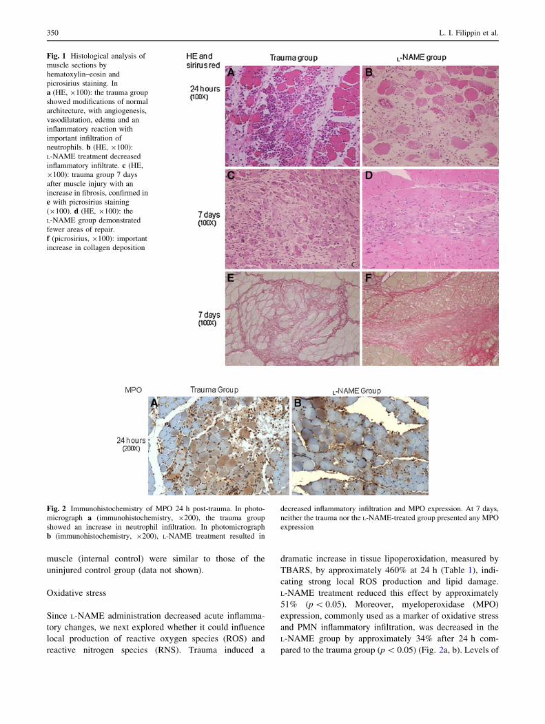
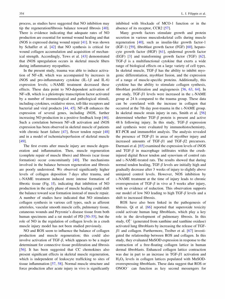
Muscle histological analysis of the trauma group after 24 h
showed typical modifications in normal architecture, with
an inflammatory reaction and edema (Fig. 1a). A single
dose of the nitric oxide synthase inhibitor L-NAME
(100 mg/kg i.p. 2 h post-trauma) markedly attenuated all
inflammatory histological abnormalities at 24 h, with fewer
cellular infiltrates and less edema than in the trauma group
(Fig. 1b). On the 7th day post-trauma, there was distinct
diffuse and poorly organized fibrosis (Fig. 2c, d) and
increased collagen concentration (Fig. 1e) in the trauma
group. L-NAME treatment was associated with more
intense and focal formation of fibrotic tissue (Fig. 1f).
Results from the sham trauma group and left gastrocnemius
Role of NO during healing of trauma to the skeletal muscle 349
muscle (internal control) were similar to those of the
uninjured control group (data not shown).
Oxidative stress
Since L-NAME administration decreased acute inflamma-
tory changes, we next explored whether it could influence
local production of reactive oxygen species (ROS) and
reactive nitrogen species (RNS). Trauma induced a
dramatic increase in tissue lipoperoxidation, measured by
TBARS, by approximately 460% at 24 h (Table 1), indi-
cating strong local ROS production and lipid damage.
L-NAME treatment reduced this effect by approximately
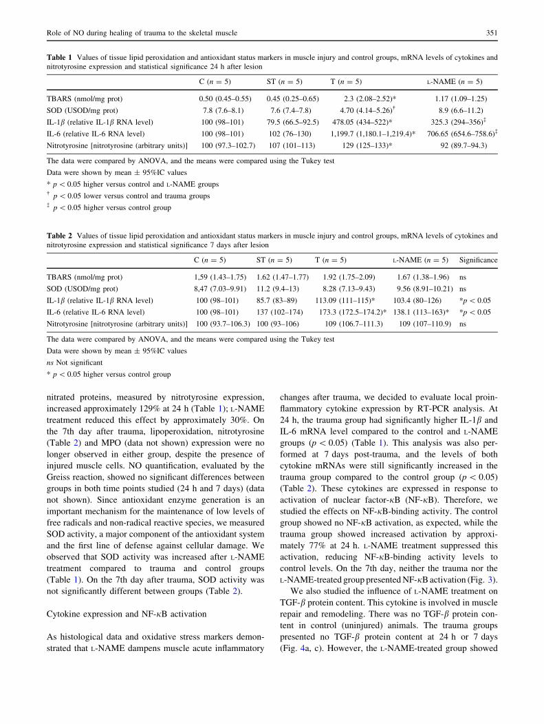
51% (p \ 0.05). Moreover, myeloperoxidase (MPO)
expression, commonly used as a marker of oxidative stress
and PMN inflammatory infiltration, was decreased in the
L-NAME group by approximately 34% after 24 h com-
pared to the trauma group (p \ 0.05) (Fig. 2a, b). Levels of
Fig. 1 Histological analysis of
muscle sections by
hematoxylin–eosin and
picrosirius staining. In
a (HE, 9100): the trauma group
showed modifications of normal
architecture, with angiogenesis,
vasodilatation, edema and an
inflammatory reaction with
important infiltration of
neutrophils. b (HE, 9100):
L-NAME treatment decreased
inflammatory infiltrate. c (HE,
9100): trauma group 7 days
after muscle injury with an
increase in fibrosis, confirmed in
e with picrosirius staining
(9100). d (HE, 9100): the
L-NAME group demonstrated
fewer areas of repair.
f (picrosirius, 9100): important
increase in collagen deposition
Fig. 2 Immunohistochemistry of MPO 24 h post-trauma. In photo-
micrograph a (immunohistochemistry, 9200), the trauma group
showed an increase in neutrophil infiltration. In photomicrograph
b (immunohistochemistry, 9200), L-NAME treatment resulted in
decreased inflammatory infiltration and MPO expression. At 7 days,
neither the trauma nor the L-NAME-treated group presented any MPO
expression
350 L. I. Filippin et al.
nitrated proteins, measured by nitrotyrosine expression,
increased approximately 129% at 24 h (Table 1); L-NAME
treatment reduced this effect by approximately 30%. On
the 7th day after trauma, lipoperoxidation, nitrotyrosine
(Table 2) and MPO (data not shown) expression were no
longer observed in either group, despite the presence of
injured muscle cells. NO quantification, evaluated by the
Greiss reaction, showed no significant differences between
groups in both time points studied (24 h and 7 days) (data
not shown). Since antioxidant enzyme generation is an
important mechanism for the maintenance of low levels of
free radicals and non-radical reactive species, we measured
SOD activity, a major component of the antioxidant system
and the first line of defense against cellular damage. We
observed that SOD activity was increased after L-NAME
treatment compared to trauma and control groups
(Table 1). On the 7th day after trauma, SOD activity was
not significantly different between groups (Table 2).
Cytokine expression and NF-jB activation
As histological data and oxidative stress markers demon-
strated that L-NAME dampens muscle acute inflammatory
changes after trauma, we decided to evaluate local proin-
flammatory cytokine expression by RT-PCR analysis. At
24 h, the trauma group had significantly higher IL-1b and
IL-6 mRNA level compared to the control and L-NAME
groups (p \ 0.05) (Table 1). This analysis was also per-
formed at 7 days post-trauma, and the levels of both
cytokine mRNAs were still significantly increased in the
trauma group compared to the control group (p \ 0.05)
(Table 2). These cytokines are expressed in response to
activation of nuclear factor-jB (NF-jB). Therefore, we
studied the effects on NF-jB-binding activity. The control
group showed no NF-jB activation, as expected, while the
trauma group showed increased activation by approxi-
mately 77% at 24 h. L-NAME treatment suppressed this
activation, reducing NF-jB-binding activity levels to
control levels. On the 7th day, neither the trauma nor the
L-NAME-treated group presented NF-jB activation (Fig. 3).
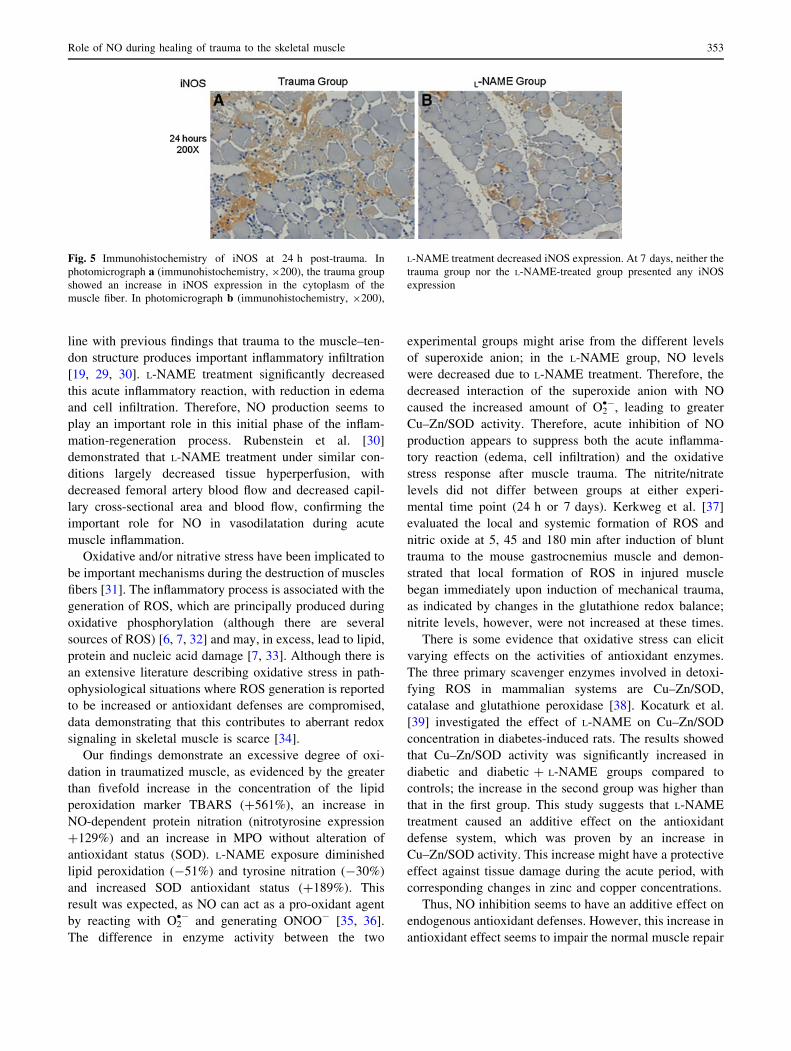
We also studied the influence of L-NAME treatment on
TGF-b protein content. This cytokine is involved in muscle
repair and remodeling. There was no TGF-b protein con-
tent in control (uninjured) animals. The trauma groups
presented no TGF-b protein content at 24 h or 7 days
(Fig. 4a, c). However, the L-NAME-treated group showed
Table 1 Values of tissue lipid peroxidation and antioxidant status markers in muscle injury and control groups, mRNA levels of cytokines and
nitrotyrosine expression and statistical significance 24 h after lesion
C (n = 5) ST (n = 5) T (n = 5) L-NAME (n = 5)
TBARS (nmol/mg prot) 0.50 (0.45–0.55) 0.45 (0.25–0.65) 2.3 (2.08–2.52)* 1.17 (1.09–1.25)
SOD (USOD/mg prot) 7.8 (7.6–8.1) 7.6 (7.4–7.8) 4.70 (4.14–5.26)� 8.9 (6.6–11.2)
IL-1b (relative IL-1b RNA level) 100 (98–101) 79.5 (66.5–92.5) 478.05 (434–522)* 325.3 (294–356)�
IL-6 (relative IL-6 RNA level) 100 (98–101) 102 (76–130) 1,199.7 (1,180.1–1,219.4)* 706.65 (654.6–758.6)�
Nitrotyrosine [nitrotyrosine (arbitrary units)] 100 (97.3–102.7) 107 (101–113) 129 (125–133)* 92 (89.7–94.3)
The data were compared by ANOVA, and the means were compared using the Tukey test
Data were shown by mean ± 95%IC values
* p \ 0.05 higher versus control and L-NAME groups� p \ 0.05 lower versus control and trauma groups� p \ 0.05 higher versus control group
Table 2 Values of tissue lipid peroxidation and antioxidant status markers in muscle injury and control groups, mRNA levels of cytokines and
nitrotyrosine expression and statistical significance 7 days after lesion
C (n = 5) ST (n = 5) T (n = 5) L-NAME (n = 5) Significance
TBARS (nmol/mg prot) 1,59 (1.43–1.75) 1.62 (1.47–1.77) 1.92 (1.75–2.09) 1.67 (1.38–1.96) ns
SOD (USOD/mg prot) 8,47 (7.03–9.91) 11.2 (9.4–13) 8.28 (7.13–9.43) 9.56 (8.91–10.21) ns
IL-1b (relative IL-1b RNA level) 100 (98–101) 85.7 (83–89) 113.09 (111–115)* 103.4 (80–126) *p \ 0.05
IL-6 (relative IL-6 RNA level) 100 (98–101) 137 (102–174) 173.3 (172.5–174.2)* 138.1 (113–163)* *p \ 0.05
Nitrotyrosine [nitrotyrosine (arbitrary units)] 100 (93.7–106.3) 100 (93–106) 109 (106.7–111.3) 109 (107–110.9) ns
The data were compared by ANOVA, and the means were compared using the Tukey test
Data were shown by mean ± 95%IC values
ns Not significant
* p \ 0.05 higher versus control group
Role of NO during healing of trauma to the skeletal muscle 351
moderate expression at 24 h with positive cells compared
to untreated mice and showed stronger staining in inter-
stitial areas at 7 days post-trauma (Fig. 4b, d).
iNOS expression
Since L-NAME exposure appears to decrease the intensity
of acute inflammation, we evaluated its impact on iNOS
tissue expression. As expected, the control group showed
no iNOS expression. In the trauma group, there was
increased iNOS expression in the cytoplasm of the muscle
fiber by approximately 67% at 24 h compared to the
L-NAME group (Fig. 5a, b). At 7 days, neither the trauma
nor the L-NAME-treated group presented iNOS expression
(data not shown).
Discussion
Inflammation of injured muscle is characterized by infil-
trating inflammatory cells, especially neutrophils, which
contribute to the removal of necrotic tissue and the release
of cytokines [26]. A potential mechanism by which
inflammatory or other cells could induce muscle injury
after acute trauma is through the generation of reactive
metabolites of nitrogen and oxygen, which can cause
additional damage to proteins, lipids and nucleic acids [27].
On the other hand, there is evidence indicating that NO can
play a significant role in normal wound repair [28],
although its role in collagen synthesis is not clear.
In our experimental work, rats were treated with
L-NAME, a competitive NOS inhibitor, in order to study
the effects of decreased NO production on the acute
inflammatory reaction and repair process of traumatized
muscle. Acute muscle trauma was accompanied by the
presence of edema and inflammatory infiltration with a
large number of neutrophils and macrophages (Fig. 1a), in
Fig. 3 NF-jB-binding activity. NF-jB-binding activity in control,
trauma and L-NAME groups at 24 h and 7 days after trauma
Fig. 4 Immunohistochemistry
of TGF-b at 24 h and 7 days
post-trauma. In
photomicrographs a and
c (immunohistochemistry,
9200), the trauma group at 24 h
and at 7 days, respectively,
presented no TGF-b expression.
In photomicrograph
b (immunohistochemistry,
9200), L-NAME treatment at
24 h showed moderate TGF-bexpression. At 7 days, the
L-NAME-treated group
presented stronger staining in
the interstitial area
352 L. I. Filippin et al.
line with previous findings that trauma to the muscle–ten-
don structure produces important inflammatory infiltration
[19, 29, 30]. L-NAME treatment significantly decreased
this acute inflammatory reaction, with reduction in edema
and cell infiltration. Therefore, NO production seems to
play an important role in this initial phase of the inflam-
mation-regeneration process. Rubenstein et al. [30]
demonstrated that L-NAME treatment under similar con-
ditions largely decreased tissue hyperperfusion, with
decreased femoral artery blood flow and decreased capil-
lary cross-sectional area and blood flow, confirming the
important role for NO in vasodilatation during acute
muscle inflammation.
Oxidative and/or nitrative stress have been implicated to
be important mechanisms during the destruction of muscles
fibers [31]. The inflammatory process is associated with the
generation of ROS, which are principally produced during
oxidative phosphorylation (although there are several
sources of ROS) [6, 7, 32] and may, in excess, lead to lipid,
protein and nucleic acid damage [7, 33]. Although there is
an extensive literature describing oxidative stress in path-
ophysiological situations where ROS generation is reported
to be increased or antioxidant defenses are compromised,
data demonstrating that this contributes to aberrant redox
signaling in skeletal muscle is scarce [34].
Our findings demonstrate an excessive degree of oxi-
dation in traumatized muscle, as evidenced by the greater
than fivefold increase in the concentration of the lipid
peroxidation marker TBARS (?561%), an increase in
NO-dependent protein nitration (nitrotyrosine expression
?129%) and an increase in MPO without alteration of
antioxidant status (SOD). L-NAME exposure diminished
lipid peroxidation (-51%) and tyrosine nitration (-30%)
and increased SOD antioxidant status (?189%). This
result was expected, as NO can act as a pro-oxidant agent
by reacting with O2•- and generating ONOO- [35, 36].
The difference in enzyme activity between the two
experimental groups might arise from the different levels
of superoxide anion; in the L-NAME group, NO levels
were decreased due to L-NAME treatment. Therefore, the
decreased interaction of the superoxide anion with NO
caused the increased amount of O2•-, leading to greater
Cu–Zn/SOD activity. Therefore, acute inhibition of NO
production appears to suppress both the acute inflamma-
tory reaction (edema, cell infiltration) and the oxidative
stress response after muscle trauma. The nitrite/nitrate
levels did not differ between groups at either experi-
mental time point (24 h or 7 days). Kerkweg et al. [37]
evaluated the local and systemic formation of ROS and
nitric oxide at 5, 45 and 180 min after induction of blunt
trauma to the mouse gastrocnemius muscle and demon-
strated that local formation of ROS in injured muscle
began immediately upon induction of mechanical trauma,
as indicated by changes in the glutathione redox balance;
nitrite levels, however, were not increased at these times.
There is some evidence that oxidative stress can elicit
varying effects on the activities of antioxidant enzymes.
The three primary scavenger enzymes involved in detoxi-
fying ROS in mammalian systems are Cu–Zn/SOD,
catalase and glutathione peroxidase [38]. Kocaturk et al.
[39] investigated the effect of L-NAME on Cu–Zn/SOD
concentration in diabetes-induced rats. The results showed
that Cu–Zn/SOD activity was significantly increased in
diabetic and diabetic ? L-NAME groups compared to
controls; the increase in the second group was higher than
that in the first group. This study suggests that L-NAME
treatment caused an additive effect on the antioxidant
defense system, which was proven by an increase in
Cu–Zn/SOD activity. This increase might have a protective
effect against tissue damage during the acute period, with
corresponding changes in zinc and copper concentrations.
Thus, NO inhibition seems to have an additive effect on
endogenous antioxidant defenses. However, this increase in
antioxidant effect seems to impair the normal muscle repair
Fig. 5 Immunohistochemistry of iNOS at 24 h post-trauma. In
photomicrograph a (immunohistochemistry, 9200), the trauma group
showed an increase in iNOS expression in the cytoplasm of the
muscle fiber. In photomicrograph b (immunohistochemistry, 9200),
L-NAME treatment decreased iNOS expression. At 7 days, neither the
trauma group nor the L-NAME-treated group presented any iNOS
expression
Role of NO during healing of trauma to the skeletal muscle 353
process, as studies have suggested that NO inhibition may
tip the regeneration/fibrosis balance toward fibrosis [40].
There is evidence indicating that adequate rates of NO
production are essential for normal wound healing and that
iNOS is expressed during wound repair [41]. It was shown
by Schaffer et al. [42] that NO synthesis is critical for
wound collagen accumulation and acquisition of mechan-
ical strength. Accordingly, Tews et al. [43] demonstrated
that iNOS upregulation occurs in skeletal muscle fibers
during inflammatory myopathies.
In the present study, trauma was able to induce activa-
tion of NF-jB, which was accompanied by increases in
iNOS and pro-inflammatory cytokine (IL-1b and IL-6)
expression levels; L-NAME treatment decreased these
effects. These data point to NO-dependent activation of
NF-jB, which is a pleiotropic transcription factor activated
by a number of immunological and pathological stimuli,
including cytokines, oxidative stress, toll-like receptors and
bacterial and viral products [44, 45]. NF-jB enhances the
expression of several genes, including iNOS, further
increasing NO production in a positive feedback loop [46].
Such a correlation between NF-jB activation and iNOS
expression has been observed in skeletal muscle of patients
with chronic heart failure [47], flexor tendon repair [48]
and in a model of ischemia/reperfusion of skeletal muscle
[49].
The first events after muscle injury are muscle degen-
eration and inflammation. Then, muscle regeneration
(complete repair of muscle fibers) and fibrosis (scar tissue
formation) occur concomitantly [40]. The mechanisms
involved in the balance between regeneration and fibrosis
are poorly understood. We observed significantly higher
levels of collagen deposition 7 days after trauma, and
L-NAME treatment induced more intense formation of
fibrotic tissue (Fig. 1f), indicating that inhibition of NO
production in the early phase of muscle healing could shift
the balance toward scar formation instead of muscle repair.
A number of studies have indicated that NO stimulates
collagen synthesis in various cell types, such as afferent
arterioles, vascular smooth muscle cells, pulmonary tissue,
cutaneous wounds and Peyronie’s disease tissue from both
human specimens and a rat model of PD [50–55], but the
role of NO in the regulation of collagen levels in a crush
muscle injury model has not been studied previously.
NO and ROS seem to influence the balance of collagen
production and muscle regeneration; this could also
involve activation of TGF-b, which appears to be a major
determinant for connective tissue proliferation and fibrosis
[56]. It has been suggested that CC chemokines also
present significant effects in skeletal muscle regeneration,
which is independent of leukocyte trafficking to sites of
tissue inflammation [57, 58]. Normal muscle structure and
force production after acute injury in vivo is significantly
inhibited with blockade of MCO-1 function or in the
absence of its receptor, CCR2 [57].
Many growth factors stimulate growth and protein
secretion in various musculoskeletal cells during muscle
regeneration [40], such as insulin-like growth factor-1
(IGF-1) [59], fibroblast growth factor (FGF) [60], hepato-
cyte growth factor (HGF) [61], epidermal growth factor
(EGF) [3] and transforming growth factor (TGF) [62].
TGF-b is a multifunctional cytokine that exerts a wide
range of biological effects on a large variety of cell types.
In skeletal muscle, TGF-b has the ability to inhibit myo-
genic differentiation, myoblast fusion, and the expression
of a range of muscle-specific proteins. Additionally, this
cytokine has the ability to stimulate collagen synthesis,
fibroblast proliferation and angiogenesis [56, 63, 64]. In
our study, TGF-b1 levels were increased in the L-NAME
group at 24 h compared to the trauma group. These data
can be correlated with the increase in collagen that
occurred at the 7th day post-trauma in the L-NAME group.
In skeletal muscle strain injury in rats, Smith et al. [56]
determined whether TGF-b protein is present and active
48 h following injury. In this study, TGF-b expression
and synthesis were evaluated by immunohistochemistry,
RT-PCR and immunoblot analysis. The analysis revealed
the presence of TGF-b1 in areas of myofiber injury and
increased amounts of TGF-b1 and TGF-b2 precursors.
Darmani et al. [65] examined the expression levels of iNOS
and TGF-b in macrophage infiltrates within the crush-
injured digital flexor tendon and synovium of control rats
and L-NAME-treated rats. The results showed that during
normal tendon healing, TGF-b levels are initially high and
gradually decrease after 3 weeks of injury to slightly above
uninjured control levels. However, NOS inhibition by
L-NAME treatment at the time of injury leads to chronic
overexpression of TGF-b in vivo at 5 weeks after injury,
with no evidence of reduction. This observation supports
our model of low NO leading to high TGF-b levels and a
shift to increased fibrosis.
ROS have also been linked in the pathogenesis of
fibrosis. Qi et al. [66] reported that superoxide toxicity
could activate human lung fibroblasts, which play a key
role in the development of pulmonary fibrosis. In this
study, O2•- (generated from xanthine and xanthine oxidase)
activated lung fibroblasts by increasing the release of TGF-
b1 and collagen. Furthermore, Treiber et al. [67] investi-
gated the relationship between ROS and collagen. In this
study, they evaluated MnSOD expression in response to the
contraction of a free-floating collagen lattice in human
dermal fibroblasts. Enhanced collagen lattice contraction
was due in part to an increase in TGF-b1 activation and
H2O2 levels in collagen lattices populated with MnSOD-
overexpressing fibroblasts. These data suggest that ROS or
ONOO- can function as key second messengers for
354 L. I. Filippin et al.
collagen lattice contraction and can perhaps act on the
TGF-b1 activation pathway.
In summary, our results point to an important role for
NO in healing processes following acute muscle trauma.
After a single early exposure to the NOS inhibitor
L-NAME, there was a reduction in oxidative and nitrative
stress markers and inflammatory reaction, a marked
decrease in pro-inflammatory cytokines and iNOS and an
increase in TGF-b expression at 24 h. After 7 days, there
was a well-defined increase in collagen deposition, point-
ing to a shift toward healing with fibrosis under conditions
of low early NO concentrations. The exact molecular
mechanisms of this clearly important role for NO in muscle
injury repair need to be studied further. They could involve
an early vasodilatation effect, intracellular signaling with
regulation of matrix protein synthesis or satellite cell
activation. Better understanding of the molecular regula-
tion of muscle repair will have significant therapeutic
implications.
Acknowledgments Coordenacao de Aperfeicoamento de Pessoal de
Ensino Superior (CAPES), Conselho Nacional de Desenvolvimento
Cientıfico e Tecnologico (CNPq), Fundo de Incentivo a Pesquisa e
Eventos (FIPE).
References
1. Kirkendall DT, Garrett WE Jr. Clinical perspectives regarding
eccentric muscle injury. Clin Orthop Relat Res. 2002;S81–9.
2. Huard J, Li Y, Fu FH. Muscle injuries and repair: current trends
in research. J Bone Jt Surg Am. 2002;84-A:822–32.
3. Jarvinen TA, Jarvinen TL, Kaariainen M, Kalimo H, Jarvinen M.
Muscle injuries: biology and treatment. Am J Sports Med.
2005;33:745–64.
4. Toumi H, F’Guyer S, Best TM. The role of neutrophils in injury
and repair following muscle stretch. J Anat. 2006;208:459–70.
5. Pierce AP, de Waal E, McManus LM, Shireman PK, Chaudhuri
AR. Oxidation and structural perturbation of redox-sensitive
enzymes in injured skeletal muscle. Free Radic Biol Med.
2007;43:1584–93.
6. Butterfield TA, Best TM, Merrick MA. The dual roles of neu-
trophils and macrophages in inflammation: a critical balance
between tissue damage and repair. J Athl Train. 2006;41:457–65.
7. Droge W. Free radicals in the physiological control of cell
function. Physiol Rev. 2002;82:47–95.
8. Seeland U, Haeuseler C, Hinrichs R, Rosenkranz S, Pfitzner T,
Scharffetter-Kochanek K, et al. Myocardial fibrosis in trans-
forming growth factor-beta(1) (TGF-beta(1)) transgenic mice is
associated with inhibition of interstitial collagenase. Eur J Clin
Invest. 2002;32:295–303.
9. Quintero AJ, Wright VJ, Fu FH, Huard J. Stem cells for the
treatment of skeletal muscle injury. Clin Sports Med. 2009;28:
1–11.
10. Li Y, Foster W, Deasy BM, Chan Y, Prisk V, Tang Y, et al.
Transforming growth factor-beta1 induces the differentiation of
myogenic cells into fibrotic cells in injured skeletal muscle: a key
event in muscle fibrogenesis. Am J Pathol. 2004;164:1007–19.
11. Hitchon CA, El-Gabalawy HS. Oxidation in rheumatoid arthritis.
Arthritis Res Ther. 2004;6:265–78.
12. Forman HJ, Fukuto JM, Torres M. Redox signaling: thiol
chemistry defines which reactive oxygen and nitrogen species can
act as second messengers. Am J Physiol Cell Physiol.
2004;287:C246–56.
13. Jones DP. Disruption of mitochondrial redox circuitry in oxida-
tive stress. Chem Biol Interact. 2006;163:38–53.
14. Kobzik L, Reid MB, Bredt DS, Stamler JS. Nitric oxide in
skeletal muscle. Nature. 1994;372:546–8.
15. Clancy RM, Leszczynska-Piziak J, Abramson SB. Nitric oxide,
an endothelial cell relaxation factor, inhibits neutrophil super-
oxide anion production via a direct action on the NADPH
oxidase. J Clin Invest. 1992;90:1116–21.
16. Connelly L, Palacios-Callender M, Ameixa C, Moncada S,
Hobbs AJ. Biphasic regulation of NF-kappa B activity underlies
the pro- and anti-inflammatory actions of nitric oxide. J Immunol.
2001;166:3873–81.
17. Nathan CF, Hibbs JB Jr. Role of nitric oxide synthesis in mac-
rophage antimicrobial activity. Curr Opin Immunol. 1991;3:
65–70.
18. LECH OLC. Efeito do uso de corticoide em tendoes previamente
traumatizados: estudo experimental. Revista Brasileira de Orto-
pedia. 1996;31:187–91.
19. Rizzi CF, Mauriz JL, Freitas Correa DS, Moreira AJ, Zettler CG,
Filippin LI, et al. Effects of low-level laser therapy (LLLT) on the
nuclear factor (NF)-kappaB signaling pathway in traumatized
muscle. Lasers Surg Med. 2006;38:704–13.
20. Buege JA, Aust SD. Microsomal lipid peroxidation. Methods
Enzymol. 1978;52:302–10.
21. Misra HP, Fridovich I. The role of superoxide anion in the
autoxidation of epinephrine and a simple assay for superoxide
dismutase. J Biol Chem. 1972;247:3170–5.
22. Misra HP, Fridovich I. The univalent reduction of oxygen by
reduced flavins and quinones. J Biol Chem. 1972;247:188–92.
23. Livak KJ, Schmittgen TD. Analysis of relative gene expression
data using real-time quantitative PCR and the 2(-Delta Delta
C(T)) method. Methods. 2001;25:402–8.
24. Dignam JD, Lebovitz RM, Roeder RG. Accurate transcription
initiation by RNA polymerase II in a soluble extract from isolated
mammalian nuclei. Nucleic Acids Res. 1983;11:1475–89.
25. Granger DL, Taintor RR, Boockvar KS, Hibbs JB Jr. Measure-
ment of nitrate and nitrite in biological samples using nitrate
reductase and Griess reaction. Methods Enzymol. 1996;268:
142–51.
26. Shi X, Garry DJ. Muscle stem cells in development, regeneration,
and disease. Genes Dev. 2006;20:1692–708.
27. Pizza FX, Hernandez IJ, Tidball JG. Nitric oxide synthase inhi-
bition reduces muscle inflammation and necrosis in modified
muscle use. J Leukoc Biol. 1998;64:427–33.
28. Shukla A, Rasik AM, Shankar R. Nitric oxide inhibits wounds
collagen synthesis. Mol Cell Biochem. 1999;200:27–33.
29. Fillipin LI, Mauriz JL, Vedovelli K, Moreira AJ, Zettler CG,
Lech O, et al. Low-level laser therapy (LLLT) prevents oxidative
stress and reduces fibrosis in rat traumatized Achilles tendon.
Lasers Surg Med. 2005;37:293–300.
30. Rubinstein I, Abassi Z, Coleman R, Milman F, Winaver J, Better
OS. Involvement of nitric oxide system in experimental muscle
crush injury. J Clin Invest. 1998;101:1325–33.
31. Tidball JG. Inflammatory processes in muscle injury and repair.
Am J Physiol Regul Integr Comp Physiol. 2005;288:R345–53.
32. Lenaz G. The mitochondrial production of reactive oxygen spe-
cies: mechanisms and implications in human pathology. IUBMB
Life. 2001;52:159–64.
33. Kageyama Y, Takahashi M, Nagafusa T, Torikai E, Nagano A.
Etanercept reduces the oxidative stress marker levels in patients
with rheumatoid arthritis. Rheumatol Int. 2008;28:245–51.
Role of NO during healing of trauma to the skeletal muscle 355
34. Jackson MJ. Redox regulation of skeletal muscle. IUBMB Life.
2008;60:497–501.
35. Salvemini D, Doyle TM, Cuzzocrea S. Superoxide, peroxynitrite
and oxidative/nitrative stress in inflammation. Biochem Soc
Trans. 2006;34:965–70.
36. Lancaster JR Jr. Nitroxidative, nitrosative, and nitrative stress:
kinetic predictions of reactive nitrogen species chemistry under
biological conditions. Chem Res Toxicol. 2006;19:1160–74.
37. Kerkweg U, Schmitz D, de Groot H. Screening for the formation
of reactive oxygen species and of NO in muscle tissue and remote
organs upon mechanical trauma to the mouse hind limb. Eur Surg
Res. 2006;38:83–9.
38. Halliwell B. The chemistry of free radicals and related ‘reactive
species’ Antioxidant defenses endogenous and diet derived. In:
Press U, editor. Free radicals in biology and medicine, vol. 4.
New York; 2007;30–185.
39. Kocaturk PA, Kavas GO. Effect of an inhibitor of nitric oxide
production on Cu–Zn/SOD and its cofactors in diabetic rats. Biol
Trace Elem Res. 2007;115:59–65.
40. Filippin LI, Moreira AJ, Marroni NP, Xavier RM. Nitric oxide
and repair of skeletal muscle injury. Nitric Oxide: Biology and
Chemistry. 2009;21:157–63.
41. Yamasaki K, Edington HD, McClosky C, Tzeng E, Lizonova A,
Kovesdi I, et al. Reversal of impaired wound repair in iNOS-
deficient mice by topical adenoviral-mediated iNOS gene trans-
fer. J Clin Invest. 1998;101:967–71.
42. Schaffer MR, Tantry U, van Wesep RA, Barbul A. Nitric oxide
metabolism in wounds. J Surg Res. 1997;71:25–31.
43. Tews DS, Goebel HH. Cell death and oxidative damage in
inflammatory myopathies. Clin Immunol Immunopathol. 1998;
87:240–7.
44. Hayden MS, Ghosh S. Shared principles in NF-kappaB signaling.
Cell. 2008;132:344–62.
45. Kawai T, Akira S. TLR signaling. Cell Death Differ. 2006;13:
816–25.
46. Adams V, Nehrhoff B, Spate U, Linke A, Schulze PC, Baur A,
et al. Induction of iNOS expression in skeletal muscle by
IL-1beta and NFkappaB activation: an in vitro and in vivo study.
Cardiovasc Res. 2002;54:95–104.
47. Adams V, Spate U, Krankel N, Schulze PC, Linke A, Schuler G,
et al. Nuclear factor-kappa B activation in skeletal muscle of
patients with chronic heart failure: correlation with the expression
of inducible nitric oxide synthase. Eur J Cardiovasc Prev Rehabil.
2003;10:273–7.
48. Tang JB, Xu Y, Ding F, Wang XT. Expression of genes for
collagen production and NF-kappaB gene activation of in vivo
healing flexor tendons. J Hand Surg Am. 2004;29:564–70.
49. Lille ST, Lefler SR, Mowlavi A, Suchy H, Boyle EM Jr, Farr AL,
et al. Inhibition of the initial wave of NF-kappaB activity in rat
muscle reduces ischemia/reperfusion injury. Muscle Nerve.
2001;24:534–41.
50. Ferrini MG, Vernet D, Magee TR, Shahed A, Qian A, Rajfer J,
et al. Antifibrotic role of inducible nitric oxide synthase. Nitric
Oxide. 2002;6:283–94.
51. Amadeu TP, Seabra AB, de Oliveira MG, Costa AM. S-nitro-
soglutathione-containing hydrogel accelerates rat cutaneous
wound repair. J Eur Acad Dermatol Venereol. 2007;21:629–37.
52. Angeli P, Prado CM, Xisto DG, Silva PL, Passaro CP, Nakazato
HD, et al. Effects of chronic L-NAME treatment lung tissue
mechanics, eosinophilic and extracellular matrix responses
induced by chronic pulmonary inflammation. Am J Physiol Lung
Cell Mol Physiol. 2008;294:L1197–205.
53. Lukivskaya O, Patsenker E, Lis R, Buko VU. Inhibition of
inducible nitric oxide synthase activity prevents liver recovery in
rat thioacetamide-induced fibrosis reversal. Eur J Clin Invest.
2008;38:317–25.
54. Amadeu TP, Seabra AB, de Oliveira MG, Monte-Alto-Costa A.
Nitric oxide donor improves healing if applied on inflammatory
and proliferative phase. J Surg Res. 2007;149(1):84–93.
55. Angeli P, Prado CM, Xisto DG, Silva PL, Passaro CP, Nakazato
HD, et al. Effects of chronic L-NAME treatment lung tissue
mechanics, eosinophilic and extracellular matrix responses
induced by chronic pulmonary inflammation. Am J Physiol Lung
Cell Mol Physiol. 2008;294(6):L1197–205.
56. Smith CA, Stauber F, Waters C, Alway SE, Stauber WT.
Transforming growth factor-beta following skeletal muscle strain
injury in rats. J Appl Physiol. 2007;102:755–61.
57. Warren G, O’Farrell L, Summan M, Hulderman T, Mishra D,
Luster M, et al. Role of CC chemokines in skeletal muscle
functional restoration after injury. Am J Physiol Cell Physiol.
2004;286:C1031–6.
58. Yahiaoui L, Gvozdic D, Danialou G, Mack M, Petrof B. CC
family chemokines directly regulate myoblast responses to skel-
etal muscle injury. J Physiol. 2008;586:3991–4004.
59. Metsios G, Stavropoulos-Kalinoglou A, Douglas K, Koutedakis Y,
Nevill A, Panoulas V, et al. Blockade of tumour necrosis factor-
alpha in rheumatoid arthritis: effects on components of rheumatoid
cachexia. Rheumatology (Oxford). 2007;46:1824–7.
60. Hodgetts S, Radley H, Davies M, Grounds M. Reduced necrosis
of dystrophic muscle by depletion of host neutrophils, or blocking
TNFalpha function with Etanercept in mdx mice. Neuromuscul
Disord. 2006;16:591–602.
61. Wang W, Pan H, Murray K, Jefferson B, Li Y. Matrix metallo-
proteinase-1 promotes muscle cell migration and differentiation.
Am J Pathol. 2009;174:541–9.
62. Rantanen J, Thorsson O, Wollmer P, Hurme T, Kalimo H. Effects
of therapeutic ultrasound on the regeneration of skeletal myofi-
bers after experimental muscle injury. Am J Sports Med. 1999;
27:54–9.
63. Liu D, Black B, Derynck R. TGF-beta inhibits muscle differen-
tiation through functional repression of myogenic transcription
factors by Smad3. Genes Dev. 2001;15:2950–66.
64. Cannon J, St Pierre B. Cytokines in exertion-induced skeletal
muscle injury. Mol Cell Biochem. 1998;179:159–67.
65. Darmani H, Crossan J, McLellan SD, Meek D, Adam C.
Expression of nitric oxide synthase and transforming growth
factor-beta in crush-injured tendon and synovium. Mediators
Inflamm. 2004;13:299–305.
66. Qi S, den Hartog GJ, Bast A. Superoxide radicals increase
transforming growth factor-beta1 and collagen release from
human lung fibroblasts via cellular influx through chloride
channels. Toxicol Appl Pharmacol. 2009;237:111–8.
67. Treiber N, Peters T, Sindrilaru A, Huber R, Kohn M, Menke A,
et al. Overexpression of manganese superoxide dismutase in
human dermal fibroblasts enhances the contraction of free float-
ing collagen lattice: implications for ageing and hyperplastic scar
formation. Arch Dermatol Res. 2009;301:273–87.
356 L. I. Filippin et al.