The role of extracellular Tau in the spreading of neurofibrillary pathology
7
CELLULAR NEUROSCIENCE REVIEW ARTICLE published: 23 April 2014 doi: 10.3389/fncel.2014.00113 The role of extracellular Tau in the spreading of neurofibrillary pathology Miguel Medina 1 and Jesús Avila 1,2 * 1 Centro de Investigación Biomédica en Red de Enfermedades Neurodegenerativas (CIBERNED), Madrid, Spain 2 Centro de Biología Molecular “Severo Ochoa” CSIC-UAM, Madrid, Spain Edited by: Ludovic Martin, Université de Nantes, France Reviewed by: Khalid Iqbal, New York State Institute for Basic Research in Developmental Disabilities, USA Amy Pooler, King’s College London, UK *Correspondence: Jesús Avila, Centro de Biología Molecular “Severo Ochoa” CSIC-UAM, Nicolás Cabrera 1, 28049 – Madrid, Spain e-mail: [email protected] The microtubule-associated protein (MAP) tau plays a critical role in the pathogenesis of Alzheimer’s disease (AD) and several related disorders collectively known as tauopathies. Development of tau pathology is associated with progressive neuronal loss and cognitive decline. In the brains of AD patients, tau pathology spreads following an anatomically defined pattern. Mounting evidence strongly suggests that accumulation of abnormal tau is mediated through spreading of seeds of the protein from cell to cell and point at the involvement of extracellular tau species as the main agent in the interneuronal propagation of neurofibrillary lesions and spreading of tau toxicity throughout different brain regions in these disorders. That would support the concept that pathology initiates in a very small part of the brain many years before becoming symptomatic, spreading progressively to the whole brain within 10–20 years. Understanding the precise molecular mechanism underlying tau propagation is crucial for the development of therapeutics for this devastating disorder. In this work, we will discuss recent research on the role of extracellular tau in the spreading of tau pathology, through synaptic and non-synaptic mechanisms. Keywords: Alzheimer, exosomes, vesicles, neurodegeneration, propagation, spreading, tau, tauopathies INTRODUCTION A common pathological feature of many neurodegenerative dis- eases, including Alzheimer’s disease (AD), Parkinson’s disease (PD), Huntington’s disease (HD), amyotrophic lateral sclerosis (ALS) or prion diseases, among others, is the abnormal deposition of proteins in the brain. Among these pathological proteins, the MAP tau forms intraneuronal filaments in a spectrum of neurological disorders collectively known as tauopathies. Tau protein is a MAP that under physiological conditions regulates microtubules (MT) assembly, dynamic behavior, and spatial organization, and has also been shown to regulate the axonal transport of organelles, including mitochondria. The gene encoding tau protein MAPT is located on chromosome 17q21.3, spans approximately 150 kb and consists of 16 exons (Pittman et al., 2006) from which six major isoforms are expressed in adult brain through alternative splicing (reviewed in Andreadis, 2012). The interaction between tau and tubulin is mediated by four imperfect repeat domains (encompassing 31–32 residues) encoded by exons 9–12 (Lee et al., 1989). Alternative splicing of exon 10 results in the production of isoforms containing 3 or 4 binding domains (3R and 4R tau) (Himmler et al., 1989). Adult human brain contains equal amounts of 3R and 4R isoforms whereas foetal brain, however, only expresses 3R tau, demonstrating developmental regulation of exon 10 splicing (Goedert et al., 1989). Different brain regions also differ in the relative levels of 3R and 4R isoforms with granule cells in the hip- pocampal formation reported to have only 3R tau. Disturbances, usually increases, in the 3R/4R ratio are a common feature in most neurodegenerative tauopathies. Furthermore, morpholog- ical differences exist among different diseases or disease types as different tau isoforms are accumulated in diseased brains, namely, six tau isoforms in AD, 3R tau isoforms in Pick’s disease, and 4R tau isoforms in profressive supranuclear palsy (PSP) and cortical basal degeneration (CBD; Goedert and Spillantini, 2011). Interestingly, a recent study has shown that the 4R/3R ration may have been underestimated in AD brains when compared with PSP or CBD, presumably due to extensive deamidation at Asn279 (Dan et al., 2013). Within neurons, tau is predominantly found in axons (Hanger et al., 2009) as a highly soluble phosphoprotein (Iqbal et al., 2009). Phosphorylation is also developmentally regulated, with a high tau phosphorylation level during embryogenesis and early development, when only the shortest of the isoforms is being expressed. By contrast, adult brain expresses all six isoforms with relatively reduced phosphorylation levels compared with the foetal one (Liu et al., 2007). The key discovery directly involving tau protein in neurode- generation and dementia came from the finding that highly penetrant, dominant mutations in the MAPT gene encoding tau cause an inherited form of frontotemporal dementia and parkinsonism (Hutton et al., 1998; Poorkaj et al., 1998; Spillantini et al., 1998). A number of neurodegenerative disorders present prominent tau pathology in the CNS, predominantly within the neuronal compartment, but also within glial cells. Because of this Frontiers in Cellular Neuroscience www.frontiersin.org April 2014 | Volume 8 | Article 113 | 1
-
Upload
independent -
Category
Documents
-
view
2 -
download
0
Transcript of The role of extracellular Tau in the spreading of neurofibrillary pathology

CELLULAR NEUROSCIENCEREVIEW ARTICLE
published: 23 April 2014doi: 10.3389/fncel.2014.00113
The role of extracellular Tau in the spreading ofneurofibrillary pathologyMiguel Medina1 and Jesús Avila1,2*1 Centro de Investigación Biomédica en Red de Enfermedades Neurodegenerativas (CIBERNED), Madrid, Spain2 Centro de Biología Molecular “Severo Ochoa” CSIC-UAM, Madrid, Spain
Edited by:Ludovic Martin, Université deNantes, France
Reviewed by:Khalid Iqbal, New York StateInstitute for Basic Research inDevelopmental Disabilities, USAAmy Pooler, King’s College London,UK
*Correspondence:Jesús Avila, Centro de BiologíaMolecular “Severo Ochoa”CSIC-UAM, Nicolás Cabrera 1,28049 – Madrid, Spaine-mail: [email protected]
The microtubule-associated protein (MAP) tau plays a critical role in the pathogenesis ofAlzheimer’s disease (AD) and several related disorders collectively known as tauopathies.Development of tau pathology is associated with progressive neuronal loss and cognitivedecline. In the brains of AD patients, tau pathology spreads following an anatomicallydefined pattern. Mounting evidence strongly suggests that accumulation of abnormaltau is mediated through spreading of seeds of the protein from cell to cell and pointat the involvement of extracellular tau species as the main agent in the interneuronalpropagation of neurofibrillary lesions and spreading of tau toxicity throughout differentbrain regions in these disorders. That would support the concept that pathology initiatesin a very small part of the brain many years before becoming symptomatic, spreadingprogressively to the whole brain within 10–20 years. Understanding the precise molecularmechanism underlying tau propagation is crucial for the development of therapeutics forthis devastating disorder. In this work, we will discuss recent research on the role ofextracellular tau in the spreading of tau pathology, through synaptic and non-synapticmechanisms.
Keywords: Alzheimer, exosomes, vesicles, neurodegeneration, propagation, spreading, tau, tauopathies
INTRODUCTIONA common pathological feature of many neurodegenerative dis-eases, including Alzheimer’s disease (AD), Parkinson’s disease(PD), Huntington’s disease (HD), amyotrophic lateral sclerosis(ALS) or prion diseases, among others, is the abnormal depositionof proteins in the brain. Among these pathological proteins,the MAP tau forms intraneuronal filaments in a spectrum ofneurological disorders collectively known as tauopathies.
Tau protein is a MAP that under physiological conditionsregulates microtubules (MT) assembly, dynamic behavior, andspatial organization, and has also been shown to regulate theaxonal transport of organelles, including mitochondria. The geneencoding tau protein MAPT is located on chromosome 17q21.3,spans approximately 150 kb and consists of 16 exons (Pittmanet al., 2006) from which six major isoforms are expressed inadult brain through alternative splicing (reviewed in Andreadis,2012). The interaction between tau and tubulin is mediated byfour imperfect repeat domains (encompassing 31–32 residues)encoded by exons 9–12 (Lee et al., 1989). Alternative splicing ofexon 10 results in the production of isoforms containing 3 or 4binding domains (3R and 4R tau) (Himmler et al., 1989).
Adult human brain contains equal amounts of 3R and 4Risoforms whereas foetal brain, however, only expresses 3R tau,demonstrating developmental regulation of exon 10 splicing(Goedert et al., 1989). Different brain regions also differ in therelative levels of 3R and 4R isoforms with granule cells in the hip-pocampal formation reported to have only 3R tau. Disturbances,
usually increases, in the 3R/4R ratio are a common feature inmost neurodegenerative tauopathies. Furthermore, morpholog-ical differences exist among different diseases or disease typesas different tau isoforms are accumulated in diseased brains,namely, six tau isoforms in AD, 3R tau isoforms in Pick’s disease,and 4R tau isoforms in profressive supranuclear palsy (PSP) andcortical basal degeneration (CBD; Goedert and Spillantini, 2011).Interestingly, a recent study has shown that the 4R/3R ration mayhave been underestimated in AD brains when compared withPSP or CBD, presumably due to extensive deamidation at Asn279(Dan et al., 2013).
Within neurons, tau is predominantly found in axons (Hangeret al., 2009) as a highly soluble phosphoprotein (Iqbal et al.,2009). Phosphorylation is also developmentally regulated, with ahigh tau phosphorylation level during embryogenesis and earlydevelopment, when only the shortest of the isoforms is beingexpressed. By contrast, adult brain expresses all six isoformswith relatively reduced phosphorylation levels compared with thefoetal one (Liu et al., 2007).
The key discovery directly involving tau protein in neurode-generation and dementia came from the finding that highlypenetrant, dominant mutations in the MAPT gene encodingtau cause an inherited form of frontotemporal dementia andparkinsonism (Hutton et al., 1998; Poorkaj et al., 1998; Spillantiniet al., 1998). A number of neurodegenerative disorders presentprominent tau pathology in the CNS, predominantly within theneuronal compartment, but also within glial cells. Because of this
Frontiers in Cellular Neuroscience www.frontiersin.org April 2014 | Volume 8 | Article 113 | 1
Medina and Avila Progression of tau pathology
shared histopathological feature, they are referred collectively astauopathies, although they constitute a group of etiologically het-erogeneous, clinically and neuropathologically overlapping dis-ease entities (Ballatore et al., 2007; Spillantini and Goedert, 2013).In tauopathies, the intracellular soluble tau forms filamentousstructures of aggregated, hyperphosphorylated tau, which areassociated with synaptic loss and neuronal death. The occurrenceof fibrillar tau inclusions in tauopathies strongly supports a keyrole in the observed clinical symptoms and pathology.
Further insights into the overlapping pathogenic and etiologicaspects of the discrete diseases will help to design (perhaps com-mon) disease-modifying treatment strategies (Medina, 2011). Toachieve that goal however, it is critical to understand the normalbiological roles of tau, the specific molecular events that inducetau to become neurotoxic, the biochemical nature of pathogenictau, the means by which pathogenic tau exerts neurotoxicity, andhow tau pathology propagates.
As mentioned, the recognition of the MAP tau as a key playerin the pathobiology of human neurodegenerative diseases hasled to major efforts to understand its biological and pathologicalfunction(s). This has resulted in an improved understanding oftau cellular functions beyond its classical role in stabilizing MT(Morris et al., 2011) to unveil novel physiological tau functionsthat may also play a role in pathogenesis. Such functions includeaxonal transport (Terwel et al., 2002; Rodríguez-Martín et al.,2013), neuronal polarization (Caceres and Kosik, 1990; Dawsonet al., 2010), axonogenesis (DiTella et al., 1994; Klein et al.,2002; Belkadi and LoPresti, 2008), interactions with the plasmamembrane (Brandt et al., 1995; Lee et al., 1998; Maas et al., 2000),signal transduction (Lee et al., 2004; Ittner et al., 2010) and cellcycle (Andorfer et al., 2005). Furthermore, despite lacking anidentified nuclear localization signal, tau has also been reportedin nuclei in a number of cell lines (Loomis et al., 1990; Wang et al.,1993) and human brain (Brady et al., 1995) where it may play arole in DNA protection (Sultan et al., 2011).
EXTRACELLULAR TAUIt has been over 20 years since the original report that intracellulartau levels are increased in the brains of AD patients when com-pared to non-demented controls (Barton et al., 1990; Khatoonet al., 1992). This increase in the amount of tau could be toxicto neurons since a reduction in the amount of intracellular tauhas indeed a protective effect in mouse models of neurodegen-eration (Rapoport et al., 2002; Roberson et al., 2007) and it hasbeen suggested that reducing tau levels may be therapeuticallybeneficial (Götz et al., 2013). However, we must be cautious sinceother studies in similar tau-deficient mice point in the oppositedirection, suggesting that loss of tau function can actually lead toneurodegeneration (Dawson et al., 2010).
Little is known about how tau synthesis is regulated althoughsome factors such as fibroblast growth factor (Tatebayashi et al.,1999), Dyrk1A (Qian et al., 2013), or the haplotype H1 have beeninvolved in increased synthesis whereas the miRNA-34 family(Dickson et al., 2013) seems to downregulate tau levels.
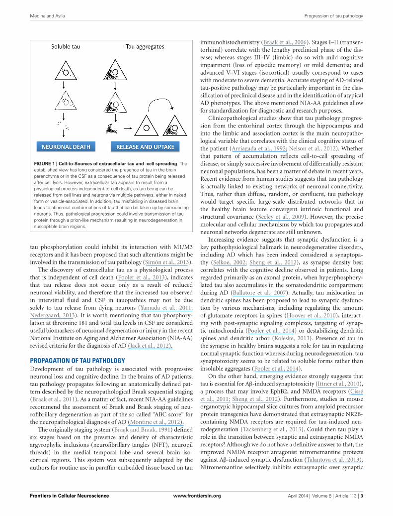
Conventional wisdom has suggested that the presence of tauin the brain parenchyma or in the cerebrospinal fluid (CSF) isa consequence of tau protein being released from dead cells.
However, this has recently been challenged by a number of studiesshowing extracellular tau being released from cell lines and neu-rons via multiple pathways, strongly supporting the notion thatsecretion of tau protein may be an important biological functionof tau protein, especially in disease. Despite the fact that taulacks a signal sequence a number of reports have now shownthat tau is released into culture medium from neuroblastomacells, tau-expressing non-neuronal cells, induced pluripotent stemcell-derived human neurons, and mouse primary neurons (Kimet al., 2010; Shi et al., 2012). Thus, tau has been reported tobe secreted unconventionally in naked form (Chai et al., 2012)or associated to exosomes (Saman et al., 2012) and/or othermembrane vesicles (Simón et al., 2012a). Since increased taucellular levels are detrimental, secretion has been proposed asa mechanism to eliminate the excess of tau protein therebyavoiding its toxicity (Simón et al., 2012b). Interestingly, while fulllength tau has been detected in the extracellular space, C-terminalcleavage of tau has been shown to enhance its secretion (Plouffeet al., 2012) which could have pathological relevance since sometruncated tau species appears to be characteristic of particulartauopathies whereas other tau fragments may be common toseveral tauopathies (Hanger and Wray, 2010; Kovacech et al.,2010).
Extracellular tau has also been detected in the brain interstitialfluid of both wild-type and P301S tau-expressing mice in micro-dialysis studies (Yamada et al., 2011), as it has also been the case inpatients following severe traumatic brain injury (Marklund et al.,2009; Magnoni et al., 2012). Actually, exosomal tau secretion hasbeen suggested to account for the elevated CSF tau levels typicallyobserved in early AD (Saman et al., 2012). Interestingly, taumutations that are associated with the development of tauopathyappears to reduce tau release (Karch et al., 2012). Interestingly,physiological secretion of endogenous tau by cortical neuronsappears to be regulated by neuronal activity, as tau release isenhanced by glutamate receptor stimulation induced by theagonist S-AMPA (Pooler et al., 2013). This process is calcium-dependent and modulated by phosphorylation and released tauis present in a relatively dephosphorylated state, compared to thatof intracellular tau.
Thus, increasing evidence point out to extracellular tau asa physiological process independent of cell death (Figure 1),although the precise relationship between tau release under phys-iological conditions and the propagation of pathology in AD andother tauopathies remains to be determined.
Tau can be toxic when applied extracellularly to culturedcells (Gómez-Ramos et al., 2006; Kopeikina et al., 2012). Sev-eral mechanisms for internalization of tau has been proposed,such as internalization of soluble, uncoated (“naked”) tau viareceptor-mediated endocytosis (Gómez-Ramos et al., 2009),dynamin-driven endocytosis of non-fibrillar, soluble tau aggre-gates (Wu et al., 2013) or even actin-dependent, proteoglycan-mediated macropynocytosis (Holmes et al., 2013). Furthermore,it has been suggested that extracellular tau might provokea receptor-activated increase in intracellular calcium throughM1/M3 muscarinic receptor stimulation (Gómez-Ramos et al.,2008; Díaz-Hernández et al., 2010) and that such receptor acti-vation could lead to endocytosis of extracellular tau. Remarkably,
Frontiers in Cellular Neuroscience www.frontiersin.org April 2014 | Volume 8 | Article 113 | 2
Medina and Avila Progression of tau pathology
FIGURE 1 | Cell-to-Sources of extracellular tau and -cell spreading. Theestablished view has long considered the presence of tau in the brainparenchyma or in the CSF as a consequence of tau protein being releasedafter cell lysis. However, extracellular tau appears to result from aphysiological process independent of cell death, as tau being can bereleased from cell lines and neurons via multiple pathways, either in nakedform or vesicle-associated. In addition, tau misfolding in diseased brainleads to abnormal conformations of tau that can be taken up by surroundingneurons. Thus, pathological progression could involve transmission of tauprotein through a prion-like mechanism resulting in neurodegeneration insusceptible brain regions.
tau phosphorylation could inhibit its interaction with M1/M3receptors and it has been proposed that such alterations might beinvolved in the transmission of tau pathology (Simón et al., 2013).
The discovery of extracellular tau as a physiological processthat is independent of cell death (Pooler et al., 2013), indicatesthat tau release does not occur only as a result of reducedneuronal viability, and therefore that the increased tau observedin interstitial fluid and CSF in tauopathies may not be duesolely to tau release from dying neurons (Yamada et al., 2011;Nedergaard, 2013). It is worth mentioning that tau phosphory-lation at threonine 181 and total tau levels in CSF are considereduseful biomarkers of neuronal degeneration or injury in the recentNational Institute on Aging and Alzheimer Association (NIA-AA)revised criteria for the diagnosis of AD (Jack et al., 2012).
PROPAGATION OF TAU PATHOLOGYDevelopment of tau pathology is associated with progressiveneuronal loss and cognitive decline. In the brains of AD patients,tau pathology propagates following an anatomically defined pat-tern described by the neuropathological Braak sequential staging(Braak et al., 2011). As a matter of fact, recent NIA-AA guidelinesrecommend the assessment of Braak and Braak staging of neu-rofibrillary degeneration as part of the so called “ABC score” forthe neuropathological diagnosis of AD (Montine et al., 2012).
The originally staging system (Braak and Braak, 1991) definedsix stages based on the presence and density of characteristicargyrophylic inclusions (neurofibrillary tangles (NFT), neuropilthreads) in the medial temporal lobe and several brain iso-cortical regions. This system was subsequently adapted by theauthors for routine use in paraffin-embedded tissue based on tau
immunohistochemistry (Braak et al., 2006). Stages I–II (transen-torhinal) correlate with the lengthy preclinical phase of the dis-ease; whereas stages III–IV (limbic) do so with mild cognitiveimpairment (loss of episodic memory) or mild dementia; andadvanced V–VI stages (isocortical) usually correspond to caseswith moderate to severe dementia. Accurate staging of AD-relatedtau-positive pathology may be particularly important in the clas-sification of preclinical disease and in the identification of atypicalAD phenotypes. The above mentioned NIA-AA guidelines allowfor standardization for diagnostic and research purposes.
Clinicopathological studies show that tau pathology progres-sion from the entorhinal cortex through the hippocampus andinto the limbic and association cortex is the main neuropatho-logical variable that correlates with the clinical cognitive status ofthe patient (Arriagada et al., 1992; Nelson et al., 2012). Whetherthat pattern of accumulation reflects cell-to-cell spreading ofdisease, or simply successive involvement of differentially resistantneuronal populations, has been a matter of debate in recent years.Recent evidence from human studies suggests that tau pathologyis actually linked to existing networks of neuronal connectivity.Thus, rather than diffuse, random, or confluent, tau pathologywould target specific large-scale distributed networks that inthe healthy brain feature convergent intrinsic functional andstructural covariance (Seeley et al., 2009). However, the precisemolecular and cellular mechanisms by which tau propagates andneuronal networks degenerate are still unknown.
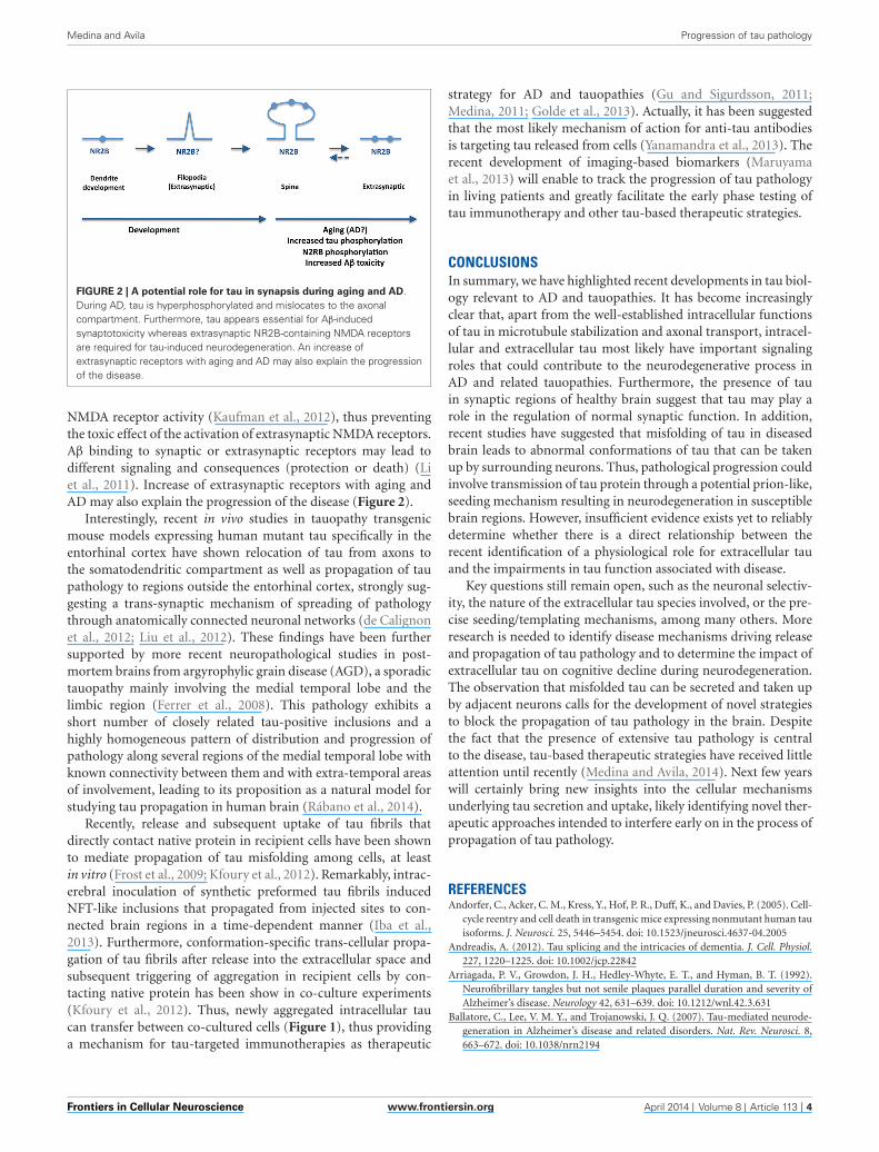
Increasing evidence suggests that synaptic dysfunction is akey pathophysiological hallmark in neurodegenerative disorders,including AD which has been indeed considered a synaptopa-thy (Selkoe, 2002; Sheng et al., 2012), as synapse density bestcorrelates with the cognitive decline observed in patients. Longregarded primarily as an axonal protein, when hyperphosphory-lated tau also accumulates in the somatodendritic compartmentduring AD (Ballatore et al., 2007). Actually, tau mislocation indendritic spines has been proposed to lead to synaptic dysfunc-tion by various mechanisms, including regulating the amountof glutamate receptors in spines (Hoover et al., 2010), interact-ing with post-synaptic signaling complexes, targeting of synap-tic mitochondria (Pooler et al., 2014) or destabilizing dendriticspines and dendritic arbor (Koleske, 2013). Presence of tau inthe synapse in healthy brains suggests a role for tau in regulatingnormal synaptic function whereas during neurodegeneration, tausynaptotoxicity seems to be related to soluble forms rather thaninsoluble aggregates (Pooler et al., 2014).
On the other hand, emerging evidence strongly suggests thattau is essential for Aβ-induced synaptotoxicity (Ittner et al., 2010),a process that may involve EphB2, and NMDA receptors (Cisséet al., 2011; Sheng et al., 2012). Furthermore, studies in mouseorganotypic hippocampal slice cultures from amyloid precurssorprotein transgenics have demonstrated that extrasynaptic NR2B-containing NMDA receptors are required for tau-induced neu-rodegeneration (Tackenberg et al., 2013). Could then tau play arole in the transition between synaptic and extrasynaptic NMDAreceptors? Although we do not have a definitive answer to that, theimproved NMDA receptor antagonist nitromemantine protectsagainst Aβ-induced synaptic dysfunction (Talantova et al., 2013).Nitromemantine selectively inhibits extrasynaptic over synaptic
Frontiers in Cellular Neuroscience www.frontiersin.org April 2014 | Volume 8 | Article 113 | 3
Medina and Avila Progression of tau pathology
FIGURE 2 | A potential role for tau in synapsis during aging and AD.During AD, tau is hyperphosphorylated and mislocates to the axonalcompartment. Furthermore, tau appears essential for Aβ-inducedsynaptotoxicity whereas extrasynaptic NR2B-containing NMDA receptorsare required for tau-induced neurodegeneration. An increase ofextrasynaptic receptors with aging and AD may also explain the progressionof the disease.
NMDA receptor activity (Kaufman et al., 2012), thus preventingthe toxic effect of the activation of extrasynaptic NMDA receptors.Aβ binding to synaptic or extrasynaptic receptors may lead todifferent signaling and consequences (protection or death) (Liet al., 2011). Increase of extrasynaptic receptors with aging andAD may also explain the progression of the disease (Figure 2).
Interestingly, recent in vivo studies in tauopathy transgenicmouse models expressing human mutant tau specifically in theentorhinal cortex have shown relocation of tau from axons tothe somatodendritic compartment as well as propagation of taupathology to regions outside the entorhinal cortex, strongly sug-gesting a trans-synaptic mechanism of spreading of pathologythrough anatomically connected neuronal networks (de Calignonet al., 2012; Liu et al., 2012). These findings have been furthersupported by more recent neuropathological studies in post-mortem brains from argyrophylic grain disease (AGD), a sporadictauopathy mainly involving the medial temporal lobe and thelimbic region (Ferrer et al., 2008). This pathology exhibits ashort number of closely related tau-positive inclusions and ahighly homogeneous pattern of distribution and progression ofpathology along several regions of the medial temporal lobe withknown connectivity between them and with extra-temporal areasof involvement, leading to its proposition as a natural model forstudying tau propagation in human brain (Rábano et al., 2014).
Recently, release and subsequent uptake of tau fibrils thatdirectly contact native protein in recipient cells have been shownto mediate propagation of tau misfolding among cells, at leastin vitro (Frost et al., 2009; Kfoury et al., 2012). Remarkably, intrac-erebral inoculation of synthetic preformed tau fibrils inducedNFT-like inclusions that propagated from injected sites to con-nected brain regions in a time-dependent manner (Iba et al.,2013). Furthermore, conformation-specific trans-cellular propa-gation of tau fibrils after release into the extracellular space andsubsequent triggering of aggregation in recipient cells by con-tacting native protein has been show in co-culture experiments(Kfoury et al., 2012). Thus, newly aggregated intracellular taucan transfer between co-cultured cells (Figure 1), thus providinga mechanism for tau-targeted immunotherapies as therapeutic
strategy for AD and tauopathies (Gu and Sigurdsson, 2011;Medina, 2011; Golde et al., 2013). Actually, it has been suggestedthat the most likely mechanism of action for anti-tau antibodiesis targeting tau released from cells (Yanamandra et al., 2013). Therecent development of imaging-based biomarkers (Maruyamaet al., 2013) will enable to track the progression of tau pathologyin living patients and greatly facilitate the early phase testing oftau immunotherapy and other tau-based therapeutic strategies.
CONCLUSIONSIn summary, we have highlighted recent developments in tau biol-ogy relevant to AD and tauopathies. It has become increasinglyclear that, apart from the well-established intracellular functionsof tau in microtubule stabilization and axonal transport, intracel-lular and extracellular tau most likely have important signalingroles that could contribute to the neurodegenerative process inAD and related tauopathies. Furthermore, the presence of tauin synaptic regions of healthy brain suggest that tau may play arole in the regulation of normal synaptic function. In addition,recent studies have suggested that misfolding of tau in diseasedbrain leads to abnormal conformations of tau that can be takenup by surrounding neurons. Thus, pathological progression couldinvolve transmission of tau protein through a potential prion-like,seeding mechanism resulting in neurodegeneration in susceptiblebrain regions. However, insufficient evidence exists yet to reliablydetermine whether there is a direct relationship between therecent identification of a physiological role for extracellular tauand the impairments in tau function associated with disease.
Key questions still remain open, such as the neuronal selectiv-ity, the nature of the extracellular tau species involved, or the pre-cise seeding/templating mechanisms, among many others. Moreresearch is needed to identify disease mechanisms driving releaseand propagation of tau pathology and to determine the impact ofextracellular tau on cognitive decline during neurodegeneration.The observation that misfolded tau can be secreted and taken upby adjacent neurons calls for the development of novel strategiesto block the propagation of tau pathology in the brain. Despitethe fact that the presence of extensive tau pathology is centralto the disease, tau-based therapeutic strategies have received littleattention until recently (Medina and Avila, 2014). Next few yearswill certainly bring new insights into the cellular mechanismsunderlying tau secretion and uptake, likely identifying novel ther-apeutic approaches intended to interfere early on in the process ofpropagation of tau pathology.
REFERENCESAndorfer, C., Acker, C. M., Kress, Y., Hof, P. R., Duff, K., and Davies, P. (2005). Cell-
cycle reentry and cell death in transgenic mice expressing nonmutant human tauisoforms. J. Neurosci. 25, 5446–5454. doi: 10.1523/jneurosci.4637-04.2005
Andreadis, A. (2012). Tau splicing and the intricacies of dementia. J. Cell. Physiol.227, 1220–1225. doi: 10.1002/jcp.22842
Arriagada, P. V., Growdon, J. H., Hedley-Whyte, E. T., and Hyman, B. T. (1992).Neurofibrillary tangles but not senile plaques parallel duration and severity ofAlzheimer’s disease. Neurology 42, 631–639. doi: 10.1212/wnl.42.3.631
Ballatore, C., Lee, V. M. Y., and Trojanowski, J. Q. (2007). Tau-mediated neurode-generation in Alzheimer’s disease and related disorders. Nat. Rev. Neurosci. 8,663–672. doi: 10.1038/nrn2194
Frontiers in Cellular Neuroscience www.frontiersin.org April 2014 | Volume 8 | Article 113 | 4
Medina and Avila Progression of tau pathology
Barton, A. J., Harrison, P. J., Najlerahim, A., Heffernan, J., McDonald, B., Robinson,J. R., et al. (1990). Increased tau messenger RNA in Alzheimer’s disease hip-pocampus. Am. J. Pathol. 137, 497–502.
Belkadi, A., and LoPresti, P. (2008). Truncated Tau with the Fyn-binding domainand without the microtubule-binding domain hinders the myelinating capacityof an oligodendrocyte cell line. J. Neurochem. 107, 351–360. doi: 10.1111/j.1471-4159.2008.05600.x
Braak, H., Alafuzoff, I., Arzberger, T., Kretzschmar, H., and Del Tredici, K. (2006).Staging of Alzheimer disease-associated neurofibrillary pathology using paraffinsections and immunocytochemistry. Acta Neuropathol. 112, 389–404. doi: 10.1007/s00401-006-0127-z
Braak, H., and Braak, E. (1991). Neuropathological stageing of Alzheimer-relatedchanges. Acta Neuropathol. 82, 239–259. doi: 10.1007/bf00308809
Braak, H., Thal, D. R., Ghebremedhin, E., and Del Tredici, K. (2011). Stagesof the pathologic process in Alzheimer disease: age categories from 1 to 100years. J. Neuropathol. Exp. Neurol. 70, 960–969. doi: 10.1097/nen.0b013e318232a379
Brady, R. M., Zinkowski, R. P., and Binder, L. I. (1995). Presence of tau in isolatednuclei from human brain. Neurobiol. Aging 16, 479–486. doi: 10.1016/0197-4580(95)00023-8
Brandt, R., Leger, J., and Lee, G. (1995). Interaction of tau with the neural plasmamembrane mediated by tau’s amino-terminal projection domain. J. Cell Biol.131, 1327–1340. doi: 10.1083/jcb.131.5.1327
Caceres, A., and Kosik, K. S. (1990). Inhibition of neurite polarity by tau antisenseoligonucleotides in primary cerebellar neurons. Nature 343, 461–463. doi: 10.1038/343461a0
Chai, X., Dage, J. L., and Citron, M. (2012). Constitutive secretion of tau protein byan unconventional mechanism. Neurobiol. Dis. 48, 356–366. doi: 10.1016/j.nbd.2012.05.021
Cissé, M., Halabisky, B., Harris, J., Devidze, N., Dubal, D. B., Sun, B., et al. (2011).Reversing EphB2 depletion rescues cognitive functions in Alzheimer model.Nature 469, 47–52. doi: 10.1038/nature09635
Dan, A., Takahashi, M., Masuda-Suzukake, M., Kametani, F., Nonaka, T.,Kondo, H., et al. (2013). Extensive deamidation at asparagine residue 279accounts for weak immunoreactivity of tau with RD4 antibody in Alzheimer’sdisease brain. Acta Neuropathol. Commun. 1:54. doi: 10.1186/2051-5960-1-54
Dawson, H. N., Cantillana, V., Jansen, M., Wang, H., Vitek, M. P., Wilcock, D. M.,et al. (2010). Loss of tau elicits axonal degeneration in a mouse model ofAlzheimer’s disease. Neuroscience 169, 516–531. doi: 10.1016/j.neuroscience.2010.04.037
de Calignon, A., Polydoro, M., Suárez-Calvet, M., William, C., Adamowicz, D. H.,Kopeikina, K. J., et al. (2012). Propagation of tau pathology in a model ofearly Alzheimer’s disease. Neuron 73, 685–697. doi: 10.1016/j.neuron.2011.11.033
Díaz-Hernández, M., Gomez-Ramos, A., Rubio, A., Gomez-Villafuertes, R.,Naranjo, J. R., Miras-Portugal, M. T., et al. (2010). Tissue-nonspecific alkalinephosphatase promotes the neurotoxicity effect of extracellular tau. J. Biol. Chem.285, 32539–32548. doi: 10.1074/jbc.m110.145003
Dickson, J. R., Kruse, C., Montagna, D. R., Finsen, B., and Wolfe, M. S. (2013).Alternative polyadenylation and miR-34 family members regulate tau expres-sion. J. Neurochem. 127, 739–749. doi: 10.1111/jnc.12437
DiTella, M., Feiguin, F., Morfini, G., and Caceres, A. (1994). Microfilament associ-ated growth cone component depends upon Tau for its intracellular localization.Cell Motil. Cytoskeleton 29, 117–130. doi: 10.1002/cm.970290204
Ferrer, I., Santpere, G., and van Leeuwen, F. W. (2008). Argyrophilic grain disease.Brain 131, 1416–1432. doi: 10.1093/brain/awm305
Frost, B., Jacks, R. L., and Diamond, M. I. (2009). Propagation of tau misfoldingfrom the outside to the inside of a cell. J. Biol. Chem. 284, 12845–12852. doi: 10.1074/jbc.m808759200
Goedert, M., and Spillantini, M. G. (2011). Pathogenesis of the tauopathies. J. Mol.Neurosci. 45, 425–431. doi: 10.1007/s12031-011-9593-4
Goedert, M., Spillantini, M. G., Potier, M. C., Ulrich, J., and Crowther, R. A. (1989).Cloning and sequencing of the cDNA encoding an isoform of microtubule-associated protein tau containing four tandem repeats: differential expressionof tau protein mRNAs in human brain. EMBO J. 8, 393–399.
Golde, T. E., Lewis, J., and McFarland, N. R. (2013). Anti-tauantibodies: hitting thetarget. Neuron 80, 254–256. doi: 10.1016/j.neuron.2013.10.009
Gómez-Ramos, A., Díaz-Hernández, M., Cuadros, R., Hernandez, F., and Avila, J.(2006). Extracellular tau is toxic to neuronal cells. FEBS Lett. 580, 4842–4850.doi: 10.1016/j.febslet.2006.07.078
Gómez-Ramos, A., Díaz-Hernández, M., Rubio, A., Díaz-Hernández, J. I., Miras-Portugal, M. T., and Avila, J. (2009). Characteristics and consequences ofmuscarinic receptor activation by tau protein. Eur. Neuropsychopharmacol. 19,708–717. doi: 10.1016/j.euroneuro.2009.04.006
Gómez-Ramos, A., Díaz-Hernández, M., Rubio, A., Miras-Portugal, M. T., andAvila, J. (2008). Extracellular tau promotes intracellular calcium increasethrough M1 and M3 muscarinic receptors in neuronal cells. Mol. Cell. Neurosci.37, 673–681. doi: 10.1016/j.mcn.2007.12.010
Götz, J., Xia, D., Leinenga, G., Chew, Y. L., and Nicholas, H. (2013). What rendersTAU toxic. Front. Neurol. 4:72. doi: 10.3389/fneur.2013.00072
Gu, J., and Sigurdsson, E. M. (2011). Immunotherapy for tauopathies. J. Mol.Neurosci. 45, 690–695. doi: 10.1007/s12031-011-9576-5
Hanger, D. P., Anderton, B. H., and Noble, W. (2009). Tau phosphorylation: thetherapeutic challenge for neurodegenerative disease. Trends Mol. Med. 15, 112–119. doi: 10.1016/j.molmed.2009.01.003
Hanger, D. P., and Wray, S. (2010). Tau cleavage and tau aggregation in neurodegen-erative disease. Biochem. Soc. Trans. 38, 1016–10120. doi: 10.1042/bst0381016
Himmler, A., Drechsel, D., Kirschner, M. W., and Martin, D. W. Jr. (1989). Tauconsists of a set of proteins with repeated C-terminal microtubule-bindingdomains and variable N-terminal domains. Mol. Cell. Biol. 9, 1381–1388.
Holmes, B. B., DeVos, S. L., Kfoury, N., Li, M., Jacks, R., Yanamandra, K., et al.(2013). Heparan sulfate proteoglycans mediate internalization and propagationof specific proteopathic seeds. Proc. Natl. Acad. Sci. U S A 110, E3138–E3147.doi: 10.1073/pnas.1301440110
Hoover, B. R., Reed, M. N., Su, J., Penrod, R. D., Kotilinek, L. A., Grant, M. K., et al.(2010). Tau mislocalization to dendritic spines mediates synaptic dysfunctionindependently of neurodegeneration. Neuron 68, 1067–1081. doi: 10.1016/j.neuron.2010.11.030
Hutton, M., Lendon, C. L., Rizzu, P., Baker, M., Froelich, S., Houlden, H., et al.(1998). Association of missense and 5’-splice-site mutations in tau with theinherited dementia FTDP-17. Nature 393, 702–705. doi: 10.1038/31508
Iba, M., Guo, J. L., McBride, J. D., Zhang, B., Trojanowski, J. Q., and Lee, V. M.(2013). Synthetic tau fibrils mediate transmission of neurofibrillary tangles ina transgenic mouse model of Alzheimer’s-like tauopathy. J. Neurosci. 33, 1024–1037. doi: 10.1523/JNEUROSCI.2642-12.2013
Iqbal, K., Liu, F., Gong, C. X., Alonso Adel, C., and Grundke-Iqbal, I. (2009).Mechanisms of tau-induced neurodegeneration. Acta Neuropathol. 118, 53–69.doi: 10.1007/s00401-009-0486-3
Ittner, L. M., Ke, Y. D., Delerue, F., Bi, M., Gladbach, A., van Eersel, J., et al. (2010).Dendritic function of tau mediates amyloid-β toxicity in Alzheimer’s diseasemouse models. Cell 142, 387–397. doi: 10.1016/j.cell.2010.06.036
Jack, C. R. Jr., Knopman, D. S., Weigand, S. D., Wiste, H. J., Vemuri, P., Lowe,V., et al. (2012). An operational approach to National Institute on Aging-Alzheimer’s association criteria for preclinical Alzheimer disease. Ann. Neurol.71, 765–775. doi: 10.1002/ana.22628
Karch, C. M., Jeng, A. T., and Goate, A. M. (2012). Extracellular Tau levels areinfluenced by variability in Tau that is associated with tauopathies. J. Biol. Chem.287, 42751–42762. doi: 10.1074/jbc.m112.380642
Kaufman, A. M., Milnerwood, A. J., Sepers, M. D., Coquinco, A., She, K., Wang,L., et al. (2012). Opposing roles of synaptic and extrasynaptic NMDA receptorsignaling in cocultured striatal and cortical neurons. J. Neurosci. 32, 3992–4003.doi: 10.1523/jneurosci.4129-11.2012
Kfoury, N., Holmes, B. B., Jiang, H., Holtzman, D. M., and Diamond, M. I. (2012).Trans-cellular propagation of Tau aggregation by fibrillar species. J. Biol. Chem.287, 19440–19451. doi: 10.1074/jbc.m112.346072
Khatoon, S., Grundke-Iqbal, I., and Iqbal, K. (1992). Brain levels of microtubule-associated protein tau are elevated in Alzheimer’s disease: a radioimmuno-slot-blot assay for nanograms of the protein. J. Neurochem. 59, 750–753. doi: 10.1111/j.1471-4159.1992.tb09432.x
Kim, W., Lee, S., Jung, C., Ahmed, A., Lee, G., and Hall, G. F. (2010). Interneuronaltransfer of human tau between Lamprey central neurons in situ. J. AlzheimersDis. 19, 647–664. doi: 10.3233/JAD-2010-1273
Klein, C., Kramer, E. M., Cardine, A. M., Schraven, B., Brandt, R., and Trotter, J.(2002). Process outgrowth of oligodendrocytes is promoted by interaction of fynkinase with the cytoskeletal protein tau. J. Neurosci. 22, 698–707.
Frontiers in Cellular Neuroscience www.frontiersin.org April 2014 | Volume 8 | Article 113 | 5
Medina and Avila Progression of tau pathology
Koleske, A. J. (2013). Molecular mechanisms of dendrite stability. Nat. Rev. Neu-rosci. 14, 536–550. doi: 10.1038/nrn3486
Kopeikina, K. J., Hyman, B. T., and Spires-Jones, T. L. (2012). Soluble formsof tau are toxic in Alzheimer’s disease. Transl. Neurosci. 3, 223–233. doi: 10.2478/s13380-012-0032-y
Kovacech, B., Skrabana, R., and Novak, M. (2010). Transition of tau protein fromdisordered to misordered in Alzheimer’s disease. Neurodegener. Dis. 7, 24–27.doi: 10.1159/000283478
Lee, G., Neve, R. L., and Kosik, K. S. (1989). The microtubule binding domain oftau protein. Neuron 2, 1615–1624. doi: 10.1016/0896-6273(89)90050-0
Lee, G., Newman, S. T., Gard, D. L., Band, H., and Panchamoorthy, G. (1998). Tauinteracts with src-family non-receptor tyrosine kinases. J. Cell Sci. 111, 3167–3177.
Lee, G., Thangavel, R., Sharma, V. M., Litersky, J. M., Bhaskar, K., Fang, S. M., et al.(2004). Phosphorylation of tau by fyn: implications for Alzheimer’s disease. J.Neurosci. 24, 2304–2312. doi: 10.1523/jneurosci.4162-03.2004
Li, S. F., Wu, M. N., Wang, X. H., Yuan, L., Yang, D., and Qi, J. S. (2011).Requirement of α7 nicotinic acetylcholine receptors for amyloid β protein-induced depression of hippocampal long-term potentiation in CA1 region ofrats in vivo. Synapse 65, 1136–1143. doi: 10.1002/syn.20951
Liu, L., Drouet, V., Wu, J. W., Witter, M. P., Small, S. A., Clelland, C., et al. (2012).Trans-synaptic spread of tau pathology in vivo. PLoS One 7:e31302. doi: 10.1371/journal.pone.0031302
Liu, F., Li, B., Tung, E. J., Grundke-Iqbal, I., Iqbal, K., and Gong, C. X. (2007). Site-specific effects of tau phosphorylation on its microtubule assembly activity andself-aggregation. Eur. J. Neurosci. 26, 3429–3436. doi: 10.1111/j.1460-9568.2007.05955.x
Loomis, P. A., Howard, T. H., Castleberry, R. P., and Binder, L. I. (1990). Identifi-cation of nuclear tau isoforms in human neuroblastoma cells. Proc. Natl. Acad.Sci. U S A 87, 8422–8426. doi: 10.1073/pnas.87.21.8422
Maas, T., Eidenmuller, J., and Brandt, R. (2000). Interaction of tau with the neuralmembrane cortex is regulated by phosphorylation sites that are modified inpaired helical filaments. J. Biol. Chem. 275, 15733–15740. doi: 10.1074/jbc.m000389200
Magnoni, S., Esparza, T. J., Conte, V., Carbonara, M., Carrabba, G., Holtzman,D. M., et al. (2012). Tau elevations in the brain extracellular space corre-late with reduced amyloid-beta levels and predict adverse clinical outcomesafter severe traumatic brain injury. Brain 135, 1268–1280. doi: 10.1093/brain/awr286
Marklund, N., Blennow, K., Zetterberg, H., Ronne-Engstrom, E., Enblad, P., andHillered, L. (2009). Monitoring of brain interstitial total tau and beta amyloidproteins by microdialysis in patients with traumatic brain injury. J. Neurosurg.110, 1227–1237. doi: 10.3171/2008.9.JNS08584
Maruyama, M., Shimada, H., Suhara, T., Shinotoh, H., Ji, B., Maeda, J., et al. (2013).Imaging of tau pathology in a tauopathy mouse model and in Alzheimer patientscompared to normal controls. Neuron 79, 1094–1108. doi: 10.1016/j.neuron.2013.07.037
Medina, M. (2011). Recent developments in tau-based therapeutics for neu-rodegenerative diseases. Recent Pat. CNS Drug Discov. 6, 20–30. doi: 10.2174/157488911794079091
Medina, M., and Avila, J. (2014). New perspectives on the role of tau in Alzheimer’sdisease. Implications for therapy. Biochem. Pharmacol. 88, 540–547. doi: 10.1016/j.bcp.2014.01.013
Montine, T. J., Phelps, C. H., Beach, T. G., Bigio, E. H., Cairns, N. J., Dickson, D. W.,et al. (2012). National Institute on Aging-Alzheimer’s association guidelines forthe neuropathologic assessment of Alzheimer’s disease: a practical approach.Acta Neuropathol. 123, 1–11. doi: 10.1007/s00401-011-0910-3
Morris, M., Maeda, S., Vossel, K., and Mucke, L. (2011). The many faces of tau.Neuron 70, 410–426. doi: 10.1016/j.neuron.2011.04.009
Nedergaard, M. (2013). Garbage truck of the brain. Science 340, 1529–1530. doi: 10.1126/science.1240514
Nelson, P. T., Alafuzoff, I., Bigio, E. H., Bouras, C., Braak, H., Cairns, N. J.,et al. (2012). Correlation of Alzheimer disease neuropathologic changes withcognitive status: a review of the literature. J. Neuropathol. Exp. Neurol. 71, 362–381. doi: 10.1097/NEN.0b013e31825018f7
Pittman, A. M., Fung, H. C., and de Silva, R. (2006). Untangling the tau geneassociation with neurodegenerative disorders. Hum. Mol. Genet. 15, R188–R195.doi: 10.1093/hmg/ddl190
Plouffe, V., Mohamed, N. V., Rivest-McGraw, J., Bertrand, J., Lauzon, M.,and Leclerc, N. (2012). Hyperphosphorylation and cleavage at D421enhance tau secretion. PLoS One 7:e36873. doi: 10.1371/journal.pone.0036873
Pooler, A. M., Noble, W., and Hanger, D. P. (2014). A role for tau at the synapsein Alzheimer’s disease pathogenesis. Neuropharmacology 76, 1–8. doi: 10.1016/j.neuropharm.2013.09.018
Pooler, A. M., Phillips, E. C., Lau, D. H., Noble, W., and Hanger, D. P. (2013).Physiological release of endogenous tau is stimulated by neuronal activity.EMBO Rep. 14, 389–394. doi: 10.1038/embor.2013.15
Poorkaj, P., Bird, T. D., Wijsman, E., Nemens, E., Garruto, R. M., Anderson, L., et al.(1998). Tau is a candidate gene for chromosome 17 frontotemporal dementia.Ann. Neurol. 43, 815–825. doi: 10.1002/ana.410430617
Qian, W., Jin, N., Shi, J., Yin, X., Jin, X., Wang, S., et al. (2013). Dual-specificity tyro-sine phosphorylation-regulated kinase 1A (Dyrk1A) enhances tau expression. J.Alzheimers Dis. 37, 529–538. doi: 10.3233/JAD-130824
Rábano, A., Rodal, I., Cuadros, R., Calero, M., Hernández, F., and Ávila, J.(2014). Argyrophylic grain pathology as a natural model of tau propagation.J. Alzheimers Dis. doi: 10.4137/jen.s12202. [Epub ahead of print].
Rapoport, M., Dawson, H. N., Binder, L. I., Vitek, M. P., and Ferreira, A. (2002).Tauis essential to beta -amyloid-induced neurotoxicity. Proc. Natl. Acad. Sci.U S A 99, 6364–6369. doi: 10.1073/pnas.092136199
Roberson, E. D., Scearce-Levie, K., Palop, J. J., Yan, F., Cheng, I. H., Wu, T., et al.(2007). Reducing endogenous tau ameliorates amyloid beta-induced deficits inan Alzheimer’s disease mouse model. Science 316, 750–754. doi: 10.1126/science.1141736
Rodríguez-Martín, T., Cuchillo-Ibáñez, I., Noble, W., Nyenya, F., Anderton, B. H.,and Hanger, D. P. (2013). Tau phosphorylation affects its axonal transport anddegradation. Neurobiol. Aging 34, 2146–2157. doi: 10.1016/j.neurobiolaging.2013.03.015
Saman, S., Kim, W., Raya, M., Visnick, Y., Miro, S., Jackson, B., et al. (2012).Exosome-associated tau is secreted in tauopathy models and is selectivelyphosphorylated in cerebrospinal fluid in early Alzheimer disease. J. Biol. Chem.287, 3842–3849. doi: 10.1074/jbc.M111.277061
Seeley, W. W., Crawford, R. K., Zhou, J., Miller, B. L., and Greicius, M. D. (2009).Neurodegenerative diseases target large-scale human brain networks. Neuron 62,42–52. doi: 10.1016/j.neuron.2009.03.024
Selkoe, D. J. (2002). Alzheimer’s disease is a synaptic failure. Science 298, 789–791.doi: 10.1126/science.1074069
Sheng, M., Sabatini, B. L., and Südhof, T. C. (2012). Synapses and Alzheimer’sdisease. Cold Spring Harb. Perspect. Biol. 4:a005777. doi: 10.1101/cshperspect.a005777
Shi, Y., Kirwan, P., Smith, J., MacLean, G., Orkin, S. H., and Livesey, F. J.(2012). A human stem cell model of early Alzheimer’s disease pathology inDown syndrome. Sci. Transl. Med. 4:124ra129. doi: 10.1126/scitranslmed.3003771
Simón, D., García-García, E., Gómez-Ramos, A., Falcón-Pérez, J. M., Díaz-Hernández, M., Hernández, F., et al. (2012b). Tau overexpression results inits secretion via membrane vesicles. Neurodegener. Dis. 10, 73–75. doi: 10.1159/000334915
Simón, D., Garcia-Garcia, E., Royo, F., Falcon-Perez, J. M., and Avila, J.(2012a). Proteostasis of tau. Tau overexpression results in its secretionvia membrane vesicles. FEBS Lett. 586, 47–54. doi: 10.1016/j.febslet.2011.11.022
Simón, D., Hernández, F., and Avila, J. (2013). The involvement of cholinergic neu-rons in the spreading of tau pathology. Front. Neurol. 4:74. doi: 10.3389/fneur.2013.00074
Spillantini, M. G., and Goedert, M. (2013). Tau pathology and neurodegeneration.Lancet Neurol. 12, 609–622. doi: 10.1016/S1474-4422(13)70090-5
Spillantini, M. G., Murrell, J. R., Goedert, M., Farlow, M. R., Klug, A., and Ghetti,B. (1998). Mutation in the tau gene in familial multiple system tauopathywith presenile dementia. Proc. Natl. Acad. Sci. U S A 95, 7737–7741. doi: 10.1073/pnas.95.13.7737
Sultan, A., Nesslany, F., Violet, M., Bégard, S., Loyens, A., Talahari, S., et al. (2011).Nuclear tau, a key player in neuronal DNA protection. J. Biol. Chem. 286, 4566–4575. doi: 10.1074/jbc.m110.199976
Tackenberg, C., Grinschgl, S., Trutzel, A., Santuccione, A. C., Frey, M. C.,Konietzko, U., et al. (2013). NMDA receptor subunit composition determines
Frontiers in Cellular Neuroscience www.frontiersin.org April 2014 | Volume 8 | Article 113 | 6

Medina and Avila Progression of tau pathology
beta-amyloid-induced neurodegeneration and synaptic loss. Cell Death Dis.4:e608. doi: 10.1038/cddis.2013.129
Talantova, M., Sanz-Blasco, S., Zhang, X., Xia, P., Akhtar, M. W., Okamoto, S., et al.(2013). Aβ induces astrocytic glutamate release, extrasynaptic NMDA receptoractivation and synaptic loss. Proc. Natl. Acad. Sci. U S A 110, E2518–E2527.doi: 10.1073/pnas.1306832110
Tatebayashi, Y., Iqbal, K., and Grundke-Iqbal, I. (1999). Dynamic regulationof expression and phosphorylation of tau by fibroblast growth factor-2in neural progenitor cells from adult rat hippocampus. J. Neurosci. 19,5245–5254.
Terwel, D., Dewachter, I., and Van Leuven, F. (2002). Axonal transport, tau proteinand neurodegeneration in Alzheimer’s disease. Neuromolecular Med. 2, 151–165.doi: 10.1385/nmm:2:2:151
Wang, Y., Loomis, P. A., Zinkowski, R. P., and Binder, L. I. (1993). A novel tautranscript in cultured human neuroblastoma cells expressing nuclear tau. J. CellBiol. 121, 257–267. doi: 10.1083/jcb.121.2.257
Wu, J. W., Herman, M., Liu, L., Simoes, S., Acker, C. M., Figueroa, H., et al.(2013). Small misfolded Tau species are internalized via bulk endocytosis andanterogradely and retrogradely transported in neurons. J. Biol. Chem. 288,1856–1870. doi: 10.1074/jbc.M112.394528
Yamada, K., Cirrito, J. R., Stewart, F. R., Jiang, H., Finn, M. B., Holmes, B. B., et al.(2011). In vivo microdialysis reveals age-dependent decrease of brain interstitial
fluid tau levels in P301S human tau transgenic mice. J. Neurosci. 31, 13110–13117. doi: 10.1523/jneurosci.2569-11.2011
Yanamandra, K., Kfoury, N., Jiang, H., Mahan, T. E., Ma, S., Maloney, S. E., et al.(2013). Anti-tauantibodies that blocktauaggregate seeding in vitro markedlydecrease pathology and improve cognition in vivo. Neuron 80, 402–414. doi: 10.1016/j.neuron.2013.07.046
Conflict of Interest Statement: The authors declare that the research was con-ducted in the absence of any commercial or financial relationships that could beconstrued as a potential conflict of interest.
Received: 28 February 2014; accepted: 05 April 2014; published online: 23 April2014.Citation: Medina M and Avila J (2014) The role of extracellular Tau in the spreading ofneurofibrillary pathology. Front. Cell. Neurosci. 8:113. doi: 10.3389/fncel.2014.00113This article was submitted to the journal Frontiers in Cellular Neuroscience.Copyright © 2014 Medina and Avila. This is an open-access article distributed underthe terms of the Creative Commons Attribution License (CC BY). The use, distributionor reproduction in other forums is permitted, provided the original author(s) or licensorare credited and that the original publication in this journal is cited, in accordance withaccepted academic practice. No use, distribution or reproduction is permitted whichdoes not comply with these terms.
Frontiers in Cellular Neuroscience www.frontiersin.org April 2014 | Volume 8 | Article 113 | 7