Detailed Project Report for School Education MMP – Draft Copy
Upload
independentCategory
view
6download
0
The requirement of zinc and calcium ions for functional MMPactivity in demineralized dentin matrices
Arzu Tezvergil-Mutluaya,b,*, Kelli A. Ageeb, Tomohiro Hoshikac, Marcela Carrilhod, LorenzoBreschie, Leo Tjäderhanef, Yoshihiro Nishitanic, Ricardo M. Carvalhog, Stephen Looneyh,Franklin R. Tayi, and David H. Pashleyb
aDepartment of Prosthetic Dentistry, Institute of Dentistry, University of Turku, Turku, FinlandbDepartment of Oral Biology, Medical College of Georgia, School of Dentistry, Augusta, GA, USAcDepartment of Operative Dentistry, Okayama University Graduate School of Medicine, Dentistryand Pharmaceutical Sciences, Okayama, Japan dDepartment of Dental Materials and OralBiochemistry, University of São Paulo, School of Dentistry, São Paulo/SP, Brazil eDepartment ofBiomedicine, University of Trieste + IGM-CNR, Unit of Bologna, c/o IOR, Bologna, Italy fInstitute ofDentistry, University of Oulu and Oulu University Hospital, Oulu, Finland g Department ofProsthodontics, Bauru School of Dentistry, University of São Paulo, Bauru, Brazil hDepartment ofBiostatistics, Medical College of Georgia, Augusta, Georgia, USA iDepartment of Endodontics,Medical College of Georgia, School of Dentistry, Augusta, GA, USA
AbstractThe progressive degradation of resin-dentin bonds is due, in part, to the slow degradation of collagenfibrils in the hybrid layer by endogenous matrix metalloproteinases (MMPs) of the dentin matrix. Inin vitro durability studies, the storage medium composition might be important because the optimumactivity of MMPs requires both zinc and calcium.
Objective—This study evaluated the effect of different storage media on changes in matrix stiffness,loss of dry weight or solubilization of collagen from demineralized dentin beams incubated invitro for up to 60 days.
Methods—Dentin beams (1×2×6mm) were completely demineralized in 10% phosphoric acid.After baseline measurements of dry mass and elastic modulus (E) (3-point bending, 15% strain) thebeams were divided into 5 groups (n=11/group) and incubated at 37°C in either media containingboth zinc and calcium designated as complete medium(CM), calcium-free medium, zinc-freemedium, a doubled-zinc medium or water. Beams were retested at 3, 7, 14, 30, and 60 days ofincubation. The incubation media was hydrolyzed with HCl for the quantitation of hydroxyproline(HOP) as an index of solubilization of collagen by MMPs. Data were analyzed using repeatedmeasures of ANOVA.
Results—Both the storage medium and storage time showed significant effects on E, mass loss andHOP release (p<0.05). The incubation in CM resulted in relatively rapid and significant (p<0.05)
© 2004 Academy of Dental Materials. Published by Elsevier Ltd. All rights reserved.*Corresponding Author: Arzu Tezvergil-Mutluay, DDS, PhD, Visiting Scientist, Department of Oral Biology, School of Dentistry,Medical College of Georgia, Augusta, Georgia, 30912-1129, USA, TEL: (706) 721-2033, FAX: (706) 721-6252, [email protected],[email protected]'s Disclaimer: This is a PDF file of an unedited manuscript that has been accepted for publication. As a service to our customerswe are providing this early version of the manuscript. The manuscript will undergo copyediting, typesetting, and review of the resultingproof before it is published in its final citable form. Please note that during the production process errors may be discovered which couldaffect the content, and all legal disclaimers that apply to the journal pertain.
NIH Public AccessAuthor ManuscriptDent Mater. Author manuscript; available in PMC 2011 November 1.
Published in final edited form as:Dent Mater. 2010 November ; 26(11): 1059–1067. doi:10.1016/j.dental.2010.07.006.
NIH
-PA Author Manuscript
NIH
-PA Author Manuscript
NIH
-PA Author Manuscript

decreases in stiffness, and increasing amounts of mass loss. The HOP content of the experimentalmedia also increased with incubation time but was significantly lower (p<0.05) than in the controlCM medium, the recommended storage medium.
Conclusions—The storage solutions used to age resin-dentin bonds should be buffered solutionsthat contain both calcium and zinc. The common use of water as an aging medium may underestimatethe hydrolytic activity of endogenous dentin MMPs.
KeywordsDentin; Matrix metalloproteinases; storage solution; elastic modulus; zinc; calcium
1. IntroductionWhen the durability of resin-dentin bonds are tested in vitro, the commonly used method is tosoak the bonded teeth in 37°C water for 24 hrs and then section the teeth in the x and y planesinto 1 mm2 beams. This preparation is considered a form of accelerated aging because waterneeds only to diffuse 0.5 mm from each cut surface to reach the center of the beam [1]. Wateralso diffuses into the adhesive and causes water sorption, plasticization of the polymers andsolubilizes unreacted monomers [2]. The other degradative mechanism in resin-dentin bondsis caused by the acid-etching step in bonding that uncovers and activates endogenous dentinmatrix metalloproteinases (MMPs) [3-5] These enzymes are neutral proteases that add wateracross specific peptide linkages in collagen peptides [6]. Metalloproteinases require calciumand zinc ions to maintain their proper tertiary structure and functional active sites, respectively[6]. These ions, that are normally constituents of body fluids [7,8] may be lost during acid-etching and water rinsing. If the resin-bonded dentin beams are incubated in water rather thancalcium and zinc containing buffers, the influence of MMPs on matrix degradation in vitromay be underestimated. Another important variable in the durability of resin-dentin bonds ishow well the adhesive monomers infiltrate the acid-etched matrix which is regarded as atechnique-sensitive procedure [9]. Recently, a simple model to study the rate of degradationof demineralized dentin matrices independent of resin, was developed by making 6×2×1 mmbeams of coronal dentin, and then completely demineralizing them in 10% phosphoric acid[10]. By using demineralized beams devoid of resin, the decrease in their modulus of elasticityby 3-point flexure before and after 30 days of incubation in experimental media at 37°C canbe measured. By not infiltrating them with resin, uncertainty about how well the collagen fibrilswere encapsulated could be eliminated [10]. When the beams are incubated in experimentalmedia containing calcium and zinc at 37°C for 30-60 days, if the MMPs are active, they willslowly solubilize collagen peptide fragments that will accumulate in the media and can becollected and quantitated for collagen release.
The purpose of this in vitro study was to test the null hypothesis that there are no differencesin matrix stiffness, loss of dry weight or solubilization of collagen from demineralized dentinmatrices when incubated in water, in complete media or media deficient in calcium or zinc.The activity of endogenous dentin matrix MMPs was assessed indirectly by measuring themodulus of elasticity of demineralized beams before and after incubation for up to 60 days at37°C. Solubilization of collagen was quantitated gravimetrically by measuring the loss of drymass over time and by measuring the amount of the hydroxyproline that was released from thesolubilized collagen into the incubation media.
Tezvergil-Mutluay et al. Page 2
Dent Mater. Author manuscript; available in PMC 2011 November 1.
NIH
-PA Author Manuscript
NIH
-PA Author Manuscript
NIH
-PA Author Manuscript

2. Materials and methods2.1 Preparation of dentin disks
Fifty-five extracted human third molars were obtained with patient's informed consent undera protocol approved by the Human Assurance Committee of the Medical College of Georgia.The teeth were stored at 4°C in 0.9% NaCl supplemented with 0.02% sodium azide for no morethan one month before use. The enamel and superficial dentin of each tooth were removedfrom the crown using a diamond-encrusted copper disk (Isomet saw, Buehler Ltd., Lake Bluff,IL, USA), by a horizontal section 1 mm below the deepest central groove. This removes thehigh concentration of MMP-2 reported to reside at the dentinoenamel junction [11]. Onemillimeter thick dentin disks of mid-coronal dentin were then prepared by moving the bladeslightly more than 1 mm apical to the first section. This second section removed the deep dentin/predentin source of MMP-2 [11] and yielded mid-coronal dentin that has been shown to havea relatively constant distribution of MMPs [11,12]. Dentin beams 6 × 2 × 1 mm were sectionedfrom each dentin disk to obtain fifty-five beams.
For demineralization, the beams were submerged in 10 wt % phosphoric acid for 18 hrs at 25°C. Digital radiography was used to document the absence of residual minerals. The initialmodulus of elasticity of the beams was measured by 3-point flexure to 15% strain. Previouswork showed no plastic deformation of the beams at 15% strain [10]. The initial modulus valuesalso confirmed complete demineralization, since mineralized beams were previously shownto have moduli of elasticity of 16-19 GPa. Beams accepted as demineralized had moduli ofelasticity <5 MPa [10]. After initial modulus of elasticity measurements, the beams weredistributed to five balanced groups (n=11/group) so that the mean initial elastic modulus ofeach group was statistically similar in all groups.
2.2 Experimental designFive incubation media were used: Group 1) complete media (CM) containing 5 mM HEPES,2.5 mM CaCl2.H2O, 0.05 mM ZnCl2, and 0.3 mM NaN3, pH 7.4 (Table 1); Group 2) a calcium-free modified media (Calcium-free) containing all the other compounds except for CaCl2;Group 3) a zinc-free modified media (Zinc-free) containing all the other compounds exceptZnCl2; Group 4) a double-zinc media (Double-zinc) containing all the other compounds, exceptthat the concentration of ZnCl2 which was doubled (0.1 mM); Group 5) distilled watercontaining 0.3 mM NaN3 (Water).
Each beam was separately incubated in one of the five incubation media, in individually labeledpropylene tubes with threaded caps containing 1 mL of incubation media. The tubes wereplaced in a shaking water bath (Reciprocal Shaking Bath, Thermo Scientific, Marietta, OH,USA) (60 cycles/min) at 37°C for 3, 7, 14, 30, 60 days of incubation.
2.3 Indirect assessment of the activity of the endogenous MMP activity in demineralizedmatrices
2.3.1 Loss of modulus of elasticity—Demineralized dentin beams were placed on the 3-point flexure jig with a 2.5 mm separation between supports, immersed under water. Using a1N load cell (Transducer Techniques, Temecula, CA, USA) mounted on a Vitrodyne universaltester (Model V1000, John Chatillon & Sons, Greensboro, NC, USA), the beams weredeformed to a 15% strain at a displacement rate of 0.5 mm min−1. After maximumdisplacement, the load was returned immediately to 0, to prevent creep of the demineralizedcollagen matrix [13,14]. After initial testing, the beams were arranged into five balanced groupsso that their mean values were not statistically different. They were then incubated in the controlor experimental media in designated polypropylene tubes, and then retested after each
Tezvergil-Mutluay et al. Page 3
Dent Mater. Author manuscript; available in PMC 2011 November 1.
NIH
-PA Author Manuscript
NIH
-PA Author Manuscript
NIH
-PA Author Manuscript

incubation period. At every time point, the beams were rinsed free of salts for 10 min withrunning distilled water, and then retested in 3-point flexure to the same degree of strain.
The modulus of elasticity (E; in MPa) of each specimen was calculated as the steepest slopeof the linear portion of the stress-strain curve using the following formula:
where m is the steepest slope of the linear portion of the load-displacement curve (N/mm), Lis the span length (2.5 mm), b is the width of the test specimen (2.0 ± 0.1 mm) and h is thebeam thickness (1.0 ± 0.05 mm).
2.3.2 Loss of dry mass over time—After the measurements of the initial modulus ofelasticity, the beams were transferred to individually labeled polypropylene tubes and placedin a sealed plastic box containing anhydrous calcium sulfate (Drierite, W.A. Hammond DrieriteCompany, Xenio, OH, USA). With the vial caps off, the beams were desiccated to a constantweight within 8 hrs. The initial dry mass was measured to the nearest 0.001 mg on an analyticalbalance (Mettler XP6 Microbalance, Mettler Toledo, Hightstown, NJ, USA). After dry massmeasurement, dried dentin beams were reimmersed in water and placed back in itscorresponding polypropylene tube containing the original incubation medium. After eachincubation period, determination of the dry mass was repeated under the same conditionsfollowing the modulus of elasticity measurements.
2.3.3 Analysis of incubation media for solubilized collagen peptides—Thecollagen in demineralized dentin is insoluble type I collagen. Demineralization of the matrixuncovers the endogenous MMPs and activates them even though they remain bound to collagen[15]. If the MMPs are incubated in media that contain calcium and zinc [3], the enzymes canslowly solubilize collagen peptide fragments that accumulate in the 1 mL of test media overthe 30-60 day experiment [10]. At the end of each incubation period, 400 μL of the media wascollected from each vial to an individually labelled ampule, diluted with an equal volume of12 N HCl to give a final concentration of 6 N HCl. Ampules were sealed using an ampulesealer (Ampulmatic, Biosciences Inc, PA, USA). The sealed ampules were hydrolyzed at 120°C in an oil bath for 18 hrs. After hydrolysis, the ampules were opened and placed in largeglass desiccators containing anhydrous calcium sulfate and trays of sodium hydroxide pelletsused to trap the HCl vapor that was released as the hydrolyzate evaporated to dryness in a mildvacuum. When dry, the hydroxyproline content of the hydrolyzate was analyzed using themethod of Jamall et al. [16] in a spectrophotometer (Model UV-A180, Shimadzu, Tokyo,Japan) at 558 nm. The measured amount of hydroxyproline was used to estimate the percentof solubilized (i.e. degraded) collagen assuming that 90% of the dry mass of the demineralizeddentin beams consisted of type I collagen [17] and that the dentin collagen contains 9.6 mass% of hydroxyproline [17]. For each specimen, the solubilized collagen from demineralizeddentin beam was expressed as μg of hydroxyproline/mg of the dry mass of the demineralizeddentin before the incubation for each incubation period.
2.4. Statistical analysisModulus of elasticity, dry mass loss and HOP data were analyzed separately. Repeatedmeasures analysis with “storage media” as the group variable and “timepoint” (3 d, 7 d, 14 d,etc.) as the repeated factor was used to analyze the data. Repeated measures analysis of variance(ANOVA) was used to test for interaction between the group and timepoint factors and, if nosignificant interaction was found, to test the main effects for group and timepoint. If significantinteraction was found, then the five groups were compared separately at each timepoint and
Tezvergil-Mutluay et al. Page 4
Dent Mater. Author manuscript; available in PMC 2011 November 1.
NIH
-PA Author Manuscript
NIH
-PA Author Manuscript
NIH
-PA Author Manuscript

timepoints were compared separately for each group. If either the normality or equal-varianceassumptions underlying the traditional repeated measures ANOVA tests were violated, rank-based ANOVA tests were used. The Tukey-Kramer method of pair-wise comparisons forrepeated measures designs was used to evaluate the significance of the difference between eachpair of groups and between each pair of timepoints. A significance level of 0.05 was used forall other statistical tests. All analyses were carried out using SAS 9.2 for Windows (SASInstitute Inc., Cary, NC, 2008).
3. ResultsFor repeated measures analysis, comparison of several possible covariance patterns for therepeated factor “timepoint” indicated that “unstructured” provided the best fit to the data,according to Schwartz Bayesian Information Criterion. This covariance pattern was assumedthroughout all remaining analyses.
The changes in the elastic moduli (E) of the beams in time are shown in Fig. 1. All groupsinitially exhibited a mean E of 2.66 (±0.01) MPa. Since the data were not normally distributed,rank-based methods were used. Analysis of covariance was used to adjust all analyses for theinitial modulus values. Both the storage media and incubation time showed significant effectson E (p<0.001) and also the interaction between the factors were significant (p<0.001), so eachstorage medium was compared to each other (Fig. 1) and each time point was compared toeach other (Fig. 1). After 3 days of incubation, the control CM group showed the highest lossin E (39%). After this reduction in E, the stiffness of this group remained relatively constantover the rest of the incubation period. In terms of comparison of individual groups at each timepoint, at 3 days none of the groups were statistically different from each other, and at 7 days,only CM group and calcium-free group were statistically different (Fig. 1). At 14 days, 30 daysand 60 days, CM and zinc-free groups each differed significantly from calcium-free, doubled-zinc and water groups. However, CM and zinc-free groups were not statistically different fromeach other, nor were the calcium-free, doubled-zinc and water groups different from each other(Fig. 1).
In the zinc-free modified group, the E modulus fell slowly over time, ending up at the samelevel of the CM control group. In the double-zinc group, the mean E modulus was similar tothe calcium-free and water groups. In the water group, the 3-day E-modulus was lower thanthe initial value, but there was no significant differences (p>0.05) detected when the specimenswere measured at subsequent incubation times (Fig. 1).
The changes in the dry mass loss over time are shown in Fig. 2. Since the data for severalcombinations of group and time were not normally distributed, rank-based methods forrepeated measures analysis were used. Both the storage media and the time showed significanteffect on mass loss (p<0.001) and also the interaction between the factors were significant(p<0.001), so each storage medium was compared to each other and each of the groupscompared separately for each timepoint.
The control CM group showed a gradual, increasing loss of dry mass of 5.5 wt% after 3 days,7.9% after 7 days, 14% after 14 days, 16.3% after 30 days and 19.6% after 60 days. Smallermass losses (ca 8%) were seen in the calcium-free group than in the zinc-free group after 30days. The mass loss in the calcium-free and double-zinc and water groups were about the sameand were significantly lower than the CM control group (p<0.05) after 60 days (Fig. 2). Interms of comparisons of individual groups at each timepoint, at 3 days, none of the groupswere significantly different from each other (Fig. 2). At 7 days, none of the groups weresignificantly different from each other except CM and calcium-free groups. At 14, 30 and 60days, CM group differed significantly from each of the other groups. In addition, the zinc-free
Tezvergil-Mutluay et al. Page 5
Dent Mater. Author manuscript; available in PMC 2011 November 1.
NIH
-PA Author Manuscript
NIH
-PA Author Manuscript
NIH
-PA Author Manuscript

group differed significantly from each of the other groups except double-zinc group. Thecalcium-free, doubled-zinc and water groups did not differ significantly from each other.
In terms of comparisons of the time points in each group, in control CM group, each of thetimepoints differed significantly from each other, with the exception of 30 and 60 days (Fig.2). In calcium-free, doubled-zinc and water groups, all the timepoints significantly differedfrom each other, with the exception of 14 and 30 days. In zinc-free group also all the timepoints differed significantly from each other.
The release of HOP over time is shown in Fig. 3. Since the data of several combinations ofgroup and timepoint were not normally distributed, rank-based methods for repeated measuresanalysis were used. Both the storage media and incubation time showed significant effects onHOP release (p<0.001) and also the interaction between the factors were significant (p<0.001),each storage medium was compared to each other, and each of the groups compared separatelyfor the various time points. When the incubation medium was hydrolyzed to release all of theamino acids from any solubilized collagen peptides, the CM control group gradually releasedmore and more HOP for the first two weeks reaching a maximum of about 5.3 μg of HOP/ mgdry weight of the matrix after 14 days. The rate of release of solubilized collagen then fell inhalf for the next 30-60 days. In terms of comparison of the timepoints in CM group, the 3 and7 day values did not differ significantly from each other, but were significantly lower than the14, 30 and 60 day values. Similarly, the 14, 30 and 60 day values did not differ from each other(Fig. 3). All other incubation groups (i.e. calcium free, zinc-free, double zinc and water)released less hydroxyproline than the CM group, indicating that the intrinsic MMP activity ofdentin matrix was not operating at their optimal level. All these groups librated between 0.5-0.9μg hyroxyproline/mg of dentin matrix at each time point. The cumulative release of HOP fromCM group over 60 days period (11.6 μg hydroxyproline/ mg of dentin) was 9 times higher thanthe cumulative release (1.29 μg hydroxyproline/ mg dentin) of water stored group.
4. DiscussionThis in vitro investigation evaluated the effect of the composition of storage medium on thematrix stiffness, dry mass and release of hydroxyproline from collagen solubilized from thedemineralized dentin matrices over a 60 day period.
When the changes in the stiffness of the matrix of various groups were compared (Fig. 1), thehighest stiffness was obtained in the calcium–free Group 2, followed by the water group (Group5). This suggests that the MMPs in the matrix specimens of these groups were least active.This result indicates that medium calcium is more important for MMP activity than mediumzinc. We speculate that zinc is more firmly bound to the matrix than is calcium. The fact thatthe media containing no zinc and media containing twice normal zinc gave similar stiffnessover time suggests that the collagen matrix retained enough zinc to maintain MMP activityregardless of the medium zinc concentration.
The significant decrease in stiffness of dentin matrices in all incubation groups except calcium-free group indicates that endogenous MMPs were cleaving collagen peptides over time in allgroups albeit in different rates. Previous work using an identical dentin matrix model showedthat incubation of demineralized beams in 2% chlorhexidine, a known MMP-inhibitor [18],prevented the decrease in E modulus and solubilization of collagen over 30 days [10]. Thatpositive control revealed that matrix-bound endogenous MMPs are responsible for slowdegradation of dentin matrices over time.
True collagenases such as MMP-8, cleave collagen peptides into ¼ and ¾ fragments [19,20].When the media in the control groups in the current study were subjected to SDS-PAGEanalysis, no ¼ or ¾ fragments could be detected (data not shown). Dentin collagen is highly
Tezvergil-Mutluay et al. Page 6
Dent Mater. Author manuscript; available in PMC 2011 November 1.
NIH
-PA Author Manuscript
NIH
-PA Author Manuscript
NIH
-PA Author Manuscript

crosslinked between peptide chains so that these peptide fragments may not solubilize as muchas expected because they remain tethered by interpeptide cross-links which are not susceptibleto collagenase activity. The significant decrease in E-modulus of most groups within 3 dayssuggests that the MMP activity is initially relatively rapid. Peptide chain scission within 3 dayssignificantly lowered the stiffness of matrix (p<0.05). The slower subsequent decreases instiffness are interpreted as being due to cleaveage of less readily accessible peptide bonds.
Solubilization of dentin matrix over time was measured indirectly as a loss of dry weight. Theloss of dry weight over time in all groups was similar over the first 3-7 days but then acceleratedin the control group (CM group) by 14 days to become significantly greater (p<0.05). All othergroups lost only around half as much dry weight as the control group. Since 90% of the dentinmatrix is type I collagen [21], most of this weight loss must have been due to the solubilizationof previously insoluble collagen by MMPs.
Removal of calcium or zinc or all ions from the media (i.e. water) did not prevent the loss ofdry mass from the matrix. We speculate that zinc remains firmly bound to the demineralizedmatrix regardless of the composition of the media. Even though a larger volume of incubationmedium could have been used to dilute any residual calcium or zinc ion concentrations fromacid-etched beams that would have also diluted the concentration of solubilized collagen inthe medium.
When solubilized collagen was hydrolyzed to amino acids, hydroxyproline analyses clearlyshowed that specimens incubated in the control CM released significantly more collagen thanany other group (p<0.05, Fig. 3). It is likely that MMP-8 bound to the matrix cleaved adjacentcollagen peptides into ¼, ¾ fragments and that MMP-2 and 9 attacked the nonhelical fragmentsand reduced them to even smaller peptide fragments. Those collagen molecules closest to theany free surface would have the highest probability of releasing fragment of collagen peptidesinto the media. Collagen molecules located more toward the middle of the 1 mm thick beamsmight not be able to release their fragments to the surface. A recent report published about thesize exclusion characteristics of type I collagen fibrils concluded that unmineralized type Icollagen fibrils could exclude molecules larger than 40 KDa but were freely permeable tomolecules <6000 Da [22]. Thus, when MMPs are degradating collagen peptides, those peptidefragments that are smaller than 6000 Da should be able to diffuse into the incubation medium,while those larger than 40 KDa will be restricted in their diffusion.
The results of this study demonstrated that demineralized dentin beams incubated in a completemedium lost more dry mass and solubilized collagen over time, than did similar beamsincubated in calcium-free media, zinc-free media, doubled-zinc media or water. These resultsrequire the rejection of the null hypothesis that the composition of incubation media has noeffect on matrix stiffness, loss of dry weight or solubilization of collagen.
The collagen-binding sites of MMPs are very close to the catalytic site of these enzymes [6].We speculate that the endogenous MMPs in the dentin matrix are bound to collagen peptidesvia their hemopexin-like domains (MMP-1, -8) and/ or their fibronectin like domains(MMPs-2, -9). The presence of a propeptide in the MMPs contains a cysteine residue, whichforms the coordination between the cysteine thiol with the zinc atom in the catalytic domain,and prevents MMPs from being active. Additionally, many MMPs are inactive due to thepresence of tissue inhibitors of MMPs (TIMPs) that also inactivate these proteases. It is notclear if all endogenous dentin MMPs in dentin are inactivated by TIMPs. In mineralizedcollagen, the presence of apatite crystallites in and on collagen fibrils and their boundnoncollagenous proteins (eg. growth factors, MMPs) probably sterically blocks access ofcollagen to the catalytic site of MMPs.
Tezvergil-Mutluay et al. Page 7
Dent Mater. Author manuscript; available in PMC 2011 November 1.
NIH
-PA Author Manuscript
NIH
-PA Author Manuscript
NIH
-PA Author Manuscript

However, during the acid-etching step of dentin bonding, the apatite crystallites are removedfrom collagen and noncollagenous proteins. Acid also disrupts the cysteine-zinc coordinationthat is lost when the propeptide is treated with acid. This seems to activate MMP-2 [23].MMP-2, in turn, may activate MMP-8 and -9 [19]. The stiffness of the collagen matrix fallsfrom 18,000 MPa in the mineralized state to 1-3 MPa in demineralized state. This low modulusof elasticity permits much more rotational and lateral movement of adjacent collagen peptides,bringing them within reach of the active site of the MMPs. This permits MMP-8 to cleave thepeptide bond immediately before a residue with a hydrophobic side chain, such as Leu, Ile,Met, Phe or Tyr. As the collagen hydrolysis continues, one would expect the number ofunhydrolyzed nearby peptide bonds to become steadily depleted in the 3-dimesional spacearound each matrix-bound collagenase. This may be why the rate of solubilization of collagenpeptides detected in the medium fell over time. Further, as collagen was cleaved, it may haveincreased the molecular mobility of the microfibrils bringing additional hydrolyzible peptidebonds within reach of the active site.
Alternatively, the hydrolytic activity of peptide bonds may cause MMPs to become solubilizedfrom the collagen matrix, allowing the enzyme much more molecular mobility. However, theactivated MMP-8 has a molecular weight of about 70 KDa. This relatively large size may trapit within water-filled space between microfibrils [15] until the collagen matrix begins to loosenup to the point that it can no longer exclude large molecular weight fragments. As helicalportions of collagen are cleaved they may begin to unravel (i.e denature). This makes themsusceptible to attack by gelatinase A and B (i.e. MMP-2 and -9).
A previous study reported the stability of EDTA-demineralized dentin matrices over 2 yearsof storage in buffer [24]. When 0.5 moles/L ethylenediaminetetricacid (EDTA) pH 7.4 is usedto demineralize dentin, it does so by chelating the calcium ions from apatite crystallites. Ascalcium is lost, the apatite crystalline lattice dissolves because the ion product of calciumphosphate falls below the solubility product constant for apatite. Chealators such as EDTApreferentially chealate divalent cations. Thus, EDTA is also effective in chealating zinc. Thatis, EDTA inhibits MMPs by removing the calcium ions from three-dimensional structure ofcollagen and noncollagenous proteins, and by removing zinc from the catalytic side of theenzyme. Without zinc, MMPs are inactive. Therefore, the 2 year stability of EDTA-demineralized dentin matrix reported previously [24] was most probably due to the removalof both calcium and zinc from the matrix. When the demineralized specimens were incubatedin a medium containing phosphate-buffered saline (PBS), there was sufficient calcium tomaintain the proper 3-D configuration of MMPs bound to the matrix, but there was no zinc inPBS to allow the catalytic site of the MMP s to be active.
The durability of amalgam restorations has been attributed, in part, to the release of zinc ionsfrom zinc-containing amalgams [25,26]. Those authors found that 15 μM ZnSO4 inhibitedMMP-2 by 52 %. Their results also showed that 0.5 mM 1,10-phenanthroline, a divalent metalchealator, could completely inhibit MMP-2 activity. These observations seem paradoxical;although metalloproteinases require zinc, 15 μM zinc, a physiological concentration inhibitedsoluble MMP-2 by 52% and MMP-9 by 40% [25]. However, doubling the zinc concentrationto 30 μM only increased the inhibition from 52 to 64.3%. When the authors doubled the zincconcentration again to 60 μM, the amount of zinc only increased the % inhibition by 13.1%.Obviously the inhibitory activity of zinc on soluble MMPs is not linear [26] and may dependon the calcium concetration of the medium. These results were obtained using purifiedrhMMPs. The dose-response between zinc concentrations and % inhibition of MMPs may bedifferent for soluble MMPs versus matrix-bound MMPs in demineralized dentin because ofthe ability of collagen to bind zinc. The normal serum range of zinc is reported to be between11.5-19.1μM [8]. Although it is possible that, complete demineralization of dentin beams in10% phosphoric acid may have removed some of the endogenous zinc in dentin, those beams
Tezvergil-Mutluay et al. Page 8
Dent Mater. Author manuscript; available in PMC 2011 November 1.
NIH
-PA Author Manuscript
NIH
-PA Author Manuscript
NIH
-PA Author Manuscript

incubated in water for 30-60 days lost about the same dry mass and solubilized about the sameamount of collagen as the zinc-free or double-zinc groups, arguing that the completelydemineralized dentin beams may contain enough residual zinc to be partially active.
An alternative explanation for the presence of residual collagenolytic activity in thedemineralized dentin beams in the various groups is the recent discovery of the presence ofcysteine cathepsins in human dentin [27]. This class of proteases is known to be capable ofhydrolyzing collagen. This speculation will be tested in future experiments by including E-64,specific inhibitor of cysteine proteinase, in the incubation medium.
Clearly, the results of this work indicate that demineralized dentin matrices degraded morerapidly in a medium containing 2.5 mM Ca and 0.05 mM Zn when the matrix was in directcontact with the media. Thus, the storage solutions used to age demineralized matrices and/orsticks of resin-dentin bonds should be incubated in buffered solutions that contain 2.5 mMcalcium ions and 20 μM soluble zinc salts such as ZnCl2. Such a storage medium more closelysimulates fluids than does water. The common use of water as an aging medium mayunderestimate the hydrolytic activity of endogenous dentin MMPs and should be discouragedbecause it would promote the loss of calcium and zinc ions from dentin matrices, rather thanrestore them.
Most simulated body fluids (SBF) attempt to approximate the composition of plasma (Table1) [28]. The major ingredient of plasma is 0.14-0.15 M NaCl, giving such solutions anosmolarity of 280-300 mOsm/Kg that is isotonic with plasma and cells. That much sodium andchloride is sufficient to displace cationic and anionic ions, including Zn++ that electrostaticallybind to ligands [29,30]. Even after adding glucose, buffer, calcium and phosphate ions, suchsolutions provide poor simulations of oral-fluids that are composed primarily of mixedunstimulated saliva. Mixed saliva has a low sodium (5 mM) and chloride (12 mM)concentration [31], giving it an osmolarity of 36 mOsm/kg. Its calcium concentration is 1.5-4mM calcium. Saliva is almost glucose-free. The composition of an ideal simulated oral fluiddepends upon its purpose. If it is to prevent demineralization or promote remineralization, itshould contain between 1.5-2.5 mM CaCl2 and 1.5-3.6 mM inorganic phosphate, pH 7.4. Noglucose is required unless it is used as a cellular medium. However, oral fluids are hypotonic(i.e. 36-50 mOsm/kg) rather than isotonic and would injure cells and dividing bacteria. In resin-enamel or resin-dentin bond durability studies, simulated oral fluids should also include at least20 μM (0.02 mM ZnCl2). This value is based on normal plasma values, not unstimulated mixedsalivary levels that are unknown. We used 50 μM zinc chloride because we anticipated collagenbinding of zinc that might lower the medium concentration of half. Clearly, more research isrequired on the zinc content of mineralized acid-demineralised and EDTA-demineralized,normal and caries-affected dentin, as well as whole unstimulated versus stimulated saliva.Known amounts of these substances should be ashed in a muffle furnace and the ash analyzedfor zinc by atomic absorption spectrophometry.
Future experiments on the durability of resin dentin bonds should include calcium and zincfree media as well as calcium and zinc containing media.
AcknowledgmentsSupported, in part, by R01 DE015306-06 from the NIDCR to (P.I David H. Pashley) and by grant# 8126472 from theAcademy of Finland to (PI. Arzu Tezvergil-Mutluay).
REFERENCES1. Pashley DH, Carvalho RM, Sano H, Nakajima M, Yoshiyama M, Shono Y, Fernandez CA, Tay FR.
The microtensile bond test: A review. J Adhes Dent 1999;1:299–309. [PubMed: 11725659]
Tezvergil-Mutluay et al. Page 9
Dent Mater. Author manuscript; available in PMC 2011 November 1.
NIH
-PA Author Manuscript
NIH
-PA Author Manuscript
NIH
-PA Author Manuscript
2. Ferracane JL. Hygroscopic and hydrolytic effects in dental polymer networks. Dent Mater2006;22:211–22. [PubMed: 16087225]
3. Pashley DH, Tay FR, Yiu C, Hashimoto M, Breschi L, Carvalho RM, Ito S. Collagen degradation byhost-derived enzymes during aging. J Dent Res 2004;83:216–21. [PubMed: 14981122]
4. Carrilho MR, Geraldeli S, Tay F, de Goes MF, Carvalho RM, Tjäderhane L, et al. In vivo preservationof the hybrid layer by chlorhexidine. J Dent Res 2007;86:529–33. [PubMed: 17525352]
5. Zhang S-C, Kern M. The role of host-derived dentinal matrix metalloproteinases in reducing dentinbonding of resin adhesive. Int J Oral Sci 2009;1:163–76. [PubMed: 20690420]
6. Visse R, Nagase H. Matrix metalloproteinases and tissue inhibitors of metalloproteinases. Structure,function and biochemistry. Cir Res 2003;92:827–39.
7. Gilbert RJ, Ingram GS. The oral disposition of zinc following the use of an anticalculus toothpastecontaining 0.5% zinc citrate. J Pharm Pharmacol 1988;40:399–402. [PubMed: 2901470]
8. Hedera P, Peltier A, Fink JK, Wilcock S, London Z, Brewer GJ. Myelopolyneuropathy andpancytopenia due to copper deficiency and high zinc levels of unknown origin: II. The denture creamis a primary source of excessive zinc. Neurotoxicity 2009;30:996–99.
9. Hashimoto M, Tay FR, Svizero NR, DeGee A, Feilzer AJ, Sano H, Kaga M, Pashley DH. The effectsof common errors on sealing ability of total etch adhesives. Dent Mater 2006;22:560–68. [PubMed:16289724]
10. Carrilho MRO, Tay FR, Donnelly AM, Agee KA, Tjäderhane L, Mazzoni A, Breschi L, Foulger S,Pashley DH. Host-derived loss of dentin stiffness associated with solubilization of collagen. J BiomedMater Res Part B: Appl Biomater 2009;90B:373–80. [PubMed: 19090493]
11. Boushell LW, Kaku M, Mochida Y, Bagnell R, Yamachi M. Immunohistochemical localization ofmatrix metalloproteinase-2 in human coronal dentin. Arch Oral Biol 2008;53:109–16. [PubMed:18001692]
12. Mazzoni A, Pashley DH, Tay FR, Gobbi P, Orsini G, Ruggeri A Jr, et al. Immunohistochemicalidentification of MMP-2 and MMP-9 in human dentin. Correlative FEI-SEM/TEM analysis. JBiomed Mater Res 2009;88A:697–703.
13. Balooch M, Wu-Magidi IC, Balazs A, Lundkvist AS, Marshall SJ, Marshall GW, et al. Viscoelasticproperties of demineralized human dentin measured in water with atomic force microscope (AFM)-based indentation. J Biomed Mater Res 1998;40:539–44. [PubMed: 9599029]
14. Pashley DH, Agee KA, Wataha JC, Rueggeberg F, Ceballos L, Itou K, et al. Viscoelastic propertiesof demineralized dentin matrix. Dent Mater 2003;19:700–06. [PubMed: 14511727]
15. Martin-Delas Heras S, Valenzuela A, Overall CM. The matrix methalloprotaine gelatinase A in humandentin. Arch Oral Biol 2000;45:757–65. [PubMed: 10869489]
16. Jamall IS, Finelli VN, Que Hee SS. A simple method to determine nanogram levels of 4-hydroxyproline in biological tissues. Anal Biochem 1981;112:70–5. [PubMed: 7258630]
17. Butler, WT. Dentin collagen: Chemical structure and role in mineralization, Chap 8. In: Linde, A.,editor. Dentin and Dentinogenesis. Vol. II. CRC Press; Boca Raton: 1984. p. 40
18. Gendron R, Grenier D, Sorsa T, Mayrand D. Inhibition of the activities of matrix metalloproteinases2, 8 and 9 by chlorhexidine. Clin Diag Lab Immunol 1999;6:437–39.
19. Massova I, Kotra L, Fridman R, Mobashery. Matrix metalloproteinases, structure, evolution anddiversification. FASEB J 1998;12:1075–95. [PubMed: 9737711]
20. Murphy G, Nagase H. Progress in matrix metalloproteinase research. Mol Aspects of Med2008;29:290–308. [PubMed: 18619669]
21. Lindhe, A. Collagenous proteins and proteoglycans in dentinogenesis, Chapter 9. In: Lindhe, A.,editor. Dentin and Dentinogenesis. Vol. II. CRC press; Boca Raton: 1984. p. 56
22. Toroian D, Lim JE, Price PA. The size exclusion characteristics of type I collagen: implications forthe role of noncollagenous bone constituents in mineralization. J Biol Chem 2007;282:22437–47.[PubMed: 17562713]
23. Sulkala M, Tervahartiala T, Sorsa T, Larmas M, Salo T, Tjäderhane L. Matrix metalloproteinase-8(MMP-8) is the major collagenase in human dentin. Arch Oral Biol 2007;52:121–27. [PubMed:17045563]
Tezvergil-Mutluay et al. Page 10
Dent Mater. Author manuscript; available in PMC 2011 November 1.
NIH
-PA Author Manuscript
NIH
-PA Author Manuscript
NIH
-PA Author Manuscript
24. Carvalho RM, Tay FR, Sano H, Yoshiyama M, Pashley DH. Long-term mechanical properties ofEDTA-demineralized dentin matrix. J Adhes Dent 2000;2:193–99. [PubMed: 11317392]
25. Souza AP, Gerlach RF, Line SRP. Inhibition of human gingival gelatinases (MMP-2 and – MMP-9)by metal salts. Dent Mater 2000;16:103–08. [PubMed: 11203530]
26. Souza AP, Gerlach RF, Line SRP. Inhibition of human gingival gelatinases by metals released fromdental amalgam. Biomater 2001;22:2025–30.
27. Tersariol IL, Gerardeli S, Minciotti CL, Nascimento FD, Pääkkönen V, Martins M, et al. Cysteinecathepsins in human dentin-pulp complex. J Endod 2010;36:475–81. [PubMed: 20171366]
28. Suddick, RP.; Hyde, RJ.; Feller, RP. Menaker, The Biologic Basis of Dental Caries. Harger & Row;New York: 1980. Salivary water and electrolytes in oral health; p. 132
29. Blackburn RS, Harvey A, Kettle LL, Manian AP, Payne JD, Russell SL. Sorption of chlorhexidineon cellulose: mechanism of binding and molecular recognition. J Phy Chem B 2007;111:8775–8784.
30. Kim J, Uchiyama T, Carrilho M, Agee KA, Mazzoni A, Breschi L, Carvalho RM, Tjäderhane L,Tezvergil-Mutluay A, Tay FR, Pashley DH. Chlorhexidine binding to human dentin. 2010 in press.
31. Dawes C. The effects of flow rate and duration of stimulation on the concentrations of protein andthe main electrolytes in human submandibular saliva. Arch Oral Biol 1974;19:887–895. [PubMed:4531847]
Tezvergil-Mutluay et al. Page 11
Dent Mater. Author manuscript; available in PMC 2011 November 1.
NIH
-PA Author Manuscript
NIH
-PA Author Manuscript
NIH
-PA Author Manuscript

Fig. 1.Changes in the modulus of elasticity of control (CM) vs calcium-free, zinc-free, double-zincmedia and water incubated beams over 60 d. In each row of the table different lowercase lettersare significantly different groups at each time point (p<0.05, Tukey-Kramer method). In eachcolumn, the capital letters show the comparison of time points of each group. The differentcapital letters in each column are significantly different time points at each group (p<0.05,Tukey-Kramer method). N = 11 per group. Height of bar is mean value. Brackets indicate ±SD.
Tezvergil-Mutluay et al. Page 12
Dent Mater. Author manuscript; available in PMC 2011 November 1.
NIH
-PA Author Manuscript
NIH
-PA Author Manuscript
NIH
-PA Author Manuscript

Fig. 2.The loss of dry mass (%) of control (CM) vs calcium-free, zinc-free, double-zinc media andwater incubated beams over 60 d. In each row of the table different lowercase letters aresignificantly different groups at each time point (p<0.05, Tukey-Kramer method). In eachcolumn, the capital letters show the comparison of time points of each group. The differentcapital letters in each column are significantly different time points at each group (p<0.05,Tukey-Kramer method). N = 11 per group. Length of bar is mean value. Brackets indicate ±SD.
Tezvergil-Mutluay et al. Page 13
Dent Mater. Author manuscript; available in PMC 2011 November 1.
NIH
-PA Author Manuscript
NIH
-PA Author Manuscript
NIH
-PA Author Manuscript
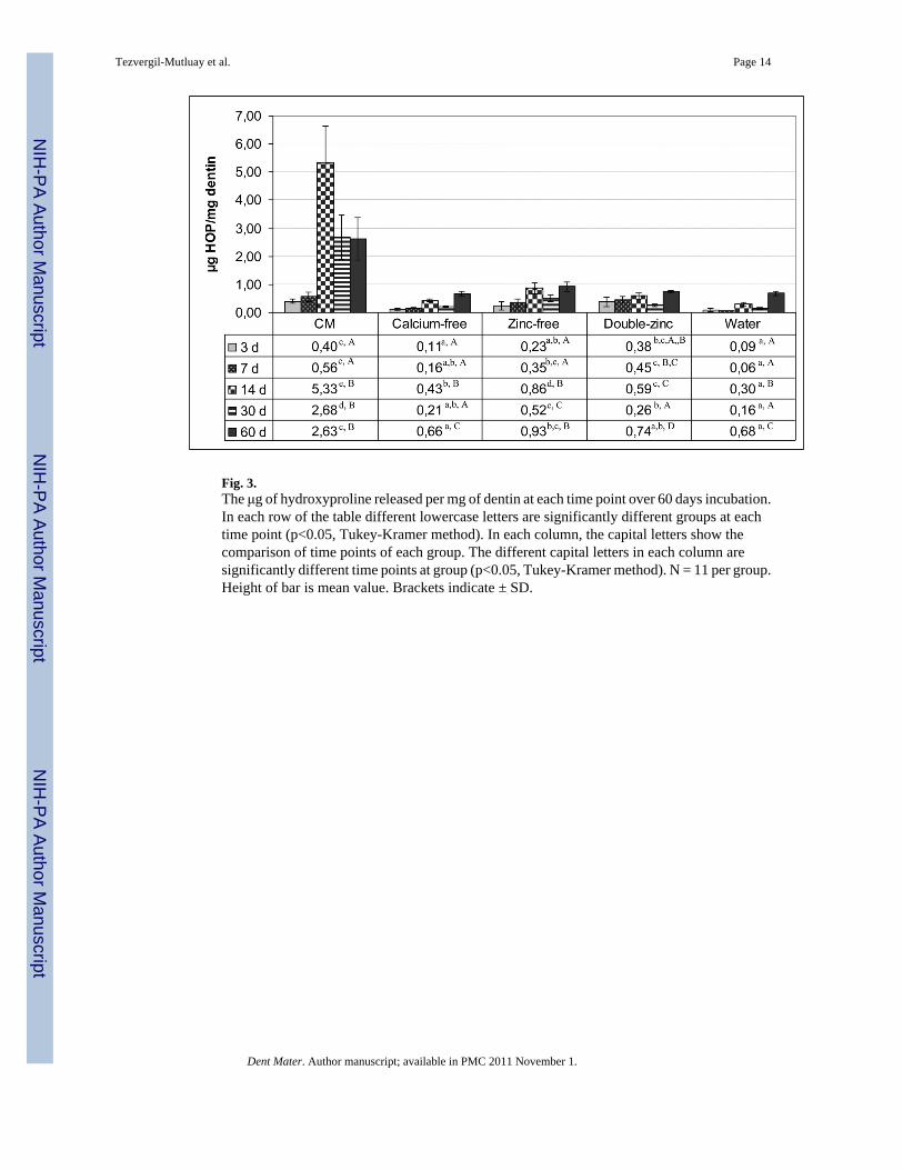
Fig. 3.The μg of hydroxyproline released per mg of dentin at each time point over 60 days incubation.In each row of the table different lowercase letters are significantly different groups at eachtime point (p<0.05, Tukey-Kramer method). In each column, the capital letters show thecomparison of time points of each group. The different capital letters in each column aresignificantly different time points at group (p<0.05, Tukey-Kramer method). N = 11 per group.Height of bar is mean value. Brackets indicate ± SD.
Tezvergil-Mutluay et al. Page 14
Dent Mater. Author manuscript; available in PMC 2011 November 1.
NIH
-PA Author Manuscript
NIH
-PA Author Manuscript
NIH
-PA Author Manuscript

NIH
-PA Author Manuscript
NIH
-PA Author Manuscript
NIH
-PA Author Manuscript
Tezvergil-Mutluay et al. Page 15
Table 1
Electrolyte composition of unsimulated whole saliva vs. simulated oral fluid vs. plasma.
Whole salivaa Complete medium(simulated oral
fluid)b
Plasmac
Sodium 4-6 0 145
Potassium 14 0 4
Calcium 1.5-4 2.5 2.5
Magnesium 0.07 0 1.5
Chloride 11.9 0 10.5
Phosphorus *3.6 0 1.5
Bicarbonate 1.0 5 mM HEPES 25
Fluoride μg% 8-25 μg/100 mL 0 10-20
Iodide μg% 4-24 μg/100 mL 0 3-8
Zn ? 0.05 0.02
NaN3 (antimicrobial) 0 0.3 mM 0
*3.6 mM phosphate should be added to simulated oral fluids if mineralized tissues are to be incubated in the solution for more than 24 h;
afrom Dawes (1974;
bcurrent paper;
cSuddick et al., 1980.
Dent Mater. Author manuscript; available in PMC 2011 November 1.
Copyright © 2022 FDOKUMEN