The relevance of tissue angiotensin-converting enzyme: manifestations in mechanistic and endpoint...
20
The Relevance of Tissue Angiotensin-Converting Enzyme: Manifestations in Mechanistic and Endpoint Data Victor J. Dzau, MD, Kenneth Bernstein, MD, David Celermajer, MBBS, PhD, Jerome Cohen MD, Bjo ¨rn Dahlo ¨f, MD, PhD, John Deanfield, MD, Javier Diez, MD, PhD, Helmut Drexler, MD, Roberto Ferrari, MD, Wiek van Gilst, PhD, Lennart Hansson, MD, Burkhard Hornig, MD, Ahsan Husain, PhD, Colin Johnston, MD, Harold Lazar, MD, Eva Lonn, MD, Thomas Lu ¨scher, MD, John Mancini, MD, Albert Mimran, MD, Carl Pepine, MD, Ton Rabelink, MD, PhD, Willem Remme, MD, PhD, Luis Ruilope, MD, Marcel Ruzicka, MD, Heribert Schunkert, MD, Karl Swedberg, MD, Thomas Unger, MD, Douglas Vaughan, MD, and Michael Weber, MD Angiotensin-converting enzyme (ACE) is primarily local- ized (>90%) in various tissues and organs, most notably on the endothelium but also within parenchyma and inflammatory cells. Tissue ACE is now recognized as a key factor in cardiovascular and renal diseases. Endo- thelial dysfunction, in response to a number of risk factors or injury such as hypertension, diabetes mellitus, hypercholesteremia, and cigarette smoking, disrupts the balance of vasodilation and vasoconstriction, vascular smooth muscle cell growth, the inflammatory and oxi- dative state of the vessel wall, and is associated with activation of tissue ACE. Pathologic activation of local ACE can have deleterious effects on the heart, vascula- ture, and the kidneys. The imbalance resulting from increased local formation of angiotensin II and in- creased bradykinin degradation favors cardiovascular disease. Indeed, ACE inhibitors effectively reduce high blood pressure and exert cardio- and renoprotective actions. Recent evidence suggests that a principal target of ACE inhibitor action is at the tissue sites. Pharmaco- kinetic properties of various ACE inhibitors indicate that there are differences in their binding characteristics for tissue ACE. Clinical studies comparing the effects of an- tihypertensives (especially ACE inhibitors) on endothelial function suggest differences. More comparative experi- mental and clinical studies should address the signifi- cance of these drug differences and their impact on clinical events. 2001 by Excerpta Medica, Inc. Am J Cardiol 2001;88(suppl):1L–20L From the Brigham Women’s Hospital, Department of Medicine, Bos- ton, Massachusetts, USA (VJD); Emory University School of Medicine, Department of Pathology and Laboratory Medicine, Atlanta, Georgia, USA (KB); University of Sydney, Department of Medicine, c/o Depart- ment of Cardiology, Royal Prince Alfred Hospital, Sydney, Australia (DC); Saint Louis University School of Medicine, Department of Internal Medicine, Saint Louis, Missouri, USA (JC); Sahlgrenska University Hospital/Ostra, Department of Medicine, Nilssonsberg, Sweden (BD); Great Ormond Street Hospital for Children, Vascular Physiology Unit, London, United Kingdom (J Deanfield); Universidad de Navarra, Unidad de Fisiopatologia Vascular, Edificio de Ciencias, Calle Iru- niarrea, Pampalona, Spain (J Diez); Medizinische Hochschule Han- nover, Department of Cardiology, Hannover, Germany (HD, BH); Nuove Cliniche, Department of Clinical and Experimental Medicine, Corso Ferrara, Italy (RF); University Hospital Groningen, Department of Clinical Pharmacology, Groningen, the Netherlands (WVG); Univer- sity of Uppsala, Clinical Hypertension Research, Uppsala, Sweden (LH); Victor Chang Cardiac Research Institute, Darlinghurst, Sydney, Australia (AH); Baker Institute, Melbourne, Australia (CJ); Boston Med- ical Center, Department of Cardiothoracic Surgery, Boston, Massa- chusetts, USA (HL); Hamilton General Hospital–McMaster Clinic, Di- vision of Cardiology, Hamilton, Ontario, Canada (EL); University Hos- pital Zurich, Division of Cardiology, Zurich, Switzerland (TL); Vancouver General Hospital, Department of Medicine, Vancouver, British Columbia, Canada (JM); Hospital Lapeyronie, Medecine In- terne et Hypertension Arterielle, Centre Hospitalier, Universitaire, Montpellier, France (AM); University of Florida Gainesville, Division of Cardiovascular Medicine, Gainesville, Florida, USA (CP); University Hospital Utrecht, Department of Internal Medicine, Utrecht, the Neth- erlands (TR); Sticares Cardiovascular Research Foundation, Rhoon, The Netherlands (WR); Unidad de Hypertension, Hospital, Madrid, Spain (LR); University of Ottawa Heart Institute, Ottawa, Ontario, Canada (MR); Klinik und Poliklinik fur Innere Medizin II, Franz-Josef- Strauss-Allee, Regensburg, Germany (HS); Sahlgrenska University Hos- pital/Ostra, Go ¨teberg, Sweden (KS); Christian-Albrechts-University Kiel, Kiel, Germany (TU); Vanderbilt University School of Medicine, Division of Nashville, Tennessee, USA (DV); Brookdale Hospital, De- partment of Medicine, Brooklyn, New York, USA (MW). This work was supported by a grant from Parke-Davis (now Pfizer Inc.). Address for reprints: Victor J. Dzau, MD, Brigham Women’s Hospital, 75 Francis Street, Suite 210, Tower 1, Boston, Massachusetts 02115. 1L ©2001 by Excerpta Medica, Inc. 0002-9149/01/$ – see front matter All rights reserved. PII S0002-9149(01)01878-1
Transcript of The relevance of tissue angiotensin-converting enzyme: manifestations in mechanistic and endpoint...

The Relevance of TissueAngiotensin-Converting Enzyme:
Manifestations in Mechanistic andEndpoint Data
Victor J. Dzau, MD, Kenneth Bernstein, MD, David Celermajer, MBBS, PhD,Jerome Cohen MD, Bjorn Dahlof, MD, PhD, John Deanfield, MD, Javier Diez, MD, PhD,Helmut Drexler, MD, Roberto Ferrari, MD, Wiek van Gilst, PhD, Lennart Hansson, MD,
Burkhard Hornig, MD, Ahsan Husain, PhD, Colin Johnston, MD, Harold Lazar, MD,Eva Lonn, MD, Thomas Luscher, MD, John Mancini, MD, Albert Mimran, MD,
Carl Pepine, MD, Ton Rabelink, MD, PhD, Willem Remme, MD, PhD, Luis Ruilope, MD,Marcel Ruzicka, MD, Heribert Schunkert, MD, Karl Swedberg, MD, Thomas Unger, MD,
Douglas Vaughan, MD, and Michael Weber, MD
Angiotensin-converting enzyme (ACE) is primarily local-ized (>90%) in various tissues and organs, most notablyon the endothelium but also within parenchyma andinflammatory cells. Tissue ACE is now recognized as akey factor in cardiovascular and renal diseases. Endo-thelial dysfunction, in response to a number of riskfactors or injury such as hypertension, diabetes mellitus,hypercholesteremia, and cigarette smoking, disrupts thebalance of vasodilation and vasoconstriction, vascularsmooth muscle cell growth, the inflammatory and oxi-dative state of the vessel wall, and is associated withactivation of tissue ACE. Pathologic activation of localACE can have deleterious effects on the heart, vascula-ture, and the kidneys. The imbalance resulting fromincreased local formation of angiotensin II and in-
creased bradykinin degradation favors cardiovasculardisease. Indeed, ACE inhibitors effectively reduce highblood pressure and exert cardio- and renoprotectiveactions. Recent evidence suggests that a principal targetof ACE inhibitor action is at the tissue sites. Pharmaco-kinetic properties of various ACE inhibitors indicate thatthere are differences in their binding characteristics fortissue ACE. Clinical studies comparing the effects of an-tihypertensives (especially ACE inhibitors) on endothelialfunction suggest differences. More comparative experi-mental and clinical studies should address the signifi-cance of these drug differences and their impact onclinical events. �2001 by Excerpta Medica, Inc.
Am J Cardiol 2001;88(suppl):1L–20L
From the Brigham Women’s Hospital, Department of Medicine, Bos-ton, Massachusetts, USA (VJD); Emory University School of Medicine,Department of Pathology and Laboratory Medicine, Atlanta, Georgia,USA (KB); University of Sydney, Department of Medicine, c/o Depart-ment of Cardiology, Royal Prince Alfred Hospital, Sydney, Australia(DC); Saint Louis University School of Medicine, Department of InternalMedicine, Saint Louis, Missouri, USA (JC); Sahlgrenska UniversityHospital/Ostra, Department of Medicine, Nilssonsberg, Sweden(BD); Great Ormond Street Hospital for Children, Vascular PhysiologyUnit, London, United Kingdom (J Deanfield); Universidad de Navarra,Unidad de Fisiopatologia Vascular, Edificio de Ciencias, Calle Iru-niarrea, Pampalona, Spain (J Diez); Medizinische Hochschule Han-nover, Department of Cardiology, Hannover, Germany (HD, BH);Nuove Cliniche, Department of Clinical and Experimental Medicine,Corso Ferrara, Italy (RF); University Hospital Groningen, Department ofClinical Pharmacology, Groningen, the Netherlands (WVG); Univer-sity of Uppsala, Clinical Hypertension Research, Uppsala, Sweden(LH); Victor Chang Cardiac Research Institute, Darlinghurst, Sydney,Australia (AH); Baker Institute, Melbourne, Australia (CJ); Boston Med-ical Center, Department of Cardiothoracic Surgery, Boston, Massa-chusetts, USA (HL); Hamilton General Hospital–McMaster Clinic, Di-
vision of Cardiology, Hamilton, Ontario, Canada (EL); University Hos-pital Zurich, Division of Cardiology, Zurich, Switzerland (TL);Vancouver General Hospital, Department of Medicine, Vancouver,British Columbia, Canada (JM); Hospital Lapeyronie, Medecine In-terne et Hypertension Arterielle, Centre Hospitalier, Universitaire,Montpellier, France (AM); University of Florida Gainesville, Division ofCardiovascular Medicine, Gainesville, Florida, USA (CP); UniversityHospital Utrecht, Department of Internal Medicine, Utrecht, the Neth-erlands (TR); Sticares Cardiovascular Research Foundation, Rhoon,The Netherlands (WR); Unidad de Hypertension, Hospital, Madrid,Spain (LR); University of Ottawa Heart Institute, Ottawa, Ontario,Canada (MR); Klinik und Poliklinik fur Innere Medizin II, Franz-Josef-Strauss-Allee, Regensburg, Germany (HS); Sahlgrenska University Hos-pital/Ostra, Goteberg, Sweden (KS); Christian-Albrechts-UniversityKiel, Kiel, Germany (TU); Vanderbilt University School of Medicine,Division of Nashville, Tennessee, USA (DV); Brookdale Hospital, De-partment of Medicine, Brooklyn, New York, USA (MW).
This work was supported by a grant from Parke-Davis (now PfizerInc.).
Address for reprints: Victor J. Dzau, MD, Brigham Women’s Hospital,75 Francis Street, Suite 210, Tower 1, Boston, Massachusetts 02115.
1L©2001 by Excerpta Medica, Inc. 0002-9149/01/$ – see front matterAll rights reserved. PII S0002-9149(01)01878-1

INTRODUCTION
Our awareness and appreciation of the role oftissue angiotensin-converting enzyme (ACE) in
endothelial function and vascular health has begun toinfluence the treatment of cardiovascular and renaldisorders. The results of experimental and clinicalresearch have provided the rationale for intervening inthe underlying pathophysiologic processes associatedwith activated tissue ACE in conditions such as con-gestive heart failure, coronary artery disease, hyper-tension, and nephrosclerosis. Extensive evidence in-dicates that ACE inhibition favorably affects the heart,
the vasculature, and the kidney, the results of whichare associated with improved patient outcomes. Thisconsensus report will provide an extensive review ofthe biology and function of tissue ACE, its role in thepathophysiology of cardiovascular disease, the impor-tance of tissue ACE as a therapeutic target, and evi-dence from clinical trials for the beneficial effects oftissue-ACE inhibition. The article will also examinethe pharmacologic properties of ACE inhibitors andexplore the potential clinical effects related to differ-ences in binding for tissue ACE.
TISSUE ANGIOTENSIN-CONVERTING ENZYME: BIOLOGY,FUNCTION, AND PATHOPHYSIOLOGY
The structure of ACE is well known, and theenzyme’s predominant localization in tissue, ratherthan plasma, was established nearly 30 years ago.Despite this knowledge and an abundance of recentexperimental data, the role of genetic variability inACE activity has yet to be fully resolved.
BIOCHEMISTRY AND GENETICSOF ACE
ACE: ACE is a zinc metallopeptidase that catalyzes1 of the main steps in the renin cascade—the conver-sion of angiotensin I (Ang I) to angiotensin II (Ang II),a potent vasoconstrictor. ACE is also involved in theinactivation of the vasodilator hormones, bradykininand substance P.1 The ACE enzyme exists in 2 forms,a high molecular weight form (170 kDa) found inendothelial, epithelial, and neuronal cells, and a lowmolecular weight form (90 kDa) found in germinalcells. The 2 forms are encoded by 2 different messen-ger RNAs corresponding to molecular sizes of 2.0kilobase (kb) and 4.3 kb. ACE is found in the plasmaand in a number of tissues including blood vessels,heart, kidney, brain, and the adrenal gland.2 SomaticACE (the form of ACE made by endothelium andother somatic tissues) is a single polypeptide chainthat contains 2 homologous protein domains. Eachdomain is independently catalytic with roughly equiv-alent affinities for Ang I. ACE is synthesized with anamino terminal signal sequence. This leads to exportof both catalytic domains from the cell, but the lastcarboxyl-terminal portion of the molecule is hydro-phobic and anchors the protein within the cell mem-brane. Thus, ACE is an ectoenzyme with both cata-lytic domains outside of the cell (Figure 1).
Plasma versus tissue ACE: Biochemical measure-ments of ACE activity illustrate that ACE is a tissue-based enzyme.3 Indeed, �10% of ACE is found cir-culating in the plasma.3 The functional importance of
tissue-based ACE has been demonstrated in geneti-cally altered mice devoid of tissue ACE but havingsubstantial plasma ACE levels. These mice have dem-onstrated an inability to activate their renin–angioten-sin system and consequently develop marked hypo-tension.4 The precise function of plasma ACE is un-clear. However, because it represents only a smallproportion of the body’s total ACE activity, its role isthought to be minimal.
Role of the genetic variations of ACE: The chromo-somal locus of the ACE gene has been linked to thevariability of ACE activity and arterial hypertension,as well as left ventricular mass (independent of bloodpressure) in several rodent breeding experiments.5,6 Inaddition, genetic factors may also regulate vasculatureACE expression and production in humans. In 1990,Rigat et al7 described an insertion/deletion polymor-phism of the ACE gene that accounted for 40% of theinterindividual variation in serum and cardiac ACEactivity.7–9 ACE levels are highest in individuals whoare homozygous for the D allele, lowest in thosehomozygous for the I allele, and intermediate in I/Dheterozygous individuals. Since 1990, the ACE Dallele has been associated with a number of diseasestates for which activation of the renin–angiotensinsystem has been implicated in playing a role, includ-ing acute myocardial infarction (MI) in low-risk pa-tients,10 left ventricular hypertrophy,11–13 and progres-sive diabetic nephropathy.14 This association has beenattributed to increased formation of Ang II in individ-uals who carry the ACE D allele. These results havenot been duplicated by other investigators.15,16 It hasbeen suggested that in healthy subjects, negative feed-back inhibition may neutralize the genetically en-hanced expression of singular components in the AngII synthetic cascade.17 By contrast, the ACE DD ge-notype may play a substantial role in the developmentof left ventricular hypertrophy when the cardiacgrowth machinery is activated. This hypothesis is il-
2L THE AMERICAN JOURNAL OF CARDIOLOGY� VOL. 88 (9A) NOVEMBER 8, 2001

lustrated by recent data from Montgomery et al18 inwhich young healthy subjects were studied before andafter a rigorous exercise protocol. Only those partici-pants who carried the ACE deletion allele displayedan increase in left ventricular mass. Thus, the ACEgenotype may act only under specific conditions, sug-gesting an interaction between altered hemodynamics,ACE, or other genetic cofactors in the modulation ofleft ventricular mass. In agreement with this notion arethe observations of Pinto et al19 and Ohmichi et al20
who both found that pathologic remodeling early afterMI occurs predominantly in those subjects with theACE DD genotype. Furthermore, transgenic rats withhigh levels of cardiac ACE expression have normal(or even smaller) hearts, as long as these animals arehoused under physiologic conditions. However, car-diac growth and diastolic dysfunction were augmentedin the same ACE transgenic rats when the animalswere stressed by abdominal aortic banding and sub-sequent cardiac pressure overload.21 However, theACE gene polymorphism has not been consistentlyassociated with hypertension or the prevalence or ex-tent of coronary artery disease or MI.22,23 Thus, therole of the genetic variability of ACE remains to befully elucidated.
TISSUE ACE, THE CARDIOVASCULARSYSTEM, AND THE KIDNEYS
The importance of tissue ACE in the pathophysi-ology of cardiovascular disease is reflected by findingsthat, despite the existence of alternative Ang II path-ways, marked ACE induction occurs in almost allmodels of cardiac injury. Within the vasculature, tis-sue ACE plays a critical role in endothelial functionthrough the direct pleiotropic actions of Ang II andalso through a bradykinin-dependent mechanism.There is also substantial evidence that in atheroscle-rosis, plaque represents an important target of ACEinhibitor action. Finally, the kidneys are especiallysusceptible to the toxic effects of chronically elevatedlevels of Ang II; thus, the exuberant response to injurymay ultimately lead to renal failure.
Tissue ACE and the heart: TISSUE SITES OF ACE EX-
PRESSION: ACE activity is distributed in a tissue andcell-type specific fashion.3 Very high levels are foundin the capillary bed of the lungs.24 Because of its highACE levels, the pulmonary vasculature—albeit a tis-sue site—is considered an integral part of the classiccirculating renin–angiotensin system.24 In contrast,some tissues, including the heart, express relativelylow levels of ACE, at least under physiologic condi-
FIGURE 1. Angiotensin-converting enzyme (ACE; EC 3.4.15.1) Schematic drawing shows thestructure of ACE. There is a catalytic site on each extracellular lobe, each of which binds azinc (Zn2�) atom. (Adapted with permission from Hypertension).159
A SYMPOSIUM: TISSUE ANGIOTENSIN-CONVERTING ENZYME 3L

tions.3,25 Within the normal heart, the right atriumelaborates a moderate density of ACE, which is higherthan that of the left atrium and the ventricles.26 Thevast majority of immunohistochemical ACE stainingis found in the endothelium of large and small cardiacarteries and arterioles, whereas only half the capillar-ies are immunoreactive, and venous vessels are almostcompletely devoid of the enzyme.26,27 Other sites ofcardiac tissue ACE expression include the endocardiallayer and the cardiac valves.26 Very little, if any, ACEis found in normal adult cardiac myocytes in situ.
LOCALIZATION AND REGULATION OF ACE IN HEARTDISEASE: After our initial observation of ACE upregu-lation in pressure-overloaded, hypertrophied hearts,25
marked ACE induction has been found in virtually allmodels of cardiac injury including volume over-load,28,29 MI,30,31 and heart failure.32 Additionally,increased cardiac ACE levels have been correlatedwith the aging process.33 Elevated wall stress is be-lieved to be a critical factor for cardiac ACE induc-tion, because elevated enzyme levels were found ex-clusively in the affected ventricle.34 Interestingly,
ACE upregulation is not restricted to the vascula-ture,27 because fibroblasts and myocytes are also re-cruited for ACE expression in injured hearts.27,31,32
Likewise, cardiac myocytes in cell culture have beenreported to express ACE and are able to generate AngII locally, especially in response to mechanicalstretch.35 Moreover, macrophages invade injuredmyocardium and carry high levels of ACE activity tointerstitial sites where Ang II, the product of ACE,accumulates.27,36,37 In addition, mast cells in cardiactissue are another source of tissue Ang II through theaction of chymase.38 The role of tissue ACE in theheart is summarized in Figure 2.
Whereas cardiac ACE increases in the failingheart, pulmonary ACE tends to decrease when pulmo-nary congestion complicates the condition.39 Theseopposing regulatory steps may protect Ang I fromconversion/degradation in the lung and increase ACEsubstrate in the heart.39
KINETICS AND MECHANISMS OF CARDIAC ANG II FOR-MATION: During a single passage through the coronarysystem, approximately 3% to 10% of Ang I is con-
FIGURE 2. Origins and actions of myocardial tissue angiotensin-converting enzyme (ACE)–angiotensin II (Ang II). AT1R � angiotensinII type 1 receptor; AT2R � angiotensin II type 2 receptor; NE � norephrine.
4L THE AMERICAN JOURNAL OF CARDIOLOGY� VOL. 88 (9A) NOVEMBER 8, 2001

verted to Ang II.25,40 However, these measurementsmay only reflect vascular conversion. More preciseinsights on the intracardiac events leading to Ang IIgeneration were revealed by experiments that usedintracoronary infusions of minute concentrations ofradiolabeled (exogenous) Ang I or Ang II followed bymeasurements of native (endogenous) as well as la-beled angiotensins in the interstitial fluid, the cellularcompartment, and the coronary effluent.8,41 These ex-periments revealed that angiotensinogen and renin areextracted from the coronary circulation.42,43 Indeed,cardiac concentrations of renin may substantially ex-ceed renin levels in the plasma, suggesting an activemechanism for cardiac renin accumulation.42,43 In ad-dition, there appears to be local generation of angio-tensinogen and renin, at least during disease condi-tions.44,45
These kinetic studies document that �80% of AngI found in the cardiac interstitium is formed locally byrenin (which is largely taken up from the circulation)cleaving angiotensinogen (which is both locallyformed and taken up from the circulation).40,41 Like-wise, most of the Ang II found in the heart is synthe-sized in situ. Specifically, the conversion of Ang I toAng II appears to be mediated by tissue ACE ratherthan blood-derived enzymes.46 Consequently, the tis-sue levels of Ang II are several times higher than thecirculating levels.36 It is conceivable, therefore, thatthe local levels of ACE activity reflect the cardiac AngII concentrations.
In experimental models and in humans, the cardiacconversion of Ang I to Ang II is largely blocked byACE inhibitors.37,46–49 By contrast, in ex vivo mem-brane preparations of cardiac tissue (human and rat),the conversion of Ang I to Ang II occurs largelyindependently of ACE.50,51 Chymase, a mast cell en-zyme with high affinity for Ang I, has been shown tocatalyze this reaction and chymase inhibitors wereeffective in its inhibition.38 This apparent discrepancybetween in vivo and in vitro data51 may have beenresolved by the findings of Kokkonen et al,52 whodemonstrated that interstitial fluid completely inhibitschymase activity, whereas ACE remains active underthese same conditions. Despite this finding, chymasemay still be important, and further investigation isnecessary to define its role in the formation of Ang IIin humans.
FUNCTIONAL ROLE OF ACE IN THE NORMAL AND FAIL-ING HEART: The normal development of the heart doesnot require the functional integrity of the cardiac re-nin–angiotensin system. Thus, genetically alteredmice lacking cardiac ACE do not experience cardiacpathology.53 Furthermore, Ang II is not required forthe maintenance of normal cardiac function.54 In thisregard, the role of the cardiac renin–angiotensin sys-tem differs from that of the renal renin–angiotensinsystem, which requires Ang II for normal kidneydevelopment.55
In the failing heart, however, activation of therenin–angiotensin system may have a series of func-tional implications. Ang II has been shown to enhanceprotein synthesis independently of load in the intact
heart as well as in the isolated myocyte.56,57 Ang II,then, is considered to be an important factor contrib-uting to the development of cardiac hypertrophy. ACEappears to be involved in this process because, on theone hand, the activity of the enzyme is enhanced inhypertrophied hearts and on the other hand, inhibitionof the enzyme may cause regression of left ventricularhypertrophy, even when the pressure or volume over-load persists.58,59 Even more strikingly, the inhibitionof cardiac ACE with a high tissue-affinity ACE inhib-itor (quinapril) prevented the development of volumeoverload hypertrophy more efficiently than an ACEinhibitor (enalapril) with low affinity for tissueACE.28,60 Moreover, tissue-ACE activity is involvedin the pathogenesis of coronary vascular and myocar-dial structural changes induced by long-term blockadeof nitric oxide synthesis.61
Ang II not only induces hypertrophy of cardiacmyocytes but also hyperplasia of cardiac fibroblasts.62
Accordingly, the activation of the cardiac renin–an-giotensin system and specifically, cardiac ACE, maycontribute to the development of cardiac fibrosis.62 Ithas been demonstrated that fibrotic areas of the heartdisplay the highest levels of cardiac ACE activity31,32
and that fibroblasts themselves generate Ang II.63
ACE inhibitors, on the other hand, may prevent theaccumulation of extracellular matrix proteins and thedevelopment of fibrosis of the heart (Figure 3), evenwhen pressure overload persists.64,65 The intimatecommunication between cardiac fibroblasts and myo-cytes was elegantly demonstrated in chimeric micethat had Ang II receptor type 1A gene null mutantcells and Ang II receptor type 1A gene intact cellsexpressing the lacZ gene. Proliferating cardiac fibro-blasts were present predominantly in areas of Agtr1aintact cardiomyocytes. Therefore, an intact cardiacrenin–angiotensin system appears to be a requirementfor local proliferation of fibroblasts and the conse-quent development of fibrosis.66
Ang II has also been shown to induce apoptosis ofcardiac myocytes, whereas cardiac fibroblasts arefairly resistant to the effects of Ang II on cell death.67
Specifically, the enhanced local renin–angiotensinsystem decreases the bcl-2-to-BAX protein ratio incardiomyocytes, thus decreasing the resistance to un-dergo programmed cell death.67 There appears to be avicious circle, given that an apoptosis-related protein,p53, induces the local renin–angiotensin system.68 Infact, the induction of cardiac ACE parallels the ap-pearance of apoptosis in the pressure-overloadedheart.69 Again, use of ACE inhibitors has been shownto prevent apoptosis of cardiac myocytes in pressure-overloaded hearts.69
The activation of tissue ACE in cardiac remodelinghas direct functional consequences. Ang II causes adepression of diastolic function in the hypertrophiedheart.25,47 Likewise, perfusion of isolated hearts withAng I, followed by intracardiac conversion to Ang II,causes an increase in left ventricular end-diastolicpressure, suggesting that local ACE may facilitate thisresponse.47 ACE inhibitors infused into the coronaryarteries of isolated experimental hearts or hearts of
A SYMPOSIUM: TISSUE ANGIOTENSIN-CONVERTING ENZYME 5L

patients with aortic stenosis caused a significant im-provement in diastolic function.47,70,71 This responseis even amplified in hearts exposed to an ischemia/reperfusion injury.72 Conversely, the effects of Ang IIon systolic function in the failing ventricles are min-imal.54
Local generation of Ang II may also increase thevascular tone of the coronary bed. Specifically, inpatients with dilated cardiomyopathy, intracoronaryenalaprilat induced a significant coronary vasodila-tion,73 and in an animal model of cardiomyopathy,long-term treatment with quinapril resulted in a sig-nificant cardioprotective effect.74 These data providestrong evidence for the functional significance of thecardiac renin–angiotensin system in both patients withheart disease and in controlled experimental situa-tions.
In 2 studies, the effects of ACE inhibition (ramiprilor fosinopril) on survival in experimental aortic ste-nosis were examined.59,71 This model allows the sep-aration of peripheral and cardiac drug effects, becauseafterload reduction is prevented by a clip at the as-cending aorta. Both studies demonstrated a survivalbenefit in animals receiving the ACE inhibitor, thussuggesting that the inhibition of cardiac ACE contrib-utes to the prognostic relevance of these agents inpatients with heart failure.75,76
Role of tissue ACE in the vasculature: REGULATION OF
VASCULAR ACE: ACE is the most important enzymecontrolling the activation of angiotensin and the deg-radation of bradykinin.77 Although ACE is widelydistributed through the tissues, it appears that ACEexpression is regulated by a number of different mech-anisms. In cultured endothelial cells, the expression ofACE is modulated by steroids, calcium ionophores,and growth factors.78 The expression of ACE in en-dothelial cells and culture is also a function of con-fluence, as ACE enzyme levels increase exponentiallyafter confluence is obtained.79 Thus, the regulation ofendothelial ACE is a determinant of vascular functionin both health and disease.
Studies of Ang I infusion into human forearm orcoronary arteries have shown that Ang I is convertedto Ang II. This conversion is blocked by ACE inhib-itor treatment. The primary vasodilatory action ofACE inhibitors is the blockade of Ang II formation.The contribution of bradykinin to the action of ACEinhibitors has been debated. With long-term adminis-tration, ACE inhibitors lower blood pressure, even inpatients with low renin hypertension, suggesting aneffect that is independent of a decrease in Ang II.Bradykinin is a potent vasodilator, acting through therelease of prostacyclin, nitric oxide, and endothelial-derived hyperpolarization factor. Accurate measure-
FIGURE 3. Angiotensin-converting enzyme (ACE) inhibition and fibrinolysis. Inhibition of ACE prevents the degradation of bradykininand the formation of angiotensin II (Ang II), thus preserving fibrinolytic balance. Ang I � angiotensin I; PAI-1 � plasminogen activa-tor inhibitor type 1. (Adapted with permission from Circulation.160)
6L THE AMERICAN JOURNAL OF CARDIOLOGY� VOL. 88 (9A) NOVEMBER 8, 2001

ment of bradykinin concentrations in plasma has beentechnically challenging; these concentrations havebeen shown to be increased or unchanged after ACEinhibition. Although it is clear that ACE inhibitionpotentiates the hemodynamic effects of exogenousbradykinin, this observation does not address whetherendogenous bradykinin plays a part in the action ofACE inhibitors. Recent studies performed by Gaineret al80 indicate that the coadministration of the brady-kinin receptor antagonist, icatibant acetate (HOE 140),significantly attenuates the hypotensive effect of cap-topril. Although HOE 140 does not alter the renalhemodynamic response to captopril, it does signifi-cantly alter the change of plasma renin activity inresponse to ACE inhibition. These effects appear to besimilar in both normotensive and hypertensive sub-jects. These data confirm that bradykinin contributesto the short-term effects of ACE inhibition in bloodpressure in normotensive and hypotensive persons andsuggest that bradykinin also contributes to the short-term effects of ACE inhibition on the renin–angioten-sin system. Similar results have been seen in theeffects of ACE inhibitors on endothelial vasodilatorfunction. Studies performed by Hornig et al81 haveshown that ACE inhibitors augment flow-dependent,endothelial-mediated dilation in humans by a brady-kinin-dependent mechanism.
ACE regulates other important vascular functions
(Figure 3). Studies in healthy human volunteers haveprovided additional support for ACE in regulatingvascular fibrinolytic balance. Specifically, examina-tion of the effect of activation of the renin–angiotensinsystem by low salt intake (10 mEq vs 200 mEq so-dium per day) on plasma fibrinolytic parameters dem-onstrated that low salt intake was associated with asignificant increase in morning plasminogen activatorinhibitor type 1 (PAI-1) levels, and plasma PAI-1correlated dramatically with serum aldosterone levels(R � 0.56, p �0.10�7). Treatment with quinaprilsignificantly lowered PAI-1 concentrations and themolar ratio of PAI-1 to tissue plasminogen activatorthroughout the day.82
PATHOPHYSIOLOGY OF VASCULAR ACE: The endothe-lium plays a crucial role in the maintenance of normalvascular tone and structure, local hemostasis, and vas-cular-wall proliferation processes (Figure 4).83,84
These processes are mediated by the reactive releaseof vasoactive substances (thromboxane A2, free radi-cals, endothelin, prostacyclin) among which nitric ox-ide is perhaps the most important. Nitric oxide (1)relaxes vascular smooth muscle through a cyclicguanosine monophosphate–mediated decrease in cy-tosolic calcium, resulting in vasodilation; (2) mediatescoagulation by the inhibition of platelet aggregationand the expression of adhesion molecules for bothmonocytes and neutrophils; and (3) prevents structural
FIGURE 4. Origins and actions of vascular tissue angiotensin-converting enzyme (ACE)-angiotensin II (Ang II). FGF � fibroblast growthfactor; IGF � insulinlike growth factor; IL-6 � interleukin-6; MCP-1 � monocyte chemoattractant protein-1; PAI-1 � plasminogen ac-tivator inhibitor type 1; PDGF � platelet-derived growth factor; TGF-� � transforming growth factor–�.
A SYMPOSIUM: TISSUE ANGIOTENSIN-CONVERTING ENZYME 7L

changes by inhibiting the growth and migration ofsmooth muscle cells. These regulatory processes areall subject to disruption by Ang II.
Ang II, elaborated by activated endothelial ACE,impairs nitric oxide bioactivity, mainly because ofoxidative stress through the Ang II–induced produc-tion of superoxide radicals (O2�) that can scavengenitric oxide and reduce endothelium-dependent vaso-dilation.85 This action is independent of the effects ofACE in degrading bradykinin and modulating theendothelial-dependent vasodilation in response to ac-tivation of the �2-receptor.
There is evidence that ACE expression is increasedin atherosclerosis and that Ang II may contribute todisease progression by increasing oxidative stress andattenuating chemoattractant and adhesion moleculeexpression, leading to inflammation. As discussed,tissue Ang II can also exert proproliferative and pro-thrombotic actions (Figure 4). Diet et al86 reported thattissue ACE in human atherosclerotic plaque localizesto regions of inflammatory cells, especially areas ofclustered macrophages and microvessel endothelialcells. The accumulation of ACE and metalloprotein-ase in the shoulder region of the vulnerable plaquemay contribute to increased local circumferentialstress and plaque instability. Thus, ACE accumulationwithin the vascular lesions may be a factor in thepathophysiology of coronary artery disease.
This hypothesis is underscored by the findings ofan elegant experiment in which endothelial nitric ox-ide synthetase gene knockout mice developed athero-sclerotic lesions in response to adventitial vessel-wallinjury.87 Wild-type mice with normal endothelialfunction were able to produce nitric oxide and weretherefore protected from this effect. The evidencecited above indicates that plaque ACE may be animportant target of ACE inhibitor action.
Tissue ACE and the kidney: The prominent role ofAng II in renal physiology, as briefly outlined below,renders the kidneys highly susceptible to injury causedby the de novo production of Ang II. The kidneys,under the regulation of Ang II and aldosterone, main-tain the electrolyte balance in the body. Sodium ho-meostasis, in particular, is maintained by the localaction of Ang II on both the proximal and distaltubules. The filtration function of the kidneys is alsopreserved during changes in systemic blood pressureby local Ang II, which acts to constrict the afferentand efferent glomerular arterioles. The efferent arte-rioles are very sensitive to Ang II, and the resultingvasoconstriction, together with prostaglandin-inducedvasodilation of the afferent arterioles, regulates intra-glomerular pressure, thereby maintaining the glomer-ular filtration rate.
Because Ang II is essential in normal kidney func-tion, increases in the level of locally elaborated Ang IIfrequently result in pathophysiologic conditions. Inrenovascular hypertension, for example, filter functionof the ischemic kidney is compensated with afferentvasodilation and efferent Ang II–induced vasocon-striction. Renin production is greatly increased aswell. This response to injury increases blood pressure,exposing the contralateral kidney to the sequelae ofsystemic hypertension. Ang II also maintains the glo-merular filtration rate in chronic renal failure regard-less of the cause of tissue damage. Despite this com-pensatory response, there is a progressive loss of renalfunction that results in further Ang II–generated in-creases in glomerular blood pressure and, therefore,continuing injury to the remaining nephrons.88 AngII–induced glomerular hypertrophy89 and renal fibro-sis90,91 escalate the response to injury into a destruc-tive cycle, which ultimately concludes with completerenal failure.
CLINICAL CONSEQUENCES OF TISSUE ANGIOTENSIN-CONVERTING ENZYME INHIBITION
Based on experimental data, hypertension may beassociated with increased local Ang II production,which may play an important role in vasoconstrictionand direct tissue pathology. Consequently, antihyper-tensive therapy with ACE inhibitors not only controlshypertension by interrupting the renin–angiotensinsystem, but it has the added benefit of reducing therisk associated with Ang II–induced disease pro-cesses, including cardiovascular disease and renal fail-ure. Thus, our evolving understanding of the role oftissue ACE in cardiovascular and renal disease culmi-nates with the therapeutic application of this knowl-edge. In this context, the beneficial consequences oftissue ACE inhibition may occur independently ofchanges in blood pressure (ie, overt renin–angiotensinsystem activation); therefore, the value of inhibiting
tissue ACE may extend to a broader range of patientsthan are currently being treated.
TISSUE ACE INHIBITION ANDHYPERTENSION, DIABETES, ANDRENAL DISEASE
The hallmark of essential hypertension is nephro-sclerosis, the first clinical sign of which is protein(chiefly albumin) in the urine. Proteinuria is a princi-pal predictor of cardiovascular disease in patientswithout diabetes mellitus and with type 2 diabetes,92
as well as in progressive renal disease in type 1diabetes, and in patients with overt diabetic nephrop-athy.93 Treatment with ACE inhibitors has beenshown to consistently reduce proteinuria in these pa-tients, as compared with other antihypertensive agentsthat appear to have milder effects.94–96
8L THE AMERICAN JOURNAL OF CARDIOLOGY� VOL. 88 (9A) NOVEMBER 8, 2001
The lack of an antiproteinuric effect by other an-tihypertensive agents that effectively reduce bloodpressure suggests that renal protection afforded byACE inhibitors may occur through a blood pressure–independent mechanism. Support for this hypothesiscan be derived from examining large clinical trials andevaluation of the high-risk groups that were treated forhypertension.
Aggressive antihypertensive treatment in patientswith type 2 diabetes mellitus was assessed in a sub-group analysis (n � 1,148) of the United KingdomProspective Diabetes study in which 758 patients(tight control group, blood pressure �150/85 mm Hg)were randomized to either an ACE inhibitor or a �-blocker (captopril or atenolol, respectively) as themain treatment.97 A total of 390 patients were treatedless aggressively (blood pressure �180/105 mm Hg)with the same antihypertensive drugs. This study dem-onstrated that aggressively treated patients had clini-cally important reductions in the risks of death orcomplications associated with diabetes compared withpatients who were treated less aggressively regardlessof the antihypertensive agent used. In light of thesedata, could further evaluation of high-risk patientswith modest reductions in blood pressure uncoveradditional beneficial effects attributable to a specificclass of antihypertensive agent?
The Captopril Prevention Project (CAPPP) wasdesigned to compare the effects of ACE inhibition andconventional therapy (diuretics and � blockers) oncardiovascular morbidity and mortality in hyperten-sive patients.98 A subgroup of �700 CAPPP patientswere at increased risk for cardiovascular complica-tions caused by diabetes mellitus. A total of 337 ofthese patients were randomized to captopril and 380 toconventional therapy. Although those patients treatedwith conventional therapy had significantly lowerblood pressure than did the patients who receivedcaptopril, conventional therapy did not result in anyadditional benefit for diabetes-related risk. Those pa-tients treated with the ACE inhibitor had a 66% re-duction in fatal and nonfatal MIs and a reduced fre-quency of all cardiac events and total mortality. More-over, within the entire study population (N � 10,085),the incidence of diabetes was lower in the captopril-treated patients than in those who received conven-tional therapy (relative risk 0.86, confidence interval0.74 to 0.99, p � 0.039).
Evidence from the Appropriate Blood PressureControl in Diabetes (ABCD) trial further supports theadvantage of ACE inhibitor therapy in high-risk pa-tients.99 ABCD was a prospective, randomized,blinded trial comparing the effects of moderate blood-pressure control (80 to 89 mm Hg, target diastolicblood pressure) with intensive control (75 mm Hg,target diastolic blood pressure) on the incidence andprogression of diabetic vascular complications in hy-pertensive patients. First-line antihypertensive therapywith a dihydropyridine calcium antagonist (nisoldip-ine) or enalapril was also evaluated. A clinically im-portant and highly statistically significant difference inthe cardiovascular event rate was observed after 67
months of treatment in the hypertensive cohort. Pa-tients treated with ACE inhibitor therapy had fewernonfatal MIs (chi-square test: p � 0.001), all MIs(chi-square test: p � 0.001), and overall cardiovascu-lar events (chi-square test: p � 0.002) than patientstreated with the calcium antagonist. This relation wasmaintained in both the moderate and intensive blood-pressure control groups.99 Because of ethical consid-erations, there was no placebo control group in thisstudy. Therefore, the difference between the ACEinhibitor group and the calcium antagonist group can-not be definitively ascribed to a beneficial effect ofACE inhibition. It is possible that calcium antagonistsexerted a deleterious effect on this study population.Comparisons with other studies,100–102 however, sug-gest that the rate of MIs in the calcium antagonistgroup is not different from these historical controls;therefore, the results of the ABCD trial may be attrib-uted to a protective effect of ACE inhibition ratherthan to a deleterious effect of calcium antagonists.99
The blood pressure–independent renoprotective ef-fects of ACE inhibition have been clearly establishedin 2 large placebo-controlled clinical trials. The firststudy was conducted to determine whether captoprilhas kidney-protecting properties independent of itseffect on blood pressure in patients with diabetic ne-phropathy.103 All patients had type 1 diabetes melli-tus, proteinuria �500 mg/day, and serum creatinineconcentration �2.5 mg/dL. Patients already on con-ventional antihypertensive therapy were randomizedto captopril (n � 207) or placebo (n � 202) and wereobserved for 4 years. Doubling of baseline serumcreatinine concentration—the primary study end-point—occurred in 43 patients who received placeboand in only 25 ACE inhibitor–treated patients (p �0.007), representing a risk reduction of 48%. Riskassociated with the combined secondary endpoints(death, dialysis, and kidney transplantation) was re-duced by 50%, and an aggregate analysis revealedsignificantly less proteinuria in the captopril-treatedpatients than in those patients who received placebo(p � 0.001). Over the course of the study, there wasno difference in blood pressure in those patients withpre-existing hypertension who were randomized toACE inhibitor therapy (n � 155) or placebo (n � 153,p � 0.16). Among patients who were normotensive atstudy entry, blood pressure was only marginallyhigher in the placebo group (p �0.001). Becauseblood pressure was not different between the groupswith hypertension, and 85% of the patients whoreached the primary endpoint were hypertensive, thedecreased progression of diabetic nephropathy mostlikely occurred through a mechanism that is not de-pendent on blood pressure reduction.
Most recently, treatment with ramipril was foundto result in vasculoprotective and renoprotective ef-fects in patients with diabetes who had a previouscardiovascular event and at least 1 other cardiovascu-lar risk factor.104 A total of 3,577 patients were ran-domized to ramipril (10 mg/day) or placebo, and vi-tamin E or placebo (2�2 factorial design). Treatmentwith ramipril reduced the risk of overt nephropathy by
A SYMPOSIUM: TISSUE ANGIOTENSIN-CONVERTING ENZYME 9L
24% (95% confidence interval 3 to 40, p � 0.027),and that of the combined primary outcome measure(MI, stroke, or cardiovascular death), even after ad-justing for changes in both systolic and diastolic bloodpressure, by 25% (confidence interval 12 to 36, p �0.004).
These results extend to patients whose renal insuf-ficiency stems from causes other than diabetic ne-phropathy. The role of ACE inhibition in the preser-vation of renal function in patients with mild-to-mod-erate renal insufficiency because of diverse causes (eg,nephrosclerosis, glomerular disease, diabetic nephrop-athy) was evaluated using benazepril, an ACE inhib-itor with high tissue-ACE affinity.105 A total of 583patients were randomized to ACE inhibitor therapy(n � 300) or placebo (n � 283). Renal insufficiencywas classified according to baseline creatinine clear-ance as either mild or moderate (46 to 60 or 30 to 45mL/min). The primary study endpoint was a doublingof the baseline creatinine concentration or the need fordialysis. At 3 years, the primary endpoint was reachedby 57 patients who received placebo and by 31 benaz-epril-treated patients (p �0.001), yielding an extraor-dinary overall risk reduction of �50%. Patients withmild or moderate renal insufficiency had risk reduc-tions of 71% and 46%, respectively. ACE inhibitionmost effectively slowed the progressive deteriorationof renal function in patients with glomerular diseases;however (and not unexpectedly), patients with poly-cystic disease (who also do not respond to low-proteindiets) benefited the least.
Statistical adjustment for changes in blood pressureamong the benazepril-treated patients and those whoreceived placebo revealed that the risk reduction couldnot be completely attributed to the antihypertensiveaction of the ACE inhibitor. Additionally, the reno-protective effect of benazepril, as reflected by reducedurinary-protein excretion, was also found to occurindependently of changes in blood pressure.
CLINICAL ASPECTS OF TISSUE ACEAND ITS RELEVANCE TOCORONARY ARTERY DISEASE
ACE inhibitors as first-line therapy in patients withheart failure, asymptomatic left ventricular dysfunction,and in post-MI patients with a low ejection fraction:More than 2 decades of experience have demonstratedthat ACE inhibitors save lives and decrease the num-ber of hospitalizations in patients with heart failure,asymptomatic left ventricular dysfunction, and thosepost-MI patients with a low left ventricular ejectionfraction (Table 1).106–108 Consequently, ACE inhibi-tors are now considered first-line therapy for thesepatients.109 Benefits have been observed with differentACE inhibitors, including captopril, enalapril, zofeno-pril, ramipril, and trandolapril, thus suggesting a classeffect.
The Cooperative Northern Scandinavian EnalaprilSurvival Study (CONSENSUS) demonstrated a sig-nificant 40% reduction in 6-month mortality in enala-pril-treated patients with severe heart failure versusthose patients who received placebo.75 Enalapril was
also found to reduce mortality in patients with less-severe congestive heart failure. In the treatment arm ofthe Studies On Left Ventricular Dysfunction(SOLVD), enalapril significantly reduced overall mor-tality by 16% versus placebo in patients with a leftventricular ejection fraction of �0.35 and New YorkHeart Association (NYHA) functional class II andIII.76 Whereas no mortality benefit was demonstratedin the prevention arm of the SOLVD trials, whichenrolled asymptomatic patients with a left ventricularejection fraction �0.35, there was a significant reduc-tion in hospitalizations for heart failure. The benefitsof ACE inhibitor therapy in heart failure are alsosubstantiated by a systematic overview of randomizedtrials of ACE inhibitors in patients with heart fail-ure.110 This meta-analysis of 32 trials, including 3,870patients with symptomatic heart failure randomized toACE inhibitor therapy and 3,235 control patients,reveals a 23% reduction in total mortality and a 35%reduction in congestive heart failure in the ACE in-hibitor group. Similar benefits were noted in thismeta-analysis across various subgroups defined byage, sex, etiology of heart failure, and NYHA class.
Trials in patients with recent MI and moderatereductions in the left ventricular ejection fraction in-cluding the Acute Infarction Ramipril Efficacy(AIRE) study,111 the Survival and Ventricular En-largement (SAVE) trial,112 and the Trandolapril Car-diac Evaluation (TRACE) trial,113 also demonstratesignificant mortality benefits for patients treated withACE inhibitors. The AIRE study evaluated ramipriltreatment in MI patients who had any sign of heartfailure subsequent to the MI.111 The risk of mortalitywas decreased in the ramipril-treated patients by 27%versus placebo. In a similar trial (SAVE), patients whoreceived captopril had a 19% reduction in mortali-ty.112 In the TRACE study, patients who had an MIwith echocardiographic evidence of left ventriculardysfunction and who were treated with trandolaprilhad a 27% increase in life expectancy as comparedwith patients given placebo.113
A recent systematic overview of long-term ACEinhibitor therapy in patients with heart failure or leftventricular dysfunction used pooled data from 12,763patients randomly assigned to ACE inhibitor treatmentor placebo for an average of 35 months.109 In the 3postinfarction trials included in this meta-analysis(SAVE, AIRE, and TRACE), patients treated with anACE inhibitor had a 26% lower mortality, a 27%lower rate of hospital admission for heart failure, anda 20% lower reinfarction rate. Similarly, when, inaddition to the trials of patients with recent MI, trialsof patients with chronic heart failure or left ventriculardysfunction were considered, significant reductions indeath, reinfarction, and heart failure rates were ob-served in patients treated with an ACE inhibitor.These benefits were observed early after the start oftherapy and persisted long term. Moreover, the bene-fits of ACE inhibitor treatment were independent ofage, sex, and baseline use of diuretics, aspirin, and �-blockers.
10L THE AMERICAN JOURNAL OF CARDIOLOGY� VOL. 88 (9A) NOVEMBER 8, 2001

Finally, the results of 2 large studies and a com-prehensive meta-analysis114 have firmly establishedthe benefits of ACE inhibition in acute MI patients.The Fourth International Study of Infarct Survival(ISIS-4) evaluated nearly 60,000 patients who wererandomized to oral mononitrate, intravenous magne-sium sulphate, or captopril. Only captopril signifi-cantly reduced mortality.115 The Third Gruppo Ital-iano per lo Studio della Sopravvivenza nell’InfartoMiocardico (GISSI-3) study randomized almost19,000 patients to lisinopril or transdermal glycerintrinitrate. Once again, only the ACE inhibitor waseffective, resulting in a 12% risk reduction in mortal-ity.116
The effect of ACE inhibitors on MI and coronaryevents: ACE inhibitors may also have the potential toprevent major acute ischemic events, perhaps througha mechanism that is independent of their ability tolower blood pressure. The SOLVD and SAVE trialshave suggested that, in addition to reducing mortalityand hospitalizations for heart failure, ACE inhibitorscan also prevent major acute ischemic events whenadministered long term in patients with a low leftventricular ejection fraction.117,118 The reductions inmajor acute ischemic events in these studies could notbe clearly explained by the acute hemodynamic ef-fects of these agents. Furthermore, the reductions weremore pronounced than expected based on the attainedblood pressure lowering in these trials, thus suggest-ing a direct tissue effect of ACE inhibitors to accountfor the reductions in MI and unstable coronary syn-dromes. Extending beyond this well-recognized classeffect, those ACE inhibitors with a high affinity fortissue ACE may be especially beneficial in patientswhose conditions are not characterized by overt renin–angiotensin system activation. In this regard, the tissueeffects of ACE inhibitors have been demonstrated inboth experimental models and human studies (1) torestore endothelial function; (2) to have antiprolifera-tive and antimigratory effects on smooth muscle cells,neutrophils, and mononuclear lymphocytes; (3) to de-crease oxidative stress; (4) to enhance endogenousfibrinolysis; (5) to have antiplatelet effects; and (6) inanimals, to be antiatherogenic and capable of stabiliz-ing plaque.119
Effects of ACE inhibition in high-risk patients withcoronary artery disease with preserved left ventricularfunction: The Quinapril Ischemic Event Trial (QUIET)trial enrolled �1,750 patients who had coronary arterydisease but normal blood pressure and no hyperlipid-emia.120 Patients were randomized to quinapril 20mg/day or placebo for 3 years. Those receivingquinapril had 13% fewer major vascular events,which, although encouraging, did not achieve statisti-cal significance. This trial, however, was hampered bylimitations, including patients who, overall, were atlow risk for major cardiovascular events at studyentry, and a high rate of drop-ins and dropouts. None-theless, a post hoc analysis determined that patientswith low-density lipoprotein cholesterol elevatedabove the study population’s median cholesterol levelTA
BLE
1A
ngio
tens
in-C
onve
rting
Enzy
me
(AC
E)In
hibi
tor
Clin
ical
Tria
lsSu
mm
ary
Tria
lA
CE
Inhi
bito
rPa
tient
Gro
upO
utco
me
CO
NSE
NSU
S(N
�25
3)En
alap
rilvs
plac
ebo
NYH
AIV
,CH
F2
Ove
rall
mor
talit
ySO
LVD
,tre
atm
enta
rm(N
�2,
569)
Enal
april
vspl
aceb
oN
YHA
II&
III,C
HF
2O
vera
llm
orta
lity
V-H
eFT
II10
7(N
�80
4)En
alap
rilvs
hydr
alaz
ine-
isos
orbi
deN
YHA
II&
III,C
HF
2O
vera
llm
orta
lity
SAVE
(N�
2,23
1)C
apto
pril
vspl
aceb
oRe
cent
MIw
ithas
ympt
omat
icLV
D2
Ove
rall
mor
talit
ySO
LVD
,pre
vent
ion
arm
(N�
4,22
8)En
alap
rilvs
plac
ebo
Asy
mpt
omat
icLV
D2
Dea
than
dho
spita
lizat
ion
due
toC
HF
AIR
E(N
�2,
006)
Ram
ipril
vspl
aceb
oRe
cent
MIw
ithov
ertC
HF
2O
vera
llm
orta
lity
ISIS
-4(N
�50
,000
)C
apto
pril
vspl
aceb
oA
cute
MI
2O
vera
llm
orta
lity
GIS
SI-3
(N�
19,3
94)
Lisin
opril
vsop
enco
ntro
lA
cute
MI
2O
vera
llm
orta
lity
TRA
CE
(N�
1,74
9)Tr
ando
lapr
ilvs
plac
ebo
Rece
ntM
Iwith
LVD
2O
vera
llm
orta
lity
SMILE
10
8(N
�1,
556)
Zofe
nopr
ilvs
plac
ebo
Acu
teM
I2
Ove
rall
mor
talit
y
AIR
E�
Acu
teIn
farc
tion
Ram
ipril
Effic
acy
trial
;CH
F�
cong
estiv
ehe
artf
ailu
re;C
ON
SEN
SUS
�C
oope
rativ
eN
ewSc
andi
navi
anEn
alap
rilSu
rviv
alSt
udy;
GIS
SI-3
�G
rupp
oIta
liano
perl
oSt
udio
della
Sopr
avvi
venz
ane
ll’In
farto
Mio
card
ica
III;I
SIS-
4�
Inte
rnat
iona
lStu
dyof
Infa
rctS
urvi
val4
;LVD
�le
ftve
ntric
ular
dysf
unct
ion;
MI�
myo
card
iali
nfar
ctio
n;N
YHA
�N
ewYo
rkH
eart
Ass
ocia
tion;
SAVE
�Su
rviv
alan
dVe
ntric
ular
Enla
rgem
entt
rial;
SOLV
D�
Stud
ieso
nLe
ftVe
ntric
ular
Dys
func
tion;
SMILE
�Su
rviv
alof
Myo
card
ialI
nfar
ctio
nLo
ng-T
erm
Eval
uatio
ntri
al;T
RAC
E�
Tran
dola
pril
Car
diac
Eval
uatio
ntri
al;V
-HeF
TII�
Vaso
dila
tor–
Hea
rtFa
ilure
Tria
lII.
Ada
pted
from
The
Phar
mac
olog
ical
Basi
sof
Ther
apeu
tics,
New
York
:McG
raw
-Hill
.10
6
A SYMPOSIUM: TISSUE ANGIOTENSIN-CONVERTING ENZYME 11L

(130 mg/dL) had a statistically significant reduction inthe progression of coronary artery disease.121
The therapeutic implications of tissue ACE inhibi-tion have been realized with the publication of theHeart Outcomes Prevention Evaluation (HOPE) studyresults.122 The HOPE study was a 2�2 factorial de-sign trial, which randomized 9,541 high-risk patients,�55 years of age with evidence of vascular disease ordiabetes plus at least 1 additional cardiovascular riskfactor, in the absence of known heart failure or a lowleft ventricular ejection fraction, to treatment with thehigh tissue-affinity ACE inhibitor ramipril or vitaminE 400 IU or placebo. The duration of therapy extendedover a mean period of 4.5 years and the primary studyoutcome was the composite of MI, stroke, or deathfrom cardiovascular causes. Additionally, the HOPEtrial evaluated the effects of ramipril on each of thecomponents of the primary endpoint, namely MI,stroke, and cardiovascular death as well as total mor-tality, and it also evaluated the effects of therapy ondevelopment of heart failure, the need for revascular-ization procedures, and diabetes-related complica-tions.
The study documented a highly statistically signif-icant 22% reduction in the composite primary end-point. Treatment with ramipril also reduced the ratesof death from cardiovascular causes by 25%, the riskof MI by 20%, and the risk of stroke by a verysignificant 31%. The risk for all-cause death was alsosignificantly reduced by 16%. Additionally, the studydemonstrated a reduction in heart failure, in revascu-larization procedures, and in macro- and microvascu-lar complications related to diabetes. Notably, therewas a 31% reduction in the diagnosis of new diabetes.
The dramatic reduction in major cardiovascularevents in the HOPE study was attained with only amodest reduction in blood pressure in a patient pop-ulation already treated with a variety of antihyperten-sive medications, and in which most patients did nothave a history of hypertension. Thus, the mean reduc-tion in systolic blood pressure was only 3.3 mm Hgand in diastolic blood pressure, 2 mm Hg.
These modest reductions in blood pressure canobviously not explain the large impact of therapy oncardiovascular endpoints. Furthermore, similar bene-fits were noted in patients with various levels of sys-tolic and diastolic pressure at baseline and throughoutthe study. These findings suggest that ramipril hasbenefits over and above blood pressure loweringalone, which may potentially be related to direct tissueeffects of the drug. Importantly, the beneficial effectof treatment with ramipril on the composite outcomewas consistently observed among all predefined sub-groups, including (1) patients with and without diabe-tes, (2) women and men, (3) those with and withoutevidence of cardiovascular disease, (4) those �65years of age and those �65 years of age, (5) thosewith hypertension at baseline and those without ahistory of hypertension, (6) those with and withoutmicroalbuminuria at baseline, (7) those with a historyof coronary artery disease and those with no suchhistory, and (8) in those patients with prior MI and
those without a history of MI. Although the relativerisk reductions among these subgroups were compa-rable, the largest absolute benefit was derived in indi-viduals with the highest baseline risk, including (1)those with a history of diabetes, (2) those �65 yearsof age, (3) those with a history of hypertension, and(4) those with a history of prior peripheral vasculardisease or coronary artery disease. The benefits oframipril were observed among patients already takinga number of effective treatments, including aspirin, �-blockers, and lipid-lowering agents, thus indicatingthat the inhibition of ACE offers an additional ap-proach to the prevention of atherothrombotic compli-cations. Results of this landmark study strongly sup-port the use of ACE inhibitor therapy in a broad rangeof patients at high risk for adverse cardiovascularevents, independent of their left ventricular ejectionfraction and whether or not they had clinical manifes-tations of heart failure. Therefore, all individuals witha history of vascular disease affecting the coronary,cerebrovascular, or peripheral vascular trees, and di-abetic patients with additional risk factors should bestrongly considered for long-term ACE inhibitor ther-apy. Among these patients, the greatest benefits maybe expected in those individuals with the highest base-line risk for adverse cardiovascular outcomes.
The clear benefits demonstrated in the HOPE trialin patients who usually do not have an activatedrenin–angiotensin system, the uniformity of benefitamong different subgroups, and the magnitude of thetreatment effect—much larger than expected based onthe observed reductions in blood pressure—suggestthat the results of this study may indeed be explainedby inhibition of tissue ACE–mediated processes thatare related to atherosclerotic and ischemic complica-tions. These findings are concordant with the results ofnumerous laboratory investigations and clinical stud-ies, such as the Trial on Reversing Endothelial Dys-function (TREND),123 the Brachial Artery Normaliza-tion of Forearm Function (BANFF),124 the Healingand Early Afterload Reducing Therapy (HEART) tri-al,125 and the effects of Quinapril on Vascular Ace andDeterminants of Ischemia (QUO VADIS) study,126
and support the use of ACE inhibitors that effectivelyinhibit tissue ACE in a wide range of patients.
The effect of ACE inhibitor therapy on cardiovas-cular outcomes in patients without heart failure andwith preserved left ventricular systolic function is alsobeing evaluated in large ongoing clinical trials. ThePrevention of Events with ACE Inhibition(PEACE)127 and European Trial of Reduction of Car-diac Events with Perindopril in Stable Coronary Ar-tery Disease (EUROPA)128,129 trials, using the hightissue-affinity ACE inhibitors trandolapril and perin-dopril, and the Ischemia Management with AccuprilPost Bypass Graft via Inhibition of Converting En-zyme (IMAGINE) study in patients with recent coro-nary bypass graft surgery will provide further evi-dence for the role of ACE inhibitor therapy in differ-ent patient subsets. If these trials confirm the largebenefits noted in the HOPE study, this will further
12L THE AMERICAN JOURNAL OF CARDIOLOGY� VOL. 88 (9A) NOVEMBER 8, 2001
support the use of long-term ACE inhibitor therapy ina wide range of patients with atherosclerotic diseasebut without systemic activation of the renin–angioten-sin system.
MECHANISTIC STUDIES WITH POSITIVE OUTCOMES:Mechanistic studies using angiographic measurementshave yielded considerable evidence that endothelialdysfunction can be altered or improved with variousACE inhibitors and that there may be differences ineffects between these agents. With regard to tissueACE and its relation to coronary artery disease, themost intriguing mechanistic studies includeTREND,123 BANFF,124 HEART trial,125 and the QUOVADIS study.126
The TREND study was the first to show improvedendothelial function in coronary artery disease patientswho were normotensive but did not have severe hyper-lipidemia or evidence of heart failure.123 A total of 105secondary prevention patients were randomized toquinapril 40 mg/day or placebo and observed for 6months. Using quantitative coronary angiography, lumi-nal diameter changes in response to acetylcholine weremeasured in both cohorts at baseline and at study com-pletion. After 6 months, patients in the quinapril groupshowed significant improvement in endothelial responseover the placebo group (p � 0.002), suggesting that ACEinhibition attenuates the vasoconstrictive and superox-ide-generating effects of Ang II while promoting endo-thelial cell release of nitric oxide consequent to theaccumulation of bradykinin.
The BANFF study124 compared the effects ofquinapril 20 mg, enalapril 10 mg, amlodipine 5 mg, andlosartan 50 mg on blood flow and dilation of the brachialartery. These doses were considered equal in antihyper-tensive efficacy. Results were assessed by measuringflow-mediated vasodilation of the brachial artery in re-sponse to hyperemia through high-resolution intravascu-lar ultrasound. Patients, who all had evidence of coro-nary artery disease confirmed by angiography, were ran-domized in a crossover design to 3 drugs for 8 weekseach, with a 2-week washout period in between. Al-though all of the agents improved blood pressure, theydiffered in their ability to improve endothelial function.Quinapril was the only agent that produced a significantimprovement (p �0.02) in endothelial function versusbaseline.
The HEART study,125 in which 120 patients wererandomized to ramipril (relatively high affinity fortissue ACE) or placebo within 24 hours of the onset ofsymptoms of MI, observed a significant decrease inPAI-1 activity levels with the administration of theACE inhibitor. This finding supports an earlier sup-position that the renin–angiotensin system plays animportant role in regulating endogenous fibrinolysisand that ACE inhibition may decrease the increase inPAI-1, yielding a clinical benefit.125 Similar resultswere also reported with the use of captopril postMI.130
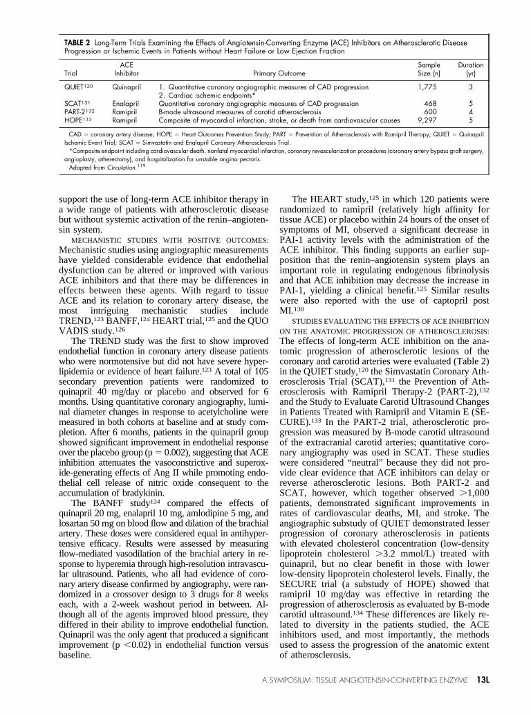
STUDIES EVALUATING THE EFFECTS OF ACE INHIBITIONON THE ANATOMIC PROGRESSION OF ATHEROSCLEROSIS:The effects of long-term ACE inhibition on the ana-tomic progression of atherosclerotic lesions of thecoronary and carotid arteries were evaluated (Table 2)in the QUIET study,120 the Simvastatin Coronary Ath-erosclerosis Trial (SCAT),131 the Prevention of Ath-erosclerosis with Ramipril Therapy-2 (PART-2),132
and the Study to Evaluate Carotid Ultrasound Changesin Patients Treated with Ramipril and Vitamin E (SE-CURE).133 In the PART-2 trial, atherosclerotic pro-gression was measured by B-mode carotid ultrasoundof the extracranial carotid arteries; quantitative coro-nary angiography was used in SCAT. These studieswere considered “neutral” because they did not pro-vide clear evidence that ACE inhibitors can delay orreverse atherosclerotic lesions. Both PART-2 andSCAT, however, which together observed �1,000patients, demonstrated significant improvements inrates of cardiovascular deaths, MI, and stroke. Theangiographic substudy of QUIET demonstrated lesserprogression of coronary atherosclerosis in patientswith elevated cholesterol concentration (low-densitylipoprotein cholesterol �3.2 mmol/L) treated withquinapril, but no clear benefit in those with lowerlow-density lipoprotein cholesterol levels. Finally, theSECURE trial (a substudy of HOPE) showed thatramipril 10 mg/day was effective in retarding theprogression of atherosclerosis as evaluated by B-modecarotid ultrasound.134 These differences are likely re-lated to diversity in the patients studied, the ACEinhibitors used, and most importantly, the methodsused to assess the progression of the anatomic extentof atherosclerosis.
TABLE 2 Long-Term Trials Examining the Effects of Angiotensin-Converting Enzyme (ACE) Inhibitors on Atherosclerotic DiseaseProgression or Ischemic Events in Patients without Heart Failure or Low Ejection Fraction
TrialACE
Inhibitor Primary OutcomeSampleSize (n)
Duration(yr)
QUIET120 Quinapril 1. Quantitative coronary angiographic measures of CAD progression2. Cardiac ischemic endpoints*
1,775 3
SCAT131 Enalapril Quantitative coronary angiographic measures of CAD progression 468 5PART-2132 Ramipril B-mode ultrasound measures of carotid atherosclerosis 600 4HOPE133 Ramipril Composite of myocardial infarction, stroke, or death from cardiovascular causes 9,297 5
CAD � coronary artery disease; HOPE � Heart Outcomes Prevention Study; PART � Prevention of Atherosclerosis with Ramipril Therapy; QUIET � QuinaprilIschemic Event Trial; SCAT � Simvastatin and Enalapril Coronary Atherosclerosis Trial.
*Composite endpoint including cardiovascular death, nonfatal myocardial infarction, coronary revascularization procedures (coronary artery bypass graft surgery,angioplasty, atherectomy), and hospitalization for unstable angina pectoris.
Adapted from Circulation.119
A SYMPOSIUM: TISSUE ANGIOTENSIN-CONVERTING ENZYME 13L

PHARMACOLOGY OF ANGIOTENSIN-CONVERTINGENZYME INHIBITORS
The ACE inhibitors currently number more than adozen different agents worldwide and have long beenrepresented by captopril and enalapril, the first ACEinhibitors to be approved. Because the mechanism ofaction of the ACE inhibitors is the same (ie, compet-itive inhibition of ACE), the documented beneficialeffects of captopril and enalapril, among others, areattributed to the class as a whole. Nevertheless, indi-vidual ACE inhibitors have unique pharmacokineticproperties that may result in differential clinical ef-fects. The most important property, perhaps, is thestrength of binding affinity to tissue ACE.
The active catalytic sites of ACE consist of hydro-phobic pockets of amino and carboxyterminal sidechains on the enzyme’s surface. The binding strengthof ACE inhibitors to ACE is dependent on the bindingof the sulfhydryl-, carboxyl-, or phosphinyl-contain-ing group at the N-terminus of the ACE inhibitor withthe coordinated Zn2� as well as the binding of thenegatively charged C-terminus of the ACE inhibitorwith the postulated positively charged carboxylatedock residue (believed to be an arginine side chain) ofACE.135 The affinity of ACE inhibitors to ACE is alsodependent on the number of auxiliary binding sites,the most important of which are the S’1 and S’2subsites.136
RELATIVE TISSUE AFFINITY OF ACEINHIBITORS
The degree of functional in vivo inhibition of tissueACE produced by an ACE inhibitor is directly depen-dent on the binding affinity of the inhibitor and theconcentration of the free inhibitor in the tissue. Theconcentration of the free inhibitor in the tissue, in turn,is dependent on the dynamic equilibrium between therate of delivery of ACE inhibitor to the tissue and itssubsequent washout into the blood. Key factors affect-ing the concentration of free inhibitor in tissues are
dose, bioavailability, half-life in blood, tissue penetra-tion, and tissue retention (or depot effect). Bioavail-ability and half-life in blood can readily be determinedand are important for decision making in initiallychoosing the correct dose of ACE inhibitor. Whenblood levels of the ACE inhibitor are consistentlyhigh—normally in the first half of the dosing period—tissue retention of the inhibitor is not likely to have asignificant effect on functional ACE inhibition. How-ever, toward the end of the dosing period, as the levelsof the ACE inhibitor in blood decreases, 2 factorsappear to be key in producing functional tissue ACEinhibition: (1) inhibitor binding affinity, and (2) tissueretention (which will directly influence the concentra-tion of the free inhibitor in the tissue).
The rank order of potency of several different ACEinhibitors has been determined by investigators usingcompetition analyses31,137–139 and by direct binding oftritium-labeled ACE inhibitors to tissue ACE.140 Thepotency is: quinaprilat � benazeprilat � ramiprilat �perindoprilat � lisinopril � enalaprilat � fosino-pril � captopril. The potency of ACE inhibitors intissue may also be ranked accordingly (Table3).31,106,138,141,142
Tissue retention of ACE inhibitor has also beenexamined. Isolated organ bath studies examining theduration of ACE inhibition after the removal of ACEinhibitor from the external milieu shows that func-tional inhibition of ACE lasts well beyond (2- to5-fold longer) the time predicted solely on the basis ofinhibitor dissociation rates or binding affinity.140 In-deed, inhibitors with similar dissociation rates fromtissue ACE show markedly different degrees of func-tional inhibition or retentiveness after washout. Therank order of tissue retentiveness is quinaprilat �lisinopril � enalaprilat � captopril and reflects boththe binding affinity and lipophilicity of these inhibitors.
CAN ANGIOTENSIN-CONVERTING ENZYME INHIBITORSBE DIFFERENTIATED?
The physiochemical differences among ACE in-hibitors that are responsible for their distinct pharma-cologic properties—binding affinity, potency, li-pophilicity, and depot effect—reveal that a divergenttrend allows the arbitrary classification of ACE inhib-itors as agents according to tissue-ACE affinity (Table3). Thus, the recognition that tissue ACE, the endo-thelium, and the natural history of cardiovascular dis-ease are interrelated leads to the question of whetherthe degree of tissue-ACE inhibition may extend to
differences in efficacy. Clearly, a reduction of Ang IIand increased nitric oxide bioavailability may repre-sent the mechanism by which ACE inhibitors confervascular protection. As a consequence, endothelialfunction may be regarded as a surrogate marker forvascular protection. The effects of ACE inhibitors onendothelium-dependent relaxation appear to differamong several reports and appear to be dependent onthe agents used and the experimental designs (Table4).123,124,126,143–148 It is intriguing that consistent im-
14L THE AMERICAN JOURNAL OF CARDIOLOGY� VOL. 88 (9A) NOVEMBER 8, 2001

provement in endothelial function is reported withthose ACE inhibitors with higher tissue-ACE affinity,such as quinapril and ramipril.
Ramipril has been shown to improve endothelialdysfunction by attenuating the toxic effects of oxi-dized low-density lipoprotein in vitro.149 More re-cently, ramiprilat was found to prevent the develop-ment of coronary endothelial dysfunction in a caninemodel. In this model, scanning electron micrographsof subepicardial arterioles from control dogs revealedendothelial leukocyte adhesion and crater formation.These markers of endothelial dysfunction were notobserved in ramiprilat-treated dogs.150 Likewise, per-indopril prevented chronic heart failure–induced en-dothelial dysfunction and reduced media cross-sec-tional area and collagen density in rats.151 Perindoprilhas also been shown to accelerate endothelial re-growth after balloon denudation in rabbits.152 In hu-mans, long-term treatment with perindopril inhibitsboth endothelial and adventitial ACE in the internalmammary arteries from patients with ischemic heartdisease.153
Several studies further extend these lines of evi-dence, including the TREND123 and the BANFF124
studies (see above), which have established that tis-sue-ACE inhibition improves endothelial function inhumans. Interestingly, the BANFF study showed thatenalapril and antihypertensive agents from otherclasses have no effect on endothelial function.
These results are strengthened by those from QUOVADIS,126 a 2-phase, parallel-arm, phase 3 study ofACE inhibition in coronary artery disease patientsscheduled to undergo coronary artery bypass graftsurgery. Patients were randomized to a double-blind,placebo-controlled treatment with quinapril (40 mg/day), or a single-blind treatment with captopril 50 mg,3 times a day (phase 1, before coronary bypass graftsurgery). Overall, 75 patients received quinapril, 37received captopril, and 74 patients received placebo,with treatment beginning, on average, 27 days beforecoronary bypass graft surgery.
Phase 1 of QUO VADIS was designed (1) todetermine the effects of ACE inhibition with quinapriland captopril on vascular tissue ACE, independent ofthe circulating renin–angiotensin system and the for-mation of Ang II; and (2) to determine whether func-tional differences existed between the 2 ACE inhibi-tors.126 During coronary bypass graft surgery, seg-ments of internal mammary arteries were harvestedfor in vitro measurements of tissue-ACE activity.Both quinapril and captopril reduced the production ofAng II. However, only the reduction in Ang II forma-tion in quinapril-treated patients was significant (p�0.05) versus placebo. This result suggests that thereis a functional difference in the respective abilities ofquinapril and captopril to inhibit endothelial ACE andthe local production of Ang II. Phase 2 of the QUOVADIS study126 evaluated the effect of chronic ACEinhibition (quinapril, 40 mg/day for 1 year) versusplacebo, on the incidence of ischemia. Treatment withquinapril significantly (p � 0.02) reduced clinicalischemic events during the 1-year period after coro-nary bypass graft surgery.
The potential importance of tissue-ACE inhibitionwas further demonstrated in a study of patients withchronic heart failure by quantitating impaired flow-dependent dilation as a measure of endothelial dys-function.147 The effects of quinaprilat (high affinity totissue ACE) were compared with those of enalaprilat.High-resolution ultrasound and Doppler were used tomeasure radial-artery diameter and blood flow in pa-tients who received intra-arterial infusions ofquinaprilat (1.6 �g/min, n � 15) and enalaprilat (5.0�g/min, n � 15) while at rest and during reactivehyperemia. Measurements were made both before andafter N-monomethly-L-arginine was used to inhibitendothelial nitric oxide synthetase and, hence, theproduction of nitric oxide. Quinaprilat improved flow-dependent dilation by �40%, whereas enalaprilat hadno effect. Moreover, although endothelial nitric oxidesynthetase was inhibited by N-monomethyl-L-arginine(the part of flow-dependent dilation mediated by nitric
TABLE 3 Pharmacological Properties of Various Angiotensin-Converting Enzyme (ACE) Inhibitors in Plasma and Tissue
TissuePotency*
ACE Inhibitor Potencies(mmol/L � 10�9, ID50)†
EnzymaticInhibition (IC50)‡
RadioligandDisplacement (DD50)‡
PlasmaHalf-Life§
Relative LipidSolubility�
HighQuinaprilat 0.07 5.5 � 10�11 4.5 � 10�11 25 ��Benazeprilat NA 1.3 � 10�9 4.8 � 10�11 11 �Ramiprilat 0.08 1.9 � 10�9 7.0 � 10�11 �50 ��Perindoprilat 0.40 NA NA 10 ��Lisinopril NA 4.5 � 10�9 1.7 � 10�10 12 NAEnalaprilat 1.00 4.5 � 10�9 1.1 � 10�9 11 �Fosinoprilat NA 1.6 � 10�8 5.1 � 10�10 11.5 ���
LowCaptopril 15.00 NA NA 2 �
NA � not available.*Radioligand binding studies using the active drug moiety.31,138,141
†ID50 is the inhibitor concentration required to displace 50% of [125I]531A bound to human plasma.142
‡Comparison of 50% inhibition of enzymatic activity (IC50) with 50% displacement of 125I-351A (DD50) from human plasma ACE.142
§Values cited for quinaprilat and ramiprilat are for dissociation from tissue ACE, ie, terminal half-life.106
�Lipid solubility based on log P logarithm of the octanol/water partition coefficient of the active drug moiety, except for captopril; � signs represent increased lipidsolubility.142
A SYMPOSIUM: TISSUE ANGIOTENSIN-CONVERTING ENZYME 15L
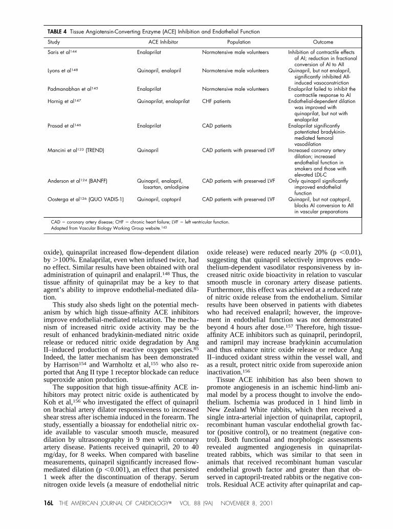
oxide), quinaprilat increased flow-dependent dilationby �100%. Enalaprilat, even when infused twice, hadno effect. Similar results have been obtained with oraladministration of quinapril and enalapril.148 Thus, thetissue affinity of quinaprilat may be a key to thatagent’s ability to improve endothelial-mediated dila-tion.
This study also sheds light on the potential mech-anism by which high tissue-affinity ACE inhibitorsimprove endothelial-mediated relaxation. The mecha-nism of increased nitric oxide activity may be theresult of enhanced bradykinin-mediated nitric oxiderelease or reduced nitric oxide degradation by AngII–induced production of reactive oxygen species.85
Indeed, the latter mechanism has been demonstratedby Harrison154 and Warnholtz et al,155 who also re-ported that Ang II type 1 receptor blockade can reducesuperoxide anion production.
The supposition that high tissue-affinity ACE in-hibitors may protect nitric oxide is authenticated byKoh et al,156 who investigated the effect of quinaprilon brachial artery dilator responsiveness to increasedshear stress after ischemia induced in the forearm. Thestudy, essentially a bioassay for endothelial nitric ox-ide available to vascular smooth muscle, measureddilation by ultrasonography in 9 men with coronaryartery disease. Patients received quinapril, 20 to 40mg/day, for 8 weeks. When compared with baselinemeasurements, quinapril significantly increased flow-mediated dilation (p �0.001), an effect that persisted1 week after the discontinuation of therapy. Serumnitrogen oxide levels (a measure of endothelial nitric
oxide release) were reduced nearly 20% (p �0.01),suggesting that quinapril selectively improves endo-thelium-dependent vasodilator responsiveness by in-creased nitric oxide bioactivity in relation to vascularsmooth muscle in coronary artery disease patients.Furthermore, this effect was achieved at a reduced rateof nitric oxide release from the endothelium. Similarresults have been observed in patients with diabeteswho had received enalapril; however, the improve-ment in endothelial function was not demonstratedbeyond 4 hours after dose.157 Therefore, high tissue-affinity ACE inhibitors such as quinapril, perindopril,and ramipril may increase bradykinin accumulationand thus enhance nitric oxide release or reduce AngII–induced oxidant stress within the vessel wall, andas a result, protect nitric oxide from superoxide anioninactivation.156
Tissue ACE inhibition has also been shown topromote angiogenesis in an ischemic hind-limb ani-mal model by a process thought to involve the endo-thelium. Ischemia was produced in 1 hind limb inNew Zealand White rabbits, which then received asingle intra-arterial injection of quinaprilat, captopril,recombinant human vascular endothelial growth fac-tor (positive control), or no treatment (negative con-trol). Both functional and morphologic assessmentsrevealed augmented angiogenesis in quinaprilat-treated rabbits, which was similar to that seen inanimals that received recombinant human vascularendothelial growth factor and greater than that ob-served in captopril-treated rabbits or the negative con-trols. Residual ACE activity after quinaprilat and cap-
TABLE 4 Tissue Angiotensin-Converting Enzyme (ACE) Inhibition and Endothelial Function
Study ACE Inhibitor Population Outcome
Saris et al144 Enalaprilat Normotensive male volunteers Inhibition of contractile effectsof AI; reduction in fractionalconversion of AI to AII
Lyons et al148 Quinapril, enalapril Normotensive male volunteers Quinapril, but not enalapril,significantly inhibited AII-induced vasoconstriction
Padmanabhan et al145 Enalaprilat Normotensive male volunteers Enalaprilat failed to inhibit thecontractile response to AI
Hornig et al147 Quinaprilat, enalaprilat CHF patients Endothelial-dependent dilationwas improved withquinaprilat, but not withenalaprilat
Prasad et al146 Enalaprilat CAD patients Enalaprilat significantlypotentiated bradykinin-mediated femoralvasodilation
Mancini et al123 (TREND) Quinapril CAD patients with preserved LVF Increased coronary arterydilation; increasedendothelial function insmokers and those withelevated LDL-C
Anderson et al124 (BANFF) Quinapril, enalapril,losartan, amlodipine
CAD patients with preserved LVF Only quinapril significantlyimproved endothelialfunction
Oosterga et al126 (QUO VADIS-1) Quinapril, captopril CAD patients with preserved LVF Quinapril, but not captopril,blocks AI conversion to AIIin vascular preparations
CAD � coronary artery disease; CHF � chronic heart failure; LVF � left ventricular function.Adapted from Vascular Biology Working Group website.143
16L THE AMERICAN JOURNAL OF CARDIOLOGY� VOL. 88 (9A) NOVEMBER 8, 2001

topril was equivalent when measured in plasma butwas significantly reduced by quinaprilat in tissuewhen compared with captopril (p �0.01).158
These results and those from HOPE122 support theimportance of tissue-ACE inhibition. Studies such as
IMAGINE, PEACE, and EUROPA, using quinapril,trandolapril, and perindopril, respectively, may con-firm the findings of the HOPE study and thus validatethe use of tissue-ACE inhibitors in clinical practiceamong high-risk patient populations.
CONCLUSION
The experimental and clinical evidence presentedhere validates earlier suppositions that long-term tis-sue-ACE inhibition would provide important clinicalbenefits to a broad population of patients with coro-nary artery disease. The cardio- and renoprotectivebenefits of this drug class appear to extend beyond thetherapeutic effects of blood pressure reduction and
may distinguish the ACE inhibitors from other anti-hypertensive agents. The next significant step is todetermine if pharmacologic differences among theACE inhibitors, such as the affinity for tissue ACEand the clinical effect on endothelial dysfunction, area differentiating factor within this class of importantcardiovascular drugs.
1. Linz W, Wiemer G, Gohlke P, Unger T, Scholkens BA. Contribution of kininsto the cardiovascular actions of angiotensin-converting enzyme inhibitors. Phar-macol Rev 1995;47:25–49.2. Nash DT. Comparative properties of angiotensin-converting enzyme inhibi-tors: relations with inhibition of tissue angiotensin-converting enzyme and po-tential clinical implications. Am J Cardiol 1992;69(suppl):26C–32C.3. Cushman DW, Cheung HS. Concentrations of angiotensin-converting enzymein tissues of the rat. Biochim Biophys Acta 1971;250:261–265.4. Esther CR, Marino EM, Howard TE, Machaud A, Corvol P, Capecchi MR,Bernstein KE. The critical role of tissue angiotensin-converting enzyme asrevealed by gene targeting in mice. J Clin Invest 1997;99:2375–2385.5. Jacob HJ, Lindpaintner K, Lincoln SE, Kusumi K, Bunker RK, Mao YP,Ganten D, Dzau VJ, Lander ES. Genetic mapping of a gene causing hypertensionin the stroke-prone spontaneously hypertensive rat. Cell 1991;67:213–224.6. Harris EL, Phelan EL, Thompson CM, Millar JA, Grigor MR. Heart mass andblood pressure have separate genetic determinants in the New Zealand geneticallyhypertensive (GH) rat. J Hypertens 1995;13:397–404.7. Rigat B, Hubert C, Alhenc-Gelas F, Cambien F, Corvol P, Soubrier F. Aninsertion/deletion polymorphism in the angiotensin I-converting enzyme geneaccounting for half the variance of serum enzyme levels. J Clin Invest 1990;86:1343–1346.8. Danser AH, Schalekamp MA, Bax WA, van den Brink AM, Saxena PR,Riegger GA, Schunkert H. Angiotensin-converting enzyme in the human heart:effect of the deletion/insertion polymorphism. Circulation 1995;92:1387–1388.9. Schunkert H. Polymorphism of the angiotensin-converting enzyme gene andcardiovascular disease. J Mol Med 1997;75:867–875.10. Cambien F, Poirier O, Lecerf L, Evans A, Cambou JP, Arveiler D, Luc G,Bard JM, Bara L, Ricard S, et al. Deletion polymorphism in the gene forangiotensin-converting enzyme is a potent risk factor for myocardial infarction.Nature 1992;359:641–644.11. Schunkert H, Hense HW, Holmer SR, Stender M, Perz S, Keil U, Lorell BH,Riegger GA. Association between a deletion polymorphism of the angiotensin-converting-enzyme gene and left ventricular hypertrophy. N Engl J Med 1994;330:1634–1638.12. Mayer B, Schunkert H. ACE gene polymorphism and cardiovascular dis-eases. Herz 2000;25:1–6.13. Iwai N, Ohmichi N, Nakamura Y, Kinoshita M. DD genotype of theangiotensin-converting enzyme gene is a risk factor for left ventricular hypertro-phy. Circulation 1994;90:2622–2628.14. Ravid M, Savin H, Jutrin I, Bental T, Lang R, Lishner M. Long-term effectof ACE inhibition on development of nephropathy in diabetes mellitus type II.Kidney Int Suppl 1994;45:S161–S164.15. Lindpaintner K, Pfeffer MA, Kreutz R, Stampfer MJ, Grodstein F, LaMotteF, Buring J, Hennekens CH. A prospective evaluation of an angiotensin-convert-ing-enzyme gene polymorphism and the risk of ischemic heart disease. N EnglJ Med 1995;332:706–711.16. Lindpaintner K, Lee M, Larson MG, Rao VS, Pfeffer MA, Ordovas JM,Schaefer EJ, Wilson AF, Wilson PW, Vasan RS, Myers RH, Levy D. Absence ofassociation or genetic linkage between the angiotensin-converting-enzyme geneand left ventricular mass. N Engl J Med 1996;334:1023–1028.17. Schunkert H. Controversial association of left ventricular hypertrophy and theACE I/D polymorphism: is the mist clearing up? Nephrol Dial Transplant1998;13:1109–1112.18. Montgomery HE, Clarkson P, Dollery CM, Prasad K, Losi MA, HemingwayH, Statters D, Jubb M, Girvain M, Varnava A, et al. Association of angiotensin-converting enzyme gene I/D polymorphism with change in left ventricular massin response to physical training. Circulation 1997;96:741–747.
19. Pinto YM, van Gilst WH, Kingma JH, Schunkert H. Deletion-type allele ofthe angiotensin-converting enzyme gene is associated with progressive ventric-ular dilation after anterior myocardial infarction: Captopril and ThrombolysisStudy Investigators. J Am Coll Cardiol 1995;25:1622–1626.20. Ohmichi N, Iwai N, Maeda K, Shimoike H, Nakamura Y, Izumi M, SugimotoY, Kinoshita M. Genetic basis of left ventricular remodeling after myocardialinfarction. Int J Cardiol 1996;53:265–272.21. Pinto YM, Buikema H, van Gilst WH, Scholtens E, van Geel PP, de GraeffPA, Wagner J, Paul M. Cardiovascular end-organ damage in Ren-2 transgenicrats compared to spontaneously hypertensive rats. J Mol Med 1997;75:371–377.22. Turner ST, Boerwinkle E, Sing CF. Context-dependent associations of theACE I/D polymorphism with blood pressure. Hypertension 1999;34:773–778.23. Pfohl M, Koch M, Prescod S, Haase KK, Haring HU, Karsch KR. Angio-tensin I-converting enzyme gene polymorphism, coronary artery disease andmyocardial infarction: an angiographically controlled study. Eur Heart J 1999;20:1318–1325.24. Ng KK, Vane JR. Fate of angiotensin I in the circulation. Nature 1968;218:144–150.25. Schunkert H, Dzau VJ, Tang SS, Hirsch AT, Apstein CS, Lorell BH.Increased rat cardiac angiotensin converting enzyme activity and mRNA expres-sion in pressure overload left ventricular hypertrophy: effects on coronary resis-tance, contractility, and relaxation. J Clin Invest 1990;86:1913–1920.26. Yamada H, Fabris B, Allen AM, Jackson B, Johnston CI, Mendelsohn AO.Localization of angiotensin converting enzyme in rat heart. Circ Res 1991;68:141–149.27. Falkenhahn M, Franke F, Bohle RM, Zhu YC, Stauss HM, Bachmann S,Danilov S, Unger T. Cellular distribution of angiotensin-converting enzyme aftermyocardial infarction. Hypertension 1995;25:219–226.28. Ruzicka M, Skarda V, Leenen FH. Effects of ACE inhibitors on circulatingversus cardiac angiotensin II in volume overload-induced cardiac hypertrophy inrats. Circulation 1995;92:3568–3573.29. Pieruzzi F, Abassi ZA, Keiser HR. Expression of renin-angiotensin systemcomponents in the heart, kidneys, and lungs of rats with experimental heartfailure. Circulation 1995;92:3105–3112.30. Hirsch AT, Talsness CE, Schunkert H, Paul M, Dzau VJ. Tissue-specificactivation of cardiac angiotensin converting enzyme in experimental heart failure.Circ Res 1991;69:475–482.31. Fabris B, Jackson B, Kohzuki M, Perich R, Johnston CI. Increased cardiacangiotensin-converting enzyme in rats with chronic heart failure. Clin Exp Phar-macol Physiol 1990;17:309–314.32. Hokimoto S, Yasue H, Fujimoto K, Yamamoto H, Nakao K, Kaikita K,Sakata R, Miyamoto E. Expression of angiotensin-converting enzyme in remain-ing viable myocytes of human ventricles after myocardial infarction. Circulation1996;94:1513–1518.33. Heymes C, Swynghedauw B, Chevalier B. Activation of angiotensinogen andangiotensin-converting enzyme gene expression in the left ventricle of senescentrats. Circulation 1994;90:1328–1333.34. Lee YA, Liang CS, Lee MA, Lindpaintner K. Local stress, not systemicfactors, regulate gene expression of the cardiac renin-angiotensin system in vivo:a comprehensive study of all its components in the dog. Proc Natl Acad Sci U SA 1996;93:11035–11040.35. Sadoshima J, Xu Y, Slayter HS, Izumo S. Autocrine release of angiotensin IImediates stretch-induced hypertrophy of cardiac myocytes in vitro. Cell 1993;75:977–984.36. Danser AH, Schalekamp MA. Is there an internal cardiac renin-angiotensinsystem? Heart 1996;76:28–32.37. Danser AH, de Lannoy LM, Saxena P, Schalekamp MD. Chymase does not
A SYMPOSIUM: TISSUE ANGIOTENSIN-CONVERTING ENZYME 17L
contribute to angiotensin I-II conversion in the interstitial fluid. Circulation1998;98:I-793.38. Urata H, Kinoshita A, Misono KS, Bumpus FM, Husain A. Identification ofa highly specific chymase as the major angiotensin II-forming enzyme in thehuman heart. J Biol Chem 1990;265:22348–22357.39. Pfeifer M, Bruckschlegel G, Holmer SR, Paul M, Riegger AJ, Schunkert H.Reciprocal regulation of pulmonary and cardiac angiotensin-converting enzymein rats with severe left ventricular hypertrophy. Cardiovasc Res 1998;38:125–132.40. Lindpaintner K, Jin M, Wilhelm M, Toth M, Ganten D. Aspects of molecularbiology and biochemistry of the cardiac renin-angiotensin system. Br J ClinPharmacol 1989;27:159S–165S.41. van Kats JP, Danser AH, van Meegen JR, Sassen LM, Verdouw PD,Schalekamp MA. Angiotensin production by the heart: a quantitative study inpigs with the use of radiolabeled angiotensin infusions. Circulation 1998;98:73–81.42. Danser AH, van Kesteren CA, Bax WA, Tavenier M, Derkx FH, Saxena PR,Schalekamp MA. Prorenin, renin, angiotensinogen, and angiotensin-convertingenzyme in normal and failing human hearts: evidence for renin binding [abstract].Circulation 1997;96:220–226.43. Danser AH, de Lannoy LM, Schalekamp MD. Uptake of renin and proreninby the isolated rat heart. Circulation 1997:96(suppl):I-280.44. Passier RC, Smits JF, Verluyten MJ, Daemen MJ. Expression and localiza-tion of renin and angiotensinogen in rat heart after myocardial infarction. Am JPhysiol 1996;271:H1040–H1048.45. Lindpaintner K, Lu W, Neidermajer N, Schieffer B, Just H, Ganten D,Drexler H. Selective activation of cardiac angiotensinogen gene expression inpost-infarction ventricular remodeling in the rat. J Mol Cell Cardiol 1993;25:133–143.46. MaassenVanDenBrink A, de Vries R, Saxena PR, Danser AH. ACE, but notchymase, generates angiotensin II in close proximity to the AT1 receptor in thehuman isolated coronary artery. Circulation 1998;98:I-606.47. Schunkert H, Jackson B, Tang SS, Schoen FJ, Smits JF, Apstein CS, LorellBH. Distribution and functional significance of cardiac angiotensin convertingenzyme in hypertrophied rat hearts. Circulation 1993;87:1328–1339.48. Zisman LS, Abraham WT, Meixell GE, Vamvakias BN, Quaife RA, LowesBD, Roden RL, Peacock SJ, Groves BM, Raynolds MV, Bristow MR, PerrymanMB. Angiotensin II formation in the intact human heart: predominance of theangiotensin-converting enzyme pathway. J Clin Invest 1995;96:1490–1498.49. Muller DN, Bohlender J, Hilgers KF, Dragun D, Costerousse O, Menard J,Luft FC. Vascular angiotensin-converting enzyme expression regulates localangiotensin II. Hypertension 1997;29:98–104.50. Urata H, Healy B, Stewart RW, Bumpus FM, Husain A. AngiotensinII-forming pathways in normal and failing human hearts. Circ Res 1990;66:883–890.51. Muller DN, Fischli W, Clozel JP, Hilgers KF, Bohlender J, Menard J,Busjahn A, Ganten D, Luft FC. Local angiotensin II generation in the rat heart:role of renin uptake. Circ Res 1998;82:13–20.52. Kokkonen JO, Saarinen J, Kovanen PT. Regulation of local angiotensin IIformation in the human heart in the presence of interstitial fluid: inhibition ofchymase by protease inhibitors of interstitial fluid and of angiotensin-convertingenzyme by Ang-(1-9) formed by heart carboxypeptidase A-like activity. Circu-lation 1997;95:1455–1463.53. Bernstein KE. ACE knockout mice: lessons for adult nephrology. NephrolDial Transplant 1998;13:2991–2994.54. Holubarsch C, Hasenfuss G, Schmidt-Schweda S, Knorr A, Pieske B, Ruf T,Fasol R, Just H. Angiotensin I and II exert inotropic effects in atrial but not inventricular human myocardium: an in vitro study under physiological experimen-tal conditions. Circulation 1993;88:1228–1237.55. Esther CR Jr, Howard TE, Marino EM, Goddard JM, Capecchi MR, Bern-stein KE. Mice lacking angiotensin-converting enzyme have low blood pressure,renal pathology, and reduced male fertility. Lab Invest 1996;74:953–965.56. Baker KM, Aceto JF. Angiotensin II stimulation of protein synthesis and cellgrowth in chick heart cells. Am J Physiol 1990;259:H610–H618.57. Schunkert H, Sadoshima J, Cornelius T, Kagaya Y, Weinberg EO, Izumo S,Riegger G, Lorell BH. Angiotensin II-induced growth responses in isolated adultrat hearts: evidence for load-independent induction of cardiac protein synthesisby angiotensin II. Circ Res 1995;76:489–497.58. Kromer EP, Riegger GA. Effects of long-term angiotensin converting enzymeinhibition on myocardial hypertrophy in experimental aortic stenosis in the rat.Am J Cardiol 1988;62:161–163.59. Bruckschlegel G, Holmer SR, Jandeleit K, Grimm D, Muders F, Kromer EP,Riegger GA, Schunkert H. Blockade of the renin-angiotensin system in cardiacpressure-overload hypertrophy in rats. Hypertension 1995;25:250–259.60. Lear W, Ruzicka M, Leenen FH. ACE inhibitors and cardiac ACE mRNA involume overload-induced cardiac hypertrophy. Am J Physiol 1997;273:H641–H646.61. Takemoto M, Egashira K, Usui M, Numaguchi K, Tomita H, Tsutsui H,Shimokawa H, Sueishi K, Takeshita A. Important role of tissue angiotensin-converting enzyme activity in the pathogenesis of coronary vascular and myo-cardial structural changes induced by long-term blockade of nitric oxide synthesisin rats. J Clin Invest 1997;99:278–287.62. Weber KT, Brilla CG. Pathological hypertrophy and cardiac interstitium:fibrosis and renin-angiotensin-aldosterone system. Circulation 1991;83:1849–1865.
63. Katwa LC, Campbell SE, Tyagi SC, Lee SJ, Cicila GT, Weber KT. Culturedmyofibroblasts generate angiotensin peptides de novo. J Mol Cell Cardiol 1997;29:1375–1386.64. Grimm D, Kromer EP, Bocker W, Bruckschlegel G, Holmer SR, Riegger GA,Schunkert H. Regulation of extracellular matrix proteins in pressure-overloadcardiac hypertrophy: effects of angiotensin converting enzyme inhibition. J Hy-pertens 1998;16:1345–1355.65. Panizo A, Pardo J, Hernandez M, Galindo MF, Cenarruzabeitia E, Diez J.Quinapril decreases myocardial accumulation of extracellular matrix componentsin spontaneously hypertensive rats. Am J Hypertens 1995;8:815–822.66. Matsusaka T, Katori H, Inagami T, Fogo A, Ichikawa I. Communicationbetween myocytes and fibroblasts in cardiac remodeling in angiotensin chimericmice. J Clin Invest 1999;103:1451–1458.67. Leri A, Claudio PP, Li Q, Wang X, Reiss K, Wang S, Malhotra A, KajsturaJ, Anversa P. Stretch-mediated release of angiotensin II induces myocyte apo-ptosis by activating p53 that enhances the local renin-angiotensin system anddecreases the Bcl-2-to-Bax protein ratio in the cell. J Clin Invest 1998;101:1326–1342.68. Pierzchalski P, Reiss K, Cheng W, Cirielli C, Kajstura J, Nitahara JA, RizkM, Capogrossi MC, Anversa P. p53 Induces myocyte apoptosis via the activationof the renin-angiotensin system. Exp Cell Res 1997;234:57–65.69. Diez J, Panizo A, Hernandez M, Vega F, Sola I, Fortuno MA, PardoJ. Cardiomyocyte apoptosis and cardiac angiotensin-converting enzyme in spon-taneously hypertensive rats. Hypertension 1997;30:1029–1034.70. Friedrich SP, Lorell BH, Rousseau MF, Hayashida W, Hess OM, Douglas PS,Gordon S, Keighley CS, Benedict C, Krayenbuehl HP, Grossman W, Pouleur H.Intracardiac angiotensin-converting enzyme inhibition improves diastolic func-tion in patients with left ventricular hypertrophy due to aortic stenosis. Circula-tion 1994;90:2761–2771.71. Weinberg EO, Schoen FJ, George D, Kagaya Y, Douglas PS, Litwin SE,Schunkert H, Benedict CR, Lorell BH. Angiotensin-converting enzyme inhibitionprolongs survival and modifies the transition to heart failure in rats with pressureoverload hypertrophy due to ascending aortic stenosis. Circulation 1994;90:1410–1422.72. Eberli FR, Apstein CS, Ngoy S, Lorell BH. Exacerbation of left ventricularischemic diastolic dysfunction by pressure-overload hypertrophy: modificationby specific inhibition of cardiac angiotensin converting enzyme. Circ Res 1992;70:931–943.73. Foult JM, Tavolaro O, Antony I, Nitenberg A. Direct myocardial andcoronary effects of enalaprilat in patients with dilated cardiomyopathy: assess-ment by a bilateral intracoronary infusion technique. Circulation 1988;77:337–344.74. Haleen SJ, Weishaar RE, Overhiser RW, Bousley RF, Keiser JA, RapundaloSR, Taylor DG. Effects of quinapril, a new angiotensin converting enzymeinhibitor, on left ventricular failure and survival in the cardiomyopathic hamster:hemodynamic, morphological, and biochemical correlates. Circ Res 1991;68:1302–1312.75. The CONSENSUS Trial Study Group. Effects of enalapril on mortality insevere congestive heart failure: results of the Cooperative North ScandinavianEnalapril Survival Study (CONSENSUS). N Engl J Med 1987;316:1429–1435.76. The SOLVD Investigators. Effect of enalapril on survival in patients withreduced left ventricular ejection fractions and congestive heart failure. N EnglJ Med 1991;325:293–302.77. Erdos EG, Skidgel RA. The angiotensin I-converting enzyme. Lab Invest1987;56:345–348.78. Fishel RS, Eisenberg S, Shai SY, Redden RA, Bernstein KE, Berk BC.Glucocorticoids induce angiotensin-converting enzyme expression in vascularsmooth muscle. Hypertension 1995;25:343–349.79. Shai SY, Fishel RS, Martin BM, Berk BC, Bernstein KE. Bovine angiotensinconverting enzyme cDNA cloning and regulation. Increased expression duringendothelial cell growth arrest. Circ Res 1992;70:1274–1281.80. Gainer JV, Morrow JD, Loveland A, King DJ, Brown NJ. Effect of brady-kinin-receptor blockade on the response to angiotensin-converting-enzyme inhib-itor in normotensive and hypertensive subjects. N Engl J Med 1998;339:1285–1292.81. Hornig B, Kohler C, Drexler H. Role of bradykinin in mediating vasculareffects of angiotensin-converting enzyme inhibitors in humans. Circulation 1997;95:1115–1118.82. Brown NJ, Agirbasli MA, Williams GH, Litchfield WR, Vaughan DE. Effectof activation and inhibition of the renin-angiotensin system on plasma PAI-1.Hypertension 1998;32:965–971.83. Britten MB, Zeiher AM, Schachinger V. Clinical importance of coronaryendothelial vasodilator dysfunction and therapeutic options. J Intern Med 1999;245:315–327.84. Drexler H, Hornig B. Endothelial dysfunction in human disease. J Mol CellCardiol 1999;31:51–60.85. Rajagopalan S, Kurz S, Munzel T, Tarpey M, Freeman BA, Griendling KK,Harrison DG. Angiotensin II-mediated hypertension in the rat increases vascularsuperoxide production via membrane NADH/NADPH oxidase activation: con-tribution to alterations of vasomotor tone. J Clin Invest 1996;97:1916–1923.86. Diet F, Pratt RE, Berry GJ, Momose N, Gibbons GH, Dzau VJ. Increasedaccumulation of tissue ACE in human atherosclerotic coronary artery disease.Circulation 1996;94:2756–2767.87. Moroi M, Zhang L, Yasuda T, Virmani R, Gold HK, Fishman MC, HuangPL. Interaction of genetic deficiency of endothelial nitric oxide, gender, and
18L THE AMERICAN JOURNAL OF CARDIOLOGY� VOL. 88 (9A) NOVEMBER 8, 2001

pregnancy in vascular response to injury in mice. J Clin Invest 1998;101:1225–1232.88. Brenner BM, Meyer TW, Hostetter TH. Dietary protein intake and theprogressive nature of kidney disease: the role of hemodynamically mediatedglomerular injury in the pathogenesis of progressive glomerular sclerosis inaging, renal ablation, and intrinsic renal disease. N Engl J Med 1982;307:652–659.89. Yoshida Y, Fogo A, Ichikawa I. Glomerular hemodynamic changes vs.hypertrophy in experimental glomerular sclerosis. Kidney Int 1989;35:654–660.90. Kagami S, Kuhara T, Okada K, Kuroda Y, Border WA, Noble NA. Dualeffects of angiotensin II on the plasminogen/plasmin system in rat mesangialcells. Kidney Int 1997;51:664–671.91. Border WA, Noble NA. Interactions of transforming growth factor-beta andangiotensin II in renal fibrosis. Hypertension 1998;31:181–188.92. Miettinen H, Haffner SM, Lehto S, Ronnemaa T, Pyorala K, Laakso M.Proteinuria predicts stroke and other atherosclerotic vascular disease events innondiabetic and non-insulin-dependent diabetic subjects. Stroke 1996;27:2033–2039.93. Breyer JA, Bain RP, Evans JK, Nahman NS Jr, Lewis EJ, Cooper M, McGillJ, Berl T. Predictors of the progression of renal insufficiency in patients withinsulin-dependent diabetes and overt diabetic nephropathy. The CollaborativeStudy Group. Kidney Int 1996;50:1651–1658.94. Ruilope LM, Miranda B, Oliet A, Millet VG, Rodicio JL, Romero JC, RaijL. Control of hypertension with the angiotensin converting enzyme inhibitorcaptopril reduces glomerular proteinuria. J Hypertens Suppl 1988;6:S467–S469.95. Kasiske BL, Kalil RS, Ma JZ, Liao M, Keane WF. Effect of antihypertensivetherapy on the kidney in patients with diabetes: a meta-regression analysis. AnnIntern Med 1993;118:129–138.96. Gansevoort RT, de Zeeuw D, de Jong PE. Additive antiproteinuric effect ofACE inhibition and a low-protein diet in human renal disease. Nephrol DialTransplant 1995;10:497–504.97. UK Prospective Diabetes Study Group. Tight blood pressure control and riskof macrovascular and microvascular complications in type 2 diabetes: UKPDS38. Br Med J 1998;317:703–713.98. Hansson L, Lindholm LH, Niskanen L, Lanke J, Hedner T, Niklason A,Luomanmaki K, Dahlof B, de Faire U, Morlin C, Karlberg BE, Wester PO,Bjorck JE. Effect of angiotensin-converting-enzyme inhibition compared withconventional therapy on cardiovascular morbidity and mortality in hypertension:the Captopril Prevention Project (CAPPP) randomised trial. Lancet 1999;353:611–616.99. Estacio RO, Schrier RW. Antihypertensive therapy in type 2 diabetes:implications of the appropriate blood pressure control in diabetes (ABCD) trial.Am J Cardiol 1998;82(suppl):9R–14R.100. Koskinen P, Manttari M, Manninen V, Huttunen JK, Heinonen OP, FrickMH. Coronary heart disease incidence in NIDDM patients in the Helsinki HeartStudy. Diabetes Care 1992;15:820–825.101. Janka HU, Dirschedl P. Systolic blood pressure as a predictor for cardio-vascular disease in diabetes: a 5-year longitudinal study. Hypertension 1985;7:II90–II94.102. Kuusisto J, Mykkanen L, Pyorala K, Laakso M. NIDDM and its metaboliccontrol predict coronary heart disease in elderly subjects. Diabetes 1994;43:960–967.103. Lewis EJ, Hunsicker LG, Bain RP, Rohde RD. The effect of angiotensin-converting-enzyme inhibition on diabetic nephropathy: the Collaborative StudyGroup. N Engl J Med 1993;329:1456–1462.104. HOPE Investigators. Effects of ramipril on cardiovascular and microvascu-lar outcomes in people with diabetes mellitus: results of the HOPE study andMICRO-HOPE substudy. Lancet 2000;355:253–259.105. Maschio G, Alberti D, Janin G, Locatelli F, Mann JFE, Motolese M,Ponticelli C, Ritz E, Zucchelli P. Effect of the angiotensin-converting-enzymeinhibitor benazepril on the progression of chronic renal insufficiency. N EnglJ Med 1996;334:939–945.106. Jackson EK, Garrison JC. Renin and angiotensin. In: Hardman JG, LimbirdL, eds. Goodman & Gilman’s The Pharmacological Basis of Therapeutics. NewYork: McGraw-Hill, 1999:743–746.107. Cohn JN, Johnson G, Ziesche S, Cobb F, Francis G, Tristani F, Smith R,Dunkman WB, Loeb H, Wong M, et al. A comparison of enalapril with hydral-azine-isosorbide dinitrate in the treatment of chronic congestive heart failure.N Engl J Med 1991;325:303–310.108. Ambrosioni E, Borghi C, Magnani B. The effect of the angiotensin-converting-enzyme inhibitor zofenopril on mortality and morbidity after anteriormyocardial infarction: the Survival of Myocardial Infarction Long-Term Evalu-ation (SMILE) Study Investigators. N Engl J Med 1995;332:80–85.109. Flather MD, Yusuf S, Kober L, Pfeffer M, Hall A, Murray G, Torp-PedersenC, Ball S, Pogue J, Moye L, Braunwald E. Long-term ACE-inhibitor therapy inpatients with heart failure or left-ventricular dysfunction: a systematic overviewof data from individual patients. ACE-Inhibitor Myocardial Infarction Collabo-rative Group. Lancet 2000;355:1575–1581.110. Garg R, Yusuf S. Overview of randomized trials of angiotensin-convertingenzyme inhibitors on mortality and morbidity in patients with heart failure:Collaborative Group on ACE Inhibitor Trials. JAMA 1995;273:1450–1456.111. Effect of ramipril on mortality and morbidity of survivors of acute myo-cardial infarction with clinical evidence of heart failure. The Acute InfarctionRamipril Efficacy (AIRE) Study Investigators. Lancet 1993;342:821–828.112. Pfeffer MA, Braunwald E, Moye LA, Basta L, Brown EJ Jr, Cuddy TE,
Davis BR, Geltman EM, Goldman S, Flaker GC, et al. Effect of captopril onmortality and morbidity in patients with left ventricular dysfunction after myo-cardial infarction: results of the survival and ventricular enlargement trial: theSAVE Investigators. N Engl J Med 1992;327:669–677.113. Torp-Pedersen C, Kober L. Effect of ACE inhibitor trandolapril on lifeexpectancy of patients with reduced left-ventricular function after acute myocar-dial infarction: TRACE Study Group. Trandolapril Cardiac Evaluation. Lancet1999;354:9–12.114. ACE Inhibitor Myocardial Infarction Collaborative Group. Indications forACE inhibitors in the early treatment of acute myocardial infarction: systematicoverview of individual data from 100,000 patients in randomized trials. Circu-lation 1998;97:2202–2212.115. ISIS-4 (Fourth International Study of Infarct Survival) Collaborative Group.ISIS-4: a randomised factorial trial assessing early oral captopril, oral mononi-trate, and intravenous magnesium sulphate in 58,050 patients with suspectedacute myocardial infarction. ISIS-4. Lancet 1995;345:669–685.116. Gruppo Italiano per lo Studio della Sopravvivenza nell’infarto Miocardico.GISSI-3: effects of lisinopril and transdermal glyceryl trinitrate singly andtogether on 6-week mortality and ventricular function after acute myocardialinfarction. Lancet 1994;343:1115–1122.117. Yusuf S, Pepine CJ, Garces C, Pouleur H, Salem D, Kostis J, Benedict C,Rousseau M, Bourassa M, Pitt B. Effect of enalapril on myocardial infarction andunstable angina in patients with low ejection fractions. Lancet 1992;340:1173–1178.118. Rutherford JD, Pfeffer MA, Moye LA, Davis BR, Flaker GC, Kowey PR,Lamas GA, Miller HS, Packer M, Rouleau JL, Braunwald E. Effects of captoprilon ischemic events after myocardial infarction: results of the Survival andVentricular Enlargement trial. SAVE Investigators. Circulation 1994;90:1731–1738.119. Lonn EM, Yusuf S, Jha P, Montague TJ, Teo KK, Benedict CR, Pitt B.Emerging role of angiotensin-converting enzyme inhibitors in cardiac and vas-cular protection. Circulation 1994;90:2056–2069.120. Texter M, Lees RS, Pitt B, Dinsmore RE, Uprichard AC. The QUinaprilIschemic Event Trial (QUIET) design and methods: evaluation of chronic ACEinhibitor therapy after coronary artery intervention. Cardiovasc Drugs Ther1993;7:273–282.121. Cashin-Hemphill L, Holmvang G, Chan RC, Pitt B, Dinsmore RE, Lees RS.Angiotensin-converting enzyme inhibition as antiatherosclerotic therapy: no an-swer yet. QUIET Investigators. QUinapril Ischemic Event Trial. Am J Cardiol1999;83:43–47.122. Yusuf S, Sleight P, Pogue J, Bosch J, Davies R, Dagenais G. Effects of anangiotensin-converting-enzyme inhibitor, ramipril, on cardiovascular events inhigh-risk patients: the Heart Outcomes Prevention Evaluation Study Investiga-tors. N Engl J Med 2000;342:145–153.123. Mancini GB, Henry GC, Macaya C, O’Neill BJ, Pucillo AL, Carere RG,Wargovich TJ, Mudra H, Luscher TF, Klibaner MI, et al. Angiotensin-convertingenzyme inhibition with quinapril improves endothelial vasomotor dysfunction inpatients with coronary artery disease: the TREND (Trial on Reversing Endothe-lial Dysfunction) Study. Circulation 1996;94:258–265.124. Anderson TJ, Elstein E, Haber H, Charbonneau F. Comparative study ofACE-inhibition, angiotensin II antagonism, and calcium channel blockade onflow-mediated vasodilation in patients with coronary disease (BANFF study).J Am Coll Cardiol 2000;35:60–66.125. Vaughan DE, Rouleau JL, Ridker PM, Arnold JM, Menapace FJ, PfefferMA. Effects of ramipril on plasma fibrinolytic balance in patients with acuteanterior myocardial infarction: HEART Study Investigators. Circulation 1997;96:442–447.126. Oosterga M, Voors AA, Buikema H, Pinto YM, Haber HE, Ebels T,Morshuis WJ, Kingma JH, Crijns HJ, van Gilst WH. Angiotensin II formation inhuman vasculature after chronic ACE inhibition: a prospective, randomized,placebo-controlled study. QUO VADIS Investigators. Cardiovasc Drugs Ther2000;14:55–60.127. Pfeffer MA, Domanski M, Rosenberg Y, Verter J, Geller N, Albert P, HsiaJ, Braunwald E. Prevention of events with angiotensin-converting enzyme inhi-bition (the PEACE study design): Prevention of Events with Angiotensin-Con-verting Enzyme Inhibition. Am J Cardiol 1998;82(suppl):25H–30H.128. Fox KM, Henderson JR, Bertrand ME, Ferrari R, Remme WJ, Simoons ML.The European trial on reduction of cardiac events with perindopril in stablecoronary artery disease (EUROPA). Eur Heart J 1998;19(suppl J):J52–J55.129. Simoons ML, Vos J, de Feyter PJ, Bots ML, Remme WJ, Grobbee DE, KluftC, de Maat MP, Fox KM, Deckers JW. EUROPA substudies, confirmation ofpathophysiological concepts: European trial on reduction of cardiac events withperindopril in stable coronary artery disease. Eur Heart J 1998;19(suppl J):J56–J60.130. Wright RA, Flapan AD, Alberti KG, Ludlam CA, Fox KA. Effects ofcaptopril therapy on endogenous fibrinolysis in men with recent, uncomplicatedmyocardial infarction. J Am Coll Cardiol 1994;24:67–73.131. Teo KK, Burton JR, Buller CE, Plante S, Catellier D, Tymchak W, DzavikV, Taylor D, Yokoyama S, Montague TJ. Long-term effects of cholesterollowering and angiotensin-converting enzyme inhibition on coronary atheroscle-rosis: the Simvastatin/Enalapril coronary atherosclerosis trial (SCAT). Circula-tion 2000;102:1748–1754.132. MacMahon S, Sharpe N, Gamble G, Clague A, Mhurchu CN, Clark T, HartH, Scott J, White H. Randomized, placebo-controlled trial of the angiotensin-converting enzyme inhibitor, ramipril, in patients with coronary or other occlu-
A SYMPOSIUM: TISSUE ANGIOTENSIN-CONVERTING ENZYME 19L
sive arterial disease. PART-2 Collaborative Research Group. Prevention of Ath-erosclerosis with Ramipril. J Am Coll Cardiol 2000;36:438–443.133. Lonn EM, Yusuf S, Dzavik V, Doris CI, Yi Q, Smith S, Moore-Cox A,Bosch J, Riley WA, Teo KK. Effects of ramipril and vitamin E on atherosclerosis:the study to evaluate carotid ultrasound changes in patients treated with ramipriland vitamin E (SECURE). Circulation 2001;103:919–925.134. Lonn E, Dzavik V, Yusuf S. Results of the study to evaluate carotidultrasound changes in patients treated with ramipril and vitamin E (SECURE).Circulation 1999;100:I-185.135. Unger T, Gohlke P. Converting enzyme inhibitors in cardiovascular therapy:current status and future potential. Cardiovasc Res 1994;28:146–158.136. Perich RB, Jackson B, Johnston CI. Structural constraints of inhibitors forbinding at two active sites on somatic angiotensin converting enzyme. EurJ Pharmacol 1994;266:201–211.137. Fabris B, Yamada H, Cubela R, Jackson B, Mendelsohn FA, Johnston CI.Characterization of cardiac angiotensin converting enzyme (ACE) and in vivoinhibition following oral quinapril to rats. Br J Pharmacol 1990;100:651–655.138. Johnston CI, Fabris B, Yamada H, Mendelsohn FA, Cubela R, Sivell D,Jackson B. Comparative studies of tissue inhibition by angiotensin convertingenzyme inhibitors. J Hypertens Suppl 1989;7:S11–S16.139. Johnston CI, Fabris B, Yoshida K. The cardiac renin-angiotensin system inheart failure. Am Heart J 1993;126:756–760.140. Kinoshita A, Urata H, Bumpus FM, Husain A. Measurement of angiotensinI converting enzyme inhibition in the heart. Circ Res 1993;73:51–60.141. Fabris B, Chen BZ, Pupic V, Perich R, Johnston CI. Inhibition of angio-tensin-converting enzyme (ACE) in plasma and tissue. J Cardiovasc Pharmacol1990;15:S6–S13.142. Opie LH. ACE Inhibitors: Specific agents and pharmacokinetics. In: OpieLH, ed. Angiotensin-Converting Enzyme Inhibitors: Scientific Basis for ClinicalUse. New York: Authors’ Publishing House, 1994:171–247.143. Pepine CJ. Clinical trials update: tissue ACE inhibition offers new hope fortreating cardiovascular disease. Vascular Biology Working Group Web site.Available at: http://www.vbwg.org. Accessed April 20, 2000.144. Saris JJ, van Dijk MA, Kroon I, Schalekamp MA, Danser AH. Functionalimportance of angiotensin-converting enzyme-dependent in situ angiotensin IIgeneration in the human forearm. Hypertension 2000;35:764–768.145. Padmanabhan N, Jardine AG, McGrath JC, Connell JM. Angiotensin-converting enzyme-independent contraction to angiotensin I in human resistancearteries. Circulation 1999;99:2914–2920.146. Prasad A, Husain S, Quyyumi AA. Effect of enalaprilat on nitric oxideactivity in coronary artery disease. Am J Cardiol 1999;84:1–6.147. Hornig B, Arakawa N, Haussmann D, Drexler H. Differential effects of
quinaprilat and enalaprilat on endothelial function of conduit arteries in patientswith chronic heart failure. Circulation 1998;98:2842–2848.148. Lyons D, Webster J, Benjamin N. Effect of enalapril and quinapril onforearm vascular ACE in man. Eur J Clin Pharmacol 1997;51:373–378.149. Berkenboom G, Langer I, Carpentier Y, Grosfils K, Fontaine J. Ramiprilprevents endothelial dysfunction induced by oxidized low-density lipoproteins: abradykinin-dependent mechanism. Hypertension 1997;30:371–376.150. Martorana PA, Ruetten H, Goebel B, Koehl D, Roegner B, Schoelkens BA,Keil M. Ramiprilat prevents the development of acute coronary endothelialdysfunction in the dog. Basic Res Cardiol 1999;94:238–245.151. Mulder P, Elfertak L, Richard V, Compagnon P, Devaux B, Henry JP,Scalbert E, Desche P, Mace B, Thuille ZC. Peripheral artery structure andendothelial function in heart failure: effect of ACE inhibition. Am J Physiol1996;271:H469–H477.152. Van Belle E, Meurice T, Tio FO, Corseaux D, Dupuis B, McFadden EP,Lablanche JM, Bauters C, Bertrand ME. ACE inhibition accelerates endothelialregrowth in vivo: a possible explanation for the benefit observed with ACEinhibitors following arterial injury. Biochem Biophys Res Commun 1997;231:577–581.153. Zhuo JL, Froomes P, Casley D, Liu JJ, Murone C, Chai SY, Buxton B,Mendelsohn FA. Perindopril chronically inhibits angiotensin-converting enzymein both the endothelium and adventitia of the internal mammary artery in patientswith ischemic heart disease. Circulation 1997;96:174–182.154. Harrison DG. Endothelial function and oxidant stress. Clin Cardiol 1997;20:II-11–II-17.155. Warnholtz A, Nickenig G, Schulz E, Macharzina R, Brasen JH, SkatchkovM, Heitzer T, Stasch JP, Griendling KK, Harrison DG, et al. Increased NADH-oxidase-mediated superoxide production in the early stages of atherosclerosis:evidence for involvement of the renin-angiotensin system. Circulation 1999;99:2027–2033.156. Koh KK, Bui MN, Hathaway L, Csako G, Waclawiw MA, Panza JA,Cannon RO III. Mechanism by which quinapril improves vascular function incoronary artery disease. Am J Cardiol 1999;83:327–331.157. O’Driscoll G, Green D, Rankin J, Stanton K, Taylor R. Improvement inendothelial function by angiotensin converting enzyme inhibition in insulin-dependent diabetes mellitus. J Clin Invest 1997;100:678–684.158. Fabre JE, Rivard A, Magner M, Silver M, Isner JM. Tissue inhibition ofangiotensin-converting enzyme activity stimulates angiogenesis in vivo. Circu-lation 1999;99:3043–3049.159. Johnston CI. Tissue angiotensin converting enzyme in cardiac and vascularhypertrophy, repair, and remodeling. Hypertension 1994;23:258–268.160. Brown NJ, Vaughan DE. Angiotensin-converting enzyme inhibitors. Cir-culation 1998;97:1411–1420.
20L THE AMERICAN JOURNAL OF CARDIOLOGY� VOL. 88 (9A) NOVEMBER 8, 2001