Filter paper performance in PCR for cutaneous leishmaniasis ...
CLINICAL MICROBIOLOGY REVIEWS, Apr. 2008, p. 334–359 Vol. 21, No. 20893-8512/08/$08.00�0 doi:10.1128/CMR.00061-07Copyright © 2008, American Society for Microbiology. All Rights Reserved.
The Relationship between Leishmaniasis and AIDS:the Second 10 Years
Jorge Alvar,1* Pilar Aparicio,1 Abraham Aseffa,2 Margriet Den Boer,3 Carmen Canavate,4Jean-Pierre Dedet,5 Luigi Gradoni,6 Rachel Ter Horst,3 Rogelio Lopez-Velez,7
and Javier Moreno8
Neglected Tropical Diseases, World Health Organization, Av. Appia 1211, Geneva 27, Switzerland1; Armauer Hansen Research Institute,P.O. Box 1005, Addis Ababa, Ethiopia2; Medecins Sans Frontieres, Amsterdam, The Netherlands3; WHO Collaborating Center for
Leishmaniasis, Centro Nacional de Microbiologıa, Instituto de Salud Carlos III, 28220 Majadahonda, Madrid, Spain4;WHO Collaborating Center for Leishmaniasis, Universite Montpellier 1-34090, Montpellier, France5; Unit of
Vector-Borne Diseases & International Health, Istituto Superiore di Sanita, 00161 Rome, Italy6;Tropical Medicine & Clinical Parasitology, Infectious Diseases, Hospital Ramon y Cajal,
28034 Madrid, Spain7; and Centro de Investigaciones Biologicas,CSIC, 28040 Madrid, Spain8
INTRODUCTION .......................................................................................................................................................335EPIDEMIOLOGICAL INFORMATION ON LEISHMANIA-HIV COINFECTION............................................335
Southern Europe .....................................................................................................................................................335South Asia................................................................................................................................................................336Brazil ........................................................................................................................................................................338Sub-Saharan Africa ................................................................................................................................................338
Burkina Faso .......................................................................................................................................................338Ethiopia ................................................................................................................................................................339Kenya, Uganda, and Somalia.........................................................................................................................................339Sudan....................................................................................................................................................................339
MICROBIOLOGY ......................................................................................................................................................340Leishmania infantum Identification during Coinfection.....................................................................................340Other Leishmania Species during HIV Coinfection ...........................................................................................341
PATHOGENESIS........................................................................................................................................................341VL-HIV Coinfection ................................................................................................................................................341CL-HIV Coinfection................................................................................................................................................343
DIAGNOSIS ................................................................................................................................................................344Parasitological Diagnosis.......................................................................................................................................344
Microscopy ...........................................................................................................................................................344Culture..................................................................................................................................................................345
Serological Diagnosis .............................................................................................................................................345Antigen Detection....................................................................................................................................................345Molecular Diagnosis...............................................................................................................................................345
CLINICAL ASPECTS OF LEISHMANIA-HIV COINFECTION............................................................................346VL-HIV Coinfection ................................................................................................................................................346CL-HIV Coinfection................................................................................................................................................347MCL-HIV Coinfection ............................................................................................................................................347PKDL and HIV Coinfection ..................................................................................................................................347IRD in Leishmania-HIV Coinfection ....................................................................................................................347
TREATMENT OF LEISHMANIASIS IN HIV-POSITIVE PATIENTS...............................................................347Antileishmania Drugs.............................................................................................................................................348
Pentavalent antimonials.....................................................................................................................................348AMB......................................................................................................................................................................348Lipid formulations of AMB...............................................................................................................................350Miltefosine ...........................................................................................................................................................350Paromomycin .......................................................................................................................................................350Pentamidine .........................................................................................................................................................350Combinations.......................................................................................................................................................350
Antiretrovirals .........................................................................................................................................................351Other Approaches ...................................................................................................................................................351
* Corresponding author. Mailing address: Control of NeglectedTropical Diseases, World Health Organization, 20 Avenue Appia,CH-1211 Geneva 27, Switzerland. Phone: 41 22 791 3870. Fax: 41 22791 4877. E-mail: [email protected].
334
on Decem
ber 17, 2015 by guesthttp://cm
r.asm.org/
Dow
nloaded from
MCL and CL ...........................................................................................................................................................351FOLLOW-UP OF PATIENTS AND MAINTENANCE THERAPY......................................................................351
Test of Cure and/or Relapse .................................................................................................................................351Antiretroviral Therapy ...........................................................................................................................................352Maintenance Therapy (Secondary Prophylaxis).................................................................................................352
PERSPECTIVES IN LEISHMANIA-HIV COINFECTION ...................................................................................353ACKNOWLEDGMENTS ...........................................................................................................................................353REFERENCES ............................................................................................................................................................353
INTRODUCTION
Updated statistics on the human immunodeficiency virus(HIV)/AIDS situation in December 2006 have shown that anestimated 39.5 million people are living with HIV/AIDS,among whom approximately 95% reside in developing coun-tries ([http://www.unaids.org/en/HIV_data/epi2006/default.asp]). Of 4.3 million new HIV infections in 2006, 2.8 million(65%) occurred in sub-Saharan Africa, which continues to bethe most severely affected part of the world. In this region,HIV has had a marked negative impact, reducing adult lifeexpectancy by 50% in several countries. Although lack of ac-cess to treatment remains a major challenge, the number ofpeople receiving highly active antiretroviral therapy (HAART)in sub-Saharan Africa recently passed 1 million for the firsttime. Weakness within the health systems has been identifiedas a key obstacle to expanding treatment, care, and preventiveservices; in addition, poor adherence to long-term therapycontributes to the expansion of AIDS.
As recognized in the resolution of the 60th World HealthAssembly (http://ftp.who.int/gb/ebwha/pdf_files/WHA60/A60_10-en.pdf), leishmaniasis is one of the most neglected tropicaldiseases, with more than 12 million people worldwide currentlyinfected and 2 million new cases reported each year; 350 mil-lion people are considered at risk, and the number of new casesis increasing. The disease affects the poorest populations in 88countries, particularly developing countries (12). Two basicclinical forms, namely, cutaneous leishmaniasis (CL), a disfig-uring and stigmatizing disease, and visceral leishmaniasis (VL)or kala-azar, which is fatal without treatment, are recognized.VL is prevalent in 70 countries, with the largest focus in SouthAsia, accounting for an estimated 300,000 cases in 2006. EastAfrica has the second largest focus of VL, with approximately30,000 cases per year, while Brazil is third, with 4,000 reportedcases in 2006. There is an alarming occurrence of new foci andan increase in the incidence of VL in east Africa (8, 214, 278).The true incidence of VL is underestimated because surveil-lance systems are lacking and misdiagnosis, especially withmalaria, is common; failure to diagnose the disease leads to anincreased case fatality rate.
CL is present in at least 82 countries, with an estimatedannual incidence of 1.5 million cases worldwide. Several clin-ical forms are possible, depending on the Leishmania speciesinvolved, including localized cutaneous leishmaniasis (LCL),which often heals without treatment; diffuse cutaneous leish-maniasis (DCL), which is very difficult to treat; and finally,mucocutaneous leishmaniasis (MCL), which is the most severeform, as it produces disfiguring lesions and mutilation of theface (204).
The first case of leishmaniasis associated with HIV infectionwas reported in 1985, while the number of cases has subse-
quently increased rapidly in southern Europe. Thirty-five coun-tries have since reported cases of coinfection. After the intro-duction of HAART, the number of coinfected cases inEuropean countries where the disease is endemic fell sharply.However, the problem has expanded to other major foci ofleishmaniasis in the world due to increased overlap of the twodiseases.
The HIV/AIDS pandemic has modified the natural historyof leishmaniasis (9). HIV infection increases the risk of devel-oping VL by 100 to 2,320 times in areas of endemicity, reducesthe likelihood of a therapeutic response, and greatly increasesthe probability of relapse (107, 141, 189, 221). At the sametime, VL promotes the clinical progression of HIV disease andthe development of AIDS-defining conditions. Both diseasesexert a synergistic detrimental effect on the cellular immuneresponse because they target similar immune cells (177, 260).
Leishmania-HIV coinfection is currently reported for 2 to9% of all VL cases in given countries of endemicity (267), butthis proportion is likely to increase dramatically. In selectedpopulations, such as in Humera, Ethiopia, the rate of coinfec-tion in VL patients is 15 to 30% (143). In general, it is acceptedthat the reported global incidence of coinfection is underesti-mated, partly because VL occurs among neglected populationsand because it is not on the CDC list of opportunistic infec-tions, so it is rarely reported in AIDS notification systems. Theintroduction of HAART has totally changed the course ofHIV/AIDS infections and the outcome of associated opportu-nistic infections. However, access to HAART in developingcountries remains inadequate. In addition, there are manyunresolved questions related to the management of Leishma-nia-HIV-coinfected patients that justify this review.
EPIDEMIOLOGICAL INFORMATION ONLEISHMANIA-HIV COINFECTION
Southern Europe
A dedicated surveillance network, consisting of 13 reportinginstitutions (268) that increased to 16 institutions in 1998(266), has been monitoring the situation of Leishmania-HIVcoinfection in southern Europe since 1994. Periodic case re-ports showed that four countries, i.e., France, Italy, Portugal,and Spain, were mainly involved (9). The largest number ofcases was consistently detected in Spain, which may be relatedto the reactivation of asymptomatic infections due to a greatergeographical overlap between leishmaniasis and HIV infec-tions compared with that for other southern European coun-tries. However, it has also been demonstrated that newly ac-quired infections occur among intravenous drug users (IVDU)sharing Leishmania-infected syringes (11, 70, 186, 187), and atthe time of these studies, Spain had five to seven times more
VOL. 21, 2008 LEISHMANIASIS AND AIDS AFTER 20 YEARS 335
on Decem
ber 17, 2015 by guesthttp://cm
r.asm.org/
Dow
nloaded from
IVDU than the European average. However, the proportion ofreactivated leishmaniasis cases in asymptomatic coinfected pa-tients compared with coinfected cases due to contaminatedsyringes has not been established. Cumulative cases recordedin different year ranges (Table 1) show that an incidence peak(n � 717) occurred during January 1996 to June 1998, repre-senting approximately 50% of the total number of cases (n �1,440) reported during the period 1990–1998 (266). By early2001, the cumulative number of cases recorded by the WHOwas 1,911, of which 57% were from Spain (83). All case recordswere analyzed by a geographical information system, whichshowed that 75% of patients lived in coastal urban areas withrelatively high population densities. The spatial pattern ofcoinfections mapped for the neighboring littorals of northeast-ern Spain and southern France suggested a progressive spreadof cases from urban to rural areas (83).
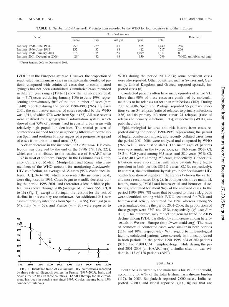
A clear decrease in the incidence of Leishmania-HIV coin-fection was observed by the end of the 1990s (79, 138, 225),which can be attributed to the routine use of HAART since1997 in most of southern Europe. In the Leishmaniasis Refer-ence Centers of Madrid, Montpellier, and Rome, which aremembers of the WHO network for monitoring Leishmania-HIV coinfection, an average of 35 cases (95% confidence in-terval [CI], 34 to 38), which represented the incidence peak,were diagnosed in 1997. Cases began to steadily decrease dur-ing the period 1998–2001, and thereafter a low-incidence pla-teau was shown through 2006 (average of 12 cases; 95% CI, 8to 16) (Fig. 1), except in Portugal; the reasons for the lack ofdecline in this country are unknown. An additional 241 newcases of primary infections from Spain (n � 95), Portugal (n �64), Italy (n � 52), and France (n � 30) were reported to
WHO during the period 2001–2006; some persistent caseswere also reported. Other countries, such as Switzerland, Ger-many, United Kingdom, and Greece, reported sporadic im-ported cases (6).
Coinfected patients often have many episodes of active VL.More than 90% of these cases are confirmed by molecularmethods to be relapses rather than reinfections (162). During2001 to 2006, Spain and Portugal reported 95 primary infec-tions versus 34 relapses (ratio of relapses to primary infections,0.36) and 64 primary infections versus 21 relapses (ratio ofrelapses to primary infections, 0.33), respectively (WHO, un-published data).
Epidemiological features and risk factors from cases re-ported during the period 1990–1998, representing the periodof higher coinfection impact, and recently collated cases fromthe period 2001–2006, were analyzed and compared by WHO(266; WHO, unpublished data). The mean ages of patientswere very similar in the two periods, i.e., 38.6 years (95% CI,38.2 to 39.0 years) among 965 cases and 38.9 years (95% CI,37.8 to 40.1 years) among 253 cases, respectively. Gender dis-tributions were also similar, with male patients being highlyprevalent in both periods (83.2% versus 88.5%, respectively).In contrast, the distributions by risk group for Leishmania-HIVcoinfection showed significant differences between the earlierand more recent cases (Fig. 2). In both periods, three main riskfactors, namely, IVDU and heterosexual and homosexual ac-tivities, accounted for about 94% of the analyzed cases. In theperiod 1990–1998, 781 cases that belonged to these risk groupswere identified, among which IVDU accounted for 76% andheterosexual activity accounted for 12%, whereas among 95cases analyzed during the period 2001–2006, the proportions ofthese groups were 67% and 23%, respectively (�2 test; P �0.01). This difference may reflect the general trend of AIDSdecline among IVDU paralleled by an increase among hetero-sexuals in Western Europe (http://www.unaids.org). The ratesof homosexual coinfected cases were similar in both periods(11% and 10%, respectively). With regard to immunologicalfactors, coinfected patients were severely immunosuppressedin both periods. In the period 1990–1998, 624 of 682 patients(91%) had �200 CD4� lymphocytes/�l, while during the pe-riod 2001–2006 (an HAART era) a similar situation was evi-dent in 113 of 128 patients (88%).
South Asia
South Asia is currently the main focus for VL in the world,accounting for 67% of the total leishmaniasis disease burden(117). In 2005, Bangladesh reported 7,000 cases, India re-ported 32,800, and Nepal reported 3,000, figures that are
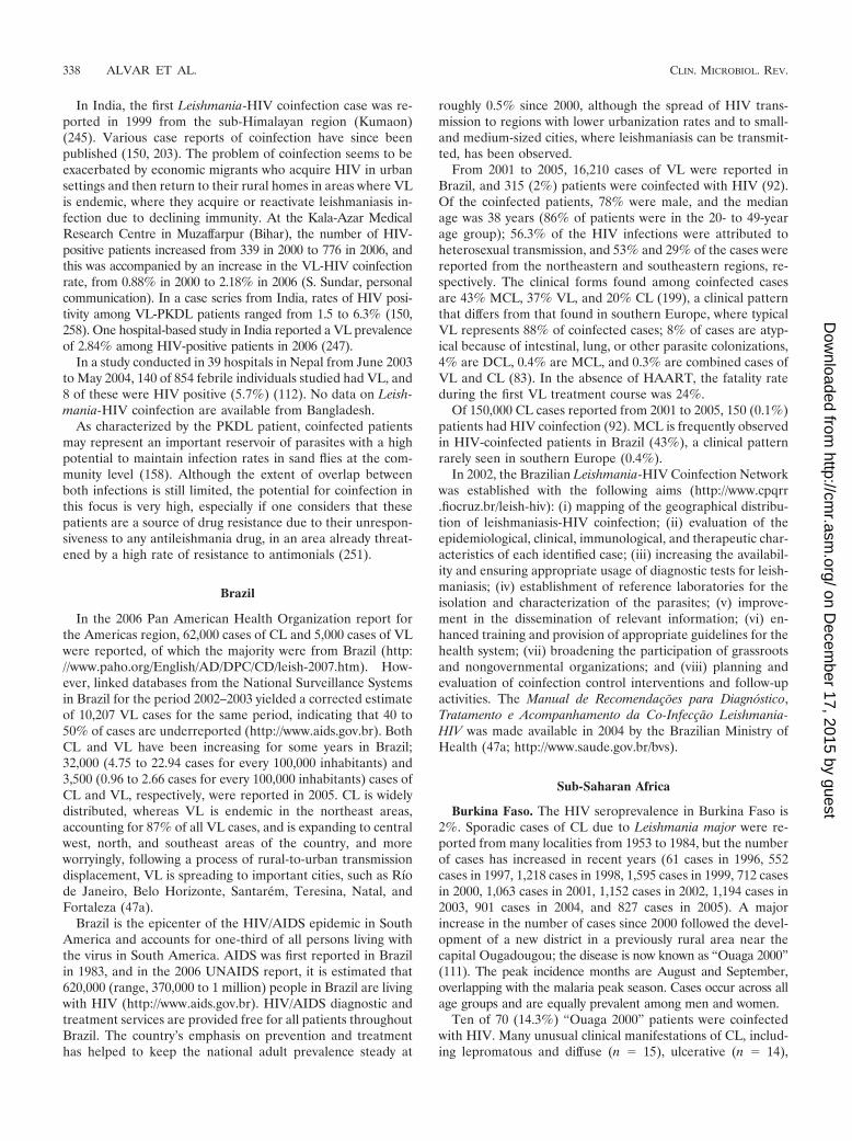
TABLE 1. Number of Leishmania-HIV coinfections recorded by the WHO for four countries in southern Europe
PeriodNo. of coinfections
ReferenceFrance Italy Portugal Spain Total
January 1990–June 1998 259 229 117 835 1,440 266January 1996–June 1998 132 85 88 412 717 266January 1990–January 2001 318 335 159 1,099 1,911 83January 2001–December 2006 29a 42 98 130 299 WHO, unpublished data
a From January 2001 to December 2005.
FIG. 1. Incidence trend of Leishmania-HIV coinfections recordedby three referral diagnosis centers, in France (1997–2005), Italy, andSpain (1997–2006). In these countries, HAART therapy for HIV treat-ment has been in routine use since 1997. Circles, means; bars, 95%confidence intervals.
336 ALVAR ET AL. CLIN. MICROBIOL. REV.
on Decem
ber 17, 2015 by guesthttp://cm
r.asm.org/
Dow
nloaded from

known to be serious underestimations (34, 123, 230). In Bihar,India, a house-to-house survey demonstrated that only one ineight VL cases was counted in official government surveillancedata (246). Additionally, there has been an upward trend in allthree countries over the last several years, where 147 millionpeople are estimated to be at risk of infection. More realisticestimates of annual incidence suggest approximately 250,000cases in India, 40,000 in Bangladesh, and 3,000 in Nepal, whichrepresents half of the VL burden in the world (41).
Interestingly, VL is concentrated in only 94 districts in allthree countries and in four states in the northeast in India(Bihar, Uttar Pradesh, West Bengal, and Jharkhand). Thestate of Bihar accounts for �90% of cases and contributes tothe spread of the disease into neighboring areas of Nepal. Thereemergence of the disease in the 1970s was most likely due toresidual post-kala-azar dermal leishmaniasis (PKDL) patientsharboring parasites in the skin. Currently, PKDL is reported tooccur in only 1% of Indian VL patients, but reliable data arelacking (228). In addition to this phenomenon, a loss of ac-quired immunity may partially explain the periodicity of VLincidence peaks every 10 to 15 years. In the past, most caseswere in adults, although now there are reported to be manymore children and adolescents, i.e., immune-naıve individuals,affected (41, 201).
The first case of HIV infection in India was reported in 1986in Chennai (Madras) and was quickly followed by reports fromall major cities; there has been a steady rise in the number ofcases since (63, 164). The new 2006 estimates by the NationalAIDS Control Organization (http://www.nacoonline.org), sup-ported by UNAIDS and WHO, indicate that the national adultHIV prevalence in India is approximately 0.36%, which corre-sponds to an estimated 2 million to 3.1 million people livingwith HIV in the country (http://www.nacoonline.org). The dis-ease is concentrated in states in the south (Tamil Nadu,Andhra Pradesh, Maharashtra, and Karnataka), where effec-tive interventions have caused HIV prevalence to begin todecline or stabilize, and to a lesser degree in the northeast(West Bengal, Orissa, Rajasthan, and Bihar), where overlapwith leishmaniasis occurs. HAART was initially introduced in25 centers in India during 2004, but because the implementa-tion of the national HIV plan has been rapid, HAART is nowwidely available all over the country.
In Bangladesh, there are 11,000 people living with HIV; anational adult prevalence of �0.1% was estimated for 2006(http://www.unaids.org/en/HIV_data/epi2006/default.asp). InNepal, an estimated 75,000 people are living with HIV, and thenational adult prevalence was estimated to be 0.5% for 2006(http://www.unaids.org/en/HIV_data/epi2006/default.asp).
FIG. 2. Algorithm for the diagnosis of VL in HIV-infected patients.
VOL. 21, 2008 LEISHMANIASIS AND AIDS AFTER 20 YEARS 337
on Decem
ber 17, 2015 by guesthttp://cm
r.asm.org/
Dow
nloaded from
In India, the first Leishmania-HIV coinfection case was re-ported in 1999 from the sub-Himalayan region (Kumaon)(245). Various case reports of coinfection have since beenpublished (150, 203). The problem of coinfection seems to beexacerbated by economic migrants who acquire HIV in urbansettings and then return to their rural homes in areas where VLis endemic, where they acquire or reactivate leishmaniasis in-fection due to declining immunity. At the Kala-Azar MedicalResearch Centre in Muzaffarpur (Bihar), the number of HIV-positive patients increased from 339 in 2000 to 776 in 2006, andthis was accompanied by an increase in the VL-HIV coinfectionrate, from 0.88% in 2000 to 2.18% in 2006 (S. Sundar, personalcommunication). In a case series from India, rates of HIV posi-tivity among VL-PKDL patients ranged from 1.5 to 6.3% (150,258). One hospital-based study in India reported a VL prevalenceof 2.84% among HIV-positive patients in 2006 (247).
In a study conducted in 39 hospitals in Nepal from June 2003to May 2004, 140 of 854 febrile individuals studied had VL, and8 of these were HIV positive (5.7%) (112). No data on Leish-mania-HIV coinfection are available from Bangladesh.
As characterized by the PKDL patient, coinfected patientsmay represent an important reservoir of parasites with a highpotential to maintain infection rates in sand flies at the com-munity level (158). Although the extent of overlap betweenboth infections is still limited, the potential for coinfection inthis focus is very high, especially if one considers that thesepatients are a source of drug resistance due to their unrespon-siveness to any antileishmania drug, in an area already threat-ened by a high rate of resistance to antimonials (251).
Brazil
In the 2006 Pan American Health Organization report forthe Americas region, 62,000 cases of CL and 5,000 cases of VLwere reported, of which the majority were from Brazil (http://www.paho.org/English/AD/DPC/CD/leish-2007.htm). How-ever, linked databases from the National Surveillance Systemsin Brazil for the period 2002–2003 yielded a corrected estimateof 10,207 VL cases for the same period, indicating that 40 to50% of cases are underreported (http://www.aids.gov.br). BothCL and VL have been increasing for some years in Brazil;32,000 (4.75 to 22.94 cases for every 100,000 inhabitants) and3,500 (0.96 to 2.66 cases for every 100,000 inhabitants) cases ofCL and VL, respectively, were reported in 2005. CL is widelydistributed, whereas VL is endemic in the northeast areas,accounting for 87% of all VL cases, and is expanding to centralwest, north, and southeast areas of the country, and moreworryingly, following a process of rural-to-urban transmissiondisplacement, VL is spreading to important cities, such as Rıode Janeiro, Belo Horizonte, Santarem, Teresina, Natal, andFortaleza (47a).
Brazil is the epicenter of the HIV/AIDS epidemic in SouthAmerica and accounts for one-third of all persons living withthe virus in South America. AIDS was first reported in Brazilin 1983, and in the 2006 UNAIDS report, it is estimated that620,000 (range, 370,000 to 1 million) people in Brazil are livingwith HIV (http://www.aids.gov.br). HIV/AIDS diagnostic andtreatment services are provided free for all patients throughoutBrazil. The country’s emphasis on prevention and treatmenthas helped to keep the national adult prevalence steady at
roughly 0.5% since 2000, although the spread of HIV trans-mission to regions with lower urbanization rates and to small-and medium-sized cities, where leishmaniasis can be transmit-ted, has been observed.
From 2001 to 2005, 16,210 cases of VL were reported inBrazil, and 315 (2%) patients were coinfected with HIV (92).Of the coinfected patients, 78% were male, and the medianage was 38 years (86% of patients were in the 20- to 49-yearage group); 56.3% of the HIV infections were attributed toheterosexual transmission, and 53% and 29% of the cases werereported from the northeastern and southeastern regions, re-spectively. The clinical forms found among coinfected casesare 43% MCL, 37% VL, and 20% CL (199), a clinical patternthat differs from that found in southern Europe, where typicalVL represents 88% of coinfected cases; 8% of cases are atyp-ical because of intestinal, lung, or other parasite colonizations,4% are DCL, 0.4% are MCL, and 0.3% are combined cases ofVL and CL (83). In the absence of HAART, the fatality rateduring the first VL treatment course was 24%.
Of 150,000 CL cases reported from 2001 to 2005, 150 (0.1%)patients had HIV coinfection (92). MCL is frequently observedin HIV-coinfected patients in Brazil (43%), a clinical patternrarely seen in southern Europe (0.4%).
In 2002, the Brazilian Leishmania-HIV Coinfection Networkwas established with the following aims (http://www.cpqrr.fiocruz.br/leish-hiv): (i) mapping of the geographical distribu-tion of leishmaniasis-HIV coinfection; (ii) evaluation of theepidemiological, clinical, immunological, and therapeutic char-acteristics of each identified case; (iii) increasing the availabil-ity and ensuring appropriate usage of diagnostic tests for leish-maniasis; (iv) establishment of reference laboratories for theisolation and characterization of the parasites; (v) improve-ment in the dissemination of relevant information; (vi) en-hanced training and provision of appropriate guidelines for thehealth system; (vii) broadening the participation of grassrootsand nongovernmental organizations; and (viii) planning andevaluation of coinfection control interventions and follow-upactivities. The Manual de Recomendacoes para Diagnostico,Tratamento e Acompanhamento da Co-Infeccao Leishmania-HIV was made available in 2004 by the Brazilian Ministry ofHealth (47a; http://www.saude.gov.br/bvs).
Sub-Saharan Africa
Burkina Faso. The HIV seroprevalence in Burkina Faso is2%. Sporadic cases of CL due to Leishmania major were re-ported from many localities from 1953 to 1984, but the numberof cases has increased in recent years (61 cases in 1996, 552cases in 1997, 1,218 cases in 1998, 1,595 cases in 1999, 712 casesin 2000, 1,063 cases in 2001, 1,152 cases in 2002, 1,194 cases in2003, 901 cases in 2004, and 827 cases in 2005). A majorincrease in the number of cases since 2000 followed the devel-opment of a new district in a previously rural area near thecapital Ougadougou; the disease is now known as “Ouaga 2000”(111). The peak incidence months are August and September,overlapping with the malaria peak season. Cases occur across allage groups and are equally prevalent among men and women.
Ten of 70 (14.3%) “Ouaga 2000” patients were coinfectedwith HIV. Many unusual clinical manifestations of CL, includ-ing lepromatous and diffuse (n � 15), ulcerative (n � 14),
338 ALVAR ET AL. CLIN. MICROBIOL. REV.
on Decem
ber 17, 2015 by guesthttp://cm
r.asm.org/
Dow
nloaded from
infiltrative (n � 12), papulonodular (n � 9), psoriasis-like (n �5), Kaposi’s sarcoma-like (n � 1), cheloid (n � 1), and histioid(n � 1) forms, were found in a prospective study of 32 coin-fected patients. Treatment with a standard dosage regimen ofmeglumine antimoniate intramuscularly for 21 days achieved a50% cure rate, but rapid relapses were observed in HIV-coin-fected patients, for whom repeated courses of treatment wererequired (111, 172). Three treatment cycles increased the curerate to 75%. Visceralization occurred in one HIV-positive pa-tient, with disseminated papulonodular leishmaniasis and abone marrow aspirate positive for L. major. Similar findingswere observed in French Guiana, where nine coinfected pa-tients had more lesions and less adenopathy or lymphangitisthan immunocompetent patients, with a poorer response totreatment and higher recurrence and/or reinfection rates (66).
Ethiopia. There is incomplete information on the epidemi-ology of leishmaniasis in Ethiopia, as no surveillance system isyet in place. However, reports of VL endemicity date back tothe 1940s. The disease is prevalent mostly in lowland aridareas, and the parasite involved is mainly L. donovani, with anestimated annual incidence of 4,000 cases. The six areas ofendemicity identified are northwestern Ethiopia (Metema,Humera, Wolkayit, and Libo/Fogera), northeastern Ethiopia(Awash Valley and Ethio-Djibouti border), south and south-west Ethiopia (Dawa, Genale, Gelana, Segen, Woito, Konso,and Omo River Valley), the Kenyan border, and the Gam-bella-Sudan border (113). However, leishmaniasis is spreadingto areas where it was previously nonendemic, as exemplified bya recent outbreak in the Libo and Fogera districts (AmharaState), a highland area close to the focus in the northwest (8).
The first HIV-positive patient was reported in 1984, and thefirst AIDS case was reported in 1986. Based on antenatalsentinel surveillance projections, the estimated national HIVprevalence is 3.5% (10% in urban areas and 1.9% in ruralareas). The urban prevalence is thought to have stabilizedduring the period 1999–2001, with a current estimation of 1.3million HIV-positive people. HAART was introduced in 2003,but programs do not yet adequately address the needs of therural poor. Approximately 65,000 HIV patients are now receiv-ing HAART.
In Humera (northwest Ethiopia), the proportion of VL pa-tients who were coinfected with HIV increased from 18.5% in1998–1999 to 40% in 2006. In a retrospective review of 791cases in Tigray, the case fatality rate among VL-HIV-coin-fected patients was four times higher than that for VL patientsnot infected with HIV (143). In Libo, 15 to 18% of all VLpatients are HIV positive (Medecins Sans Frontieres (MSF)—Greece, personal communication). The groups at highest riskof coinfection are seasonal migrant laborers, sex workers,young adults in resettlement areas (particularly in the north-west and along the Sudan border), truck and other publictransport drivers, and military personnel deployed in borderareas (19).
Kenya, Uganda, and Somalia. Data quality is poor for Kenya,Uganda, and Somalia. There are well-established foci in semi-arid, poor, and remote districts where termite hills are colo-nized by the sandfly vector (Phlebotomus martini).
In Kenya, there are several historical foci, in West Pokot(representing 70% of all patients in Kenya), Baringo, Man-dera, Garissa, and Mwingi. There was a recent outbreak in
2006 in northeast Kenya, at Wajir and Issiolo, which mainlyaffected children (n � 48; 95% of patients were �5 years ofage). In 2006, 15 cases of VL-HIV coinfection were reported,but the risk of coinfection is increasing (268a).
In Uganda, the only documented area where VL is endemicis Pokot County, in the Nakapiripirit district in the northeast-ern region of Karamoja. This is an extension of the focus ofendemicity of the West Pokot and Baringo districts of the RiftValley province in Kenya. There is no national leishmaniasiscontrol program, and most VL cases are monitored by MSF—Switzerland, who treated more than 2,500 patients in AmudatHospital from 2000 to 2006. Approximately 70% of treated VLpatients came from Kenya (West Pokot and Baringo districts).The male-to-female ratio of the patients was 3:1, and �60% ofthe patients were under 15 years of age. Eight cases of VL-HIVcoinfection were confirmed (MSF—Switzerland, personalcommunication); no other data on VL-HIV coinfection areavailable. The HIV prevalence in Karamoja, on the Ugan-dan-Kenyan border, reached 3% in 2006; local conflicts,with associated rape, and cattle trading in towns may exac-erbate the situation and elevate the potential for VL-HIVcoinfection.
In Somalia, MSF—Belgium is currently dealing with an out-break of VL and treated 263 and 1,300 cases in 2005 and 2006,respectively (MSF—Belgium, personal communication). Nocoinfection has been reported yet.
Sudan. The main area of endemicity is the southeast of thecountry, bordering Ethiopia. There have been recurrent epi-demics with high mortality rates, most recently in 1989 (236).The number of VL cases in 2006 was unexpectedly low in thestates of Gedaref (n � 1,918) and Sennar (n � 476), andsimilar figures were observed in Upper Nile, but the meanannual incidence in the whole country is approximately 15,000to 20,000 VL cases (S. H. El-Safi, personal communication).The parasite identified is mainly L. donovani, although L.infantum also occurs (81). Transmission is basically anthro-ponotic, with PKDL cases occurring in up to 50% of VL pa-tients, forming an important human reservoir (91).
The first case of AIDS was reported in 1986, and by Decem-ber 2006, the adult national prevalence was 1.6%, indicatingthat Sudan is now in a generalized epidemic (http://www.unaids.org/en/HIV_data/epi2006/default.asp). The estimatednumber of HIV/AIDS cases is around 350,000 (range, 170,000to 580,000). The transmission is mainly heterosexual (97%),and vertical transmission from mother to child also plays arole. The most affected areas are Khartoum and the easternand southern states, due to population movements (refugeesand internally displaced populations). The 19- to 39-year agegroup is the most affected, and among registered patients,males predominate (male-to-female ratio, 3:1). The prevalenceof HIV infection in the surveillance sentinel clinics in 2002 and2004 was 1.1% and 1.9%, respectively, for sexually transmittedinfection clinics, 1.6% and 2.3%, respectively, for tuberculosispatients, and 2% and 0.95%, respectively, for antenatal careclinics (Sudan National AIDS Program). The prevalence inother groups was as follows: street children, 2.3%; universitystudents, 1.1%; refugees, 4%; prisoners, 2.5%; and sex work-ers, 4.4%. The strategic objectives of the National Policies onHIV/AIDS are to maintain the prevalence of HIV/AIDS below2% and to provide care, treatment, and support to infected
VOL. 21, 2008 LEISHMANIASIS AND AIDS AFTER 20 YEARS 339
on Decem
ber 17, 2015 by guesthttp://cm
r.asm.org/
Dow
nloaded from
patients. In 2006, there were 40 centers for treatment services(voluntary counseling and testing units) and around 1,000 pa-tients receiving HAART. Screening of military personnel andstudents indicates that the number of cases of HIV isincreasing.
By 1998, three cases of coinfection had been identified.Since then, the reported prevalence in hospital-based studieswas 5% (3/60) in Khartoum between 1998 and 1999, 9.4%(5/53) in Khartoum in 2002, and 8.1% (3/37) and 3.6% (3/84)in 2002 and 2003, respectively, in Gedaref State (Sudan Na-tional AIDS Program).
MICROBIOLOGY
Since the first case of Leishmania-HIV coinfection (76),methods to identify Leishmania have greatly evolved. Besidemultilocus enzyme electrophoresis, a large number of molec-ular methods have been introduced for the characterization ofLeishmania from the genus to strain levels, but most of themlack standardization.
Three main cryobanks and identification centers, in France(Montpellier), Italy (Rome), and Spain (Madrid), which usethe same reference identification technique by 15 enzymaticsystems with starch gel electrophoresis, according to Rioux etal. (213), have identified a large series of isolates from Leish-mania-HIV coinfection cases since 1986, on which the presentreview is focused. The data presented are based on a com-pilation of several papers, mainly three papers published ina 2003 special issue of Annals of Tropical Medicine andParasitology, namely, those by Pratlong et al. (195) forsouthern France, Chicharro et al. (59) for Spain, and Gram-iccia (108) for Italy, and a recent paper by Campino et al.(54) for Portugal.
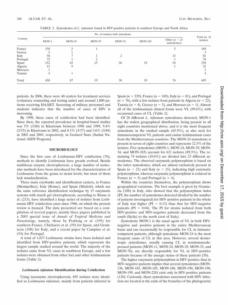
A total of 1,037 Leishmania strains have been isolated andidentified from HIV-positive patients, which represents thelargest sample studied around the world. The majority of theisolates came from VL cases in southwest Europe, and a fewisolates were obtained from other foci and other leishmaniasisforms (Table 2).
Leishmania infantum Identification during Coinfection
Using isoenzyme electrophoresis, 695 isolates were identi-fied as Leishmania infantum, mainly from patients infected in
Spain (n � 339), France (n � 169), Italy (n � 81), and Portugal(n � 76), with a few isolates from patients in Algeria (n � 22),Tunisia (n � 4), Greece (n � 3), and Morocco (n � 1). Almostall of the leishmaniasis clinical forms were VL (99.6%), withoccasional cases of CL (Table 2).
Of 28 different L. infantum zymodemes detected, MON-1has the widest geographical distribution, being present in alleight countries mentioned above, and it is the most frequentzymodeme in the studied sample (65.9%), as also seen forimmunocompetent VL patients and canine leishmaniasis casesfrom the Mediterranean countries. The MON-24 zymodeme ispresent in seven of eight countries and represents 12.5% of theisolates. Five zymodemes (MON-1, MON-24, MON-29, MON-34, and MON-183) account for 621 isolates (89.3%). The re-maining 74 isolates (10.6%) are divided into 23 different zy-modemes. The observed enzymatic polymorphism is based onthe latter zymodemes, which are almost exclusively present inSpain (n � 21) and Italy (n � 15), indicating high enzymaticpolymorphism, whereas enzymatic polymorphism is reduced inFrance (n � 5) and Portugal (n � 4).
Within the countries themselves, the polymorphism showsgeographical variations. The best example is given by Gramic-cia (108) in Italy, who showed that the polymorphism index(PI; the number of zymodemes detected divided by the numberof patients investigated) for HIV-positive patients in the wholeof Italy was higher (PI � 0.13) than that for HIV-negativepatients (PI � 0.04). The PI for strains isolated from bothHIV-positive and HIV-negative patients decreased from thesouth (Sicily) to the north (rest of Italy).
Zymodeme MON-1 is the usual agent of VL in both HIV-negative and -positive patients in the entire Mediterraneanbasin and can occasionally be responsible for CL in immuno-competent patients, although zymodeme MON-24 is the mostfrequent cause of CL in this area. However, several dermo-tropic zymodemes, usually causing CL in nonimmunode-pressed patients (MON-11, MON-24, MON-29, MON-33, andMON-78), are directly responsible for VL in HIV-positivepatients because of the anergic status of these patients (58).
The higher enzymatic polymorphism in HIV-positive than inHIV-negative patients implies that several zymodemes (MON-136, MON-183, MON-185, MON-188, MON-190, MON-198,MON-199, and MON-228) exist only in HIV-positive patients(120). Curiously, these zymodemes associated with HIV infec-tion are located at the ends of the branches of the phylogenetic
TABLE 2. Zymodemes of L. infantum found in HIV-positive patients in southern Europe and North Africa
Country
No. of isolates with zymodemeTotal no. of
isolatesMON-1 MON-24 MON-29 MON-34 MON-183 Other (n � 23zymodemes)
France 158 5 1 5 169Greece 2 1 3Italy 58 7 1 2 13 81Portugal 72 1 2 1 76Spain 152 63 15 36 19 54 339Algeria 12 9 1 22Morocco 1 1Tunisia 3 1 4
Total 458 87 19 38 19 74 695
340 ALVAR ET AL. CLIN. MICROBIOL. REV.
on Decem
ber 17, 2015 by guesthttp://cm
r.asm.org/
Dow
nloaded from
tree, as if they had recently appeared. The question is whetherthese zymodemes can survive and cause symptomatic diseasein immunocompetent patients (195).
VL occurring during HIV infection cannot be cured, evenafter extensive antileishmanial treatment. The likelihood of arelapse occurring is dependent on the level of CD4� cells inthe patient. In patients (n � 80) from southern Europe, two orthree stocks were isolated from each patient following succes-sive episodes of relapse, with time intervals of 1 to 4 years, andwere subsequently identified by enzymatic electrophoresis. Allof the isolates were L. infantum, and 18 zymodemes werefound repeatedly during the various episodes. They weremainly MON-1 (58.1%) and MON-24 (13.3%), but a largenumber of other zymodemes were also identified in the globalsample (Table 3). These results suggest that there is no specificzymodeme related to a particular severity of VL, as severalzymodemes are expressed in further relapses. However, all buttwo patients had the same L. infantum zymodeme during thesuccessive episodes. In two patients, two distinct zymodemeswere obtained, at time intervals of 20 months (MON-1 andMON-34) and 29 months (MON-1 and MON-33) (100). Byusing a more powerful tool for molecular tracking (seminestedPCR-restriction fragment length polymorphism analysis) thatis capable of discriminating within the same zymodeme, Mo-rales et al. (162) showed that 7.5% of the so-called relapses in40 patients with VL-HIV coinfection had a different bandpattern from that found in the primary attack. From thesestudies, it appears that repeated leishmaniasis episodes inHIV-positive patients could be due mainly to relapses of thefirst infection. For the two cases of different zymodemes, theseoccurred due to either a reinfection or the existence of a mixedinfection by two zymodemes which were differently selectedduring culture of the two episodes.
More recently, L. infantum-L. major hybrid strains weredescribed for HIV-coinfected patients from Portugal, by se-quencing of the gene for polymerase II RNA and by isoenzy-matic identification (202).
Other Leishmania Species during HIV Coinfection
No data from anthroponotic VL foci are available, with theexception of a single strain from Djibouti isolated and identi-fied as L. donovani MON-268 (194).
Although Leishmania-HIV coinfection concerns mainly VL,some cases of CL have been reported from countries where itis endemic in Africa and South America. In the sub-SaharanSahelian zone of Africa, several L. major isolates obtainedfrom patients in Mali (n � 4), Burkina Faso (n � 7), Senegal
(n � 1), and Mauritania (n � 1) belonged to zymodemeMON-26 and its small variants, MON-74 and MON-117, agroup of zymodemes extending from west Africa to centralAsia. An outbreak of CL in Ouagadougou (Burkina Faso) dueto L. major MON-74 included HIV-positive patients (111).
Among Brazilian HIV-coinfected patients, Rabello et al.(199) reported eight cases of CL by L. braziliensis and L.guyanensis, three cases of MCL by L. braziliensis, and one caseof VL by L. chagasi. In French Guiana, five strains isolatedfrom HIV-positive patients with CL were typed by multilocusenzyme electrophoresis and shown to belong to L. guyanensis,and four zymodemes were detected, including MON-43 (n �1), MON-45 (n � 1), MON-131 (n � 2), and MON-148 (n �1) (66).
Moreover, various New World Leishmania dermotropic spe-cies, such as L. braziliensis, L. mexicana, and L. amazonensis,have been identified as causes of VL in HIV-positive patients(23, 24, 200, 241), while conversely, viscerotropic variants of L.infantum/L. chagasi have been found in cutaneous lesions oreven in the healthy skin of HIV-positive patients (44, 105, 191).
The Trypanosomatidae family includes the protozoa ofplants, insects, and mammals, but only Leishmania andTrypanosoma species are usually found in humans. However, insome cases, nonhuman monoxenous trypanosomatids havebeen isolated from HIV-positive patients, mimicing both VL(121, 180) and CL (40, 75, 227). These infections suggest thatthe immunocompromised patient could be vulnerable to othercurrently non-human-pathogenic trypanosomatids due to theiranergic status (58, 74).
PATHOGENESIS
VL-HIV Coinfection
Early after the appearance and report of the first cases ofLeishmania-HIV coinfection, it was evident that the presenceof both pathogens concomitantly in the same host cell (themacrophage) might have enhanced reciprocal effects that in-fluence the expression and multiplication of either one or bothpathogens (36, 269). It is thought that the parasite infectionfound concurrently with HIV induces chronic immune activa-tion, and therefore an increased HIV load and acceleratedprogression of AIDS (27), whereas immunological disturbancescaused by HIV are particularly favorable for the uncontrolledmultiplication of the parasite.
During the last 10 years, attempts have been made to dis-cover those mechanisms that are involved in the immuno-pathogenesis of Leishmania-HIV coinfection and to confirm
TABLE 3. Numbers of isolates identified according to zymodemes found during VL relapses
RegionaNo. of isolates with zymodeme
1 11 24 28 29 33 34 72 78 108 136 183 185 188 201 228 253 283
A 13 2 2 1 1 1B 8 1 1 1 1 1C 20 10 1 5 1 2 1 2 1D 20 1 1 1 1 1 1 1 1 1Total 61 1 14 3 4 2 6 1 1 2 1 2 1 1 1 1 2 1
a Region A, south of France (195); region B, Barcelona, Spain (100); region C, Madrid, Spain (59); and region D, Italy (108).
VOL. 21, 2008 LEISHMANIASIS AND AIDS AFTER 20 YEARS 341
on Decem
ber 17, 2015 by guesthttp://cm
r.asm.org/
Dow
nloaded from

that there is a specific Leishmania-HIV interaction at the cel-lular level that might affect the course of infection for eachpathogen. It has been determined that such interactions arepatent in the macrophage as well in other cells, such as den-dritic cells (DCs). The involvement of other cells might explainthe systemic effects observed in coinfected patients.
The main problem of coinfection is that parasitic infectionmay induce the activation of latent virus. Clinical studies haveshown that leishmaniasis promotes an increase in serum HIVtype 1 (HIV-1) load (49, 196) and a more rapid progression toAIDS that reduces life expectancy in HIV-infected patients(72). Experimental studies have demonstrated the participa-tion of Leishmania in the pathogenesis of HIV-1 through theinduction of viral replication and disease progression. Initialreports proved that Leishmania can upregulate virus expres-sion in two monocytoid cell lines latently infected with HIV-1(36), and evidence was provided that Leishmania promasti-gotes (LPG) and the intramembrane structural component ofLPG (core-PI) present in amastigotes were both capable ofpromoting virus replication in T cells through complex bio-chemical pathways that involve the translocation of the tran-scriptional factor NF-�� to the nucleus (35). Although theexact mechanism remained unclear, it was thought that LPGmolecules could interact with the HIV-infected T cell duringantigenic presentation between the Leishmania-infected mac-rophage and the T cell (177). Furthermore, Leishmania mayinduce HIV replication in infected cells through antigen-spe-cific and non-antigen-specific mechanisms in which tumor ne-crosis factor alpha (TNF-) plays a key role (270).
More recently, it has been shown with human tonsillar tissuecultured ex vivo (275) and primary human monocyte-derivedmacrophages (274) that Leishmania does not directly affectvirus gene expression but instead modulates the life cycle ofHIV-1 through an indirect phenomenon that is linked to theinduction or elevation of TNF- and interleukin-1a (IL-1a)and that may function in an autocrine/paracrine manner toupregulate virus gene expression mediated through inductionof NF-�� (109).
For patients coinfected with HIV-1 and Leishmania, it hasbeen proposed that the increase in cytokine production in-duced by the parasite will affect virus production not only inmacrophages but also in HIV-1-infected CD4� T cells (275).
Different clinical studies performed recently illustrate thecomplexity of the interplay between the pathogens. It has beenobserved that Leishmania infection may affect the life cycle ofthe virus through the increased expression of specific chemo-kine receptors. Leishmania-HIV-coinfected patients have asignificantly higher level of CCR5� CD3� T cells than doHIV-negative patients with VL or HIV-positive patients with-out VL (176). CCR5 is a major coreceptor for HIV entry intarget cells, and its high expression in subjects is related to ahigh virus load and accelerated progression to HIV disease(206). Another study with coinfected patients showed amarked increase in circulating levels of the soluble form of thehuman leukocyte antigen G (HLA-G), a nonclassical histo-compatibility complex class I molecule with immunosuppres-sive properties (88).
Interactions between HIV and Leishmania are not only re-stricted to the replication of the virus. Clinical studies havedemonstrated that the high incidence of disseminated leish-
maniasis in AIDS patients (141) and the high peripheral par-asitemia (45) are indicative of uncontrolled parasite growth. Inconcordance with this condition, experimental in vitro coinfec-tion of monocyte-derived macrophages with HIV-1 and L.donovani (271) or L. infantum (276) promastigotes showed asignificant enhancement of intracellular parasite growth com-pared with parasite infection alone. The lack of control ofintracellular multiplication is probably related to the HIV-1-mediated impairment of an effector function carried out bymacrophages, such as phagocytosis, intracellular killing, che-motaxis, and cytokine production (126). Despite the fact thatHIV-1-infected monocyte-derived macrophages display an im-paired capacity to phagocytose numerous pathogens, Leishma-nia uptake is increased following virus infection (276).
In immunocompetent individuals, the protective Leishma-nia-specific immune response is associated with a Th1 cytokineprofile, while susceptibility to Leishmania infection and diseaseprogression are related to a Th2 cytokine response (127). It hasbeen confirmed that HIV-1 inhibits the proliferative responseto L. donovani (269); this reduced cellular response might bebecause the known inductive signal for gamma interferon(IFN-) is lacking or could be due to the direct influence ofanti-inflammatory Th2 cytokines. Wolday et al. (272) con-firmed that HIV-1 can modulate in vitro and ex vivo cytokineproduction in response to Leishmania; the addition of HIV-1to cultures decreased IFN-, IL-12, and IL-18 production andincreased IL-4 and IL-10 production in response to Leishma-nia by peripheral blood monocytic cells from healthy donors,while cells from HIV-1-infected and coinfected patients failedto secrete IFN- in response to Leishmania. Analysis of the exvivo production of cytokines by peripheral blood monocyticcells from coinfected patients showed that greater quantities ofIL-4 and IL-10 were released than those from HIV-positivepatients not infected with Leishmania, and production ofIFN- showed no changes after stimulation with phytohemag-glutinin (174). In addition, patients with VL and HIV-1 infec-tion have decreased circulating levels of IL-15, a cytokine thatenhances the Th1 response and potentiates the immune re-sponse to intracellular human pathogens (85). All of these datasuggest that a shift toward the Th2 type of specific T-cellimmune response to Leishmania occurs in patients with T-lymphocyte defects induced by HIV-1, which would partly ex-plain the high susceptibility to Leishmania infection, the highseverity of the disease, and why reactivations occur in thesepatients (49).
Barreto-de-Souza et al. (25) recently studied the role of thetranscriptional transactivator (Tat) protein, an essential viralgene product for HIV-1 replication, in the exacerbation ofLeishmania proliferation in human macrophages. Tat is se-creted in large amounts by cells infected by HIV-1 and is ableto stimulate secretion of IL-1�, IL-4, IL-6, IL-8, transforminggrowth factor beta (TGF-�), TNF-, and TNF-�. The resultsshowed that Tat protein was able to override the leishmani-cidal effect of IFN- and to enhance Leishmania replication inhuman macrophages through the production of prostaglandinE2 in the macrophage, which in turn increases the synthesis ofTGF-�1 and the augmentation of Leishmania growth.
A review of the literature confirms that there is a multifac-eted interaction between Leishmania and HIV-1 in the mac-rophage host cell. But the macrophage is not the only cell type
342 ALVAR ET AL. CLIN. MICROBIOL. REV.
on Decem
ber 17, 2015 by guesthttp://cm
r.asm.org/
Dow
nloaded from
capable of hosting both pathogens; DCs can also be infected byLeishmania parasites and HIV-1 (168, 250). There is no infor-mation on the consequence of both pathogens being present inDCs, but it has been proposed that such coinfection mightmodify the course of infection for each pathogen (102). Fur-thermore, the ability of both Leishmania and HIV-1 to bind tothe same DC receptor, DC-SIGN (DC-specific ICAM-3-grab-bing nonintegrin), to gain entry into DCs (51, 65) might affectthe life cycle of the other pathogen. It has been demonstratedthat HIV-1 transmission to CD4� T cells is diminished whenDC-SIGN-expressing cells are preincubated with Leishmaniaamastigotes before being pulsed with virions (273). Neverthe-less, the consequence of both parasite and virus competing forthe same DC receptor is unknown, and the importance of DCsas a virus and parasite reservoir, as well as the impact of bothpathogens being present in DCs at once in coinfected patients,remains to be determined.
CL-HIV Coinfection
CL occurs in humans as a localized (LCL), mucocutaneous(MCL), or diffuse (DCL) form, depending on the infectingspecies of Leishmania (2, 115) and the human host. The pa-thology of LCL in humans is similar to the pathology observedin experimental infections of resistant inbred mouse strains.The presence of amastigotes within infected macrophages isthe hallmark of the disease. In early lesions, the dermal infil-trate consists mainly of macrophages filled with amastigotes(4), with lymphocytes and plasma cells appearing later, in as-sociation with edema in the superficial dermis and hyperkera-tosis of the epidermis, followed by ulceration. Over thefollowing months, a granulomatous infiltrate consisting oflymphocytes, epitheloid cells, and multinucleated giant cellsemerges as the number of amastigotes and macrophages de-clines. Healing follows in most cases, occurring early (within 3months) in lesions caused by species such as L. major or L.mexicana or being delayed (over 10 months) in lesions of L.tropica or L. braziliensis. Infection with a small number ofparasites might lead to natural healing and immunization (2),which is the strategy applied in “leishmanization.”
Resolution of disease correlates with the development of asuccessful Th1 cellular immune response driven by IL-12- andIFN--mediated expression of TNF- (17) and inducible nitricoxide synthase (iNOS) (240), with production of NO and par-asite killing (198). It is likely that sterile immunity is notachieved and parasite persistence promotes protection againstreinfection. T regulatory cells have been demonstrated in LCLlesions due to L. braziliensis (52).
Atypical forms of CL appear in unusual locations not nor-mally exposed to sandfly bites, such as the genitals, palms, andfeet, and CL can exhibit unusual morphology (erysipeloid,verrucoid, psoriasiform, eczematoid, or zosteriform). Failureof the immune system to control parasite multiplication mightlead to infection in the superficial layer of the papillary dermisin the eczematoid, erysipeloid, and psoriasiform forms or inthe reticular dermis in sporotrichoid forms of CL (2). Multiplelesions of LCL can occur because of multiple inoculations byinfected sand flies but can also result from parasite dissemina-tion (130). A chronic form of LCL develops in cases where the
protective response is insufficient and lesions persists for over1 year (2).
CL has been compared to leprosy as a “spectral disease”(263). CL recidivans (2) and LCL would correspond to tuber-culoid forms of leprosy, with a well-developed cell-mediatedimmune response characterized by a positive skin test, lowlevels of antibody, small numbers of parasites in the lesions,and a Th1 cytokine pattern dominated by IFN- and the pres-ence of NO in lesions. At the other extreme of the cutaneousforms would be DCL, analogous to lepromatous leprosy, witha failure to mount a delayed-type hypersensitivity responsecharacterized by a negative skin test, dissemination of lesionsin the skin with large numbers of parasites, and a Th2 regula-tory type of cellular response with increased IL-4 (14, 242),IL-10, and TGF-� (86), with the associated inflammatory der-mal chemokines (217). Nevertheless, an explanation for thevariable spectrum of CL based on the host response may onlybe valid within an area where the same species of Leishmaniaare distributed. Even then, the virulence of the parasite wouldbe expected to play a role. It has been reported that CL causedby L. aethiopica manifests as either LCL or DCL depending onthe propensity of the parasite strain to induce IFN- or IL-10(3, 4), although this could not be substantiated in subsequentstudies investigating the genetic variability of L. aethiopicastrains (231).
HIV infection promotes a Th2 immune response (61) whichis detrimental to leishmaniasis. In Leishmania-HIV coinfec-tion, particularly in later stages of HIV disease with markedCD4� T-cell depletion and reduced production of IFN-, le-sion healing may be compromised because of the impairedcapacity of macrophages to kill amastigotes. Appropriate rec-ognition of Leishmania antigens and stimulation of B lympho-cytes depend on an effective CD4� T-cell response which,when compromised, manifests in an inhibition of the in vitroproliferative response to leishmanial antigens and a failure tomount a delayed-type hypersensitivity response in vivo, with apredominant humoral response from an oligoclonal B-cellpopulation that favors parasite multiplication, dissemination,and unusual localization.
Most of the literature on the clinical course of CL-HIVcoinfection highlights atypical presentations, usually of severeand unusual forms. It is likely that the impact of the HIV-driven Th2 shift on the pathogenesis of CL in coinfected pa-tients depends on the degree of immunosuppression and theloss of CD4� T cells. For example, the probability of ulcerationis reduced in coinfected patients with low CD4� T-cell counts(256). However, the relationship is not necessarily linear.Among six African HIV-positive patients with cutaneous L.major infection seen in an outpatient clinic in France between1997 and 2002, five had DCL and none showed parasite dis-semination beyond the lesion, despite a high viral load and verylow CD4 levels in the peripheral blood (�10 cells/�l) (99). InDCL, where the tendency for a Th2 response is already estab-lished, synergy could emerge early with severe negative conse-quences for the course of leishmaniasis.
Unusual clinical presentations of CL have been reportedfrom sites of endemicity for probably all Leishmania species.These are consistently associated with clinical polymorphismand difficulty in differential diagnoses (80) in the differentgeographical areas (111, 137, 151, 219, 244). Some patients
VOL. 21, 2008 LEISHMANIASIS AND AIDS AFTER 20 YEARS 343
on Decem
ber 17, 2015 by guesthttp://cm
r.asm.org/
Dow
nloaded from
manifest with several forms (papulonodular, ulcerative, infil-trative, lepromatous and diffuse, psoriasis-like, cheloid, his-tioid, or Kaposi’s sarcoma-like) at the same time. “Dermo-tropic” variants of L. infantum as well as L. braziliensis, L.mexicana, and L. amazonensis have been reported to causevisceral disease in HIV-positive patients. Cutaneous involve-ment occurs in 8 to 12% of VL-HIV-coinfected patients (268).“Viscerotropic” Leishmania sp. variants, such as L. infantum orL. chagasi, can be found in cutaneous lesions and even inapparently healthy skin (181). In general, the dermatologicalmanifestations of coinfection (reviewed in reference 197) tendtoward dissemination and abundance of amastigotes in thedermis and in keratinocytes, with various histopathologicalfindings depending on immune status, often with scarce mono-nuclear and lymphocyte infiltrates, and delayed healing.
For L. aethiopica, recurrence was reported from healed LCLlesions in two cases, one where the LCL reappeared as MCL after12 years and another where a successfully treated LCL lesionreappeared 5 years after healing (30). Recurrence of lesions inLCL due to L. aethiopica is estimated to occur in �1% of cases(20). It is not known whether the frequency of recurrence(reactivation or reinfection) is higher in HIV-coinfected pa-tients.
Leishmania major was reported to cause DCL in a child withAIDS (104), in four coinfected patients with CD4 levels below10/�l, with no visceralization and no relapse after successfultreatment (99), and in a severely immunocompromised patientin Burkina Faso, who failed to respond to treatment courses ofamphotericin B deoxycholate (AMB), sodium stibogluconate,and liposomal AMB (L-AMB) but responded to miltefosine(234). A longitudinal study in the same country reported clin-ical polymorphism and a variable treatment response for 32CL-HIV patients followed over a period of 16 months, withsome patients presenting with more than one clinical form(173). DCL in an HIV patient with a CD4 count of 98/�lresponded dramatically to meglumine antimoniate treatment(171). In Sudan, L. major was identified in a Kaposi’s sarcomalesion in an HIV-coinfected patient (90).
In L. guyanensis infection, higher rates of recurrence (orreinfection) were observed for CL-HIV-coinfected patients.Coinfected patients with moderate immunosuppression (CD4levels above 200 cells/�l) had more lesions and a lower rate ofrecovery after one treatment cycle with pentamidine than theirHIV-negative counterparts (66). Relapse was reported afterrepeated cycles of treatment for one coinfected patient (CD4count of 582/�l) who had mucosal and cutaneous involvementwith abundant amastigotes, even in clinically healed lesions,and a negative skin test (84).
PKDL, a form of CL that generally appears after successfultreatment of VL, is characterized by maculopapular or nodularlesions on the face, limbs, or trunk, with a heavy infiltrate ofplasma cells and lymphocytes (118), and occurs in associationwith L. donovani. Leishmania infantum has also been incrimi-nated in PKDL lesions in coinfected patients (223, 249).
In summary, HIV coinfection has the potential to negativelyaffect the course of CL as immunosuppression progresses bypromoting parasite dissemination, atypical localization, un-usual and more severe clinical presentations, delayed healing,poor responses to treatment, more frequent recurrences, anddifficulty in diagnosis, irrespective of the etiologic species of
Leishmania involved. The major pathophysiologic basis for thisappears to involve an enhanced shift to a predominant Th2response driven by the HIV-mediated immune dysregulationin the human host, which undermines the mainly Th1 responsethat is characteristic of CL in patients who are not immuno-compromised. On the other hand, CL might be unmasked orshow deterioration during the course of immune recovery fol-lowing HAART (192) in HIV-coinfected patients.
DIAGNOSIS
For HIV patients, the diagnosis of leishmaniasis may bedifficult because the clinical manifestations typical of the dis-ease are not always present and/or patients may present withnonspecific clinical signs. Moreover, splenomegaly is less fre-quent in coinfected patients (189), and 42 to 68% of thesepatients have other associated opportunistic infections withsimilar symptoms, which further complicates the clinical diag-nosis (224). The prevalence of different forms of leishmaniasisdepends on the particular geographical area. VL is the mostprevalent clinical form, especially in southwestern Europe andEthiopia, where the majority of coinfection cases have beenrecorded, although cases of CL and MCL, with or without anassociation to VL, have been reported.
Parasitological Diagnosis
The detection of leishmanial parasites by microscopy or cul-ture in different tissue samples is considered the gold standardfor the diagnosis of leishmaniasis in HIV-coinfected patientsand may be a useful way of detecting treatment failures.
Microscopy. Bone marrow aspirate microscopy has been themost frequently employed diagnostic technique for VL-HIVcases, with reported sensitivities of 67 to 94% (7, 82, 160, 189).Observation of amastigotes in peripheral blood-stained smearsis a noninvasive method that yields a positivity rate of 50 to53% for coinfected patients (146, 153). Splenic aspiration isconsidered the most sensitive method for the diagnosis of VL,but the risk of hemorrhages cannot be discounted (approxi-mately 0.1%). Therefore, this procedure has to be performedin equipped settings by experienced clinicians (124). Demon-stration of the parasite in spleen or lymph node aspirates is acommon procedure in countries where leishmaniasis is en-demic and has also been applied to the diagnosis of coinfectedpatients (30, 143). Lymph node aspiration is less sensitive butstill frequently used in Sudan, where lymph node enlargementis a common sign in VL patients (21, 277). Microscopic exam-ination of hepatic biopsies has shown a sensitivity of 87%(160). Amastigotes can also be found in unusual locations, suchas the lungs, larynx, tonsils, digestive tract, rectum, spinal fluid,and others (9, 175, 220).
A good microscopic diagnosis should be done on the basis ofthe observation of two slides of smears or touch preps fromsamples obtained by needle aspiration or biopsy stained withGiemsa stain or other Romanowsky-type stains. They shouldbe examined for at least 1 hour, looking for amastigotes. Theexperience of the microscopist is crucial to the accurate diag-nosis of VL. Nevertheless, false-negative results can be ob-tained due to the small number of Leishmania-infected cells asa consequence of pancytopenia. Parasitic load can also be
344 ALVAR ET AL. CLIN. MICROBIOL. REV.
on Decem
ber 17, 2015 by guesthttp://cm
r.asm.org/
Dow
nloaded from
affected by previous treatments with pentamidine or AMBagainst concomitant infections.
Culture. The culture of splenic aspirates has shown a highsensitivity (63 to 100%) (141, 189) for Leishmania-HIV-coin-fected patients, but due to the risk of internal bleeding, bonemarrow aspiration is recommended because it is safer and hasa similar sensitivity (50 to 100%) (9, 188). The culture ofmononuclear peripheral blood cells reaches a sensitivity of 64to 67% (140, 149). Cultures have to be examined every week bylight microscopy, looking for promastigote forms, and subcul-tured into fresh medium. Cultures are considered negative ifno flagellates are observed after 4 weeks.
One of the most remarkable features of coinfected patientsis their tendency to relapse, which often causes them to refusesuccessive bone marrow aspirations, and therefore the optionof noninvasive samples, such as blood, is extremely useful inthe diagnosis of leishmaniasis for these patients.
Serological Diagnosis
Serological tests are simple, noninvasive methods but havelimited diagnostic value because over 40% of coinfected indi-viduals have no detectable specific antibody levels againstLeishmania (9). This is probably the consequence of an oligo-clonal B-cell response due to the absence of T cells that canrecognize specific Leishmania antigens and make the presen-tation to the B cells. Nevertheless, seropositivity varies with theserological test employed. Therefore, it is recommended thatat least two different serological tests are used for each patientin order to significantly increase the sensitivity of antibodydetection (83).
The sensitivities of different serological techniques for thediagnosis of coinfected patients reported in the literature canbe summarized as follows: 11 to 67% for indirect immunofluo-rescent antibody test (152, 163), 76 to 89% for enzyme-linkedimmunosorbent assay using crude antigen (80), 22 to 62% forenzyme-linked immunosorbent assay using rK39 antigen (80,152), and 74 to 85% for immunoblotting (80, 152, 163). A highsensitivity (95%) of the direct agglutination test (DAT) forEthiopian VL-HIV-coinfected patients has also been reported(114). Similar DAT results for Ethiopian patients were ob-tained by Oskam et al. (179) and Schoone et al. (233), using afast agglutination screening test (a modified version of DATperformed within 3 hours with only one serum dilution), withsensitivities of 89% and 90% reported in the respective studies.The DAT has been validated in several areas of endemicity(57). Unfortunately, the technical requirements of the DATprocedure (e.g., micropipettes, microtiter plates, etc.) restrictits use to referral hospitals or well-supported health centers.
The rK39 dipstick test is a simple, fast, noninvasive serolog-ical method that has shown high sensitivity and specificity inthe diagnosis of immunocompetent VL patients from differentcountries. Unfortunately, two different rK39 dipstick formatsshowed poor sensitivities, of 20% (CORIXA-IDRI, Seattle,WA) and 22% (ACON Laboratories, Bethlehem, PA), respec-tively, in the diagnosis of European coinfected patients (80).Studies on Leishmania-HIV-coinfected patients from coun-tries where leishmaniasis is endemic are needed in order toassess the diagnostic value of the dipsticks under field condi-tions.
Serological cross-reactivity between Leishmania and otherinfectious agents, such as Trypanosoma and mycobacteria, hasbeen reported widely for immunocompetent patients. Patientswith visceral leishmaniasis can even have false-positive HIVtest results. Nevertheless, a few false-positive results are less ofa problem than are false-negative results, taking into accountthe high percentage of coinfected patients showing a partial,weak, or absent antibody response to Leishmania, as for otherinfections (152).
Antigen Detection
A new latex agglutination test (KAtex; Kalon Biological,United Kingdom), designed to detect Leishmania antigens inurine, was recently developed. KAtex was highly sensitive dur-ing clinical episodes when the parasitic load was high: in twostudies of coinfected patients in Spain, the sensitivity was85.7% and 100%, and the specificity in the second study was96% (209, 264).
Riera et al. (209) found that KAtex was negative for coin-fected patients who were considered cured (without amasti-gotes in bone marrow aspirates or blood after treatment),indicating that this test could be used as an infection marker.Furthermore, antigenuria becomes negative after successfulchemotherapy, and no relapses were observed in the majorityof these patients, showing that KAtex can be a useful tool formonitoring the efficacy of treatment. Coinfected patients usu-ally suffer numerous relapses, and it is very difficult for them toachieve parasitological cure. Nevertheless, when antigen de-tection in urine was repeatedly negative after treatment, theprobability of relapse was only 5% at 6 months. KAtex alsodetected Leishmania antigens in asymptomatic periods be-tween relapses, indicating subclinical infection. These resultsare in agreement with those obtained by Sundar et al. (252) forimmunocompetent VL patients from India, by El-Safi et al.(93) for Sudan, and by Cruz et al. (69) for pediatric patientsfrom Spain. In a recent study undertaken in Spain, Leishmaniaantigen was undetected in the urine of HIV-positive asymp-tomatic Leishmania carriers by PCR, probably because of lowlevels of circulating parasites in these individuals (101).
Molecular Diagnosis
PCR is a useful technique that has been used for the detec-tion of different Leishmania species. PCR protocols can beapplied to numerous clinical samples, with a better perfor-mance than the classical parasitological methods. The analyt-ical sensitivity is around 0.01 parasite per reaction, and home-made protocols are routinely used in many laboratoriesworldwide.
Leishmania PCR assays using bone marrow samples andperipheral blood as clinical specimens proved to be a reliabletool for the diagnosis of VL in HIV-positive patients. PCRsensitivity for bone marrow samples ranges from 82 to 100%(68, 131, 185), while blood samples offer a noninvasive alter-native, with sensitivities ranging from 72 to 100% (53, 68, 97,190). PCR is a useful technique for monitoring long-term ef-ficacy of treatment and for prediction of relapses due to itshigh performance with noninvasive samples (16, 68, 210).
PCR has been applied to the detection and identification of
VOL. 21, 2008 LEISHMANIASIS AND AIDS AFTER 20 YEARS 345
on Decem
ber 17, 2015 by guesthttp://cm
r.asm.org/
Dow
nloaded from
Leishmania at different taxonomic levels and can even be useddirectly for clinical samples (232). Infections with other mo-noxenous trypanosomatids have been described for immuno-compromised patients who displayed symptoms of CL or VL(58), which could contribute to false-positive results obtainedby microscopy, culture, or even serological techniques, such asthe indirect immunofluorescent antibody test. However, theexcellent specificity associated with PCR overcomes this prob-lem (58).
Real-time quantitative PCR (RTQ-PCR) was recently ap-plied to the diagnosis and monitoring of Leishmania infections.Its main advantages include a reduction in the time needed forthe assay and the possibility to quantify the parasitic load of theclinical sample. Based on small-subunit (SSU) rRNA geneamplification, Bossolasco et al. (45) obtained an analyticalsensitivity of 0.625 parasite/ml and estimated that an increasein the parasitic load in peripheral blood above 10 parasites/mlpreceded clinical relapse in Leishmania-HIV-coinfected pa-tients after treatment. Using kinetoplast DNA as a target,Mary et al. (147) developed an RTQ-PCR with an analyticalsensitivity of 0.001 parasite DNA equivalents/ml. RTQ-PCRanalysis of blood samples from asymptomatic carriers of Leish-mania in the Mediterranean area was able to detect disease in58% of healthy subjects, compared with 16% detected by im-munoblotting. Q-PCR with blood samples constitutes a non-invasive alternative to bone marrow aspiration in the diagnosisof leishmaniasis and represents a very sensitive tool for mon-itoring drug therapy and for follow-up in coinfected patients toconfirm relapses.
An algorithm (Fig. 2) was recommended for the diagnosis ofleishmaniasis in HIV-infected patients (WHO, presented atthe Fifth Consultative Meeting on Leishmania-HIV Coinfec-tion, Addis Ababa, Ethiopia, March 2007 [WHO/CDS/NTD/IDM/2007-7]).
CLINICAL ASPECTS OFLEISHMANIA-HIV COINFECTION
Clinical manifestations of leishmaniasis and Leishmania-HIV coinfection depend on both the infecting species of Leish-mania and the cell-mediated immune response of the host.Data and studies on VL-HIV coinfection come mainly fromEurope, where L. infantum is predominant: L. donovani is theinfecting species in Africa and the Indian subcontinent. Dataoutside Europe are scarce, despite the increasing incidence ofVL-HIV in these regions. Leishmania infection may remainasymptomatic, restricted to the skin in LCL and DCL and tothe mucous membranes in MCL, but more commonly spreadssystemically in VL. All forms, most notably VL, behave asopportunistic infections in immunocompromised HIV-infectedpeople (9, 181).
In Southern Europe, 94% of patients with Leishmania-HIVcoinfection suffered from VL, and 4% had CL (266). In EastAfrica and the Indian subcontinent, VL is the commonestform, but exact data are lacking. In Brazil, 43%, 37%, and 20%of the burden of coinfection are due to MCL, VL, and CL,respectively (199).
VL-HIV Coinfection
Clinical manifestations of VL caused by L. infantum and L.donovani in HIV-infected people are not significantly differentfrom those in non-HIV-infected people (71, 133, 141, 143, 160,166, 181, 188, 189, 205, 220, 224, 266). Classical features arefever, weight loss, hepatosplenomegaly, and pancytopenia. Lo-calization of parasites in VL-HIV coinfection is broadly similarto that described for immunocompetent subjects (188, 189),although whether the frequency of leishmaniasis found in atyp-ical locations is higher for VL-HIV-coinfected patients has notyet been confirmed with large comparative studies (220, 224).
At the time of diagnosis of the first episode of VL, 79 to100% of HIV-positive patients coinfected with L. infantum hada CD4� cell count of �200 cells/�l (133, 141, 166, 188, 189,205, 224), and 42 to 72% of patients met the AIDS-definingcriteria before or during the first episode of VL (133, 141, 160,166, 189, 205). The CD4 counts of those with L. donovaniinfection are not published. The clinical presentations of VL incoinfected patients who have relapsed are comparable withinitial episodes of VL (141, 189) and have not changed sinceHAART was introduced, except for the fact that the percent-age of concomitant opportunistic infections has decreased (96,265). Patients receiving HAART showed significantly lower fre-quencies of thrombocytopenia and leukopenia, as well as a lowerfrequency of increased liver enzyme levels, than did patients whodid not receive HAART during episodes of VL (96).
In contrast to the clinical manifestations, the clinical courseand prognosis of VL in HIV-infected individuals differ impor-tantly from those in non-HIV-infected individuals. VL-HIVcoinfection is characterized by significantly lower cure rates(143, 189, 216), higher drug toxicity (189, 215), higher relapserates (9, 141, 188, 189, 215), and higher mortality rates (141,189, 216) than those for VL in non-HIV-infected individuals.
In the period before HAART was available, cure rates inEurope and Africa after the first episode of VL in HIV-in-fected patients ranged from 56 to 90% (133, 141, 143, 189), butthe impact of HAART on cure rates of VL in HIV-infectedpatients has not been established clearly.
Drug toxicity in HIV-related VL has become a well-docu-mented problem. In an evaluation of pentavalent antimoniatetreatment, adverse effects were observed in 56% of patients,and therapy had to be discontinued permanently due to seriousadverse effects (mainly acute pancreatitis) in 28% of patients.A further 12% of patients died due to severe pancreatitisdirectly attributable to treatment (78). Although data fromAfrica and India are lacking, the high frequency of vomitingand mortality among antimonial-treated HIV-positive patientsin Ethiopia suggests that similar toxicity occurs (215, 216). Inaddition, it has been demonstrated in vitro that sodium stibo-gluconate stimulates HIV-1 replication (22).
Relapses are an important feature of VL-HIV coinfection. Ifa patient does not receive HAART, relapse rates increase to60% within 6 to 9 months posttreatment and to 90% within 12months, with the mean interval length between subsequentrelapses becoming shorter (138). In a comparative study byPintado et al., a relapse rate of 56.6% occurred within 3months after apparently successful treatment of VL (189).HAART with immune reconstitution and undetectable viralRNA loads does not prevent VL relapse (55, 96, 265). In
346 ALVAR ET AL. CLIN. MICROBIOL. REV.
on Decem
ber 17, 2015 by guesthttp://cm
r.asm.org/
Dow
nloaded from
patients receiving HAART, 80.6% had undetectable viralloads at the time of VL relapse (96). The exact impact ofHAART and other factors on reducing relapse rates and thelength of the interval between relapses has still to be deter-mined. Some studies indicate that the period between relapsesis prolonged by HAART (138, 156). Suffering a relapse in-creases the risk of a further relapse (265). This may be relatedto the poor increase in CD4� cells observed in relapsing pa-tients compared with that in nonrelapsing patients, despitecomplete suppression of HIV replication (77, 156).
HAART has greatly reduced the incidence of symptomatic firstepisodes of VL in Europe (89, 96, 222, 262), but this effect has notyet been seen in affected regions in South Asia or Africa, whereaccess to free HAART is very limited. Demonstrated risk factorsindependently associated with the emergence of first episodes ofVL, besides HAART, are CDC clinical category C and a CD4�
cell count of �300 cells/�l (262).The mortality rate for VL-HIV-coinfected patients is much
higher than that for HIV-negative VL patients. During a1-year follow-up in a comparative study, 53.7% of HIV-posi-tive patients died, compared with 7.5% of HIV-negative pa-tients (P � 0.001). Factors that predicted survival of HIV-positive patients were HAART, a high CD4� cell count, andthe use of secondary prophylaxis for VL (189). Mortality dur-ing VL treatment is significantly higher among coinfected pa-tients; a relative risk of 9.25 (P � 0.00005) was found inEthiopia (216). Mortality during treatment is fourfold higherwhen antimonials are used than when miltefosine is used (215).
CL-HIV Coinfection
HIV-positive patients with CL should be examined carefullyfor the coexistence of VL. LCL in HIV-infected individualstends to be associated with the initial stages of HIV infectionand minimal immunosuppression and is clinically and his-topathologically identical to LCL in non-HIV-infected individ-uals, with isolated papules or plaques observed on exposedareas. HIV-induced immunosuppression facilitates dissemina-tion of amastigotes, leading to DCL that is usually, but notalways (50, 62, 89), associated with VL (116, 170).
LCL and DCL are characterized by a higher rate of recur-rence or reinfection and are more difficult to treat than CL inHIV-negative patients (29, 66). The incubation period doesnot seem to be affected by HIV status (99). The impact ofHAART has not been studied.
MCL-HIV Coinfection
MCL is prevalent mainly in South America, but reports ofcoinfection with HIV are scarce. Sporadic cases have beendescribed in Brazil (13, 42, 84) and other South Americancountries, as well as India (244), Spain (157), and Ethiopia(29). MCL may be severe in HIV-infected patients, with dis-semination and a relapsing course.
PKDL and HIV Coinfection
PKDL is common in HIV-negative patients, appearingshortly after or even during treatment in 50% of VL cases inSudan and, after a longer period, in 5 to 10% of VL cases in
India (279). PKDL manifests as maculopapular or nodularlesions on the face, trunk, and limbs. Only a few cases of PKDLin HIV-positive patients have been reported in the literature(26, 211, 249). In Ethiopia, severe PKDL was more common inHIV-positive (27.3%) than in HIV-negative (13.3%) VL pa-tients at a 6-month follow-up (216). Case reports of VL-HIV-coinfected patients have described cutaneous lesions that pre-cede (159), accompany (18, 44, 64, 105), and follow (50, 56) VLafter treatment. It is unclear whether these are forms of PKDLor parasitized skin lesions formed during the course of VL. Inaddition, an association between PKDL and immune reconsti-tution disease (IRD) soon after the start of HAART has beendescribed (26, 103, 208). Interestingly, PKDL-IRD also occursafter VL caused by L. chagasi (38) and L. infantum (47, 208,249), which are usually not associated with PKDL.
IRD in Leishmania-HIV Coinfection
One recent study suggests that patients with untreated sub-clinical VL do not develop symptomatic disease when startedon HAART (77). There are, however, small numbers of re-ported possible cases of IRD. In France, two asymptomaticpatients developed VL (L. infantum) within 2 weeks ofHAART (37), while another report described a patient whohad not been in a area of leishmaniasis endemicity for 8 yearsbut who developed a first episode of VL following initiation ofHAART (5). Furthermore, first episodes of VL were describedfor three severely immunocompromised patients, all with un-detectable viral loads, who had just started HAART (122). Afew possible cases of IRD with DCL (192), CL (128), andPKDL (26, 103, 208) have been described.
TREATMENT OF LEISHMANIASIS INHIV-POSITIVE PATIENTS
The treatment of leishmaniasis in HIV-positive patients is char-acterized by a reduced number of therapeutic options. Drugs areinaccessible because of their price, lack of registration, toxicity, orineffectiveness or because they have not yet been tested in thesepatients (Table 4) (10). There are few clinical trials analyzing theefficacy of the different treatments for Leishmania-HIV-coin-fected patients (Table 5). In general, these patients have lowercure rates, higher drug toxicity rates, and higher fatality rates forleishmaniasis than do immunocompetent patients. Low CD4�
cell counts may prevent a full recovery, and multiple relapsesoften become unresponsive to all the previously used drugs. Thetreatment of coinfected patients is confounded by the scarcity offormalized clinical data. Another potential pitfall occurs whenextrapolating treatment for L. donovani in Africa or Asia to theexperience in Europe, where L. infantum probably has differentdrug susceptibilities and less virulence and seems to occur atlower CD4� cell counts. Optimal treatment regimens have yet tobe established for any region. A parasitological response to treat-ment is not indicative of an absence of future relapses (134, 135,189), while a clinical response does not necessarily indicate par-asitological clearance (141). Because recovery without relapses inimmunodepressed patients has not yet been achieved, secondaryprophylaxis must be considered, but there is little evidence of itsefficacy.
VOL. 21, 2008 LEISHMANIASIS AND AIDS AFTER 20 YEARS 347
on Decem
ber 17, 2015 by guesthttp://cm
r.asm.org/
Dow
nloaded from
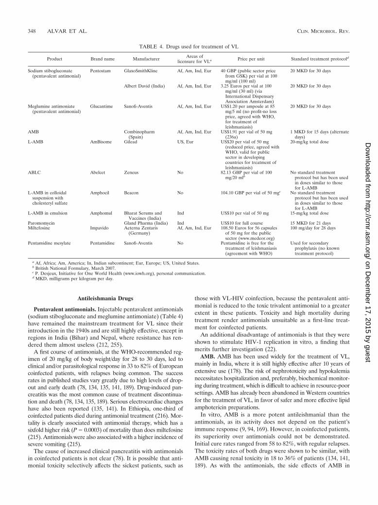
Antileishmania Drugs
Pentavalent antimonials. Injectable pentavalent antimonials(sodium stibogluconate and meglumine antimoniate) (Table 4)have remained the mainstream treatment for VL since theirintroduction in the 1940s and are still highly effective, except inregions in India (Bihar) and Nepal, where resistance has ren-dered them almost useless (212, 255).
A first course of antimonials, at the WHO-recommended reg-imen of 20 mg/kg of body weight/day for 28 to 30 days, led toclinical and/or parasitological response in 33 to 82% of Europeancoinfected patients, with relapses being common. The successrates in published studies vary greatly due to high levels of drop-out and early death (78, 134, 135, 141, 189). Drug-induced pan-creatitis was the most common cause of treatment discontinua-tion and death (78, 134, 135, 189). Serious electrocardiac changeshave also been reported (135, 141). In Ethiopia, one-third ofcoinfected patients died during antimonial treatment (216). Mor-tality is clearly associated with antimonial therapy, which has asixfold higher risk (P � 0.0003) of mortality than does miltefosine(215). Antimonials were also associated with a higher incidence ofsevere vomiting (215).
The cause of increased clinical pancreatitis with antimonialsin coinfected patients is not clear (78). It is possible that anti-monial toxicity selectively affects the sickest patients, such as
those with VL-HIV coinfection, because the pentavalent anti-monial is reduced to the toxic trivalent antimonial to a greaterextent in these patients. Toxicity and high mortality duringtreatment render antimonials unsuitable as a first-line treat-ment for coinfected patients.
An additional disadvantage of antimonials is that they wereshown to stimulate HIV-1 replication in vitro, a finding thatmerits further investigation (22).
AMB. AMB has been used widely for the treatment of VL,mainly in India, where it is still highly effective after 10 years ofextensive use (178). The risk of nephrotoxicity and hypokalemianecessitates hospitalization and, preferably, biochemical monitor-ing during treatment, which is difficult to achieve in resource-poorsettings. AMB has already been abandoned in Western countriesfor the treatment of VL, in favor of safer and more effective lipidamphotericin preparations.
In vitro, AMB is a more potent antileishmanial than theantimonials, as its activity does not depend on the patient’simmune response (9, 94, 169). However, in coinfected patients,its superiority over antimonials could not be demonstrated.Initial cure rates ranged from 58 to 82%, with regular relapses.The toxicity rates of both drugs were shown to be similar, withAMB causing renal toxicity in 18 to 36% of patients (134, 141,189). As with the antimonials, the side effects of AMB in
TABLE 4. Drugs used for treatment of VL
Product Brand name Manufacturer Areas oflicensure for VLa Price per unit Standard treatment protocold
Sodium stibogluconate(pentavalent antimonial)
Pentostam GlaxoSmithKline Af, Am, Ind, Eur 40 GBP (public sector pricefrom GSK) per vial at 100mg/ml (100 ml)
20 MKD for 30 days
Albert David (India) Af, Am, Ind, Eur 3.25 Euros per vial at 100mg/ml (30 ml) (viaInternational DispensaryAssociation Amsterdam)
20 MKD for 30 days
Meglumine antimoniate(pentavalent antimonial)
Glucantime Sanofi-Aventis Af, Am, Ind, Eur US$1.20 per ampoule at 85mg/5 ml (no profit-no lossprice, agreed with WHO,for treatment ofleishmaniasis)
20 MKD for 30 days
AMB Combinopharm(Spain)
Af, Am, Ind, Eur US$1.91 per vial of 50 mg(236a)
1 MKD for 15 days (alternatedays)
L-AMB AmBisome Gilead US, Eur US$20 per vial of 50 mg(reduced price, agreed withWHO, valid for publicsector in developingcountries for treatment ofleishmaniasis)
20-mg/kg total dose
ABLC Abelcet Zeneus No 82.13 GBP per vial of 100mg/20 mlb
No standard treatmentprotocol but has been usedin doses similar to thosefor L-AMB
L-AMB in colloidalsuspension withcholesteryl sulfate
Amphocil Beacon No 104.10 GBP per vial of 50 mgc No standard treatmentprotocol but has been usedin doses similar to thosefor L-AMB
L-AMB in emulsion Amphomul Bharat Serums andVaccines (India)
Ind US$10 per vial of 50 mg 15-mg/kg total dose
Paromomycin Gland Pharma (India) Ind US$10 for full course 15 MKD for 21 daysMiltefosine Impavido Aeterna Zentaris
(Germany)Af, Am, Ind, Eur 108.50 Euros for 56 capsules
of 50 mg for the publicsector (www.medeor.org)
100 mg/day for 28 days
Pentamidine mesylate Pentamidine Sanofi-Aventis No Pentamidine is free for thetreatment of leishmaniasis(agreement with WHO)
Used for secondaryprophylaxis (no knowntreatment protocol)
a Af, Africa; Am, America; In, Indian subcontinent; Eur, Europe; US, United States.b British National Formulary, March 2007.c P. Desjeux, Initiative for One World Health (www.iowh.org), personal communication.d MKD, milligrams per kilogram per day.
348 ALVAR ET AL. CLIN. MICROBIOL. REV.
on Decem
ber 17, 2015 by guesthttp://cm
r.asm.org/
Dow
nloaded from
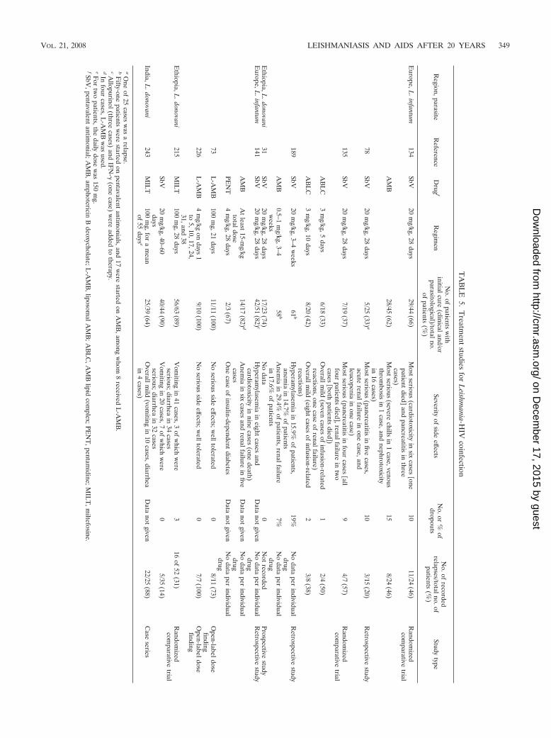
TA
BL
E5.
Treatm
entstudies
forL
eishmania-H
IVcoinfection
Region,parasite
Reference
Drug
fR
egimen
No.of
patientsw
ithinitialcure
(clinicaland/orparasitological)/totalno.
ofpatients
(%)
Severityof
sideeffects
No.or
%of
dropouts
No.of
recordedrelapses/totalno.of
patients(%
)Study
type
Europe,L
.infantum134
SbV20
mg/kg,28
days29/44
(66)M
ostserious
(cardiotoxicityin
sixcases
�onepatient
died�
andpancreatitis
inthree
cases)
1011/24
(46)R
andomized
comparative
trial
AM
B28/45
(62)M
ostserious
(severechills
in1
case,venousthrom
bosisin
1case,and
nephrotoxicityin
16cases)
158/24
(46)
78SbV
20m
g/kg,28days
5/25(33) a
Most
serious(pancreatitis
infive
cases,acute
renalfailurein
onecase,and
leucopeniain
onecase)
103/15
(20)R
etrospectivestudy
135SbV
20m
g/kg,28days
7/19(37)
Most
serious(pancreatitis
infour
cases�all
fourpatients
died�,renalfailure
intw
ocases
�bothpatients
died�)
94/7
(57)R
andomized
comparative
trial
AB
LC
3m
g/kg,5days
6/18(33)
Overallm
ild(seven
casesof
infusion-relatedreactions,one
caseof
renalfailure)1
2/4(50)
AB
LC
3m
g/kg,10days
8/20(42)
Overallm
ild(eight
casesof
infusion-relatedreactions)
23/8
(38)
189SbV
20m
g/kg,3–4w
eeks61
bH
yperamylasem
iain
15.9%of
patients,anem
iain
14.7%of
patients19%
No
dataper
individualdrug
Retrospective
study
AM
B0.5–1
mg/kg,3–4
weeks
58b
Anem
iain
29.4%of
patients,renalfailurein
17.6%of
patients7%
No
dataper
individualdrug
Ethiopia,L
.donovani31
SbV20
mg/kg,28
days17/23
(74)N
odata
0N
otrecorded
Prospectivestudy
Europe,L
.infantum141
SbV20
mg/kg,28
days42/51
(82) cH
yperamylasem
iain
eightcases
andcardiotoxicity
innine
cases(one
death)D
atanot
givenN
odata
perindividual
drugR
etrospectivestudy
AM
BA
tleast
15-mg/kg
totaldose14/17
(82) dA
nemia
insix
casesand
renalfailurein
fivecases
Data
notgiven
No
dataper
individualdrug
PEN
T4
mg/kg,28
days2/3
(67)O
necase
ofinsulin-dependent
diabetesD
atanot
givenN
odata
perindividual
drug73
L-A
MB
100m
g,21days
11/11(100)
No
seriousside
effects;welltolerated
08/11
(73)O
pen-labeldosefinding
226L
-AM
B4
mg/kg
ondays
1to
5,10,17,24,31,and
38
9/10(100)
No
seriousside
effects;welltolerated
07/7
(100)O
pen-labeldosefinding
Ethiopia,L
.donovani215
MIL
T100
mg,28
days56/63
(89)V
omiting
in41
cases,3of
which
were
serious;diarrheain
34cases
316
of52
(31)R
andomized
comparative
trialSbV
20m
g/kg,40–60days
40/44(90)
Vom
itingin
20cases,7
ofw
hichw
ereserious;diarrhea
in32
cases0
5/35(14)
India,L.donovani
243M
ILT
100m
g,fora
mean
of55
days e25/39
(64)O
verallmild
(vomiting
in10
cases,diarrheain
4cases)
Data
notgiven
22/25(88)
Case
series
aO
neof
25cases
was
arelapse.
bF
ifty-onepatients
were
startedon
pentavalentantim
onials,and17
were
startedon
AM
B,am
ongw
hom8
receivedL
-AM
B.
cAllopurinol(three
cases)and
IFN
-(one
case)w
ereadded
totherapy.
dIn
fourcases,L
-AM
Bw
asused.
eFor
two
patients,thedaily
dosew
as150
mg.
fSbV,pentavalent
antimonial;A
MB
,amphotericin
Bdeoxycholate;L
-AM
B,liposom
alAM
B;A
BL
C,A
MB
lipidcom
plex;PEN
T,pentam
idine;MIL
T,m
iltefosine.
VOL. 21, 2008 LEISHMANIASIS AND AIDS AFTER 20 YEARS 349
on Decem
ber 17, 2015 by guesthttp://cm
r.asm.org/
Dow
nloaded from
coinfected patients are more severe than those in noncoin-fected patients.
Lipid formulations of AMB. All lipid formulations of AMBshow a better safety profile than that of AMB, with L-AMB(AmBisome; Gilead USA) considered superior to the otherformulations. L-AMB has the highest therapeutic index of allantileishmanial drugs, including the other lipid AMB formu-lations (33). It is therefore indicated for the treatment of coin-fected patients. The limited data on the use of L-AMB incoinfected patients were generated mostly in Europe. In otherregions, L-AMB is prohibitively expensive, even though it isnow available for a nonprofit cost of $20 per vial for VL in thedeveloping world (Table 4).
For immunocompetent patients, no treatment failures havebeen observed with the currently recommended total dose of20 mg/kg (33), except in Sudan. Sudanese patients withoutHIV were less responsive to L-AMB than were Europeans orIndians: the cure rates for total doses of �15 mg/kg, 16 to 20mg/kg, and �20 mg/kg were 50%, 64%, and 88%, respectively(235). In another study, 10 of 64 Sudanese patients did notrespond to treatment with L-AMB (20 mg/kg). This wasthought to be related to high initial parasite loads and immu-nosuppressing underlying diseases (HIV infection and tuber-culosis), and it was suggested that higher doses should havebeen used (165). In Europe, total doses of up to 30 to 40 mg/kghave been evaluated in small numbers of coinfected patientsand were well tolerated, but they did not prevent relapses (73,226). A dosing recommendation for coinfected patients cannotbe made based on the limited data available. Since the half-lifeof L-AMB that has accumulated in the spleen, liver, and bonemarrow is estimated to be at least 2 to 3 weeks (106), a pro-longed high-dose regimen may be necessary to avoid the se-lection of resistant parasites.
Of the other lipid forms of AMB, AMB lipid complex(ABLC) has been used for secondary prophylaxis, adminis-tered every 21 days for 12 months. ABLC was well tolerated,and after 1 year, 22% of patients had relapsed, compared with50% of patients without secondary prophylaxis (142). Data onsecondary prophylaxis with L-AMB are anecdotal (161). Aninexpensive lipid emulsion is manufactured in India (Ampho-mul; Bharat Sera and Vaccines) (Table 4), which is encourag-ing news for the treatment of VL, but it has not yet beenevaluated in coinfected patients.
Miltefosine. Treatment with miltefosine has consistentlybeen shown to be safe and effective (255a). Relatively mildgastrointestinal side effects are common. A study of 299 Indianpatients showed that vomiting occurred in 38% and diarrheaoccurred in 20% of patients and generally lasted 1 to 2 days(253); none of these patients were coinfected. A major pitfallof miltefosine is its potential teratogenic effect, which impedesits use in pregnant or lactating women and women of child-bearing potential not taking contraceptives.
In Ethiopia, side effects with miltefosine are worse in HIV-positive patients; vomiting occurred in 65% of coinfectedpatients, compared to 45% of noncoinfected patients. Milte-fosine was used in 39 coinfected patients from Europe who hadrelapsed on previous treatments. Cure was achieved in 64% ofpatients, but almost all patients relapsed again and were re-treated, with moderate success. It was found that prolongedtreatment was safe, with one patient receiving treatment for 2
years (243). In Ethiopia, miltefosine was found to be less effectivethan antimonials in coinfected patients, with 18% versus 2% ofpatients failing initial treatment and 25% versus 11% relapsingwithin 6 months in the miltefosine and antimonial groups, respec-tively. Overall, mortality in the miltefosine group (6%) was lowerthan that in the antimonial group (12%) (215).
Miltefosine has a half-life of 1 week, so especially in re-lapsing coinfected patients, there is a risk of resistant parasitesdeveloping (32), as generated in vitro (239). As with otherdrugs, the use of miltefosine in coinfected patients should beprolonged until parasite clearance is achieved. In India, wherelarge-scale miltefosine distribution is planned within the con-text of an elimination program, controlled use and knowledgeof the HIV status of patients are of crucial importance (255a).In Europe and other zones where VL transmission is zoonotic,the use of miltefosine (and other first-line drugs) should not beallowed in dogs to prevent the development of primary resis-tant strains (255a).
Paromomycin. Paromomycin sulfate was recently licensedfor the treatment of VL on the basis of a large phase III trial,where it was shown to be as effective as AMB and was very welltolerated; the main side effects were a reversible elevation ofliver enzymes, in approximately 1% of patients, and pain at theinjection site (254). Paromomycin is the cheapest of all anti-leishmanial drugs (Table 4). Paromomycin has a good safetyprofile and is a broad-spectrum antibiotic active against somegram-positive and most gram-negative bacteria, many proto-zoa, and Mycobacterium tuberculosis (125). Its activity in coin-fected patients has yet to be established. Acquired resistancemay easily develop (67, 98, 144) and has been generated invitro. There has been extensive, favorable experience withcombinations of paromomycin plus sodium stibogluconate,with 224 patients receiving a combination in three randomizedclinical trials (60, 237, 257), as have thousands of patientstreated under field conditions (154). However, data for coin-fected patients are lacking.
Pentamidine. Pentamidine was used to treat VL mainly inIndia but has now been abandoned due to the severity of itsside effects and to poor efficacy. Irreversible diabetes mellitusis a serious complication, occurring in 4 to 12% of cases (178).After a decade of use, the efficacy has declined to �70% (67).The scope of safe drugs to treat coinfected patients is limited,and thus these drugs are not to be used for secondary prophy-laxis but reserved for treatment of acute cases. Pentamidine,although toxic in treatment doses, has been considered for usein secondary prophylaxis (183), as intermittent low-dose main-tenance therapy is unlikely to show the toxicity seen with atherapeutic dosing regimen.
Following a recent agreement between WHO and the man-ufacturer (Sanofi-Aventis), pentamidine is now available forfree for the treatment of all neglected diseases, including leish-maniasis (Table 4).
Combinations. There is consensus that combination therapyfor VL should urgently be introduced to minimize the devel-opment of resistant strains (48, 110). This is especially true forcoinfected patients, as repeated exposure to single antileish-manial drugs will inevitably generate resistance, and incurableVL patients (158) or PKDL patients (1) may transmit drug-resistant parasites. Although no clinical trials to date haveinvestigated combination therapies in coinfected patients, phy-
350 ALVAR ET AL. CLIN. MICROBIOL. REV.
on Decem
ber 17, 2015 by guesthttp://cm
r.asm.org/
Dow
nloaded from
sicians may use combination therapy in patients with multiplerelapses on a compassionate basis. Combinations of miltefosineplus paromomycin, pentavalent antimonials, and AMB were stud-ied in vivo and in vitro; miltefosine plus paromomycin and milte-fosine plus AMB were synergistic and should progress to clinicaltesting (238). In coinfected patients, even if these combinationsdo not prevent relapse, they may preserve drug sensitivity inindividual patients and therefore prolong survival.
Antiretrovirals
Since 1996, the introduction of HAART in Europe has ledto far fewer new VL infections in HIV-positive patients.HAART has reduced the risk of relapse in coinfected patients,although it is not clear to what extent (55, 77, 96, 138, 156,265). These results cannot be extrapolated to the developingworld, where the condition of patients is much worse and theL. donovani species is highly virulent. Nonetheless, it is clearthat HAART reduces the morbidity and mortality of coin-fected patients in all regions. In some areas with a high inci-dence of coinfection (Bihar, India; Ethiopia; and EasternSudan), access to HAART remains minimal. HAART isfrequently restricted to nongovernmental organizations(NGOs), programs that cover small numbers of patients, whilegovernment programs are absent, malfunctioning, or at thefirst stage of implementation (e.g., in Ethiopia). Only relativelyfew coinfected patients in developing countries will be in-cluded in well-functioning HAART programs. Where CD4�
cell counts are difficult to obtain, VL should therefore beregarded as a valid entry point for HAART. For the Africanregion, VL has been defined as an AIDS-defining disease,which is a pragmatic approach that should be adopted globally(http://www.who.int/hiv/pub/guidelines/clinicalstaging.pdf).
Indinavir and saquinavir show in vitro activity against L.infantum and L. major (33, 193, 229), but there is currently noclinical evidence of any HAART being more effective thanothers in preventing VL relapses in coinfected patients. Thus,the choice of HAART for coinfected patients does not differfrom that in national programs for HIV.
Other Approaches
It has been suggested that in the absence of an adequateimmune response, the addition of IFN- or recombinantIL-2 to VL therapy could be useful. However, both have notyet shown any additional benefit (46, 132). Selective sple-nectomy has been evaluated for relapsing coinfected pa-tients, but despite being effective in restoring hematologicalparameters and reducing the need for blood transfusions, itdid not prevent further relapses (261); in addition, this pro-cedure should be avoided because of the risk of overwhelm-ing sepsis and severe malaria.
MCL and CL
Evidence to support different treatments is very limited forHIV-negative CL patients (129, 145) and is absent for immu-nocompromised HIV-coinfected patients.
In general, to reduce the probability of dissemination andrelapse in CL-HIV-coinfected patients, systemic treatment is
recommended over local treatment, with close monitoring andfollow-up of the patient. As long as evidence is lacking, thesame treatment used for immunocompetent individuals will beoffered to localized CL-HIV-coinfected patients. Reports ofthe treatment of HIV-associated MCL are scarce, although thecure of MCL following treatment with L-AMB has been de-scribed for a coinfected patient failing antimonial treatment(13). Miltefosine has been used effectively for treatment ofPKDL in HIV-infected patients (26).
FOLLOW-UP OF PATIENTS ANDMAINTENANCE THERAPY
Leishmania infection exacerbates the evolution of HIVthrough an increase of HIV replication and a decrease of theCD4� lymphocyte count. In fact, about half of coinfected pa-tients develop full AIDS criteria within 2 months after thediagnosis of VL. Furthermore, the course of VL in HIV-pos-itive patients is characterized by the appearance of numerousrelapses despite effective antileishmanial treatment. The num-ber of relapses and the average time between them dependmainly on the immunological status of the patient (mainlydefined by the CD4� cell count and definitely influenced byHAART) and on the use of antileishmanial maintenance ther-apy. However, the different evolution of VL among patientssuggests that other factors, such as parasitological variability,pathogenic strength of the infecting strain, or the existence ofany resistance to antileishmanial drugs, can influence treat-ment success (87, 95, 218).
The mortality rate for European patients during a first epi-sode of VL is 10 to 19%, which can increase to 24% during themonth following the end of treatment, and is related to thetoxicity of the treatment, complications arising during the VLepisode, and concomitant opportunistic infections. The sur-vival time varies from 3 to 24 months (average survival rate of60% after 12 months) but can triple if the patient is treatedwith highly active HAART. After a correctly treated initialepisode of VL, 25 to 61% of patients will relapse before 6 to 9months, and 79 to 97% will relapse during the patient’s lifetime(independent of the drug used to treat the first VL episode),with the average period between relapses becoming increas-ingly shorter (141, 188, 189).
There are three main points to consider in following up anHIV-positive patient who was recently diagnosed and treatedfor an episode of VL, as follows: (i) the need for test of cureafter treatment and prediction/diagnosis of relapse, (ii) promptinitiation of effective HAART, and (iii) the convenience ofsecondary prophylaxis with an antileishmanial drug as mainte-nance therapy.
Test of Cure and/or Relapse
In treating immunocompetent patients, it is recognized thattests of cure are performed 1 month and 6 months after thefinish of antileishmanial treatment in order to assess parasito-logical cure. Bone marrow or spleen aspiration is the techniqueusually performed, and diagnosis of VL is made when Leish-mania sp. amastigotes are seen in Giemsa-stained samples orpromastigotes are cultured in specific medium. For HIV-pos-itive patients, though, parasitological cure is very difficult to
VOL. 21, 2008 LEISHMANIASIS AND AIDS AFTER 20 YEARS 351
on Decem
ber 17, 2015 by guesthttp://cm
r.asm.org/
Dow
nloaded from

achieve and relapses are very common, so the need for re-peated invasive tests of cure could be very uncomfortable ordangerous for these patients. Repeated invasive diagnostictests should be performed if there is no clinical improvementafter treatment or whenever there is a suspicion of VL relapse.
PCR-based assays are useful tools for the diagnosis of VL incoinfected patients (16, 45, 68, 131, 185, 190, 210). The analysisof peripheral blood samples using PCR can be a useful non-invasive technique for monitoring patients posttreatment,which avoids unnecessary pain to the patient in evaluating theparasitic evolution during follow-up (16, 45, 68, 131, 185, 190,210). PCR is a sensitive technique for detecting relapses sev-eral weeks before the clinical onset of disease. LeishmaniaDNA levels correlate with the clinical course of VL, and theirmeasurement at diagnosis and during and after treatmentseems to be useful in the clinical management of VL-HIV-coinfected patients. Positive nested PCR results during asymp-tomatic periods indicate the presence of the parasite, but notnecessarily relapse (16, 45, 68, 131, 185, 190, 210). However,the presence of viable parasites during posttreatment fol-low-up increases the probability of relapse and could be a goodmarker of relapse (16, 45, 68, 131, 185, 190, 210). Duringrelapse of VL, the parasite is detected more effectively in bloodsamples by nested PCR than by culture.
Antigen detection in urine with a latex agglutination testcould represent an alternative noninvasive and easy-to-per-form test for the diagnosis of leishmaniasis (sensitivity of 86%)and posttreatment follow-up in coinfected patients. In the ab-sence of clinical symptoms, the detection of parasite antigensin urine samples is not associated with clinical disease in morethan one-half of cases, and when antigen detection in urine isrepeatedly negative, the probability of relapse is very low (5%at 6 months) (209).
Antiretroviral Therapy
HAART has dramatically modified the natural history ofHIV infection and associated opportunistic infections. Despitethe positive data associated with the use of these drugs, thebenefits of HAART apply to only a minority of coinfectedpatients in the developing world. The effects of HAART oncoinfected patients can be summarized as follows (77, 138, 139,148, 156, 225, 262, 265). (i) The incidence of new VL cases inHIV-positive patients dropped about 50 to 65% in the Medi-terranean area after HAART was introduced. HAART pre-vents future clinical VL in asymptomatic Leishmania sp.-in-fected patients. (ii) Coinfected patients who receive HAARThave a higher survival rate than those who do not receive it(mean survival of 66 months). (iii) HAART modifies the pat-tern of VL relapse but is not sufficient to prevent the relapse ofthe disease. Relapsing VL is possible in HIV-infected patientsreceiving successful HAART, mainly when CD4� cell levelsare �200 cells/�l, but even if a very low viral load is achieved.About 40% (25 to 70%) of coinfected patients receivingHAART without VL maintenance therapy experience relapse,but HAART delays VL relapse, with a longer time betweenVL episodes (average of 7 months), in patients who receiveHAART compared with those not receiving HAART. (iv) Thewhole possible clinical spectrum of leishmaniasis has been ob-served in HIV-positive patients, and atypical clinical forms
have been observed no more frequently in patients receivingHAART. There is no correlation between the introduction ofHAART and the occurrence of relapses, and the phenomenonof immune restoration syndrome is rarely observed (39, 122).
Maintenance Therapy (Secondary Prophylaxis)
Primary prophylaxis is not indicated. On the other hand, itseems advisable to use maintenance therapy systematically andsoon after the first episode of VL to prevent early relapse,which would perpetuate a Th2-type response in cellular immu-nity and prevent the control of parasitization by Leishmaniaspp. despite effective HAART. The data about maintenancetherapy come from the Mediterranean area, where VL is azoonotic disease caused by L. infantum and where there aremedical facilities for the follow-up of patients (136). Severalquestions arise when maintenance therapy is prescribed inother settings where medical care is insufficient for monitoringtoxicity (mainly with antimonials) or where VL is an anthro-ponotic disease (Sudan, Ethiopia, and India) and there is therisk of inducing drug resistance (67). If maintenance therapy isprovided in such situations, it should be conducted under adirectly observed treatment program (particularly if miltefos-ine is the chosen drug).
There are enough accumulated data about the benefits ofmaintenance therapy in coinfected patients (156, 182, 188, 189,207), but they are based on open-label, noncontrolled studies.In studies which failed to demonstrate the benefit of prophy-laxis, patients tended to have more serious infection or hadrelapsed more often (133). In the only prospective and ran-domized study to investigate the effectiveness of a maintenancetherapy, ABLC (3 mg/kg every 21 days) was compared withnonmaintenance therapy. At 12 months of follow-up, the ben-efit of the maintenance therapy was evident in the intention-to-treat analysis; the probability of being free of illness at 12months was 50% and 22% for the groups with and withoutmaintenance therapy, respectively (142).
Different regimens with several drugs have been used,mainly with pentavalent antimonials (20 mg/kg/day, given ev-ery 3 to 4 weeks), AMB (either L-AMB or ABLC) (3 to 5mg/kg/day, given every 3 to 4 weeks), or pentamidine (4 mg/kg/day [300 mg for an adult], given every 3 to 4 weeks) (43).
Miltefosine is a very effective oral drug against VL (119, 234,253, 259). There are no data about maintenance therapy withthis drug for humans, but once- or twice-weekly oral adminis-tration to T-cell-deficient nude mice infected with L. donovaniwas useful as maintenance therapy (167). However, miltefosinerequires a long treatment course (28 days) and has a longhalf-life (around 150 h), which might accelerate the emergenceof drug resistance (184). Other oral drugs, such as itraconazoleand allopurinol, have some therapeutic efficacy against Leish-mania spp. and could hypothetically be used in maintenancetherapy, together with miltefosine, to increase efficacy or pre-vent the appearance of resistance (15).
Once a patient’s immune function has recovered withHAART and VL is quiescent, prophylaxis therapy could besuspended provided that the CD4� cell count is maintained(�200 cells/�l) for more than 6 months (28, 248).
352 ALVAR ET AL. CLIN. MICROBIOL. REV.
on Decem
ber 17, 2015 by guesthttp://cm
r.asm.org/
Dow
nloaded from

PERSPECTIVES IN LEISHMANIA-HIV COINFECTION
The spread and overlap of both leishmaniasis and HIV in-fections in the major foci of leishmaniasis (India, Brazil, andEastern Africa) make VL-HIV coinfection a serious worldwideconcern. HIV and VL programs should develop integrated andwell-coordinated intervention activities, particularly in areaswhere VL is endemic. Collaboration of the two programsshould particularly be strengthened by local government at thedistrict level to focus on the specific circumstances of each areaof endemicity. The mobility of people, such as seasonal mi-grant laborers, urban migration from rural areas, resettlementactivities from areas where disease is nonendemic to thosewhere it is endemic, and the presence of refugees and internalpopulation displacements due to war and conflicts constitutemajor factors for the spread of both diseases. There is a needto develop locally applicable strategies for appropriate inter-vention in migrant communities.
In areas where transmission is anthroponotic, VL-HIV-coin-fected patients should be targeted as potential reservoirs ofinfection. Preventing the transmission of L. donovani is man-datory by providing bed nets. Furthermore, it may be prudentto encourage HIV-positive patients to move from areas of VLendemicity to prevent them from becoming infected with VL.In anthroponotic transmission areas, more studies are requiredto provide sufficient evidence to recommend maintenance/sec-ondary prophylaxis treatment, and the risk of resistance mustalso be considered in this situation.
Both HIV and Leishmania infections induce a deficit in hosthumoral and cellular responses that limits the diagnostic valueof serological tests for coinfected patients. Only 40 to 50% ofVL-HIV-coinfected patients have detectable specific antibodylevels against Leishmania. There is a need for comparativeanalysis studies from different geographical areas, using differ-ent serological techniques, including CD4� counts, to charac-terize serum samples. The high parasitic burden of leishman-iasis in HIV-infected individuals permits the detection ofLeishmania antigens, but there is only one technique currentlyavailable for antigen detection (KAtex, used for urine sam-ples). Research into other antigen detection tests is needed toevaluate their use as active phase markers that allow the de-tection of parasitological cure and relapse. In addition, moreresearch is needed to link the clinical and parasitological con-ditions and to establish better criteria for cure, considering thepoor prognosis of coinfection and the high risk of relapse.
VL is an opportunistic infection in HIV-infected individualsand should be considered in the definition of stage IV in theWHO HIV/AIDS classification (stage C of the CDC classifi-cation). It is crucial to try to restore cell-mediated immunitywith HAART as soon as possible to prevent further relapses ofVL, even though HAART alone is not enough to prevent VLrelapse.
All antileishmanial therapies are less effective in HIV-posi-tive patients. There is a high mortality rate due to concurrentillness, complications, and drug toxicity. Pentavalent antimo-nials and AMB are more toxic to HIV patients, who requireclose monitoring for pancreatitis, cardiotoxicity, and nephro-toxicity, and these drugs should be avoided if better drugs, suchas liposomal formulations of AMB, are available. In consider-ing combination therapy, drugs should be administered se-
quentially (with the safest given first) rather than concurrently.Miltefosine is a safer drug for coinfected patients, althoughthere are still problems of teratogenicity and adherence toconsider. In general, patients who fail to clear the parasite willrelapse, independent of the drug used. Relapses should betreated appropriately, and combination therapy may representthe best approach to avoid drug resistance while balancingbenefit with drug toxicity. Drugs used for relapse treatmentshould be avoided for secondary prophylaxis, although thedrug panoply is very limited. Secondary prophylaxis to preventrelapses in areas with zoonotic transmission of L. infantum hasbeen reported in several publications, but more evidence fromclinical trials is needed to establish a beneficial effect. Treat-ment options for secondary prophylaxis in VL-HIV-coinfectedpatients in anthroponotic transmission areas (L. donovani) arelimited, and the risk of resistance must be taken into account.The use of promising new drugs, such as miltefosine, for canineleishmaniasis can also accelerate the development of resis-tance, and thus their use in dogs should be avoided to protecttheir efficacy in humans.
The priorities of treatment-related research should includethe evaluation of efficacy and tolerability of alternative drugs,such as paromomycin, pentamidine (maintenance therapyonly), and combinations with currently available drugs, in coin-fected patients. Possible benefits to achieve include a short-ened duration of treatment, improved efficacy, cost reduction,protection of drug resistance, and improved compliance. Thetoxicity of combination therapy may be enhanced in coinfectedpatients and so should be evaluated thoroughly in well-con-ducted clinical trials. The only combination that has demon-strated a positive result so far is sodium stibogluconate plusparomomycin. Confirmatory clinical trials are currently ongo-ing in East Africa to establish clear criteria on the use ofmaintenance therapy.
ACKNOWLEDGMENTS
We are indebted to all of the participants of the Fifth InformalConsultative Meeting on Leishmania-HIV Coinfection, held in AddisAbaba, Ethiopia, in April 2007, and special thanks go to L. Campino,Portugal, A. N. Elkhoury, Brazil, S. Sundar, India, R. Guiguemde,Burkina Faso, K. Ritmeijer, Holland, and R. Davidson, United King-dom, for providing useful information or comments.
Special recognition is given to the Spanish Agency for InternationalCooperation (AECI) for supporting this initiative.
REFERENCES
1. Addy, M., and A. Nandy. 1992. Ten years of kala-azar in west Bengal. I. Didpost-kala-azar dermal leishmaniasis initiate the outbreak in 24-Parganas?Bull. W. H. O. 70:341–346.
2. Akilov, O. E., A. Khachemoune, and T. Hasan. 2007. Clinical manifestationsand classification of Old World cutaneous leishmaniasis. Int. J. Dermatol.46:132–142.
3. Akuffo, H., K. Maasho, M. Blostedt, B. Hojeberg, S. Britton, and M. Ba-khiet. 1997. Leishmania aethiopica derived from diffuse leishmaniasis pa-tients preferentially induce mRNA for interleukin-10 while those fromlocalized leishmaniasis patients induce interferon-gamma. J. Infect. Dis.175:737–741.
4. Akuffo, H., E. Schurr, G. Andersson, T. Yamaneberhan, and S. Britton.1987. Responsiveness in diffuse versus local cutaneous leishmaniasis is dueto parasite differences. Scand. J. Immunol. 26:717–721.
5. Albrecht, H. 1998. Leishmaniosis—new perspectives on an underappreci-ated opportunistic infection. AIDS 12:2225–2226.
6. Albrecht, H., I. Sobottka, C. Emminger, H. Jablonowski, G. Just, A. Stoehr,T. Kubin, B. Salzberger, T. Lutz, and J. van Lunzen. 1996. Visceral leish-maniasis emerging as an important opportunistic infection in HIV-infectedpersons living in areas nonendemic for Leishmania donovani. Arch. Pathol.Lab. Med. 120:189–198.
VOL. 21, 2008 LEISHMANIASIS AND AIDS AFTER 20 YEARS 353
on Decem
ber 17, 2015 by guesthttp://cm
r.asm.org/
Dow
nloaded from

7. Altes, J., A. Salas, M. Riera, M. Udina, A. Galmes, J. Balanzat, A. Balles-teros, J. Buades, F. Salva, and C. Villalonga. 1991. Visceral leishmaniasis:another HIV-associated opportunistic infection? Report of eight cases andreview of the literature. AIDS 5:201–207.
8. Alvar, J., S. Bashaye, D. Argaw, I. Cruz, P. Aparicio, A. Kassa, G. Orfanos,F. Parreno, O. Babaniyi, N. Gudeta, C. Canavate, and C. Bern. 2007.Kala-azar outbreak in Libo Kemkem, Ethiopia: epidemiologic and parasi-tologic assessment. Am. J. Trop. Med. Hyg. 77:275–282.
9. Alvar, J., C. Canavate, B. Gutierrez-Solar, M. Jimenez, F. Laguna, R.Lopez-Velez, R. Molina, and J. Moreno. 1997. Leishmania and humanimmunodeficiency virus coinfection: the first 10 years. Clin. Microbiol. Rev.10:298–319.
10. Alvar, J., S. Croft, and P. Olliaro. 2006. Chemotherapy in the treatment andcontrol of leishmaniasis. Adv. Parasitol. 61:223–274.
11. Alvar, J., B. Gutierrez-Solar, R. Molina, R. Lopez-Velez, A. Garcia-Camacho,P. Martinez, F. Laguna, E. Cercenado, and A. Galmes. 1992. Prevalence ofLeishmania infection among AIDS patients. Lancet 339:1427.
12. Alvar, J., S. Yactayo, and C. Bern. 2006. Leishmaniasis and poverty. TrendsParasitol. 22:552–557.
13. Amato, V. S., A. C. Nicodemo, J. G. Amato, M. Boulos, and V. A. Neto. 2000.Mucocutaneous leishmaniasis associated with HIV infection treated suc-cessfully with liposomal amphotericin B (AmBisome). J. Antimicrob. Che-mother. 46:341–342.
14. Andrade-Narvaez, F. J., S. Medina-Peralta, A. Vargas-Gonzalez, S. B.Canto-Lara, and S. Estrada-Parra. 2005. The histopathology of cutaneousleishmaniasis due to Leishmania (Leishmania) mexicana in the YucatanPeninsula, Mexico. Rev. Inst. Med. Trop. Sao Paulo 47:191–194.
15. Angarano, G., P. Maggi, S. L. Coppola, and R. L. Cavaliere. 1998. Itracon-azole as maintenance therapy for visceral leishmaniasis in HIV-infectedpatients. Eur. J. Clin. Microbiol. Infect. Dis. 17:365–367.
16. Antinori, S., S. Calattini, E. Longhi, G. Bestetti, R. Piolini, C. Magni, G.Orlando, M. Gramiccia, V. Acquaviva, A. Foschi, S. Corvasce, C. Colomba,L. Titone, C. Parravicini, A. Cascio, and M. Corbellino. 2007. Clinical useof polymerase chain reaction performed on peripheral blood and bonemarrow samples for the diagnosis and monitoring of visceral leishmaniasisin HIV-infected and HIV-uninfected patients: a single-center, 8-year expe-rience in Italy and review of the literature. Clin. Infect. Dis. 44:1602–1610.
17. Antonelli, L. R., W. O. Dutra, R. P. Almeida, O. Bacellar, E. M. Carvalho,and K. J. Gollob. 2005. Activated inflammatory T cells correlate with lesionsize in human cutaneous leishmaniasis. Immunol. Lett. 101:226–230.
18. Ara, M., C. Maillo, G. Peon, A. Clavel, J. Cuesta, M. P. Grasa, and F. J.Carapeto. 1998. Visceral leishmaniasis with cutaneous lesions in a patientinfected with human immunodeficiency virus. Br. J. Dermatol. 139:114–117.
19. Arya, S. C., and N. Agarwal. 2005. HIV/AIDS and leishmaniasis coinfectionin Ethiopia. CMAJ 173:1067.
20. Ashford, R. W., M. A. Bray, M. P. Hutchinson, and R. S. Bray. 1973. Theepidemiology of cutaneous leishmaniasis in Ethiopia. Trans. R. Soc. Trop.Med. Hyg. 67:568–601.
21. Babiker, Z. O., R. Davidson, C. Mazinda, S. Kipngetich, and K. Ritmeijer.2007. Utility of lymph node aspiration in the diagnosis of visceral leishman-iasis in Sudan. Am. J. Trop. Med. Hyg. 76:689–693.
22. Barat, C., C. Zhao, M. Ouellette, and M. J. Tremblay. 2007. HIV-1 repli-cation is stimulated by sodium stibogluconate, the therapeutic mainstay inthe treatment of leishmaniasis. J. Infect. Dis. 195:236–245.
23. Barral, A., R. Badaro, M. Barral-Netto, G. Grimaldi, Jr., H. Momem, andE. M. Carvalho. 1986. Isolation of Leishmania mexicana amazonensis fromthe bone marrow in a case of American visceral leishmaniasis. Am. J. Trop.Med. Hyg. 35:732–734.
24. Barral, A., D. Pedral-Sampaio, G. Grimaldi Junior, H. Momen, D. McMahon-Pratt, A. Ribeiro de Jesus, R. Almeida, R. Badaro, M. Barral-Netto, E. M.Carvalho, et al. 1991. Leishmaniasis in Bahia, Brazil: evidence that Leish-mania amazonensis produces a wide spectrum of clinical disease. Am. J.Trop. Med. Hyg. 44:536–546.
25. Barreto-de-Souza, V., G. J. Pacheco, A. R. Silva, H. C. Castro-Faria-Neto,P. T. Bozza, E. M. Saraiva, and D. C. Bou-Habib. 2006. Increased Leish-mania replication in HIV-1-infected macrophages is mediated by Tat pro-tein through cyclooxygenase-2 expression and prostaglandin E2 synthesis.J. Infect. Dis. 194:846–854.
26. Belay, A. D., Y. Asafa, J. Mesure, and R. N. Davidson. 2006. Successfulmiltefosine treatment of post-kala-azar dermal leishmaniasis occurring dur-ing antiretroviral therapy. Ann. Trop. Med. Parasitol. 100:223–227.
27. Bentwich, Z. 2003. Concurrent infections that rise the HIV viral load. J.HIV Ther. 8:72–75.
28. Berenguer, J., J. Cosin, P. Miralles, J. C. Lopez, and B. Padilla. 2000.Discontinuation of secondary anti-leishmania prophylaxis in HIV-infectedpatients who have responded to highly active antiretroviral therapy. AIDS14:2946–2948.
29. Berhe, N., A. Hailu, and T. Gemetchu. 1995. Human immunodeficiencyvirus and recurrence of cutaneous leishmaniasis long after healed localizedcutaneous leishmaniasis due to Leishmania aethiopica. Trans. R. Soc. Trop.Med. Hyg. 89:400–401.
30. Berhe, N., A. Hailu, D. Wolday, Y. Negesse, P. Cenini, and D. Frommel.
1995. Ethiopian visceral leishmaniasis patients co-infected with human im-munodeficiency virus. Trans. R. Soc. Trop. Med. Hyg. 89:205–207.
31. Berhe, N., D. Wolday, A. Hailu, Y. Abraham, A. Ali, T. Gebre-Michael, P.Desjeux, A. Sonnerborg, H. Akuffo, and S. Britton. 1999. HIV viral load andresponse to antileishmanial chemotherapy in co-infected patients. AIDS13:1921–1925.
32. Berman, J., A. D. Bryceson, S. Croft, J. Engel, W. Gutteridge, J. Karbwang,H. Sindermann, J. Soto, S. Sundar, and J. A. Urbina. 2006. Miltefosine:issues to be addressed in the future. Trans. R. Soc. Trop. Med. Hyg.100(Suppl. 1):S41–S44.
33. Bern, C., J. Adler-Moore, J. Berenguer, M. Boelaert, M. den Boer, R. N.Davidson, C. Figueras, L. Gradoni, D. A. Kafetzis, K. Ritmeijer, E.Rosenthal, C. Royce, R. Russo, S. Sundar, and J. Alvar. 2006. Liposomalamphotericin B for the treatment of visceral leishmaniasis. Clin. Infect. Dis.43:917–924.
34. Bern, C., and R. Chowdhury. 2006. The epidemiology of visceral leishman-iasis in Bangladesh: prospects for improved control. Indian J. Med. Res.123:275–288.
35. Bernier, R., B. Barbeau, M. J. Tremblay, and M. Olivier. 1998. The lipo-phosphoglycan of Leishmania donovani up-regulates HIV-1 transcriptionin T cells through the nuclear factor-kappaB elements. J. Immunol. 160:2881–2888.
36. Bernier, R., S. J. Turco, M. Olivier, and M. Tremblay. 1995. Activation ofhuman immunodeficiency virus type 1 in monocytoid cells by the protozoanparasite Leishmania donovani. J. Virol. 69:7282–7285.
37. Berry, A., B. Abraham, J. Dereure, V. Pinzani, P. Bastien, and J. Reynes.2004. Two case reports of symptomatic visceral leishmaniasis in AIDSpatients concomitant with immune reconstitution due to antiretroviral ther-apy. Scand. J. Infect. Dis. 36:225–227.
38. Bittencourt, A., N. Silva, A. Straatmann, V. L. Nunes, I. Follador, and R.Badaro. 2002. Post-kala-azar dermal leishmaniasis associated with AIDS.Braz. J. Infect. Dis. 6:313–316.
39. Blanche, P., B. Gombert, O. Rivoal, S. Abad, D. Salmon, and A. Brezin.2002. Uveitis due to Leishmania major as part of HAART-induced immunerestitution syndrome in a patient with AIDS. Clin. Infect. Dis. 34:1279–1280.
40. Boisseau-Garsaud, A. M., D. Cales-Quist, N. Desbois, J. Jouannelle, A.Jouannelle, F. Pratlong, and J. P. Dedet. 2000. A new case of cutaneousinfection by a presumed monoxenous trypanosomatid in the island of Mar-tinique (French West Indies). Trans. R. Soc. Trop. Med. Hyg. 94:51–52.
41. Bora, D. 1999. Epidemiology of visceral leishmaniasis in India. Natl. Med.J. India 12:62–68.
42. Borges, A. S., A. A. Machado, M. S. Ferreira, J. F. de Castro Figueiredo,G. F. Silva, S. Cimerman, H. A. Bacha, and M. C. Teixeira. 1999. Concur-rent leishmaniasis and human immunodeficiency virus (HIV) infection: astudy of four cases. Rev. Soc. Bras. Med. Trop. 32:713–719.
43. Borrelli, P., A. Imperato, G. Murdaca, and M. Scudeletti. 2000. Liposomalamphotericin B as first line and secondary prophylactic treatment for vis-ceral leishmaniasis in a patient infected with HIV. Ann. Ital. Med. Int.15:169–171.
44. Bosch, R. J., A. B. Rodrigo, P. Sanchez, M. V. de Galvez, and E. Herrera.2002. Presence of Leishmania organisms in specific and non-specific skinlesions in HIV-infected individuals with visceral leishmaniasis. Int. J. Der-matol. 41:670–675.
45. Bossolasco, S., G. Gaiera, D. Olchini, M. Gulletta, L. Martello, A. Bestetti,L. Bossi, L. Germagnoli, A. Lazzarin, C. Uberti-Foppa, and P. Cinque.2003. Real-time PCR assay for clinical management of human immunode-ficiency virus-infected patients with visceral leishmaniasis. J. Clin. Micro-biol. 41:5080–5084.
46. Bossolasco, S., S. Nozza, G. Gaiera, A. Bestetti, A. Lazzarin, and P. Cinque.2007. Lack of immune recovery in HIV/Leishmania co-infection treatedwith human recombinant IL-2. AIDS 21:1223–1225.
47. Boumis, E., P. Chinello, C. Della Rocca, M. G. Paglia, M. F. Proietti, andN. Petrosillo. 2006. Atypical disseminated leishmaniasis resembling post-kala-azar dermal leishmaniasis in an HIV-infected patient. Int. J. STDAIDS 17:351–353.
47a.Brazilian Ministry of Health. 2004. Manual de recomendacoes para diag-nostico, tratamento e acompanhamento da co-infeccao Leishmania-HIV.Brazilian Ministry of Health, Brasilia, Brazil.
48. Bryceson, A. 2001. A policy for leishmaniasis with respect to the preventionand control of drug resistance. Trop. Med. Int. Health 6:928–934.
49. Cacopardo, B., L. Nigro, W. Preiser, A. Fama, M. I. Satariano, J. Braner,B. M. Celesia, B. Weber, R. Russo, and H. W. Doerr. 1996. Prolonged Th2cell activation and increased viral replication in HIV-Leishmania co-in-fected patients despite treatment. Trans. R. Soc. Trop. Med. Hyg. 90:434–435.
50. Calza, L., A. D’Antuono, G. Marinacci, R. Manfredi, V. Colangeli, B.Passarini, R. Orioli, O. Varoli, and F. Chiodo. 2004. Disseminated cutane-ous leishmaniasis after visceral disease in a patient with AIDS. J. Am. Acad.Dermatol. 50:461–465.
51. Cambi, A., F. de Lange, N. M. van Maarseveen, M. Nijhuis, B. Joosten,E. M. van Dijk, B. I. de Bakker, J. A. Fransen, P. H. Bovee-Geurts, F. N. van
354 ALVAR ET AL. CLIN. MICROBIOL. REV.
on Decem
ber 17, 2015 by guesthttp://cm
r.asm.org/
Dow
nloaded from
Leeuwen, N. F. Van Hulst, and C. G. Figdor. 2004. Microdomains of theC-type lectin DC-SIGN are portals for virus entry into dendritic cells.J. Cell Biol. 164:145–155.
52. Campanelli, A. P., A. M. Roselino, K. A. Cavassani, M. S. Pereira, R. A.Mortara, C. I. Brodskyn, H. S. Goncalves, Y. Belkaid, M. Barral-Netto, A.Barral, and J. S. Silva. 2006. CD4�CD25� T cells in skin lesions ofpatients with cutaneous leishmaniasis exhibit phenotypic and functionalcharacteristics of natural regulatory T cells. J. Infect. Dis. 193:1313–1322.
53. Campino, L., S. Cortes, R. Pires, L. Oskam, and P. Abranches. 2000.Detection of Leishmania in immunocompromised patients using peripheralblood spots on filter paper and the polymerase chain reaction. Eur. J. Clin.Microbiol. Infect. Dis. 19:396–398.
54. Campino, L., F. Pratlong, P. Abranches, J. A. Rioux, G. Santos-Gomes, C.Alves-Pires, S. Cortes, J. Ramada, J. M. Cristovao, M. O. Afonso, and J. P.Dedet. 2006. Leishmaniasis in Portugal: enzyme polymorphism of Leishma-nia infantum based on the identification of 213 strains. Trop. Med. Int.Health 11:1708–1714.
55. Casado, J. L., R. Lopez-Velez, V. Pintado, C. Quereda, A. Antela, and S.Moreno. 2001. Relapsing visceral leishmaniasis in HIV-infected patientsundergoing successful protease inhibitor therapy. Eur. J. Clin. Microbiol.Infect. Dis. 20:202–205.
56. Catorze, G., J. Alberto, A. Afonso, R. Vieira, S. Cortes, and L. Campino.2006. Leishmania infantum/HIV co-infection: cutaneous lesions followingtreatment of visceral leishmaniasis. Ann. Dermatol. Venereol. 133:39–42.
57. Chappuis, F., S. Rijal, A. Soto, J. Menten, and M. Boelaert. 2006. Ameta-analysis of the diagnostic performance of the direct agglutination testand rK39 dipstick for visceral leishmaniasis. BMJ 333:723.
58. Chicharro, C., and J. Alvar. 2003. Lower trypanosomatids in HIV/AIDSpatients. Ann. Trop. Med. Parasitol. 97(Suppl. 1):75–78.
59. Chicharro, C., M. I. Jimenez, and J. Alvar. 2003. Iso-enzymatic variabilityof Leishmania infantum in Spain. Ann. Trop. Med. Parasitol. 97(Suppl.1):57–64.
60. Chunge, C. N., J. Owate, H. O. Pamba, and L. Donno. 1990. Treatment ofvisceral leishmaniasis in Kenya by aminosidine alone or combined withsodium stibogluconate. Trans. R. Soc. Trop. Med. Hyg. 84:221–225.
61. Clerici, M., and G. M. Shearer. 1993. A TH13TH2 switch is a critical stepin the etiology of HIV infection. Immunol. Today 14:107–111.
62. Cnudde, F., C. Raccurt, F. Boulard, B. Terron-Aboud, M. Nicolas, and B.Juminer. 1994. Diffuse cutaneous leishmaniasis with visceral disseminationin an AIDS patient in Guadeloupe, West Indies. AIDS 8:559–560.
63. Cohen, J. 2007. HIV/AIDS. India slashes estimate of HIV-infected people.Science 317:179–181.
64. Colebunders, R., K. Depraetere, T. Verstraeten, J. Lambert, E. Hauben, E.Van Marck, T. Maurer, A. L. Banuls, and J. C. Dujardin. 1999. Unusualcutaneous lesions in two patients with visceral leishmaniasis and HIV in-fection. J. Am. Acad. Dermatol. 41:847–850.
65. Colmenares, M., A. Puig-Kroger, O. M. Pello, A. L. Corbi, and L. Rivas.2002. Dendritic cell (DC)-specific intercellular adhesion molecule 3(ICAM-3)-grabbing nonintegrin (DC-SIGN, CD209), a C-type surface lec-tin in human DCs, is a receptor for Leishmania amastigotes. J. Biol. Chem.277:36766–36769.
66. Couppie, P., E. Clyti, M. Sobesky, F. Bissuel, P. Del Giudice, D. Sainte-Marie, J. P. Dedet, B. Carme, and R. Pradinaud. 2004. Comparative studyof cutaneous leishmaniasis in human immunodeficiency virus (HIV)-in-fected patients and non-HIV-infected patients in French Guiana. Br. J.Dermatol. 151:1165–1171.
67. Croft, S. L., S. Sundar, and A. H. Fairlamb. 2006. Drug resistance inleishmaniasis. Clin. Microbiol. Rev. 19:111–126.
68. Cruz, I., C. Canavate, J. M. Rubio, M. A. Morales, C. Chicharro, F. Laguna,M. Jimenez-Mejias, G. Sirera, S. Videla, and J. Alvar. 2002. A nestedpolymerase chain reaction (Ln-PCR) for diagnosing and monitoring Leish-mania infantum infection in patients co-infected with human immunodefi-ciency virus. Trans. R. Soc. Trop. Med. Hyg. 96(Suppl. 1):S185–S189.
69. Cruz, I., C. Chicharro, J. Nieto, B. Bailo, C. Canavate, M. C. Figueras, andJ. Alvar. 2006. Comparison of new diagnostic tools for management ofpediatric Mediterranean visceral leishmaniasis. J. Clin. Microbiol. 44:2343–2347.
70. Cruz, I., M. A. Morales, I. Noguer, A. Rodriguez, and J. Alvar. 2002.Leishmania in discarded syringes from intravenous drug users. Lancet 359:1124–1125.
71. Cruz, I., J. Nieto, J. Moreno, C. Canavate, P. Desjeux, and J. Alvar. 2006.Leishmania/HIV co-infections in the second decade. Indian J. Med. Res.123:357–388.
72. Davidson, R. N. 1998. Practical guide for the treatment of leishmaniasis.Drugs 56:1009–1018.
73. Davidson, R. N., L. Di Martino, L. Gradoni, R. Giacchino, R. Russo, G. B.Gaeta, R. Pempinello, S. Scott, F. Raimondi, A. Cascio, et al. 1994. Lipo-somal amphotericin B (AmBisome) in Mediterranean visceral leishmania-sis: a multi-centre trial. Q. J. Med. 87:75–81.
74. Dedet, J. P., and F. Pratlong. 2000. Leishmania, Trypanosoma and mo-noxenous trypanosomatids as emerging opportunistic agents. J. Eukaryot.Microbiol. 47:37–39.
75. Dedet, J. P., B. Roche, F. Pratlong, D. Cales-Quist, J. Jouannelle, J. C.Benichou, and M. Huerre. 1995. Diffuse cutaneous infection caused by apresumed monoxenous trypanosomatid in a patient infected with HIV.Trans. R. Soc. Trop. Med. Hyg. 89:644–646.
76. de la Loma, A., J. Alvar, E. Martinez Galiano, J. Blazquez, A. AlcalaMunoz, and R. Najera. 1985. Leishmaniasis or AIDS? Trans. R. Soc. Trop.Med. Hyg. 79:421–422.
77. de La Rosa, R., J. A. Pineda, J. Delgado, J. Macias, F. Morillas, J. A. Mira,A. Sanchez-Quijano, M. Leal, and E. Lissen. 2002. Incidence of and riskfactors for symptomatic visceral leishmaniasis among human immunodefi-ciency virus type 1-infected patients from Spain in the era of highly activeantiretroviral therapy. J. Clin. Microbiol. 40:762–767.
78. Delgado, J., J. Macias, J. A. Pineda, J. E. Corzo, M. P. Gonzalez-Moreno,R. de la Rosa, A. Sanchez-Quijano, M. Leal, and E. Lissen. 1999. Highfrequency of serious side effects from meglumine antimoniate given withoutan upper limit dose for the treatment of visceral leishmaniasis in humanimmunodeficiency virus type-1-infected patients. Am. J. Trop. Med. Hyg.61:766–769.
79. del Giudice, P., M. Mary-Krause, C. Pradier, S. Grabar, P. Dellamonica, P.Marty, J. A. Gastaut, D. Costagliola, and E. Rosenthal. 2002. Impact ofhighly active antiretroviral therapy on the incidence of visceral leishmani-asis in a French cohort of patients infected with human immunodeficiencyvirus. J. Infect. Dis. 186:1366–1370.
80. Deniau, M., C. Canavate, F. Faraut-Gambarelli, and P. Marty. 2003. Thebiological diagnosis of leishmaniasis in HIV-infected patients. Ann. Trop.Med. Parasitol. 97(Suppl. 1):115–133.
81. Dereure, J., S. H. El-Safi, B. Bucheton, M. Boni, M. M. Kheir, B. Davoust,F. Pratlong, E. Feugier, M. Lambert, A. Dessein, and J. P. Dedet. 2003.Visceral leishmaniasis in eastern Sudan: parasite identification in humansand dogs; host-parasite relationships. Microbes Infect. 5:1103–1108.
82. Dereure, J., J. Reynes, F. Pratlong, I. Lamaury, J. A. Rioux, F. Janbon, andJ. P. Dedet. 1995. Visceral leishmaniasis in HIV-infected patients in thesouth of France. Bull. W. H. O. 73:245–246.
83. Desjeux, P., and J. Alvar. 2003. Leishmania/HIV co-infections: epidemiol-ogy in Europe. Ann. Trop. Med. Parasitol. 97(Suppl. 1):3–15.
84. de Souza e Souza, I., R. D. Naiff, T. C. Guimaraes, M. F. Naiff, E. Cupolillo,W. A. Rodrigues, and A. P. Schettini. 1998. American cutaneous leishman-iasis due to Leishmania (Viannia) guyanensis as an initial clinical presen-tation of human immunodeficiency virus infection. J. Eur. Acad. Dermatol.Venereol. 10:214–217.
85. d’Ettorre, G., G. Ceccarelli, M. Carnevalini, G. Forcina, L. Zaffiri, A. P.Massetti, C. M. Mastroianni, and V. Vullo. 2006. Central role of interleu-kin-15 in human immunodeficiency virus (HIV)-infected patients with vis-ceral leishmaniasis. Acta Trop. 99:83–87.
86. Diaz, N. L., O. Zerpa, L. V. Ponce, J. Convit, A. J. Rondon, and F. J. Tapia.2002. Intermediate or chronic cutaneous leishmaniasis: leukocyte immuno-phenotypes and cytokine characterisation of the lesion. Exp. Dermatol.11:34–41.
87. Di Giorgio, C., F. Faraut-Gambarelli, A. Imbert, P. Minodier, M. Gasquet,and H. Dumon. 1999. Flow cytometric assessment of amphotericin B sus-ceptibility in Leishmania infantum isolates from patients with visceral leish-maniasis. J. Antimicrob. Chemother. 44:71–76.
88. Donaghy, L., F. Gros, L. Amiot, C. Mary, A. Maillard, C. Guiguen, and J. P.Gangneux. 2007. Elevated levels of soluble non-classical major histocom-patibility class I molecule human leucocyte antigen (HLA)-G in the bloodof HIV-infected patients with or without visceral leishmaniasis. Clin. Exp.Immunol. 147:236–240.
89. Durand, I., M. Beylot-Barry, F. X. Weill, M. S. Doutre, and C. Beylot. 1998.Disseminated cutaneous leishmaniasis revealing human immunodeficiencyvirus infection. Ann. Dermatol. Venereol. 125:268–270.
90. Elamin, E. M., S. Guerbouj, A. M. Musa, I. Guizani, E. A. Khalil, M. M.Mukhtar, A. M. Elkadaro, H. S. Mohamed, M. E. Ibrahim, M. M. AbdelHamid, M. El Azhari, and A. M. El Hassan. 2005. Uncommon clinicalpresentations of cutaneous leishmaniasis in Sudan. Trans. R. Soc. Trop.Med. Hyg. 99:803–808.
91. el-Hassan, A. M., F. A. Hashim, M. S. Ali, H. W. Ghalib, and E. E. Zijlstra.1993. Kala-azar in western Upper Nile province in the southern Sudan andits spread to a nomadic tribe from the north. Trans. R. Soc. Trop. Med.Hyg. 87:395–398.
92. Elkhoury, A., M. Sousa, F. Lucena, W. Alves, J. Sena, and L. Paz. 2007.Co-infeccao leishmaniose visceral e AIDS no Brasil. Rev. Soc. Bras. Med.Trop. 40:124.
93. El-Safi, S. H., A. Abdel-Haleem, A. Hammad, I. El-Basha, A. Omer, H. G.Kareem, M. Boelaert, M. Chance, and M. Hommel. 2003. Field evaluationof latex agglutination test for detecting urinary antigens in visceral leish-maniasis in Sudan. East Mediterr. Health J. 9:844–855.
94. Escobar, P., V. Yardley, and S. L. Croft. 2001. Activities of hexadecylphospho-choline (miltefosine), AmBisome, and sodium stibogluconate (Pentostam)against Leishmania donovani in immunodeficient scid mice. Antimicrob.Agents Chemother. 45:1872–1875.
95. Faraut-Gambarelli, F., R. Piarroux, M. Deniau, B. Giusiano, P. Marty, G.Michel, B. Faugere, and H. Dumon. 1997. In vitro and in vivo resistance of
VOL. 21, 2008 LEISHMANIASIS AND AIDS AFTER 20 YEARS 355
on Decem
ber 17, 2015 by guesthttp://cm
r.asm.org/
Dow
nloaded from
Leishmania infantum to meglumine antimoniate: a study of 37 strains col-lected from patients with visceral leishmaniasis. Antimicrob. Agents Che-mother. 41:827–830.
96. Fernandez Cotarelo, M. J., J. Abellan Martinez, J. M. Guerra Vales, P.Martinez Sanchez, M. Rodrigo Gomez De La Barcena, and E. Salto Fer-nandez. 2003. Effect of highly active antiretroviral therapy on the incidenceand clinical manifestations of visceral leishmaniasis in human immunode-ficiency virus-infected patients. Clin. Infect. Dis. 37:973–977.
97. Fisa, R., C. Riera, E. Ribera, M. Gallego, and M. Portus. 2002. A nestedpolymerase chain reaction for diagnosis and follow-up of human visceralleishmaniasis patients using blood samples. Trans. R. Soc. Trop. Med. Hyg.96(Suppl. 1):S191–S194.
98. Fong, D., M. M. Chan, R. Rodriguez, L. J. Gately, J. D. Berman, and M.Grogl. 1994. Paromomycin resistance in Leishmania tropica: lack of corre-lation with mutation in the small subunit ribosomal RNA gene. Am. J.Trop. Med. Hyg. 51:758–766.
99. Foulet, F., A. Cosnes, S. Dellion, L. Blum, C. Bachmeyer, G. Raguin, C.Tourte-Schaeffer, M. Deniau, and S. Bretagne. 2006. Leishmania majorcutaneous leishmaniasis in HIV-positive patients does not spread to ex-tralesional sites. Arch. Dermatol. 142:1368–1369.
100. Gallego, M., F. Pratlong, C. Riera, C. Munoz, E. Ribera, R. Fisa, J. A.Rioux, J. P. Dedet, and M. Portus. 2002. Isoenzymatic identification ofLeishmania isolates from repeated clinical human leishmaniasis episodes inCatalonia (Spain). Trans. R. Soc. Trop. Med. Hyg. 96:45–47.
101. Garcia-Garcia, J. A., J. Martin-Sanchez, M. Gallego, A. Rivero-Roman, A.Camacho, C. Riera, F. Morillas-Marquez, S. Vergara, J. Macias, and J. A.Pineda. 2006. Use of noninvasive markers to detect Leishmania infection inasymptomatic human immunodeficiency virus-infected patients. J. Clin.Microbiol. 44:4455–4458.
102. Garg, R., N. Trudel, and M. J. Tremblay. 2007. Consequences of the naturalpropensity of Leishmania and HIV-1 to target dendritic cells. Trends Para-sitol. 23:317–324.
103. Gilad, J., A. Borer, D. Hallel-Halevy, K. Riesenberg, M. Alkan, and F.Schlaeffer. 2001. Post-kala-azar dermal leishmaniasis manifesting after ini-tiation of highly active anti-retroviral therapy in a patient with humanimmunodeficiency virus infection. Isr. Med. Assoc. J. 3:451–452.
104. Gillis, D., S. Klaus, L. F. Schnur, P. Piscopos, S. Maayan, E. Okon, and D.Engelhard. 1995. Diffusely disseminated cutaneous Leishmania major in-fection in a child with acquired immunodeficiency syndrome. Pediatr. In-fect. Dis. J. 14:247–249.
105. Gonzalez-Beato, M. J., B. Moyano, C. Sanchez, M. T. Gonzalez-Beato, J. A.Perez-Molina, P. Miralles, and P. Lazaro. 2000. Kaposi’s sarcoma-likelesions and other nodules as cutaneous involvement in AIDS-related vis-ceral leishmaniasis. Br. J. Dermatol. 143:1316–1318.
106. Gradoni, L., R. N. Davidson, S. Orsini, P. Betto, and M. Giambenedetti.1993. Activity of liposomal amphotericin B (AmBisome) against Leishma-nia infantum and tissue distribution in mice. J. Drug Target. 1:311–316.
107. Gradoni, L., A. Scalone, M. Gramiccia, and M. Troiani. 1996. Epidemio-logical surveillance of leishmaniasis in HIV-1-infected individuals in Italy.AIDS 10:785–791.
108. Gramiccia, M. 2003. The identification and variability of the parasitescausing leishmaniasis in HIV-positive patients in Italy. Ann. Trop. Med.Parasitol. 97(Suppl. 1):65–73.
109. Griffin, G. E., K. Leung, T. M. Folks, S. Kunkel, and G. J. Nabel. 1989.Activation of HIV gene expression during monocyte differentiation byinduction of NF-kappa B. Nature 339:70–73.
110. Guerin, P. J., P. Olliaro, S. Sundar, M. Boelaert, S. L. Croft, P. Desjeux,M. K. Wasunna, and A. D. Bryceson. 2002. Visceral leishmaniasis: currentstatus of control, diagnosis, and treatment, and a proposed research anddevelopment agenda. Lancet Infect. Dis. 2:494–501.
111. Guiguemde, R. T., O. S. Sawadogo, C. Bories, K. L. Traore, D. Nezien, L.Nikiema, F. Pratlong, P. Marty, R. Houin, and M. Deniau. 2003. Leishma-nia major and HIV co-infection in Burkina Faso. Trans. R. Soc. Trop. Med.Hyg. 97:168–169.
112. Gurubacharya, R. L., S. M. Gurubacharya, D. L. Gurubacharya, J.Quinkel, and V. L. Gurubacharya. 2006. Prevalence of visceral leishmaniaand HIV co-infection in Nepal. Indian J. Med. Res. 123:473–475.
113. Haile, T., and S. D. Anderson. 2006. Visceral leishmaniasis in northernEthiopia. East Afr. Med. J. 83:389–392.
114. Hailu, A., and N. Berhe. 2002. The performance of direct agglutination tests(DAT) in the diagnosis of visceral leishmaniasis among Ethiopian patientswith HIV co-infection. Ann. Trop. Med. Parasitol. 96:25–30.
115. Hepburn, N. C. 2003. Cutaneous leishmaniasis: an overview. J. Postgrad.Med. 49:50–54.
116. Herrera, E., P. Sanchez, and R. J. Bosch. 1995. Disseminated cutaneousleishmaniasis in an HIV-infected patient. Int. J. STD AIDS 6:125–126.
117. Hotez, P. J., J. H. Remme, P. Buss, G. Alleyne, C. Morel, and J. G. Breman.2004. Combating tropical infectious diseases: report of the Disease ControlPriorities in Developing Countries Project. Clin. Infect. Dis. 38:871–878.
118. Ismail, A., A. F. Gadir, T. G. Theander, A. Kharazmi, and A. M. El Hassan.2006. Pathology of post-kala-azar dermal leishmaniasis: a light microscop-
ical, immunohistochemical, and ultrastructural study of skin lesions anddraining lymph nodes. J. Cutan. Pathol. 33:778–787.
119. Jha, T. K., S. Sundar, C. P. Thakur, P. Bachmann, J. Karbwang, C. Fischer,A. Voss, and J. Berman. 1999. Miltefosine, an oral agent, for the treatmentof Indian visceral leishmaniasis. N. Engl. J. Med. 341:1795–1800.
120. Jimenez, M., M. Ferrer-Dufol, C. Canavate, B. Gutierrez-Solar, R. Molina,F. Laguna, R. Lopez-Velez, E. Cercenado, E. Dauden, J. Blazquez, et al.1995. Variability of Leishmania (Leishmania) infantum among stocks fromimmunocompromised, immunocompetent patients and dogs in Spain.FEMS Microbiol. Lett. 131:197–204.
121. Jimenez, M. I., R. Lopez-Velez, R. Molina, C. Canavate, and J. Alvar. 1996.HIV co-infection with a currently non-pathogenic flagellate. Lancet 347:264–265.
122. Jimenez-Exposito, M. J., C. Alonso-Villaverde, P. Sarda, and L. Masana.1999. Visceral leishmaniasis in HIV-infected patients with non-detectableHIV-1 viral load after highly active antiretroviral therapy. AIDS 13:152–153.
123. Joshi, D. D., M. Sharma, and S. Bhandari. 2006. Visceral leishmaniasis inNepal during 1980–2006. J. Commun. Dis. 38:139–148.
124. Kager, P. A., and P. H. Rees. 1983. Splenic aspiration. Review of theliterature. Trop. Geogr. Med. 35:111–124.
125. Kanyok, T. P., M. V. Reddy, J. Chinnaswamy, L. H. Danziger, and P. R.Gangadharam. 1994. In vivo activity of paromomycin against susceptibleand multidrug-resistant Mycobacterium tuberculosis and M. avium complexstrains. Antimicrob. Agents Chemother. 38:170–173.
126. Kedzierska, K., and S. M. Crowe. 2002. The role of monocytes and mac-rophages in the pathogenesis of HIV-1 infection. Curr. Med. Chem.9:1893–1903.
127. Kemp, K. 2000. Cytokine-producing T cell subsets in human leishmaniasis.Arch. Immunol. Ther. Exp. (Warsaw) 48:173–176.
128. Kerob, D., J. D. Bouaziz, C. Sarfati, J. Pavie, M. D. Vignon-Pennamen, J.Menotti, S. Hamane, F. Pralong, P. Buffet, P. Morel, and C. Lebbe. 2006.First case of cutaneous reconstitution inflammatory syndrome associatedwith HIV infection and leishmaniasis. Clin. Infect. Dis. 43:664–666.
129. Khatami, A., A. Firooz, F. Gorouhi, and Y. Dowlati. 2007. Treatment ofacute Old World cutaneous leishmaniasis: a systematic review of the ran-domized controlled trials. J. Am. Acad. Dermatol. 57:335.e1–335.e29.
130. Kubba, R., Y. al-Gindan, A. M. el-Hassan, A. H. Omer, M. K. Kutty, andM. B. Saeed. 1988. Dissemination in cutaneous leishmaniasis. II. Satellitepapules and subcutaneous induration. Int. J. Dermatol. 27:702–706.
131. Lachaud, L., J. Dereure, E. Chabbert, J. Reynes, J. M. Mauboussin, E.Oziol, J. P. Dedet, and P. Bastien. 2000. Optimized PCR using patientblood samples for diagnosis and follow-up of visceral leishmaniasis, withspecial reference to AIDS patients. J. Clin. Microbiol. 38:236–240.
132. Laguna, F. 2003. Treatment of leishmaniasis in HIV-positive patients. Ann.Trop. Med. Parasitol. 97(Suppl. 1):135–142.
133. Laguna, F., M. Adrados, J. Alvar, V. Soriano, M. E. Valencia, V. Moreno,R. Polo, J. Verdejo, M. I. Jimenez, P. Martinez, M. L. Martinez, and J. M.Gonzalez-Lahoz. 1997. Visceral leishmaniasis in patients infected with thehuman immunodeficiency virus. Eur. J. Clin. Microbiol. Infect. Dis. 16:898–903.
134. Laguna, F., R. Lopez-Velez, F. Pulido, A. Salas, J. Torre-Cisneros, E.Torres, F. J. Medrano, J. Sanz, G. Pico, J. Gomez-Rodrigo, J. Pasquau, andJ. Alvar. 1999. Treatment of visceral leishmaniasis in HIV-infected patients:a randomized trial comparing meglumine antimoniate with amphotericin B.AIDS 13:1063–1069.
135. Laguna, F., S. Videla, M. E. Jimenez-Mejias, G. Sirera, J. Torre-Cisneros,E. Ribera, D. Prados, B. Clotet, M. Sust, R. Lopez-Velez, and J. Alvar. 2003.Amphotericin B lipid complex versus meglumine antimoniate in the treat-ment of visceral leishmaniasis in patients infected with HIV: a randomizedpilot study. J. Antimicrob. Chemother. 52:464–468.
136. Laguna-del-Estal, P., and M. S. Moya-Mir. 1997. Prophylaxis of visceralleishmaniasis in human immunodeficiency virus-infected patients. Am. J.Med. 102:222.
137. Lartey, M., L. Adusei, L. Hanson-Nortey, and J. Addy. 2006. Coinfection ofcutaneous leishmaniasis and HIV infection. Ghana Med. J. 40:110–112.
138. Lopez-Velez, R. 2003. The impact of highly active antiretroviral therapy(HAART) on visceral leishmaniasis in Spanish patients who are co-infectedwith HIV. Ann. Trop. Med. Parasitol. 97(Suppl. 1):143–147.
139. Lopez-Velez, R., J. L. Casado, and V. Pintado. 2001. Decline of a visceralleishmaniasis epidemic in HIV-infected patients after the introduction ofhighly active antiretroviral therapy (HAART). Clin. Microbiol. Infect.7:394–395.
140. Lopez-Velez, R., F. Laguna, J. Alvar, J. A. Perez-Molina, R. Molina, P.Martinez, and J. Villarrubia. 1995. Parasitic culture of buffy coat for diag-nosis of visceral leishmaniasis in human immunodeficiency virus-infectedpatients. J. Clin. Microbiol. 33:937–939.
141. Lopez-Velez, R., J. A. Perez-Molina, A. Guerrero, F. Baquero, J. Villarru-bia, L. Escribano, C. Bellas, F. Perez-Corral, and J. Alvar. 1998. Clinico-epidemiologic characteristics, prognostic factors, and survival analysis ofpatients coinfected with human immunodeficiency virus and Leishmania inan area of Madrid, Spain. Am. J. Trop. Med. Hyg. 58:436–443.
356 ALVAR ET AL. CLIN. MICROBIOL. REV.
on Decem
ber 17, 2015 by guesthttp://cm
r.asm.org/
Dow
nloaded from
142. Lopez-Velez, R., S. Videla, M. Marquez, V. Boix, M. E. Jimenez-Mejias, M.Gorgolas, J. R. Arribas, A. Salas, F. Laguna, M. Sust, C. Canavate, and J.Alvar. 2004. Amphotericin B lipid complex versus no treatment in thesecondary prophylaxis of visceral leishmaniasis in HIV-infected patients. J.Antimicrob. Chemother. 53:540–543.
143. Lyons, S., H. Veeken, and J. Long. 2003. Visceral leishmaniasis and HIV inTigray, Ethiopia. Trop. Med. Int. Health 8:733–739.
144. Maarouf, M., M. T. Adeline, M. Solignac, D. Vautrin, and M. Robert-Gero.1998. Development and characterization of paromomycin-resistant Leish-mania donovani promastigotes. Parasite 5:167–173.
145. Mahajan, V. K., and N. L. Sharma. 2007. Therapeutic options for cutane-ous leishmaniasis. J. Dermatol. Treat. 18:97–104.
146. Martinez, P., E. de la Vega, F. Laguna, V. Soriano, S. Puente, V. Moreno,M. J. Sentchordi, C. Garcia-Aguado, and J. Gonzalez-Lahoz. 1993. Diag-nosis of visceral leishmaniasis in HIV-infected individuals using peripheralblood smears. AIDS 7:227–230.
147. Mary, C., F. Faraut, M. P. Drogoul, B. Xeridat, N. Schleinitz, B. Cuisenier,and H. Dumon. 2006. Reference values for Leishmania infantum para-sitemia in different clinical presentations: quantitative polymerase chainreaction for therapeutic monitoring and patient follow-up. Am. J. Trop.Med. Hyg. 75:858–863.
148. Mastroianni, C. M., G. d’Ettorre, G. Forcina, A. Corpolongo, S. Dell’Isola,M. Lichtner, C. D’Agostino, V. Trinchieri, P. Santopadre, and V. Vullo.2000. Long-term remission of human immunodeficiency virus-associatedvisceral leishmaniasis after initiation of potent combination antiretroviraltreatment: report of two cases. J. Infect. 40:94–96.
149. Mathis, A., and P. Deplazes. 1995. PCR and in vitro cultivation for detec-tion of Leishmania spp. in diagnostic samples from humans and dogs.J. Clin. Microbiol. 33:1145–1149.
150. Mathur, P., J. C. Samantaray, M. Vajpayee, and P. Samanta. 2006. Visceralleishmaniasis/human immunodeficiency virus co-infection in India: the fo-cus of two epidemics. J. Med. Microbiol. 55:919–922.
151. Mattos, M., A. Caiza, O. Fernandes, A. J. Goncalves, C. Pirmez, C. S.Souza, and M. P. Oliveira-Neto. 1998. American cutaneous leishmaniasisassociated with HIV infection: report of four cases. J. Eur. Acad. Dermatol.Venereol. 10:218–225.
152. Medrano, F. J., C. Canavate, M. Leal, C. Rey, E. Lissen, and J. Alvar. 1998.The role of serology in the diagnosis and prognosis of visceral leishmaniasisin patients coinfected with human immunodeficiency virus type-1. Am. J.Trop. Med. Hyg. 59:155–162.
153. Medrano, F. J., E. Jimenez-Mejias, E. Calderon, C. Regordan, and M. Leal.1993. An easy and quick method for the diagnosis of visceral leishmaniasisin HIV-1-infected individuals. AIDS 7:1399.
154. Melaku, Y., S. M. Collin, K. Keus, F. Gatluak, K. Ritmeijer, and R. N.Davidson. 2007. Treatment of kala-azar in southern Sudan using a 17-dayregimen of sodium stibogluconate combined with paromomycin: a retro-spective comparison with 30-day sodium stibogluconate monotherapy.Am. J. Trop. Med. Hyg. 77:89–94.
155. Reference deleted.156. Mira, J. A., J. E. Corzo, A. Rivero, J. Macias, F. L. De Leon, J. Torre-
Cisneros, J. Gomez-Mateos, R. Jurado, and J. A. Pineda. 2004. Frequencyof visceral leishmaniasis relapses in human immunodeficiency virus-in-fected patients receiving highly active antiretroviral therapy. Am. J. Trop.Med. Hyg. 70:298–301.
157. Miralles, E. S., M. Nunez, Y. Hilara, A. Harto, R. Moreno, and A. Ledo.1994. Mucocutaneous leishmaniasis and HIV. Dermatology 189:275–277.
158. Molina, R., J. M. Lohse, F. Pulido, F. Laguna, R. Lopez-Velez, and J. Alvar.1999. Infection of sand flies by humans coinfected with Leishmania infan-tum and human immunodeficiency virus. Am. J. Trop. Med. Hyg. 60:51–53.
159. Mondain-Miton, V., M. Toussaint-Gari, P. Hofman, P. Marty, M. Carles, F.De Salvador, F. Miton, Y. Le Fichoux, and P. Dellamonica. 1995. Atypicalleishmaniasis in a patient infected with human immunodeficiency virus.Clin. Infect. Dis. 21:663–665.
160. Montalban, C., J. L. Calleja, A. Erice, F. Laguna, B. Clotet, D. Podzamczer,J. Cobo, J. Mallolas, M. Yebra, and A. Gallego. 1990. Visceral leishmaniasisin patients infected with human immunodeficiency virus. J. Infect. 21:261–270.
161. Montana, M., N. Chochoi, P. Monges, I. Ravaux, F. Faraut, S. Gensollen,M. C. Bongrand, P. Timon-David, and H. Gallais. 2004. Liposomal am-photericin B in secondary prophylaxis of visceral leishmaniasis in HIV-infected patients: report of five clinical cases. Pathol. Biol. (Paris) 52:66–75.
162. Morales, M. A., I. Cruz, J. M. Rubio, C. Chicharro, C. Canavate, F. Laguna,and J. Alvar. 2002. Relapses versus reinfections in patients coinfected withLeishmania infantum and human immunodeficiency virus type 1. J. Infect.Dis. 185:1533–1537.
163. Moreno, J., C. Canavate, C. Chamizo, F. Laguna, and J. Alvar. 2000.HIV-Leishmania infantum co-infection: humoral and cellular immune re-sponses to the parasite after chemotherapy. Trans. R. Soc. Trop. Med. Hyg.94:328–332.
164. Mudur, G. 2007. India reduces estimated count of people with HIV. BMJ335:67.
165. Mueller, M., K. Ritmeijer, M. Balasegaram, Y. Koummuki, M. R. Santana,
and R. Davidson. 2007. Unresponsiveness to AmBisome in some Sudanesepatients with kala-azar. Trans. R. Soc. Trop. Med. Hyg. 101:19–24.
166. Murray, H. W. 1999. Kala-azar as an AIDS-related opportunistic infection.AIDS Patient Care STDs 13:459–465.
167. Murray, H. W. 2000. Suppression of posttreatment recurrence of experi-mental visceral leishmaniasis in T-cell-deficient mice by oral miltefosine.Antimicrob. Agents Chemother. 44:3235–3236.
168. Murray, H. W., J. D. Berman, C. R. Davies, and N. G. Saravia. 2005.Advances in leishmaniasis. Lancet 366:1561–1577.
169. Murray, H. W., J. Hariprashad, and R. E. Fichtl. 1993. Treatment ofexperimental visceral leishmaniasis in a T-cell-deficient host: response toamphotericin B and pentamidine. Antimicrob. Agents Chemother.37:1504–1505.
170. Ndiaye, P. B., M. Develoux, M. T. Dieng, and M. Huerre. 1996. Diffusecutaneous leishmaniasis and acquired immunodeficiency syndrome in aSenegalese patient. Bull. Soc. Pathol. Exot. 89:282–286.
171. Niamba, P., O. Goumbri-Lompo, A. Traore, F. Barro-Traore, and R. T.Soudre. 2007. Diffuse cutaneous leishmaniasis in an HIV-positive patient inwestern Africa. Australas. J. Dermatol. 48:32–34.
172. Niamba, P., A. Traore, O. Goumbri-Lompo, C. Labreze, F. Traore-Barro,M. Bonkoungou, L. Ilboudo, A. Gaulier, and B. R. Soudre. 2006. Cutaneousleishmania in HIV patient in Ouagadougou: clinical and therapeutic as-pects. Ann. Dermatol. Venereol. 133:537–542.
173. Niamba, P. A., A. Traore, O. Goumbri-Lompo, B. F. Traore, M. Bonkoun-gou, L. Ilboudo, A. Gaulier, and C. Boileau. 2005. Cutaneous leishmaniaand HIV co-infection: clinical and evolutionary aspects, abstr. TuPe1.4C17.Third Int. AIDS Conf. HIV Pathog. Treat., Rio de Janiero, Brazil, 24 to 27July 2005.
174. Nigro, L., B. Cacopardo, W. Preiser, J. Braner, J. Cinatl, F. Palermo, R.Russo, H. W. Doerr, and A. Nunnari. 1999. In vitro production of type 1 andtype 2 cytokines by peripheral blood mononuclear cells from subjects coin-fected with human immunodeficiency virus and Leishmania infantum.Am. J. Trop. Med. Hyg. 60:142–145.
175. Nigro, L., A. Montineri, R. La Rosa, M. Zuccarello, C. Iacobello, C. Iaco-bello, C. Vinci, R. Pulizia, and F. Fatuzzo. 2003. Visceral leishmaniasis andHIV co-infection: a rare case of pulmonary and oral localization. Infez.Med. 11:93–96.
176. Nigro, L., M. L. Rizzo, C. Vancheri, R. La Rosa, C. Mastruzzo, V. Toma-selli, A. Ragusa, R. Manuele, and B. Cacopardo. 2007. CCR5 and CCR3expression on T CD3� lymphocytes from HIV/Leishmania co-infectedsubjects. Med. Microbiol. Immunol. 196:253–255.
177. Olivier, M., R. Badaro, F. J. Medrano, and J. Moreno. 2003. The patho-genesis of Leishmania/HIV co-infection: cellular and immunological mech-anisms. Ann. Trop. Med. Parasitol. 97(Suppl. 1):79–98.
178. Olliaro, P. L., P. J. Guerin, S. Gerstl, A. A. Haaskjold, J. A. Rottingen, andS. Sundar. 2005. Treatment options for visceral leishmaniasis: a systematicreview of clinical studies done in India, 1980–2004. Lancet Infect. Dis.5:763–774.
179. Oskam, L., J. L. Nieuwenhuijs, and A. Hailu. 1999. Evaluation of the directagglutination test (DAT) using freeze-dried antigen for the detection ofanti-Leishmania antibodies in stored sera from various patient groups inEthiopia. Trans. R. Soc. Trop. Med. Hyg. 93:275–277.
180. Pacheco, R. S., M. C. Marzochi, M. Q. Pires, C. M. Brito, F. Madeira Mde,and E. G. Barbosa-Santos. 1998. Parasite genotypically related to a mo-noxenous trypanosomatid of dog’s flea causing opportunistic infection in anHIV positive patient. Mem. Inst. Oswaldo Cruz 93:531–537.
181. Paredes, R., J. Munoz, I. Diaz, P. Domingo, M. Gurgui, and B. Clotet. 2003.Leishmaniasis in HIV infection. J. Postgrad. Med. 49:39–49.
182. Pasquau, F., J. Ena, R. Sanchez, J. M. Cuadrado, C. Amador, J. Flores, C.Benito, C. Redondo, J. Lacruz, V. Abril, and J. Onofre. 2005. Leishmaniasisas an opportunistic infection in HIV-infected patients: determinants ofrelapse and mortality in a collaborative study of 228 episodes in a Medi-terreanean region. Eur. J. Clin. Microbiol. Infect. Dis. 24:411–418.
183. Perez-Molina, J. A., R. Lopez-Velez, P. Montilla, and A. Guerrero. 1996.Pentamidine isethionate as secondary prophylaxis against visceral leishman-iasis in HIV-positive patients. AIDS 10:237–238.
184. Perez-Victoria, F. J., M. P. Sanchez-Canete, K. Seifert, S. L. Croft, S.Sundar, S. Castanys, and F. Gamarro. 2006. Mechanisms of experimentalresistance of Leishmania to miltefosine: implications for clinical use. DrugResist. Updat. 9:26–39.
185. Piarroux, R., F. Gambarelli, H. Dumon, M. Fontes, S. Dunan, C. Mary, B.Toga, and M. Quilici. 1994. Comparison of PCR with direct examination ofbone marrow aspiration, myeloculture, and serology for diagnosis of vis-ceral leishmaniasis in immunocompromised patients. J. Clin. Microbiol.32:746–749.
186. Pineda, J. A., J. Macias, F. Morillas, J. Fernandez-Ochoa, J. Cara, R. de LaRosa, J. A. Mira, J. Martin-Sanchez, M. Gonzalez, J. Delgado, C. Acedo,and E. Lissen. 2001. Evidence of increased risk for Leishmania infantuminfection among HIV-seronegative intravenous drug users from southernSpain. Eur. J. Clin. Microbiol. Infect. Dis. 20:354–357.
187. Pineda, J. A., J. Martin-Sanchez, J. Macias, and F. Morillas. 2002. Leish-mania spp. infection in injecting drug users. Lancet 360:950–951.
VOL. 21, 2008 LEISHMANIASIS AND AIDS AFTER 20 YEARS 357
on Decem
ber 17, 2015 by guesthttp://cm
r.asm.org/
Dow
nloaded from
188. Pintado, V., and R. Lopez-Velez. 2001. HIV-associated visceral leishmani-asis. Clin. Microbiol. Infect. 7:291–300.
189. Pintado, V., P. Martin-Rabadan, M. L. Rivera, S. Moreno, and E. Bouza.2001. Visceral leishmaniasis in human immunodeficiency virus (HIV)-in-fected and non-HIV-infected patients. A comparative study. Medicine (Bal-timore) 80:54–73.
190. Pizzuto, M., M. Piazza, D. Senese, C. Scalamogna, S. Calattini, L. Corsico,T. Persico, B. Adriani, C. Magni, G. Guaraldi, G. Gaiera, A. Ludovisi, M.Gramiccia, M. Galli, M. Moroni, M. Corbellino, and S. Antinori. 2001.Role of PCR in diagnosis and prognosis of visceral leishmaniasis in patientscoinfected with human immunodeficiency virus type 1. J. Clin. Microbiol.39:357–361.
191. Ponce, C., E. Ponce, A. Morrison, A. Cruz, R. Kreutzer, D. McMahon-Pratt,and F. Neva. 1991. Leishmania donovani chagasi: new clinical variant ofcutaneous leishmaniasis in Honduras. Lancet 337:67–70.
192. Posada-Vergara, M. P., J. A. Lindoso, J. E. Tolezano, V. L. Pereira-Chioc-cola, M. V. Silva, and H. Goto. 2005. Tegumentary leishmaniasis as amanifestation of immune reconstitution inflammatory syndrome in 2 pa-tients with AIDS. J. Infect. Dis. 192:1819–1822.
193. Pozio, E., and M. A. Morales. 2005. The impact of HIV-protease inhibitorson opportunistic parasites. Trends Parasitol. 21:58–63.
194. Pratlong, F., T. Debord, E. Garnotel, E. Garrabe, P. Marty, G. Raphenon,and J. P. Dedet. 2005. First identification of the causative agent of visceralleishmaniasis in Djibouti: Leishmania donovani. Ann. Trop. Med. Parasitol.99:21–25.
195. Pratlong, F., J. Dereure, M. Deniau, P. Marty, F. Faraut-Gambarelli, andJ. P. Dedet. 2003. Enzymatic polymorphism during Leishmania/HIV co-infection: a study of 381 Leishmania strains received between 1986 and2000 at the international cryobank in Montpellier, France. Ann. Trop. Med.Parasitol. 97(Suppl. 1):47–56.
196. Preiser, W., B. Cacopardo, L. Nigro, J. Braner, A. Nunnari, H. W. Doerr,and B. Weber. 1996. Immunological findings in HIV-Leishmania coinfec-tion. Intervirology 39:285–288.
197. Puig, L., and R. Pradinaud. 2003. Leishmania and HIV co-infection: derma-tological manifestations. Ann. Trop. Med. Parasitol. 97(Suppl. 1):107–114.
198. Qadoumi, M., I. Becker, N. Donhauser, M. Rollinghoff, and C. Bogdan.2002. Expression of inducible nitric oxide synthase in skin lesions of patientswith American cutaneous leishmaniasis. Infect. Immun. 70:4638–4642.
199. Rabello, A., M. Orsini, and J. Disch. 2003. Leishmania/HIV co-infection inBrazil: an appraisal. Ann. Trop. Med. Parasitol. 97(Suppl. 1):17–28.
200. Ramos-Santos, C., O. Hernandez-Montes, G. Sanchez-Tejeda, and A. Monroy-Ostria. 2000. Visceral leishmaniasis caused by Leishmania (L.) mexicana ina Mexican patient with human immunodeficiency virus infection. Mem.Inst. Oswaldo Cruz 95:733–737.
201. Ranjan, A., D. Sur, V. P. Singh, N. A. Siddique, B. Manna, C. S. Lal, P. K.Sinha, K. Kishore, and S. K. Bhattacharya. 2005. Risk factors for Indiankala-azar. Am. J. Trop. Med. Hyg. 73:74–78.
202. Ravel, C., S. Cortes, F. Pratlong, F. Morio, J. P. Dedet, and L. Campino.2006. First report of genetic hybrids between two very divergent Leishmaniaspecies: Leishmania infantum and Leishmania major. Int. J. Parasitol. 36:1383–1388.
203. Redhu, N. S., A. Dey, V. Balooni, and S. Singh. 2006. Leishmania-HIVco-infection: an emerging problem in India. AIDS 20:1213–1215.
204. Reithinger, R., J. C. Dujardin, H. Louzir, C. Pirmez, B. Alexander, and S.Brooker. 2007. Cutaneous leishmaniasis. Lancet Infect. Dis. 7:581–596.
205. Reus, S., R. Sanchez, J. Portilla, V. Boix, M. Priego, E. Merino, and F.Roman. 1999. Visceral leishmaniasis: a comparative study of patients withand without human immunodeficiency virus infection. Enferm. Infecc. Mi-crobiol. Clin. 17:515–520.
206. Reynes, J., P. Portales, M. Segondy, V. Baillat, P. Andre, O. Avinens, M. C.Picot, J. Clot, J. F. Eliaou, and P. Corbeau. 2001. CD4 T cell surface CCR5density as a host factor in HIV-1 disease progression. AIDS 15:1627–1634.
207. Ribera, E., I. Ocana, J. de Otero, E. Cortes, I. Gasser, and A. Pahissa. 1996.Prophylaxis of visceral leishmaniasis in human immunodeficiency virus-infected patients. Am. J. Med. 100:496–501.
208. Ridolfo, A. L., C. Gervasoni, S. Antinori, M. Pizzuto, S. Santambrogio, D.Trabattoni, M. Clerici, and M. Galli. 2000. Post-kala-azar dermal leish-maniasis during highly active antiretroviral therapy in an AIDS patientinfected with Leishmania infantum. J. Infect. 40:199–202.
209. Riera, C., R. Fisa, P. Lopez, E. Ribera, J. Carrio, V. Falco, I. Molina, M.Gallego, and M. Portus. 2004. Evaluation of a latex agglutination test(KAtex) for detection of Leishmania antigen in urine of patients withHIV-Leishmania coinfection: value in diagnosis and post-treatment follow-up. Eur. J. Clin. Microbiol. Infect. Dis. 23:899–904.
210. Riera, C., R. Fisa, E. Ribera, J. Carrio, V. Falco, M. Gallego, L. Moner, I.Molina, and M. Portus. 2005. Value of culture and nested polymerase chainreaction of blood in the prediction of relapses in patients co-infected withleishmania and human immunodeficiency virus. Am. J. Trop. Med. Hyg.73:1012–1015.
211. Rihl, M., M. Stoll, K. Ulbricht, F. C. Bange, and R. E. Schmidt. 2006.Successful treatment of post-kala-azar dermal leishmaniasis (PKDL) in a
HIV infected patient with multiple relapsing leishmaniasis from WesternEurope. J. Infect. 53:e25–e27.
212. Rijal, S., F. Chappuis, R. Singh, P. A. Bovier, P. Acharya, B. M. Karki,M. L. Das, P. Desjeux, L. Loutan, and S. Koirala. 2003. Treatment ofvisceral leishmaniasis in south-eastern Nepal: decreasing efficacy of sodiumstibogluconate and need for a policy to limit further decline. Trans. R. Soc.Trop. Med. Hyg. 97:350–354.
213. Rioux, J. A., G. Lanotte, E. Serres, F. Pratlong, P. Bastien, and J. Perieres.1990. Taxonomy of Leishmania. Use of isoenzymes. Suggestions for a newclassification. Ann. Parasitol. Hum. Comp. 65:111–125.
214. Ritmeijer, K., and R. N. Davidson. 2003. Royal Society of Tropical Medi-cine and Hygiene joint meeting with Medecins Sans Frontieres at MansonHouse, London, 20 March 2003: field research in humanitarian medicalprogrammes. Medecins Sans Frontieres interventions against kala-azar inthe Sudan, 1989–2003. Trans. R. Soc. Trop. Med. Hyg. 97:609–613.
215. Ritmeijer, K., A. Dejenie, Y. Assefa, T. B. Hundie, J. Mesure, G. Boots, M.den Boer, and R. N. Davidson. 2006. A comparison of miltefosine andsodium stibogluconate for treatment of visceral leishmaniasis in an Ethio-pian population with high prevalence of HIV infection. Clin. Infect. Dis.43:357–364.
216. Ritmeijer, K., H. Veeken, Y. Melaku, G. Leal, R. Amsalu, J. Seaman, andR. N. Davidson. 2001. Ethiopian visceral leishmaniasis: generic and propri-etary sodium stibogluconate are equivalent; HIV co-infected patients havea poor outcome. Trans. R. Soc. Trop. Med. Hyg. 95:668–672.
217. Ritter, U., and H. Korner. 2002. Divergent expression of inflammatory dermalchemokines in cutaneous leishmaniasis. Parasite Immunol. 24:295–301.
218. Rivas, L., J. Moreno, C. Canavate, and J. Alvar. 2004. Virulence anddisease in leishmaniasis: what is relevant for the patient? Trends Parasitol.20:297–301.
219. Rosatelli, J. B., C. S. Souza, F. A. Soares, N. T. Foss, and A. M. Roselino.1998. Generalized cutaneous leishmaniasis in acquired immunodeficiencysyndrome. J. Eur. Acad. Dermatol. Venereol. 10:229–232.
220. Rosenthal, E., P. Marty, P. del Giudice, C. Pradier, C. Ceppi, J. A. Gastaut,Y. Le Fichoux, and J. P. Cassuto. 2000. HIV and Leishmania coinfection:a review of 91 cases with focus on atypical locations of Leishmania. Clin.Infect. Dis. 31:1093–1095.
221. Rosenthal, E., P. Marty, I. Poizot-Martin, J. Reynes, F. Pratlong, A. La-feuillade, D. Jaubert, O. Boulat, J. Dereure, F. Gambarelli, et al. 1995.Visceral leishmaniasis and HIV-1 co-infection in southern France. Trans.R. Soc. Trop. Med. Hyg. 89:159–162.
222. Rosenthal, E., S. Tempesta, P. Del Giudice, P. Marty, P. Desjeux, C.Pradier, Y. Le Fichoux, and J. P. Cassuto. 2001. Declining incidence ofvisceral leishmaniasis in HIV-infected individuals in the era of highly activeantiretroviral therapy. AIDS 15:1184–1185.
223. Roustan, G., J. A. Jimenez, B. Gutierrez-Solar, J. L. Gallego, J. Alvar, andM. Patron. 1998. Post-kala-azar dermal leishmaniasis with mucosal involve-ment in a kidney transplant recipient: treatment with liposomal amphotericinB. Br. J. Dermatol. 138:526–528.
224. Russo, R., F. Laguna, R. Lopez-Velez, F. J. Medrano, E. Rosenthal, B.Cacopardo, and L. Nigro. 2003. Visceral leishmaniasis in those infectedwith HIV: clinical aspects and other opportunistic infections. Ann. Trop.Med. Parasitol. 97(Suppl. 1):99–105.
225. Russo, R., L. Nigro, G. Panarello, and A. Montineri. 2003. Clinical surveyof Leishmania/HIV co-infection in Catania, Italy: the impact of highlyactive antiretroviral therapy (HAART). Ann. Trop. Med. Parasitol.97(Suppl. 1):149–155.
226. Russo, R., L. C. Nigro, S. Minniti, A. Montineri, L. Gradoni, L. Caldeira,and R. N. Davidson. 1996. Visceral leishmaniasis in HIV infected patients:treatment with high dose liposomal amphotericin B (AmBisome). J. Infect.32:133–137.
227. Sabbatani, S., A. Isuierdo Calzado, A. Ferro, A. M. Lopez Goudiaby, V.Borghi, G. P. Zanchetta, and O. E. Varnier. 1991. Atypical leishmaniasis inan HIV-2-seropositive patient from Guinea-Bissau. AIDS 5:899–901.
228. Salotra, P., and R. Singh. 2006. Challenges in the diagnosis of post kala-azar dermal leishmaniasis. Indian J. Med. Res. 123:295–310.
229. Savoia, D., T. Allice, and P. A. Tovo. 2005. Antileishmanial activity of HIVprotease inhibitors. Int. J. Antimicrob. Agents 26:92–94.
230. Schenkel, K., S. Rijal, S. Koirala, S. Koirala, V. Vanlerberghe, P. Van derStuyft, M. Gramiccia, and M. Boelaert. 2006. Visceral leishmaniasis insoutheastern Nepal: a cross-sectional survey on Leishmania donovani in-fection and its risk factors. Trop. Med. Int. Health 11:1792–1799.
231. Schonian, G., H. Akuffo, S. Lewin, K. Maasho, S. Nylen, F. Pratlong, C. L.Eisenberger, L. F. Schnur, and W. Presber. 2000. Genetic variability withinthe species Leishmania aethiopica does not correlate with clinical variationsof cutaneous leishmaniasis. Mol. Biochem. Parasitol. 106:239–248.
232. Schonian, G., A. Nasereddin, N. Dinse, C. Schweynoch, H. D. Schallig, W.Presber, and C. L. Jaffe. 2003. PCR diagnosis and characterization ofLeishmania in local and imported clinical samples. Diagn. Microbiol. In-fect. Dis. 47:349–358.
233. Schoone, G. J., A. Hailu, C. C. Kroon, J. L. Nieuwenhuys, H. D. Schallig,and L. Oskam. 2001. A fast agglutination screening test (FAST) for the
358 ALVAR ET AL. CLIN. MICROBIOL. REV.
on Decem
ber 17, 2015 by guesthttp://cm
r.asm.org/
Dow
nloaded from
detection of anti-Leishmania antibodies. Trans. R. Soc. Trop. Med. Hyg.95:400–401.
234. Schraner, C., B. Hasse, U. Hasse, D. Baumann, A. Faeh, G. Burg, F.Grimm, A. Mathis, R. Weber, and H. F. Gunthard. 2005. Successful treat-ment with miltefosine of disseminated cutaneous leishmaniasis in a severelyimmunocompromised patient infected with HIV-1. Clin. Infect. Dis. 40:e120–e124.
235. Seaman, J., C. Boer, R. Wilkinson, J. de Jong, E. de Wilde, E. Sondorp, andR. Davidson. 1995. Liposomal amphotericin B (AmBisome) in the treat-ment of complicated kala-azar under field conditions. Clin. Infect. Dis.21:188–193.
236. Seaman, J., A. J. Mercer, and E. Sondorp. 1996. The epidemic of visceralleishmaniasis in western Upper Nile, southern Sudan: course and impactfrom 1984 to 1994. Int. J. Epidemiol. 25:862–871.
237. Seaman, J., D. Pryce, H. E. Sondorp, A. Moody, A. D. Bryceson, and R. N.Davidson. 1993. Epidemic visceral leishmaniasis in Sudan: a randomizedtrial of aminosidine plus sodium stibogluconate versus sodium stiboglu-conate alone. J. Infect. Dis. 168:715–720.
238. Seifert, K., and S. L. Croft. 2006. In vitro and in vivo interactions betweenmiltefosine and other antileishmanial drugs. Antimicrob. Agents Che-mother. 50:73–79.
239. Seifert, K., S. Matu, F. Javier Perez-Victoria, S. Castanys, F. Gamarro, andS. L. Croft. 2003. Characterisation of Leishmania donovani promastigotesresistant to hexadecylphosphocholine (miltefosine). Int. J. Antimicrob.Agents 22:380–387.
240. Serarslan, G., and E. Atik. 2005. Expression of inducible nitric oxide syn-thase in human cutaneous leishmaniasis. Mol. Cell Biochem. 280:147–149.
241. Silva, E. S., R. S. Pacheco, C. M. Gontijo, I. R. Carvalho, and R. P. Brazil.2002. Visceral leishmaniasis caused by Leishmania (Viannia) braziliensis ina patient infected with human immunodeficiency virus. Rev. Inst. Med.Trop. Sao Paulo 44:145–149.
242. Silveira, F. T., R. Lainson, and C. E. Corbett. 2004. Clinical and immuno-pathological spectrum of American cutaneous leishmaniasis with specialreference to the disease in Amazonian Brazil: a review. Mem. Inst. OswaldoCruz 99:239–251.
243. Sindermann, H., K. R. Engel, C. Fischer, and W. Bommer. 2004. Oral milte-fosine for leishmaniasis in immunocompromised patients: compassionate usein 39 patients with HIV infection. Clin. Infect. Dis. 39:1520–1523.
244. Singh, S. 2004. Mucosal leishmaniasis in an Indian AIDS patient. LancetInfect. Dis. 4:660–661.
245. Singh, S., A. Biswas, N. Wig, P. Aggarwal, R. Sood, and J. P. Wali. 1999. Anew focus of visceral leishmaniasis in sub-Himalayan (Kumaon) region ofnorthern India. J. Commun. Dis. 31:73–77.
246. Singh, S. P., D. C. Reddy, M. Rai, and S. Sundar. 2006. Serious underre-porting of visceral leishmaniasis through passive case reporting in Bihar,India. Trop. Med. Int. Health 11:899–905.
247. Sinha, P. K., V. N. Rabidas, K. Pandey, N. Verma, A. K. Gupta, A. Ranjan,P. Das, and S. K. Bhattacharya. 2003. Visceral leishmaniasis and HIVcoinfection in Bihar, India. J. Acquir. Immune Defic. Syndr. 32:115–116.
248. Soriano, V., C. Dona, R. Rodriguez-Rosado, P. Barreiro, and J. Gonzalez-Lahoz. 2000. Discontinuation of secondary prophylaxis for opportunisticinfections in HIV-infected patients receiving highly active antiretroviraltherapy. AIDS 14:383–386.
249. Stark, D., S. Pett, D. Marriott, and J. Harkness. 2006. Post-kala-azardermal leishmaniasis due to Leishmania infantum in a human immunode-ficiency virus type 1-infected patient. J. Clin. Microbiol. 44:1178–1180.
250. Stebbing, J., B. Gazzard, and D. C. Douek. 2004. Where does HIV live?N. Engl. J. Med. 350:1872–1880.
251. Sundar, S. 2001. Drug resistance in Indian visceral leishmaniasis. Trop.Med. Int. Health 6:849–854.
252. Sundar, S., S. Agrawal, K. Pai, M. Chance, and M. Hommel. 2005. Detec-tion of leishmanial antigen in the urine of patients with visceral leishman-iasis by a latex agglutination test. Am. J. Trop. Med. Hyg. 73:269–271.
253. Sundar, S., T. K. Jha, C. P. Thakur, J. Engel, H. Sindermann, C. Fischer,K. Junge, A. Bryceson, and J. Berman. 2002. Oral miltefosine for Indianvisceral leishmaniasis. N. Engl. J. Med. 347:1739–1746.
254. Sundar, S., T. K. Jha, C. P. Thakur, P. K. Sinha, and S. K. Bhattacharya.2007. Injectable paromomycin for visceral leishmaniasis in India. N. Engl.J. Med. 356:2571–2581.
255. Sundar, S., D. K. More, M. K. Singh, V. P. Singh, S. Sharma, A. Makharia,P. C. Kumar, and H. W. Murray. 2000. Failure of pentavalent antimony invisceral leishmaniasis in India: report from the center of the Indian epi-demic. Clin. Infect. Dis. 31:1104–1107.
255a.Sundar, S., and P. L. Olliaro. Miltefosine in the treatment of leishmaniasis:clinical evidence for informed clinical risk management. Ther. Clin. RiskManag., in press.
256. Terabe, M., T. Kuramochi, M. Ito, T. Hatabu, C. Sanjoba, K. P. Chang, T.Onodera, and Y. Matsumoto. 2000. CD4� cells are indispensable for ulcerdevelopment in murine cutaneous leishmaniasis. Infect. Immun. 68:4574–4577.
257. Thakur, C. P., T. P. Kanyok, A. K. Pandey, G. P. Sinha, A. E. Zaniewski,H. H. Houlihan, and P. Olliaro. 2000. A prospective randomized, compar-
ative, open-label trial of the safety and efficacy of paromomycin (amino-sidine) plus sodium stibogluconate versus sodium stibogluconate alone forthe treatment of visceral leishmaniasis. Trans. R. Soc. Trop. Med. Hyg.94:429–431.
258. Thakur, C. P., S. Narayan, and A. Ranjan. 2003. Kala-azar (visceral leish-maniasis) and HIV coinfection in Bihar, India: is this combination increas-ing? J. Acquir. Immune Defic. Syndr. 32:572–573.
259. Thakur, C. P., P. K. Sinha, R. K. Singh, S. M. Hassan, and S. Narain. 2000.Miltefosine in a case of visceral leishmaniasis with HIV co-infection; andrising incidence of this disease in India. Trans. R. Soc. Trop. Med. Hyg.94:696–697.
260. Tremblay, M., M. Olivier, and R. Bernier. 1996. Leishmania and the patho-genesis of HIV infection. Parasitol. Today 12:257–261.
261. Troya, J., A. Casquero, G. Muniz, M. L. Fernandez-Guerrero, and M.Gorgolas. 2007. The role of splenectomy in HIV-infected patients withrelapsing visceral leishmaniasis. Parasitology 134:621–624.
262. Tumbarello, M., E. Tacconelli, S. Bertagnolio, and R. Cauda. 2000. Highlyactive antiretroviral therapy decreases the incidence of visceral leishmani-asis in HIV-infected individuals. AIDS 14:2948–2949.
263. Turk, J. L., and A. D. Bryceson. 1971. Immunological phenomena in leprosyand related diseases. Adv. Immunol. 13:209–266.
263a.UNICEF/UNAIDS/WHO/MSF. June 2005. Sources and prices of selected med-icines and diagnostics for people living with HIV/AIDS: a joint UNICEF,UNAIDS, WHO, MSF project, 6th ed. WHO, Geneva, Switzerland.
264. Vilaplana, C., S. Blanco, J. Dominguez, M. Gimenez, V. Ausina, C. Tural,and C. Munoz. 2004. Noninvasive method for diagnosis of visceral leish-maniasis by a latex agglutination test for detection of antigens in urinesamples. J. Clin. Microbiol. 42:1853–1854.
265. Villanueva, J. L., A. Alarcon, M. Bernabeu-Wittel, E. Cordero, D. Prados,C. Regordan, and J. Alvar. 2000. Prospective evaluation and follow-up ofEuropean patients with visceral leishmaniasis and HIV-1 coinfection in theera of highly active antiretroviral therapy. Eur. J. Clin. Microbiol. Infect.Dis. 19:798–801.
266. WHO. 2000. Leishmania/HIV co-infection in south-western Europe 1990–1998: restrospective analysis of 965 cases, p. 14, vol. WHO/LEISH/2000.42.World Health Organization, Geneva, Switzerland.
267. WHO. 1997. Leishmania/HIV co-infection. Epidemiological analysis of 692restrospective cases. Wkly. Epidemiol. Rec. 72:49–54.
268. WHO. 1995. Report on the consultative meeting on Leishmania/HIV co-infection, p. 1–14, vol. WHO/LEISH/95.35. World Health Organization,Geneva, Switzerland.
268a.WHO. May 2000. WHO fact sheet no. 116. World Health Organization,Geneva, Switzerland.
269. Wolday, D., H. Akuffo, S. Britton, A. Hathaway, and B. Sander. 1994. HIV-1inhibits Leishmania-induced cell proliferation but not production of inter-leukin-6 and tumour necrosis factor alpha. Scand. J. Immunol. 39:380–386.
270. Wolday, D., H. Akuffo, A. Demissie, and S. Britton. 1999. Role of Leishmaniadonovani and its lipophosphoglycan in CD4� T-cell activation-induced humanimmunodeficiency virus replication. Infect. Immun. 67:5258–5264.
271. Wolday, D., H. Akuffo, G. Fessahaye, A. Valantine, and S. Britton. 1998.Live and killed human immunodeficiency virus type-1 increases the intra-cellular growth of Leishmania donovani in monocyte-derived cells. Scand.J. Infect. Dis. 30:29–34.
272. Wolday, D., N. Berhe, S. Britton, and H. Akuffo. 2000. HIV-1 alters Thelper cytokines, interleukin-12 and interleukin-18 responses to the proto-zoan parasite Leishmania donovani. AIDS 14:921–929.
273. Zhao, C., R. Cantin, M. Breton, B. Papadopoulou, and M. J. Tremblay.2005. DC-SIGN-mediated transfer of HIV-1 is compromised by the abilityof Leishmania infantum to exploit DC-SIGN as a ligand. J. Infect. Dis.191:1665–1669.
274. Zhao, C., B. Papadopoulou, and M. J. Tremblay. 2004. Leishmania infan-tum enhances human immunodeficiency virus type-1 replication in primaryhuman macrophages through a complex cytokine network. Clin. Immunol.113:81–88.
275. Zhao, C., B. Papadopoulou, and M. J. Tremblay. 2004. Leishmania infan-tum promotes replication of HIV type 1 in human lymphoid tissue culturedex vivo by inducing secretion of the proinflammatory cytokines TNF-alphaand IL-1 alpha. J. Immunol. 172:3086–3093.
276. Zhao, C., S. Thibault, N. Messier, M. Ouellette, B. Papadopoulou, andM. J. Tremblay. 2006. In primary human monocyte-derived macrophagesexposed to human immunodeficiency virus type 1, does the increased in-tracellular growth of Leishmania infantum rely on its enhanced uptake?J. Gen. Virol. 87:1295–1302.
277. Zijlstra, E. E., M. S. Ali, A. M. el-Hassan, I. A. el-Toum, M. Satti, H. W.Ghalib, and P. A. Kager. 1992. Kala-azar: a comparative study of parasi-tological methods and the direct agglutination test in diagnosis. Trans. R.Soc. Trop. Med. Hyg. 86:505–507.
278. Zijlstra, E. E., and A. M. el-Hassan. 2001. Leishmaniasis in Sudan. Visceralleishmaniasis. Trans. R. Soc. Trop. Med. Hyg. 95(Suppl. 1):S27–S58.
279. Zijlstra, E. E., A. M. Musa, E. A. Khalil, I. M. el-Hassan, and A. M. el-Hassan.2003. Post-kala-azar dermal leishmaniasis. Lancet Infect. Dis. 3:87–98.
VOL. 21, 2008 LEISHMANIASIS AND AIDS AFTER 20 YEARS 359
on Decem
ber 17, 2015 by guesthttp://cm
r.asm.org/
Dow
nloaded from
Copyright © 2022 FDOKUMEN