Exploring Fold Space Preferences of New-born and Ancient Protein Superfamilies
Upload
christuniversityCategory
view
0download
0
Photosynthesis Research55: 363–368, 1998.© 1998Kluwer Academic Publishers. Printed in the Netherlands.
363
The reaction centre of the photounit ofRhodospirillum rubrum is anchoredto the light-harvesting complex with four-fold rotational disorder
Henning Stahlberg1,2, Jacques Dubochet1, Horst Vogel2 & Robin Ghosh3,∗1Laboratory for Ultrastructural Research (LAU), Biology Department, University of Lausanne (UNIL), CH-1015Lausanne, Switzerland;2Laboratory of Physical Chemistry of Polymers and Membranes (LCPPM), ChemistryDepartment, Swiss Federal Institute of Technology (EPFL), CH-1015 Lausanne, Switzerland;3Laboratory ofBioenergetics, University of Geneva, Ch. des Embouchis, CH-1254 Jussy-Lullier, Switzerland. Present address:Department of Bioenergetics, Institute of Biology, University of Stuttgart, D-70550 Stuttgart, Germany;∗Authorfor correspondence
Received 15 July 1997; accepted in revised form 12 November 1997
Key words:2D crystals, image processing, membrane protein structure, reaction centre,Rhodospirillum rubrum,transmission electron microscopy
Abstract
The minimal photounit of the photosynthetic membranes of the purple non-sulphur bacteriumRhodospirillumrubrum, comprising the reaction centre and the light-harvesting complex has been purified and crystallised intwo dimensions in the presence of added phospholipids, and subsequently visualised by electron microscopy afternegatively-staining. The position of the reaction centres within the light-harvesting ring has been determined at lowresolution by the application of a new analysis for rotationally disordered identical units (here the reaction centres)within a two-dimensional crystalline lattice comprised of perfectly aligned unit cells (here the light-harvestingcomplexes). The reaction centre was found to preferentially occupy one of four orientations within the light-harvesting complex. The light-harvesting complex appears to be distorted to C4 symmetry, thus assuming a squarishshape when visualised by negative staining. A tentative structural model of the reaction centre-light-harvestingcomplex photounit which fits the experimental data is proposed.
Abbreviations:2D – two-dimensional; BPh – bacteriopheophytin;βOG –β-octyl-D-glucoside; DHPC – 1,2-di-heptanoyl-sn-phosphatidylcholine; DOPC – 1,2-dioleoyl-sn-phosphatidylcholine; LHI – light-harvesting complexI; RC – reaction centre; RC-LHI – reaction centre associated with the light-harvesting complex I; TEM –transmission electron microscopy
Introduction
The elucidation of the mechanisms of light energycapture and utilisation are of great interest at thepresent time. All photosynthetic organisms possessspecific protein structures, the light-harvesting (LH)complexes for efficiently collecting light-energy andtransferring it to the reaction centres for further utili-sation. In photosynthetic purple non-sulphur and sul-phur bacteria, at least 2 LH complexes are observed,characterised by their near-IR absorption maxima at880 nm (B880 or LHI), and 800–850 nm (B850 or
LHII). In both cases the complexes are composed ofnon-identical polypeptides,α andβ, which bind thetwo major photosynthetic pigments, bacteriochloro-phyll and carotenoids.
For the LHII complexes from the purple bacteriaRhodopseudomonas acidophilaand Rhodospirillummolischianum, respectively, two structures have beensolved in three dimensions at atomic resolution andhave been shown to be built from circularly interact-ing dimers of the basicαβ unit, with ring sizes of 9and 8αβ dimers respectively (McDermott et al. 1995;Koepke et al. 1996). For the LHI complex fromRho-
*158553* pres850.tex; 30/06/1998; 15:22; p.1
PIPS NO.:158553 (M) (preskap:bio2fam) v.1.15
364
dospirillum rubrum, an 0.85 nm projection map hasbeen obtained by cryoelectron microscopy which re-vealed once again a circular arrangement of dimerictransmembrane subunits, presumablyαβ dimers, butthis time with 16αβ dimers as the oligomeric unit(Karrasch et al. 1995). Although the ring-like arrange-ment of LHI oligomers agreed with early studies usingnegatively-stained 2D LHI crystals (Meckenstock etal. 1992; Ghosh et al. 1993), 12αβ subunits weredetermined to be present in these preparations. Thelower determination of the number ofαβ oligomersin these latter studies is now thought to lie with thelower resolution obtained, leading to the dominanceof pseudo-6-fold symmetry in the diffraction patterns.Recently, 2D crystals have also been obtained froma reaction centre-light-harvesting photounit (RC-LHI)from a carotenoid-less mutant ofR. rubrum whichshowed the same geometrical arrangement of ringsubunits, this time surrounding a central diffuse pro-jection, assigned to the reaction centre (Walz et al.1997). Although only a single reaction centre was as-signed per LHI ring, this latter study was unable toresolve the reaction centre unambiguously. In contrastto the structural data, kinetic studies have sometimesindicated that two reaction centres per photounit maybe interacting (Joliot et al. 1990). A further openquestion, not yet resolved, concerns the mechanismof transfer from a membrane-bound quinone moleculeacross an apparently closed LHI ring.
In this study, we have employed a new rotationalanalysis of 2D crystals of the RC-LHI photounit fromR. rubrumand have determined with reasonable cer-tainty the stoichiometry of the reaction centre to theLHI ring. In addition, we have determined that the LHIring of the carotenoid-less mutant appears to be squar-ish rather than round, which may indicate the presenceof a protein component in addition to theα and βpolypeptides. It is tempting to assume that this com-ponent may play a role in quinone transfer. A proteincandidate for this third component, the� polypep-tide, detected in the random carotenoid-less mutantR.rubrumG9+ (Ghosh et al. 1994) is shown here to bealso present in the Tn5-induced carotenoid-less mutantST2 (Wiggli et al. 1996).
Materials and methods
Chemicals
All chemicals were of the highest purity and in gen-eral were obtained from Fluka Chemie AG (Buchs,Switzerland). Exceptions were: 1,2-diheptanoyl-sn-phosphatidylcholine (DHPC) and 1,2-dioleoyl-sn-phosphatidylcholine (DOPC) (Avanti Polar Lipids,Alabaster, Alabama, USA) andβ-octyl-D-glucoside(βOG) (BACHEM, Bubendorf, Switzerland).
Preparation of purified RC-LHI complexes
The carotenoid-less Tn5 mutantR. rubrumST2 (Wig-gli et al. 1996) was grown in 20 l bottles in dimlight and under anaerobic heterotrophic conditions us-ing Sistrom medium A (Sistrom 1977) as described(Saegesser et al. 1992) and harvested in the lateexponential phase. Chromatophore membranes wereprepared as described (Wiggli et al. 1996). The pu-rification of the RC-LHI complexes and the prepara-tion of single detergent-free RC-LHI complexes wasperformed as described (Stahlberg et al. 1997).
2D crystallisation
2D membrane crystals were grown with the dialy-sis method (Jap et al. 1992) as described (Stahlberget al. 1997). In short: 200µg of 1,2-diheptyl-sn-phosphatidylcholine (DHPC)-solubilised and puri-fied RC-LHI photounits plus the phospholipid 1,2-dioleoyl-sn-phosphatidylcholine (DOPC) at a molarratio of about 1:1 (DOPC:LHI) were adjusted to 100µl volume with 10 mM TrisHCl pH 8.0, contain-ing 200 mM NaCl and 2 mM MgCl2. Dialysis wasperformed for 6 days at 8◦C in microdialysis setupsagainst 2.4 ml of 50 mM NH4HCO3 pH 7.9 contain-ing 200 mM NaCl and 10 mM MgCl2 (Buffer A) andcontaining 0.8% (w/v)βOG. A second dialysis of 7days followed against 2.4 ml Buffer A at 8◦C.
SMARTr (Pharmacia) chromatography of isolatedLHI complexes
SMARTr (Pharmacia) chromatography of isolatedLHI complexes was performed using the same con-ditions as described for FPLCr (Pharmacia) chro-matography in Ghosh et al. (1994). Selective fractionsfrom the SMARTr elution profiles were analysed forthe amino acid composition as described in Ghosh etal. (1994).
pres850.tex; 30/06/1998; 15:22; p.2
365
Transmission electron microscopy
Negatively stained grids for transmission electron mi-croscopy (TEM) were prepared within 4 h after samplecollection as described (Stahlberg et al. 1997). TEMwas carried out with a Philips CM12, operated at 80kV. Images were recorded under low-dose conditionsat nominal magnifications of 45 000× on Kodak SO-169 plates, which were developed in D19 standarddeveloper for 10 min. The electron dose for acquiringan image was between 5 and 10 electrons/�A2.
Image treatment
Micrographs were examined with an optical diffrac-tometer (Aebi et al. 1973) and suitable areas weredigitised using an Eikonix 850 CCD imaging camera.The pixel size corresponded to 0.36 nm in the speci-men plane. Images of 2D crystals were treated usingthe MRC-programs developed by R. Henderson et al.(1986, 1990).
Within the images of negatively stained 2D crystalthe differing orientations of the RCs in each LHI ringwere corrected with the program SPIDER (Frank etal. 1985; Radermacher 1988) by rotational alignmentas described (Stahlberg et al. 1997).
Results and discussion
We have recently developed a new rotational analy-sis (Stahlberg et al. 1997) for 2D crystals containingidentical rotationally disordered units (here the re-action centre) placed within a perfect crystal lattice(here the LHI complex). This analysis utilises the pro-gram SPIDER, which was originally developed for
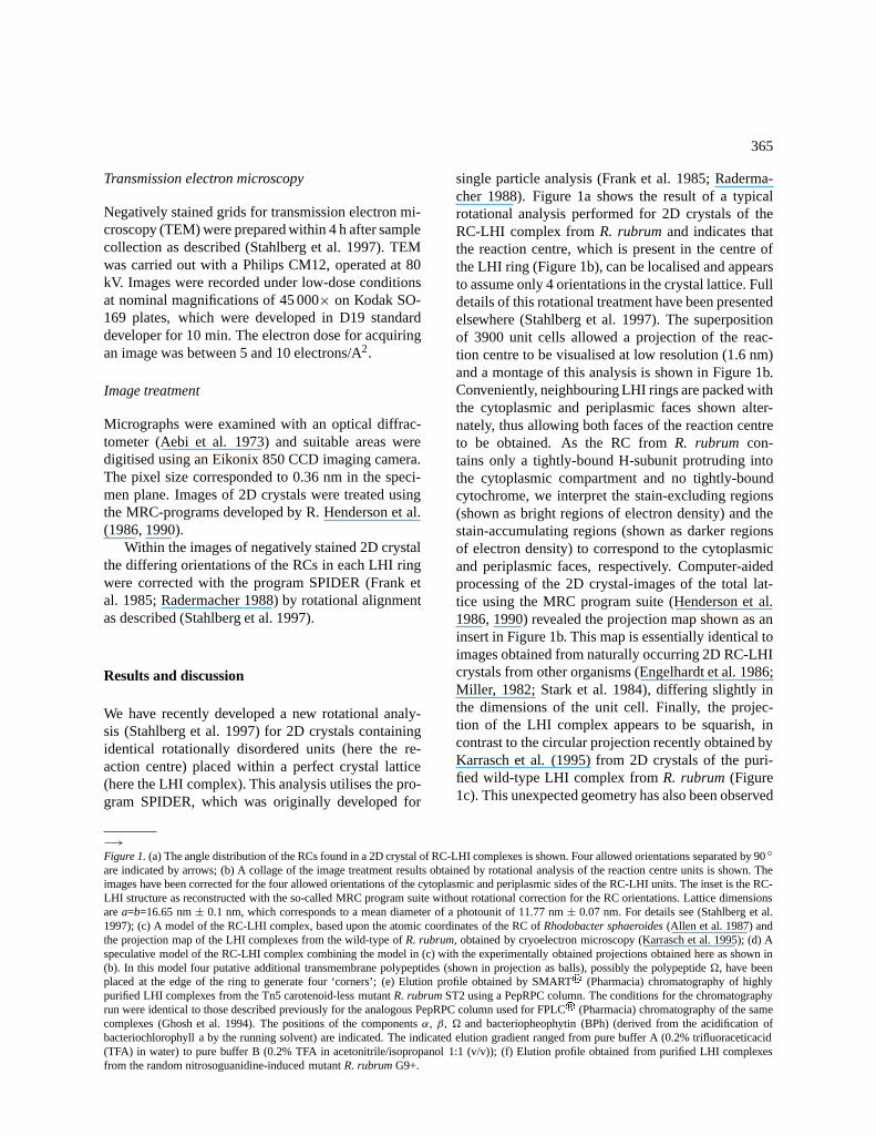
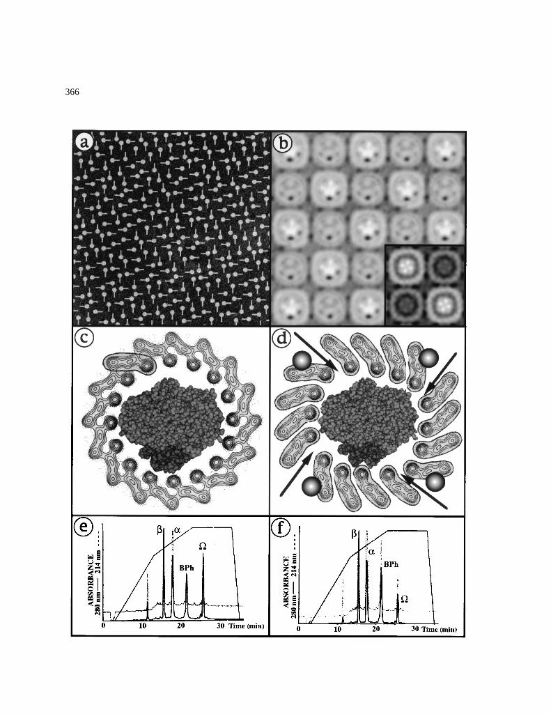
−→Figure 1.(a) The angle distribution of the RCs found in a 2D crystal of RC-LHI complexes is shown. Four allowed orientations separated by 90◦are indicated by arrows; (b) A collage of the image treatment results obtained by rotational analysis of the reaction centre units is shown. Theimages have been corrected for the four allowed orientations of the cytoplasmic and periplasmic sides of the RC-LHI units. The inset is the RC-LHI structure as reconstructed with the so-called MRC program suite without rotational correction for the RC orientations. Lattice dimensionsarea=b=16.65 nm± 0.1 nm, which corresponds to a mean diameter of a photounit of 11.77 nm± 0.07 nm. For details see (Stahlberg et al.1997); (c) A model of the RC-LHI complex, based upon the atomic coordinates of the RC ofRhodobacter sphaeroides(Allen et al. 1987) andthe projection map of the LHI complexes from the wild-type ofR. rubrum,obtained by cryoelectron microscopy (Karrasch et al. 1995); (d) Aspeculative model of the RC-LHI complex combining the model in (c) with the experimentally obtained projections obtained here as shown in(b). In this model four putative additional transmembrane polypeptides (shown in projection as balls), possibly the polypeptide�, have beenplaced at the edge of the ring to generate four ‘corners’; (e) Elution profile obtained by SMARTr (Pharmacia) chromatography of highlypurified LHI complexes from the Tn5 carotenoid-less mutantR. rubrumST2 using a PepRPC column. The conditions for the chromatographyrun were identical to those described previously for the analogous PepRPC column used for FPLCr (Pharmacia) chromatography of the samecomplexes (Ghosh et al. 1994). The positions of the componentsα, β, � and bacteriopheophytin (BPh) (derived from the acidification ofbacteriochlorophyll a by the running solvent) are indicated. The indicated elution gradient ranged from pure buffer A (0.2% trifluoraceticacid(TFA) in water) to pure buffer B (0.2% TFA in acetonitrile/isopropanol 1:1 (v/v)); (f) Elution profile obtained from purified LHI complexesfrom the random nitrosoguanidine-induced mutantR. rubrumG9+.
single particle analysis (Frank et al. 1985; Raderma-cher 1988). Figure 1a shows the result of a typicalrotational analysis performed for 2D crystals of theRC-LHI complex fromR. rubrumand indicates thatthe reaction centre, which is present in the centre ofthe LHI ring (Figure 1b), can be localised and appearsto assume only 4 orientations in the crystal lattice. Fulldetails of this rotational treatment have been presentedelsewhere (Stahlberg et al. 1997). The superpositionof 3900 unit cells allowed a projection of the reac-tion centre to be visualised at low resolution (1.6 nm)and a montage of this analysis is shown in Figure 1b.Conveniently, neighbouring LHI rings are packed withthe cytoplasmic and periplasmic faces shown alter-nately, thus allowing both faces of the reaction centreto be obtained. As the RC fromR. rubrum con-tains only a tightly-bound H-subunit protruding intothe cytoplasmic compartment and no tightly-boundcytochrome, we interpret the stain-excluding regions(shown as bright regions of electron density) and thestain-accumulating regions (shown as darker regionsof electron density) to correspond to the cytoplasmicand periplasmic faces, respectively. Computer-aidedprocessing of the 2D crystal-images of the total lat-tice using the MRC program suite (Henderson et al.1986, 1990) revealed the projection map shown as aninsert in Figure 1b. This map is essentially identical toimages obtained from naturally occurring 2D RC-LHIcrystals from other organisms (Engelhardt et al. 1986;Miller, 1982; Stark et al. 1984), differing slightly inthe dimensions of the unit cell. Finally, the projec-tion of the LHI complex appears to be squarish, incontrast to the circular projection recently obtained byKarrasch et al. (1995) from 2D crystals of the puri-fied wild-type LHI complex fromR. rubrum(Figure1c). This unexpected geometry has also been observed
pres850.tex; 30/06/1998; 15:22; p.3
366
pres850.tex; 30/06/1998; 15:22; p.4
367
by electron microscopy of single RC-LHI particles(Stahlberg et al. 1997). An interpretation of the C4symmetry might be that an additional protein compo-nent, possibly a single transmembraneα-helix whichcan bind negative stain, is present at the ‘corners’of the LHI ring. Our interpretation of the presentstructural data is shown in Figure 1d.
A candidate for the additional protein componentmay be the polypeptide� (Ghosh et al. 1994). The�polypeptide is a highly hydrophobic peptide of mole-cular weight 4 kDa containing about 28 amino acidsand is found in a molar stoichiometric ratio to theαandβ LHI polypeptides of approximately 1�/10αβin purified LHI complexes from the carotenoid-lessmutantR. rubrumG9+ (Ghosh et al. 1994). However,in the latter study the interpretation of the data wascomplicated by the fact that the carotenoid-less mutantR. rubrumG9+, which was obtained using random ni-trosoguanidine mutagenesis, may contain polypeptidefragments generated by the creation of stop codons,possibly leading to the production of small polypep-tides which copurify with the LHI complex. However,SMARTr chromatography profiles of purified LHIcomplexes from the Tn5-directed mutantR. rubrumST2, which contains only a single chromosomal le-sion (Wiggli et al. 1996) also shows the presence ofthe same polypeptide (Figure 1d) as observed forR.rubrum G9+ (Figure 1e) and in the same molar ra-tio as determined by amino acid analysis (data notshown). We have also observed the� polypeptide inSMARTr profiles of LHI complexes obtained fromthe wild-type organism (data not shown). Althoughwe have not yet been successful in microsequencingthe� polypeptide, due to high hydrophobicity and N-terminal blockage, the SMARTr data are neverthelesssuggestive that it is not artefactual.
The present work suggests the following conclu-sions for the RC-LHI complex fromR. rubrum: (1)The LHI ring from the carotenoid-less mutant con-tains 16 subunits, as was found for the wild-typecomplex (Karrasch et al. 1995); (2) the LHI ringsurrounds a single reaction centre; (3) the LHI ringfrom the carotenoid-less complex is closed and ap-pears to have a squarish shape, possibly due to thepresence of another protein component in additionto the α and β polypeptides; (4) a possible candi-date for this additional component, the hydrophobicpolypeptide� (Ghosh et al. 1994) has also beenshown to be present in highly purified LHI prepara-tions of the Tn5-directed carotenoid-less mutantR.
rubrum, as well in those obtained from the wild-typecarotenoid-containing organism.
Acknowledgements
We thank Per Bullough and Thomas Walz for theirhelp with the image analysis with the MRC softwareand for stimulating discussions. We also thank Marie-France Blanc for expert technical assistance and RetoJ. Strasser for encouragement and support. We ac-knowledge the Swiss National Science FoundationPriority Program on Biotechnology, projects 5002-35180 to H.S. and H.V. and projects 5002-41801 and5002-39816 to R.G. for generous financial support.
References
Aebi U, Smith PR, Dubochet J, Henry C and Kellenberger E (1973)A study of the structure of the T-layer ofBacillus brevis. JSupramol Struct 1(6): 498–522
Allen JP, Feher G, Yeates TO, Komiya H and Rees DC (1987) Struc-ture of the reaction center fromRhodobacter sphaeroidesR-26:The cofactors. Proc Natl Acad Sci USA 84: 5730–5734
Engelhardt H, Engel A and Baumeister W (1986) Stoichiometricmodel of the photosynthetic unitEctothiorhodospira halochloris.Proc Natl Acad Sci USA 83: 8972–8976
Frank J, Verschoor A and Wagenknecht T. (1985). Processing ofelectron-microscopic images of single macromolecules. In: WuTT (ed) New methodologies in Studies of Protein Conformation,pp 36–89. Van Nostrand-Reinold Inc., New York
Ghosh R, Ghosh-Eicher S, DiBerardino M and Bachofen R (1994)Protein phosphorylation inRhodospirillum rubrum: Purifica-tion and characterization of a water-soluble B873 protein kinaseand a new component of the B873 complex,�, which can bephosphorylated. Biochim Biophys Acta 1184: 28–36
Ghosh R, Hoenger A, Hardmeyer A, Mihailescu D, Bachofen R,Engel A and Rosenbusch JP (1993) Two-dimensional crystal-lization of the light-harvesting complex fromRhodospirillumrubrum. J Mol Biol 231(2): 501–504
Henderson R, Baldwin JM, Ceska TA, Zemlin F, Beckmann Eand Downing KH (1990) Model for the structure ofBacteri-orhodopsinbased on high-resolution electron cryo-microscopy.J Mol Biol 213: 899–929
Henderson R, Baldwin JM, Downing KH, Lepault J and ZemlinF (1986) Structure of purple membrane fromHalobacteriumhalobium: Recording, measurement and evaluation of electronmicrographs at 3.5�A resolution. Ultramicroscopy 19: 147–178
Jap BK, Zulauf M, Scheybani T, Hefti A, Baumeister W, Aebi Uand Engel A (1992) 2D crystallization: From art to science.Ultramicroscopy 46(1-4): 45–84
Joliot P, Vermeglio A and Joliot A (1990) Electron transfer betweenprimary and secondary donors inRhodospirillum rubrum: Evi-dence for a dimeric association of reaction centers. Biochemistry29: 4031–4037
Karrasch S, Bullough PA and Ghosh R (1995) The 8.5�A projec-tion map of the light-harvesting complex I fromRhodospirillumrubrum reveals a ring composed of 16 subunits. EMBO J 14(4):631–638
pres850.tex; 30/06/1998; 15:22; p.5
368
Koepke J, Hu X, Münke C, Schulten K and Michel H (1996) Thecrystal structure of the light-harvesting complex II (B800–B850)from Rhodospirillum molischianum. Structure 4(5): 581–597
McDermott G, Prince SM, Freer AA, Hawthornthwaite-LawlessAM, Papiz MZ, Cogdell RJ and Isaacs NW (1995) Crystal struc-ture of an integral membrane light-harvesting complex fromphotosynthetic bacteria. Nature 374: 517–521
Meckenstock RU, Krusche K, Brunisholz RA and Zuber H (1992)The light-harvesting core-complex and the B820-subunit fromRhodopseudomonas marina. Part II. Electron microscopic char-acterisation. FEBS Lett 311(2): 135–138
Miller KR (1982) Three-dimensional structure of a photosyntheticmembrane. Nature 300: 53–55
Radermacher M (1988) Three-dimensional reconstruction of sin-gle particles from random and nonrandom tilt series. J ElectronMicrosc Tech 9: 359–394
Saegesser R, Ghosh R and Bachofen R (1992) Stability of broadhost range cloning vectors in the phototrophic bacteriumRho-dospirillum rubrum. FEMS Microbiol Lett 95: 7–12
Sistrom WR (1977) Transfer of chromosomal genes mediated byplasmid R68.45 inRhodopseudomondas sphaeroides. J Bacteriol131(2): 526–533
Stahlberg H, Dubochet J, Vogel H and Ghosh R (1997) Are light-harvesting I complexes fromRhodospirillum rubrumarrangedaround the reaction centre in a square geometry ? (submitted)
Stark W, Kühlbrandt W, Wildhaber I, Wehrli E and Müh-lethaler K (1984) The structure of the photoreceptor unit ofRhodopseudomonas viridis. EMBO J 3: 77–783
Walz T and Ghosh R (1997) Two-dimensional crystallization of thelight-harvesting I-reaction centre photounit fromRhodospirillumrubrum. J Mol Biol 265(2): 107–111
Wiggli M, Cornacchia L, Saegesser R, Bachofen R and GhoshR (1996) Characterisation ofRhodospirillum rubrumST2. Anew Tn5-induced carotenoid-less mutant for functional studies.Microbiol Res 151: 1–5
pres850.tex; 30/06/1998; 15:22; p.6
Copyright © 2022 FDOKUMEN