The Proliferation of Pre-Pubertal Porcine Spermatogonia in ...
15
International Journal of Molecular Sciences Article The Proliferation of Pre-Pubertal Porcine Spermatogonia in Stirred Suspension Bioreactors Is Partially Mediated by the Wnt/β-Catenin Pathway Sadman Sakib 1,2 , Anna Voigt 2 , Nathalia de Lima e Martins Lara 2 , Lin Su 2 , Mark Ungrin 2,3 , Derrick Rancourt 1,4 and Ina Dobrinski 1,2, * Citation: Sakib, S.; Voigt, A.; de Lima e Martins Lara, N.; Su, L.; Ungrin, M.; Rancourt, D.; Dobrinski, I. The Proliferation of Pre-Pubertal Porcine Spermatogonia in Stirred Suspension Bioreactors Is Partially Mediated by the Wnt/β-Catenin Pathway. Int. J. Mol. Sci. 2021, 22, 13549. https:// doi.org/10.3390/ijms222413549 Academic Editor: Mahmoud Huleihel Received: 10 November 2021 Accepted: 13 December 2021 Published: 17 December 2021 Publisher’s Note: MDPI stays neutral with regard to jurisdictional claims in published maps and institutional affil- iations. Copyright: © 2021 by the authors. Licensee MDPI, Basel, Switzerland. This article is an open access article distributed under the terms and conditions of the Creative Commons Attribution (CC BY) license (https:// creativecommons.org/licenses/by/ 4.0/). 1 Department of Biochemistry and Molecular Biology, Cumming School of Medicine, University of Calgary, Calgary, AB T2N 4N1, Canada; [email protected] (S.S.); [email protected] (D.R.) 2 Department of Comparative Biology and Experimental Medicine, Faculty of Veterinary Medicine, University of Calgary, Calgary, AB T2N 1N4, Canada; [email protected] (A.V.); [email protected] (N.d.L.e.M.L.); [email protected] (L.S.); [email protected] (M.U.) 3 Biomedical Engineering Graduate Program, University of Calgary, Calgary, AB T2N 1N4, Canada 4 Department of Oncology and Medical Genetics, Cumming School of Medicine, University of Calgary, Calgary, AB T2N 4N2, Canada * Correspondence: [email protected]; Tel.: +1-403-210-6532 Abstract: Male survivors of childhood cancer are at risk of suffering from infertility in adulthood because of gonadotoxic chemotherapies. For adult men, sperm collection and preservation are routine procedures prior to treatment; however, this is not an option for pre-pubertal children. From young boys, a small biopsy may be taken before chemotherapy, and spermatogonia may be propagated in vitro for future transplantation to restore fertility. A robust system that allows for scalable expansion of spermatogonia within a controlled environment is therefore required. Stirred suspension culture has been applied to different types of stem cells but has so far not been explored for spermatogonia. Here, we report that pre-pubertal porcine spermatogonia proliferate more in bioreactor suspension culture, compared with static culture. Interestingly, oxygen tension provides an avenue to modulate spermatogonia status, with culture under 10% oxygen retaining a more undifferentiated state and reducing proliferation in comparison with the conventional approach of culturing under ambient oxygen levels. Spermatogonia grown in bioreactors upregulate the Wnt/ β-catenin pathway, which, along with enhanced gas and nutrient exchange observed in bioreactor culture, may synergistically account for higher spermatogonia proliferation. Therefore, stirred suspension bioreactors provide novel platforms to culture spermatogonia in a scalable manner and with minimal handling. Keywords: bioreactor; stirred suspension bioreactor; germ cell; spermatogonia; culture 1. Introduction Advances in cancer therapies have led to increased survival rates of pediatric cancer patients [1,2]. As a result, there is a growing number of childhood cancer survivors who suffer from the late effects of gonadotoxic chemotherapy, which include hypogonadism and infertility due to loss of the spermatogonia pool [3,4]. Although cryopreservation of sperm is a routine fertility preservation technique for adult men undergoing chemotherapy, there is no such option for pre-pubertal boys before the onset of spermatogenesis. This has led to an interest in the isolation and cryopreservation of testicular biopsies, to allow subsequent isolation, expansion, and transplantation of spermatogonia back into the testes to restore spermatogenesis [5]. Although the culture of rodent spermatogonia is well established [6], scalable expan- sion of non-rodent spermatogonia in culture has had limited success [7–10]. Most of the research reported so far has employed conventional static culture systems, which are labor Int. J. Mol. Sci. 2021, 22, 13549. https://doi.org/10.3390/ijms222413549 https://www.mdpi.com/journal/ijms
-
Upload
khangminh22 -
Category
Documents
-
view
2 -
download
0
Transcript of The Proliferation of Pre-Pubertal Porcine Spermatogonia in ...

International Journal of
Molecular Sciences
Article
The Proliferation of Pre-Pubertal Porcine Spermatogonia inStirred Suspension Bioreactors Is Partially Mediated by theWnt/β-Catenin Pathway
Sadman Sakib 1,2, Anna Voigt 2 , Nathalia de Lima e Martins Lara 2 , Lin Su 2, Mark Ungrin 2,3 ,Derrick Rancourt 1,4 and Ina Dobrinski 1,2,*
�����������������
Citation: Sakib, S.; Voigt, A.; de Lima
e Martins Lara, N.; Su, L.; Ungrin, M.;
Rancourt, D.; Dobrinski, I. The
Proliferation of Pre-Pubertal Porcine
Spermatogonia in Stirred Suspension
Bioreactors Is Partially Mediated by
the Wnt/β-Catenin Pathway. Int. J.
Mol. Sci. 2021, 22, 13549. https://
doi.org/10.3390/ijms222413549
Academic Editor: Mahmoud Huleihel
Received: 10 November 2021
Accepted: 13 December 2021
Published: 17 December 2021
Publisher’s Note: MDPI stays neutral
with regard to jurisdictional claims in
published maps and institutional affil-
iations.
Copyright: © 2021 by the authors.
Licensee MDPI, Basel, Switzerland.
This article is an open access article
distributed under the terms and
conditions of the Creative Commons
Attribution (CC BY) license (https://
creativecommons.org/licenses/by/
4.0/).
1 Department of Biochemistry and Molecular Biology, Cumming School of Medicine, University of Calgary,Calgary, AB T2N 4N1, Canada; [email protected] (S.S.); [email protected] (D.R.)
2 Department of Comparative Biology and Experimental Medicine, Faculty of Veterinary Medicine,University of Calgary, Calgary, AB T2N 1N4, Canada; [email protected] (A.V.);[email protected] (N.d.L.e.M.L.); [email protected] (L.S.); [email protected] (M.U.)
3 Biomedical Engineering Graduate Program, University of Calgary, Calgary, AB T2N 1N4, Canada4 Department of Oncology and Medical Genetics, Cumming School of Medicine, University of Calgary,
Calgary, AB T2N 4N2, Canada* Correspondence: [email protected]; Tel.: +1-403-210-6532
Abstract: Male survivors of childhood cancer are at risk of suffering from infertility in adulthoodbecause of gonadotoxic chemotherapies. For adult men, sperm collection and preservation areroutine procedures prior to treatment; however, this is not an option for pre-pubertal children.From young boys, a small biopsy may be taken before chemotherapy, and spermatogonia may bepropagated in vitro for future transplantation to restore fertility. A robust system that allows forscalable expansion of spermatogonia within a controlled environment is therefore required. Stirredsuspension culture has been applied to different types of stem cells but has so far not been exploredfor spermatogonia. Here, we report that pre-pubertal porcine spermatogonia proliferate more inbioreactor suspension culture, compared with static culture. Interestingly, oxygen tension providesan avenue to modulate spermatogonia status, with culture under 10% oxygen retaining a moreundifferentiated state and reducing proliferation in comparison with the conventional approach ofculturing under ambient oxygen levels. Spermatogonia grown in bioreactors upregulate the Wnt/β-catenin pathway, which, along with enhanced gas and nutrient exchange observed in bioreactorculture, may synergistically account for higher spermatogonia proliferation. Therefore, stirredsuspension bioreactors provide novel platforms to culture spermatogonia in a scalable manner andwith minimal handling.
Keywords: bioreactor; stirred suspension bioreactor; germ cell; spermatogonia; culture
1. Introduction
Advances in cancer therapies have led to increased survival rates of pediatric cancerpatients [1,2]. As a result, there is a growing number of childhood cancer survivors whosuffer from the late effects of gonadotoxic chemotherapy, which include hypogonadismand infertility due to loss of the spermatogonia pool [3,4]. Although cryopreservation ofsperm is a routine fertility preservation technique for adult men undergoing chemotherapy,there is no such option for pre-pubertal boys before the onset of spermatogenesis. Thishas led to an interest in the isolation and cryopreservation of testicular biopsies, to allowsubsequent isolation, expansion, and transplantation of spermatogonia back into the testesto restore spermatogenesis [5].
Although the culture of rodent spermatogonia is well established [6], scalable expan-sion of non-rodent spermatogonia in culture has had limited success [7–10]. Most of theresearch reported so far has employed conventional static culture systems, which are labor
Int. J. Mol. Sci. 2021, 22, 13549. https://doi.org/10.3390/ijms222413549 https://www.mdpi.com/journal/ijms

Int. J. Mol. Sci. 2021, 22, 13549 2 of 15
intensive and can be challenging to scale. Further, as “open” culture systems that involveextensive handling, they are particularly vulnerable to contamination and variability be-tween batches, factors that can further reduce their utility in clinical applications [11,12].Another important avenue of cell culture that is often neglected in the majority of reportsis O2 tension [13,14]. In most cases, it is assumed that incubators set up for ambient O2tension deliver 20.9% O2 to the cells. In reality, the O2 levels the cells experience dependon a number of factors such as altitude, media height, type of cell being cultured, andabsence or presence of agitation [13,14]. Diffusive barriers in static culture further reduceO2 levels, often quite dramatically [13]. Previous research has shown that a reduced O2tension environment is more supportive to several types of stem cells than culture underambient O2 tension [15–17]. Recent studies also indicated that low O2 tension of 10% ismore supportive for mouse and cattle spermatogonia proliferation in vitro [10,18].
We, therefore, assessed the potential for stirred suspension bioreactors (SSBs) toaddress the need for scalable expansion of spermatogonia within a controlled environmentat ambient and low (10%) O2 tension. As the availability of immature human testis tissuefor research is severely limited, we used spermatogonia from pre-pubertal pigs, which arewidely available and share many physiological similarities with humans [19–22]. SSBs aresealed glass chambers with a central (generally magnetically driven) impeller, as well asports for gas and medium exchange. Speed-controlled rotation of the impeller providesmixing and controls the shear forces to which the culture is exposed. The result is ascalable, controlled culture environment, with enhanced homogeneity, oxygen and masstransfer, and reduced variability [12]. We and others have demonstrated efficient andeffective expansion of pluripotent stem cells in SSBs [23–25] and a role for upregulationof the Wnt/β-catenin pathway via the shear forces produced in SSB culture [25–28]. Thisis consistent with reports that fluid shear forces increase E-cadherin expression, induceadherens junctions [29], and upregulate the Wnt/β-catenin pathway and downstreamC-myc expression [28]. Notably, Wnt/β-catenin signaling has also been shown to supportthe proliferation of murine spermatogonia [30].
Here, we report a novel suspension culture model for the scalable expansion of porcinepre-pubertal spermatogonia using the SSB. We found that while 10% O2 tension supportsless proliferation than ambient O2 levels, it retains more undifferentiated spermatogonia.We also found that shear forces generated in the SSB upregulate the Wnt/β-catenin path-way, which is partially responsible for the increased proliferation of porcine spermatogoniain suspension culture.
2. Results
2.1. Rotation Speed of 120 rpm (3.9 dyne/cm2) Supports the Highest Level ofSpermatogonia Proliferation
Cell populations from 1-week-old porcine testes were enriched for spermatogonia(82.2 ± 1.6%) positive for the spermatogonia marker ubiquitin C-terminal hydrolase L1(UCHL1) [31,32] and cultured in 10 mL SSBs (Figure S1A) at 60, 80, 100, 120, and 140 rpm,alongside static-culture controls, for 14 days. These rotation speeds were selected basedon previously reported studies [23,25,33–36] and translate to maximum shear forces of1.5, 2.2, 3.0, 3.9, and 4.8 dyne/cm2, respectively (Figure S1B). The viability of the cells instatic, 60, 80, 100, 120, and 140 rpm bioreactors were, respectively, 87.6 ± 2.6% (n = 15),42.2 ± 7.7%, 90.4 ± 1.5%, 92.5 ± 2.3%, 93.7 ± 2.6% and 50 ± 5.6% (n = 3). As the viabilityof cells cultured at 60 and 140 rpm SSBs were low, these rotation speeds were excludedfrom future experiments and analyses. The number of spermatogonia and percentages ofproliferative spermatogonia was analyzed by immunofluorescence for UCHL1 and EdUincorporation. Compared with the respective static cultures, SSB cultures (80, 100, and120 rpm) had overall higher total spermatogonia yields (n = 3, p < 0.05) (Figure 1B,D,F).Similarly, the percentage of proliferative spermatogonia (EdU+ve UCHL1+ve cells) washigher in all SSB cultures except 80 rpm (n = 3, p < 0.05) (Figure 1A,C,E,G). Of the three SSBrotation speeds, 120 rpm that had a maximum shear force of 3.9 dyne/ cm2, supported the

Int. J. Mol. Sci. 2021, 22, 13549 3 of 15
highest degree of proliferation (Figure S1C), and as a result, only 120 rpm rotation speedSSB cultures were used for further experiments.
Int. J. Mol. Sci. 2021, 22, x FOR PEER REVIEW 3 of 15
had overall higher total spermatogonia yields (n = 3, p < 0.05) (Figure 1 B,D,F). Similarly, the percentage of proliferative spermatogonia (EdU+ve UCHL1+ve cells) was higher in all SSB cultures except 80 rpm (n = 3, p < 0.05) (Figure 1A,C,E,G). Of the three SSB rotation speeds, 120 rpm that had a maximum shear force of 3.9 dyne/ cm2, supported the highest degree of proliferation (Figure S1C), and as a result, only 120 rpm rotation speed SSB cul-tures were used for further experiments.
Figure 1. Stirred suspension bioreactor culture at 120 rpm (3.9 dyne/cm2) supports the highest level of spermatogonia proliferation: (A) immunofluorescence images of spermatogonia after static, 80, 100, and 120 rpm SSB culture. Scale bars 50 µm; (B,D,F) number of spermatogonia harvested after 2 weeks of SSB and static culture at 80 (B) 100 (D), and 120 (F) rpm; (C,E,G) percentage of proliferating spermatogonia after 2 weeks of SSB and static culture 80 (C), 100 (E), and 120 (G) rpm. n = 3, mean ± SD. p > 0.05 (ns), * p ≤ 0.05, ** p ≤ 0.01.
Interestingly, mouse spermatogonia cultured under similar conditions had substan-tially reduced viability (static culture: 92.1–93.4%, 120 rpm: 8–10%, n = 2; 60 rpm: 10%, 80 rpm: 12.1%, n = 1) and proliferation (static culture: 15.2–18.2%, 120 rpm: 4.1–8.3%, n = 2; 60 rpm: 5.3%, 80 rpm: 4.8%, n = 1) in SSB. As a result, mouse spermatogonia cultures in SSBs were not pursued further.
2.2. In Suspension Culture, Porcine Spermatogonia Proliferate More in Ambient O2 Tension but Contain Fewer Undifferentiated Spermatogonia
Mouse and cattle spermatogonia cultures at 10% O2 support better expansion in vitro [10,18]. It should be noted that incubators filled with ambient air are widely referred to as containing 20.9% O2; however, dilution with CO2 and water vapor results in actual oxygen content that is somewhat lower. At sea level, this reduction is to ~18.6% O2 in the gas phase; in our laboratory in Calgary, elevation (~1084 m) reduced air pressure and conse-quently oxygen levels as well (see Supplementary Methods for detailed calculations) [13,14,37,38]. To explore if low O2 tension would be beneficial for porcine spermatogonia culture, cells from 1-week-old pigs (containing 82.2 ± 1.6% UCHL1+ve cells) and from 8-
Figure 1. Stirred suspension bioreactor culture at 120 rpm (3.9 dyne/cm2) supports the highest level of spermatogoniaproliferation: (A) immunofluorescence images of spermatogonia after static, 80, 100, and 120 rpm SSB culture. Scale bars50 µm; (B,D,F) number of spermatogonia harvested after 2 weeks of SSB and static culture at 80 (B) 100 (D), and 120 (F) rpm;(C,E,G) percentage of proliferating spermatogonia after 2 weeks of SSB and static culture 80 (C), 100 (E), and 120 (G) rpm.n = 3, mean ± SD. p > 0.05 (ns), * p ≤ 0.05, ** p ≤ 0.01.
Interestingly, mouse spermatogonia cultured under similar conditions had substan-tially reduced viability (static culture: 92.1–93.4%, 120 rpm: 8–10%, n = 2; 60 rpm: 10%,80 rpm: 12.1%, n = 1) and proliferation (static culture: 15.2–18.2%, 120 rpm: 4.1–8.3%, n = 2;60 rpm: 5.3%, 80 rpm: 4.8%, n = 1) in SSB. As a result, mouse spermatogonia cultures inSSBs were not pursued further.
2.2. In Suspension Culture, Porcine Spermatogonia Proliferate More in Ambient O2 Tension butContain Fewer Undifferentiated Spermatogonia
Mouse and cattle spermatogonia cultures at 10% O2 support better expansionin vitro [10,18]. It should be noted that incubators filled with ambient air are widelyreferred to as containing 20.9% O2; however, dilution with CO2 and water vapor resultsin actual oxygen content that is somewhat lower. At sea level, this reduction is to ~18.6%O2 in the gas phase; in our laboratory in Calgary, elevation (~1084 m) reduced air pres-sure and consequently oxygen levels as well (see Supplementary Methods for detailedcalculations) [13,14,37,38]. To explore if low O2 tension would be beneficial for porcinespermatogonia culture, cells from 1-week-old pigs (containing 82.2 ± 1.6% UCHL1+ve cells)and from 8-week-old pigs (containing 80.6 ± 1.4% UCHL1+ve cells) were cultured in staticplates (control) and in 120 rpm SSBs at 10% and ambient O2 tensions. The 10% O2 andthe ambient culture conditions translate to 3.45 mM and 6.34 mM of O2 in the gas phase(Table 1).

Int. J. Mol. Sci. 2021, 22, 13549 4 of 15
Table 1. Calculation of O2 tension and concentration in gas phase at ~1084 m elevation (see Supple-mentary Methods).
Conditions O2 Tension Pre-Setfor the Incubator
PartialPressure of O2
True O2 Tension (at1084 m Altitude)
Concentrationin Gas Phase
Ambient 20.9% 16.34 kPa 18.4% 6.34 mM10% O2 10% 8.89 kPa 10% 3.45 mM
The O2 tension and concentration in the liquid phase (media) at 48 h as measuredvia Oxygen Sensor Spots SP-PSt3-NAU (PreSens Regensburg, Germany) (Figure S2C,Supplementary Methods) [32] are shown in Table 2.
Table 2. O2 measurements in media via spot sensors (Supplementary Methods).
Conditions O2 Tension Concentration
Static ambient O2 14.9 ± 0.04% 5.2 ± 0.01 mMSSB ambient O2 18.2 ± 0.17% 6.3 ± 0.06 mM
Static 10% O2 3.28 ± 0.22% 1.1 ± 0.07 mMSSB 10% O2 3.74 ± 0.01% 1.3 ± 0.003 mM
Both 1- and 8-week-old spermatogonia were examined to evaluate the effect of SSBculture on early and late pre-pubertal spermatogonia, as we reported previously that thesecells have different metabolic requirements [32]. For 1-week-old spermatogonia, total cellyield after 2 weeks of culture was higher in both 10% and ambient O2 (n = 3, p < 0.05)tension, compared with static culture (Figure 2C). However, no significant difference incell yield was observed between SSB cultures in ambient and 10% O2 tension (Figure 2C).Similarly, proliferation levels (% of EdU+ve UCHL+ve cells) were higher for SSB cultures,compared with static controls for both O2 tensions (n = 3, p < 0.05). In addition, spermato-gonia proliferation was higher in ambient O2 SSB culture, compared with 10% O2 culture(n = 3, p < 0.05) (Figure 2A,D).
For 8-week-old spermatogonia, similar to 1-week-old cells, cell yield after 2 weeks ofculture was higher in SSBs at both 10% and ambient O2 (n = 3, p < 0.05) tension, comparedwith the static. Cell yield between SSB cultures in 10% and ambient O2 tension wasnot significantly different (Figure 2C). Spermatogonia proliferation was higher for SSBcultures, compared with static controls at both 10% and ambient (n = 3, p < 0.05) O2 cultures(Figure 2B,D). Compared with 10% O2 tension, spermatogonia proliferation was higher inambient O2 tension for SSB cultures (n = 3, p < 0.05) (Figure 2D).
It has been reported that mouse spermatogonia culture at ambient O2 results indifferentiation into progenitor cells and loss of stem cell potential [6,18,39]. To eluci-date if the spermatogonia in SSB culture are undergoing differentiation, the transcriptionof GDNF family receptor alpha 1 (GFRα1) [40] and promyelocytic leukemia zinc finger(PLZF), as markers of undifferentiated spermatogonia, and deleted in azoospermia-like(DAZL), a marker of differentiating spermatogonia [41], was evaluated. Although no sig-nificant difference in transcription levels was observed for PLZF and DAZL (Figure S2A,B),GFRα1 transcription levels were 3.72-fold higher in 1-week-old cells and 3.18-fold higher in8-week-old cells (n = 3, p < 0.05) cultured at 10% O2 tension, compared with cells culturedunder ambient O2 (Figure 2E) indicating retention of a more undifferentiated state, whichis consistent with previous reports [6,18,39]. Subsequent analyses were therefore carriedout at 10% O2 tension.
Int. J. Mol. Sci. 2021, 22, 13549 5 of 15Int. J. Mol. Sci. 2021, 22, x FOR PEER REVIEW 5 of 15
Figure 2. Spermatogonia cultured at 10% O2 tension proliferate less than those cultured at ambient O2 tension but contain more undifferentiated cells: (A,B) immunofluorescence images of 1-week-old and 8-week-old spermatogonia after 14 days of culture: red, UCHL1; grey, EdU; blue, DAPI. Scale bars 75 µm; (C) number of 1-week-old and 8-week-old spermatogonia (SG) harvested after 14 days of culture; (D) percentage of proliferating 1-week-old and 8-week-old spermatogonia (SG) after 14 days of culture; (E) relative mRNA fold change of GFRα1 for 1-week-old and 8-week-old spermatogonia (SG). n = 3, mean ± SD. p > 0.05 (ns), p ≤ 0.05 (*), p ≤ 0.01 (**), p ≤ 0.001 (***).
2.3. Shear Forces Generated in SSB Culture Activate the Wnt/β-Catenin Pathway Fluid shear forces are known to activate the Wnt/β-catenin pathway by nuclear trans-
location of β-catenin [25,28,42–44], likely mediated by the mechano-sensing properties of E-cadherin [45]. β-catenin binds to the cytoplasmic tail of E-cadherin [46], which, upon exposure to fluid shear in SSBs, is dislodged from the adherens junction and accumulates in the cytoplasm resulting in nuclear translocation and pathway activation [25]. To inves-tigate Wnt/β-catenin pathway activation, 1- and 8-week-old cells cultured in static condi-tions and SSBs at 120 rpm under 10% O2 tension were stained for β-catenin (1:200, Abcam) and vimentin, a marker for Sertoli and interstitial cells (1:500, Sigma) (Figure S3A,B), and the number of vimentin-ve cells with nuclear β-catenin were counted to determine the % of spermatogonia displaying nuclear β-catenin localization. For both age groups, the per-centages of spermatogonia with nuclear β-catenin were higher in SSB cultures, compared with static controls (n = 3, p < 0.05) (Figure 3A, Figure S3C–H). Spermatogonia express E-cadherin [47] and grow as clusters in the SSBs (Figure S3I,J). This clustering is mediated by E-cadherin [48], which likely serves as an upstream mediator for the nuclear localiza-tion of β-catenin in SSBs. To further characterize the Wnt/β-catenin pathway activation, qPCR for E-cadherin and Axin2, a downstream target of the Wnt/β-catenin pathway [49],
Figure 2. Spermatogonia cultured at 10% O2 tension proliferate less than those cultured at ambient O2 tension but containmore undifferentiated cells: (A,B) immunofluorescence images of 1-week-old and 8-week-old spermatogonia after 14 daysof culture: red, UCHL1; grey, EdU; blue, DAPI. Scale bars 75 µm; (C) number of 1-week-old and 8-week-old spermatogonia(SG) harvested after 14 days of culture; (D) percentage of proliferating 1-week-old and 8-week-old spermatogonia (SG)after 14 days of culture; (E) relative mRNA fold change of GFRα1 for 1-week-old and 8-week-old spermatogonia (SG). n = 3,mean ± SD. p > 0.05 (ns), p ≤ 0.05 (*), p ≤ 0.01 (**), p ≤ 0.001 (***).
2.3. Shear Forces Generated in SSB Culture Activate the Wnt/β-Catenin Pathway
Fluid shear forces are known to activate the Wnt/β-catenin pathway by nucleartranslocation of β-catenin [25,28,42–44], likely mediated by the mechano-sensing propertiesof E-cadherin [45]. β-catenin binds to the cytoplasmic tail of E-cadherin [46], which, uponexposure to fluid shear in SSBs, is dislodged from the adherens junction and accumulates inthe cytoplasm resulting in nuclear translocation and pathway activation [25]. To investigateWnt/β-catenin pathway activation, 1- and 8-week-old cells cultured in static conditionsand SSBs at 120 rpm under 10% O2 tension were stained for β-catenin (1:200, Abcam)and vimentin, a marker for Sertoli and interstitial cells (1:500, Sigma) (Figure S3A,B),and the number of vimentin-ve cells with nuclear β-catenin were counted to determinethe % of spermatogonia displaying nuclear β-catenin localization. For both age groups,the percentages of spermatogonia with nuclear β-catenin were higher in SSB cultures,compared with static controls (n = 3, p < 0.05) (Figures 3A and S3C–H). Spermatogoniaexpress E-cadherin [47] and grow as clusters in the SSBs (Figure S3I,J). This clustering ismediated by E-cadherin [48], which likely serves as an upstream mediator for the nuclearlocalization of β-catenin in SSBs. To further characterize the Wnt/β-catenin pathwayactivation, qPCR for E-cadherin and Axin2, a downstream target of the Wnt/β-catenin

Int. J. Mol. Sci. 2021, 22, 13549 6 of 15
pathway [49], was performed. Transcription data revealed that both E-cadherin and Axin2were upregulated in SSB culture, compared with conventional static culture, with E-cadherinincreasing 5.3-fold for 1-week-old and 2.7-fold for 8-week-old cells, while Axin2 wasincreased 4-fold and 3-fold, respectively (n = 3, p < 0.05) (Figure 3B,C).
Int. J. Mol. Sci. 2021, 22, x FOR PEER REVIEW 6 of 15
was performed. Transcription data revealed that both E-cadherin and Axin2 were upregu-lated in SSB culture, compared with conventional static culture, with E-cadherin increasing 5.3-fold for 1-week-old and 2.7-fold for 8-week-old cells, while Axin2 was increased 4-fold and 3-fold, respectively (n = 3, p < 0.05) (Figure 3B,C).
Figure 3. Suspension bioreactor culture elicits Wnt/β-catenin pathway activation: (A) percentage of 1- and 8-week-old spermatogonia (SG) with nuclear β-catenin; (B) relative mRNA fold change of E-cadherin for 1- and 8-week-old spermato-gonia (SG); (C) relative mRNA fold change of Axin2 for 1- and 8-week-old spermatogonia (SG). n = 3, mean ± SD. p > 0.05 (ns), p ≤ 0.05 (*), ** p ≤ 0.01, **** p ≤ 0.0001.
2.4. Activation of the Wnt/β-Catenin Pathway in Static Culture Does Not Fully Mimic the Effect of Suspension Culture
If Wnt/β-catenin is solely responsible for the enhanced proliferation seen in SSB cul-ture, then activating the pathway in static culture would lead to similar levels of prolifer-ation. To test that hypothesis, spermatogonia from 1- and 8-week-old pigs were cultured in SSBs at 120 rpm and in static cultures at 10% O2 tension with CHIR99021, a Glycogen Synthase Kinase 3β (GSK3β) inhibitor (Wnt activator) [50] for 14 days. Preliminary dose–response experiments showed that 3 µM CHIR99021 was the highest dose that could be used without negatively affecting cell viability. Upon treatment with 3 µM CHIR99021, transcription of Axin2 was increased 3.2-fold for the treatment group and 4.3-fold for SSB, compared with control (n = 3, p < 0.05) for the 1-week-old spermatogonia (Figure 4A). For 8-week-old cells, Axin2 transcription level increased 2.3-fold for treatment and 3.4-fold for SSB 120 rpm, compared with the control group (n = 3, p < 0.05) (Figure 4A). Proliferation levels were found to be higher in the treatment groups than in the control but were still lower than in SSB cultures (n = 3, p < 0.05) (Figure 4B).
Figure 3. Suspension bioreactor culture elicits Wnt/β-catenin pathway activation: (A) percentage of 1- and 8-week-old spermatogonia (SG) with nuclear β-catenin; (B) relative mRNA fold change of E-cadherin for 1- and 8-week-oldspermatogonia (SG); (C) relative mRNA fold change of Axin2 for 1- and 8-week-old spermatogonia (SG). n = 3, mean ± SD.p > 0.05 (ns), p ≤ 0.05 (*), ** p ≤ 0.01, **** p ≤ 0.0001.
2.4. Activation of the Wnt/β-Catenin Pathway in Static Culture Does Not Fully Mimic the Effectof Suspension Culture
If Wnt/β-catenin is solely responsible for the enhanced proliferation seen in SSBculture, then activating the pathway in static culture would lead to similar levels of prolif-eration. To test that hypothesis, spermatogonia from 1- and 8-week-old pigs were culturedin SSBs at 120 rpm and in static cultures at 10% O2 tension with CHIR99021, a GlycogenSynthase Kinase 3β (GSK3β) inhibitor (Wnt activator) [50] for 14 days. Preliminary dose–response experiments showed that 3 µM CHIR99021 was the highest dose that could beused without negatively affecting cell viability. Upon treatment with 3 µM CHIR99021,transcription of Axin2 was increased 3.2-fold for the treatment group and 4.3-fold for SSB,compared with control (n = 3, p < 0.05) for the 1-week-old spermatogonia (Figure 4A). For8-week-old cells, Axin2 transcription level increased 2.3-fold for treatment and 3.4-fold forSSB 120 rpm, compared with the control group (n = 3, p < 0.05) (Figure 4A). Proliferationlevels were found to be higher in the treatment groups than in the control but were stilllower than in SSB cultures (n = 3, p < 0.05) (Figure 4B).

Int. J. Mol. Sci. 2021, 22, 13549 7 of 15Int. J. Mol. Sci. 2021, 22, x FOR PEER REVIEW 7 of 15
Figure 4. Activation of Wnt/β-catenin pathway with CHIR 99021 in static culture does not fully mimic the effects of stirred suspension bioreactor culture: (A) relative mRNA fold change of Axin2 for 1- and 8-week-old spermatogonia (SG); (B) percentage of proliferating 1- and 8-week-old spermatogonia (SG). n = 3, mean ± SD. p > 0.05 (ns), *p ≤ 0.05, **p ≤ 0.01, ***p ≤ 0.001, ****p ≤ 0.0001.
3. Discussion Stirred suspension bioreactors have proven to be a robust culture system for the ex-
pansion of various stem cells [23,33,34,36]. Bioreactors can, in most cases, minimize or eliminate the shortcomings of conventionally employed static culture systems, allowing for easily scalable cell culture with minimal handling [12]. This is of particular importance when cells are grown for cell therapy applications [11]. With the advent of computational fluid dynamics modeling, it has become easier to fine-tune the shear environment allow-ing for more accurate scale-up strategies from smaller to larger bioreactors [51]. Despite these benefits, the application of a dynamic suspension culture system to spermatogonia remains relatively unexplored. In 2014, Zhang et al. reported the use of a dynamically simulated microgravity rotating cell culture system (RCCS) for culturing mouse spermat-ogonia for 14 days [52]. The cells were co-cultured with mitotically inactivated Sertoli cell feeders on fibrin scaffolds and grew as aggregates. However, this RCCS system did not allow opportunities for scalability and requires the use of feeder cells. We attempted to overcome these limitations with stirred suspension bioreactors.
In stirred suspension bioreactors, cells grow as aggregates [23,25], and the aggregate size is determined by shear stress. Shear stress can be controlled by increasing or decreas-ing the rotation speed of the impeller, which correlates to the size of eddies [53]. The size of turbulent eddies is inversely proportional to rotation speed. Slower rotation speed leads to larger eddies, which are unable to break down larger aggregates. This allows the ag-gregates to grow too large and, as a result, fall out of suspension negating the beneficial effects of suspension culture. Too high of a rotation speed can generate eddies that are too small, leading to cell damage and death. At optimal rotation speed, the eddies are large enough to keep aggregates in suspension but small enough to impart shear stress to break down larger aggregates into smaller ones without damaging the cells [35]. According to
Figure 4. Activation of Wnt/β-catenin pathway with CHIR 99021 in static culture does not fully mimic the effects ofstirred suspension bioreactor culture: (A) relative mRNA fold change of Axin2 for 1- and 8-week-old spermatogonia (SG);(B) percentage of proliferating 1- and 8-week-old spermatogonia (SG). n = 3, mean ± SD. p > 0.05 (ns), * p ≤ 0.05, ** p ≤ 0.01,*** p ≤ 0.001, **** p ≤ 0.0001.
3. Discussion
Stirred suspension bioreactors have proven to be a robust culture system for theexpansion of various stem cells [23,33,34,36]. Bioreactors can, in most cases, minimize oreliminate the shortcomings of conventionally employed static culture systems, allowingfor easily scalable cell culture with minimal handling [12]. This is of particular importancewhen cells are grown for cell therapy applications [11]. With the advent of computationalfluid dynamics modeling, it has become easier to fine-tune the shear environment allowingfor more accurate scale-up strategies from smaller to larger bioreactors [51]. Despite thesebenefits, the application of a dynamic suspension culture system to spermatogonia remainsrelatively unexplored. In 2014, Zhang et al. reported the use of a dynamically simulatedmicrogravity rotating cell culture system (RCCS) for culturing mouse spermatogonia for14 days [52]. The cells were co-cultured with mitotically inactivated Sertoli cell feederson fibrin scaffolds and grew as aggregates. However, this RCCS system did not allowopportunities for scalability and requires the use of feeder cells. We attempted to overcomethese limitations with stirred suspension bioreactors.
In stirred suspension bioreactors, cells grow as aggregates [23,25], and the aggregatesize is determined by shear stress. Shear stress can be controlled by increasing or decreasingthe rotation speed of the impeller, which correlates to the size of eddies [53]. The size ofturbulent eddies is inversely proportional to rotation speed. Slower rotation speed leads tolarger eddies, which are unable to break down larger aggregates. This allows the aggregatesto grow too large and, as a result, fall out of suspension negating the beneficial effects ofsuspension culture. Too high of a rotation speed can generate eddies that are too small,leading to cell damage and death. At optimal rotation speed, the eddies are large enoughto keep aggregates in suspension but small enough to impart shear stress to break down
Int. J. Mol. Sci. 2021, 22, 13549 8 of 15
larger aggregates into smaller ones without damaging the cells [35]. According to ourstudy, 120 rpm, which generated a maximum shear force of 3.9 dyne/cm2, was the optimalrotational speed to maintain spermatogonia in suspension and allow for proliferation.While the rotational speed of 80 rpm or 2.2 dyne/cm2 of maximum shear force was notdetrimental to cell survival, it failed to elicit spermatogonia proliferation. In contrast, both60 (1.5 dyne/cm2) and 140 rpm (4.8 dyne/cm2) were detrimental to cell survival, likelydue to too low and too high of shear stress, respectively.
One key parameter in cell culture systems, which is often not reported but is criticalfor reproducibility, is the O2 tension that cells experience in the liquid phase. We measuredO2 concentration in the liquid phase to be 5.2± 0.01 mM for static culture, which was lowerthan 6.3 ± 0.06 mM O2 concentration seen in bioreactor culture under atmospheric condi-tions. In 10% O2 cultures, O2 concentration in the bioreactor was around 1.3 ± 0.003 mMand in static culture was 1.1 ± 0.07 mM. Calgary is located at approximately 1084 m abovesea level, and therefore, the O2 concentration in the gas phase of a tissue culture incubatoroperated at 37 ◦C under atmospheric conditions is 6.34 mM, and under 10% O2 conditions,it is 3.45 mM [14]. The solubility of O2, which is proportional to partial pressure, is ratherlow in a liquid phase (culture media). In addition, diffusion of O2 through the media, start-ing from the gas–liquid interphase to the bottom of the plate (where cells are in adherentculture) for non-stirred cultures is also considerably slow. The partial pressure of O2 inthe ambient and 10% O2 incubators are, respectively, 16.34 kPa and 8.89 kPa; therefore,reduced O2 concentration in the static conditions, compared with the gas phase, is notsurprising [14]. What is noteworthy here is that the O2 concentration in the liquid phaseof bioreactors is higher than their static counterparts. Since the media in bioreactors iscontinuously stirred, the distribution of O2 throughout the liquid phase is accelerated, andas a result, the oxygen tension in bioreactors tends to be higher than in static culture [38].
As there is no established static culture system for pig spermatogonia, we comparedmouse spermatogonia culture in bioreactors with the mouse static culture system. To thebest of our knowledge, an established static culture system is only available for mousespermatogonia. However, different from our observations with porcine spermatogonia,the stirred suspension bioreactor culture did not support the survival or proliferation ofcultured mouse spermatogonia. One factor that may account for this difference could bethat the mouse spermatogonia that we cultured in the bioreactors were not primary cells.To obtain sufficient cell numbers to load SSBs, these cells were expanded initially in staticculture on mitotically inactivated STO feeder cells [6]. As a result, they may have becomedependent on the presence of a feeder layer rendering them unsuitable for suspensionculture. It may be necessary to transition primary mouse testicular cells directly onto afeeder-free suspension system without an intermediate static culture. Another way toaddress this issue would be to incorporate microcarriers, inert plastic beads that can betreated with different substrates such as gelatin or laminin to promote cell adhesion. Cellsthat do not actively form aggregates with each other may be adhered initially to suchmicrocarriers and then cultured in bioreactors [33]. If feeder cells are essential, they canalso be adhered to microcarrier beads and then mitotically inactivated to be incorporatedin spermatogonia cultures.
Spermatogonial stem cells (SSCs), precursors of spermatogonia, originate from bipo-tent primordial germ cells, which depend on oxidative phosphorylation (OXPHOS) [54–57],while adult cells primarily rely on high glycolytic flux and low mitochondrial activity fortheir metabolic needs [18,58–60]. We have shown that the transition to a more maturephenotype is initiated at 8 weeks of age, while 1-week-old pig spermatogonia mainly relyon OXPHOS fuelled by pyruvate consumption [32]. Glycolytic metabolism is required tosecure carbon atoms for nucleotide, amino acid, and lipid biosynthesis, all required forrapid cell proliferation. At the same time, differences in metabolic flux cause a change inmetabolite abundance within the cell, functioning as co-factors to alternate the epigenomeand therefore transcriptional profile and ultimately cell function [61].

Int. J. Mol. Sci. 2021, 22, 13549 9 of 15
Lower oxygen tensions generally cause a relative shift in the contribution of glycolyticflux to the energy production of the cell. In SSCs, glycolysis has been shown to contributeto the enhancement of self-renewing circuits in vivo and in vitro via an increase in AKTactivity [18,58]. AKT phosphorylation is suggested to be downstream of GFRα1 activation.Therefore, initiating a shift toward anaerobic metabolism via the decrease in oxygen partialpressure increases SSC maintenance circuits via AKT, similarly to what has been describedin mouse models. In mice, an increase in AKT pathway activity has been shown to increaseglycolytic flux and expression of GFRa1 [58]. PLZF and DAZL have a much broader expres-sion pattern, and their regulation is most likely mainly regulated via post-transcriptionalregulation, which can explain the fact that no change was observed on a transcriptionallevel. Aside from the physical benefits, bioreactors have also been shown to upregulateand maintain pluripotency markers while downregulating the differentiation potential ofpluripotent stem cells [23,24,62] This is mediated by the bioreactors’ ability in modulatingthe Wnt/β-catenin pathway [25,34]. Previous studies have shown that the Wnt/β-cateninpathway is involved in the survival and proliferation of spermatogonia and is requiredfor spermatogenesis [30,63,64]. Chassot et al. reported that constitutive activation of theWnt pathway led to the proliferation of gonocytes [64]. Deletion of β-catenin in adultmouse testes caused a reduction of PLZF+ve undifferentiated spermatogonia [30]. Similarto pluripotent cells [25], porcine spermatogonia also responded to shear forces in biore-actors by upregulating the Wnt/β-catenin pathway, characterized by nuclear β-catenintranslocation and elevated Axin2 transcription level. Although activation of the pathwayin static culture with a small molecule activator CHIR 99021 caused increased proliferation,compared with controls, the proliferation levels observed after treatment were lower thanin bioreactor culture. It is possible that, under the conditions tested, pathway activationwith 3 um CHIR 99021 was incomplete [65]. In SSBs, exposure to fluid shear forces releasesβ-catenin from adherens junctions and also increases the levels of phosphorylated glycogensynthase kinase 3β, thus inhibiting β-catenin degradation. This inactivation of GSK3β islikely missing in static culture, even with CHIR 99021 treatment, causing both reducedAxin2 transcription and spermatogonia proliferation. In addition, the physical benefits—namely, higher nutrient and gas exchange within the spermatogonia aggregates due tocontinuous hydrodynamic modulation, may also play an important role in spermatogoniasurvival and expansion in suspension culture.
Human pre-pubertal spermatogonia culture is challenging due to the scarcity ofpre-pubertal tissue. It is also not clearly understood when metabolic transitions occur inhumans. However, we have shown that early pre-pubertal human spermatogonia (1 year ofage) have similar mitochondrial ultrastructure as 1-week-old pig spermatogonia [61]; there-fore, human spermatogenic metabolic development might be similar to the pre-pubertaldevelopment in the pig. As the metabolic requirements of human pre-pubertal spermato-gonia are still ambiguous, any culture techniques and especially bioreactor culture are stillin their infancy. To summarize, we report a novel culture system for undifferentiated, non-rodent spermatogonia using stirred suspension bioreactors. Stirred suspension bioreactorsallowed a scalable expansion of spermatogonia by both enhancing oxygen and mass trans-fer, as well as by modulating the Wnt/ β-catenin pathway within a controlled environment.Therefore, bioreactors could be an invaluable tool for future clinical applications such ascell therapy for treating infertility.
4. Materials and Methods4.1. Spermatogonia Isolation and Enrichment
Testes from 1- and 8-week-old piglets were obtained from Sunterra Farms Ltd (Acme,AB, Canada) and the University of Alberta, Edmonton, AB. All procedures were performedwith approval and under the oversight of the Animal Care Committee of the University ofCalgary. Spermatogonia were harvested using a two-step enzymatic digestion process aspreviously described [66,67]. Briefly, testes were decapsulated and minced into ~1–2 mmpieces. These tissue pieces were then digested using collagenase IV (Sigma, Oakville, ON,

Int. J. Mol. Sci. 2021, 22, 13549 10 of 15
Canada) (2 mg/mL), 0.25% trypsin–EDTA (Sigma, Oakville, ON, Canada), and DNase I(Sigma, Oakville, ON, Canada) (7 mg/mL) to obtain the starting cell population. Differen-tial plating was performed with the starting cell population to enrich for spermatogonia.This enriched spermatogonia population was assigned to different experimental groups.All experiments were repeated using a minimum of three independently prepared cellsuspensions.
4.2. Spermatogonia Culture
Enriched porcine spermatogonia from 1- and 8-week-old piglets were suspended inspermatogonia culture media: Advanced Minimum Essential medium (Thermo FisherScientific, Mississauga, ON, Canada) supplemented with glial cell-derived neurotrophicfactor (40 ng/mL), GFRα1 (25 ng/mL), and epidermal growth factor (20 ng/mL) [32]and were seeded into 10 mL SSBs (Corning Style Spinner Flask; NDS Technologies Inc.,Vineland, NJ, USA) [25,51] at a concentration of 500 × 103 cells per mL (which translatedto 5 × 10⁶ cells suspended in 10 mL media in each SSB) and into 6-well plates (whichtranslated to 1 × 10⁶ cells in 2 mL media in each well, a total of 5 wells were prepared foreach sample totaling in 5 × 10⁶ cells) as static control. The SSBs were placed onto magneticstirrers inside a cell culture incubator (37 ◦C which is 2 ◦C lower than the average bodytemperature (39 ◦C) of a domestic pig), which was preset for ambient O2 tension. The1-week-old spermatogonia were stirred at 60, 80, 100, 120, and 140 rpm, while 8-week-oldspermatogonia were cultured only at 120 rpm. Additionally, 120 rpm SSB and controlstatic culture were performed in a tri-gas incubator, which was preset for O2 tension of10%. The cells in SSBs and static control plates were cultured for a total of 14 days, withmedia changes at every 48 h. The cells were exposed to EdU (5-ethynyl-2′-deoxyuridine)for the last 12 h of culture, to quantify the proportion actively synthesizing DNA duringthat period, and were harvested using 0.25% trypsin EDTA. Cell viability was assessedusing trypan blue exclusion assay.
4.3. Oxygen Measurement
Oxygen measurements were performed by using Oxygen Sensor Spots SP-PSt3-NAU(PreSens, Regensburg, Germany). The sensor was calibrated according to the manufac-turer’s instructions. Measurements were performed for 1-week-old porcine spermatogoniacultured in static and 120 rpm SSBs at both ambient and 10% O2 tensions [32] for 48 h.
4.4. Mouse Spermatogonia Culture
Spermatogonia from C57Bl/6 mice were cultured on a mitotically inactivated feederlayer of STO (SIM mouse embryo-derived thioguanine and ouabain resistant) embryonicfibroblasts [68] and were harvested with 0.25% trypsin–EDTA. The cells were then placedonto a 100 mm tissue culture dish with mouse spermatogonia culture media and incubatedin a humidified tissue culture incubator (37 ◦C, 5% CO2) to allow the feeder cells to attach.After 1 h, only the non-adhered spermatogonia were collected and seeded into a 10 mLSSB (5 × 10⁶ cells/ SSB). A conventional feeder-based culture was used as a static control.The mouse spermatogonia culture medium was composed of minimum essential mediumα (Fisher Scientific, Ottawa, ON, Canada) supplemented with 0.2 % bovine serum albumin,transferrin (10 µg/mL), insulin (5 µg/mL), free fatty acid mixture (7.6 µ eq/mL), sodiumselenite (3 × 10⁻⁸ M), 2-mercaptoethanol (50 µM), HEPES (10 mM), putrescine (60 µM),glutamine (2 mM), 1% penicillin/streptomycin, recombinant human GDNF (20 ng/mL),recombinant rat GFRA1 (75 ng/mL), and FGF2 (1 ng/mL) [39]. The SSBs were stirred at120 rpm and cultured for 14 days as before.
4.5. CHIR Treatment
Porcine spermatogonia from 1- and 8-week-old piglets were seeded into a 6-wellplate, and the media was supplemented with 3 µM CHIR 99021 (Bio Techne, Toronto, ON,Canada) dissolved in DMSO (0.03% DMSO in medium). The control group was treated
Int. J. Mol. Sci. 2021, 22, 13549 11 of 15
with an equal amount of DMSO. The culture was carried out in a hypoxic incubator. Thetotal duration of culture was 14 days with media changes every day. Each time media wassupplemented with 3 µM CHIR 99021. The cells were then harvested and analyzed forproliferation and Axin2 transcription level.
4.6. EdU Incorporation Assay
Cells were pulsed with 5 µM EdU (5-ethynyl-2′-deoxyuridine), a nucleoside analogincorporated into DNA during replication, for the last 12 h of all cultures. EdU uptake wasvisualized using Click-iT EdU Alexa Fluor 647 imaging kit as per manufacturers protocol(Thermo Fisher Scientific, Mississauga, ON, Canada).
4.7. Immunofluorescence
Testis tissue was fixed in 4% paraformaldehyde (PFA) at 4 ◦C overnight, dehydratedwith a gradient series of ethanol, and embedded in paraffin wax to prepare sections of5 µm thickness. Cells were fixed with 2% PFA at room temperature for 15 min and stainedfor EdU according to instructions by the manufacturer. Both the cells and the tissue werethen blocked with CAS-Block (Thermo Fisher Scientific, Mississauga, ON, Canada) andincubated overnight with anti-UCH-L1 (ubiquitin C-terminal hydrolase L1; 1:100 dilution;Abcam, Waltham, MA, USA), anti-vimentin (1:500, Sigma, Oakville, ON, Canada) or anti-β-catenin (1:200 dilution; Abcam, Waltham, MA, USA). The samples were then incubatedwith secondary antibodies conjugated with Alexa Fluor 488 and 555. The nuclei werestained with DAPI (4′,6-diamidino-2-phenylindole), and the samples were analyzed usingfluorescence microscopy. The number and percentage of cells were quantified by countingapproximately 350 cells per sample.
4.8. Quantitative Reverse Transcription Polymerase Chain Reaction (qRT-PCR)
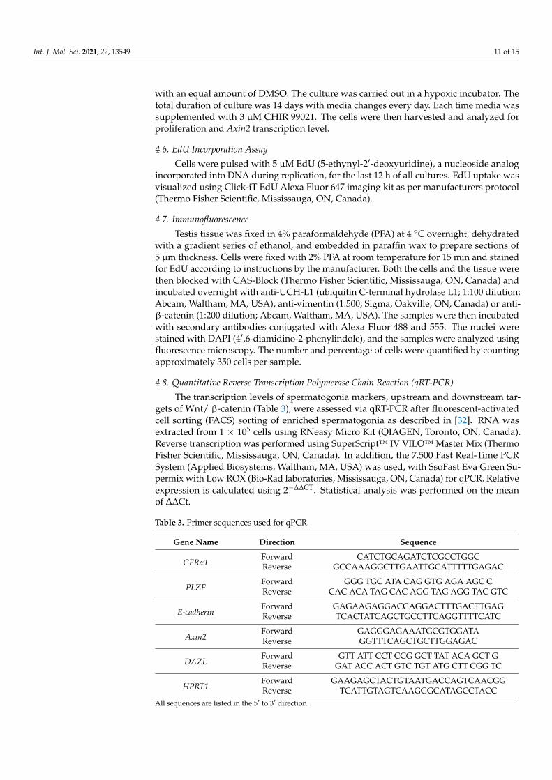
The transcription levels of spermatogonia markers, upstream and downstream tar-gets of Wnt/ β-catenin (Table 3), were assessed via qRT-PCR after fluorescent-activatedcell sorting (FACS) sorting of enriched spermatogonia as described in [32]. RNA wasextracted from 1 × 10⁵ cells using RNeasy Micro Kit (QIAGEN, Toronto, ON, Canada).Reverse transcription was performed using SuperScript™ IV VILO™ Master Mix (ThermoFisher Scientific, Mississauga, ON, Canada). In addition, the 7.500 Fast Real-Time PCRSystem (Applied Biosystems, Waltham, MA, USA) was used, with SsoFast Eva Green Su-permix with Low ROX (Bio-Rad laboratories, Mississauga, ON, Canada) for qPCR. Relativeexpression is calculated using 2−∆∆CT. Statistical analysis was performed on the meanof ∆∆Ct.
Table 3. Primer sequences used for qPCR.
Gene Name Direction Sequence
GFRα1Forward CATCTGCAGATCTCGCCTGGCReverse GCCAAAGGCTTGAATTGCATTTTTGAGAC
PLZFForward GGG TGC ATA CAG GTG AGA AGC CReverse CAC ACA TAG CAC AGG TAG AGG TAC GTC
E-cadherinForward GAGAAGAGGACCAGGACTTTGACTTGAGReverse TCACTATCAGCTGCCTTCAGGTTTTCATC
Axin2Forward GAGGGAGAAATGCGTGGATAReverse GGTTTCAGCTGCTTGGAGAC
DAZLForward GTT ATT CCT CCG GCT TAT ACA GCT GReverse GAT ACC ACT GTC TGT ATG CTT CGG TC
HPRT1Forward GAAGAGCTACTGTAATGACCAGTCAACGGReverse TCATTGTAGTCAAGGGCATAGCCTACC
All sequences are listed in the 5′ to 3′ direction.

Int. J. Mol. Sci. 2021, 22, 13549 12 of 15
4.9. Maximum Shear Stress Calculation
Maximum shear stress in a 10 mL bioreactor was calculated using equations fromGareau et al. 2014 [34]. The required dimensions of 10 mL bioreactors are stated in Table 4.
Table 4. Dimensions of 10 mL stirred suspension bioreactor.
Parameters Dimensions
Media volume 0.00001 m3
Impeller width 0.0054 mImpeller diameter 0.0254 m
Bioreactor diameter 0.032 m
4.10. Statistical Analysis
All data reported are from at least three independent experiments performed withseparately prepared enriched spermatogonia populations from different animals (n = 3).The data were analyzed by unpaired two-tailed t-tests to compare two groups and one-wayANOVA for more than two groups with the Tukey multiple comparisons test. A value ofp < 0.05 was set as the limit of statistical significance. Statistical analyses were performedusing the GraphPad Prism 8 software (San Diego, CA, USA).
Supplementary Materials: The following are available online at https://www.mdpi.com/article/10.3390/ijms222413549/s1.
Author Contributions: Conceptualization, S.S. and I.D.; methodology, S.S., A.V., N.d.L.e.M.L., L.S.and M.U.; software, S.S.; validation, S.S. and I.D.; formal analysis, S.S., M.U. and I.D.; investigation,S.S. and I.D.; resources, M.U. and D.R.; writing—original draft preparation, S.S.; writing—reviewand editing, S.S., A.V., N.d.L.e.M.L., M.U., D.R. and I.D.; visualization, S.S.; supervision, I.D.; projectadministration, I.D.; funding acquisition, I.D. All authors have read and agreed to the publishedversion of the manuscript.
Funding: This work was supported by NIH/NICHD HD091068-01 to I.D.; a graduate studentscholarship to S.S. from the Alberta Children’s Hospital Research Institute (ACHRI); and a CanadianInstitutes of Health Research (CIHR) fellowship (MFE-176542) and an ACHRI postdoctoral fellowshipto N.d.L.e.M.L.
Institutional Review Board Statement: The study was conducted according to the guidelines of theDeclaration of Helsinki and approved by the Animal Care Committee of the University of Calgary(protocol code: AC20-0143, date of approval: 3 December 2021).
Informed Consent Statement: Not applicable.
Data Availability Statement: The data that support the findings of this study are available on requestfrom the corresponding author.
Acknowledgments: We thank Douglas Kondro for the technical assistance provided in setting upthe oxygen spot sensors.
Conflicts of Interest: The authors declare no conflict of interest.
References1. Mulder, R.L.; Font-Gonzalez, A.; Green, D.M.; Loeffen, E.A.H.; Hudson, M.M.; Loonen, J.; Yu, R.; Ginsberg, J.P.; Mitchell, R.T.;
Byrne, J.; et al. Fertility Preservation for Male Patients with Childhood, Adolescent, and Young Adult Cancer: Recommendationsfrom the Pancarelife Consortium and the International Late Effects of Childhood Cancer Guideline Harmonization Group. LancetOncol. 2021, 22, e57–e67. [CrossRef]
2. Phillips, S.M.; Padgett, L.S.; Leisenring, W.M.; Stratton, K.K.; Bishop, K.; Krull, K.R.; Alfano, C.M.; Gibson, T.M.; de Moor, J.S.;Hartigan, D.B.; et al. Survivors of Childhood Cancer in the United States: Prevalence and Burden of Morbidity. Cancer Epidemiol.Biomark. Prev. 2015, 24, 653–663. [CrossRef] [PubMed]
3. Green, D.M.; Kawashima, T.; Stovall, M.; Leisenring, W.; Sklar, C.A.; Mertens, A.C.; Donaldson, S.S.; Byrne, J.; Robison, L.L.Fertility of Male Survivors of Childhood Cancer: A Report from the Childhood Cancer Survivor Study. J. Clin. Oncol. 2010, 28,332–339. [CrossRef]
Int. J. Mol. Sci. 2021, 22, 13549 13 of 15
4. Basaria, S. Male Hypogonadism. Lancet 2014, 383, 1250–1263. [CrossRef]5. Forbes, C.M.; Flannigan, R.; Schlegel, P.N. Spermatogonial Stem Cell Transplantation and Male Infertility: Current Status and
Future Directions. Arab J. Urol. 2018, 16, 171–180. [CrossRef]6. Kanatsu-Shinohara, M.; Ogonuki, N.; Inoue, K.; Miki, H.; Ogura, A.; Toyokuni, S.; Shinohara, T. Long-Term Proliferation in
Culture and Germline Transmission of Mouse Male Germline Stem Cells. Biol. Reprod. 2003, 69, 612–616. [CrossRef]7. Izadyar, F.; den Ouden, K.; Creemers, L.B.; Posthuma, G.; Parvinen, M.; de Rooij, D.G. Proliferation and Differentiation of Bovine
Type a Spermatogonia During Long-Term Culture. Biol. Reprod. 2003, 68, 272–281. [CrossRef] [PubMed]8. Bahadorani, M.; Hosseini, S.M.; Abedi, P.; Abbasi, H.; Nasr-Esfahani, M.H. Glial Cell Line-Derived Neurotrophic Factor
in Combination with Insulin-Like Growth Factor 1 and Basic Fibroblast Growth Factor Promote in Vitro Culture of GoatSpermatogonial Stem Cells. Growth Factors 2015, 33, 181–191. [CrossRef] [PubMed]
9. Cai, H.; Wu, J.Y.; An, X.L.; Zhao, X.X.; Wang, Z.Z.; Tang, B.; Yue, Z.P.; Li, Z.Y.; Zhang, X.M. Enrichment and Culture ofSpermatogonia from Cryopreserved Adult Bovine Testis Tissue. Anim. Reprod. Sci. 2016, 166, 109–115. [CrossRef] [PubMed]
10. Oatley, M.J.; Kaucher, A.V.; Yang, Q.E.; Waqas, M.S.; Oatley, J.M. Conditions for Long-Term Culture of Cattle UndifferentiatedSpermatogonia1. Biol. Reprod. 2016, 95, 1–10. [CrossRef]
11. Nath, S.C.; Harper, L.; Rancourt, D.E. Rancourt. Cell-Based Therapy Manufacturing in Stirred Suspension Bioreactor: Thoughtsfor Cgmp Compliance. Front. Bioeng. Biotechnol. 2020, 8, 599674. [CrossRef]
12. Burrell, K.; Dardari, R.; Goldsmith, T.; Toms, D.; Villagomez, D.A.F.; King, W.A.; Ungrin, M.D.; West, F.; Dobrinski, I. StirredSuspension Bioreactor Culture of Porcine Induced Pluripotent Stem Cells. Stem Cells Dev. 2019, 28, 1264–1275. [CrossRef]
13. Al-Ani, A.; Toms, D.; Kondro, D.; Thundathil, J.; Yu, Y. Oxygenation in Cell Culture: Critical Parameters for Reproducibility AreRoutinely Not Reported. PLoS ONE 2018, 13, e0204269.
14. Wenger, R.H.; Kurtcuoglu, V.; Scholz, C.C.; Marti, H.H.; Hoogewijs, D. Frequently Asked Questions in Hypoxia Research. Hypoxia2015, 3, 35–43. [CrossRef]
15. Badger, J.L.; Byrne, M.L.; Veraitch, F.S.; Mason, C.; Wall, I.B.; Caldwell, M.A. Hypoxic Culture of Human Pluripotent Stem CellLines Is Permissible Using Mouse Embryonic Fibroblasts. Regen. Med. 2012, 7, 675–683. [CrossRef]
16. Hsu, S.H.; Chen, C.T.; Wei, Y.H. Inhibitory Effects of Hypoxia on Metabolic Switch and Osteogenic Differentiation of HumanMesenchymal Stem Cells. Stem Cells 2013, 31, 2779–2788. [CrossRef]
17. Renault, V.M.; Rafalski, V.A.; Morgan, A.A.; Salih, D.A.; Brett, J.O.; Webb, A.E.; Villeda, S.A.; Thekkat, P.U.; Guillerey, C.; Denko,N.C.; et al. Foxo3 Regulates Neural Stem Cell Homeostasis. Cell Stem Cell 2009, 5, 527–539. [CrossRef]
18. Helsel, A.R.; Oatley, M.J.; Oatley, J.M. Glycolysis-Optimized Conditions Enhance Maintenance of Regenerative Integrity in MouseSpermatogonial Stem Cells During Long-Term Culture. Stem Cell Rep. 2017, 8, 1430–1441. [CrossRef]
19. Vodicka, P.; Smetana, K., Jr.; Dvorankova, B.; Emerick, T.; Xu, Y.Z.; Ourednik, J.; Ourednik, V.; Motlik, J. The Miniature Pig as anAnimal Model in Biomedical Research. Ann. N.Y. Acad. Sci. 2005, 1049, 161–171. [CrossRef]
20. Bode, G.; Clausing, P.; Gervais, F.; Loegsted, J.; Luft, J.; Nogues, V.; Sims, J. The Utility of the Minipig as an Animal Model inRegulatory Toxicology. J. Pharmacol. Toxicol. Methods 2010, 62, 196–220. [CrossRef]
21. França, L.R.; Silva, V.A., Jr.; Chiarini-Garcia, H.; Garcia, S.K.; Debeljuk, L. Cell Proliferation and Hormonal Changes DuringPostnatal Development of the Testis in the Pig. Biol. Reprod. 2000, 63, 1629–1636. [CrossRef]
22. Foster, D.L.; Hileman, S.M. Chapter 31-Puberty in the Sheep. In Knobil and Neill’s Physiology of Reproduction, 4th ed.; Plant, T.,Zeleznik, A., Eds.; Academic Press: San Diego, CA, USA, 2015; pp. 1441–1485.
23. Shafa, M.; Day, B.; Yamashita, A.; Meng, G.; Liu, S.; Krawetz, R.; Rancourt, D.E. Derivation of Ipscs in Stirred SuspensionBioreactors. Nat. Methods 2012, 9, 465–466. [CrossRef]
24. Fluri, D.A.; Tonge, P.D.; Song, H.; Baptista, R.P.; Shakiba, N.; Shukla, S.; Clarke, G.; Nagy, A.; Zandstra, W.P. Derivation, Expansionand Differentiation of Induced Pluripotent Stem Cells in Continuous Suspension Cultures. Nat. Methods 2012, 9, 509–516.[CrossRef]
25. Nath, S.C.; Day, B.; Harper, L.; Yee, J. Fluid Shear Stress Promotes Embryonic Stem Cell Pluripotency Via Interplay betweenB-Catenin and Vinculin in Bioreactor Culture. Stem Cells 2021, 39, 1166–1177. [CrossRef]
26. Tang, W.R.; Liu, Y.; Li, L.H.; Wu, Z.B.; He, Y. Fluid Shear Stress and Raloxifene Stimulates the Proliferation of Osteoblast throughRegulating the Expresstion of Beta-Catenin and Estrogen Receptor Alpha. Sichuan Da Xue Xue Bao Yi Xue Ban 2014, 45, 913–918.
27. Li, C.; Zeng, Y.; Hu, J.; Yu, H. Effects of Fluid Shear Stress on Expression of Proto-Oncogenes C-Fos and C-Myc in CulturedHuman Umbilical Vein Endothelial Cells. Clin. Hemorheol. Microcirc. 2002, 26, 117–123.
28. Norvell, S.M.; Alvarez, M.; Bidwell, J.P.; Pavalko, F.M. Fluid Shear Stress Induces Beta-Catenin Signaling in Osteoblasts. Calcif.Tissue Int. 2004, 75, 396–404. [CrossRef]
29. Verma, D.; Ye, N.; Hua, S.Z. Role of Fluid Shear Stress on E-Cadherin Dynamics and Cytoskeletal Stresses. In Proceedings of thePaper Presented at the 2015 41st Annual Northeast Biomedical Engineering Conference (NEBEC), Troy, NY, USA, 17–19 April2015.
30. Takase, H.M.; Nusse, R. Paracrine Wnt/Beta-Catenin Signaling Mediates Proliferation of Undifferentiated Spermatogonia in theAdult Mouse Testis. Proc. Natl. Acad. Sci. USA 2016, 113, E1489–E1497. [CrossRef]
31. Luo, J.; Megee, S.; Rathi, R.; Dobrinski, I. Protein Gene Product 9.5 Is a Spermatogonia-Specific Marker in the Pig Testis:Application to Enrichment and Culture of Porcine Spermatogonia. Mol. Reprod. Dev. 2006, 73, 1531–1540. [CrossRef]
Int. J. Mol. Sci. 2021, 22, 13549 14 of 15
32. Voigt, A.L.; Kondro, D.A.; Powell, D.; Valli-Pulaski, H.; Ungrin, M.; Stukenborg, J.B.; Klein, C.; Lewis, I.A.; Orwig, K.E.; Dobrinski,I. Unique Metabolic Phenotype and Its Transition During Maturation of Juvenile Male Germ Cells. FASEB J. 2021, 35, e21513.[CrossRef]
33. Kehoe, D.E.; Jing, D.; Lock, L.T.; Tzanakakis, E.S. Scalable Stirred-Suspension Bioreactor Culture of Human Pluripotent StemCells. Tissue Eng. Part A 2010, 16, 405–421. [CrossRef]
34. Gareau, T.; Lara, G.G.; Shepherd, R.D.; Krawetz, R.; Rancourt, D.E.; Rinker, K.D.; Kallos, M.S. Shear Stress Influences thePluripotency of Murine Embryonic Stem Cells in Stirred Suspension Bioreactors. J. Tissue Eng. Regen. Med. 2014, 8, 268–278.[CrossRef]
35. Sen, A.; Kallos, M.S.; Behie, L.A. Expansion of Mammalian Neural Stem Cells in Bioreactors: Effect of Power Input and MediumViscosity. Dev. Brain Res. 2002, 134, 103–113. [CrossRef]
36. Cormier, J.T.; Nieden, N.I.z.; Rancourt, D.E.; Kallos, M.S. Expansion of Undifferentiated Murine Embryonic Stem Cells asAggregates in Suspension Culture Bioreactors. Tissue Eng. 2006, 12, 3233–3245. [CrossRef]
37. Canadian Climate Normals 1981–2010 Station Data. Available online: https://climate.weather.gc.ca/climate_normals/results_1981_2010_e.html?searchType=stnProx&txtRadius=25&optProxType=city&selCity=51%7C2%7C114%7C4%7CCalgary&selPark=&txtCentralLatDeg=&txtCentralLatMin=0&txtCentralLatSec=0&txtCentralLongDeg=&txtCentralLongMin=0&txtCentralLongSec=0&txtLatDecDeg=&txtLongDecDeg=&stnID=2205&dispBack=0 (accessed on 3 November 2020).
38. Place, T.L.; Domann, F.E.; Case, A.J. Limitations of Oxygen Delivery to Cells in Culture: An Underappreciated Problem in Basicand Translational Research. Free Radic. Biol. Med. 2017, 113, 311–322. [CrossRef]
39. Kubota, H.; Avarbock, M.R.; Brinster, R.L. Growth Factors Essential for Self-Renewal and Expansion of Mouse SpermatogonialStem Cells. Proc. Natl. Acad. Sci. USA 2004, 101, 16489–16494. [CrossRef]
40. He, Z.; Jiang, J.; Hofmann, M.C.; Dym, M. Gfra1 Silencing in Mouse Spermatogonial Stem Cells Results in Their DifferentiationVia the Inactivation of Ret Tyrosine Kinase. Biol. Reprod. 2007, 77, 723–733. [CrossRef]
41. Tseng, Y.T.; Liao, H.F.; Yu, C.Y.; Mo, C.F.; Lin, S.P. Epigenetic Factors in the Regulation of Prospermatogonia and SpermatogonialStem Cells. Reproduction 2015, 150, R77–R91. [CrossRef]
42. Avvisato, C.L.; Yang, X.; Shah, S.; Hoxter, B.; Li, W.; Gaynor, R.; Pestell, R.; Tozeren, A.; Byers, S.W. Mechanical Force ModulatesGlobal Gene Expression and Beta-Catenin Signaling in Colon Cancer Cells. J. Cell Sci. 2007, 120, 2672–2682. [CrossRef]
43. Cha, B.; Geng, X.; Mahamud, M.R.; Fu, J.; Mukherjee, A.; Kim, Y.; Jho, E.H.; Kim, T.H.; Kahn, M.L.; Xia, L.; et al. Mechanotrans-duction Activates Canonical Wnt/B-Catenin Signaling to Promote Lymphatic Vascular Patterning and the Development ofLymphatic and Lymphovenous Valves. Genes. Dev. 2016, 30, 1454–1469. [CrossRef]
44. Fernández-Sánchez, M.E.; Barbier, S.; Whitehead, J.; Béalle, G.; Michel, A.; Latorre-Ossa, H.; Rey, C.; Fouassier, L.; Claperon, A.;Brullé, L.; et al. Mechanical Induction of the Tumorigenic B-Catenin Pathway by Tumour Growth Pressure. Nature 2015, 523,92–95. [CrossRef]
45. le Duc, Q.; Shi, Q.; Blonk, I.; Sonnenberg, A.; Wang, N.; Leckband, D.; de Rooij, J. Vinculin Potentiates E-Cadherin Mechanosensingand Is Recruited to Actin-Anchored Sites within Adherens Junctions in a Myosin Ii-Dependent Manner. J. Cell Biol. 2010, 189,1107–1115. [CrossRef]
46. Kemler, R. From Cadherins to Catenins: Cytoplasmic Protein Interactions and Regulation of Cell Adhesion. Trends Genet. 1993, 9,317–321. [CrossRef]
47. Tolkunova, E.N.; Malashicheva, A.B.; Chikhirzhina, E.V.; Kostyleva, E.I.; Zeng, W.; Luo, J.; Dobrinskiı̆, I.; Hierholzer, A.; Kemler,R.; Tomilin, A.N. E-Cadherin as a Novel Surface Marker of Spermatogonial Stem Cells. Tsitologiia 2009, 51, 212–218. [CrossRef]
48. Thompson, C.J.; Su, Z. Cadherin Clusters Stabilized by a Combination of Specific and Nonspecific Cis-Interactions. eLife 2020, 9,e59035. [CrossRef]
49. Wu, Z.Q.; Brabletz, T.; Fearon, E.; Willis, A.L.; Hu, C.Y.; Li, X.Y.; Weiss, S.J. Canonical Wnt Suppressor, Axin2, Promotes ColonCarcinoma Oncogenic Activity. Proc. Natl. Acad. Sci. USA 2012, 109, 11312. [CrossRef]
50. Lam, A.Q.; Freedman, B.S.; Morizane, R.; Lerou, P.H.; Valerius, M.T.; Bonventre, J.V. Rapid and Efficient Differentiation of HumanPluripotent Stem Cells into Intermediate Mesoderm That Forms Tubules Expressing Kidney Proximal Tubular Markers. J. Am.Soc. Nephrol. 2014, 25, 1211–1225. [CrossRef]
51. Borys, B.S.; Roberts, E.L.; Le, A.; Kallos, M.S. Scale-up of Embryonic Stem Cell Aggregate Stirred Suspension Bioreactor CultureEnabled by Computational Fluid Dynamics Modeling. Biochem. Eng. J. 2018, 133, 157–167. [CrossRef]
52. Zhang, X.; Li, L.; Bai, Y.; Shi, R.; Wei, H.; Zhang, S. Mouse Undifferentiated Spermatogonial Stem Cells Cultured as Aggregatesunder Simulated Microgravity. Andrologia 2014, 46, 1013–1021. [CrossRef]
53. King, J.A.; Miller, W.M. Bioreactor Development for Stem Cell Expansion and Controlled Differentiation. Curr. Opin. Chem. Biol.2007, 11, 394–398. [CrossRef]
54. Hayashi, Y.; Otsuka, K.; Ebina, M.; Igarashi, K.; Takehara, A.; Matsumoto, M.; Kanai, A. Distinct Requirements for EnergyMetabolism in Mouse Primordial Germ Cells and Their Reprogramming to Embryonic Germ Cells. Proc. Natl. Acad. Sci. USA2017, 114, 8289–8294. [CrossRef]
55. Brinster, R.L.; Harstad, H. Energy Metabolism in Primordial Germ Cells of the Mouse. Exp. Cell Res. 1977, 109, 111–117. [CrossRef]56. Tischler, J.; Gruhn, W.H.; Reid, J.; Allgeyer, E. Metabolic Regulation of Pluripotency and Germ Cell Fate through A-Ketoglutarate.
EMBO J. 2019, 38, e99518. [CrossRef] [PubMed]

Int. J. Mol. Sci. 2021, 22, 13549 15 of 15
57. Verdikt, R.; Allard, P. Metabolo-Epigenetics: The Interplay of Metabolism and Epigenetics During Early Germ Cells Development.Biol. Reprod. 2021, 105, 616–624. [CrossRef]
58. Kanatsu-Shinohara, M.; Tanaka, T.; Ogonuki, N.; Ogura, A.; Morimoto, H.; Cheng, P.F.; Eisenman, R.N.; Trumpp, A.; Shinohara,T. Myc/Mycn-Mediated Glycolysis Enhances Mouse Spermatogonial Stem Cell Self-Renewal. Genes. Dev. 2016, 30, 2637–2648.[CrossRef]
59. Lord, T.; Nixon, B. Metabolic Changes Accompanying Spermatogonial Stem Cell Differentiation. Dev. Cell 2020, 52, 399–411.[CrossRef]
60. Hermann, B.P.; Cheng, K.; Singh, A.; Roa-De La Cruz, L.; Mutoji, K.N.; Chen, I.C.; Gildersleeve, H.; Lehle, J.D.; Mayo, M.;Westernströer, B.; et al. The Mammalian Spermatogenesis Single-Cell Transcriptome, from Spermatogonial Stem Cells toSpermatids. Cell Rep. 2018, 25, 1650–1667. [CrossRef] [PubMed]
61. Voigt, A.L.; Thiageswaran, S.; de Lima e Martins Lara, N.; Dobrinski, I. Metabolic Requirements for Spermatogonial Stem CellEstablishment and Maintenance in vivo and in vitro. Int. J. Mol. Sci. 2021, 22, 1998. [CrossRef]
62. Taiani, J.T.; Krawetz, R.J.; Nieden, N.I.Z.; Wu, Y.E.; Kallos, M.S.; Matyas, J.R.; Rancourt, D.E. Reduced Differentiation Efficiency ofMurine Embryonic Stem Cells in Stirred Suspension Bioreactors. Stem Cells Dev. 2010, 19, 989–998. [CrossRef] [PubMed]
63. Kerr, G.E.; Young, J.C.; Horvay, K.; Abud, H.E.; Loveland, K.L. Regulated Wnt/Beta-Catenin Signaling Sustains Adult Spermato-genesis in Mice. Biol. Reprod. 2014, 90, 1–12. [CrossRef]
64. Chassot, A.A.; Le Rolle, M.; Jourden, M.; Taketo, M.M.; Ghyselinck, N.B.; Chaboissier, M.C. Constitutive Wnt/Ctnnb1 ActivationTriggers Spermatogonial Stem Cell Proliferation and Germ Cell Depletion. Dev. Biol. 2017, 426, 17–27. [CrossRef]
65. O’Flaherty, L.; Shnyder, S.D.; Cooper, P.A.; Cross, S.J.; Wakefield, J.G.; Pardo, O.E.; Seckl, M.J.; Tavaré, J.M. Tumor GrowthSuppression Using a Combination of Taxol-Based Therapy and Gsk3 Inhibition in Non-Small Cell Lung Cancer. PLoS ONE 2019,14, e0214610. [CrossRef] [PubMed]
66. Honaramooz, A.; Snedaker, A.; Boiani, M.; Scholer, H.; Dobrinski, I.; Schlatt, S. Sperm from Neonatal Mammalian Testes Graftedin Mice. Nature 2002, 418, 778–781. [CrossRef] [PubMed]
67. Sakib, S.; Yu, Y.; Voigt, A.; Ungrin, M.; Dobrinski, I. Generation of Porcine Testicular Organoids with Testis Specific ArchitectureUsing Microwell Culture. J. Vis. Exp. 2019, 152, e60387. [CrossRef] [PubMed]
68. Yeh, J.R.; Zhang, X.; Nagano, M.C. Establishment of a Short-Term in Vitro Assay for Mouse Spermatogonial Stem Cells. Biol.Reprod. 2007, 77, 897–904. [CrossRef] [PubMed]