characterization of porcine nlrp3 inflammasome- mediated ...
202
CHARACTERIZATION OF PORCINE NLRP3 INFLAMMASOME- MEDIATED INTERLEUKIN-1 BETA PRODUCTION IN RESPONSE TO INFLUENZA A VIRUSES A Thesis Submitted to the College of Graduate and Postdoctoral Studies in Partial Fulfillment of the Requirements for the Degree of Doctor of Philosophy in the Department of Veterinary Microbiology, Western College of Veterinary Medicine, University of Saskatchewan, Saskatoon, Canada By HONG-SU PARK © Copyright Hong-Su Park, December 2018. All rights reserved.
-
Upload
khangminh22 -
Category
Documents
-
view
0 -
download
0
Transcript of characterization of porcine nlrp3 inflammasome- mediated ...

CHARACTERIZATION OF PORCINE NLRP3 INFLAMMASOME-
MEDIATED INTERLEUKIN-1 BETA PRODUCTION IN RESPONSE TO
INFLUENZA A VIRUSES
A Thesis
Submitted to the College of Graduate and Postdoctoral Studies
in Partial Fulfillment of the Requirements for
the Degree of Doctor of Philosophy
in the
Department of Veterinary Microbiology,
Western College of Veterinary Medicine,
University of Saskatchewan,
Saskatoon,
Canada
By
HONG-SU PARK
© Copyright Hong-Su Park, December 2018. All rights reserved.

i
PERMISSION TO USE
In presenting this thesis in partial fulfillment of the requirements for a postgraduate degree from
the University of Saskatchewan, I agree that the libraries of this university may make it freely
available for inspection. I further agree that permission for copying of this thesis in any manner,
whole or in part, for scholarly purposes may be granted by the professors who supervised my
thesis work or in their absence, the Head of the Department or the Dean of the College in which
my thesis work was done. It is understood that any copying or publication or use of this thesis or
parts thereof for financial gain shall not be allowed without any written permission. It is also
understood that due recognition shall be given to me and to the University of Saskatchewan in
any scholarly use which may be made of any material in my thesis.
Request for permission to copy or to make other use of material in this thesis in whole or part
should be addressed to:
Head of the Department of Veterinary Microbiology,
Western College of Veterinary Medicine,
University of Saskatchewan,
52 Campus Drive,
Saskatoon, SK S7N 5B4
Canada
Or
Dean of the College of Graduate and Postdoctoral Studies,
University of Saskatchewan,
116 Thorvaldson Building, 110 Science Place,
Saskatoon, SK S7N 5C9
Canada
ii
ABSTRACT
Influenza A virus (IAV) causes respiratory infections in humans and animals. Pigs are
infected not only by swine influenza virus (SIV), but also by human and avian influenza viruses.
Pigs serve as the mixing vessel for the generation of novel reassortant viruses with pandemic
potential as evidenced by the emergence of 2009 human pandemic IAV (pdm09). Therefore,
IAV infection in pigs poses both an economic concern and a threat to public health. Virus-host
interplay dictates the outcome of virus infection. The host innate immune system can sense the
virus infection and produce multiple cytokines and chemokines to control inflammation as well
as to mount antiviral responses. On the other hand, the host innate immune response can be
antagonized by IAV-encoded proteins, one being the non-structural protein 1 (NS1). Thus, a
better understanding of the porcine innate immunity to IAV infection and the innate immune
evasion strategies by IAV is required for a rational design to combat IAV infections in pigs.
Innate immune cells in lungs, particularly the alveolar macrophages, are indispensable for
host protection against IAV infection. They produce pro-inflammatory cytokines including
interleukin-1 beta (IL-1β) that recruits other immune cells and promotes phagocytic activities.
IL-1β production is tightly regulated by a cytosolic multiprotein complex called the nucleotide-
binding domain and leucine-rich repeat-containing protein 3 (NLRP3) inflammasome. The
NLRP3 inflammasome is comprised of NLRP3, apoptosis-associated speck-like protein
containing caspase activation and recruitment domain (ASC), and pro-caspase-1. The molecular
mechanism underlying the NLRP3 inflammasome-mediated IL-1β production upon IAV
infection in the swine host, and the mechanism by which IAV NS1 protein counteracts the
porcine IL-1β response remain largely unknown. This PhD project was centered on the
characterization of the NLRP3 inflammasome-mediated IL-1β production in response to IAV
infection in the swine host; specifically, I first characterized the IL-1β production in primary
porcine alveolar macrophages (PAMs) in response to different isolates of IAV. Next, I
investigated the molecular mechanisms by which viral NS1 protein modulates the porcine
NLRP3 inflammasome activity, and finally I examined the molecular pathways that are involved
in inflammasome activation upon IAV infection in PAMs.
This study showed that while various SIV strains could induce NLRP3 inflammasome-
mediated IL-1β production in primary PAMs, the human pdm09 virus induced much less IL-1β
than did SIVs. Subsequent analyses revealed that the NS1 C-terminus of pdm09, but not that of

iii
SIV, was responsible for the significant inhibition of the NLRP3 inflammasome-mediated IL-1β
production. Mechanistically, the NS1 C-terminus of pdm09 disrupted the interaction between
NLRP3 and ASC, a prerequisite for the NLRP3 inflammasome assembly; this led to an impaired
formation of ASC specks, a hallmark of NLRP3 inflammasome activation. NS1 C-terminus of
pdm09 also suppressed the ubiquitination of porcine ASC. Furthermore, two lysine (K) residues,
K110 and K140, on porcine ASC were identified as the ubiquitination acceptor sites; mutation of
these two lysine residues diminished the ubiquitination of ASC and significantly reduced the IL-
1β production in response to the pdm09 virus with the NS1 C-terminal deletion. These results
revealed that the NS1 C-terminus of pdm09 suppresses ASC ubiquitination to support the
immune evasion by the virus.
Further attempts were made to dissect the upstream mechanism of the NLRP3
inflammasome-mediated IL-1β production upon SIV infection, focusing on the mitochondrial
dynamics regulated by dynamin-related protein 1 (DRP1). This study showed that SIV infection
induced not only phosphorylation of DRP1 at serine 579 (S579) that is required for
mitochondrial fission activity of DRP1, but also mitochondrial fission in PAMs. The reactive
oxygen species produced from mitochondrial fission was also related to the IL-1β production.
Phospho-mimetic mutation at S579 on DRP1 could upreguate the NLRP3 inflammasome
activity, leading to an increased IL-1β production. Furthermore, the requirement of the kinase
activity of receptor-interacting protein kinase 1 (RIPK1) for the IL-1β production and the
association of RIPK1 with DRP1 suggested that RIPK1 is an upstream kinase for DRP1
phosphorylation. These results indicated an integral role of the RIPK1/DRP1 signaling axis in
modulating the porcine NLRP3 inflammasome-mediated IL-1β production in SIV-infected
PAMs.
Taken together, this study defined the mechanism by which SIV induces porcine NLRP3
inflammasome-mediated IL-1β production and elucidated the strategies pdm09 employs to
circumvent the host innate immunity. The obtained information will enhance our knowledge of
the innate immunity to IAV infection in the swine host.

iv
ACKNOWLEDGEMENTS
First of all, I would like to express the deepest gratitude to my supervisor, Dr. Yan Zhou,
for her thoughtful guidance and support that enabled me to complete the study. Without her
dedicated mentorship, it would have been much harder for me to get through this journey. I want
to extend my appreciation to the advisory committee members, Dr. Suresh Tikoo, Dr. Joyce
Wilson, and Dr. Alexander Zakhartchouk for their constructive criticism with suggestions, and
the graduate chair, Dr. Janet Hill for keeping me on the right track.
I would like to acknowledge my former and current colleagues, Dr. Hyun-Mi Pyo, Dr. Li
Lin, Dr. Fang Xu, Dr. Sathya N. Thulasi-Raman, Dr. Amit Gaba, Leo Guanqun Liu, Magda
Langton, Fangzheng Wang, Yao Lu, and Shelby Landreth for their assistance and inspiration.
Among others, I give special thanks to Leo for stimulating me not only with the way of doing
science, but also through helpful discussions, and to Sathya for providing valuable resources and
advice. I also feel thankful to the VIDO-InterVac scientists and staff, especially Dr. Qiang Liu,
Donna Dent and Ken Lai, and to the faculty and staff in the Department of Veterinary
Microbiology, Western College of Veterinary Medicine for their kind help and support.
Further, I feel indebted to the funding sources, the Natural Sciences and Engineering
Research Council of Canada (NSERC) grant to Dr. Yan Zhou as well as the Western College of
Veterinary Medicine, University of Saskatchewan that provided scholarship/fellowship to me.
Finally and most importantly, I thank my parents for all their love and support.

v
TABLE OF CONTENTS
PERMISSION TO USE ................................................................................................................. i
ABSTRACT ................................................................................................................................... ii
ACKNOWLEDGEMENTS ........................................................................................................ iv
TABLE OF CONTENTS ............................................................................................................. v
LIST OF FIGURES ..................................................................................................................... ix
LIST OF TABLES ........................................................................................................................ x
LIST OF ABBREVIATIONS ..................................................................................................... xi
CHAPTER 1 LITERATURE REVIEW ................................................................................ 1 1.1 Influenza A virus (IAV) ................................................................................................ 1
1.1.1 Overview ..................................................................................................................... 1 1.1.2 Life cycle of IAV ........................................................................................................ 2 1.1.3 Immunology of IAV infection .................................................................................... 4
1.1.4 Swine influenza virus (SIV)........................................................................................ 7 1.1.5 2009 human pandemic IAV (pdm09) ......................................................................... 9
1.1.6 Non-structural protein 1 (NS1) of IAV ....................................................................... 9 1.1.6.1 NS1 acting on the synthesis of viral proteins .................................................... 10 1.1.6.2 NS1-mediated restriction of the host gene expression ...................................... 10
1.2 Inflammasomes and interleukin-1 beta (IL-1β) ....................................................... 12 1.2.1 Inflammasomes ......................................................................................................... 12
1.2.1.1 Apoptosis-associated speck-like protein containing caspase activation and
recruitment domain (ASC) ................................................................................................ 13
1.2.1.2 Caspase-1 .......................................................................................................... 15 1.2.1.3 Inflammasomes formed by nucleotide-binding oligomerization domain-like
receptors (NLRs) ............................................................................................................... 15
1.2.1.3.1 NLRP3 inflammasome ................................................................................ 16 1.2.1.3.1.1 Mechanisms of NLRP3 inflammasome activation............................... 16
1.2.1.3.1.2 Ubiquitination and NLRP3 inflammasome .......................................... 19 1.2.1.3.1.3 Mitochondria acting on the NLRP3 inflammasome ............................ 20
1.2.1.4 Inflammasomes formed by non-NLRs.............................................................. 22 1.2.2 IL-1β ......................................................................................................................... 22
1.2.2.1 Regulation of IL-1β production by IAV proteins ............................................. 25
1.3 Innate immune cells and sensors ............................................................................... 26 1.3.1 Innate immune cells .................................................................................................. 26
1.3.1.1 Macrophages ..................................................................................................... 27 1.3.1.2 Dendritic cells (DCs) ........................................................................................ 28 1.3.1.3 Neutrophils ........................................................................................................ 28 1.3.1.4 Mast cells .......................................................................................................... 29
1.3.2 Innate immune sensors .............................................................................................. 30
1.3.2.1 Nucleotide-binding oligomerization domain-like receptors (NLRs) ................ 30 1.3.2.1.1 NLRs with pyrin domain (NLRP) ............................................................... 31 1.3.2.1.2 NLRs with caspase activation and recruitment domain (NLRC) ................ 34

vi
1.3.2.1.3 NLR with baculovirus inhibitor of apoptosis protein repeat (NLRB) ........ 35 1.3.2.1.4 NLR with acidic transactivation domain (NLRA) ...................................... 36
1.3.2.2 Toll-like receptors (TLRs) ................................................................................ 38 1.3.2.3 Retinoic acid-inducible gene I-like receptors (RLRs) ...................................... 38
1.3.2.4 Other sensors ..................................................................................................... 39
1.4 Mitochondria and innate immune responses ........................................................... 40 1.4.1 Mitochondrial control of the innate immunity .......................................................... 40 1.4.2 Mitochondrial dynamics modulated by viruses ........................................................ 40
1.5 Innate immune evasion ............................................................................................... 42 1.5.1 Immune evasion strategies by RNA viruses ............................................................. 42 1.5.2 Immune evasion by non-structural proteins of IAV ................................................. 44
1.5.2.1 NS1 protein ....................................................................................................... 44
1.5.2.1.1 NS1 antagonizing the host immune signaling ............................................. 44 1.5.2.1.2 NS1 regulating the host cell death .............................................................. 45
1.5.2.2 PB1-F2 and PA-X ............................................................................................. 46
1.6 Conclusions of the literature review .......................................................................... 46
CHAPTER 2 RATIONALE, HYPOTHESIS AND OBJECTIVE..................................... 48 2.1 Rationale ...................................................................................................................... 48 2.2 Hypothesis and objective ............................................................................................ 48
CHAPTER 3 INFLUENZA A VIRUS INDUCES NLRP3 INFLAMMASOME-
MEDIATED IL-1β PRODUCTION IN PORCINE ALVEOLAR MACROPHAGES. ....... 50
3.1 Abstract ........................................................................................................................ 51 3.2 Introduction ................................................................................................................. 51 3.3 Materials and methods ............................................................................................... 53
3.3.1 Isolation of porcine alveolar macrophages (PAMs) ................................................. 53 3.3.2 Cell lines and viruses ................................................................................................ 54
3.3.3 Generation of mutant viruses .................................................................................... 54 3.3.4 Antibodies and reagents ............................................................................................ 56 3.3.5 Plasmid construction ................................................................................................. 57
3.3.6 Infection and treatment of PAMs .............................................................................. 59 3.3.7 Virus purification ...................................................................................................... 59
3.3.8 UV inactivation of viruses ........................................................................................ 60 3.3.9 RNA interference ...................................................................................................... 60
3.3.10 Quantitative PCR .............................................................................................. 60 3.3.11 Antigen-specific IgG ELISA ............................................................................ 61 3.3.12 Porcine IL-1β ELISA ........................................................................................ 62
3.3.13 NLRP3 inflammasome reconstitution assay ..................................................... 62 3.3.14 Western blotting ................................................................................................ 63 3.3.15 Co-immunoprecipitation (co-IP) ....................................................................... 63 3.3.16 Immunofluorescence and confocal microscopy................................................ 63
3.3.17 Statistical analysis ............................................................................................. 64
3.4 Results .......................................................................................................................... 64 3.4.1 SIV infection induces IL-1β production in PAMs. ................................................... 64 3.4.2 SIV-induced IL-1β production is mediated by TLR3. .............................................. 66 3.4.3 SIV-induced IL-1β production is mediated by NLRP3 inflammasome. .................. 67 3.4.4 2009 human pandemic IAV (pdm09) induces less IL-1β than does the SIV. .......... 69

vii
3.4.5 NS1 C-terminus of pdm09, but not that of SIV, inhibits the NLRP3 inflammasome
activation. .............................................................................................................................. 71 3.4.6 Mutant SIVs with NS1 C-terminus derived from pdm09 inhibits the NLRP3
inflammasome activation. ..................................................................................................... 74
3.4.7 NS1 C-terminus of pdm09 inhibits the interaction between NLRP3 and ASC. ....... 78 3.4.8 NS1 C-terminus of pdm09 inhibits the ASC speck formation. ................................ 80
3.5 Discussion..................................................................................................................... 82
TRANSITION BETWEEN CHAPTER 3 AND CHAPTER 4 ............................................... 85
CHAPTER 4 NS1 C-TERMINUS OF 2009 PANDEMIC INFLUENZA VIRUS
INHIBITS PORCINE NLRP3 INFLAMMASOME BY SUPRESSING ASC
UBIQUITINATION. ................................................................................................................... 86
4.1 Abstract ........................................................................................................................ 87 4.2 Introduction ................................................................................................................. 87 4.3 Materials and methods ............................................................................................... 89
4.3.1 Cells and viruses ....................................................................................................... 89
4.3.2 Antibodies and reagents ............................................................................................ 89 4.3.3 Plasmid construction ................................................................................................. 90
4.3.4 Ubiquitination assay.................................................................................................. 92 4.3.5 NLRP3 inflammasome reconstitution assay ............................................................. 93 4.3.6 Porcine IL-1β ELISA ................................................................................................ 93
4.3.7 Human IL-1β ELISA ................................................................................................ 94 4.3.8 Cycloheximide (CHX) chase assay .......................................................................... 94
4.3.9 Western blotting ........................................................................................................ 94 4.3.10 Immunofluorescence and confocal microscopy................................................ 95
4.3.11 Statistical analysis ............................................................................................. 95
4.4 Results .......................................................................................................................... 95 4.4.1 Porcine ASC is ubiquitinated and NS1 C-terminus of pdm09 inhibits the
ubiquitination. ....................................................................................................................... 95 4.4.2 NS1 C-terminus of pdm09 inhibits the ubiquitination of human ASC. .................... 98
4.4.3 ASC undergoes mixed lysine-linked ubiquitination in response to NS1-mutant
pdm09 virus. ......................................................................................................................... 99
4.4.4 K110 and K140 are ubiquitination sites on porcine ASC. ...................................... 101 4.4.5 Inhibition of ASC ubiquitination on K110 and K140 does not impair the ASC speck
formation or ASC stability. ................................................................................................. 103 4.4.6 Ubiquitination of ASC on K110 and K140 is required for caspase-1 activation in
response to NS1-mutant pdm09 virus. ................................................................................ 105
4.5 Discussion................................................................................................................... 108
TRANSITION BETWEEN CHAPTER 4 AND CHAPTER 5 ............................................. 111
CHAPTER 5 SWINE INFLUENZA VIRUS INDUCES NLRP3 INFLAMMASOME-
MEDIATED INTERLEUKIN-1 BETA PRODUCTION THROUGH THE RIPK1/DRP1
SIGNALING. 112 5.1 Abstract ...................................................................................................................... 113 5.2 Introduction ............................................................................................................... 113 5.3 Materials and methods ............................................................................................. 115
5.3.1 Cells and viruses ..................................................................................................... 115

viii
5.3.2 Antibodies and reagents .......................................................................................... 115 5.3.3 Plasmid construction ............................................................................................... 116 5.3.4 Infection and treatment of PAMs ............................................................................ 117 5.3.5 RNA interference and quantitative PCR ................................................................. 118
5.3.6 NLRP3 inflammasome reconstitution assay ........................................................... 118 5.3.7 Co-IP ....................................................................................................................... 119 5.3.8 Porcine IL-1β ELISA .............................................................................................. 119 5.3.9 Western blotting ...................................................................................................... 120 5.3.10 Confocal Microscopy ...................................................................................... 120
5.3.11 Statistical analysis ........................................................................................... 121
5.4 Results ........................................................................................................................ 121 5.4.1 SIV infection induces dynamin-related protein 1 (DRP1) phosphorylation and
mitochondrial fission. ......................................................................................................... 121 5.4.2 SIV-induced porcine IL-1β production is dependent on reactive oxygen species
(ROS) production. ............................................................................................................... 123
5.4.3 SIV-induced porcine IL-1β production is mediated by DRP1. .............................. 124 5.4.4 Kinase activity of receptor-interacting protein 1 (RIPK1) is critical for SIV-induced
IL-1β production. ................................................................................................................ 127 5.4.5 RIPK1 interacts with DRP1. ................................................................................... 131
5.5 Discussion................................................................................................................... 133
CHAPTER 6 GENERAL DISCUSSION ........................................................................... 136
6.1 General discussion .................................................................................................... 136 6.1.1 Alveolar macrophages (AMs) as the model for the IL-1β signaling research ........ 136 6.1.2 Signal 1 and 2 activation by SIV and pdm09 ......................................................... 137
6.1.3 Interaction of NS1 with its host cellular partners for the inhibition of NLRP3
inflammasome activity ........................................................................................................ 138
6.1.4 Possible engagement of tripartite motif-containing protein 25 (TRIM25) in the IL-1β
signaling .............................................................................................................................. 138 6.1.5 Post-translational modifications in regulating the NLRP3 inflammasome activity 139
6.1.6 Additional events involved in the mitochondrial dynamics ................................... 140 6.1.7 Possible sensors upstream of the NLRP3 inflammasome....................................... 141
6.1.8 The roles of IL-1β in the swine host ....................................................................... 141 6.1.9 Possible reasons for different IL-1β secretion phenotypes of SIV and pdm09 ...... 142
6.2 Future directions ....................................................................................................... 143
REFERENCES .......................................................................................................................... 144
APPENDIX ................................................................................................................................ 178 A. SUPPLEMENTARY TABLES ......................................................................................... 178 B. ACHIEVEMENTS DURING THE STUDY .................................................................... 183
B.1 Publications .................................................................................................................... 183 B.2 Presentations .................................................................................................................. 183 B.3 Scholarships and fellowships ......................................................................................... 184 B.4 Awards ........................................................................................................................... 184

ix
LIST OF FIGURES
Figure 1.1 Life cycle of IAV. .......................................................................................................... 3
Figure 1.2 Innate recognition of IAV infection. ............................................................................. 5 Figure 1.3 Multiple functions of IAV NS1 protein....................................................................... 11 Figure 1.4 Two-signal model for the inflammasome activation. .................................................. 13 Figure 1.5 Mechanisms of the NLRP3 inflammasome activation. ............................................... 18 Figure 1.6 Domain architecture of the NLR family proteins. ....................................................... 31
Figure 3.1 SIV infection induces IL-1β production in PAMs. ..................................................... 66 Figure 3.2 SIV-induced IL-1β production is mediated by TLR3. ................................................ 67 Figure 3.3 SIV-induced IL-1β production is mediated by NLRP3 inflammasome. ..................... 69 Figure 3.4 pdm09 induces less IL-1β than does SIV. ................................................................... 70
Figure 3.5 NS1 C-terminus of pdm09, but not that of SIV, inhibits porcine NLRP3
inflammasome activation. ............................................................................................................. 73 Figure 3.6 Mutant SIVs with NS1 C-terminus derived from pdm09 inhibits the NLRP3
inflammasome. .............................................................................................................................. 77
Figure 3.7 NS1 C-terminus of pdm09 inhibits the interaction between NLRP3 and ASC. ......... 79 Figure 3.8 NS1 C-terminus of pdm09 inhibits the ASC speck formation. ................................... 81 Figure 4.1 Porcine ASC is ubiquitinated and NS1 C-terminus of pdm09 inhibits the
ubiquitination. ............................................................................................................................... 97 Figure 4.2 NS1 C-terminus of pdm09 inhibits the ubiquitination of human ASC. ...................... 99 Figure 4.3 ASC undergoes mixed lysine-linked ubiquitination in response to NS1-mutant pdm09
virus............................................................................................................................................. 100 Figure 4.4 K110 and K140 are ubiquitination sites on porcine ASC. ........................................ 103
Figure 4.5 Inhibition of ASC ubiquitination on K110 and K140 does not impair the ASC speck
formation or ASC stability. ......................................................................................................... 104
Figure 4.6 Ubiquitination of ASC on K110 and K140 is required for caspase-1 activation in
response to NS1-mutant pdm09 virus. ........................................................................................ 106
Figure 4.7 A proposed model of the mechanisms by which NS1 C-terminus of pdm09 inhibits
the porcine NLRP3 inflammasome. ............................................................................................ 107 Figure 5.1 SIV infection induces DRP1 phosphorylation and mitochondrial fission................. 122
Figure 5.2 SIV-induced porcine IL-1β production is dependent on ROS production. ............... 124 Figure 5.3 SIV-induced porcine IL-1β production is mediated by DRP1. ................................. 126
Figure 5.4 Kinase activity of RIPK1 is critical for SIV-induced IL-1β production. .................. 130 Figure 5.5 RIPK1 interacts with DRP1....................................................................................... 131 Figure 5.6 A proposed model of RIPK1/DRP1-mediated IL-1β production in SIV-infected
PAMs. ......................................................................................................................................... 132

x
LIST OF TABLES
Table 1.1 IL-1 family members with their receptors and functions. ............................................ 23
Table 1.2 NLR members with their activators and modes of action. ........................................... 37 Table 3.1 List of WT and mutant viruses used in this study. ........................................................ 56 Table 3.2 List of expression plasmids used in Chapter 3. ............................................................ 58 Table 3.3 List of primers used for quantitative PCR in Chapter 3. .............................................. 61 Table 4.1 List of expression plasmids used in Chapter 4. ............................................................ 91
Table 5.1 List of expression plasmids used in Chapter 5. .......................................................... 117 Table 5.2 List of primers used for quantitative PCR in Chapter 5. ............................................ 118 Supplementary Table 1 List of primers used to generate NS constructs in the pHW2000 vector in
Chapter 3. .................................................................................................................................... 178 Supplementary Table 2 List of primers used to generate expression plasmids in Chapter 3. .... 179 Supplementary Table 3 List of primers used to generate expression plasmids in Chapter 4. .... 180 Supplementary Table 4 List of primers used to generate expression plasmids in Chapter 5. .... 182

xi
LIST OF ABBREVIATIONS
aa amino acid
AIM2 absent in melanoma 2
AM alveolar macrophage
AMP adenosine monophosphate
ANOVA analysis of variance
AP-1 activator protein 1
ASC apoptosis-associated speck-like protein containing caspase activation and
recruitment domain
ATD acidic transactivation domain
ATP adenosine triphosphate
BALF bronchoalveolar lavage fluid
BIR baculovirus inhibitor of apoptosis protein repeat
bp base pairs
BM18 influenza A/Brevig Mission/1/1918/H1N1
BSA bovine serum albumin
CARD caspase activation and recruitment domain
CDK1 cyclin-dependent kinase 1
cGAS cyclic GMP-AMP synthase
CHX cycloheximide
CIITA MHC class II transactivator
CPE cytopathic effect
CPSF30 30 kDa subunit of cleavage and polyadenylation specificity factor
cRNA complementary RNA
DAMP danger-associated molecular pattern
DAPI 4',6-diamidino-2-phenylindole
DC dendritic cell
DDX DEAD-box helicase
DED death effector domain
DENV dengue virus
DHX DEAH-box helicase

xii
DMEM Dulbecco’s modified Eagle’s medium
DMSO dimethyl sulfoxide
DPBS Dulbecco’s phosphate-buffered saline
DRP1 dynamin-related protein 1
dsDNA double-stranded DNA
dsRNA double-stranded RNA
EDTA ethylenediaminetetraacetic acid
eIF2a eukaryotic translation factor 2 subunit alpha
eIF4B eukaryotic translation initiation factor 4B
eIF4GI eukaryotic translation initiation factor 4 gamma 1
ELISA enzyme-linked immunosorbent assay
ER endoplasmic reticulum
FBS fetal bovine serum
FIIND function to find domain
GFP green fluorescent protein
GMP guanosine monophosphate
GTP guanosine triphosphate
GTPase GTP hydrolase
HA hemagglutinin
HCV hepatitis C virus
HEK293T human embryonic kidney 293T
Hf09 influenza A/Halifax/210/2009/H1N1
HIN200 hematopoietic IFN-inducible nuclear antigens with 200 amino acid repeats
hpi hours post-infection
HPRT1 hypoxanthine phosphoribosyltransferase 1
hpt hours post-transfection
hStaufen human homologue of Drosophila melanogaster Staufen
IAV influenza A virus
IFITM3 IFN-induced transmembrane protein 3
IFN interferon
IκB inhibitor of NF-κB

xiii
IKK IκB kinase
IL interleukin
IL-1α IL-1 alpha
IL-1β IL-1 beta
IL-1R IL-1 receptor
IL-1Ra IL-1 receptor antagonist
IP immunoprecipitation
IPS-1 IFN-β promoter stimulator protein 1
IRF IFN regulatory factor
ISG IFN-stimulated gene
JAK Janus kinase
LPS lipopolysaccharide
LRR leucine-rich repeat
LUBAC linear ubiquitin assembly complex
MAPK mitogen-activated protein kinase
MAVS mitochondrial antiviral signaling protein
MDA5 melanoma differentiation-associated protein 5
MDCK Madin-Darby canine kidney
MEM minimum essential medium
Mfn mitofusin
MHC major histocompatibility complex
MNS 3,4-methylenedioxy-β-nitrostyrene
MOI multiplicity of infection
MyD88 myeloid differentiation primary response protein 88
NA neuraminidase
NAC N-acetyl L-cysteine
NACHT neuronal apoptosis inhibitor protein, MHC class II transactivator, plant het gene
product involved in vegetative incompatibility, and telomerase-associated protein
1
NAIP neuronal apoptosis inhibitor protein
Nec-1 Necrostatin-1

xiv
NEK7 NIMA-related kinase 7
NEMO NF-κB essential modulator
NEP nuclear export protein
NF-B nuclear factor kappa-light-chain-enhancer of activated B cells
NIMA never in mitosis gene A
NLR nucleotide-binding oligomerization domain-like receptor
NLRA NLR family ATD-containing protein
NLRB NLR family BIR domain-containing protein
NLRC NLR family CARD-containing protein
NLRP NLR family PYD-containing protein
NLRP3 NLR family PYD-containing protein 3 (or nucleotide-binding domain and
leucine-rich repeat-containing protein 3)
NOD nucleotide-binding oligomerization domain-containing protein
NP nucleoprotein
NS1 non-structural protein 1
OAS 2’-5’-oligoadenylate synthetase
OD optical density
ORF open reading frame
PABPII poly(A)-binding protein II
PACT protein activator of the IFN-induced protein kinase
PAGE polyacrylamide gel electrophoresis
PAM porcine alveolar macrophage
PAMP pathogen-associated molecular pattern
pdm09 2009 human pandemic IAV
PFU plaque-forming unit
PBS phosphate-buffered saline
PI3K phosphatidylinositol-4,5-bisphosphate 3-kinase
PIP3 phosphatidylinositol (3,4,5)-trisphosphate
PKR protein kinase R
PNPP p-nitrophenyl phosphate
PR8 influenza A/Puerto Rico/8/1934/H1N1
xv
PRR pattern recognition receptor
PRRSV porcine reproductive and respiratory syndrome virus
PYD pyrin domain
RIG-I retinoic acid-inducible gene-I
RIPK receptor-interacting protein kinase
RLR RIG-I-like receptor
RNase ribonuclease
ROS reactive oxygen species
RSV respiratory syncytial virus
SD standard deviation
SDS sodium dodecyl sulfate
siRNA small interfering RNA
SIV swine influenza virus
Sk02 influenza A/swine/Saskatchewan/18789/2002/H1N1
ssRNA single-stranded RNA
STAT signal transducer and activator of transcription
STING stimulator of IFN genes
SUMO small ubiquitin-like modifier
Syk spleen tyrosine kinase
TAK1 transforming growth factor beta-activated kinase 1
TANK TRAF family member-associated NF-κB activator
TBK1 TANK-binding kinase 1
TBS Tris-buffered saline
TBST Tris-buffered saline with Tween 20
TIR Toll-interleukin-1 receptor
TLR toll-like receptor
TNF tumor necrosis factor
TOM20 translocase of outer mitochondrial membrane 20
TRAF TNF receptor-associated factor
TRIF TIR-domain-containing adapter-inducing IFN-
TRIM25 tripartite motif-containing protein 25
xvi
TSE Tris-buffered saline with EDTA
Tx98 influenza A/swine/Texas/4199-2/1998/H1N1
TXNIP thioredoxin-interacting protein
UTR untranslated region
vRNA viral RNA
vRNP viral ribonucleoprotein
VSV vesicular stomatitis virus
WNV West Nile virus
WSN33 influenza A/WSN/1933/H1N1
WT wild-type
ZBP1 Z-DNA binding protein 1

1
CHAPTER 1 LITERATURE REVIEW
1.1 Influenza A virus (IAV)
1.1.1 Overview
IAV is a contagious pathogen that causes airway infection in a wide range of mammalian
and avian hosts. IAV is the only species in the genus Alphainfluenzavirus, which is one of the
seven genera in the family Orthomyxoviridae (Ferhadian et al., 2018, Shaw and Palese, 2013).
This enveloped virus contains eight segments of negative-sense, single-stranded RNAs (ssRNAs)
for its genome, which enable antigenic changes by mutation and reassortment events. IAVs are
divided into subtypes based on the surface antigens, hemagglutinin (HA) and neuraminidase
(NA); there are currently 18 HA subtypes (H1 to H18) and 11 NA subtypes (N1 to N11)
(Hutchinson, 2018). IAV has the ability to cause pandemics and is the causative agent of
seasonal flu along with the genus Betainfluenzavirus (Teitzel, 2018), while other genera of
influenza viruses, Gammainfluenzavirus and Deltainfluenzavirus do not pose as significant of a
threat to public health as IAV (Zhou et al., 2018). With rapid evolution and interspecies
transmission, IAV is one of the most important global health threats today. It is responsible for
annual flu epidemic affecting human populations including several million severe cases and
estimated up to 646,000 deaths worldwide (Girard et al., 2005, Iuliano et al., 2018). In Canada, a
death toll caused by IAV infection and its complications is reported to be more than 5,000 per
year (Thommes et al., 2017). The 2009 pandemic H1N1 has affected millions of people with an
estimated fatality of up to 575,000 in the world (Dawood et al., 2012). The emergence of this
novel swine-derived virus through the reassortment of swine influenza virus (SIV) with human
and avian strains has emphasized the importance of research with SIV and immunity to SIV in
pigs, since pigs are the intermediate host to generate new viruses with pandemic capacity
(Nelson and Vincent, 2015). There is possibility of further reassortment among SIVs and human
IAV strains in pigs leading to the production of new strains with more pathogenicity and better
transmissibility among humans. Due to public health concerns, there has been a need for
effective vaccines and therapeutics to cope with the potential pandemic. In parallel, a better
understanding of the swine host immunity to IAV is required, since the outcome of IAV
infection depends on the host immune response against infection.

2
1.1.2 Life cycle of IAV
The IAV genome consists of eight segments of ssRNAs: PB2, PB1 and PA segments
encode the three subunits of RNA polymerase complex, PB2, PB1 and PA proteins, respectively;
HA and NA segments encode the two surface glycoproteins, HA and NA, respectively; NP
segment encodes the nucleoprotein (NP); M segment encodes the matrix protein M1 and
envelope protein M2 that is expressed via alternative splicing; NS segment encodes the non-
structural protein 1 (NS1) and nuclear export protein (NEP), which is also an alternative splicing
product. Each RNA segment is associated with the RNA polymerase complex and encapsidated
by viral NP (Bouvier and Palese, 2008, Shaw and Palese, 2013), forming viral ribonucleoprotein
(vRNP); this is the minimum unit for IAV replication.
As shown in Figure 1.1, IAV attaches to the host cell by receptor binding activity of viral
glycoprotein, HA. The globular head region of this protein binds to the cell surface receptor,
sialic acid (also called N-acetylneuraminic acid), and then, the virus can be endocytosed to be
located in the endosomal compartment (Shaw and Palese, 2013). After host cellular proteases
cleave HA0, which refers to the precursor form of HA, into HA1 and HA2, viral membranes are
fused with endosomal membranes at acidic pH (Hamilton et al., 2012). As a result, the fusion
peptide in the HA2 is exposed and inserted into the endosome, thereby inducing the alignment of
viral and endosomal membranes (Hamilton et al., 2012). Next, uncoating is initiated by the viral
ion channel protein, M2 that lowers the pH inside the virion by transporting protons from the
endosome into the virus (Pinto and Lamb, 2006). Consequently, the vRNP complex for each
gene segment is dissociated from M1 and released into the cytoplasm (Shaw and Palese, 2013).
Transcription and replication of the viral genome take place in the nucleus. Thus, nuclear entry
of vRNP is required and is dependent on the nuclear localization signal on NP and three
polymerase subunits (PB2, PB1 and PA) consisting the vRNP (Martín-Benito and Ortín, 2013).
Viral mRNA synthesis requires 5’ cap structures from host mRNA, which is generated by host
RNA polymerase II. PB2 binds to the cap structure on the 5’ end of host pre-mRNA and PB1
binds to the 5’ and 3’ ends of viral RNA (vRNA), while PA cleaves 10 to 13 nucleotides
downstream of the 5’ cap. Using this cleaved fragment as the primer, viral mRNA synthesis is
catalyzed by PB1 (Martín-Benito and Ortín, 2013, Shaw and Palese, 2013). Synthesized viral
mRNA is transported to the cytoplasm to be translated. Incoming vRNA from the cytoplasm also
serves as the template for the synthesis of complementary RNA (cRNA), which in turn, is

3
required for the generation of new vRNA (Shaw and Palese, 2013). After vRNA is assembled as
vRNP, the vRNP is exported to cytoplasm with the help of M1 and NEP, both of which interact
with a cellular export receptor (Samji, 2009, Shaw and Palese, 2013). Unlike NP, M1 and NEP
that move to the nucleus to associate with newly produced vRNA, HA, NA and M2 are modified
in endoplasmic reticulum (ER) and Golgi apparatus, and are transported to the cell membrane
(Shaw and Palese, 2013). For virion assembly, selective packaging of vRNP is believed to occur
based on the packaging signals on each vRNA (Shaw and Palese, 2013). HA and NA associate
with plasma membrane domains called lipid rafts where budding takes place (Nayak et al.,
2004). Finally, virion release occurs with the destruction of sialic acid by NA (Shaw and Palese,
2013).
Reprinted from Nature 459: 931–939 (Neumann et al., 2009) with permission.
Figure 1.1 Life cycle of IAV.
IAV attaches to the host receptor, sialic acid and is endocytosed. After cleavage of HA, the
fusion between viral membranes and endosomes occurs. Uncoating allows the vRNPs to be
released into cytoplasm, followed by its entry into the nucleus. With vRNAs as the template,
viral mRNAs are transcribed using the 5’ cap snatched from host pre-mRNAs, and cRNAs are
synthesized for the vRNA replication. Viral proteins are produced and modified, and newly

4
assembled vRNPs are packaged for virion assembly. Upon budding, the sialic acid destruction by
NA allows the virions to be released.
1.1.3 Immunology of IAV infection
In IAV-infected animals, the epithelial cells in the airway mucosa are the first to be
infected (Pulendran and Maddur, 2014). IAV infection then further spreads to the lung
macrophages and dendritic cells (DCs) in alveoli or within the lung epithelium, which are the
pivotal cells to trigger inflammation (Iwasaki and Pillai, 2014, Short et al., 2012). The innate
immunity to IAV begins with the recognition of viral genome via host immune sensors in the
infected cells (Figure 1.2). The main sensors to detect IAV infection includes toll-like receptors
(TLRs) and retinoic acid-inducible gene-I (RIG-I) (Ichinohe, 2010). Viral ssRNA or double-
stranded RNA (dsRNA) is recognized by TLR3 or TLR7 in the endosome, and the 5’-
triphosphate dsRNA is recognized by RIG-I both in the cytosol and nucleus (Iwasaki and Pillai,
2014, Liu et al., 2018). Activation of both of sensors turns on a cascade of signaling events
leading to the activation of transcription factors, nuclear factor kappa-light-chain-enhancer of
activated B cells (NF-B), interferon (IFN) regulatory factors 3 (IRF3) and 7 (IRF7). This results
in the production of pro-inflammatory cytokines, type I IFNs and a variety of IFN-stimulated
genes (ISGs) that support the host antiviral status (Iwasaki and Pillai, 2014). Viral RNA of IAV
can be sensed by another cytosolic sensor called the nucleotide-binding domain and leucine-rich
repeat-containing protein 3 (NLRP3) (Allen et al., 2009, Thomas et al., 2009), but whether
NLRP3 can sense viral RNA by a direct binding is still under debate. Recent evidence suggests
that NLRP3 recognizes diverse cellular changes induced by viral infection as reviewed in the
section 1.2.1.3.1. Among pro-inflammatory cytokines, interleukin (IL)-1β and IL-18 are
synthesized as precursors via NF-B pathway; then, the precursors become biologically active
after processed by inflammasome complexes consisted of nucleotide-binding oligomerization
domain-like receptors (NLRs) or non-NLRs along with their adaptor and effector molecules
(Iwasaki and Pillai, 2014). Recently, Z-DNA binding protein 1 (ZBP1) is proposed as another
important sensor; the recognition of IAV proteins by ZBP1 triggers NLRP3 inflammasome
activation and cell deaths (Kuriakose et al., 2016).
Among innate immune cells, the lung macrophages are crucial in eliminating IAV particles
and controlling inflammation, which largely depends on the action of pro-inflammatory

5
cytokines including IL-1β, IL-6 and tumor necrosis factor (TNF)-. These cytokines can be
involved in the inflammatory responses as well as the pathogenesis during the course of IAV
infection (Van Reeth, 2000). IAV-induced pathological signs in the lung can be derived from the
combination of direct effects of viral replication and the effects of host responses via
inflammatory cytokines produced from immune cells including alveolar macrophages (Gordon
and Read, 2002).
Reprinted from Trends Immunol. 32(1):34-41 (Pang and Iwasaki, 2011) with permission.
Figure 1.2 Innate recognition of IAV infection. Viral ssRNA is sensed by TLR7 in the endosome (i) or viral 5’-triphosphate RNA is sensed by
RIG-I (ii). TLR7 signaling mediated its adaptor protein, myeloid differentiation primary
response protein 88 (MyD88) activates IRF7 and NF-κB, whereas RIG-I signals mediated by its
adaptor, IFN-β promoter stimulator protein 1 (IPS-1, also called mitochondrial antiviral signaling
protein [MAVS]) on the mitochondria activates IRF3 and NF-κB. IRF7 and IRF3 translocate to
the nucleus, thereby inducing the type I IFN, while NF-κB induces the expression of pro-
inflammatory cytokines including pro-IL-1β and pro-IL-18. IAV infection also induces the

6
NLRP3 inflammasome activation (iii), leading to pro-caspase-1 cleavage that is critical for the
conversion of pro-IL-1β and pro-IL-18 into their mature forms.
DCs are critical in linking innate immunity to adaptive immunity in response to viral
infection. Upon antigen uptake in IAV-infected lungs, respiratory DCs mature and migrate to the
mediastinal lymph node where they present processed antigens to naïve T cells (Ho et al., 2011,
Ingulli et al., 1997). T cells are classified into two subgroups, CD4+ and CD8
+ T cells. After
antigen recognition, they possess effector functions to support antibody production and to
eliminate infected cells. IAV-specific CD4+ T cells can migrate to the lung. They produce
cytokines like TNF-, IL-2 and IFN-γ to induce cytotoxicity by CD8+ T cells, and to activate
alveolar macrophages. They also secrete IL-4 and IL-13 to activate B cells (Kreijtz et al., 2011,
Zens and Farber, 2014). After the infection is resolved, some of the CD4+ T cells acquire
memory and can clear IAV particles more effectively in case of reinfection. Memory CD4+ T
cells can secrete more cytokines in a more rapid manner than naïve CD4+ T cells, particularly for
IFN-γ (Lai et al., 2011). Memory CD4+ T cell-mediated protection against further IAV infection
is executed independently of B cells or CD8+ T cells; and this protection is cross-reactive to
different strains (Richards et al., 2010, Zens and Farber, 2014).
With signals provided by DCs, CD8+ T cells proliferate in IAV-infected lungs (Lawrence
and Braciale, 2004). CD8+ T cells also secrete cytokines including IFN-γ, but their main function
is to eliminate IAV-infected epithelial cells through cytotoxicity (Topham et al., 1997). When
recognizing antigens presented by major histocompatibility complex (MHC) class I, CD8+ T
cells induce apoptosis of target cells by releasing their enzymes (pore-forming proteins and
granzymes), or by upregulating the TNF family members (such as Fas ligand) that bind to the
target cell ligand, Fas to initiate the apoptotic signaling (Hufford et al., 2014). Following
recovery from infection, some portions of CD8+ T cells also remain as long-lived memory CD8
+
T cells in preparation for re-infection (Hufford et al., 2014, Kreijtz et al., 2011).
For humoral immunity, the surface glycoproteins HA and NA are the major targets of
antibody production. Naïve B cells recognize these antigens provided by DCs in the mediastinal
lymph nodes and the respiratory mucosa-associated lymphoid tissues, thereby becoming
antibody-secreting cells (Chiu et al., 2014). Alternatively, some B cells can capture antigens by
themselves in the lymph nodes and at the site of infection (Waffarn and Baumgarth, 2011).
While antibody secretion by B cells can occur independently of T cells, it is known that CD4+ T

7
cell-mediated B cell responses are critical for more effective antibody production against IAV
infection (Mozdzanowska et al., 2005). HA-specific antibodies can neutralize IAV, and NA-
binding antibodies interfere with the enzymatic activity of NA, rather than directly neutralizing
(Kreijtz et al., 2011). Binding of antibodies targeting both HA and NA to infected cells can lead
to antibody-dependent cell-mediated cytotoxicity executed by natural killer cells (Kreijtz et al.,
2011). IgG and IgM are the predominant antibody classes produced in the lung and in particular,
IgG is thought to be responsible for preventing systemic dissemination of IAV; IgA secreted
from the respiratory mucosa inhibits the neutralized antigens from adhering to the epithelium
(Chiu et al., 2014). After acquiring memory, B cells maintain the ability to generate both IgG and
IgA, which also serves for cross-protection against other IAV strains (Waffarn and Baumgarth,
2011).
1.1.4 Swine influenza virus (SIV)
Influenza infection in pigs causes a mild or subclinical disease in the respiratory tract.
Symptoms of the acute illness include fever, anorexia, lethargy, nasal discharge and coughing
(Van Reeth et al., 2012). These symptoms are similarly observed upon infection by different
viral subtypes, and major difference in virulence is not reported by infection with different
strains (Van Reeth et al., 2012). While high morbidity is seen, mortality is generally low and
affected animals can recover in 5 to 7 days. However, secondary infection with other respiratory
viruses or bacteria can exacerbate the condition (Van Reeth et al., 2012). SIV transmission
occurs through direct contact or by aerosols. Epithelial cells on the airway are the main sites for
viral replication, while viruses preferentially target the lower respiratory tract (Van Reeth et al.,
2012). The disease outcome can be affected by inflammatory cytokines such as IL-1, IL-6 and
TNF-α along with IFN-α. Although they have antiviral activities, high levels of these cytokines
are responsible for lung inflammation (Van Reeth et al., 2012, Van Reeth et al., 1998, Van Reeth
et al., 2002). Bronchopneumonia is prominent in infected pigs and microscopic lesions include
necrotizing bronchitis; bronchiolitis; airway obstruction by necrotic epithelial cells and
inflammatory cells such as neutrophils (Janke, 2014, Van Reeth et al., 2012).
Although swine influenza does not often lead to fatal outcomes, it has been a concern of
economic loss in the swine production industry (Schultz-Cherry et al., 2013). Among different
influenza species, influenza A, C and D viruses are isolated from pigs (Su et al., 2017). However,

8
only IAV is considered as a critical pathogen for swine influenza (Van Reeth et al., 2012). The
first case of swine influenza was reported during the pandemic in 1918, while SIV was first
isolated in the United States in 1930 (Lorusso et al., 2013). As a result of infection of pigs with
the 1918 pandemic strain, classical swine H1N1 was established (Zell et al., 2013). Since then,
this lineage of SIV has stably circulated in North America for decades. In the late 1990s, two
different genotypes of H3N2 subtype emerged, and one of them, a triple reassortant derived from
avian, swine and human seasonal H3N2 strains, has spread throughout the United States. While
this virus was endemic in pigs, its reassortment with the classical swine H1N1 also occurred,
generating novel variant SIVs of H1N1 and H1N2 subtypes (Brockwell-Staats et al., 2009,
Lorusso et al., 2013). As evidenced by the 2009 pandemic H1N1, the reassortment of IAVs from
different hosts in pigs highlights the important roles played by pigs for the emergence of new
viruses. The contribution of pigs to the generation of new viruses can be presented by the mixing
vessel theory (Scholtissek et al., 1985) with several observations supporting this idea. Pigs are
permissive to avian and human isolates (Kida et al., 1994), and reassortment among viruses of
different origins has been displayed for isolates from pigs (Peiris et al., 2001). Another important
factor is the receptor binding activity of HA, one of the critical determinants in the host
restriction of IAVs. The binding specificity of HA depends on the types of host cell receptors
differentially distributed in host animals. The molecular structures of this receptor, sialic acid
linked to galactose, are divided into two types: avian-type alpha 2,3-linkage and human-type
alpha 2,6-linkage that are preferably recognized by avian and human isolates, respectively (Zell
et al., 2013). Swine isolates recognize both structures and pigs express both types of sialic acids
(Ito et al., 1998), rendering them susceptible to infection by both avian and human strains. Later,
human cells were also found to harbor both types of these receptors (Shinya et al., 2006),
showing that the receptor binding activity alone may not fully support this theory. An additional
model is such that the adaptation of avian strains in swine hosts, as avian-like SIVs, through
repeated replication in pigs, is able to generate variants that can recognize the human-type sialic
acid (Ito et al., 1998). Overall, further reassortment among IAV strains in swine hosts may lead
to the generation of new strains with pandemic capacity.

9
1.1.5 2009 human pandemic IAV (pdm09)
The pdm09 emerged as a pandemic through the reassortment of IAVs of different origins
in the intermediate hosts, pigs. The genome segments of pdm09 are derived from multiple
lineages: PB2 and PA from avian strain of North American lineage; PB1 from human seasonal
H3N2 strain; HA, NP and NS from classical swine H1N1 of North American lineage; NA and M
from avian-like swine H1N1 of Eurasian lineage (Neumann et al., 2009). This virus is thus,
antigenically close to swine H1N1, while distinct from human seasonal strains (Garten et al.,
2009). The initial emergence of this H1N1 subtype virus in Mexico was quickly followed by the
worldwide spread through human-to-human transmission, leading to the first IAV pandemic in
the 21st century. However, it is suggested that the introduction and circulation of the original
gene segments in pigs took place years before the pdm09 outbreak in humans (Girard et al.,
2010, Vijaykrishna et al., 2010). In humans, the pdm09 generally induced higher rates of
infection in the younger population rather than the old; this may be attributable to the possession
of cross-reactive antibodies initially raised against the 1918 pandemic-like viruses in the older
generation (Girard et al., 2010, Tscherne and García-Sastre, 2011). Genetic signatures for human
adaptation or increased pathogenicity characteristic for highly pathogenic strains were not found
in the pdm09 strains. For example, the pdm09 PB2 protein harbours glutamic acid at the 627th
amino acid (aa), common for avian strains, instead of lysine that is typical for human strains.
While PB1-F2 protein and C-terminal region of NS1 protein encoded by 1918 pandemic IAV
strains are involved in pathogenicity, the PB1 and NS segments of pdm09 encode a truncated
version of PB1-F2 and NS1 proteins, respectively, compared to those of 1918 pandemic strains
(Garten et al., 2009, Tscherne and García-Sastre, 2011). Although the pdm09 infection elicited
symptoms that are comparable to seasonal IAVs in humans (Tscherne and García-Sastre, 2011),
it may acquire virulence by further reassortment, and this potential threat underscores the
importance of ongoing surveillance in pigs.
1.1.6 Non-structural protein 1 (NS1) of IAV
NS1 protein is encoded by the NS segment of IAV and is between 202 and 237 aa in
length. NS1 exerts extensive functions such as supporting viral replication and counteracting the
host antiviral responses (Figure 1.3), although many of these functions are virus strain- or host-
dependent (Ayllon and García-Sastre, 2014). It has two functional domains: an N-terminal RNA-

10
binding domain that can bind several species of RNA such as dsRNA and mRNA in a sequence-
independent manner; and a C-terminal effector domain that interacts with host cellular proteins
and stabilizes the RNA-binding domain (Hale et al., 2008). NS1 has one or two nuclear
localization signals that are required for the nuclear import of NS1 (Hale et al., 2008). In infected
cells, NS1 mainly localizes to the nucleus, while it is also found in cytoplasm indicating that NS1
functions in both compartments (Hale et al., 2008, Krug, 2015).
1.1.6.1 NS1 acting on the synthesis of viral proteins
Upon IAV infection, viral mRNAs are preferentially translated over cellular mRNAs
(Hale et al., 2008). NS1 is one of many proteins that binds the 5’ untranslated region (UTR) of
viral mRNAs for selective translation (Garfinkel and Katze, 1993); NS1 promotes the translation
of viral mRNAs, but not that of non-viral mRNAs (de la Luna et al., 1995). While NS1 does not
impact viral mRNA transcription, it enhances viral mRNA translation that is dependent on the 5’
UTR of mRNAs (Enami et al., 1994). NS1 can bind proteins involved in translation such as the
eukaryotic translation initiation factor 4 gamma 1 (eIF4GI) (Aragón et al., 2000), and it is
suggested that NS1 recruits eIF4GI to the 5’ UTR of viral mRNAs (Hale et al., 2008). NS1 also
binds human homologue of Drosophila melanogaster Staufen (hStaufen), a dsRNA-binding
protein involved in the transport of viral mRNA to polyribosomes where multiple ribosomes
form a complex for active translation (Falcón et al., 1999). Protein kinase R (PKR) is activated
either by the recognition of viral dsRNA or via a cellular protein, PACT (which refers to ‘protein
activator of the IFN-induced protein kinase’) (Li et al., 2006). PKR activation induces the
phosphorylation of eukaryotic translation factor 2 subunit alpha (eIF2α), thereby suppressing the
viral RNA and protein synthesis in infected cells (Hatada et al., 1999). However, binding of NS1
to PKR can subvert either dsRNA- or PACT-mediated PKR activation (Li et al., 2006, Min et al.,
2007).
1.1.6.2 NS1-mediated restriction of the host gene expression
NS1 inhibits the transportation of polyadenylated mRNAs from the nucleus to the
cytoplasm and this action is exerted by binding of NS1 to the poly(A) tails of cellular mRNAs
(Hale et al., 2008, Qiu and Krug, 1994). The effector domain of NS1 binds to the 30 kDa subunit
of cleavage and polyadenylation specificity factor (CPSF30) and poly(A)-binding protein II

11
(PABPII) (Chen et al., 1999, Twu et al., 2006). CPSF30 recognizes the AAUAAA sequence
upstream of the cleavage site for polyadenylation of cellular mRNAs (Das et al., 2008). NS1
binding to CPSF30 or sequestration of CPSF30 by NS1 prevents cleavage and polyadenylation
of cellular mRNAs, while viral mRNAs are polyadenylated by viral polymerases and not
affected by this inhibition (Hale et al., 2008). NS1 binding to PABPII blocks the nuclear export
of mature cellular mRNAs that could avoid the inhibition step at the polyadenylation level (Chen
et al., 1999). NS1 also blocks the export of cellular mRNAs from the nucleus to the cytoplasm,
by interacting with cellular proteins involved in nuclear export machinery (Satterly et al., 2007).
Reprinted from J Gen Virol. 89(10):2359-2376 (Hale et al., 2008) with permission.
Figure 1.3 Multiple functions of IAV NS1 protein.
(a) NS1 limits IFN induction by binding to viral RNA and RIG-I. (b) NS1 limits the antiviral
status established by the PKR and 2’-5’-oligoadenylate synthetase (OAS)/ribonuclease (RNase)
L pathways. (c) NS1 restricts the maturation and nuclear export of cellular mRNAs. (d) NS1

12
promotes the translation of viral mRNA. (e) NS1 limits apoptosis by activating the
phosphatidylinositol-4,5-bisphosphate 3-kinase (PI3K)/Akt pathway.
1.2 Inflammasomes and interleukin-1 beta (IL-1β)
1.2.1 Inflammasomes
Inflammasomes are cytosolic multiprotein complexes that govern inflammation by
regulating the pro-inflammatory cytokines, IL-1β and IL-18. Different immune sensors (either
NLRs or non-NLRs) can assemble into the inflammasomes with apoptosis-associated speck-like
protein containing caspase activation and recruitment domain (ASC) and inflammatory caspases
in response to diverse stimuli. Upon this assembly, the inflammatory caspases are modified by
proteolytic cleavage for their enzymatic activity that is required to process pro-IL-1β and pro-IL-
18 (Pétrilli and Martinon, 2011). Inflammasomes consisting of caspase-1 are considered as
canonical, while non-canonical inflammasomes involve caspase-4 and -5 (or caspase-11 in
mouse) (Viganò et al., 2015). The key steps in inflammasome activation is represented by a two-
signal model (Figure 1.4): signal 1 for the enhanced transcription of the inflammasome-forming
immune sensors and substrates (pro-IL-1β or pro-IL-18) by NF-κB activation; signal 2 for the
recruitment of adaptor and effector proteins for inflammasome assembly (Mariathasan and
Monack, 2007). By finding that dysregulation of inflammasomes is related to auto-inflammatory
disorders and metabolic diseases (Guo et al., 2015), the role of inflammasomes is considered to
be the maintenance of homeostasis as well as the host defense against pathogen-associated
molecular patterns (PAMPs) and danger-associated molecular patterns (DAMPs).

13
Reprinted from Antiviral Res. 148:32-42 (Sarvestani and McAuley, 2017) with permission.
Figure 1.4 Two-signal model for the inflammasome activation. A) Signal 1: Upon endocytosis of PAMPs or phagocytosis of DAMPs, TLR3 and TLR7
signaling involving their adaptors are activated. Upon sensing RNA, RIG-I and melanoma
differentiation-associated protein 5 (MDA5) signaling involving their adaptor is activated as
well. These activate the NF-κB pathway, which upregulates the expression of inflammasome
receptor (NLR or non-NLR) and pro-IL-1β or pro-IL-18. B) Signal 2: The inflammasome
receptor undergoes a conformational change upon sensing stimuli, leading to the assembly of
inflammasome complex with ASC and pro-caspase-1. Upon auto-proteolytic cleavage of pro-
caspase-1, active caspase-1 converts pro-IL-1β or pro-IL-18 into IL-1β or IL-18.
1.2.1.1 Apoptosis-associated speck-like protein containing caspase activation and
recruitment domain (ASC)
ASC is a component of inflammasomes. It was initially discovered as a pro-apoptotic
protein that forms speck-like aggregates in apoptotic cells (Masumoto et al., 1999). It is
constitutively expressed in many myeloid cells, and functions as a bridge between immune
sensors (mostly NLRs) and the effector molecule, pro-caspase-1 (Hoss et al., 2017). ASC has
two domains: N-terminal pyrin domain (PYD) that interacts with another PYD of NLRs and C-
terminal caspase activation and recruitment domain (CARD) that interacts with CARD of pro-
caspase-1. Upon activation, NLRs oligomerize and recruit ASC molecules, which also
oligomerize and form long filaments. Diffusely distributed ASC throughout the cytoplasm and
nucleus cluster to a spot where NLRs form a seed. This leads to the formation of a perinuclear
speck of 1 μm in diameter per each cell (Hoss et al., 2017, Stutz et al., 2013). While ASC PYD
alone can form filaments, spherical speck formation requires ASC CARD that connects between

14
filaments (Dick et al., 2016). Moreover, the interaction between ASC CARDs provides an
additional seed for the filament formation by pro-caspase-1 CARD (Dick et al., 2016, Schmidt et
al., 2016). Conclusively, ASC specks are where pro-caspase-1 recruitment and activation occur
(Franklin et al., 2018), therefore, they are a hallmark of inflammasome activation. Interestingly,
ASC specks can be secreted outside the cells where pro-caspase-1 recruitment and IL-1β
maturation can also occur. The extracellular specks can disseminate inflammatory signals to
phagocytes that then engulf them (Baroja-Mazo et al., 2014, Franklin et al., 2014).
Post-translational modifications on ASC play important roles in the regulation of
inflammasome activity. Absent in melanoma 2 (AIM2) inflammasome activation upon double-
stranded DNA (dsDNA) is restricted by means of the ubiquitination of ASC, which is subject to
degradation via autophagy (Shi et al., 2012). NLRP3 inflammasome activity can be inhibited by
an autophagy protein that upregulates the activity of an E3 ligase, TNF receptor-associated factor
(TRAF) 6, which ubiquitinates ASC (Chiu et al., 2016). Linear ubiquitination of ASC is essential
for the activation of NLRP3 inflammasome (Rodgers et al., 2014), whereas deubiquitination of
ASC is required for the same event (Lee et al., 2017c). Ubiquitination of ASC by an E3 ligase,
TRAF3 upon infection with vesicular stomatitis virus (VSV) is critical for inflammasome-
mediated IL-1β production (Guan et al., 2015). Phosphorylation of ASC also exerts controls on
the inflammasome activity. ASC speck formation is dependent on c-Jun N-terminal kinase and
spleen tyrosine kinase (Syk), and upon inflammasome activation ASC is phosphorylated by
those kinases (Hara et al., 2013). This is supported by another finding that phosphorylation of
ASC by Syk is required for ASC oligomerization and NLRP3 inflammasome activity (Lin et al.,
2015). ASC is also phosphorylated by another kinase, proline-rich tyrosine kinase 2 that interacts
and colocalizes with ASC specks to activate the NLRP3 inflammasome (Chung et al., 2016).
Spatial regulation of ASC by inhibitor of κB kinases (IKKs) is also suggested. IKKα
negatively regulates the NLRP3 inflammasome by phosphorylating serine residues on ASC,
which is bound to nuclear IKKα in the resting state, whereas upon lipopolysaccharide (LPS)
treatment, ASC is released from IKKα. Moreover, IKKε, by phosphorylating a different serine of
ASC, promotes the translocation of ASC from the nucleus to the cytosol for NLRP3
inflammasome activation (Martin et al., 2014).

15
1.2.1.2 Caspase-1
Caspase-1 is a component of canonical inflammasomes. Caspases (refers to cysteine-
aspartic proteases) are a family of enzymes, most of which are involved in programmed cell
deaths or inflammation. Caspases are expressed as a precursor, pro-caspase, which generally
consists of the N-terminal pro-domain, either CARD or death effector domain (DED), the large
subunit (p20) and the small subunit (p10). The proteolytic cleavage between pro-domain and p20
as well as between p20 and p10 subunit leads to the formation of active caspase (Sollberger et
al., 2014, Winkler and Rösen-Wolff, 2015). Caspases are classified into either apoptotic caspases
or inflammatory caspases. Apoptotic caspase are further divided into initiator caspases (caspase-
2, -8, -9, and -10) and executioner caspases (caspase-3, -6, and -7). Initiator caspases, through
either CARD (for caspase-2 and -9) or DED (for caspase-8 and -10), are recruited to adaptor
proteins that react to cell death signals such as cytochrome c, TNF-α or Fas ligand. Executioner
caspases, which lack the pro-domain, are cleaved by macromolecular complexes containing
initiator caspases, and perform apoptosis by processing their target molecules (Shalini et al.,
2015, Sollberger et al., 2014). Inflammatory caspases (caspase-1, -4, -5, -11 and -12) are pivotal
in the regulation of inflammatory responses, and caspase-1, the prototype in this group, is
integral to the cleavage of pro-IL-1β and pro-IL-18. As with other caspases, caspase-1 is
expressed as pro-caspase-1, which is enzymatically inactive. Through its N-terminal CARD, pro-
caspase-1 interacts with another CARD of ASC, which is the adaptor protein of NLR or non-
NLR proteins. The recruitment of pro-caspase-1 to ASC is essential for the inflammasome
formation and subsequently, the auto-proteolysis of pro-caspase-1. Active caspase-1 is a tetramer
composed of two molecules each of p20 and p10 (Shalini et al., 2015, Sollberger et al., 2014). In
addition to the cytokine regulation, active caspase-1 may contribute to NF-κB activation,
independent of its enzyme activity (Lamkanfi et al., 2004). Caspase-1 also executes an
inflammatory cell death called pyroptosis by cleaving the downstream effector, gasdermin D
(Shi et al., 2015).
1.2.1.3 Inflammasomes formed by nucleotide-binding oligomerization domain-
like receptors (NLRs)
NLRs are cytosolic PAMP/DAMP sensors. Among them, NLRP1 is the first identified
inflammasome-forming innate sensor. Its assembly with ASC, caspase-1 and caspase-5 was
16
found to be required for IL-1β production in LPS-stimulated human monocytes (Martinon et al.,
2002). Later, a two-step mechanism of NLRP1 inflammasome activation was suggested by
showing that the priming and oligomerization of NLRP1 is induced by muramyl dipeptide and
adenosine triphosphate (ATP), respectively (Faustin et al., 2007). Another NLR member, NLRP3
was shown to form a complex with ASC and caspase-1 for IL-1β production, and more IL-1β
secretion was observed from cells with the mutant NLRP3 (Agostini et al., 2004), indicating the
involvement of inflammasome activation in abnormal inflammation. A two-signal model for
NLRP3 inflammasome activation was also proposed: priming signal (signal 1) by TLR agonists
and activation signal (signal 2) by ATP (Netea et al., 2009). A wide variety of PAMPs, DAMPs
or cell stress have been displayed to activate the NLRP3 inflammasome. While NLRP7 can
negatively regulate IL-1β production (Kinoshita et al., 2005), activation of NLRP7
inflammasome can restrict intracellular bacteria (Khare et al., 2012). NLRP12 inflammasome-
mediated IL-18 production can provide host resistance against Yersinia pestis (Vladimer et al.,
2012). Unlike other inflammasome-forming receptors, two NLRs, the NLR family CARD-
containing protein 4 (NLRC4) and the neuronal apoptosis inhibitor protein (NAIP) form the
inflammasome together upon sensing of bacterial ligands by NAIP (Yang et al., 2013, Zhao et
al., 2011). NLRC4 inflammasome assembly, possibly with NAIP, is required for IFN-γ
production from CD8+ T cells in response to bacterial infection (Kupz et al., 2012).
1.2.1.3.1 NLRP3 inflammasome
Among inflammasome-forming NLRs, NLRP3 has been most widely investigated. A
variety of stimuli have been known to induce the formation and activation of NLRP3
inflammasome leading to secretion of IL-1β or IL-18. Importantly, NLRP3 inflammasome-
mediated IL-1β production confers protection in IAV-infected mice (Allen et al., 2009, Thomas
et al., 2009).
1.2.1.3.1.1 Mechanisms of NLRP3 inflammasome activation
Upon recognition of ligands by pattern recognition receptors (PRRs), i.e. TLRs or
RLRs, signal transduction is facilitated through multiple adaptor proteins of the PRRs, leading to
the activation of the NF-κB pathway. As a result, transcription of NLRP3 and pro-IL-1β is
initiated (Kattah et al., 2017) and this priming step is called “signal 1” (Figure 1.4). Adaptor

17
proteins of TLRs such as MyD88 and TIR-domain-containing adapter-inducing IFN- (TRIF)
are known to play roles on the transcriptional and post-transcriptional regulation of NLRP3 (He
et al., 2016a). Priming by some stimuli may cause NLRP3 activation (He et al., 2016a), while
priming alone can trigger IL-1β secretion in human monocytes (Netea et al., 2009). Diverse
stimuli provide “signal 2” that is needed for NLRP3 inflammasome assembly (Figure 1.4).
Unlike NLRP3, ASC is constitutively expressed in many types of myeloid cells (Hoss et al.,
2017). After NLRP3 senses stimuli, it oligomerizes and its N-terminal PYD interacts with N-
terminal PYD of ASC. This interaction is followed by aggregation between the two, forming a
supramolecular structure termed a speck. Multiple pro-caspase-1 molecules are recruited to
speck-formed ASC through CARD-CARD interactions between the ASC and pro-caspase-1
(Hoss et al., 2017). Auto-cleavage of pro-caspase-1 generates two subunits of active caspase-1,
p20 and p10. Two sets of a heterodimer of p20 and p10 form a homodimer that can cleave pro-
IL-1β (Walker et al., 1994). Since caspase-1 is an inflammatory caspase, NLRP3 inflammasome
activation is implicated in inflammatory cell death (Fink and Cookson, 2006, Shi et al., 2015).
Different mechanisms have been suggested to elucidate the signal 2 required for
the NLRP3 inflammasome activation (Figure 1.5). An early study demonstrated that IL-1β
secretion is promoted by ATP or nigericin that induce the depletion of intracellular K+
(Perregaux and Gabel, 1994). This is further supported by studies showing that K+ efflux can be
a common stimulus to induce the NLRP3 inflammasome activation (Franchi et al., 2014, Muñoz-
Planillo et al., 2013, Pétrilli et al., 2007). Although K+ efflux-mediated NLRP3 inflammasome
activation can occur independent of Ca2+
influx (Katsnelson et al., 2015), the importance of
calcium sensing mechanisms has been demonstrated. Extracellular Ca2+
or elevated intracellular
Ca2+
sensed by calcium sensing receptors activates the NLRP3 inflammasome (Lee et al., 2012,
Rossol et al., 2012). In addition to the Ca2+
influx, Ca2+
release from ER due to phagolysosomal
rupture can augment the NLRP3 inflammasome activation (Murakami et al., 2012). In fact, the
contribution of lysosomal disruption was displayed earlier. Phagocytosis of crystals induces the
leakage of lysosomal contents such as cathepsin B from damaged lysosomes, thereby activating
the NLRP3 inflammasome (Duewell et al., 2010, Hornung et al., 2008). Reactive oxygen species
(ROS) has been proposed as another common trigger of the NLRP3 inflammasome activation.
Cytosolic ROS generated after phagocytosis of particulates activates the NLRP3 inflammasome
(Dostert et al., 2008). Subsequent reports revealed that NLRP3 inflammasome requires

18
mitochondrial ROS for its activation (Nakahira et al., 2011, Zhou et al., 2011). Furthermore, the
release of mitochondrial DNA can cause NLRP3 inflammasome activation (Nakahira et al.,
2011, Shimada et al., 2012).
Reprinted from Nat Med. 21(7):677-87 (Guo et al., 2015) with permission.
Figure 1.5 Mechanisms of the NLRP3 inflammasome activation. A stimulus (e.g. LPS sensed by TLR4) activates the NF-κB pathway, promoting the mRNA
expression of NLRP3 and IL-1β. This priming step is followed by post-translational
modifications such as deubiquitination of NLRP3 and ubiquitination/phosphorylation of ASC.
NLRP3 inflammasome activation requires signal 2 that include the relocalization of NLRP3 to
the mitochondria, the release of mitochondrial ROS/DNA or cardiolipin, ion fluxes (e.g. K+
efflux) and cathepsin release from disturbed lysosomes. For NLRP3 inflammasome assembly,
NLRP3 nucleates ASC into filaments through PYD-PYD interaction forming the ASC speck.
Pro-caspase-1 is recruited to ASC through CARD-CARD interaction, leading to the auto-
proteolytic cleavage of pro-caspase-1. Active subunits of caspase-1 (p20 and p10) cleave
precursor forms of IL-1β or IL-18. Domain structures of NLRP3 inflammasome components are
shown in the inset.

19
1.2.1.3.1.2 Ubiquitination and NLRP3 inflammasome
Ubiquitination is a post-translational modification process by which ubiquitin
proteins are covalently conjugated to substrate proteins. Ubiquitin is an 8.6-kDa protein, which is
highly conserved among eukaryotes (Zuin et al., 2014). Its linkage to substrates was initially
known to be required for proteasomal degradation of the substrate proteins; however it has been
further identified to play crucial roles in a variety of cell signaling pathways. Ubiquitination is a
three-step process mediated by three enzymes: E1 activating enzymes, E2 conjugating enzymes
and E3 ligases. In addition, ubiquitins can be recycled after being removed from target proteins
by deubiquitinases (Kattah et al., 2017). The substrate selectivity is generally determined by E3
ligases that link the C-terminal glycine of an ubiquitin molecule to the ɛ-amino group of a lysine
(K) residue on the substrate. Monoubiquitination occurs by binding of a single ubiquitin to each
lysine in the substrate; subsequent linkages of more ubiquitins to lysine residues located on the
first ubiquitin leads to different types of polyubiquitination (Davis and Gack, 2015). Additional
ubiquitin molecules can be linked on a lysine residue among seven lysines (K6, K11, K27, K29,
K33, K48 and K63) or on the first methionine (M1) residue, eventually leading to different
consequences. M1-linked ubiquitination, also called linear ubiquitination is mediated by an E3
ligase complex, a linear ubiquitin assembly complex (LUBAC), and is involved in signal
transduction such as the NF-κB activation (Davis and Gack, 2015). K48-linked ubiquitination
can target the substrates for proteasomal degradation, while K63-linked ubiquitination leads to
signal transduction by affecting the stability or localization of target proteins; other linkages are
involved in rather diverse activities including cell development, signaling regulation, autophagy
or protein degradation (Davis and Gack, 2015, Kattah et al., 2017). In some cases, unanchored
ubiquitin chains that are not linked to the substrate can participate in signaling events (Kattah et
al., 2017).
Given that protein modifications by ubiquitins occur for a variety of cellular
functions, it is of no surprise that ubiquitination and its related enzymes play pivotal roles in
immune modulation. Ubiquitination and deubiquitination have been described to regulate the
inflammasome activity (Bednash and Mallampalli, 2016). Deubiquitinases are required for
inflammasome-dependent IL-1β production (Lopez-Castejon et al., 2013) and more specifically,
deubiquitination of NLRP3 is reported to be important for the activation of the NLRP3

20
inflammasome (Guo et al., 2016, Han et al., 2015, Juliana et al., 2012, Kawashima et al., 2017,
Py et al., 2013, Yen et al., 2015). On the contrary, autophagy-mediated or proteasomal
degradation of ubiquitinated NLRP3 leads to suppression of the NLRP3 inflammasome (Song et
al., 2016, Yan et al., 2015). Upon activation of several different inflammasomes, caspase-1 can
be ubiquitinated, which may downregulate the inflammasome activity (Van Opdenbosch et al.,
2014). A recent report shows that ubiquitination-mediated degradation of caspase-1 is prevented
by Zika virus NS1 protein leading to NLRP3 inflammasome activation (Zheng et al., 2018). Pro-
IL-1β also undergoes ubiquitination, which leads to either its maturation into IL-1β or its
degradation (Ainscough et al., 2014, Duong et al., 2015, Harris et al., 2011). Interestingly, upon
human papillomavirus infection, ubiquitination of pro-IL-1β followed by the proteasomal
degradation is mediated by a viral oncoprotein that hijacks the cellular E3 ligase (Niebler et al.,
2013). ASC ubiquitination was initially reported in a study of AIM2 inflammasome-related
autophagy in response to dsDNA; for the inhibition of AIM2 inflammasome-mediated IL-1β,
ubiquitinated ASC is delivered to an autophagic adaptor, p62 and subject to autophagy for
degradation (Shi et al., 2012). Similarly, NLRP3 inflammasome activity is restricted by p62 that
interacts with an E3 ligase, TRAF6, which ubiquitinates ASC (Chiu et al., 2016). Linear
ubiquitination of ASC is essential for the activation of NLRP3 inflammasome (Rodgers et al.,
2014), whereas a deubiquitinase, ubiquitin-specific peptidase 50 interacts with ASC and
deubiquitinates the ASC for NLRP3 inflammasome activation (Lee et al., 2017c). Notably,
ubiquitination of ASC by an E3 ligase, TRAF3 upon infection with VSV is critical for
inflammasome-mediated IL-1β production (Guan et al., 2015).
1.2.1.3.1.3 Mitochondria acting on the NLRP3 inflammasome
Mitochondria-mediated pathophysiological changes positively or negatively
regulate the NLRP3 inflammasome. Mitochondria that generate ROS are readily eliminated by
mitophagy, whereas inhibition of this event causes the ROS accumulation and the loss of
mitochondrial membrane potential. The correlation between autophagy and ROS-mediated
NLRP3 inflammasome activation is demonstrated. While autophagy proteins can inhibit the
NLRP3 inflammasome, depletion of them enhances caspase-1 activity by damaging
mitochondrial homeostasis (Nakahira et al., 2011). Another study shows that upon agonist
treatment, NLRP3 can localize to the ER-mitochondria that enables NLRP3 to sense ROS (Zhou

21
et al., 2011). ROS sensing mechanism by NLRP3 is proposed by the same group. In the resting
state, thioredoxin-interacting protein (TXNIP) is bound to the antioxidant molecule, thioredoxin.
Inflammasome activators let the TXNIP dissociate from thioredoxin upon ROS production; in
turn, TXNIP binds to NLRP3 to activate the NLRP3 inflammasome (Zhou et al., 2010).
However, NLRP3 inflammasome activation can be triggered independent of ROS, as shown by
an NLRP3 agonist that induces mitochondrial dysfunction, but does not induce ROS (Iyer et al.,
2013). Mitochondrial DNA released into cytosol activates the NLRP3 inflammasome as well
(Nakahira et al., 2011). Another study explains this phenomenon in relation to mitochondrial
membrane potential, which is a proton gradient in the inner mitochondrial membrane. NLRP3
agonists can dissipate the mitochondrial membrane potential during apoptosis; this leads to the
release of oxidized mitochondrial DNA that binds to NLRP3 to activate the NLRP3
inflammasome (Shimada et al., 2012). However, it is contradicted by another finding showing
that the maintenance of the intact mitochondrial membrane potential is required for the
inflammasome activation upon infection with RNA viruses, including IAV. Mitochondrial
uncoupling protein 2, responsible for transporting anions from the inner to the outer
mitochondrial membrane, reduces the mitochondrial membrane potential, thereby suppressing
the NLRP3 inflammasome activity (Ichinohe et al., 2013).
Mitochondrial components are also implicated in the regulation of the NLRP3
inflammasome. Several isoforms of voltage-dependent anion channels that are outer
mitochondrial proteins are required for NLRP3 inflammasome activation (Zhou et al., 2011),
whereas a mitochondrial protease, HtrA serine peptidase 2 restricts the activation (Rodrigue-
Gervais et al., 2018). Cardiolipin, an inner mitochondrial membrane phospholipid has been
demonstrated to interact with NLRP3 and activate the NLRP3 inflammasome (Iyer et al., 2013).
Interestingly, cytochrome c released from the inner mitochondrial membrane during apoptosis
inhibits the cardiolipin-NLRP3 interaction and NLRP3 inflammasome activity (Shi and Kehrl,
2016). An outer mitochondrial membrane protein, mitofusin (Mfn) 2 interacts with NLRP3 upon
RNA virus infection and activates the NLRP3 inflammasome activation (Ichinohe et al., 2013).
Inactive NLRP3 is cytosolic, but upon activation, NLRP3 is recruited to mitochondria by
MAVS; this translocation and NLRP3 oligomerization are important for the NLRP3
inflammasome activation (Park et al., 2013b, Subramanian et al., 2013). In response to cytosolic
dsRNA, MAVS can activate the NLRP3 inflammasome by inducing membrane permeabilization

22
(Franchi et al., 2014).
1.2.1.4 Inflammasomes formed by non-NLRs
Non-NLR proteins can also form inflammasomes. The best example is a protein called
AIM2 that belongs to an IFN-inducible protein class. Unlike NLRs, it consists of the N-terminal
PYD and the C-terminal domain, HIN200 (that refers to ‘hematopoietic IFN-inducible nuclear
antigens with 200 amino acid repeats’). Upon sensing of cytosolic dsDNA or DNA virus by
HIN200 domain, the AIM2 inflammasome is activated through assembly with ASC and pro-
caspase-1 (Fernandes-Alnemri et al., 2009, Hornung et al., 2009). Initially, the AIM2
inflammasome was known to be triggered by cytosolic dsDNA, but not inducers of other
inflammasomes (Rathinam et al., 2010). However, further findings suggest that bacterial or
fungal pathogens can activate both AIM2 and NLRP3 inflammasomes (Karki et al., 2015, Park
et al., 2014, Wu et al., 2010). In spite of different structures, AIM2 and NLRP3 are commonly
involved in some signaling pathways (Sagulenko et al., 2013). Recent studies have demonstrated
the activation of AIM2 inflammasome upon RNA virus infection. Induction of AIM2
inflammasome upon enterovirus 71 limits viral replication (Yogarajah et al., 2017), whereas
activation of AIM2 inflammasome is implicated in IAV-induced mortality (Zhang et al., 2017a).
RIG-I is an inflammasome-forming RLR. Independent of NLRP3, it forms the inflammasome by
binding to ASC to regulate IL-1β production upon RNA virus infection, while MDA5 only
upregulates NLRP3 (Poeck et al., 2010). Its role to activate NLRP3 inflammasome via
interaction with ASC and caspase-1 upon IAV infection is also described (Pothlichet et al.,
2013). Pyrin is not a classical member of NLR, but it also assembles into the inflammasome that
is analogous to NLRP3 inflammasome, while not affecting the NLRP3 inflammasome (Yu et al.,
2006). Critical roles of pyrin inflammasome on IL-1β production upon bacterial infection have
been demonstrated (Gavrilin et al., 2009, Xu et al., 2014).
1.2.2 IL-1β
The IL-1 superfamily is a group of IL-1-related proteins involved in pro- or anti-
inflammatory responses. This superfamily is divided into three subfamilies, IL-1, IL-18 and IL-
36 (Table 1.1). IL-1β belongs to the IL-1 subfamily along with IL-1α, IL-33 and IL-1 receptor
(IL-1R) antagonist (IL-1Ra) (Dinarello, 2018). IL-1 cytokines (IL-1α and IL-1β) have specific

23
receptors including IL-1R1 and IL-1R2, and a co-receptor, IL-1R3. IL-1β binds either IL-1R1 to
induce pro-inflammatory action or a decoy receptor, IL-1R2 to block the IL-1 signaling. For pro-
inflammatory responses, IL-1β binding to IL-1R1 induces a structural change of IL-1R1 that
further allows the binding of the co-receptor, IL-1R3 to IL-1R1. As a result, their adaptor
protein, MyD88 is recruited, which then turns on pro-inflammatory signals via the NF-κB
pathway (Dinarello, 2018).
Table 1.1 IL-1 family members with their receptors and functions.
Subfamily Member Receptor Function
IL-1
IL-1α IL-1R1 Pro-inflammatory
IL-1β IL-1R1 Pro-inflammatory
IL-1R2 Anti-inflammatory
IL-1Ra IL-1R1 Anti-inflammatory
IL-33 IL-1R4 Pro-inflammatory
IL-18
IL-18 IL-1R5 Pro-inflammatory
IL-37 IL-1R5 Anti-inflammatory
IL-36
IL-36α, IL-36β, IL-36γ IL-1R6 Pro-inflammatory
IL-36Ra IL-1R6 Anti-inflammatory
IL-38 IL-1R6 Anti-inflammatory
Adapted from Immunol Rev. 281(1):8-27 (Dinarello, 2018) with permission.
IL-1β is primarily produced by immune cells of the myeloid lineage such as monocytes,
macrophages and DCs, and its expression is induced during disease conditions. However, IL-1α
is constitutively produced from epithelial/endothelial cells in the case of sterile inflammation,
which is induced by non-microbial subjects or cell injury (Dinarello, 2018, Garlanda et al.,
2013). IL-1β is expressed as an inactive form and needs to be processed for biological activity;
IL-1α is active either as its precursor or mature form. Transcription of IL-1β is initiated by pro-

24
inflammatory stimuli (including microbial ligands), inflammatory cytokines (such as TNF or IL-
1 itself) or physiological changes in cells (Allan et al., 2005, Fenton, 1992). Upon activation of
downstream signaling cascades, transcription factors bind the IL-1β gene promoter and the DNA
regulatory region, which consists of multiple elements, for example, activator protein 1 (AP-1)
and NF-κB binding sites, and the LPS response enhancer (Watkins et al., 1999). While the 5’ cap
and poly (A) tail prevent mRNA degradation, the stability of IL-1β mRNA can be enhanced by
granulocyte-macrophage colony-stimulating factor, TNF or LPS (Watkins et al., 1999). Since IL-
1β mRNA does not have a signal sequence that is required to be transported to ER-bound
ribosomes, it is transported to free ribosomes for translation (Watkins et al., 1999). Post-
translational processing of pro-IL-1β is necessary for its maturation into IL-1β. The proteolytic
cleavage of pro-IL-1β is conducted by caspase-1, whose activation largely depends on the signal
sensing by inflammasome-forming PRRs, although proteases like elastase, cathepsin G and
chymase derived from neutrophils and mast cells can also cleave pro-IL-1β (Afonina et al.,
2015). Different models for the IL-1β secretion pathway are proposed. While other inflammatory
cytokines are secreted through a conventional pathway, i.e. transportation through the ER-Golgi
route, IL-1β is not secreted in that way due to not having a signal peptide (Lopez-Castejon and
Brough, 2011). The unconventional secretion pathways suggested are as follows: IL-1β can be
localized to lysosomes that are exocytosed; IL-1β can be released with microvesicles that detach
from plasma membranes or released with other small vesicles called exosomes that surround IL-
1β; IL-1β can be released upon cell lysis (Eder, 2009, Lopez-Castejon and Brough, 2011).
IL-1β exerts a variety of functions on the immune system. Its action on vascular
endothelial cells upregulates adhesion molecules and increases the vascular permeability to help
leukocytes adhere to blood vessel walls (Dinarello, 2009). Macrophages stimulated with IL-1β
produce cyclooxygenase-2, which is responsible for generating prostaglandins and leukotrienes
that regulate the constriction and dilation of blood vessels (Tizard, 2009b). These changes are
required for the recruitment and infiltration of inflammatory cells to the site of infection.
Secretion of IL-1β itself along with other pro-inflammatory cytokines like TNF-, IL-6 and IL-8
by monocytes and macrophages can be induced by IL-1β (Dinarello, 1991). In addition, IL-1β
promotes phagocytosis and antigen presentation by macrophages and DCs. IL-1β also stimulates
liver cells to produce acute-phase proteins that support inflammatory responses and pathogen
destruction (Tizard, 2009b). Pro-inflammatory responses lead by IL-1β contribute to causing

25
critical signs of inflammation such as fever, redness and swelling (Tizard, 2009b). IL-1β, in
synergy with other interleukins, enhances the differentiation of IL-17-producing helper T cells,
which are pro-inflammatory (Acosta-Rodriguez et al., 2007, Sutton et al., 2006). IL-1β can
promote B cell- and T cell-mediated antibody production as well (Dinarello, 2009, Nakae et al.,
2001).
1.2.2.1 Regulation of IL-1β production by IAV proteins
While viral RNA itself has the ability to activate the NLRP3 inflammasome (Allen et
al., 2009, Thomas et al., 2009), different viral proteins of IAVs have been reported to regulate
the NLRP3 inflammasome-mediated IL-1β production. An early study demonstrated that the
NS1 protein of the mouse-adapted strain, influenza A/Puerto Rico/08/1934/H1N1 (PR8) inhibits
caspase-1 activation and IL-1β production in human macrophages (Stasakova et al., 2005). NS1
of the 1918 pandemic strain, by binding to RIG-I, can inhibit RIG-I-mediated IL-1β production,
leading to increased virulence in ferrets (Pothlichet et al., 2013). Furthermore, NS1 inhibits
NLRP3 inflammasome-mediated IL-1β production by downregulating pro-IL-1β transcription
and by interacting with NLRP3 (Cheong et al., 2015). More recently, the residues critical for
NS1 protein’s RNA-binding and tripartite motif-containing protein 25 (TRIM25)-binding
activities, R38/K41 and E96/E97, respectively, are found to be required for NS1 to inhibit the
NLRP3 inflammasome activity (Moriyama et al., 2016). IAV M2 protein exerts an ion channel
activity required for virion uncoating (Pinto et al., 1992). Perturbation of cellular ionic
concentrations mediated by transmembrane region of this protein contributes to the NLRP3
inflammasome-mediated IL-1β production (Ichinohe et al., 2010). PB1-F2 is a non-structural
protein, which is a virulence factor (Zamarin et al., 2006); studies have been performed since the
effects of PB1-F2 on IL-1β production from different animals were evaluated (Meunier and von
Messling, 2012). By challenging mice with PB1-F2 peptides, the roles of PB1-F2 to activate the
NLRP3 inflammasome and recruit leukocytes were identified; this function was mapped to the
C-terminus of PB1-F2 from pathogenic strains including PR8 (McAuley et al., 2013). A further
study by the same group showed that PB1-F2 from PR8 and avian H7N9 strains can cause
NLRP3 inflammasome-mediated IL-1β production by inducing the mitochondrial ROS (Pinar et
al., 2017). In contrast, PB1-F2 that targets the mitochondria functions to inhibit the NLRP3
26
inflammasome by blocking the recruitment of NLRP3 to the mitochondria (Yoshizumi et al.,
2014).
1.3 Innate immune cells and sensors
The innate immune system is the first line of host defense against pathogens and
endogenous/exogenous danger signals. It is important for the effective clearance of pathogens
and the healing process, and also for the onset of adaptive immunity. Inflammation is one of the
first and the most critical responses in the innate immune system. Inflammation is intended to
protect the host by eliminating the pathogens/danger signals and restoring the damaged tissues,
however it has to be terminated timely to prevent prolonged inflammation as it can bring about
both positive and negative consequences for the host health. Due to its double-edged sword-like
roles, the tight control of inflammation is essential for the resolution of infection. Pathogens have
genomes or structures that are distinguished from the normal host components. Their structural
motifs or characteristic signatures of nucleic acids are called pathogen-associated molecular
patterns (PAMPs). Sensing the PAMPs or uncommon physiological changes in and around the
host cells by pattern recognition receptors (PRRs) triggers the innate immune signaling required
for inflammation (Iwasaki and Pillai, 2014). Signals from activated PRRs are transduced to the
adaptor molecules and transcription factors, so that the pro-inflammatory cytokines/chemokines,
such as IFNs and ISGs are secreted and/or expressed. Subsequent events include a set of
reactions to recruit other immune cells, eliminate the pathogens, process the infected or dead
cells, and repair damaged tissues (Iwasaki and Pillai, 2014). Innate immune cells are specialized
for these functions, although non-immune cells also express PRRs and mediate the immune
signaling (Takeuchi and Akira, 2010). Successful innate immunity is accomplished through
interplay between immune cells and non-immune cells. Some types of innate immune cells are
responsible for antigen processing and presentation playing as a bridge between innate and
adaptive immunity (Vyas et al., 2008).
1.3.1 Innate immune cells
Immune cells are leukocytes that are either from myeloid or lymphoid progenitors derived
from multipotent hematopoietic stem cells in the bone marrow. Based on the shapes, leukocytes
can be classified into two populations: mononuclear leukocytes and polymorphonuclear

27
granulocytes (neutrophils, eosinophils, basophils and mast cells) (Tizard, 2009d). Mononuclear
leukocytes are further divided into two subpopulations: (phagocytic) monocytes and (non-
phagocytic) lymphocytes (Tizard, 2009d). Monocytes are further differentiated into macrophages
or DCs. Among others, macrophages, DCs, neutrophils and mast cells are the representative
members in the innate immune system by playing crucial roles such as phagocytosis, production
of immune mediators and antigen presentation (Janeway and Medzhitov, 2002). These functions
lead to the protection against pathogens and prevention of injury by danger signals.
1.3.1.1 Macrophages
Phagocytosis is an important event in innate immunity, which is played by phagocytic
leukocytes such as macrophages, DCs and neutrophils. While some macrophages are derived
from their progenitors in yolk sac or fetal liver, most of the macrophages responding to microbial
infection are derived from stem cells in the bone marrow (Gordon and Plüddemann, 2017).
Monocytes developed from these stem cells circulate in the blood. The maturation of monocytes
into macrophages occurs as they enter into tissues, where differentiated macrophages are
distributed to exert their roles (Tizard, 2009b). At the site of infection, neutrophils are the first to
show up to engulf pathogens, which is then followed by the appearance of macrophages, which
can phagocytose more effectively than neutrophils. Macrophages are specialized in phagocytosis
and the clearance of pathogens or dead neutrophils, but their functions are not restricted to those.
They are also responsible for the immune surveillance to trigger inflammation as well as anti-
inflammatory actions required for tissue repair and healing. These two-faceted functions are
possible with the presence of two different populations of macrophages: M1 cells for pro-
inflammatory responses and M2 cells for anti-inflammatory roles (Tizard, 2009c).
In the lungs, different types of macrophages including alveolar macrophages (AMs),
interstitial macrophages and exudate macrophages reside (Nicol and Dutia, 2014). Among them,
AMs are one of the first cells to respond to respiratory pathogens (Tumpey et al., 2005). These
cells are located in the alveolar space formed by the epithelium consisting of type I and II
pneumocytes, which are responsible for the maintenance of the space and the secretion of
surfactant substances, respectively (Short et al., 2014). AMs are predominant immune cells in
this space (Duan et al., 2017), and are also involved in homeostasis of the surfactant proteins
(Halstead and Chroneos, 2015). Upon IAV infection, AMs produce pro-inflammatory cytokines

28
including IL-1β, IL-6 and TNF-α that mediate inflammation and recruit other immune cells; they
produce type I IFNs that limit the spread of IAV. In addition, AMs are critical for activating the
T cell response as well as antibody response upon IAV infection (He et al., 2017, Wijburg et al.,
1997); more pathogenicity is observed in different animals that are deficient in AMs (Cardani et
al., 2017, Kim et al., 2013, Kim et al., 2008, Purnama et al., 2014, Schneider et al., 2014). IAV
infection can deplete AMs by inducing their death, which may increase the susceptibility to
secondary bacterial infection (Ghoneim et al., 2013), while AM depletion upon co-infection with
IAV and bacteria can be prevented by IL-1 signaling (Bansal et al., 2018). Furthermore, infection
of type I pneumocytes by IAV can be inhibited by AMs (Cardani et al., 2017).
1.3.1.2 Dendritic cells (DCs)
DCs are critical for linking the innate immunity to adaptive immunity by functioning in
both antigen processing and antigen presentation. Two subpopulations of DCs exist: myeloid
DCs that are derived from blood monocytes, and plasmacytoid DCs that are derived from
lymphoid precursors (Tizard, 2009a). Myeloid DCs are distributed in tissues and are responsible
for immune surveillance; plasmacytoid DCs are located in lymphoid organs, and are responsible
for type I IFN production and T cell activation. After produced from bone marrow progenitors,
DCs exist as immature cells (Tizard, 2009a). Immature DCs are activated upon recognition of
antigens via their PRRs or in response to pro-inflammatory cytokines; this activation enhances
their recruitment to the site of infection and promotes phagocytosis (Tizard, 2009a). Their
maturation by encountering antigens also leads to the migration to lymph nodes in which they
interact with T cells and B cells for antigen presentation (Clark et al., 2000). Importantly, mature
DCs are the only type of antigen-presenting cells that can stimulate naïve T cells (Tizard, 2009a).
Critical involvement of DCs on protection against IAV infection has been well demonstrated
(Bhardwaj et al., 1994, Fonteneau et al., 2003).
1.3.1.3 Neutrophils
Neutrophils are a class of polymorphonuclear granulocytes that contain multilobular
nuclei and granule-filled cytoplasm. They are derived from the same progenitor in bone marrow
as macrophages and constitute the major population of leukocytes (Tizard, 2009d). From the
bloodstream, they are recruited to the site of infection by chemotaxis, thereby becoming the first

29
cells that respond to infection. Due to the lifespan of only a few days and lack of ability to
repeatedly ingest, neutrophils are frequently replaced with new cells. Phagocytic activity of
neutrophils is enhanced by opsonization, a process by which pathogens are coated by opsonins.
By producing reactive oxygen species (ROS) or releasing antimicrobial enzymes from granules,
neutrophils can destroy the ingested pathogens (Tizard, 2009d). Additionally, the cells can
release their own proteins and chromatin to form extracellular networks called neutrophil
extracellular traps, which are designed to kill extracellular microbes without phagocytosis
(Brinkmann et al., 2004). Although neutrophils express different TLRs along with RIG-I-like
receptors (RLRs), the inflammasome-forming NLRs are not defined yet (Clarke et al., 2010,
Hayashi et al., 2003, Tamassia et al., 2008). In addition to microbicidal functions, they
participate in the immune regulation by secreting both pro-inflammatory and anti-inflammatory
cytokines (Mantovani et al., 2011). Neutrophils can attract and interplay with T cells to promote
adaptive immunity, and in turn, cytokines from activated T cells can support the functions of
neutrophils (Pelletier et al., 2010a, Pelletier et al., 2010b). Upon IAV infection, a number of
neutrophils are recruited to the airways where they play protective roles on the host. Together
with AMs, neutrophils can alleviate the IAV-induced pathology and reduce mortality (Fujisawa,
2008, Tate et al., 2008, Tumpey et al., 2005). Neutrophil-mediated IL-1β production from AMs
upon IAV infection is also reported (Peiró et al., 2018).
1.3.1.4 Mast cells
Mast cells originate in the bone marrow and then migrate and mature in specific
locations (Abraham and St John, 2010). Their distribution in connective tissue, the skin and
respiratory/digestive mucosa allows them to encounter and respond to pathogens in the early
phase of infection (St John and Abraham, 2013). The cytoplasm of mast cells is filled with
granules from which regulatory molecules are released upon inflammation. Mast cells quickly
initiate inflammation by secreting these molecules such as histamine and serotonin, and later,
they also secrete cytokines and chemokines (Abraham and St John, 2010). Like other innate
immune cells, mast cells induce inflammation upon sensing pathogens with a variety of PRRs or
signals from other cells. Moreover, the cells harbor receptors for the Fc region of IgE molecules
enabling mast cells to release their granule components upon recognition of antigen/allergen-
bound IgE antibodies (Tizard, 2009b). Due to this behavior, mast cells can not only cause allergy

30
and anaphylaxis, but also play protective roles against parasites (Abraham and St John, 2010).
Mast cells are activated upon IAV infection through RIG-I signaling and their roles in lung
pathology are reported (Graham et al., 2013, Hu et al., 2012, Josset et al., 2012).
1.3.2 Innate immune sensors
Innate immune sensors are receptors that recognize extracellular and intracellular PAMPs
or DAMPs. There are three main classes based on the domain structures: nucleotide-binding
oligomerization domain-like receptors (NLRs) are cytosolic sensors to detect a wide range of
molecules and cellular stress; toll-like receptors (TLRs) are membrane-bound sensors detecting
both PAMPs and DAMPs in either cell surface or endosomal compartments; retinoic acid-
inducible gene I-like receptors (RLRs) are cytosolic RNA sensors (Brubaker et al., 2015).
1.3.2.1 Nucleotide-binding oligomerization domain-like receptors (NLRs)
NLRs are primarily expressed in immune cells, while some NLR family proteins are
expressed ubiquitously (Shaw et al., 2008). NLRs are a class of PRRs that contain three major
domains: a variable N-terminal domain, a C-terminal leucine-rich repeat (LRR) domain and a
central domain called NACHT, which was named after the five proteins that contain this domain
(NAIP [neuronal apoptosis inhibitor protein], CIITA [MHC class II transactivator], HET‐ E
[plant het gene product involved in vegetative incompatibility], and TP1 [telomerase-associated
protein 1]) (Albrecht et al., 2003). The LRR domain can sense ligands or PAMPs; the N-terminal
domain, which is either pyrin domain (PYD), caspase activation and recruitment domain
(CARD) or baculovirus inhibitor of apoptosis protein repeat (BIR) domain, interacts with
different adaptor proteins to mediate downstream signaling (Proell et al., 2008). The NACHT
domain is responsible for self-oligomerization, which is further required for the signaling
cascade through the N-terminal domain (Martinon and Tschopp, 2005). Based on the N-terminal
domain structures, the NLRs are divided into four subfamilies: NLRs with PYD (NLRP, i.e.
NLRP1 to NLRP14); NLRs with CARD (NLRC, i.e. NOD1, NOD2, NLRC3 to NLRC5); NLR
with BIR (NLRB, i.e. NAIP); NLR with acidic transactivation domain (ATD) (NLRA, i.e.
CIITA) (Figure 1.6). The diversity in the N-terminal domain can lead to different signaling
events. The activators of NLR members and their modes of action are shown in Table 1.2.

31
Figure 1.6 Domain architecture of the NLR family proteins. NLRs are divided into four subfamilies (NLRP, NLRC, NLRB and NLRA) based on the N-
terminal domains. PYD, pyrin domain; CARD, caspase activation and recruitment domain; BIR,
baculovirus inhibitor of apoptosis protein repeat; ATD, acidic transactivation domain; NACHT,
for NAIP, CIITA, HET-E and TP1; LRR, leucine-rich repeat; FIIND, function to find domain.
1.3.2.1.1 NLRs with pyrin domain (NLRP)
Among NLRs with PYD, NLRP1 and NLRP3 have been most widely investigated.
These two members function by forming the protein complex, inflammasome with ASC and pro-
caspase-1 to induce pro-inflammatory cytokines such as IL-1β and IL-18. NLRP1 is the first of
its kind that is known to act through the assembly of the inflammasome. In LPS-stimulated
human monocytes, the inflammasome consisting of NLRP1, ASC, caspase-1 and caspase-5 was
discovered (Martinon et al., 2002). A structurally characteristic feature for NLRP1 is that it has

32
two more C-terminal domains, the ‘function to find’ domain (FIIND) and CARD besides the
LRR domain. Interestingly, cleavage of the FIIND is needed for NLRP1 inflammasome
activation (Finger et al., 2012, Frew et al., 2012). NLRP1-mediated caspase-1 activation upon
anthrax toxin in mouse macrophages was reported (Boyden and Dietrich, 2006); a subsequent
study showed the formation of NLRP1 inflammasome upon the toxin (Nour et al., 2009). Later,
bacterial muramyl dipeptide was identified as a ligand for the NLRP1 inflammasome (Faustin et
al., 2007). In response to both ligands, NLRP1 association with the nucleotide-binding
oligomerization domain-containing protein (NOD) 2 is critical for IL-1β secretion (Hsu et al.,
2008). In addition to responding to microbial products, the NLRP1 inflammasome is also
activated by cellular stress such as ATP depletion (Liao and Mogridge, 2013).
Since initial discoveries have shown that mutations in the gene encoding NLRP3 are
implicated in several auto-inflammatory diseases in humans (Hoffman et al., 2001), researchers
have focused on the roles of NLRP3 in inflammation. Early studies displayed that NLRP3 alone
can inhibit the NF-κB pathway (O'Connor et al., 2003), but by interacting with ASC, it is able to
activate the pathway (Manji et al., 2002). Like NLRP1, NLRP3 forms the inflammasome with
ASC and pro-caspase-1; in macrophages from an auto-inflammatory disorder patient with an
NLRP3 mutation, more IL-1β secretion is observed (Agostini et al., 2004). With a growing
number of studies illustrating the importance of ASC on caspase-1 activity (Mariathasan et al.,
2004, Srinivasula et al., 2002, Stehlik et al., 2003), more details on the activation of the NLRP3
inflammasome started to be unveiled. Later, the specific roles of NLRP3 and ASC on NLRP3
inflammasome-mediated IL-1β production in response to bacterial RNA (Kanneganti et al.,
2006b), or upon stimulation with TLR agonists followed by bacterial infection or toxin treatment
(Mariathasan et al., 2006, Sutterwala et al., 2006) were demonstrated. Further, malarial
hemozoin and fungal products were added to the list of agonists that can activate the NLRP3
inflammasome (Lamkanfi et al., 2009, Shio et al., 2009). Importantly, dependency of caspase-1
activity on the expression of NLRP3 and ASC upon infection with RNA viruses including IAV
is reported (Kanneganti et al., 2006a). This is supported by in vivo studies showing that
protective roles are exerted by the NLRP3 inflammasome against IAV-induced pathology (Allen
et al., 2009, Thomas et al., 2009). Another study indicates that T cell responses upon IAV
infection are mediated by ASC and caspase-1, while NLRP3 is not critical for the protection
against IAV infection (Ichinohe et al., 2009). NLRP3 inflammasome activation by viral DNA is

33
demonstrated using adenovirus; this complex is activated by mammalian genomic DNA or
bacterial DNA as well (Muruve et al., 2008). Afterwards, a variety of different pathogens have
been reported to activate the NLRP3 inflammasome (Ito et al., 2012, Lawrence et al., 2013,
Rajan et al., 2011, Segovia et al., 2012, Wang et al., 2018, Wang et al., 2017). Stimuli of NLRP3
are not restricted to microbial products, since various danger-associated particulates (Cassel et
al., 2008, Dostert et al., 2008, Eisenbarth et al., 2008, Hornung et al., 2008) or even endogenous
danger molecules can induce the NLRP3 inflammasome activation (Duewell et al., 2010,
Martinon et al., 2006, Yamasaki et al., 2009). Because the aforementioned NLRP3
inflammasome activators are not chemically related, studies have focused on the upstream
mechanism of NLRP3 inflammasome activation. For example, the production of ROS, lysosomal
damage, ER stress, and ion fluxes have been described to play roles on the activation of the
NLRP3 inflammasome (Duewell et al., 2010, Hornung et al., 2008, Menu et al., 2012, Murakami
et al., 2012, Pétrilli et al., 2007).
NLRP2 inhibits NF-B by interacting with the inhibitor of κB kinase (IKK), but can
induce IL-1β by interacting with ASC (Bruey et al., 2004). NLRP2 is known to form an
inflammasome to regulate IL-1β production in astrocytes (Minkiewicz et al., 2013). NLRP6, in
association with ASC, activates the NF-B pathway and contributes to caspase-1-mediated IL-1β
production (Grenier et al., 2002). In contrast, another study indicates that NLRP6 is not involved
in IL-1β production, and rather inhibits the NF-κB activation; NLRP6 deficiency can confer
resistance against bacterial infection in mice (Anand et al., 2012). A finding that NLRP6 binds
viral RNA to activate the IFN signaling in response to enteric viruses in the intestines suggests
its role as a viral RNA sensor (Wang et al., 2015c). NLRP7 either negatively regulates caspase-
1-mediated IL-1β production (Kinoshita et al., 2005), or inhibits the pro-IL-1β synthesis
(Messaed et al., 2011). NLRP7 senses intracellular bacteria, and NLRP7 inflammasome-
mediated IL-1β production in response to microbial lipopeptides can limit bacterial replication
(Khare et al., 2012). NLRP10, the only NLR member without the LRR domain, inhibits the NF-
κB pathway and IL-1β production by associating with ASC and caspase-1 (Wang et al., 2004).
According to a mouse study, NLRP10 is critical for antigen presentation by DCs rather than
modulation of inflammasome activity (Eisenbarth et al., 2012). Contribution of NLRP10 to the
adaptive immunity is also shown by an investigation on NLRP10-mediated helper T cell
responses against fungal infection (Joly et al., 2012), while this was not the case for parasite
34
infection, because inflammation and adaptive immunity were upregulated by NLRP10 deficiency
(Clay et al., 2017). Upon bacterial infection, inflammatory cytokine production was positively
regulated by NLRP10 and this was regardless of inflammasome activity (Lautz et al., 2012).
NLRP12 is defined as a negative regulator of inflammatory cytokines upon bacterial infection
(Silveira et al., 2017, Zaki et al., 2014). A contradicting report shows that NLRP12
inflammasome-mediated IL-18 production recruits IFN-γ, thereby conferring protection against
Yersinia pestis infection (Vladimer et al., 2012). NLRP12 is also responsible for enhanced
neutrophil infiltration and contributes to the pathogenesis in IAV-infected mice (Hornick et al.,
2018).
Interestingly, implications of NLRP proteins in the downregulation of the IFN
signaling have been reported. NLRP4 recruits an E3 ligase responsible for the degradation of
TANK (which refers to ‘TNF receptor-associated factor family member-associated NF-κB
activator’)-binding kinase 1 (TBK1), thereby suppressing type I IFN induction in response to
dsRNA (Cui et al., 2012). A subsequent study indicates that NLRP4 forms a signaling complex
with multiple ubiquitin-modifying enzymes to inhibit the TBK1-mediated IFN induction (Lin et
al., 2016). NLRP11 translocates to the mitochondria and inhibits MAVS-mediated IFN signaling
upon RNA virus infection (Qin et al., 2017); it also limits TLR signaling by degrading TRAF6
(Wu et al., 2017).
1.3.2.1.2 NLRs with caspase activation and recruitment domain (NLRC)
NOD1 is ubiquitously expressed, while NOD2 is primarily expressed in immune
cells (Inohara et al., 2005). NOD1 and NOD2 sense bacterial peptidoglycan after degradation of
bacterial cell walls (Caruso et al., 2014). This sensing is followed by self-oligomerization of
NODs, and the recruitment of receptor-interacting serine/threonine-protein kinase 2 (RIPK2)
mediated by the CARD-CARD interaction between RIPK2 and NODs (Caruso et al., 2014).
RIPK2 further activates the NF-κB signaling or mitogen-activated protein kinase (MAPK)
signaling that results in the expression of pro-inflammatory cytokines (Caruso et al., 2014). In
addition to bacterial components, NODs can also recognize viral RNAs. Upon sensing ssRNA of
RNA viruses, NOD2 associates with MAVS, thereby activating the IRF3-mediated type I IFN
production (Sabbah et al., 2009), while NOD1 activation upon viral dsRNA detection induces the
innate immune responses independent of MAVS (Vegna et al., 2016). The importance of NOD2

35
in the control of CD8+ T cell or regulatory T cell response upon IAV infection is also revealed
(Egarnes and Gosselin, 2018, Lupfer et al., 2014).
NLRC3 was initially known to inhibit T cell functions (Conti et al., 2005).
Degradation of TRAF6 by NLRC3 can induce the downregulation of NF-κB pathway and the
suppression of pro-inflammatory cytokines (Schneider et al., 2012). NLRC3 also interferes with
the type I IFN signaling mediated by the stimulator of interferon genes (STING) in response to
cytosolic DNA or a DNA virus, herpes simplex virus 1 (Zhang et al., 2014b). Further, its
function to inhibit NLRP3 inflammasome through the interaction with ASC and pro-caspase-1 is
also demonstrated (Eren et al., 2017, Gültekin et al., 2015). NLRC5 is a transcriptional activator
of MHC I genes (Meissner et al., 2010) and is important for viral clearance (Lupfer et al., 2017).
However, its function as a regulator of antiviral and inflammatory response is under debate.
Some studies have shown NLRC5 to negatively regulate the RIG-I-mediated type I IFN and
inflammatory cytokines (Benko et al., 2010, Cui et al., 2010). In contrast, other studies have
shown NLRC5 to enhance antiviral gene expression (Kuenzel et al., 2010), as well as its
interaction with RIG-I to promote the IFN response against IAV infection (Ranjan et al., 2015).
Interestingly, it is also known to be required for NLRP3 inflammasome activation upon bacterial
infection (Davis et al., 2011), showing that its signaling pathway may overlap with other NLRs.
NLRC4 functions by forming an inflammasome; its conformation is changed by NAIP that binds
to bacterial proteins, and as a result, the NAIP/NLRC4 inflammasome can be activated (Yang et
al., 2013, Zhao et al., 2011). As in the case of NLRC5, interplay and association between
NLRC4 and NLRP3 upon bacterial infection have also been determined (Man et al., 2014, Qu et
al., 2016). Enteric bacteria-induced NLRC4 inflammasome activation can provide protection
from rotavirus infection in the intestines (Zhang et al., 2014a), however there is no direct
evidence that the NAIP/NLRC4 inflammasome is critical for the inflammation upon viral
infection. Several studies demonstrate that NLRC4 is not involved in inflammatory response
during IAV infection (Allen et al., 2009, Ichinohe et al., 2009).
1.3.2.1.3 NLR with baculovirus inhibitor of apoptosis protein repeat (NLRB)
NAIP senses bacterial ligands and assembles with NLRC4, which recruits ASC and
pro-caspase-1 to form the NAIP/NLRC4 inflammasome. A mouse study suggested that there are
variants for NAIP, which determine the specificity to different kinds of bacterial ligands for the

36
inflammasome activation (Kofoed and Vance, 2011). In contrast, a single NAIP in humans is
responsible for inflammasome activation by recognizing different bacterial ligands (Reyes Ruiz
et al., 2017). Recently, it is reported that NLRC4 inflammasome-mediated protection against
bacterial pathogens requires IRF8 that regulates the transcription of NAIP (Karki et al., 2018).
1.3.2.1.4 NLR with acidic transactivation domain (NLRA)
CIITA is the only member of NLRA. It has a similar structure to NLRC5 and
activates the MHC I and II genes (Martin et al., 1997, Steimle et al., 1993). Further, its roles on
the negative regulation of cytokines such as IL-4 and IL-10 are described (Gourley et al., 1999,
Yee et al., 2005). While its function as an innate sensor is not defined, CIITA is known to inhibit
virus-induced NF-κB activation (Forlani et al., 2016).

37
Table 1.2 NLR members with their activators and modes of action.
The nomenclature of NLR subfamilies and members is based on human proteins.
Subfamily Member Activator Mode of action
NLRP
NLRP1 LPS, toxin, muramyl dipeptide Inflammasome formation
NLRP2 Unknown Inflammasome formation,
negative regulation of NF-B
NLRP3
Bacterial/fungal products, toxin,
viral/bacterial nucleic acid,
particulate matters, ATP, ROS,
lysosomal damage, ER stress, ion
fluxes
Inflammasome formation
NLRP4 Unknown Negative regulation of IFN
signaling
NLRP6 Viral RNA Inflammasome formation,
negative regulation of NF-B
NLRP7 Bacterial lipopeptide Inflammasome formation
NLRP10 Unknown Regulation of antigen
presentation by DCs
NLRP12 Yersinia pestis Inflammasome formation,
negative regulation of NF-B
NLRC
NOD1 Bacterial peptidoglycan, viral
dsRNA Positive regulation of NF-B
and MAPK
NOD2 Muramyl dipeptide, viral ssRNA Positive regulation of NF-B
and MAPK
NLRC3 Cytosolic DNA Negative regulation of NF-B
and IFN signaling
NLRC4 Bacterial proteins Inflammasome formation with
NAIP
NLRC5 Unknown Positive or negative regulation
of IFN signaling
NLRB NAIP Bacterial proteins Inflammasome formation with
NLRC4
NLRA CIITA Unknown
Activation of MHC I and II
genes, negative regulation of
NF-B

38
1.3.2.2 Toll-like receptors (TLRs)
TLRs are membrane-associated molecules that sense PAMPs and DAMPs; they are
expressed in a broad range of both immune cells and non-immune cells (Leifer and Medvedev,
2016). Three main domains that comprise the TLRs are 1) LRR which is a ligand-binding
ectodomain; 2) transmembrane domain; 3) cytoplasmic Toll-interleukin-1 receptor (TIR) domain
that mediates downstream signaling (Satoh and Akira, 2016). Among others, TLR3 and TLR7/8
are the main sensors for viral dsRNA and ssRNA, respectively; they function in early endosomes
and their signaling induces both pro-inflammatory cytokines and IFNs. TLR3 is expressed in
most innate immune cells with the exception of plasmacytoid DCs and neutrophils (Chow et al.,
2018). Upon recognition of viral dsRNA or intermediate RNAs, TLR3 recruits its adaptor
protein, TRIF (Oshiumi et al., 2003). Binding of TRIF to TRAF3 leads to the activation of
transforming growth factor beta-activated kinase 1 (TAK1). Additionally, TRIF associates with
TBK1 and IKKε (Sato et al., 2003), which activate the transcription factors, IRF3/7 and NF-κB,
respectively. TLR7 is expressed in monocytes, macrophages, plasmacytoid DCs and B cells; and
TLR8 is expressed in monocytes, macrophages and myeloid DCs (Chow et al., 2018). Their
ligand specificity is slightly different; for example, TLR7 has specificity for GU-rich ssRNA,
while TLR8 is specific for AU-rich ssRNA (Vabret et al., 2017). TLR7 and TLR8 are similar in
their mode of signaling transduction. Unlike TLR3, activation of TLR7/8 recruits its adaptor
protein, myeloid differentiation primary response protein 88 (MyD88). MyD88 associates with
TRAF6 that ubiquitinates the NF-κB essential modulator (NEMO) (Chen et al., 2006).
Ubiquitinated NEMO recruits IKKα and IKKβ that phosphorylate the inhibitor of κB (IκB).
Then, NF-κB translocates to the nucleus for induction of cytokines and IFNs. In addition,
MyD88 binds IRF7 to form a complex with TRAF6, TRAF3 and IKKα. Phosphorylated IRF7
dissociates from this complex and translocates to the nucleus for the IFN induction (Kawai et al.,
2004).
1.3.2.3 Retinoic acid-inducible gene I-like receptors (RLRs)
RLRs are cytosolic RNA sensors and are expressed in most cell types. RLRs consist of
a DExD/H-box helicase domain and C-terminal repressor domain; among them, RIG-I and
MDA5 have two tandem CARDs at the N-terminus, additionally (Chow et al., 2018). Expression
of RLRs is generally maintained at a low level, but it is upregulated upon viral infection or IFN

39
stimulation (Chow et al., 2018). RIG-I senses viral 5’-triphosphate dsRNA generated by different
RNA viruses, while long dsRNA is preferably recognized by MDA5, whose critical role in
detecting picornaviruses is revealed (Chiang et al., 2014, Feng et al., 2012). Upon ligand sensing,
RIG-I undergoes a conformational change by exposing its N-terminal CARDs (Takahasi et al.,
2008). This domain is activated through lysine 63-linked poly-ubiquitination by E3 ligases,
TRIM25 or Riplet (Gack et al., 2008, Gack et al., 2007, Oshiumi et al., 2010). Activated RIG-I
interacts with its adaptor protein, MAVS, which further undergoes self-polymerization to form
aggregates (Hou et al., 2011). MAVS recruits E3 ligases such as TRAF2, -5 or -6 to activate
cytosolic kinases, TBK1 or IKKε (Liu et al., 2013a). As a result, IRF3, IRF7 and NF-κB
pathways are activated for the production of IFNs, ISGs and pro-inflammatory cytokines
(Beachboard and Horner, 2016, Chatterjee et al., 2016). Independent of the MAVS-mediated
signaling cascade, RIG-I can interact with ASC to form an inflammasome without NLRs,
thereby leading to production of IL-1β and IL-18 (Poeck et al., 2010, Pothlichet et al., 2013).
1.3.2.4 Other sensors
PKR is an RNA-dependent kinase in the cytoplasm. It is involved in the activation of
the NF-κB pathway (Kumar et al., 1994) as well as the stabilization of the IFN mRNAs (Schulz
et al., 2010). In IAV-infected cells, its activation upon recognition of viral dsRNA leads to the
phosphorylation of eIF2α, which suppresses viral protein translation and prevents viral
replication (Li et al., 2006). Recent evidence has shown the roles of Z-DNA binding protein 1
(ZBP1), which has been known to sense cytosolic DNA (Takaoka et al., 2007), as an RNA
sensor. ZBP1 recognition of genomic RNA of IAV or newly synthesized viral RNA of DNA
virus is required for necroptosis and restriction of viral replication (Maelfait et al., 2017, Thapa
et al., 2016). Sensing IAV proteins by ZBP1 leads to the RIPK1-RIPK3 mediated NLRP3
inflammasome activation and cell deaths (Kuriakose et al., 2016). STING is known to regulate
type I IFN induction in response to RNA viruses with different mechanisms. After sensing viral
RNA, RIG-I interacts with MAVS and STING, thereby activating the STING pathway. Another
scenario is that cyclic guanosine monophosphate (GMP)-adenosine monophosphate (AMP)
synthase (cGAS) senses viral RNA with an elusive mechanism, and produces cyclic GMP-AMP
as the second messenger molecule, which binds and activates STING (Maringer and Fernandez-
Sesma, 2014). The activated STING is phosphorylated and ubiquitinated. Then, STING
40
dimerizes and interacts with TBK1 to phosphorylate IRF3, which leads to type I IFN induction
(Maringer and Fernandez-Sesma, 2014). Upon IAV infection, viral membrane fusion induces the
STING-mediated IFN response, independently of cGAS, whereas the HA fusion peptide
counteracts this action by inhibiting STING dimerization (Holm et al., 2016).
1.4 Mitochondria and innate immune responses
1.4.1 Mitochondrial control of the innate immunity
Mitochondria have a variety of functions essential for cell survival, homeostasis and host
immune responses. Emerging evidence emphasizes the roles mitochondria play as the key
platform for innate immune regulation. MAVS, a protein that is inserted on the outer
mitochondrial membrane, is one of the best-known signaling molecules in mitochondria. As an
adaptor protein of RLRs, MAVS activates the transcription factors NF-κB and IRFs to induce
pro-inflammatory cytokines and IFNs (Kawai et al., 2005, Seth et al., 2005). MAVS can also
localize to peroxisomes, and IRF1-mediated IFN-λ is induced when peroxisomal MAVS is
activated (Odendall et al., 2014). Mitochondrial ROS and DNA are considered to be DAMPs that
trigger innate immunity. ROS released from mitochondria has been shown to activate
transcription factors like AP-1 and NF-κB (Sena and Chandel, 2012). Further, mitochondrial
ROS can induce MAPK-mediated pro-inflammatory cytokines such as IL-6 and TNF (Bulua et
al., 2011). Mitochondrial DNA triggers TLR9-mediated MAPK signaling for cytokine
production (Caielli et al., 2016, Zhang et al., 2010); it can also be sensed by cGAS, which
activates STING for type I IFN response (Rongvaux et al., 2014, West et al., 2015).
1.4.2 Mitochondrial dynamics modulated by viruses
Mitochondria undergo continuous changes in its dynamics represented by fission, fusion
and mitophagy. Dynamin-related protein 1 (DRP1) plays a vital role in fission and fusion events.
For fission, DRP1 is phosphorylated at serine 616 (S616) by different kinases including receptor-
interacting protein kinase 1 (RIPK1), cyclin-dependent kinase 1 (CDK1), and MAPK1 (Kashatus
et al., 2015, Taguchi et al., 2007, Wang et al., 2014c), or dephosphorylated at serine 637 (S637)
by the phosphatase, calcineurin (Cereghetti et al., 2008). After DRP1 is recruited to the
mitothondrial fission factor, oligomerized DRP1 surrounds and constricts the mitochondrial
membranes (Kim et al., 2018). Since fission occurs to process unhealthy mitochondria,

41
fragmented mitochondria can be subject to mitophagy. In contrast, dephosphorylation at S616 or
phosphorylation at S637 on DRP1 leads to fusion. Mitochondrial proteins Mfn1/2 and optic
atrophy 1 are involved in the fusion of outer and inner mitochondrial membranes, respectively
(Kim et al., 2018).
Viruses can alter the mitochondrial dynamics to regulate the host innate immune responses
or the host cell death. Some RNA viruses are reported to induce DRP1-mediated mitochondrial
fission to regulate cell deaths. Classical swine fever virus, porcine reproductive and respiratory
syndrome virus (PRRSV) and hepatitis C virus (HCV) cause mitochondrial fission to inhibit
apoptosis (Gou et al., 2017, Kim et al., 2014, Li et al., 2016c), whereas rotavirus-induced
mitochondrial fission stimulates apoptosis (Mukherjee et al., 2018). Downregulation of IFN
signaling can be the result of either suppressed or activated mitochondrial fusion upon dengue
virus (DENV) infection. DENV proteases cleave Mfn1 to suppress mitochondrial fusion and this
leads to the attenuated IFN induction (Yu et al., 2015). In contrast, DENV enhances
mitochondrial fusion by disrupting DRP1-mediated mitochondrial fission, which supports viral
replication or attenuates the RIG-I-mediated antiviral response (Barbier et al., 2017, Chatel-
Chaix et al., 2016). As to DRP1 phosphorylation upon viral infection, RIPK1 phosphorylates
DRP1 leading to mitochondrial fission in response to VSV infection (Wang et al., 2014c), while
CDK1 is required for DRP1 phosphorylation upon rotavirus infection (Mukherjee et al., 2018).
RIPK1 forms a complex with RIPK3 to induce necroptosis and inflammation (Cho et al., 2009),
and this complex also plays roles in the host defense and pathogenesis against viral infection
(Daniels et al., 2017, Downey et al., 2017, Kuriakose et al., 2016). An initial study on DRP1-
mediated NLRP3 inflammasome activation upon viral infection revealed that in response to
RNA viruses such as VSV, the RIPK1/RIPK3 signaling can induce necroptosis, while it can also
promote the NLRP3 inflammasome-mediated IL-1β production by inducing DRP1-involved
mitochondrial damage (Wang et al., 2014c). Regardless of DRP1, a mitochondrial phosphatase,
phosphoglycerate mutase family member 5, which mediates mitochondrial fission, can induce
the NLRP3 inflammasome activation upon VSV infection (Moriwaki et al., 2016). Upon IAV
infection, PB1-F2 protein, which translocates to mitochondria, can cause mitochondrial fission
that is correlated with the defective NLRP3 inflammasome activation (Yoshizumi et al., 2014).

42
1.5 Innate immune evasion
1.5.1 Immune evasion strategies by RNA viruses
Viruses have evolved ways to counteract the host defense at multiple levels. Their
countermeasures can target innate immune sensing and signaling pathways, control the
production of antiviral or inflammatory molecules, suppress the MHC gene expression, interfere
with cytotoxic T cell and B cell responses and modulate apoptosis of infected cells (Alcami and
Koszinowski, 2000, Smith, 1994, Tortorella et al., 2000). Among a variety of strategies, how
RNA viruses evade the host innate immunity represented by type I IFN production and pro-
inflammatory responses is reviewed as following examples:
i) Viruses can sequester viral RNA to escape from host responses. DENV has a strategy to
hide its dsRNA in the intracellular membrane to avoid IFN induction (Uchida et al., 2014). An
Ebola virus protein binds viral dsRNA to prevent it from being recognized by RIG-I (Cárdenas et
al., 2006).
ii) Viral proteins can directly interact with the host PRRs/adaptor proteins. Respiratory
syncytial virus (RSV) non-structural proteins interact either with RIG-I to inhibit IFN
transcription or with MAVS to dissociate the RIG-I-MAVS interaction (Boyapalle et al., 2012,
Ling et al., 2009). Enterovirus 71 protease 3C associates with RIG-I and inhibits the recruitment
of MAVS (Lei et al., 2010), while foot-and-mouth disease virus 3A protein interacts with RIG-I
and MAVS to inhibit IFN-β signaling (Li et al., 2016b).
iii) Viruses degrade or cleave host PRRs/adaptor proteins. This mechanism is well-
established in picornaviruses that express viral proteases. Encephalomyocarditis virus induces
the degradation of RIG-I (Papon et al., 2009), while hepatitis A virus proteases cleave MAVS
and NEMO to inhibit IFN induction (Wang et al., 2014a, Yang et al., 2007). Cleavage of TRIF
by proteases of hepatitis A virus, coxsackievirus B3 and enterovirus 68 is known to suppress IFN
induction (Mukherjee et al., 2011, Qu et al., 2011, Xiang et al., 2014). For other viruses rather
than picornaviruses, rotavirus and classical swine fever virus proteins induce the proteasomal
degradation of IRF3 (Barro and Patton, 2005, Bauhofer et al., 2007).
iv) Viruses inhibit the downstream signaling proteins of PRRs. NS3/4A protease of HCV
inhibits the phosphorylation of IRF3 and IRF3-mediated promoter activation (Foy et al., 2003).
Non-structural proteins of PRRSV inhibit the phosphorylation and nuclear translocation of IRF3
(Beura et al., 2010, Li et al., 2010), which is also demonstrated by non-structural proteins of
43
DENV and West Nile virus (WNV) (Angleró-Rodríguez et al., 2014, Dalrymple et al., 2015).
HCV NS3 protein interacts with TBK1, thereby inhibiting the association between TBK1 and
IRF3 (Otsuka et al., 2005).
v) Viruses can suppress ubiquitination-mediated antiviral responses. Ubiquitination is
involved in the activation of the host IFN induction pathway, and cysteine proteases from viruses
can function as deubiquitinases to suppress the ubiquitination-dependent IFN induction (Liu et
al., 2013b). The cysteine protease domain in NSP2 of PRRSV inhibits the ubiquitination of IκBα
and prevents IκBα degradation and subsequently, IFN induction (Sun et al., 2010). Also,
hepatitis E virus ORF1 has a cysteine protease domain that functions as a deubiquitinase for
RIG-I and TBK1, whose ubiquitination is critical for IFN induction (Nan et al., 2014).
vi) Viruses inhibit the IFN-induced JAK/STAT pathway. Non-structural proteins of RSV
mediate the inhibition of STAT2 expression and type I IFN responsiveness (Lo et al., 2005).
Chikungunya virus NSP2 inhibits the IFN-stimulated Janus kinases (JAK)/signal transducer and
activator of transcription (STAT) signaling by blocking STAT1 phosphorylation and nuclear
translocation (Fros et al., 2010). In addition, NSP5 of porcine deltacoronavirus cleaves STAT2 to
impair IFN signaling (Zhu et al., 2017). Multiple non-structural proteins of flaviviruses prevent
the activation of STAT1 and/or STAT2. NS5 proteins of both DENV and WNV inhibit the
accumulation of phosphorylated STAT1 (Laurent-Rolle et al., 2010, Mazzon et al., 2009);
NS2A, NS4A and NS4B proteins of DENV inhibit STAT1 phosphorylation, while NS4B of
WNV blocks the auto-phosphorylation and activation of JAK1 (Gack and Diamond, 2016); Zika
virus NS5 causes the proteasomal degradation of STAT2 (Grant et al., 2016, Kumar et al., 2016).
vii) Viruses can interfere with the cGAS/STING pathway. DENV protease complex
cleaves STING to inhibit type I IFN induction (Aguirre et al., 2012). Coronavirus papain-like
proteases suppress the dimerization of STING and assembly of the STING-MAVS-TBK1/IKKε
complex (Sun et al., 2012). The same protein disrupts the STING-TRAF3-TBK1 interaction and
inhibits the ubiquitination of the STING-TRAF3-TBK1 complex (Chen et al., 2014).
viii) Viruses inhibit NF-κB activity and/or secretion of pro-inflammatory cytokines. NF-κB
activation is blocked by HCV NS5B (Choi et al., 2006) or enterovirus 68 3C protease (Xiang et
al., 2014). Multiple proteins of porcine epidemic diarrhea virus can suppress NF-κB-mediated
pro-inflammatory cytokines (Zhang et al., 2017b). Poliovirus 3A protein inhibits the secretion of
IL-6 and IL-8 (Dodd et al., 2001), and Rift Valley Fever virus NSs are responsible for blocking
44
the production of pro-inflammatory cytokines including TNF- (McElroy and Nichol, 2012).
ix) Viruses inhibit NLRP3 inflammasome-mediated IL-1β production. Viral proteases of
enterovirus 71 suppress NLRP3 inflammasome activity by cleaving NLRP3 (Wang et al.,
2015b). V proteins of paramyxoviruses (measles virus, Sendai virus, Nipah virus and human
parainfluenza virus type 2) interact with NLRP3, thereby inhibiting the NLRP3 inflammasome-
mediated IL-1β production (Komatsu et al., 2018, Komune et al., 2011). C protein of another
paramyxovirus, human parainfluenza virus type 3 also interacts with NLRP3 to downregulate the
NLRP3 inflammasome (Shil et al., 2018).
1.5.2 Immune evasion by non-structural proteins of IAV
1.5.2.1 NS1 protein
1.5.2.1.1 NS1 antagonizing the host immune signaling
NS1 blocks the nuclear accumulation of IRF3 (Talon et al., 2000); this is performed
at the pre-transcriptional level and the residues (arginine 38 and lysine 41) critical for NS1’s
RNA-binding activity are required for this inhibition (Talon et al., 2000). The dsRNA-binding
activity of NS1 also suppresses NF-κB-mediated IFN induction (Wang et al., 2000). A cytosolic
antiviral protein, OAS that detects viral dsRNA is also targeted by NS1. Activated OAS
produces 2’-5’-oligoadenylate that binds and activates RNase L, which can cleave viral RNA
(Silverman, 2007); the degraded products activate RIG-I and induce IFNs. Through the dsRNA-
binding activity, NS1 sequesters viral RNA from OAS, thereby suppressing the RNase L-
mediated restriction of IAV replication (Min and Krug, 2006).
NS1 can also counteract the antiviral responses played by PKR, a dsRNA-dependent
antiviral protein (Bergmann et al., 2000). PKR activation by dsRNA leads to the phosphorylation
of its target proteins (Hale et al., 2008). Importantly, phosphorylation of eIF2α leads to the
inhibition of protein synthesis in infected cells (Hatada et al., 1999). In IAV-infected cells,
binding of NS1 to PKR diminishes the PKR activation (Min et al., 2007).
While viral 5’-triphosphate dsRNA is sensed by RIG-I that is important for IFN
signaling pathway, NS1 mediates the pre-transcriptional block of IFN-β induction by interacting
with RIG-I (Guo et al., 2007, Pichlmair et al., 2006). After binding to viral RNA, RIG-I
undergoes a conformational change by exposing its N-terminal CARDs, which are subject to

45
ubiquitination by E3 ligases, TRIM25 or Riplet; this action is required for IRF3 activation and
IFN induction (Ayllon and García-Sastre, 2014). Binding of NS1 to TRIM25 blocks the
formation of oligomerized RIG-I and represses the TRIM25-mediated ubiquitination on the
CARD of RIG-I (Gack et al., 2009, Mibayashi et al., 2007). Interaction of NS1 with another E3
ligase, Riplet blocks its activation on RIG-I as well (Rajsbaum et al., 2012).
Phosphorylation and nuclear translocation of STAT proteins, which are required for
the IFN response, are also blocked by NS1 (Jia et al., 2010). In addition, IFN-induced
transmembrane protein 3 (IFITM3) can block the IAV entry and replication; its expression is
regulated by the eukaryotic translation initiation factor 4B (eIF4B). NS1 induces the degradation
of eIF4B to limit the action of IFITM3 (Wang et al., 2014b).
NS1 participates in the regulation of pro-inflammatory cytokines, with IL-1β that is
known to be beneficial for the host (Allen et al., 2009, Schmitz et al., 2005) being one of them.
NS1 inhibits NLRP3 inflammasome-mediated IL-1β production by interacting with NLRP3
(Cheong et al., 2015, Moriyama et al., 2016).
1.5.2.1.2 NS1 regulating the host cell death
Apoptosis is one of antiviral strategies used by the host to suppress viral replication,
although some pro-apoptotic factors have roles to promote viral propagation (Hale et al., 2008).
The suppression of apoptosis during early infection can promote viral replication, while
apoptosis during late infection can support the release of progeny viruses, hence the regulation of
both pro- and anti-apoptotic mechanisms can be critical for viruses (Hale et al., 2008). NS1 has
both pro- and anti-apoptotic roles (Ehrhardt et al., 2006, Lam et al., 2008, Shin et al., 2007a,
Zhirnov et al., 2002) and NS1 is thought to contribute to the suppression and induction of
apoptosis during the early and late phases of infection, respectively (Hale et al., 2008). PKR
plays a role in apoptosis during IAV infection, with the inhibition of PKR by NS1 possibly
leading to the establishment of an anti-apoptotic status (Takizawa et al., 1996).
Phosphatidylinositol-4,5-bisphosphate 3-kinase (PI3K) produces the messenger molecule,
phosphatidylinositol (3,4,5)-trisphosphate (PIP3); PIP3 binding by a serine/threonine protein
kinase, Akt is involved in the maintenance of anti-apoptotic status (Hale et al., 2008). During the
early phase of infection with IAV, PI3K activation is turned on by the NS1 interaction with the
p85β subunit of PI3K, leading to the restriction of apoptosis (Ehrhardt et al., 2006, Li et al.,

46
2008, Shin et al., 2007c), suggesting that IAV-induced early apoptosis is inhibited by NS1.
1.5.2.2 PB1-F2 and PA-X
While IAV polymerase proteins, PB2, PB1 and PA can downregulate host innate
immunity (Graef et al., 2010, Iwai et al., 2010, Yi et al., 2017), non-structural proteins derived
from IAV polymerase segments can also exert inhibitory functions. PB1-F2 is encoded by +1
open reading frame (ORF) in the PB1 segment and its presence is strain-dependent (Chen et al.,
2001). In addition to being pro-apoptotic, it has multiple functions such as the contribution to
virulence, and the regulation of inflammatory or antiviral activities (Klemm et al., 2018). To
antagonize the IFN signaling, PB1-F2 interacts with MAVS or inhibits the RIG-I-MAVS
complex formation (Dudek et al., 2011, Varga et al., 2012). Dissipated mitochondrial membrane
potential by mitochondrial localization of PB1-F2 not only blocks the RIG-I signaling, but also
interferes with NLRP3 inflammasome activation (Yoshizumi et al., 2014). PB1-F2, in a strain-
dependent way, also modulates the production of pro-inflammatory cytokines (Lee et al., 2017a,
Pena et al., 2012). PA-X is expressed from the PA segment as a result of ribosomal frameshifting
(Jagger et al., 2012). With its N-terminal endonuclease domain shared with PA, it exerts host
shutoff effects by degrading host mRNAs (Desmet et al., 2013, Khaperskyy et al., 2016). PA-X
inhibits the host IFN or pro-inflammatory responses, but promotes viral growth or increases
virulence (Hayashi et al., 2015, Lee et al., 2017b, Xu et al., 2017). On the contrary, PA-X can
repress both host immune responses and viral replication, which lead to decreased pathogenicity
(Gao et al., 2015, Hu et al., 2015).
1.6 Conclusions of the literature review
Innate immune responses are critical in determining the consequences of disease outcomes
in virus-infected hosts. Among different pathways that regulate the host response, NLRP3
inflammasome-mediated IL-1β production is a key process in inflammation that is required for
the elimination of viruses. This literature review has provided knowledge on the immunology of
IAV infection and innate immunity focusing on inflammasomes and IL-1β. The mechanisms of
NLRP3 inflammasome-mediated IL-1β production, particularly the ones regulated by
ubiquitination and the roles of mitochondria on the NLRP3 inflammasome activation were
47
discussed. In addition, the immune evasion strategies exerted by non-structural proteins of IAVs
were explained.
How IAV infection induces NLRP3 inflammasome-mediated IL-1β production in the
swine host is elusive, and as a part of immune evasion, whether IAV proteins, particularly NS1
protein, contribute to the regulation of porcine NLRP3 inflammasome activation awaits
elucidation. Moreover, how ubiquitination fits in the modulation of the porcine NLRP3
inflammasome requires clarification. Answering these questions further expands the
understanding of the host innate immunity upon IAV infection in pigs.

48
CHAPTER 2 RATIONALE, HYPOTHESIS AND OBJECTIVE
2.1 Rationale
Influenza A virus (IAV) causes annual outbreaks of respiratory infections worldwide and
novel reassortant strains can emerge as pandemic viruses. Host responses against the IAV
infection, particularly the innate immune responses, determine the outcome of infection.
Activation of the innate immune system not only clears the virus, but also leads to the effective
protection by adaptive immune responses with virus-specific antibodies. In response to the IAV
infection, which initially targets the respiratory epithelium, alveolar macrophages (AMs) play an
important role in controlling the IAV-induced inflammation. AMs secrete cytokines such as IL-
1β to mediate the inflammation and recruit other immune cells upon recognition of the IAV
infection. While IAV-induced IL-1β production is known to be NLRP3 inflammasome-mediated
in human and mouse, it is overlooked in swine. Particularly, the underlying mechanisms of the
NLRP inflammasome regulation in the swine host, and how viral proteins control the porcine IL-
1β signaling are poorly understood. Given the important role of AMs in control of IAV infection
and the well-known function of viral NS1 protein in antagonizing the host innate immune
responses, I believe that investigation of how IAV infection triggers the NLRP3 inflammasome-
mediated IL-1β production in porcine alveolar macrophages (PAMs) is a key to the
understanding of the innate immunity to IAV infection. Further, elucidating the roles of viral
proteins, specifically NS1, in regulating the porcine NLRP3 inflammasome will provide the
essential knowledge on the IAV-host cell interactions. The information obtained through this
study will pave the way to a deeper insight into the immunology of influenza infection in pigs.
2.2 Hypothesis and objective
Based on the literature, I hypothesized that NLRP3 inflammasome-mediated IL-1β
production occurs in PAMs upon IAV infection and viral NS1 protein contributes to the
inhibition of the NLRP3 inflammasome. The main objective of this study is to characterize the
NLRP3 inflammasome-mediated IL-1β production in response to IAVs including swine
influenza virus (SIV) as well as to identify the signaling pathway involved in IAV-induced IL-1β
production, and to define the mechanisms exerted by viral NS1 proteins on the porcine NLRP3
inflammasome. To achieve the objective, I proposed the following specific aims:

49
Aim 1. Characterization of IL-1β production from PAMs infected with different IAV
strains.
Aim 2. Investigation of the mechanisms by which viral NS1 protein regulates porcine
NLRP3 inflammasome activation.
Aim 3. Identification of the signaling pathway through which SIV infection induces
porcine NLRP3 inflammasome-mediated IL-1β production.

50
CHAPTER 3 INFLUENZA A VIRUS INDUCES NLRP3 INFLAMMASOME-
MEDIATED IL-1β PRODUCTION IN PORCINE ALVEOLAR MACROPHAGES.
Portions of the data presented in this chapter were published in
NS1 Protein of 2009 Pandemic Influenza A Virus Inhibits Porcine NLRP3 Inflammasome-
Mediated Interleukin-1 Beta Production by Suppressing ASC Ubiquitination.
HS Park, G Liu, SN Thulasi Raman, SL Landreth, Q Liu and Y Zhou. (2018)
Journal of Virology, 92(8), e00022-18.

51
3.1 Abstract
The inflammasome is a molecular platform critical for regulating pro-inflammatory
cytokine production. NLR-family pyrin domain-containing protein 3 (NLRP3) inflammasome is
comprised of NLRP3, apoptosis-associated speck-like protein containing caspase activation and
recruitment domain (ASC) and pro-caspase-1. For NLRP3 inflammasome activation, NLRP3
aggregates with ASC to form ASC specks where pro-caspase-1 is recruited, and then caspase-1
is activated for the conversion of pro-IL-1β into IL-1β. Influenza A virus (IAV) infection can
induce NLRP3 inflammasome-mediated IL-1β secretion in human and mouse, and multiple N-
terminal amino acid clusters of NS1 protein of different IAV strains are reported to suppress the
NLRP3 inflammasome. However, whether IAV infection induces NLRP3 inflammasome-
mediated IL-1β production in porcine macrophages is not defined. Therefore, in this study,
porcine IL-1β production in response to different IAVs including swine influenza viruses (SIVs)
and the 2009 human pandemic H1N1 (pdm09) was characterized. The results showed SIV-
induced IL-1β production from porcine alveolar macrophages (PAMs) was mediated by toll-like
receptor 3 and NLRP3 inflammasome. While SIV and pdm09 induced comparable levels of pro-
IL-1β expression and NLRP3 transcription, the pdm09 induced less IL-1β than SIV did. Further
study revealed that NS1 C-terminus of pdm09, but not that of SIV, could significantly inhibit the
NLRP3 inflammasome-mediated IL-1β production. This inhibition was attributed to the limited
interaction between NLRP3 and ASC, which led to the impaired ASC speck formation. These
results showed how IAVs induce IL-1β production in PAMs and revealed a novel mechanism by
which NS1 protein of the pdm09 suppresses the NLRP3 inflammasome activation.
3.2 Introduction
Pattern recognition receptors (PRRs) play important roles in host innate immune responses
to pathogens (Chen and Ichinohe, 2015). Nucleotide-binding domain and leucine-rich repeat-
containing proteins (NLRs) are one of the families of PRRs, among which the NLR-family pyrin
domain-containing protein 3 (NLRP3) has been extensively investigated. With apoptosis-
associated speck-like protein containing caspase activation and recruitment domain (ASC) and
pro-caspase-1, the NLRP3 forms a cytosolic protein complex called NLRP3 inflammasome
(Chen and Ichinohe, 2015, Guo et al., 2015). When NLRP3 inflammasome is activated in
response to a variety of pathogen-associated molecular patterns (PAMPs), it can then regulate the
52
secretion of inflammatory cytokines including IL-1β, which is a major mediator for
inflammation (Chen and Ichinohe, 2015, Guo et al., 2015). IL-1β is synthesized as an immature
form, pro-IL-1β; maturation of pro-IL-1β relies on the NLRP3 inflammasome activation. This
activation occurs by the formation of ASC specks and activation of caspase-1, which is
converted from pro-caspase-1 through an auto-processing mechanism (Chen and Ichinohe, 2015,
Guo et al., 2015).
NLRP3 inflammasome-mediated IL-1β production plays a critical role in modulating host
immunity to IAV infection and IAV-induced lung pathology (Allen et al., 2009, Kanneganti et
al., 2006a, Thomas et al., 2009). Sensing viral RNAs by toll-like receptors (TLRs) or retinoic
acid-inducible gene-I (RIG-I)-like receptors leads to the expression of pro-IL-1β and NLRP3
through the NF-κB pathway (signal 1) (Iwasaki and Pillai, 2014, Ong et al., 2016). Recently, Z-
DNA binding protein 1 (ZBP1) is identified as another sensor of IAV for the NLRP3
inflammasome activation and inflammatory responses (Kuriakose et al., 2016). Activation of
NLRP3 inflammasome requires the second signal (signal 2) that includes the recognition of viral
components or virus-induced intracellular processes through mechanisms that are less clearly
understood (Iwasaki and Pillai, 2014, Ong et al., 2016). While viral RNA itself is shown to
activate NLRP3 inflammasome (Allen et al., 2009, Thomas et al., 2009), different viral proteins
of IAVs are reported to regulate the NLRP3 inflammasome. For example, PB1-F2 protein can
activate or inhibit the NLRP3 inflammasome in a viral strain-dependent manner (McAuley et al.,
2013, Pinar et al., 2017, Yoshizumi et al., 2014), whereas the M2 protein contributes to the
NLRP3 inflammasome-mediated IL-1β production through its ion channel activity (Ichinohe et
al., 2010). NS1 protein, which is well known for its interferon (IFN) antagonistic function (Krug,
2015), is reported to inhibit both caspase-1 activation (Stasakova et al., 2005) and NLRP3-
mediated IL-1β production (Cheong et al., 2015, Moriyama et al., 2016, Pothlichet et al., 2013).
However, there is still a lack of knowledge on how NLRP3 inflammasome-mediated IL-1β
production is regulated in porcine cells upon IAV infection.
In this chapter, the NLRP3 inflammasome-mediated IL-1β production in primary porcine
alveolar macrophages (PAMs) upon infection with different IAVs was characterized. The
negative regulation of porcine NLRP3 inflammasome activity by pdm09 NS1 protein was
demonstrated as well.

53
3.3 Materials and methods
3.3.1 Isolation of porcine alveolar macrophages (PAMs)
Sera from 4- to 7-week-old piglets in SIV-free farms were verified by antigen-specific
enzyme-linked immunosorbent assay (ELISA) using purified SIVs as antigens. The pre-screened
piglets that were confirmed to be sero-negative against H1N1 and H3N2 subtypes of SIVs were
selected for PAM isolation from bronchoalveolar lavage fluid (BALF), where most of the cells
are comprised of alveolar macrophages (Ait-Ali et al., 2007, Gordon and Read, 2002). The purity
of cells isolated this way was assessed by flow cytometry using mouse anti-pig macrophage
antibody conjugated with fluorescein isothiocyanate (MCA2317F, Bio-Rad) as described (Liu et
al., 2015). All experimental procedures were in accordance with the guidelines of the Canadian
Council on Animal Care and were approved by the University of Saskatchewan Animal
Research Ethics Committee. The selected piglets were euthanized and sacrificed by the VIDO-
InterVac animal care staff according to the approved protocol. Phosphate-buffered saline (PBS)
with antibiotics (PBS-Anti) was prepared by combining PBS with 50 μg/ml gentamicin (BS724,
Bio Basic Canada) and 1 × Antibiotic-Antimycotic (15240-062, Thermo Fisher). The BALF
from each piglet was collected by adding 50 ml of cold PBS-Anti into trachea and gently
squeezing both sides of lungs under sterile conditions. Mucus and debris were excluded from
BALF by using 40 μm cell strainers (352340, Corning). After centrifuging at 400 × g for 10 min
at 4°C, the cell pellets were resuspended in 20 ml cold PBS-Anti on ice. After another
centrifugation, the cells were resuspended in 2 ml ammonium chloride lysis buffer (139 mM
NH4Cl, 17 mM Tris, pH 7.2) and left for 1 min to lyse erythrocytes. PBS-Anti was added up to
20 ml and the cells were centrifuged as above. Lysed erythrocytes were removed by
micropipetting. The cells were further resuspended with 20 ml PBS-Anti and pelleted down as
above conditions once more. The cells were resuspended in complete RPMI 1640 media
(HyClone RPMI 1640 [SH30027.01, GE Healthcare], 20% fetal bovine serum [FBS, 16000-044,
Thermo Fisher], 10 mM HEPES [15630-080, Thermo Fisher], 50 μg/ml gentamicin, 1 ×
Antibiotic-Antimycotic) and counted. After another round of centrifugation, the cells were
resuspended in complete RPMI 1640 media with 10% dimethyl sulfoxide (DMSO, D2660,
Sigma). The cells were frozen at -80°C overnight in freezing containers (5100-0001, Nalgene)
and moved to a liquid nitrogen tank for long-term storage, while small aliquots of the cells from

54
each individual piglet were seeded to test the background levels of IL-1β. Based on the results of
porcine IL-1β ELISA, the cells with less background of IL-1β were selected for further use.
3.3.2 Cell lines and viruses
Madin-Darby canine kidney (MDCK) cells were cultured with minimum essential medium
(MEM, M4655, Sigma) supplemented with 10% FBS and 50 μg/ml gentamicin. Human
embryonic kidney 293T (HEK293T) cells were cultured with Dulbecco’s modified Eagle’s
medium (DMEM, D5796, Sigma) supplemented with 10% FBS and 50 μg/ml gentamicin. The
wild-type (WT) IAVs used were influenza A/swine/Saskatchewan/18789/2002/H1N1 (Sk02),
influenza A/swine/Texas/4199-2/1998/H3N2 (Tx98) and influenza A/Halifax/210/2009/H1N1
(Hf09) (Table 3.1). A mutant Sk02 virus (Sk02-NS1/1-99) containing only aa 1 to 99 of the N-
terminal NS1 was described previously (Liu et al., 2015). Additional mutant viruses were
generated as described in the section 3.3.3. All mutant viruses with truncated NS1 were
propagated in MDCK cells stably expressing NS1 protein of influenza A/Puerto
Rico/8/1934/H1N1 (PR8) strain (MDCK-NS1), while others were propagated in MDCK cells.
Viral titres were determined by plaque assay in either MDCK or MDCK-NS1 cells.
3.3.3 Generation of mutant viruses
Mutant viruses were rescued by the eight-plasmid reverse genetics system as previously
established and conducted (Hoffmann et al., 2000, Masic et al., 2009a). This system requires the
WT or mutant cDNA of each IAV segment cloned into the pHW2000 vector that is driven by
RNA polymerase I and II (Hoffmann et al., 2000). Four mutant viruses were generated in this
study as indicated in Table 3.1.
Hf09-816 and Hf09-817 are two isogenic Hf09 viruses that encode truncated NS1, i.e. aa
1-73 or 1-99, respectively. The mutant Hf09 NS segments harbouring the truncated NS1 coding
sequence were generated by site-directed mutagenesis. Briefly, three stop codons (for each ORF)
were inserted after the 73rd or the 99th aa of NS1 (while not changing the NEP coding sequence)
by overlapping PCR using appropriate mutagenic primers (Supplementary Table 1) and pHW-
Hf09-NS as the template. The fragments were cloned into SphI and NcoI restriction sites of the
pHW2000 vector and the positive clones were designated as pHW-Hf09-NS-816 and pHW-
Hf09-NS-817 for NS11-73 and NS11-99, respectively. Each of these NS segments and other seven

55
Hf09 segments in the pHW2000 vector were used to rescue the Hf09-816 and Hf09-817 viruses
(Table 3.1).
Sk02-616, an isogenic Sk02 virus that contains the NS segment of Hf09 was rescued using
pHW-Hf09-NS and other seven Sk02 segments in the pHW2000 vector. Sk02-930, is another
isogenic Sk02 virus, but contains a chimeric NS segment encoding NS1 with aa 1 to 99 from
Sk02 and aa 100 to 219 from Hf09. Two PCR products containing either Sk02 NS11-99 or Hf09
NS1100-219 were amplified and joined by overlapping PCR using mutagenic primers
(Supplementary Table 1) with the WT NS segment of Sk02 (pHW-Sk02-NS) or Hf09 (pHW-
Hf09-NS) as the templates. The chimeric fragment was cloned into SalI and NaeI restriction sites
of the pHW2000 vector and the successful clone was designated as pHW-Sk02/Hf09-NS-930.
This chimeric NS segment and other seven Sk02 segments in the pHW2000 vector were used to
rescue the Sk02-930 virus (Table 3.1).
The viruses were rescued by co-transfection system as follows. On 35 mm × 10 mm dishes
(#3294, Corning), 2.2 × 10⁵ cells/dish of MDCK cells and 3.0 × 10
5 cells/dish of HEK293T cells
were seeded in 1.2 ml per dish of complete DMEM. Next day, the cells were transfected by
adding a mixture of 1 μg each of the eight pHW2000-derived constructs plus 16 μl of TransIT-
LT1 (MIR2300, Mirus Bio) in 240 μl Opti-MEM (31985-070, Thermo Fisher). After 6 h, the
media was replaced by 1 ml Opti-MEM. At 24 h post-transfection (hpt), another 1 ml of Opti-
MEM containing 2 μl of 1 mg/ml tosyl phenylalanyl chloromethyl ketone-treated trypsin (22725,
Affymetrix) and 80 μl of 5% bovine serum albumin (BSA) (A8806, Sigma) in distilled water
was added into each dish. When the cells show prominent cytopathic effects (CPEs) between 48
and 72 hpt, the supernatants containing the viruses were harvested. The rescued viruses were
passaged twice on MDCK cells and titrated by plaque assay.

56
Table 3.1 List of WT and mutant viruses used in this study.
Viruses from previous studies
Virus NS1 aa in length Reference
Sk02 (WT) 1 to 230 (Masic et al., 2009b)
Tx98 (WT) 1 to 219 (Masic et al., 2009b)
Hf09 (WT) 1 to 219 (Lin et al., 2012)
Sk02-NS1/1-99 1 to 99 (Liu et al., 2015)
Viruses generated in this study
Virus NS1 composition
and aa in length
NS construct used
to rescue the virus
Hf09-816 Hf09 1 to 73 pHW-Hf09-NS-816
Hf09-817 Hf09 1 to 99 pHW-Hf09-NS-817
Sk02-616 Hf09 1 to 219 pHW-Hf09-NS
Sk02-930 Sk02 1 to 99 and
Hf09 100 to 219 pHW-Sk02/Hf09-NS-930
3.3.4 Antibodies and reagents
Rabbit polyclonal antibodies against IAV NP and NS1 were previously generated in our
laboratory (Shin et al., 2007b). Goat polyclonal anti-porcine IL-1β antibody (BAF681, R&D
Systems), rabbit polyclonal anti-porcine caspase-1 (p20) antibody (PAB592Po01, Cloud-Clone
Corp), mouse monoclonal anti-β-actin antibody (3700, Cell Signaling Technology, CST), mouse
monoclonal anti-Myc-tag antibody (2276, CST), mouse monoclonal anti-FLAG M2 antibody
(F3165, Sigma) and rabbit polyclonal anti-ASC antibody (AG-25B-006, Adipogen) were
purchased from different sources. For the secondary antibodies, IRDye 680RD donkey anti-
rabbit (926-68073), IRDye 800CW donkey anti-mouse (926-32212) and IRDye 800CW donkey
anti-goat (926-32214) antibodies were purchased from LI-COR Biosciences. Transfection was
conducted using TransIT-LT1 Transfection Reagent. Ethylenediaminetetraacetic acid (EDTA)-

57
free protease inhibitor cocktail tablets (4693132001, Roche) were used in cell lysis and
immunoprecipitation (IP). An NLRP3 inhibitor, 3,4-methylenedioxy-β-nitrostyrene (MNS)
(574713, EMD Millipore) was used as previously reported (He et al., 2014) and DMSO was used
as the vehicle.
3.3.5 Plasmid construction
Total RNA was extracted from PAMs stimulated with LPS (L3024, Sigma) and ATP
(A1852, Sigma) by using RNeasy Mini Kit (74104, QIAGEN), and then cDNA was synthesized
using 2.5 μM oligo (dT), 50 ng random hexamers (N8080127, Thermo Fisher), 0.5 mM dNTP
mix (18427088, Thermo Fisher) and SuperScript III Reverse Transcriptase (18080044, Thermo
Fisher). The full-length genes of porcine NLRP3, ASC, pro-caspase-1 and pro-IL-1β were
amplified using HotStar HiFidelity Polymerase Kit (202602, QIAGEN) and primers that were
designed based on the GenBank sequences (accession numbers NM_001256770 [NLRP3],
AB873106 [ASC], AK231984 [pro-caspase-1] and NM_214055 [pro-IL-1β]). The genes were
cloned into pcDNA3.1 or pCMV-3×Flag (N-terminal tag) vectors and the resulting plasmids
were designated as pcDNA-NLRP3, pcDNA-ASC, pCMV-Flag-pro-caspase-1 and pcDNA-pro-
IL-1β. Additionally, the full-length genes of porcine NLRP3 and ASC were cloned into pCMV-
3×Flag (N-terminal tag) or pcDNA3.1-3×Myc (C-terminal tag) vector by amplifying the genes
from pcDNA-NLRP3 or pcDNA-ASC. The resulting plasmids were designated as pcDNA-
NLRP3-Myc, pCMV-Flag-NLRP3, pcDNA-ASC-Myc and pCMV-Flag-ASC. WT and truncated
NS1 of Hf09 were cloned into pCMV-3×Flag vector and the resulting plasmids were designated
as pCMV-Flag-Hf09 NS1, pCMV-Flag-Hf09 NS11-73 and pCMV-Flag-Hf09 NS11-99, encoding
the full-length, N-terminal aa 1 to 73 and aa 1 to 99 of NS1, respectively. NS1 C-termini of Hf09
and Sk02 were cloned into pcDNA3.1-3×Myc and designated as pcDNA-Hf09 NS1100-219-Myc
and pcDNA-Sk02 NS1100-230-Myc, respectively. The successful construction of plasmids was
confirmed by DNA sequencing. The expression plasmids generated for this chapter are listed in
Table 3.2 and the primers are listed in Supplementary Table 2.

58
Table 3.2 List of expression plasmids used in Chapter 3.
Construct Template used to clone Purpose
pcDNA-NLRP3 cDNA from PAMs NLRP3 inflammasome reconstitution
pcDNA-ASC cDNA from PAMs NLRP3 inflammasome reconstitution,
NLRP3-ASC interaction
pCMV-Flag-pro-caspase-1 cDNA from PAMs NLRP3 inflammasome reconstitution
pcDNA-pro-IL-1β cDNA from PAMs NLRP3 inflammasome reconstitution
pcDNA-NLRP3-Myc pcDNA-NLRP3 NLRP3-ASC interaction,
ASC speck formation
pCMV-Flag-NLRP3 pcDNA-NLRP3 NLRP3-ASC interaction
pcDNA-ASC-Myc pcDNA-ASC NLRP3-ASC interaction
pCMV-Flag-ASC pcDNA-ASC ASC speck formation
pCMV-Flag-Hf09 NS1 pcDNA-Hf09-NS1-
Myc
NLRP3 inflammasome reconstitution,
NLRP3-ASC interaction
pCMV-Flag-Hf09-NS11-73 pcDNA-Hf09-NS1-
Myc
NLRP3 inflammasome reconstitution,
NLRP3-ASC interaction
pCMV-Flag-Hf09-NS11-99 pcDNA-Hf09-NS1-
Myc
NLRP3 inflammasome reconstitution,
NLRP3-ASC interaction
pcDNA-Hf09 NS1100-219-
Myc pHW-Hf09-NS NLRP3 inflammasome reconstitution
pcDNA-Sk02 NS1100-230-
Myc pHW-Sk02-NS NLRP3 inflammasome reconstitution

59
3.3.6 Infection and treatment of PAMs
PAMs were seeded on 24-well plates (#3524, Corning) at 1 × 10⁶ cells per well
considering the low proliferating capacity of alveolar macrophages (Bowden and Adamson,
1972, Nyunoya et al., 2003). The cells were infected with viruses at a multiplicity of infection
(MOI) of 1. At indicated time points, cell-free supernatants were collected for ELISA, while the
cells were lysed with 1 × sodium dodecyl sulfate (SDS) sample buffer and boiled at 95°C for 5
min for Western blotting. For time-course experiments, the supernatants collected at 8, 16 and 24
h post-infection (hpi) were used for both ELISA and plaque assay in MDCK cells. To check the
mRNA expression of porcine NLRP3 upon infection, PAMs were infected with Sk02 or Hf09 at
an MOI of 1 for 12 h. As a positive control, the cells were stimulated with 200 ng/ml LPS for 12
h.
3.3.7 Virus purification
MDCK cells were seeded at 3 × 10⁶ cells/dish on ten 100 mm × 20 mm dishes (430167,
Corning) and were infected with Sk02 or Tx98 at an MOI of 0.001. Viral fluid was harvested at
full CPEs at around 48 hpi. After centrifugation at 300 × g for 10 min, the supernatants were
transferred to ultracentrifuge tubes (Thinwall, Ultra-Clear, 25 × 89 mm, 344058, Beckman
Coulter) and centrifuged at 25,000 rpm for 2.5 h using a SW 28 rotor. The supernatants were
discarded and 100 μl Tris-buffered saline (TBS) with EDTA (20 mM Tris, pH 7.8, 150 mM
NaCl, 2 mM EDTA disodium salt) (TSE) was added to resuspend the pellets followed by
incubation at 4°C overnight. Next day, 2.5 ml 60% sucrose in TSE, 5 ml 30% sucrose in TSE
and the resuspended pellets in TSE were added into ultracentrifuge tubes (Thinwall, Ultra-Clear,
14 × 89 mm, 344059, Beckman Coulter) and centrifuged at 25,000 rpm for 2.5 h using a SW 41
rotor. Viral band was collected and transferred to new ultracentrifuge tubes (Thinwall, Ultra-
Clear, 14 × 89 mm, 344059, Beckman Coulter) that were filled by 8 ml TSE. After centrifugation
at 35,000 rpm for 1.5 h, the supernatants were discarded and 200 μl TSE were added followed by
incubation at 4°C overnight. The resuspended virus was transferred to a microcentrifuge tube and
the bottom of ultracentrifuge tubes was further washed with additional 500 μl TSE. The purified
viruses were stored at -80°C until further use.

60
3.3.8 UV inactivation of viruses
WT SIVs, Sk02 and Tx98 were inactivated by UV irradiation as described previously
(Shin et al., 2007b) with modifications. Briefly, 0.5 ml of each virus on a 35-mm dish without a
lid was placed on ice. The viruses were left with a distance of 20 cm from a 30-watt UV lamp for
20 min.
3.3.9 RNA interference
Small interfering RNAs (siRNAs) targeting porcine TLR3 or TLR7, and a negative control
siRNA were purchased from QIAGEN. PAMs were seeded on 24-well plates at 7 × 10⁵ cells per
well. Immediately after seeding, a mixture of 20 nM of each siRNA with 2 μl Lipofectamine
RNAiMax Reagent (13778-030, Thermo Fisher) prepared in 50 μl Opti-MEM was added into
each well. At 24 hpt, the cells were infected with Sk02 at an MOI of 1. At 24 hpi, cell-free
supernatants were collected for IL-1β ELISA. Knockdown efficiency was measured by
quantitative PCR as described in the section 3.3.10.
3.3.10 Quantitative PCR
Total RNA from PAMs was extracted with RNeasy Mini kit (74104, QIAGEN) according
to the manufacturer’s protocol. Two hundred ng of RNA were treated with 1 U of DNase I
(18068-015, Thermo Fisher) at room temperature for 15 min and incubated with 1 μl of 25 mM
EDTA at 65°C for 10 min. Half of the above reaction volume was used for the denaturation step
using 2.5 μM oligo (dT) and 0.5 mM dNTP mix (18427088, Thermo Fisher) by incubating at
65°C for 5 min followed by placing on ice. Then, the first-strand cDNA synthesis was completed
by adding 5 mM dithiothreitol, 40 U of RNaseOUT RNase Inhibitor (10777-019, Thermo Fisher)
and 200 U of SuperScript III Reverse Transcriptase (18080044, Thermo Fisher) and incubating
at 50°C for 50 min followed by 85°C for 5 min. For quantitative real-time PCR, each cDNA (2
μl from the above reaction) was mixed with 0.2 μM each of forward and backward primers, and
Power SYBR Green PCR Master Mix (4367659, Thermo Fisher). Porcine hypoxanthine
phosphoribosyltransferase 1 (HPRT1) was selected as the housekeeping gene and primers based
on the GenBank sequence (accession number DQ845175) were used as reported (Nygard et al.,
2007). Primers for porcine TLR3, TLR7 and NLRP3 were designed based on the GenBank
sequences (accession numbers KC011280, JQ664686 and NM_001256770, respectively). Primer
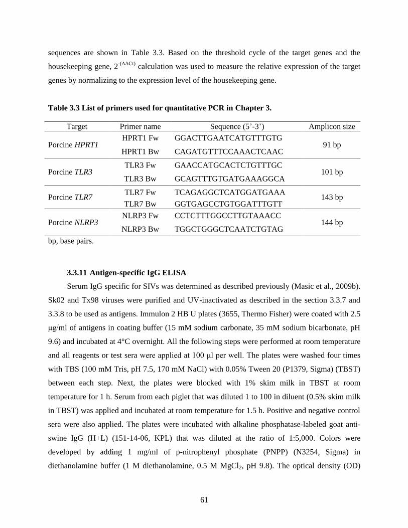
61
sequences are shown in Table 3.3. Based on the threshold cycle of the target genes and the
housekeeping gene, 2-(ΔΔCt)
calculation was used to measure the relative expression of the target
genes by normalizing to the expression level of the housekeeping gene.
Table 3.3 List of primers used for quantitative PCR in Chapter 3.
Target Primer name Sequence (5’-3’) Amplicon size
Porcine HPRT1 HPRT1 Fw GGACTTGAATCATGTTTGTG
91 bp HPRT1 Bw CAGATGTTTCCAAACTCAAC
Porcine TLR3 TLR3 Fw GAACCATGCACTCTGTTTGC
101 bp TLR3 Bw GCAGTTTGTGATGAAAGGCA
Porcine TLR7 TLR7 Fw TCAGAGGCTCATGGATGAAA
143 bp TLR7 Bw GGTGAGCCTGTGGATTTGTT
Porcine NLRP3 NLRP3 Fw CCTCTTTGGCCTTGTAAACC
144 bp NLRP3 Bw TGGCTGGGCTCAATCTGTAG
bp, base pairs.
3.3.11 Antigen-specific IgG ELISA
Serum IgG specific for SIVs was determined as described previously (Masic et al., 2009b).
Sk02 and Tx98 viruses were purified and UV-inactivated as described in the section 3.3.7 and
3.3.8 to be used as antigens. Immulon 2 HB U plates (3655, Thermo Fisher) were coated with 2.5
μg/ml of antigens in coating buffer (15 mM sodium carbonate, 35 mM sodium bicarbonate, pH
9.6) and incubated at 4°C overnight. All the following steps were performed at room temperature
and all reagents or test sera were applied at 100 μl per well. The plates were washed four times
with TBS (100 mM Tris, pH 7.5, 170 mM NaCl) with 0.05% Tween 20 (P1379, Sigma) (TBST)
between each step. Next, the plates were blocked with 1% skim milk in TBST at room
temperature for 1 h. Serum from each piglet that was diluted 1 to 100 in diluent (0.5% skim milk
in TBST) was applied and incubated at room temperature for 1.5 h. Positive and negative control
sera were also applied. The plates were incubated with alkaline phosphatase-labeled goat anti-
swine IgG (H+L) (151-14-06, KPL) that was diluted at the ratio of 1:5,000. Colors were
developed by adding 1 mg/ml of p-nitrophenyl phosphate (PNPP) (N3254, Sigma) in
diethanolamine buffer (1 M diethanolamine, 0.5 M MgCl2, pH 9.8). The optical density (OD)

62
was measured by reading at 405 nm with reference at 490 nm using xMark Microplate
Absorbance Spectrophotometer (Bio-Rad). The highest dilution at which the OD of the test
serum was higher than the cut-off value, which is the mean OD of the negative control sera plus
two times the standard deviation (SD), was determined as the titre.
3.3.12 Porcine IL-1β ELISA
Immulon 2 HB U plates (3655, Thermo Fisher) were coated with 2 μg/ml of mouse
monoclonal anti-porcine IL-1β antibody (MAB6811, R&D Systems) in PBS at room temperature
overnight. All the following steps were performed at room temperature and all reagents or
samples were applied at 100 μl per well. The plates were washed four times with TBST between
each step. Blocking was performed with 1% BSA (A7030, Sigma) in PBS for 1 h. Two-fold
serial dilutions of recombinant porcine IL-1β protein (681-PI-010, R&D Systems) in diluent
(0.1% BSA in TBST) was used as the standard. The plates were incubated with samples or the
standard for 2 h. The plates were then incubated with 50 ng/ml of goat polyclonal anti-porcine
IL-1β biotinylated antibody (BAF681, R&D Systems) in diluent for 1 h, followed by the
incubation with alkaline phosphatase streptavidin (016-050-084, Jackson ImmunoResearch)
diluted at the ratio of 1:5,000 in diluent for another 1 h. For color development, the plates were
incubated with 1 mg/ml of PNPP in diethanolamine buffer until the OD of the first dilution of the
standard reached around 2.0. The plates were read at 405 nm with reference at 490 nm using
xMark Microplate Absorbance Spectrophotometer (Bio-Rad).
3.3.13 NLRP3 inflammasome reconstitution assay
HEK293T cells were seeded at 1.5 × 10⁵ cells per well on 24-well plates and transfected
with pcDNA-NLRP3 (30 ng), pcDNA-ASC (20 ng), pCMV-Flag-pro-caspase-1 (20 ng) and
pcDNA-pro-IL-1β (100 ng) along with other constructs (300 ng of Flag-vector or Flag-tagged
WT/mutant Hf09 NS1 constructs, or 300 ng of Myc-vector or Myc-tagged mutant Sk02 or Hf09
NS1 constructs). Transfection was conducted by adding the mixture of plasmids and TransIT-
LT1 Transfection Reagent. At 16 hpt, cell-free supernatants were collected for porcine IL-1β
ELISA and cells were lysed with 1 × SDS sample buffer for Western blotting. To monitor the
NLRP3 inflammasome activation levels in response to viral infection, at 12 hpt, the cells were

63
mock-infected or infected with viruses at an MOI of 5. At 12 hpi, cell-free supernatants were
collected and cells were lysed as mentioned.
3.3.14 Western blotting
Cell lysates or IP samples were fractionated by SDS-polyacrylamide gel electrophoresis
(PAGE) and blotted onto 0.45 μm nitrocellulose membranes (1620115, Bio-Rad). The
membranes were blocked with 5% skim milk in TBS with 0.1% Tween 20 for 1 h and incubated
with primary antibodies in TBS with 0.1% Tween 20 at 4°C overnight. After incubation with the
secondary antibodies in TBS with 0.1% Tween 20 at room temperature for 1 h, the membranes
were scanned with an Odyssey infrared imager (LI-COR Biosciences).
3.3.15 Co-immunoprecipitation (co-IP)
HEK293T cells were seeded at 1 × 10⁶ per well on 6-well plates (#3506, Corning) and
were transfected with 1 μg of each plasmid indicated. Transfection was conducted by adding the
mixture of plasmids and TransIT-LT1 Transfection Reagent. To examine the effects of viral
infection on the NLRP3-ASC interaction, the transfected cells were infected with Hf09 at an
MOI of 1 for 12 h. To observe the effects of WT/mutant NS1 of Hf09 on the NLRP3-ASC
interaction, the cells were incubated for 48 h after transfection. The cells were lysed with 500 μl
lysis buffer (50 mM Tris, pH 7.4, 150 mM NaCl, 0.5% Nonidet P-40 substitute, 1 × protease
inhibitor cocktail) and clarified by centrifugation at 12,000 × g for 10 min at 4°C. For input, 10%
of the cell lysates were stored separately. Dynabeads Protein G (10004D, Thermo Fisher) were
first conjugated with 1 μg of the respective antibodies by incubation at room temperature for 1 h
and then, were incubated with the cell lysates at 4°C overnight with agitation. After washed
thrice with TBS, the beads were resuspended in 2 × SDS sample buffer and boiled at 98°C for 5
min to be analyzed by Western blotting.
3.3.16 Immunofluorescence and confocal microscopy
HEK293T cells were seeded at 6 × 10⁴ cells per well on LabTek II CC2 chamber slides
(154941, Thermo Fisher). The cells were transfected with 50 ng of pcDNA-NLRP3-Myc or
Myc-vector and 30 ng of pCMV-Flag-ASC or Flag-vector for 24 h to validate the specificity of
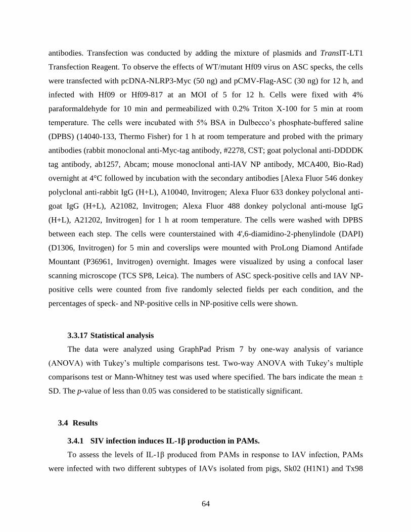
64
antibodies. Transfection was conducted by adding the mixture of plasmids and TransIT-LT1
Transfection Reagent. To observe the effects of WT/mutant Hf09 virus on ASC specks, the cells
were transfected with pcDNA-NLRP3-Myc (50 ng) and pCMV-Flag-ASC (30 ng) for 12 h, and
infected with Hf09 or Hf09-817 at an MOI of 5 for 12 h. Cells were fixed with 4%
paraformaldehyde for 10 min and permeabilized with 0.2% Triton X-100 for 5 min at room
temperature. The cells were incubated with 5% BSA in Dulbecco’s phosphate-buffered saline
(DPBS) (14040-133, Thermo Fisher) for 1 h at room temperature and probed with the primary
antibodies (rabbit monoclonal anti-Myc-tag antibody, #2278, CST; goat polyclonal anti-DDDDK
tag antibody, ab1257, Abcam; mouse monoclonal anti-IAV NP antibody, MCA400, Bio-Rad)
overnight at 4°C followed by incubation with the secondary antibodies [Alexa Fluor 546 donkey
polyclonal anti-rabbit IgG (H+L), A10040, Invitrogen; Alexa Fluor 633 donkey polyclonal anti-
goat IgG (H+L), A21082, Invitrogen; Alexa Fluor 488 donkey polyclonal anti-mouse IgG
(H+L), A21202, Invitrogen] for 1 h at room temperature. The cells were washed with DPBS
between each step. The cells were counterstained with 4',6-diamidino-2-phenylindole (DAPI)
(D1306, Invitrogen) for 5 min and coverslips were mounted with ProLong Diamond Antifade
Mountant (P36961, Invitrogen) overnight. Images were visualized by using a confocal laser
scanning microscope (TCS SP8, Leica). The numbers of ASC speck-positive cells and IAV NP-
positive cells were counted from five randomly selected fields per each condition, and the
percentages of speck- and NP-positive cells in NP-positive cells were shown.
3.3.17 Statistical analysis
The data were analyzed using GraphPad Prism 7 by one-way analysis of variance
(ANOVA) with Tukey’s multiple comparisons test. Two-way ANOVA with Tukey’s multiple
comparisons test or Mann-Whitney test was used where specified. The bars indicate the mean ±
SD. The p-value of less than 0.05 was considered to be statistically significant.
3.4 Results
3.4.1 SIV infection induces IL-1β production in PAMs.
To assess the levels of IL-1β produced from PAMs in response to IAV infection, PAMs
were infected with two different subtypes of IAVs isolated from pigs, Sk02 (H1N1) and Tx98

65
(H3N2) at indicated MOIs (Figure 3.1A). At 24 hpi, cell-free supernatants were collected for
porcine IL-1β ELISA and the cells were lysed for Western blotting to measure the expression of
pro-IL-1β and viral proteins. Although PAMs from different piglets showed variable levels of
IL-1β production in response to SIV infection, the differences in IL-1β levels in response to SIVs
at indicated MOIs were reproducible when cells from different piglets were used, and the
representative data using PAMs isolated from a piglet with the lowest background level of IL-1β
is shown. Pro-IL-1β levels from PAMs infected at an MOI of 1 or 10 were comparable and were
significantly higher than those in mock cells or cells infected at an MOI of 0.1 (Figure 3.1A). In
contrast, IL-1β levels in virus-infected cells were significantly higher than that of mock infected
cells and were viral dose-dependent. Accordingly, the expression of viral NP and NS1 proteins
were also viral dose-dependent, confirming that PAMs were infected with SIVs. Based on these
results, an MOI of 1 for 24 h was chosen in the following infection experiments with PAMs,
except for an experiment to check mRNA expression of NLRP3. Since the above results
suggested that viral replication is required for IL-1β production in PAMs, experiments were
conducted to further confirm this finding by inactivating the two virus strains with UV
irradiation (Figure 3.1B). The effect of UV inactivation on virus replication was confirmed by
monitoring the NS1 protein expression levels. Compared to WT viruses, UV-inactivated viruses
lost the ability to induce IL-1β production in PAMs (Figure 3.1B), indicating viral replication is
necessary for the conversion of pro-IL-1β into mature IL-1β.

66
Figure 3.1 SIV infection induces IL-1β production in PAMs.
(A) PAMs were infected with two SIV strains, Sk02 and Tx98 at an MOI of 0.1, 1 or 10. At 24
hpi, cell-free supernatants were collected for porcine IL-1β ELISA and cells were lysed for
Western blotting to measure the expression of pro-IL-1β and viral proteins. ***, p<0.001. (B)
PAMs were infected with WT or UV-inactivated viruses of two SIV strains at an MOI of 1 for
24 h. IL-1β production and pro-IL-1β expression were measured as in (A). ***, p<0.001.
3.4.2 SIV-induced IL-1β production is mediated by TLR3.
To identify which cytosolic sensor is mainly involved in IL-1β production from PAMs
infected with SIV, endosomal TLRs for dsRNA or ssRNA were knocked down by siRNAs.
PAMs were transfected with a negative control siRNA or siRNA specific for porcine TLR3 or
TLR7 and infected with Sk02. Knockdown efficiency confirmed by comparing the mRNA
expression levels of TLR3 and TLR7 to that of the housekeeping gene, HPRT1 was more than
50% (Figure 3.2A). Compared to treatment with a negative control or TLR7 siRNA, TLR3
silencing led to reduced IL-1β production upon Sk02 infection (Figure 3.2B), indicating that
TLR3 functions as a sensor of SIV infection for porcine IL-1β production.

67
Figure 3.2 SIV-induced IL-1β production is mediated by TLR3.
(A) PAMs were transfected with 20 nM of a negative control siRNA (siControl) or siRNAs
targeting porcine TLR3 (siTLR3) or TLR7 (siTLR7) for 24 h. Knockdown efficiency was
measured by checking the mRNA expression levels of porcine TLR3 and TLR7 compared to that
of porcine HPRT1 by quantitative PCR. ***, p<0.001. (B) PAMs were transfected with
siControl, siTLR3 or siTLR7 for 24 h. The cells were infected with SIV Sk02 at an MOI of 1 for
24 h. Cell-free supernatants were collected for porcine IL-1β ELISA. **, p<0.01.
3.4.3 SIV-induced IL-1β production is mediated by NLRP3 inflammasome.
Next, whether the NLRP3 inflammasome is involved in IL-1β production in PAMs upon
SIV infection was tested. PAMs were first treated with an NLRP3 inhibitor, MNS, which inhibits
the NLRP3 inflammasome activation by binding to NLRP3 (He et al., 2014), and were then
infected with Sk02 at an MOI of 1. In virus-infected PAMs, compared to vehicle-treated cells, a
dose dependent decrease of IL-1β induction upon the inhibitor treatment was observed (Figure
3.3A), indicating that porcine IL-1β production in SIV-infected PAMs is mediated by NLRP3
inflammasome. At the tested concentration of MNS, cells did not show noticeable cytotoxicity
(data not shown). To reaffirm the NLRP3 inflammasome-mediated IL-1β production upon IAV
infection, the porcine NLRP3 inflammasome was reconstituted in HEK293T cells that are
deficient in endogenous human NLRP3 inflammasome (Bryan et al., 2009). Porcine NLRP3,
ASC, pro-caspase-1 and pro-IL-1β genes were cloned from PAMs and the corresponding
mammalian expression plasmids were constructed. Co-transfection of all four plasmids led to a
moderate level of IL-1β in the supernatant owing to the engagement of NLPR3, ASC and

68
caspase-1. Only basal level of IL-1β was detected when pro-IL-1β was expressed alone (Figure
3.3B). These results validated the successful setup of the reconstitution system, showing that IL-
1β maturation and secretion predominantly rely on the porcine NLRP3 inflammasome. To
confirm the dependency of IL-1β production on NLRP3 inflammasome in the context of IAV
infection, porcine NLRP3 inflammasome was reconstituted in HEK293T cells for 12 h and then
were infected with Sk02 at an MOI of 5 for another 12 h. In the presence of all the NLRP3
inflammasome components and pro-IL-1β, infection with Sk02 induced over 5,000 pg/ml of IL-
1β and a noticeable level of cleaved caspase-1, p20, which was much higher than its counterpart
in the absence of virus infection (Figure 3.3B). In all samples, viral protein expression and
cellular β-actin levels were monitored. Together, these results demonstrate that SIV infection
induces porcine NLRP3 inflammasome-mediated IL-1β production.
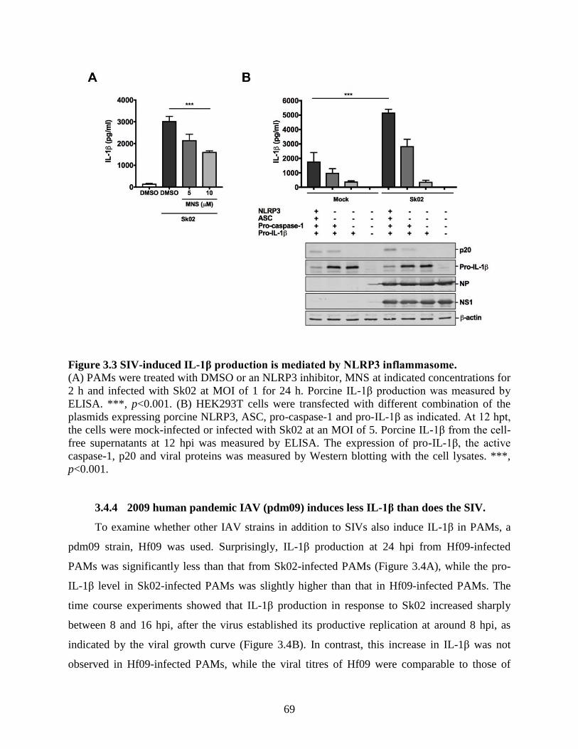
69
Figure 3.3 SIV-induced IL-1β production is mediated by NLRP3 inflammasome.
(A) PAMs were treated with DMSO or an NLRP3 inhibitor, MNS at indicated concentrations for
2 h and infected with Sk02 at MOI of 1 for 24 h. Porcine IL-1β production was measured by
ELISA. ***, p<0.001. (B) HEK293T cells were transfected with different combination of the
plasmids expressing porcine NLRP3, ASC, pro-caspase-1 and pro-IL-1β as indicated. At 12 hpt,
the cells were mock-infected or infected with Sk02 at an MOI of 5. Porcine IL-1β from the cell-
free supernatants at 12 hpi was measured by ELISA. The expression of pro-IL-1β, the active
caspase-1, p20 and viral proteins was measured by Western blotting with the cell lysates. ***,
p<0.001.
3.4.4 2009 human pandemic IAV (pdm09) induces less IL-1β than does the SIV.
To examine whether other IAV strains in addition to SIVs also induce IL-1β in PAMs, a
pdm09 strain, Hf09 was used. Surprisingly, IL-1β production at 24 hpi from Hf09-infected
PAMs was significantly less than that from Sk02-infected PAMs (Figure 3.4A), while the pro-
IL-1β level in Sk02-infected PAMs was slightly higher than that in Hf09-infected PAMs. The
time course experiments showed that IL-1β production in response to Sk02 increased sharply
between 8 and 16 hpi, after the virus established its productive replication at around 8 hpi, as
indicated by the viral growth curve (Figure 3.4B). In contrast, this increase in IL-1β was not
observed in Hf09-infected PAMs, while the viral titres of Hf09 were comparable to those of

70
Sk02 during the time course. These results showed that less IL-1β production by Hf09 was not
due to the inefficient replication of Hf09 in PAMs, which was also confirmed by viral NS1
expression levels (Figure 3.4A). The signal 1 required for NLRP3 inflammasome-mediated IL-
1β production induces the transcriptional upregulation of NLRP3 in addition to pro-IL-1β. Thus,
the mRNA expression of NLRP3 in PAMs infected with Sk02 or Hf09 was also assessed.
Porcine NLRP3 transcription was around 2.5 times increased upon viral infection, while this
increase was comparable between Sk02 and Hf09-infected PAMs (Figure 3.4C). LPS-induced
upregulation of NLRP3 expression was shown as a positive control. These indicated that the
remarkable difference in IL-1β levels was possibly attributed to the suppression of NLRP3
inflammasome activity in cells infected by pdm09.
Figure 3.4 pdm09 induces less IL-1β than does SIV.
(A) PAMs were infected with an SIV strain, Sk02 or a human pandemic strain, Hf09 at an MOI
of 1. At 24 hpi, cell-free supernatants were collected for IL-1β ELISA and the cells were lysed
for Western blotting to measure the expression of pro-IL-1β and viral NS1 protein. ns, not
significant, ***, p<0.001. (B) PAMs were infected with Sk02 or Hf09 at an MOI of 1, and the
supernatants were collected at 8, 16 and 24 hpi. IL-1β was measured by ELISA and viral titres
were determined by plaque assay on MDCK cells. Two-way ANOVA with Tukey’s multiple
comparisons test (B, upper panel), ***, p<0.001; Mann-Whitney test (B, lower panel), ns, not
significant. (C) PAMs were either infected with viruses (Sk02 or Hf09) at an MOI of 1 for 12 h
or stimulated with 200 ng/ml LPS for 12 h. Expression of porcine NLRP3 mRNA was compared
to that of porcine HPRT1 by conducting quantitative PCR. ns, not significant.

71
3.4.5 NS1 C-terminus of pdm09, but not that of SIV, inhibits the NLRP3
inflammasome activation.
Based on the above results, viral proteins were postulated to contribute to the regulation of
NLRP3 inflammasome activation. Among others, NS1 protein of IAV is well known for its
function of antagonizing host immune responses. However, the NS1 protein of pdm09 was
reported to be inefficient in controlling the host gene expression (Hale et al., 2010). Unexpected
results obtained above that Hf09 has a strong inhibitory effect on porcine IL-1β production led to
a further study on whether Hf09 NS1 regulates the NLRP3 inflammasome activity. NS1 C-
terminus has been shown to interact with multiple cellular proteins (Hale et al., 2008), which led
to a hypothesis that NS1 C-terminus may exert an inhibitory function on the porcine IL-1β
production. To investigate this, mutant viruses derived from WT Sk02 or Hf09 containing
mutations in the NS1 protein were constructed (illustrated in Figure 3.5A). Sk02-NS1/1-99 virus
encoding N-terminal NS1 from aa 1 to 99 was previously generated in our lab; the Sk02-NS1/1-
99 induced more type I IFN secretion from PAMs than its WT did (Liu et al., 2015). In addition,
two Hf09 mutant viruses encoding truncated NS1 proteins (Hf09-816 and Hf09-817 encoding
NS1 aa 1 to 73 and aa 1 to 99, respectively) were generated in the current study. They showed
smaller-sized plaques than that of the WT virus (Figure 3.5B) and grew to titers 0.7 to 1 log
lower on MDCK-NS1 cells than the WT Hf09 on MDCK cells titers were 7 × 10⁶ plaque-
forming unit (PFU)/ml for Hf09-816 and 1.25 × 10⁷ PFU/ml for Hf09-817 versus 7.5 × 10
7
PFU/ml for WT Hf09 (Figure 3.5B). Infection of PAMs with WT Sk02 or Sk02-NS1/1–99 virus
showed that the deletion of NS1 C-terminal aa 100 to 230 in Sk02 led to a slightly increased pro-
IL-1β expression, but did not alter the IL-1β level. Specifically, Sk02-NS1/1–99 virus induced
only 1.07-fold more IL-1β than that induced by WT Sk02 (Figure 3.5C). In contrast, upon
infection of PAMs, the Hf09-816 and Hf09-817 viruses induced 2.53- and 3.05-fold more IL-1β
production than WT Hf09, respectively, yet they did not significantly affect pro-IL-1β expression
levels (Figure 3.5D). In the reconstitution system, Hf09-816 and Hf09-817 infection led to a
striking increase of IL-1β production above the level by WT Hf09 infection, although they did
not markedly alter the pro-IL-1β level (Figure 3.5E). The level of an active caspase-1 subunit,
p20, an indicator for NLRP3 inflammasome activation, was also consistently increased in the

72
cells infected with NS1-mutant viruses. These suggested that NS1 C-terminus of pdm09
suppresses the porcine NLRP3 inflammasome-mediated IL-1β production.
(Figure 3.5 to be continued)

73
Figure 3.5 NS1 C-terminus of pdm09, but not that of SIV, inhibits porcine NLRP3
inflammasome activation.
(A) A schematic representation of genome composition of WT and mutant viruses. The NS1
protein encoded by different viruses is depicted. (B) Plaque assay results of WT virus in MDCK
cells and NS1-mutant viruses in MDCK-NS1 cells at indicated dilutions shown above the plaque
images. (C) PAMs were infected with Sk02 WT or Sk02-NS1/1-99 virus at an MOI of 1. At 24
hpi, cell-free supernatants were collected for IL-1β ELISA and the cells were lysed for Western
blotting to measure the expression of pro-IL-1β and viral proteins. ns, not significant. (D) PAMs
were infected with Hf09 WT or two NS1-mutant virus (Hf09-816 or Hf09-817) at an MOI of 1
for 24 h. IL-1β ELISA and Western blotting were performed as in (B). *, p=0.0201, **,
p=0.0015. (E) HEK293T cells were transfected with plasmids expressing porcine NLRP3, ASC,
pro-caspase-1 and pro-IL-1β. At 12 hpt, the cells were infected with Hf09 WT or NS1-mutant
viruses at an MOI of 5. IL-1β production at 12 hpi was measured by ELISA and the expression
of p20, pro-IL-1β and viral proteins was measured by Western blotting. ***, p<0.001.

74
3.4.6 Mutant SIVs with NS1 C-terminus derived from pdm09 inhibits the NLRP3
inflammasome activation.
To demonstrate conclusively that the C-terminus of pdm09 NS1 is responsible for the
inhibition of IL-1β production, additional two mutant viruses in the background of Sk02 were
generated. Sk02-616 has the entire NS segment from Hf09, and Sk02-930 has a chimeric NS1
with the N-terminus from Sk02 and C-terminus from Hf09 as illustrated in Figure 3.6A. Plaque
assay showed that the mutant viruses produced similar-sized plaques to those produced by the
WT virus and their titres were comparable to that of WT Sk02: 6.25 × 10⁷ PFU/ml for Sk02-616
and 8.75 × 10⁷ PFU/ml for Sk02-930 versus 4 × 10
7 PFU/ml for WT Sk02 (Figure 3.6B).
Infection of PAMs with Sk02-616 or Sk02-930 led to a reduction of IL-1β, while pro-IL-1β
expression was not significantly impaired (Figure 3.6C). To support that the C-terminus of Hf09
NS1 has a greater effect on the inflammasome activation rather than on the pro-IL-1β level, the
expression of pro-IL-1β and p20 upon infection with Sk02 WT and Sk02-930 was monitored in
the reconstitution system. Indeed, Sk02-930 could inhibit both caspase-1 activation and IL-1β
production, but did not affect the pro-IL-1β expression (Figure 3.6D).
Next, caspase-1 activity and IL-1β production in the reconstitution system were examined
by overexpressing Flag- or Myc-tagged WT or truncated NS1. This experiment showed that co-
expression of the full-length Hf09 NS1 significantly inhibited caspase-1 activation and IL-1β
production, whereas co-expression of the NS1 with C-terminal deletion (NS11-73 or NS11-99) had
little or no inhibitory effects (Figure 3.6E). Further, co-expression of the NS1 C-terminal aa 100
to 219 derived from Hf09 led to a reduction of IL-1β due to less caspase-1 activation, whereas
this effect was not observed with the NS1 C-terminal aa 100 to 230 derived from Sk02 virus
(Figure 3.6F). All of these results evidently demonstrated that the C-terminus of the Hf09 NS1
has an inhibitory effect on NLRP3 inflammasome activation.
Efforts were also made to map the responsible residues in the C-terminus of pdm09 NS1
for the inhibition of IL-1β production. NS1 C-terminal aa sequences among SIVs (Sk02 and
Tx98) and a pdm09 strain, Hf09 that induced high and low levels of porcine IL-1β production,
respectively, were aligned (Figure 3.6G). In addition, a 1918 pandemic strain, influenza
A/Brevig Mission/1/1918/H1N1 (BM18), whose NS1 protein is known to inhibit IL-1β
production in primary human cells (Pothlichet et al., 2013), was included. Sk02 and BM18 had
230 aa, while Tx98 and Hf09 had 219 aa in full lengths of NS1 protein, implying that the

75
presence of additional 11 aa at the C-terminal end did not contribute to the up- or down-
regulation of IL-1β production. Identification of unique residues for Hf09 or BM18 NS1 to
inhibit the IL-1β production was not successful, because among variable aa residues, those
contained by Hf09 NS1 were also harboured by Tx98 NS1, or those contained by BM18 NS1
were shared with Sk02 or Tx98 NS1. It suggested that multiple aa clusters in the NS1 C-terminus
of pdm09 are responsible for the inhibition of IL-1β production.
(Figure 3.6 to be continued)

76
(Figure 3.6 to be continued)
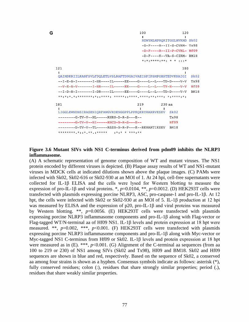
77
Figure 3.6 Mutant SIVs with NS1 C-terminus derived from pdm09 inhibits the NLRP3
inflammasome.
(A) A schematic representation of genome composition of WT and mutant viruses. The NS1
protein encoded by different viruses is depicted. (B) Plaque assay results of WT and NS1-mutant
viruses in MDCK cells at indicated dilutions shown above the plaque images. (C) PAMs were
infected with Sk02, Sk02-616 or Sk02-930 at an MOI of 1. At 24 hpi, cell-free supernatants were
collected for IL-1β ELISA and the cells were lysed for Western blotting to measure the
expression of pro-IL-1β and viral proteins. *, p=0.0104, **, p=0.0012. (D) HEK293T cells were
transfected with plasmids expressing porcine NLRP3, ASC, pro-caspase-1 and pro-IL-1β. At 12
hpt, the cells were infected with Sk02 or Sk02-930 at an MOI of 5. IL-1β production at 12 hpi
was measured by ELISA and the expression of p20, pro-IL-1β and viral proteins was measured
by Western blotting. **, p=0.0056. (E) HEK293T cells were transfected with plasmids
expressing porcine NLRP3 inflammasome components and pro-IL-1β along with Flag-vector or
Flag-tagged WT/N-terminal aa of Hf09 NS1. IL-1β levels and protein expression at 18 hpt were
measured. **, p=0.002, ***, p<0.001. (F) HEK293T cells were transfected with plasmids
expressing porcine NLRP3 inflammasome components and pro-IL-1β along with Myc-vector or
Myc-tagged NS1 C-terminus from Hf09 or Sk02. IL-1β levels and protein expression at 18 hpt
were measured as in (E). ***, p<0.001. (G) Alignment of the C-terminal aa sequences (from aa
100 to 219 or 230) of NS1 among SIVs (Sk02 and Tx98), Hf09 and BM18. Sk02 and Hf09
sequences are shown in blue and red, respectively. Based on the sequence of Sk02, a conserved
aa among four strains is shown as a hyphen. Consensus symbols indicate as follows: asterisk (*),
fully conserved residues; colon (:), residues that share strongly similar properties; period (.),
residues that share weakly similar properties.
78
3.4.7 NS1 C-terminus of pdm09 inhibits the interaction between NLRP3 and ASC.
In an effort to define the mechanism behind the inhibitory function of pdm09 NS1 on the
porcine NLRP3 inflammasome, the NLRP3-ASC interaction was examined. First, whether Hf09
infection can inhibit the NLRP3-ASC interaction was tested by transfecting HEK293T cells with
plasmids expressing Myc-tagged ASC and Flag-tagged NLRP3 followed by infection of the cells
with Hf09. Co-IP with anti-Myc antibody followed by Western blotting showed that Flag-
NLRP3 was not in the IP complex when expressed alone (Figure 3.7A, lane 1), whereas it was
successfully pulled down when ASC-Myc was co-expressed (Figure 3.7A, lane 2). However,
when comparing between the mock-infected and Hf09-infected conditions, the pulled-down level
of Flag-NLRP3 was lower in Hf09-infected cells (Figure 3.7A, lane 4), indicating that the
NLRP3-ASC interaction is inhibited by Hf09 infection. Next, the effects of WT and truncated
NS1 of Hf09 on the NLRP3-ASC interaction were compared. HEK293T cells were transfected
with plasmids expressing Myc-tagged NLRP3 and ASC along with Flag-tagged WT or truncated
NS1, and were subjected to co-IP. Association between NLRP3-Myc and ASC was clearly
noticeable (Figure 3.7B, lane 2). The NLRP3-ASC interaction was obviously inhibited by the
expression of Hf09 WT NS1 (Figure 3.7B, lane 4), while the interaction was restored with the
expression of Hf09 NS11-73 or NS11-99 (Figure 3.7B, lanes 6 and 8). Therefore, the C-terminal
region, particularly aa 100 to 219 of Hf09 NS1 plays a critical role in interfering with the
NLRP3-ASC interaction that is essential for the activation of NLRP3 inflammasome.

79
Figure 3.7 NS1 C-terminus of pdm09 inhibits the interaction between NLRP3 and ASC.
(A) HEK293T cells were transfected with Flag-NLRP3 construct along with Myc-vector or
ASC-Myc construct, and either mock-infected or infected with Hf09 at an MOI of 1 for 12 h.
Cell lysates were subjected to co-IP with anti-Myc antibody. IP and input samples were analyzed
with Western blotting using anti-Myc, anti-Flag and anti-NS1 antibodies. (B) HEK293T cells
were transfected with ASC construct and Myc-vector or NLRP3-Myc construct in addition to
plasmids expressing Flag-tagged WT or mutant NS1 of Hf09. At 48 hpt, the cells were harvested
and cell lysates were subjected to co-IP with mouse anti-Myc antibody. IP and input samples
were analyzed with Western blotting using anti-ASC, anti-Myc and anti-Flag antibodies. IB,
immunoblot.

80
3.4.8 NS1 C-terminus of pdm09 inhibits the ASC speck formation.
Interference of NLRP3-ASC interaction by pdm09 NS1 C-terminus prompted further
investigation on whether ASC specks formed by aggregation of NLRP3 with ASC, a hallmark of
NLRP3 inflammasome activation, are affected by NS1 protein. In non-infected cells, transfection
of either ASC or NLRP3 alone did not induce a noticeable number of specks (Figure 3.8A, top
three rows). In contrast, co-transfection of porcine ASC and NLRP3 led to the aggregation of
two proteins, forming the specks in the perinuclear area. Furthermore, in virus-infected cells as
indicated by NP staining, Hf09-817 encoding NS1 aa 1-99 led to 2-fold more ASC speck
formation than WT Hf09 (Figure 3.8A, bottom three rows). Specifically, while 6.8% of NP-
positive cells displayed ASC specks in Hf09-infected cells, 14.3% of NP-positive cells exhibited
ASC specks in Hf09-817-infected cells (Figure 3.8B). These results demonstrated that the pdm09
NS1 C-terminus suppresses the ASC speck formation, concomitantly with the disruption of
NLRP3-ASC association.

81
Figure 3.8 NS1 C-terminus of pdm09 inhibits the ASC speck formation.
(A) HEK293T cells were transfected with Myc-tagged NLRP3 or Myc-vector and Flag-tagged
ASC or Flag-vector for 24 h and left uninfected (upper three rows). The cells were transfected
with Myc-tagged NLRP3 and Flag-tagged ASC for 12 h and infected with Hf09 or Hf09-817 at
an MOI of 5 for 12 h (lower three rows). The cells were fixed, permeabilized, blocked and
probed with appropriate antibodies followed by DAPI staining. NLRP3-Myc (gray), Flag-ASC
(green), viral NP (red) and nuclei (blue) were visualized by confocal microscopy. The scale bars
indicate 10 μm. (B) ASC speck-forming cells and NP-stained cells infected by Hf09 or Hf09-817
were quantified from five randomly selected fields and the percentages of ASC speck- and NP-
positive cells in total NP-positive cells were shown. Mann-Whitney test, **, p=0.0025.

82
3.5 Discussion
NLRP3 inflammasome exerts indispensable roles in regulating the innate immunity. In this
study, porcine NLRP3 inflammasome regulation in response to IAV infection was investigated.
While SIVs generally induce inflammatory responses in pigs (Khatri et al., 2010, Pomorska-Mól
et al., 2014), Ma et al. showed pdm09-infected pigs had upregulated expression of genes
involved in the inflammatory and immune responses (Ma et al., 2011). In terms of the cytokine
response in human cells infected by pdm09, there have been some controversial reports. Both
upregulation of inflammatory cytokines (Lee et al., 2011, Sakabe et al., 2011, Yu et al., 2011)
and poor inflammatory responses were reported (Osterlund et al., 2010, Zeng et al., 2011).
Nevertheless, there is little information concerning the regulation of IL-1β production, which
requires the activation of NLRP3 inflammasome upon IAV infection in pigs. Since alveolar
macrophages are essential for the regulation of lung inflammation and are required for the
protection against IAV infection (Cardani et al., 2017, Kim et al., 2008, Schneider et al., 2014),
primary PAMs were utilized to characterize how porcine IL-1β is produced in response to IAV
infection.
In this study, swine isolates of IAVs induced IL-1β production, which was mediated by
TLR3 and NLRP3 inflammasome, in a viral dose- and replication-dependent manner. In contrast,
Hf09, a pdm09 strain, induced much lower level of IL-1β production. The fact that Hf09 and
Sk02 replicated to similar titres in PAMs and that they induced similar levels of pro-IL-1β
suggested that the NLRP3 inflammasome was less active or was suppressed in Hf09-infected
cells. Similar levels of NLRP3 mRNA transcription induced by Sk02 and Hf09 infection further
supported that the intensity of signal 1 provided by the two viruses was comparable.
IAV NS1 protein has been reported to inhibit the NLRP3 inflammasome-mediated IL-1β
production in mouse or human macrophages (Cheong et al., 2015, Moriyama et al., 2016). In
addition, NS1 from a highly pathogenic, 1918 pandemic strain, BM18 suppresses RIG-I/NLRP3
inflammasome-dependent IL-1β production from primary human lung cells (Pothlichet et al.,
2013). Cheong et al. reported that NS1 encoded by human H5N1 or H1N1 strain physically
interacts with NLRP3 and suppresses the inflammasome activation in THP-1 cells (Cheong et
al., 2015). Based on an early study using PR8 strain, which showed that deletion of NS1 aa 40 to
80 increases human IL-1β production (Stasakova et al., 2005), they tested NS1 from a low
pathogenic strain, influenza A/WSN/1933/H1N1 (WSN33) and its mutant with aa 40-80 deleted
83
(Cheong et al., 2015), however, the mutant NS1 without aa 40-80 could still bind NLRP3.
Interestingly, another study with the same virus shows that deletion of 28 aa at the NS1 C-
terminal end of the WSN33 virus results in the production of more IL-1β, but less TNF-α and
chemokines in mice (Anastasina et al., 2015), suggesting that critical regions for the inhibition of
IL-1β production may be strain-dependent and/or host-dependent. Moriyama et al. demonstrated
that PR8 NS1 also interacts with NLRP3, thereby inhibiting human or mouse IL-1β production.
The inhibitory function was mapped to the RNA-binding domain (R38 and K41) and TRIM25-
binding domain (E96 and E97) within the N-terminus of NS1, since PR8 NS1 constructs with
mutations on those residues were unable to bind NLRP3 (Moriyama et al., 2016), meaning that
multiple regions of NS1 are involved in the NLRP3 inflammasome regulation. In contrary to the
aforementioned reports, the current study showed that the mutant pdm09 virus carrying NS1 C-
terminal deletion from aa 100 to 219 induced strikingly higher level of IL-1β than did the WT
virus. Also, the ectopic expression of the NS1 C-terminal aa 100-219 from Hf09, but not that
from Sk02 led to a decreased level of IL-1β secretion. In accordance with it, NS1 C-terminus
derived from Hf09 was responsible for disrupting the interaction between NLRP3 and ASC, and
suppressing the formation of ASC specks. Thus, the C-terminus of pdm09 NS1 obviously
possesses inhibitory functions on the porcine NLRP3 inflammasome activation. Intriguingly,
NS1 C-terminal aa 126-225 of avian H5N1 downregulates the RIG-I-mediated IFN signaling by
interacting with TRAF3 (Qian et al., 2017). Whether pdm09 NS1 can interact with any of
porcine NLRP3 inflammasome components or even other proteins that may participate in the
regulation of the inflammasome activity requires further elucidation.
NS1 C-terminus contains multiple motifs critical for binding host cellular partners.
For example, by binding to CPSF30 and PABPII, NS1 can repress the polyadenylation of host
cellular pre-mRNAs and the nuclear export of cellular mRNAs, respectively (Hale et al., 2008).
However, PABPII-binding motif (NS1 aa 223 to 230) is not encoded by pdm09 due to a natural
truncation of 11 aa at the C-terminal end (Tu et al., 2011), and pdm09 NS1 is defective in
binding CPSF30 (Hale et al., 2010). This prompted an analysis to locate the potential residues in
pdm09 NS1 involved in regulating the NLRP3 inflammasome. BM18 NS1 was included in the
NS1 aa alignment with a speculation that C-terminus of this protein may be involved in the
downregulation of IL-1β production. Unfortunately, aligning the NS1 C-terminal aa 100 to 219
(or 230) of three IAV strains used in the current study (Sk02, Tx98 and Hf09) and BM18 did not

84
provide any clue in identifying critical residues in Hf09 NS1 for the inhibition of IL-1β.
Therefore, it will be required to obtain the porcine IL-1β secretion profiles of other IAV strains
and compare the C-terminal aa sequences together with the additional strains.
Collectively, NLRP3 inflammasome-mediated IL-1β production was potently induced in
primary PAMs infected with SIVs, while it was suppressed in pdm09-infected cells due to the
inhibition of NLRP3 inflammasome activity by NS1. NS1 C-terminus of pdm09 exhibited strong
inhibitory functions on porcine IL-1β production by interference with NLRP3-ASC interaction
and ASC speck formation, all of which are critical for the NLRP3 inflammasome activation. This
unveiled an important mechanism played by pdm09 NS1 to evade the inflammatory responses in
pigs.

85
TRANSITION BETWEEN CHAPTER 3 AND CHAPTER 4
In Chapter 3, IL-1β production from PAMs upon infection with SIVs was found to be
TLR3- and NLRP3 inflammasome-mediated. Testing with additional IAV strains led to a finding
that a pdm09 strain, Hf09 induced less IL-1β than SIVs. Further study revealed that NS1 C-
terminus of Hf09, but not that of SIV, was able to significantly inhibit the NLRP3
inflammasome-mediated IL-1β production. Interfered interaction between NLRP3 and ASC
leading to the suppressed ASC speck formation by NS1 C-terminus of Hf09 was suggested as
one of the regulatory events behind the inhibition of it. In the following chapter, to dissect the
mechanism in depth, further investigation is conducted regarding the modifications on ASC,
which functions as a bridge between the sensor protein, NLRP3 and the effector molecule,
caspase-1. Aiming to elucidate whether any post-translational modification of ASC is inhibited
by pdm09 NS1 and if it is related to the ASC speck phenotypes, the study in Chapter 4
particularly focuses on how ASC ubiquitination is regulated by NS1 C-terminus of Hf09.

86
CHAPTER 4 NS1 C-TERMINUS OF 2009 PANDEMIC INFLUENZA VIRUS INHIBITS
PORCINE NLRP3 INFLAMMASOME BY SUPRESSING ASC UBIQUITINATION.
Portions of the data presented in this chapter were published in
NS1 Protein of 2009 Pandemic Influenza A Virus Inhibits Porcine NLRP3 Inflammasome-
Mediated Interleukin-1 Beta Production by Suppressing ASC Ubiquitination.
HS Park, G Liu, SN Thulasi Raman, SL Landreth, Q Liu and Y Zhou. (2018)
Journal of Virology, 92(8), e00022-18.
Acknowledgement specific for this chapter:
The human ASC construct was provided by Dr. Takeshi Ichinohe (University of Tokyo) and the
ubiquitin constructs were provided by Dr. Dirk Bohmann (University of Rochester Medical
Center) and Dr. Edward Harhaj (Johns Hopkins University School of Medicine).

87
4.1 Abstract
NLRP3 inflammasome is regulated at multiple levels, with one level being post-
translational modification. Among others, ubiquitination of ASC has been reported to be
indispensable for the activation of NLRP3 inflammasome. IAV infection induces NLRP3
inflammasome-dependent IL-1β production, which is critical in the host innate immunity. Based
on the results that NS1 C-terminus of pdm09, but not that of SIV, inhibited the NLRP3-ASC
interaction and ASC speck formation in the previous chapter, the in-depth mechanism behind the
inhibitory functions was explored. Studies in this chapter showed that porcine ASC could
undergo ubiquitination upon IAV infection; NS1 C-terminus of pdm09 suppressed the
ubiquitination on both porcine and human ASC. Moreover, two target lysine residues K110 and
K140 that are essential for porcine ASC ubiquitination were identified. The importance of these
two residues in regulating the NLRP3 inflammasome-mediated IL-1β production was also
demonstrated. These results showed how NS1 protein of the pdm09 suppresses a post-
translational modification to downregulate the NLRP3 inflammasome activation.
4.2 Introduction
NLRP3 inflammasome is regulated by post-translational modifications. Among different
types of modifications, ubiquitination and deubiquitination play diverse roles in the regulation of
inflammasomes (Bednash and Mallampalli, 2016). Ubiquitination is a process by which
ubiquitin proteins are conjugated to substrate proteins. In addition to proteasomal degradation of
the substrates, this linkage plays crucial roles in a variety of cell signaling pathways.
Ubiquitination is a three-step process mediated by E1 activating enzymes, E2 conjugating
enzymes and E3 ligases. Ubiquitins can also be recycled after being removed from substrates by
deubiquitinases (Kattah et al., 2017). The substrate selectivity is generally determined by E3
ligases that link the C-terminal glycine of a ubiquitin molecule to a lysine (K) on the substrate.
Monoubiquitination occurs by binding of a single ubiquitin to each lysine in the substrate; a
subsequent linkage of more ubiquitins to lysine residues located on the first ubiquitin leads to
different types of polyubiquitination (Davis and Gack, 2015). Additional ubiquitin molecules can
be linked on a lysine residue among seven lysines (K6, K11, K27, K29, K33, K48 and K63) or
on the first methionine (M1) residue, eventually leading to different consequences. K48-linked
ubiquitination can target the substrates for proteasomal degradation, while K63-linked

88
ubiquitination leads to signal transduction by affecting the stability or localization of target
proteins; other linkages are involved in rather diverse activities including cell development,
signaling regulation, autophagy or protein degradation (Davis and Gack, 2015, Kattah et al.,
2017).
Deubiquitinases are required for inflammasome-dependent IL-1β production (Lopez-
Castejon et al., 2013) and more specifically, deubiquitination of NLRP3 is reported to be
important for the activation of the NLRP3 inflammasome (Guo et al., 2016, Han et al., 2015,
Juliana et al., 2012, Kawashima et al., 2017, Py et al., 2013, Yen et al., 2015). On the contrary,
autophagy-mediated or proteasomal degradation of ubiquitinated NLRP3 leads to suppression of
the NLRP3 inflammasome (Song et al., 2016, Yan et al., 2015). Upon activation of several
different inflammasomes, caspase-1 can be ubiquitinated, which may downregulate the
inflammasome activity (Van Opdenbosch et al., 2014). Also, pro-IL-1β undergoes
ubiquitination, which leads to either its maturation into IL-1β or its degradation (Ainscough et
al., 2014, Duong et al., 2015, Harris et al., 2011). ASC ubiquitination was first reported in a
study of AIM2 inflammasome-related autophagy in response to double-stranded DNA (Shi et al.,
2012). More recent studies observed ASC ubiquitination upon NLRP3 inflammasome activation.
While ASC ubiquitination by an E3 ligase, TRAF6 can limit the NLRP3 inflammasome activity
(Chiu et al., 2016), deubiquitination of ASC is required for the NLRP3 inflammasome activation
(Lee et al., 2017c). ASC can also be modified by linear ubiquitin assembly complex (LUBAC),
which is required for the activation of NLRP3 inflammasome (Rodgers et al., 2014). In the
context of RNA virus infection, ubiquitination of ASC by E3 ligase TRAF3 upon infection with
vesicular stomatitis virus (VSV) is critical for inflammasome-mediated IL-1β production (Guan
et al., 2015). In spite of those studies, it remains unknown how ASC ubiquitination regulates
inflammasome activity during IAV infection.
In this chapter, ubiquitin modification on ASC upon WT or NS1-mutant pdm09 virus
infection was examined. It showed that the NS1 C-terminus of the pdm09 strain, Hf09 inhibits
NLRP3 inflammasome-mediated IL-1β production by suppressing the ubiquitination of porcine
ASC. Mutational analyses identified the target lysine residues, K110 and K140 found in porcine
ASC to be critical for the NLRP3 inflammasome-mediated IL-1β production. Thus, these results
revealed a new mechanism of the innate immune evasion achieved by the NS1 protein of pdm09.

89
4.3 Materials and methods
4.3.1 Cells and viruses
HEK293T cells were cultured with DMEM supplemented with 10% FBS and 50 μg/ml
gentamicin. Human monocytic THP-1 cells were cultured with RPMI 1640 media (30-2001,
ATCC) supplemented with 10% FBS, 50 μM 2-mercaptoethanol (21985-023, Thermo Fisher), 1
× penicillin-streptomycin (100 U/ml penicillin, 100 μg/ml streptomycin) (15140122, Thermo
Fisher). Hf09, a pdm09 strain, was propagated in MDCK cells. The mutant viruses, Hf09-816
and Hf09-817 are two isogenic Hf09 viruses that encode truncated NS1, i.e. aa 1-73 or 1-99,
respectively. Generation and propagation of these viruses were described in the sections 3.3.2
and 3.3.3. THP-1 cells were seeded at 3 × 10⁵ cells/well on 24-well plates and differentiated into
adherent macrophages by treatment with 100 ng/ml phorbol 12-myristate-13-acetate (P8139,
Sigma) for 24 h. The cells were infected with Hf09 or Hf09-817 at an MOI of 5. At 24 hpi, cell-
free supernatants were collected for human IL-1β ELISA.
4.3.2 Antibodies and reagents
Rabbit anti-NP and anti-NS1 antibodies were generated as described (Shin et al., 2007b).
Goat polyclonal anti-porcine IL-1β antibody (BAF681, R&D Systems), rabbit polyclonal anti-
porcine caspase-1 (p20) antibody (PAB592Po01, Cloud-Clone Corp), mouse monoclonal anti-β-
actin antibody (#3700, CST), mouse monoclonal anti-green fluorescent protein (GFP) antibody
(#2955, CST), mouse monoclonal anti-Myc-tag antibody (#2276, CST), mouse monoclonal anti-
FLAG M2 antibody (F3165, Sigma), rabbit polyclonal anti-HA tag antibody (ab9110, Abcam),
mouse monoclonal anti-ubiquitin antibody (sc-8017, Santa Cruz) were purchased from different
sources. For the secondary antibodies, IRDye 680RD donkey anti-rabbit (926-68073), IRDye
800CW donkey anti-mouse (926-32212) and IRDye 800CW donkey anti-goat (926-32214)
antibodies were purchased from LI-COR Biosciences. Transfection was conducted using
TransIT-LT1 Transfection Reagent (MIR2300, Mirus-Bio). EDTA-free protease inhibitor
cocktail tablets (4693132001, Roche) were used in cell lysis and IP.

90
4.3.3 Plasmid construction
Expression plasmids for porcine NLRP3, ASC, pro-caspase-1 and pro-IL-1β used for
NLRP3 inflammasome reconstitution and epitope-tagged NLRP3 or ASC were generated as
described in the section 3.3.5. WT and truncated NS1 of Hf09 were cloned into pcDNA3.1-
3×Myc vector and the resulting plasmids were designated as pcDNA-Hf09-NS1-Myc and
pcDNA-Hf09 NS11-99-Myc, encoding the full-length and N-terminal aa 1 to 99 of NS1,
respectively. Mutant ASC constructs with lysines replaced by arginines were generated by site-
directed mutagenesis using pCMV-Flag-ASC as the template. Truncated ASC with aa 1 to 109
or 1 to 158 was cloned into pCMV-3×Flag vector to generate pCMV-Flag-ASC1-109 and pCMV-
Flag-ASC1-158. The full-length gene of human ASC was cloned into pCMV-3×Flag vector to
generate pCMV-Flag-huASC using pCA7-ASC (Komune et al., 2011) as the template and
primers designed based on the GenBank sequence (accession number NM_013258). The HA-
tagged ubiquitin construct, pCMV-HA-ubiquitin, with eight tandem sequences of HA-ubiquitin
(Treier et al., 1994) was used in ubiquitination assays. pcDNA-HA-ubiquitin (Xiao et al., 2001)
was used as the template to generate ubiquitin constructs with a mutation K48R (pcDNA-HA-
ubiquitin [K48R]) or K63R (pcDNA-HA-ubiquitin [K63R]). For mutagenesis, primers were
designed with the online tool, NEBaseChanger provided by New England BioLabs (NEB), and
PCR was performed with Q5 Hot Start High-Fidelity 2 × Master Mix (M0494S, NEB). Purified
PCR products were phosphorylated with 10 mM ATP and T4 Polynucleotide Kinase (M0201S,
NEB) followed by ligation, DpnI digestion and transformation with standard protocols.
Mutagenesis of some constructs was conducted in classical method. PCR was performed with
overlapping primer sets containing the mutant sequences using HotStar HiFidelity Polymerase
Kit. PCR products were subjected to the digestion of parental plasmids with DpnI before used for
transformation. The mutations and truncations were confirmed by DNA sequencing. The
expression plasmids generated for this chapter are listed in Table 4.1 and the primers are listed in
Supplementary Table 3.

91
Table 4.1 List of expression plasmids used in Chapter 4.
The constructs for NLRP3 inflammasome components and pro-IL-1β used for NLRP3
inflammasome reconstitution assay were described in Table 3.2.
Construct Template used to clone Purpose
pcDNA-NLRP3-Myc pcDNA-NLRP3 ASC speck formation
pCMV-Flag-NLRP3 pcDNA-NLRP3 Ubiquitination assay
pCMV-Flag-ASC pcDNA-ASC
ASC speck formation,
Ubiquitination assay,
NLRP3 inflammasome reconstitution
pcDNA-GFP Ubiquitination assay
pcDNA-Hf09-NS1-Myc pHW-Hf09-NS Ubiquitination assay
pcDNA-Hf09-NS11-99-Myc pHW-Hf09-NS Ubiquitination assay
pCMV-Flag-huASC pCA7-ASC
(Komune et al., 2011) Ubiquitination assay
pCMV-Flag-ASC
(K21R/K22R/K24R/K26R) pCMV-Flag-ASC Ubiquitination assay
pCMV-Flag-ASC (K55R) pCMV-Flag-ASC Ubiquitination assay
pCMV-Flag-ASC (K79R) pCMV-Flag-ASC Ubiquitination assay
pCMV-Flag-ASC (K87R) pCMV-Flag-ASC Ubiquitination assay
pCMV-Flag-ASC (K91R) pCMV-Flag-ASC Ubiquitination assay
pCMV-Flag-ASC (K96R) pCMV-Flag-ASC Ubiquitination assay
pCMV-Flag-ASC (K101R) pCMV-Flag-ASC Ubiquitination assay
pCMV-Flag-ASC (K106R) pCMV-Flag-ASC Ubiquitination assay
pCMV-Flag-ASC (K110R) pCMV-Flag-ASC Ubiquitination assay,
NLRP3 inflammasome reconstitution
pCMV-Flag-ASC (K140R) pCMV-Flag-ASC Ubiquitination assay,
NLRP3 inflammasome reconstitution
pCMV-Flag-ASC
(K110R/K140R)
pCMV-Flag-ASC
(K110R)
Ubiquitination assay,
ASC speck formation,
NLRP3 inflammasome reconstitution

92
pCMV-Flag-ASC (K159R) pCMV-Flag-ASC Ubiquitination assay
pCMV-Flag-ASC (K175R) pCMV-Flag-ASC Ubiquitination assay
pCMV-Flag-ASC (K183R) pCMV-Flag-ASC Ubiquitination assay
pCMV-Flag-ASC1-109 pCMV-Flag-ASC Ubiquitination assay
pCMV-Flag-ASC1-158 pCMV-Flag-ASC Ubiquitination assay
pCMV-HA-ubiquitin
(Treier et al., 1994) Ubiquitination assay
pcDNA-HA-ubiquitin
(Xiao et al., 2001)
Ubiquitination assay,
NLRP3 inflammasome reconstitution
pcDNA-HA-ubiquitin
(K48R) pcDNA-HA-ubiquitin Ubiquitination assay
pcDNA-HA-ubiquitin
(K63R) pcDNA-HA-ubiquitin Ubiquitination assay
4.3.4 Ubiquitination assay
HEK293T cells were seeded at 1 × 10⁶ cells per well on 6-well plates and were transfected
with 1 μg of pCMV-HA-ubiquitin or pcDNA-HA-WT/mutant ubiquitin together with one of the
following constructs encoding target proteins: Flag-tagged NLRP3 (pCMV-Flag-NLRP3),
WT/mutant ASC (pCMV-Flag-WT/mutant ASC) or human ASC (pCMV-Flag-huASC). At 24
hpt, the cells were infected with viruses at an MOI of 10 for 9 h. For the compensation assay, the
cells were transfected with 1 μg of pCMV-HA-ubiquitin and pCMV-Flag-ASC along with either
a plasmid expressing GFP, pcDNA-Hf09-NS1-Myc or pcDNA-Hf09-NS11-99-Myc. At 24 hpt,
the cells were infected with Hf09-817 as above. Cell lysis and IP were performed as previously
described with some modifications (Palazón-Riquelme and López-Castejón, 2016). Briefly, the
cells were lysed with 200 μl of SDS-lysis buffer (20 mM Tris, pH 8.0, 250 mM NaCl, 3 mM
EDTA, 10% glycerol, 1% SDS, 0.5% Nonidet P-40 substitute) freshly supplemented with 20
mM N-ethylmaleimide (E3876, Sigma), 5 mM 1,10-phenanthroline monohydrate (P9375,
Sigma) and 1 × protease inhibitor cocktail, while 5% of the cells were lysed with 1 × SDS
sample buffer for input. The clarified cell lysates were mixed with 600 μl of SDS-IP buffer (20
mM Tris, pH 8.0, 250 mM NaCl, 3 mM EDTA, 10% glycerol, 0.1% SDS, 0.5% Nonidet P-40
substitute) freshly supplemented with 20 mM N-ethylmaleimide, 5 mM 1,10-phenanthroline

93
monohydrate and 1 × protease inhibitor cocktail and incubated with 40 μl slurry of mouse anti-
FLAG M2 affinity agarose (A2220, Sigma) at 4°C for 4 h with agitation. After washed four
times with 500 μl of SDS-IP buffer, the agarose was mixed with 60 μl of 2 × SDS sample buffer
and boiled at 95°C for 5 min for elution. The eluted proteins were subjected to SDS-PAGE and
Western blotting.
4.3.5 NLRP3 inflammasome reconstitution assay
HEK293T cells were seeded at 1.5 × 10⁵ cells per well on 24-well plates. To examine the
effect of ubiquitin on the NLRP3 inflammasome activity, cells were transfected with pcDNA-
NLRP3 (30 ng), pcDNA-ASC (20 ng), pCMV-Flag-pro-caspase-1 (20 ng) and pcDNA-pro-IL-
1β (100 ng) along with 100 ng of HA-vector or HA-tagged ubiquitin construct. At 24 hpt, cell-
free supernatants were collected for porcine IL-1β ELISA and cells were lysed with 1 × SDS
sample buffer for Western blotting. To examine the NLRP3 inflammasome activity upon viral
infection, the cells were transfected with pcDNA-NLRP3 (30 ng), pCMV-Flag-WT/mutant ASC
(10 ng), pCMV-Flag-pro-caspase-1 (20 ng) and pcDNA-pro-IL-1β (100 ng). At 12 hpt, cells
were infected with Hf09-817 at an MOI of 5. Cell-free supernatants and cells harvested at 12 hpi
were analyzed as mentioned.
4.3.6 Porcine IL-1β ELISA
Porcine IL-1β from cell-free supernatants was measured as described in the section 3.3.12.
Briefly, Immulon 2 HB U plates were coated with mouse monoclonal anti-porcine IL-1β
antibody in PBS at room temperature overnight. The plates were washed four times with TBST
between each step. Blocking was performed with 1% BSA in PBS for 1 h. Serial dilutions of
recombinant porcine IL-1β protein in diluent were used as the standard. The plates were
incubated with samples or the standard for 2 h. The plates were then incubated with goat
polyclonal anti-porcine IL-1β biotinylated antibody in diluent for 1 h, followed by the incubation
with alkaline phosphatase streptavidin in diluent for another 1 h. After additional incubation with
PNPP in diethanolamine buffer for color development, the plates were read at 405 nm with
reference at 490 nm using xMark Microplate Absorbance Spectrophotometer.

94
4.3.7 Human IL-1β ELISA
IL-1β secreted from THP-1 cells was measured by human IL-1β DuoSet ELISA kit
(DY201-05, R&D Systems) according to the manufacturer’s protocol. All the procedures were
performed at room temperature. A 96-well microplate included in the kit was coated with 100 μl
per well of mouse anti-human IL-1β capture antibody (Part #840168) that was reconstituted with
PBS (137 mM NaCl, 2.7 mM KCl, 8.1 mM Na2HPO4, 1.5 mM KH2PO4, pH 7.2-7.4, 0.2 µm
filtered). After incubation overnight, the plate was washed thrice with 400 μl Wash Buffer
(0.05% Tween 20 in PBS, pH 7.2-7.4) per well. Blocking was performed by an hour’s incubation
with 300 μl Reagent Diluent (1% BSA in PBS, pH 7.2-7.4, 0.2 µm filtered) per well.
Recombinant human IL-1β standard (Part #840170) was reconstituted with distilled water and
two-fold serial dilution was performed in Reagent Diluent for the seven-point standard curve.
Samples or each dilution of the standard were applied at 100 μl per well and left for 2 h. After
washing as above, the plate was incubated for additional 2 h with 100 μl per well of biotinylated
goat anti-human IL-1β detection antibody (Part #840169) that was reconstituted and diluted in
Reagent Diluent. Streptavidin-horseradish peroxidase (Part #843975) diluted in Reagent Diluent
was added at 100 μl per well followed by incubation for 20 min in the dark. After washing, the
plate was further incubated with 100 μl per well of Substrate Solution (1:1 mixture of H2O2 and
tetramethylbenzidine) for 20 min in the dark. Upon adding 100 μl Stop Solution (2 N H2SO4) to
each well, the OD was measured by reading at 450 nm with reference at 540 nm using xMark
Microplate Absorbance Spectrophotometer (Bio-Rad).
4.3.8 Cycloheximide (CHX) chase assay
HEK293T cells were seeded at 1.8 × 10⁵ cells per well on 24-well plates and transfected
with 200 ng of plasmids expressing Flag-tagged WT ASC or ASC with K110R/K140R mutation.
At 24 hpt, the cells were treated with 50 μg/ml CHX (C7698, Sigma) for 2, 4, 6 or 8 h before
harvested. Untreated cells were harvested at 24 hpt. The cells were lysed with 1 × SDS sample
buffer for Western blotting using anti-Flag and anti-β-actin antibodies.
4.3.9 Western blotting
Cell lysates or IP samples were fractionated by SDS-PAGE and blotted onto nitrocellulose
membranes. The membranes were blocked with 5% skim milk in TBS with 0.1% Tween 20 for 1

95
h and incubated with primary antibodies at 4°C overnight. After incubation with the secondary
antibodies at room temperature for 1 h, the membranes were scanned with an Odyssey infrared
imager. Image J software (National Institutes of Health, United States) was used for the
normalization of expression levels.
4.3.10 Immunofluorescence and confocal microscopy
ASC specks were visualized as described in the section 3.3.16. Briefly, HEK293T cells
were seeded at 6 × 10⁴ cells per well on LabTek II CC2 chamber slides. The cells were
transfected with pcDNA-NLRP3-Myc (50 ng) and pCMV-Flag-WT/mutantASC (30 ng) for 24
h. Cells were fixed with 4% paraformaldehyde for 10 min and permeabilized with 0.2% Triton
X-100 for 5 min at room temperature. The cells were incubated with 5% BSA in DPBS for 1 h at
room temperature and probed with rabbit anti-Myc and goat anti-Flag antibodies overnight at
4°C followed by incubation with the secondary antibodies, Alexa Fluor 546 donkey anti-rabbit
IgG and Alexa Fluor 633 donkey anti-goat IgG for 1 h at room temperature. The cells were
counterstained with DAPI for 5 min and the coverslip was mounted with ProLong Diamond
Antifade Mountant overnight. Images were obtained with a confocal microscope.
4.3.11 Statistical analysis
The data were analyzed using GraphPad Prism 7 (version 7.0a) by one-way ANOVA with
Tukey’s multiple comparisons test. Two-tailed unpaired t-test was used where specified. The
bars indicate the mean ± SD. The p-value of less than 0.05 was considered to be statistically
significant.
4.4 Results
4.4.1 Porcine ASC is ubiquitinated and NS1 C-terminus of pdm09 inhibits the
ubiquitination.
To examine whether ubiquitin modifications are involved in porcine NLRP3
inflammasome activity, NLRP3 inflammasome reconstitution assay was conducted by co-
transfecting HA-vector or HA-tagged ubiquitin. When ubiquitin was co-expressed, there was
increased caspase-1 activation and more IL-1β production (Figure 4.1A), indicating that

96
ubiquitination on the NLRP3 inflammasome components or pro-IL-1β may upregulate the
inflammasome activity. Based on this finding, two questions were asked: i) whether NLRP3 or
ASC ubiquitination occurs upon viral infection; ii) whether Hf09 NS1 C-terminus contributes to
the regulation of this event. HEK293T cells were co-transfected with plasmids expressing HA-
tagged ubiquitin and Flag-tagged NLRP3 or Flag-tagged ASC, and were then infected with Hf09
WT, Hf09-816 or Hf09-817 virus. NLRP3 was ubiquitinated in this setting; however, the
ubiquitination levels were not affected by infection with the Hf09 WT, NS1-mutant viruses
(Figure 4.1B). In contrast, while ASC was slightly ubiquitinated in mock- or WT Hf09-infected
cells, the ubiquitination levels of ASC were increased in cells infected with NS1-mutant viruses
(Figure 4.1C). The total cellular ubiquitination levels were not considerably altered by viral
infection. These results suggested that the NS1 C-terminus of Hf09 plays a role in inhibiting the
ubiquitination of ASC, but has no effect on NLRP3 ubiquitination. To further confirm the
inhibitory function of NS1 on ASC ubiquitination, a compensation assay was conducted. In this
assay, Myc-tagged WT NS1 or truncated NS1 (NS11-99) was pre-expressed together with ASC
and ubiquitin in HEK293T cells. After 24 h, cells were infected with an NS1-mutant virus, Hf09-
817, to induce the ubiquitination of ASC. While pre-expression of GFP or NS11–99 protein did
not have any impact on ASC ubiquitination, WT NS1 protein could diminish the ubiquitination
induced by the Hf09-817 virus (Figure 4.1D), reaffirming that the NS1 C-terminus of Hf09
suppresses porcine ASC ubiquitination.
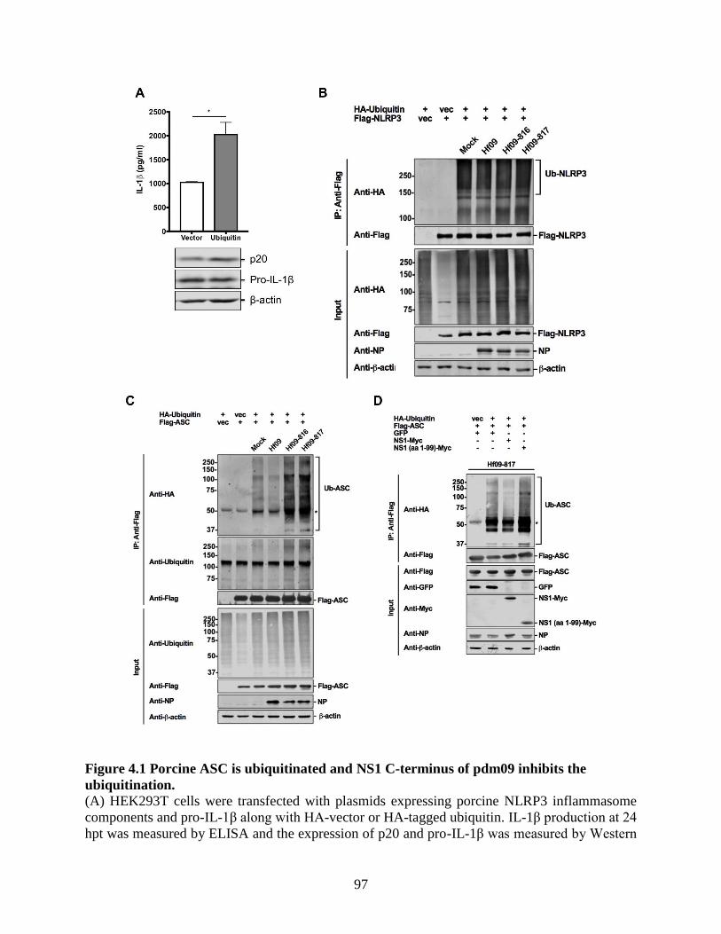
97
Figure 4.1 Porcine ASC is ubiquitinated and NS1 C-terminus of pdm09 inhibits the
ubiquitination.
(A) HEK293T cells were transfected with plasmids expressing porcine NLRP3 inflammasome
components and pro-IL-1β along with HA-vector or HA-tagged ubiquitin. IL-1β production at 24
hpt was measured by ELISA and the expression of p20 and pro-IL-1β was measured by Western

98
blotting. Unpaired t-test, two-tailed, p=0.0279. (B and C) HEK293T cells were transfected with
HA-vector or HA-ubiquitin and Flag-vector or Flag-NLRP3/ASC constructs. At 24 hpt, the cells
were infected with WT or NS1-mutant Hf09 viruses at an MOI of 10 for 9 h. Cell lysates in
denaturing condition were subjected to co-IP with beads conjugated with anti-Flag antibody. IP
and input samples were used for Western blotting with antibodies indicated. (D) HEK293T cells
were transfected with HA-vector or HA-ubiquitin and Flag-ASC constructs along with plasmids
expressing GFP, Myc-tagged WT or mutant NS1 of Hf09. At 24 hpt, the cells were infected with
Hf09-817 at an MOI of 10 for 9 h. Co-IP was conducted as in (B) and Western blotting was
performed with antibodies indicated. The asterisks in (C) and (D) indicate IgG heavy chains.
4.4.2 NS1 C-terminus of pdm09 inhibits the ubiquitination of human ASC.
To explore whether the ability of Hf09 NS1 to suppress ASC ubiquitination is limited to
porcine immunity, ubiquitination assay using a Flag-tagged human ASC construct (Flag-huASC)
was performed. Interestingly, the ubiquitination level of human ASC was increased in cells
infected with Hf09-817 compared to that in mock-infected or WT Hf09-infected cells (Figure
4.2A), showing that Hf09 NS1 C-terminus can inhibit the ubiquitination of both porcine and
human ASC. To answer an arising question if human IL-1β production is also suppressed by C-
terminal NS1 of Hf09, THP-1 cells were infected by Hf09 or Hf09-817. Indeed, human IL-1β
produced from cells infected with Hf09-817 was more than that from Hf09 WT-infected cells
(Figure 4.2B), suggesting that the inhibition of human IL-1β production may be attributed to the
suppressed ASC ubiquitination by pdm09 NS1 C-terminus.

99
Figure 4.2 NS1 C-terminus of pdm09 inhibits the ubiquitination of human ASC.
(A) HEK293T cells were transfected with HA-ubiquitin and Flag-huASC constructs for 24 h and
infected with WT Hf09 or Hf09-817 at an MOI of 10 for 9 h. Cell lysates in denaturing condition
were subjected to co-IP with beads conjugated with anti-Flag antibody. IP and input samples
were used for Western blotting with antibodies indicated. The asterisk indicates IgG heavy
chains. (B) THP-1 cells were infected with Hf09 or Hf09-817 at an MOI of 1 for 24 h. Human
IL-1β levels in cell-free supernatants were analyzed by ELISA. *, p=0.021.
4.4.3 ASC undergoes mixed lysine-linked ubiquitination in response to NS1-mutant
pdm09 virus.
Which lysine residue(s) among seven in the ubiquitin molecules is involved in the
ubiquitination of target proteins may determine the fate of target proteins. Ubiquitination assay
was conducted using mutant ubiquitin constructs with each lysine (K) replaced by arginine (R)
among seven lysines on a ubiquitin molecule. Ubiquitination levels of porcine ASC upon Hf09-
817 infection were not diminished when either K48 or K63 was replaced by arginine in the
ubiquitin (Figure 4.3), suggesting that K48 or K63 alone is not linked to ASC ubiquitination,
rather, mixed lysines are involved.
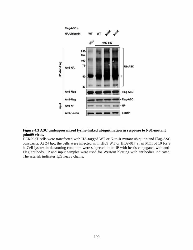
100
Figure 4.3 ASC undergoes mixed lysine-linked ubiquitination in response to NS1-mutant
pdm09 virus.
HEK293T cells were transfected with HA-tagged WT or K-to-R mutant ubiquitin and Flag-ASC
constructs. At 24 hpt, the cells were infected with Hf09 WT or Hf09-817 at an MOI of 10 for 9
h. Cell lysates in denaturing condition were subjected to co-IP with beads conjugated with anti-
Flag antibody. IP and input samples were used for Western blotting with antibodies indicated.
The asterisk indicates IgG heavy chains.

101
4.4.4 K110 and K140 are ubiquitination sites on porcine ASC.
Ubiquitination generally occurs by conjugation of ubiquitin molecules on the lysine(s) in
target proteins. To investigate which lysine(s) in porcine ASC is targeted for ubiquitination, the
positions of lysine residues in porcine ASC were compared to those in human ASC. As
illustrated in Figure 4.4A, porcine ASC has 16 lysines, among which 7 lysines (K79, K87, K91,
K96, K101, K106, and K183) (identified above the bar in Figure 4.4A) are unique for the porcine
ASC. The K175 residue in porcine ASC corresponds to K174 in human ASC, which was
reported to be the target site for ubiquitination upon an RNA virus infection (Guan et al., 2015).
Thus, whether these sites are targeted for ubiquitination was tested first. For that, single lysine-
to-arginine (K-to-R) mutation constructs were made; however, none of the mutations led to a
reduced level of ASC ubiquitination upon Hf09-817 infection (Figure 4.4B, right panel). The
mutations were expanded to the remaining lysines. While individual mutations were made on
K55, K110, K140, and K159, a cluster of lysines (K21, K22, K24, and K26) was simultaneously
mutated due to their close proximity. However, the replacement of these remaining lysines with
arginines did not reduce the ubiquitination levels (Figure 4.4B, left panel). ASC has an N-
terminal pyrin domain (PYD) and a C-terminal caspase activation and recruitment domain
(CARD). Based on the reports that CARD is targeted for different modifications (Guan et al.,
2015, Hara et al., 2013, Lin et al., 2015), whether multiple lysines in the CARD of porcine ASC
are targeted for ubiquitination was tested. Thus, two truncated ASCs, ASC consisting of aa 1 to
109 (ASC1–109) and ASC consisting of aa 1 to 158 (ASC1–158) were constructed (Figure 4.4A).
Upon Hf09-817 infection, the ubiquitination level of ASC1–158 was not altered, while the level on
ASC1–109 was significantly reduced compared to that of the WT ASC (Figure 4.4C). This finding
suggested that the two lysines, K110 and K140, within aa 110 to 158, may be the target for
ubiquitination. Indeed, ASC with a K110R/K140R mutation had an appreciably reduced level of
ubiquitination compared to that of WT ASC in response to Hf09-817 infection (Figure 4.4D).

102
(Figure 4.4 to be continued)

103
Figure 4.4 K110 and K140 are ubiquitination sites on porcine ASC.
(A) A schematic representation of WT and two mutant ASC constructs (aa 1-109 and aa 1-158)
is illustrated with lysine (K) residues. (B, C and D) HEK293T cells were transfected with HA-
ubiquitin and Flag-tagged WT or mutant ASC constructs. At 24 hpt, the cells were infected with
Hf09 WT or Hf09-817 at an MOI of 10 for 9 h. Cell lysates in denaturing condition were
subjected to co-IP with beads conjugated with anti-Flag antibody. IP and input samples were
used for Western blotting with antibodies indicated. The asterisks indicate IgG heavy chains.
4.4.5 Inhibition of ASC ubiquitination on K110 and K140 does not impair the ASC
speck formation or ASC stability.
To define the effects of ubiquitination at K110/K140 on the ASC protein itself, ASC
specks formed by porcine NLRP3 along with either WT or K-to-R mutant ASC were examined
by immunofluorescence staining. Elimination of ASC ubiquitination by the K110R/K140R
mutation did not alter the overall number or the perinuclear localization pattern of ASC specks
(Figure 4.5A). Since ubiquitination may affect the stability of target proteins, the stability of
ASC was monitored by treating the cells transiently expressing WT or K-to-R mutant ASC with

104
CHX, which blocks the translation of new proteins, for different time points before being
subjected to Western blotting. The expression intensity of either WT or mutant ASC was not
reduced by CHX treatment (Figure 4.5B), suggesting that the ASC stability is not critically
affected by preventing the ubiquitination on K110 and K140.
Figure 4.5 Inhibition of ASC ubiquitination on K110 and K140 does not impair the ASC
speck formation or ASC stability.
(A) HEK293T cells were transfected with plasmids expressing Myc-tagged NLRP3 and Flag-
tagged WT/mutant ASC. At 24 hpt, the cells were fixed, permeabilized, immunostained with
appropriate antibodies, and were counterstained with DAPI. ASC specks formed by
colocalization of NLRP3-Myc (gray) and Flag-ASC (green) were seen in perinuclear region.
Nuclei were visualized in blue. The scale bars indicate 5 μm. (B) HEK293T cells were
transfected with 200 ng of plasmids expressing Flag-tagged WT/mutant ASC. At 24 hpt, the cells
were treated with CHX for 2, 4, 6 or 8 h before harvested, while untreated cells were harvested at
24 hpt. The cells were lysed and analyzed by Western blotting with antibodies indicated.

105
4.4.6 Ubiquitination of ASC on K110 and K140 is required for caspase-1 activation
in response to NS1-mutant pdm09 virus.
In an attempt to observe the impact of ubiquitination at K110/K140 on the NLRP3
inflammasome activity, NLRP3 inflammasome reconstitution was conducted using WT/mutant
ASC. In NLRP3 inflammasome reconstitution followed by Hf09-817 infection, the NLRP3
inflammasome consisting of WT ASC could induce an appreciable level of active caspase-1,
p20, and a large amount of IL-1β (Figure 4.6, left panel). In contrast, the inflammasome
consisting of ASC K110R/K140R produced lower caspase-1 activity, as evidenced by the
reduced level of both p20 and IL-1β. None of the single mutations (K110R or K140R) on ASC
had significant effects on caspase-1 activation and IL-1β maturation. The p20 level was almost
half diminished in the ASC K110R/K140R condition compared to other conditions, while the
pro-IL-1β expression was not significantly impaired as the normalized data showed (Figure 4.6,
right panel).
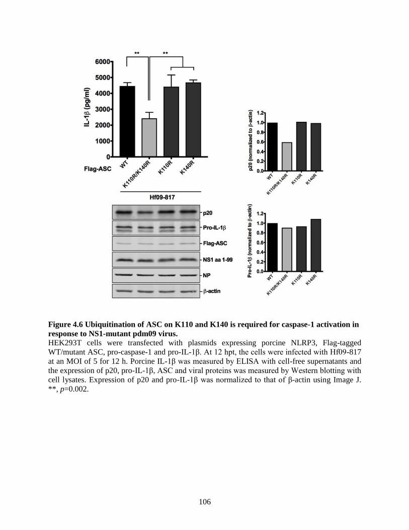
106
Figure 4.6 Ubiquitination of ASC on K110 and K140 is required for caspase-1 activation in
response to NS1-mutant pdm09 virus.
HEK293T cells were transfected with plasmids expressing porcine NLRP3, Flag-tagged
WT/mutant ASC, pro-caspase-1 and pro-IL-1β. At 12 hpt, the cells were infected with Hf09-817
at an MOI of 5 for 12 h. Porcine IL-1β was measured by ELISA with cell-free supernatants and
the expression of p20, pro-IL-1β, ASC and viral proteins was measured by Western blotting with
cell lysates. Expression of p20 and pro-IL-1β was normalized to that of β-actin using Image J.
**, p=0.002.

107
Figure 4.7 A proposed model of the mechanisms by which NS1 C-terminus of pdm09
inhibits the porcine NLRP3 inflammasome.
Viral dsRNA is sensed by TLR3 in the endosome of pdm09-infected alveolar macrophages. This
activates the NF-κB pathway, which is required for the transcriptional upregulation of NLRP3
and pro-IL-1β (Signal 1). Cellular stress provoked by viral infection is expected to induce the
conformational change of NLRP3 (e.g. oligomerization) (Signal 2), while the roles of pdm09
viral proteins in this process are not fully understood. This results in the PYD-PYD interaction
between NLRP3 and ASC leading to the formation of ASC specks. Further recruitment of pro-
caspase-1 to ASC by CARD-CARD interaction completes the assembly of NLRP3
inflammasome. Auto-proteolytic cleavage of pro-caspase-1 into active subunits of caspase-1 is
required to process the pro-IL-1β, leading to IL-1β secretion. NS1 C-terminus of pdm09 inhibits
the porcine NLRP3 inflammasome-mediated IL-1β production with two mechanisms. First, it
suppresses the ubiquitination of porcine ASC on CARD, which is crucial for caspase-1
activation, but not involved in the formation of ASC specks, while whether this inhibition occurs
before or upon NLRP3 activation is unclear. Second, it disrupts the NLRP3-ASC interaction,
thereby impairing the ASC speck formation. The domain structure of NLRP3 inflammasome
components is shown in the inset.

108
4.5 Discussion
Ubiquitination and deubiquitination on NLRP3 inflammasome components mediate the
inflammasome activity. To seek for the in-depth mechanism by which pdm09 NS1 suppresses
porcine NLRP3 inflammasome activity, whether the NS1 modulates the ASC or NLRP3
ubiquitination was investigated. Infection with mutant Hf09 viruses with NS1 C-terminal
deletion caused more ubiquitination of ASC than did the WT virus, but not affecting the
ubiquitination of NLRP3, implying an inhibitory role of Hf09 NS1 C-terminus on porcine ASC
ubiquitination. A speculation that NS1 C-terminus plays an enhancing role on ASC
deubiquitination was eliminated, since WT virus infection induced a low level of ubiquitination,
which was similar to that in mock-infected cells. One possible explanation could be that the
pdm09 NS1 influences E3 ligase activity required for ASC ubiquitination. This type of
regulation is reported in RIG-I ubiquitination by an E3 ligase, TRIM25 that is targeted by NS1 to
suppress the IFN production (Gack et al., 2009). NS1 C-terminal aa 126-225 of avian H5N1 is
recently reported to downregulate the RIG-I-mediated IFN signaling through interacting with
TRAF3 and inhibiting the K63-linked ubiquitination of TRAF3 (Qian et al., 2017). This shows
that the inhibitory function of NS1 C-terminus on the host immunity is not limited to the
suppression of NLRP3 inflammasome. Guan et al. discovered that ASC undergoes ubiquitination
via the E3 ligase TRAF3 by forming the complex of ASC-TRAF3-MAVS upon VSV infection
(Guan et al., 2015). In that study, MAVS contributes to the stability of ASC and increases the
interaction of ASC with TRAF3. In this context, it will be of interest to examine if NS1 interacts
with another adaptor protein that associates with an E3 ligase responsible for the ubiquitination
of porcine ASC. In addition to K63-linked or linear ubiquitination as previously reported (Guan
et al., 2015, Rodgers et al., 2014), ASC was modified by mixed lysine-linked ubiquitination.
This does not seem to be a rare event, since NLRP3 is modified by both K48- and K63-linked
ubiquitination, and is also deubiquitinated upon activation (Guo et al., 2016, Kawashima et al.,
2017, Py et al., 2013, Song et al., 2016). Different types of ubiquitination are thought to
modulate the activities of inflammasomes for tight regulation of the innate immune signalling.
K110 and K140 in porcine ASC were the ubiquitin acceptor sites, suggesting that
modifications of the C-terminal CARD of ASC, which interacts with another CARD of pro-
caspase-1 (Srinivasula et al., 2002, Wang et al., 2002), are crucial for the inflammasome
activation. Removal of ASC ubiquitination by K-to-R mutation on the acceptor sites impaired the

109
caspase-1 activity during virus infection, but did not affect the stability of ASC, localization and
number of ASC specks, suggesting the ubiquitination of porcine ASC contributes to the fully
functional activation of NLRP3 inflammasome. What remains elusive is whether the
ubiquitination on ASC CARD has any impacts on the CARD-CARD interaction between ASC
and pro-caspase-1 upon infection with NS1-mutant virus, or the interaction occurs regardless of
the ASC ubiquitination. It is noteworthy that deubiquitination of ASC can lead to the
inflammasome activation (Lee et al., 2017c), which contradicts to the observation in the current
study. The ambivalence may be due to the diverse interplay between virus and the host signalling
pathways that is dissimilar to how conventional agonists stimulate the immune system. IAV
infection can manipulate not only the host gene expression, but also the host ubiquitin system
(Tisoncik et al., 2011).
In addition to ASC ubiquitination, other post-translational modifications may also occur on
any components of the porcine NLRP3 inflammasome to regulate its activation. SUMOylation or
deSUMOylation, a process by which small ubiquitin-like modifier (SUMO) proteins are
conjugated to or deconjugated from target proteins can be one of them. Unlike ubiquitination,
SUMOylation occurs on a lysine residue in the consensus motif as well as other lysines, which
do not constitute the motif on the target protein (Wilkinson and Henley, 2010). A recent study
shows the elevated activation of NLRP3 inflammasome upon deSUMOylation on NLRP3 (Barry
et al., 2018). Interestingly, porcine ASC, but not human ASC, contains the SUMO consensus
motif, Ψ-K-x-D/E (where Ψ is a hydrophobic residue and x is any residue). Whether porcine
ASC undergoes SUMOylation/deSUMOylation upon IAV infection and if it is relevant to the
NLRP3 inflammasome activation are outstanding concerns.
Hf09 NS1 C-terminus not only suppresses ASC ubiquitination on the CARD, but also
interferes with the PYD-PYD homotypic interaction between porcine NLRP3 and ASC, which
leads to the reduced ASC specks as shown in the previous chapter and in Figure 4.7. Therefore,
the two inhibitory effects by pdm09 NS1 exerted on different domains of ASC are thought to be
independent each other, and pdm09 NS1 may be equipped with redundant strategies to block
NLRP3 inflammasome activation. Considering that ubiquitination of human ASC along with
human IL-1β secretion was also repressed by pdm09 NS1 C-terminus, it is suggested that pdm09
has acquired the ability to evade the host immunity of both pigs and humans. Further

110
investigation is needed to identify the ubiquitination sites on human ASC and to define whether
this strategy is also played by other swine-origin human IAVs.
In summary, porcine ASC could be modified by ubiquitination upon IAV infection, and
NS1 C-terminus of pdm09 suppressed the ubiquitination on K110/K140, which was critical for
NLRP3 inflammasome-mediated IL-1β production. Further, the inhibitory function played by
pdm09 NS1 was also executed on human ASC. These provide new insights into NS1-mediated
immune evasion in the swine and human hosts infected with IAVs.

111
TRANSITION BETWEEN CHAPTER 4 AND CHAPTER 5
In Chapter 4, the ubiquitination of porcine ASC was shown to be suppressed by NS1 C-
terminus of pdm09. Further analyses identified that K110 and K140 on CARD of porcine ASC
were target sites of ubiquitination. Ubiquitination on those residues was indispensable for
caspase-1 activation, while it is not directly involved in the ASC speck formation or ASC
stability. Along with observations in Chapter 3, this revealed novel mechanisms by which pdm09
NS1 exerts to evade the host inflammatory responses. Considering that a high level of IL-1β was
produced upon swine influenza virus (SIV) infection in Chapter 3, an important question about
the underlying mechanism for that event is not resolved yet. Thus, the upstream mechanism by
which SIV induces NLRP3 inflammasome-mediated IL-1β production is explored in the
following chapter. Based on the knowledge that mitochondria play a central role in controlling
the innate immunity including the NLRP3 inflammasome activation, how mitochondrial
dynamics and its related signalling are involved in SIV-induced IL-1β production is a main
subject in Chapter 5.

112
CHAPTER 5 SWINE INFLUENZA VIRUS INDUCES NLRP3 INFLAMMASOME-
MEDIATED INTERLEUKIN-1 BETA PRODUCTION THROUGH THE
RIPK1/DRP1 SIGNALING.
The data presented in this chapter were published as
Swine Influenza Virus Induces RIPK1/DRP1-Mediated Interleukin-1 Beta Production.
HS Park, G Liu, Q Liu and Y Zhou. (2018)
Viruses, 10(8), 419.
Acknowledgement specific for this chapter:
The WT and mutant human RIPK1 constructs were provided by Dr. Xin Lin (MD Anderson
Cancer Center) and Dr. Francis Chan (University of Massachusetts Medical School),
respectively.

113
5.1 Abstract
Nucleotide-binding domain and leucine-rich repeat-containing protein 3 (NLRP3)
inflammasome plays a pivotal role in modulating lung inflammation in response to the influenza
A virus infection. Based on the previous finding that swine influenza virus (SIV) infection
induced NLRP3 inflammasome-mediated IL-1β production in primary porcine alveolar
macrophages (PAMs), the upstream signaling events that are involved in this process were
examined. In this study, results showed that SIV infection led to dynamin-related protein 1
(DRP1) phosphorylation at serine 579 (S579) and mitochondrial fission in PAMs.
Concomitantly, IL-1β production was dependent on the reactive oxygen species (ROS)
production, and NLRP3 inflammasome activity was upregulated by phospho-mimetic mutation
at S579 on DRP1. Furthermore, the requirement of the kinase activity of receptor-interacting
protein kinase 1 (RIPK1) for the IL-1β production and the association of RIPK1 with DRP1
suggested that RIPK1 is an upstream kinase for DRP1 phosphorylation. These results showed a
critical role of the RIPK1/DRP1 signaling axis, whose activation leads to mitochondrial fission
and ROS release, in modulating porcine NLRP3 inflammasome-mediated IL-1β production in
SIV-infected PAMs.
5.2 Introduction
Interleukin-1 beta (IL-1β) is a pro-inflammatory cytokine that contributes to the effective
modulation of the host innate and adaptive immunity upon influenza A virus (IAV) infection
(Allen et al., 2009, Ichinohe et al., 2009, Thomas et al., 2009). IL-1β is tightly regulated by
pattern recognition receptors (PRRs) such as nucleotide-binding domain and leucine-rich repeat-
containing protein 3 (NLRP3), which functions by forming the NLRP3 inflammasome with its
adaptor protein, apoptosis-associated speck-like protein containing caspase activation and
recruitment domain (ASC), and pro-caspase-1. The activation of NLRP3 leads to the conversion
of pro-caspase-1 into caspase-1, which cleaves pro-IL-1β to generate mature IL-1β and also
induces pyroptosis (Kuriakose and Kanneganti, 2017, Sarvestani and McAuley, 2017).
Mitochondria are well appreciated for their roles in innate immunity. Mitochondrial
proteins are involved in the NLRP3 inflammasome regulation (Ichinohe et al., 2013, Moriwaki et
al., 2016, Park et al., 2013b). Pathophysiological changes mediated by mitochondria including
the imbalance in mitochondrial dynamics and the release of cytochrome c, mitochondrial DNA

114
or reactive oxygen species (ROS) can either activate or inhibit the NLRP3 inflammasome
(Nakahira et al., 2011, Park et al., 2015, Shi and Kehrl, 2016, Yoshizumi et al., 2014, Zhou et al.,
2011). The phosphorylation of dynamin-related protein 1 (DRP1), a guanosine triphosphate
(GTP) hydrolase (GTPase), on different residues determines mitochondrial dynamics, namely
fission and fusion. For human DRP1 (transcript variant 1), phosphorylation at serine 616 (S616)
or dephosphorylation at serine 637 (S637) is linked to mitochondrial fission, whereas
phosphorylation at S637 is associated with fusion (Zemirli et al., 2018). In order for fission to
occur, DRP1 is recruited to mitochondria upon its phosphorylation at S616 by kinases including
receptor-interacting protein kinase 1 (RIPK1) or dephosphorylation at S637 (Cereghetti et al.,
2008, Kashatus et al., 2015, Taguchi et al., 2007, Wang et al., 2014c). The association of DRP1
with mitochondrial fission proteins leads to mitochondrial fission, which depends on GTP
hydrolysis by DRP1 (Elgass et al., 2013). The suppression of mitochondrial fission by diverse
molecules leads to the inhibition of NLRP3 inflammasome activity (Li et al., 2016a, Li et al.,
2016d, Zhou et al., 2016), while this is contradicted by a finding that the impaired mitochondrial
fission can upregulate the NLRP3 inflammasome activity (Park et al., 2015).
Viruses can alter the mitochondrial dynamics to subvert the host’s innate immunity or to
modulate the host cell survival. Various RNA viruses are reported to induce DRP1-mediated
mitochondrial fission to regulate apoptosis (Gou et al., 2017, Kim et al., 2014, Li et al., 2016c,
Mukherjee et al., 2018), whereas the dengue virus is reported to regulate mitochondrial fusion
(Barbier et al., 2017, Chatel-Chaix et al., 2016, Yu et al., 2015). An initial study on DRP1-
mediated NLRP3 inflammasome activation upon viral infection discovered that in response to
vesicular stomatitis virus (VSV), RIPK1 forms a complex with RIPK3 to induce necroptosis,
while it also phosphorylates DRP1. The latter event leads to mitochondrial fission, and
subsequently, ROS can activate NLRP3 leading to IL-1β production (Wang et al., 2014c). In
contrast, another report showed that the NLRP3 inflammasome activation upon VSV infection
does not require RIPK1 or DRP1 (Kang et al., 2015). In addition, there are reports showing that
cyclin-dependent kinase 1 is required for DRP1 phosphorylation and mitochondrial fission upon
infection with RNA viruses such as hepatitis C virus and rotavirus (Kim et al., 2014, Mukherjee
et al., 2018). Upon IAV infection, a mitochondrial fusion protein, mitofusin 2, can regulate the
NLRP3 inflammasome activation (Ichinohe et al., 2013). The IAV PB1-F2 protein, which
translocates to mitochondria, can cause mitochondrial fission that is associated with the defective
115
NLRP3 inflammasome activation (Yoshizumi et al., 2014). However, there is still a lack of
information on how DRP1-mediated mitochondrial fission fits in the innate immune signaling in
IAV-infected cells.
In the previous chapter, swine influenza virus (SIV) infection induced NLRP3
inflammasome activation leading to the IL-1β production in primary porcine alveolar
macrophages (PAMs). In this chapter, whether DRP1-mediated mitochondrial dynamics are
involved in this process was investigated. The results suggest that upon SIV infection of PAMs,
mitochondrial fission occurred through the RIPK1/DRP1 signaling axis, which is involved in the
NLRP3 inflammasome-mediated IL-1β production.
5.3 Materials and methods
5.3.1 Cells and viruses
PAMs were isolated from lungs of 4-week-old, SIV-negative piglets by collecting
bronchoalveolar lavage fluid, where most of the cells are comprised of alveolar macrophages
(Ait-Ali et al., 2007, Gordon and Read, 2002). The PAMs were characterized by flow cytometry
using mouse anti-pig macrophage antibody conjugated with fluorescein isothiocyanate
(MCA2317F, Bio-Rad) (Liu et al., 2015). PAMs were cultured with HyClone RPMI 1640
medium (SH30027.01, GE Healthcare) supplemented with 20% FBS (16000-044, Thermo
Fisher), 50 μg/mL gentamicin (BS724, Bio Basic Canada), and 1 × Antibiotic-Antimycotic
(15240-062, Thermo Fisher). HEK293T cells were maintained in DMEM (D5796, Sigma)
supplemented with 10% FBS and 50 μg/mL gentamicin. The SIV strain, Sk02 was propagated in
MDCK cells and titrated by plaque assay as mentioned in the previous chapters.
5.3.2 Antibodies and reagents
Rabbit polyclonal NP and NS1 antibodies were produced as described (Shin et al., 2007b).
The commercial antibodies used are as follows. Goat anti-porcine IL-1β antibody (BAF681):
R&D Systems; rabbit anti-porcine caspase-1 (p20) antibody (PAB592Po01): Cloud-Clone Corp.;
mouse anti-Myc-tag antibody (#2276), mouse anti-β-actin antibody (#3700), rabbit anti-DRP1
antibody (#8570) and rabbit anti-phospho-DRP1 (S616) antibody (#3455): Cell Signaling
Technology; mouse anti-FLAG M2 antibody (F3165): Sigma; IRDye 680RD donkey anti-rabbit

116
(926-68073), IRDye 800CW donkey anti-mouse (926-32212) and IRDye 800CW donkey anti-
goat (926-32214) antibodies: LI-COR Biosciences. The following reagents were used: LPS
(L3024) and N-acetyl L-cysteine (NAC) (A9165): Sigma; Necrostatin-1 (Nec-1) (BML-AP309):
Enzo Life Sciences; Mdivi-1 (ab144589): Abcam.
5.3.3 Plasmid construction
Plasmids expressing porcine NLRP3, ASC, procaspase-1, and pro-IL-1β were generated
using cDNA synthesized from the total RNA of PAMs that were stimulated with LPS as
described in the previous chapters. Using the same cDNA, the full-length porcine DRP1 was
cloned into pcDNA3.1-3×Myc (C-terminal tag) generating pcDNA-DRP1-Myc. Primers were
designed based on the GenBank sequence of porcine DRP1 transcript variant X1 (accession
number: XM_021092056). Myc-tagged DRP1 constructs with S579D or S579A mutation
(pcDNA-DRP1 (S579D)-Myc or pcDNA-DRP1 (S579A)-Myc) were generated by site-directed
mutagenesis using pcDNA-DRP1-Myc as the template. Porcine RIPK1 was cloned into
pcDNA3.1-3×Myc or pCMV-3×Flag (N-terminal tag) using cDNA from a porcine alveolar
macrophage cell line, 3D4/2, generating pcDNA-RIPK1-Myc or pCMV-Flag-RIPK1. Using the
above construct as the template, the mutant RIPK1 construct with K41A/K42A (pcDNA-RIPK1
(K41A/K42A)-Myc) was generated by mutagenesis. Myc-tagged human RIPK1 (huRIPK1)
(#44159, Addgene) was a gift from Xin Lin (Blonska et al., 2004). HA/GFP-tagged human
RIPK1 (K45A) (#41389, Addgene) was a gift from Francis Chan (Cho et al., 2009) and used as
the template to generate Myc-tagged human RIPK1 (K45A). All the mutations were confirmed
by nucleotide sequencing. The expression plasmids generated for this chapter are listed in Table
5.1 and the primers are listed in Supplementary Table 4.

117
Table 5.1 List of expression plasmids used in Chapter 5.
The constructs for NLRP3 inflammasome components and pro-IL-1β used for NLRP3
inflammasome reconstitution assay were described in Table 3.2.
Construct Template used to clone Purpose
pcDNA-DRP1-Myc cDNA from PAMs NLRP3 inflammasome reconstitution,
RIPK1-DRP1 interaction
pcDNA-DRP1 (S579D)-
Myc pcDNA-DRP1-Myc NLRP3 inflammasome reconstitution
pcDNA-DRP1 (S579A)-
Myc pcDNA-DRP1-Myc NLRP3 inflammasome reconstitution
huRIPK1-Myc
(Blonska et al., 2004) NLRP3 inflammasome reconstitution
huRIPK1 (K45A)-Myc
HA/GFP-huRIPK1
(K45A)
(Cho et al., 2009)
NLRP3 inflammasome reconstitution
pcDNA-RIPK1-Myc cDNA from 3D4/2 cells NLRP3 inflammasome reconstitution
pcDNA-RIPK1
(K41A/K42A)-Myc pcDNA-RIPK1-Myc NLRP3 inflammasome reconstitution
pCMV-Flag-RIPK1 pcDNA-RIPK1-Myc NLRP3 inflammasome reconstitution,
RIPK1-DRP1 interaction
5.3.4 Infection and treatment of PAMs
PAMs seeded at 1 × 10⁶ cells per well on 24-well plates were either infected with the Sk02
virus for 20 h at an MOI of 1 or stimulated with 200 ng/mL LPS for 12 h. The cells were pre-
treated with vehicles or inhibitors for 2 h, and were then infected with the virus or stimulated
with LPS in the presence of the vehicles or inhibitors. Nec-1 was used at 80 or 160 μM for
RIPK1 inhibition; Mdivi-1 was used at 5 or 20 μM for DRP1 inhibition, while DMSO was used
as the vehicle. As a ROS scavenger, NAC was used at 2 or 10 mM and distilled water was used
as the vehicle. Cell-free supernatants were collected for ELISA. Cells were lysed with 1 × SDS
sample buffer and boiled for Western blotting. To detect the phosphorylated DRP1 (phospho-
DRP1), the PAMs were seeded at 3 × 10⁶ cells per well on 6-well plates. The cells were infected
with the Sk02 virus for 4 h at an MOI of 1 or stimulated with 200 ng/mL LPS for 12 h. The cells
in each well were lysed with 120 μL of M-PER Mammalian Protein Extraction Reagent (78503,

118
Thermo Fisher) containing 1× Halt Protease and Phosphatase Inhibitor Cocktail (78440, Thermo
Fisher). After centrifugation, the cell lysates were mixed with 5 × SDS sample buffer and were
subjected to Western blotting.
5.3.5 RNA interference and quantitative PCR
siRNA targeting porcine RIPK1 was designed by the siRNA design tool provided by
Dharmacon. The RIPK1-targeting siRNA (5′-UGGAAGAGGAUGUGAAGAAUU-3′) and a
negative control siRNA (D-001810-01-05, 5′-UGGUUUACAUGUCGACUAA-3′) were
purchased from Dharmacon. PAMs seeded at 1 × 10⁶ cells per well on 24-well plates were
transfected with 100 nM of the control siRNA or RIPK1-targeting siRNA using Lipofectamine
RNAiMAX (13778-030, Thermo Fisher). At 24 hpt, the cells were infected with the Sk02 virus
for 16 h at an MOI of 1. To show the knockdown efficiency, the relative mRNA expression of
RIPK1 compared to that of porcine HPRT1 gene was measured by quantitative PCR using cDNA
from total RNA with a standard protocol. Primers for porcine RIPK1 were designed based on the
GenBank sequence (accession number XM_005665536) using an online tool, Primer3
(Untergasser et al., 2012) and those for the housekeeping gene, HPRT1 were used as previously
reported (Nygard et al., 2007). Primer sequences are indicated in Table 5.2.
Table 5.2 List of primers used for quantitative PCR in Chapter 5.
Target Primer name Sequence (5’-3’) Amplicon size
Porcine HPRT1 HPRT1 Fw GGACTTGAATCATGTTTGTG
91 bp HPRT1 Bw CAGATGTTTCCAAACTCAAC
Porcine RIPK1 RIPK1 Fw TTCGGAAAGGTGTCCTTGTG
91 bp RIPK1 Bw CGTTGTACTCATTGCGCTTG
bp, base pairs.
5.3.6 NLRP3 inflammasome reconstitution assay
The porcine NLRP3 inflammasome reconstitution assay was conducted to examine the
effects of ectopically expressed proteins on NLRP3 inflammasome activity as described in the
previous chapters. Briefly, the HEK293T cells seeded at 1.5 × 10⁵ cells per well on 24-well
plates were co-transfected with expression plasmids for porcine NLRP3 inflammasome

119
components and pro-IL-1β (pcDNA-NLRP3 (30 ng), pcDNA-ASC (20 ng), pCMV-Flag-
procaspase-1 (20 ng), and pcDNA-pro-IL-1β (100 ng)) using TransIT-LT1 Transfection Reagent
(MIR2300, Mirus Bio). Other plasmids (50 ng of Myc-vector or Myc-tagged human RIPK1
WT/mutant, 100 ng of Myc-vector or Myc-tagged porcine RIPK1 WT/mutant, 100 ng of Myc-
vector or Myc-tagged DRP1 WT/mutant) were co-transfected to study their effects on NLRP3
inflammasome activity. At 16 hpt, supernatants were harvested for IL-1β ELISA and the pelleted
cells were lysed for Western blotting.
5.3.7 Co-IP
HEK293T cells were seeded at 9 × 10⁵ per well on 6-well plates and were transfected with
1 μg of Flag-tagged vector or Flag-tagged RIPK1 and Myc-tagged DRP1. At 24 hpt, the cells
were lysed with 500 μL lysis buffer (50 mM Tris, pH 7.4, 150 mM NaCl, 0.5% Nonidet P-40
substitute, 1 × protease inhibitor cocktail) and clarified by centrifugation at 12,000 × g at 4 °C
for 10 min. For input, 10% of the cell lysates were mixed with 5× SDS sample buffer and boiled
at 95 °C for 5 min. For each sample, 35 μL of Dynabeads Protein G (10004D, Thermo Fisher)
were first conjugated with 1 μg of mouse monoclonal anti-FLAG M2 antibody (F3165, Sigma)
in 300 μL phosphate-buffered saline (PBS) with 0.02% Tween 20 by agitation at room
temperature for 1 h. Then, the beads were incubated with the cell lysates at room temperature for
2 h with agitation. After washed thrice with Tris-buffered saline (50 mM Tris, pH 7.4, 150 mM
NaCl), the beads were resuspended in 60 μL of 2× SDS sample buffer and boiled as above to be
analyzed by SDS-PAGE and Western blotting.
5.3.8 Porcine IL-1β ELISA
Porcine IL-1β in the cell-free supernatants was determined using ELISA as described in the
previous chapters. Briefly, Immulon 2 HB U plates (#3655, Thermo Fisher) were coated with a
mouse anti-porcine IL-1β antibody (MAB6811) at 2 μg/mL in PBS overnight. The plates were
blocked with 1% BSA (A7030, Sigma) in PBS for 1 h, and were incubated with either samples
or the standard for 2 h. Two-fold serial dilutions of the recombinant porcine IL-1β protein (681-
PI-010, R&D Systems) in diluent (0.1% BSA in Tris-buffered saline with 0.05% Tween 20) was
used for the standard curve. Next, the plates were incubated with goat anti-porcine IL-1β
biotinylated antibody (BAF681, R&D Systems) at 50 ng/mL in the diluent for 1 h, and further

120
incubated for 1 h with alkaline phosphatase-streptavidin (016-050-084, Jackson
ImmunoResearch) that was diluted in a ratio of 1:5,000 by the diluent. After incubation with 1
mg/mL of PNPP in the diethanolamine buffer (1 M diethanolamine, 0.5 M MgCl2, pH 9.8),
optical densities were measured at 405 nm with a reference at 490 nm using the xMark
Microplate Absorbance Spectrophotometer (Bio-Rad).
5.3.9 Western blotting
Cell lysates or IP samples were subjected to SDS-PAGE followed by blotting on
nitrocellulose membranes. The membranes were blocked with 5% skim milk (or 5% BSA for the
detection of phospho-DRP1) in TBS with 0.1% Tween 20 for 1 h and incubated with primary
antibodies at 4 °C overnight. The membranes were further incubated with secondary antibodies
at room temperature for 1 h and were scanned with an Odyssey Infrared Imager (LI-COR
Biosciences).
5.3.10 Confocal Microscopy
To check the mitochondrial integrity, PAMs were seeded at 2 × 10⁵ cells per well on
LabTek II CC2 chamber slides (154941, Thermo Fisher). The cells were infected with the Sk02
virus at an MOI of 1 for 7 h or they were stimulated with 200 ng/mL LPS for 16 h. After fixed
with 4% paraformaldehyde for 10 min and permeabilized with 0.1% Triton X-100 in DPBS for 5
min, the cells were incubated with 5% BSA in DPBS for 1 h. The cells were probed with mouse
monoclonal anti-TOM20 antibody (sc-17764, Santa Cruz) and goat polyclonal anti-IAV
antibody (AB1074, EMD Millipore) overnight at 4 °C, and the secondary antibodies (Alexa
Fluor 488 donkey polyclonal anti-mouse IgGH+L (A21202; Invitrogen) and Alexa Fluor 633
donkey polyclonal anti-goat IgGH+L (A21082; Invitrogen)) for 1 h at room temperature.
Counterstaining was performed with DAPI (D1306, Invitrogen) for 5 min and the coverslips
were mounted with ProLong Diamond Antifade Mountant (P36961, Invitrogen) overnight at
room temperature. Images were obtained by using a confocal laser scanning microscope (TCS
SP8, Leica).

121
5.3.11 Statistical analysis
ANOVA with Tukey’s multiple comparison tests or unpaired t-test with Welch’s
correction was performed using GraphPad Prism 7. The error bars indicate the mean ± SD. The
p-values of less than 0.05 were considered to be statistically significant.
5.4 Results
5.4.1 SIV infection induces dynamin-related protein 1 (DRP1) phosphorylation and
mitochondrial fission.
It has been reported that virus-induced DRP1 phosphorylation at S616 leads to the
mitochondrial fission in the early stages of viral infection (Keck et al., 2017, Mukherjee et al.,
2018, Wang et al., 2014c). Amino acid alignment indicated that S616 (location based on the
transcript variant X1 of human DRP1) corresponds to S579 of porcine DRP1 (transcript variant
X8) and is conserved among different isoforms of human DRP1 and porcine DRP1. It was of
interest whether phosphorylation at S579 on porcine DRP1 and the subsequent mitochondrial
fission would occur upon SIV infection. PAMs were infected with the Sk02 virus for 4 h at an
MOI of 1 and the cell lysates were analyzed by Western blotting with antibodies specific for
phospho-DRP1 (S616) or the total form of DRP1. The SIV infection indeed resulted in
phosphorylation of DRP1 at S579, while the expression of total form was not changed (Figure
5.1A). As a positive control, PAMs were treated with LPS, which is known to induce DRP1
S616 phosphorylation or DRP1 S637 dephosphorylation-mediated mitochondrial fission in
murine cells (Park et al., 2013a, Shen et al., 2018). As expected, LPS stimulation led to DRP1
phosphorylation in the PAMs (Figure 5.1B).
To test whether mitochondrial fission also occurs by SIV infection, the PAMs were
infected with Sk02 for 7 h at an MOI of 1. Immunofluorescence staining was conducted by
probing the cells with the antibody against translocase of outer mitochondrial membrane 20
(TOM20), to observe the mitochondrial morphology. Confocal imaging revealed that in Sk02-
infected cells, mitochondria exhibited short and round forms owing to fragmentation; however,
in mock-infected cells, normal tubular and networked mitochondria were observed (Figure
5.1C). Likewise, mitochondrial fission was demonstrated in LPS-treated PAMs (Figure 5.1D).
These data show that upon SIV infection, porcine DRP1 phosphorylation at S579 followed by
mitochondrial fission occurs in PAMs.
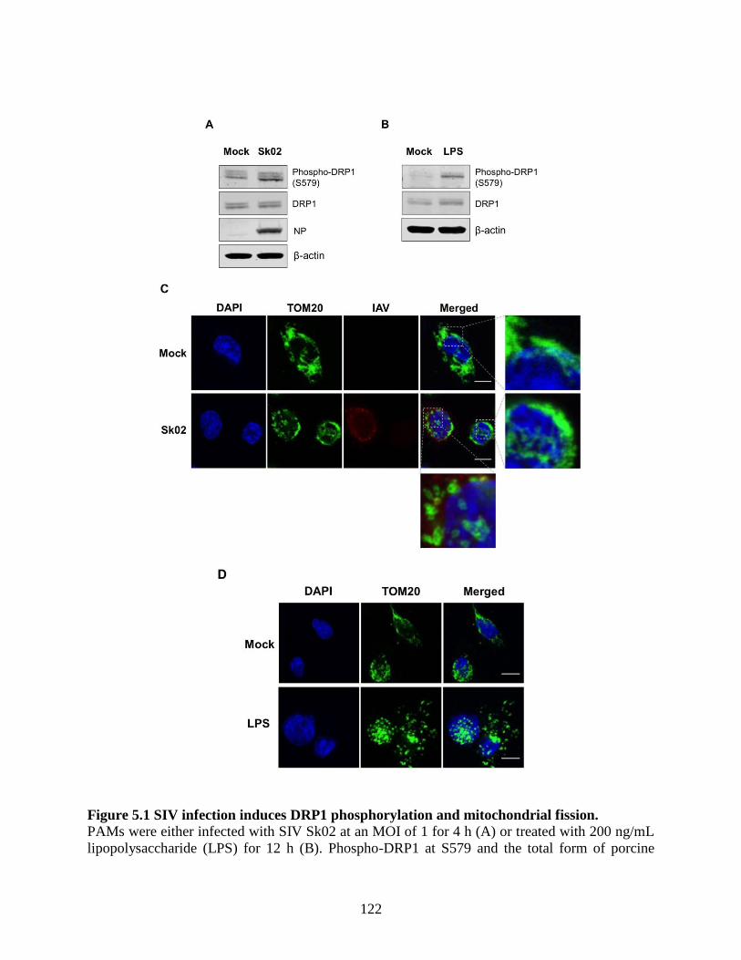
122
Figure 5.1 SIV infection induces DRP1 phosphorylation and mitochondrial fission.
PAMs were either infected with SIV Sk02 at an MOI of 1 for 4 h (A) or treated with 200 ng/mL
lipopolysaccharide (LPS) for 12 h (B). Phospho-DRP1 at S579 and the total form of porcine

123
DRP1 were detected by Western blotting. The viral NP protein was also monitored by Western
blotting in infected cells. PAMs were either infected with Sk02 at an MOI of 1 for 7 h (C) or
treated with 200 ng/mL LPS for 16 h (D). Immunofluorescence and confocal microscopy were
performed using an antibody against the mitochondrial protein, TOM20, and the influenza A
virus (IAV) antibody, when infected. Nuclei were stained by DAPI. Boxed areas were magnified
and displayed on the right side (for a mock or un-infected cell) or below (for an infected cell) the
corresponding panels in (C). Scale bar, 10 μm.
5.4.2 SIV-induced porcine IL-1β production is dependent on reactive oxygen species
(ROS) production.
Mitochondrial ROS from damaged mitochondria can induce NLRP3 inflammasome
activation (Allen et al., 2009, Zhou et al., 2011) and whether IL-1β production by SIV infection
is ROS-dependent was further examined. PAMs were infected with Sk02 in the presence of a
ROS scavenger, NAC, or a vehicle for 20 h. The IL-1β level detected from the supernatant was
considerably higher in SIV-infected cells than that in mock-infected cells (Figure 5.2A).
Treatment of NAC significantly decreased IL-1β production in a dose-dependent manner. The
expression of pro-IL-1β and the viral protein was not impaired, indicating the NAC eliminates a
signal required for inflammasome activity. As a control, a decline in IL-1β levels was also
achieved when the cells were treated with LPS in the presence of NAC (Figure 5.2B). This
suggests that ROS generated from fragmented mitochondria enhances NLRP3 inflammasome-
dependent IL-1β production in PAMs upon SIV infection.

124
Figure 5.2 SIV-induced porcine IL-1β production is dependent on ROS production. PAMs were either infected with Sk02 at an MOI of 1 for 20 h (A) or stimulated with 200 ng/mL
LPS for 16 h (B) in the presence of vehicle (distilled water) or increasing concentrations (2 and
10 mM) of an ROS scavenger, NAC. Porcine IL-1β levels from the supernatants were measured
by ELISA and the expression of pro-IL-1β, viral NS1, and β-actin was analyzed by Western
blotting. Statistical analysis was performed with one-way ANOVA. **, p< 0.01; ***, p< 0.001.
5.4.3 SIV-induced porcine IL-1β production is mediated by DRP1.
The aforementioned data suggested that DRP1-mitochondrial fission axis is involved in the
SIV-induced IL-1β production. To further confirm the role of DRP1, PAMs were treated with
Mdivi-1, which inhibits the GTPase activity of DRP1 and mitochondrial fission (Cassidy-Stone
et al., 2008), and were then infected with Sk02 at an MOI of 1 for 20 h. As seen before, the IL-1β
level was appreciably elevated in SIV-infected cells compared to that in mock cells. The
treatment with Mdivi-1 led to a decreased IL-1β production upon Sk02 infection in a dose-
dependent manner. Pro-IL-1β expression and viral replication as indicated by NS1 expression
were not much affected (Figure 5.3A). LPS-induced inflammatory cytokine expression is shown
to be mediated by DRP1 activity (Park et al., 2013a), hence the LPS-treated cells were also used
as a control. With the pro-IL-1β expression unaffected, the IL-1β production upon LPS was
declined by the Mdivi-1 treatment (Figure 5.3B).

125
To demonstrate the role of S579-phosphorylated DRP1 on pro-caspase-1 cleavage, a
hallmark of inflammasome activation, NLRP3 inflammasome reconstitution was performed
using HEK293T cells that are deficient for endogenous human NLRP3 inflammasome (Bryan et
al., 2009). In this assay, the first signal required for NLRP3 inflammasome activation is provided
by the overexpression of NLRP3 inflammasome components and pro-IL-1β; the regulatory effect
on the second signal by the proteins and particular amino acids of interest can be examined by
monitoring the levels of cleaved caspase-1 and/or mature IL-1β. Despite some limitations, as for
all in vitro assays, such as excluding the roles of undefined co-factors, the system is especially
useful when the cells, in this study PAMs, are difficult to be transfected. Among the different
transcript variants of DRP1, porcine DRP1, which was the most identical to the variant X8
(GenBank accession number XM_021092064), was cloned. S579 in porcine DRP1 corresponds
to S616 in human DRP1 as explained above and as depicted in Figure 5.3C. Using this as the
template, the phospho-mimetic (S579D) and phospho-deficient (S579A) mutants of DRP1 were
generated. HEK293T cells were transfected with plasmids expressing NLRP3 inflammasome
components and pro-IL-1β along with Myc-tagged vector or WT/mutant DRP1. The co-
expression of phospho-mimetic DRP1 (S579D) could significantly increase the caspase-1
activity as shown by the increased level of an active form of caspase-1, p20, while not affecting
the expression of pro-IL-1β (Figure 5.3D). In contrast, this was not observed with WT DRP1 or
phospho-deficient DRP1 (S579A). Concomitantly, the porcine IL-1β level was also higher in the
supernatant of cells co-expressing DRP1 (S579D) compared to other conditions. This data
strongly indicate that porcine DRP1 phosphorylation at S579, which is required for
mitochondrial fission, enhances the NLRP3 inflammasome activation.
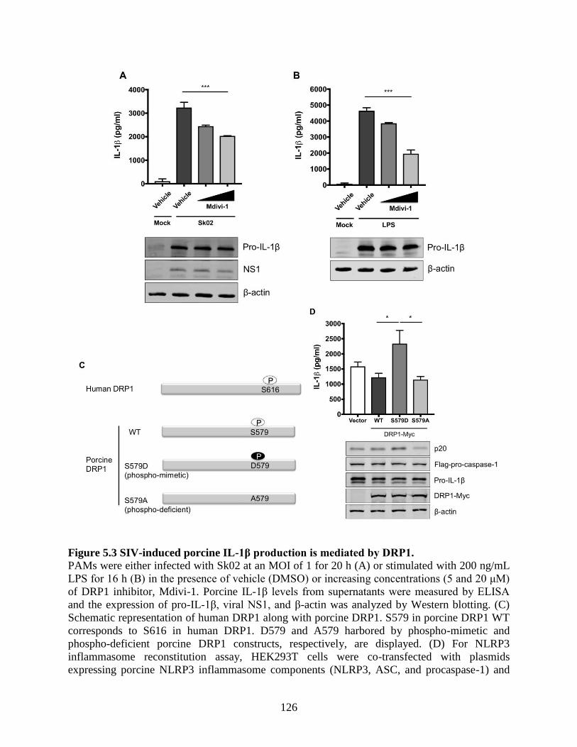
126
Figure 5.3 SIV-induced porcine IL-1β production is mediated by DRP1.
PAMs were either infected with Sk02 at an MOI of 1 for 20 h (A) or stimulated with 200 ng/mL
LPS for 16 h (B) in the presence of vehicle (DMSO) or increasing concentrations (5 and 20 μM)
of DRP1 inhibitor, Mdivi-1. Porcine IL-1β levels from supernatants were measured by ELISA
and the expression of pro-IL-1β, viral NS1, and β-actin was analyzed by Western blotting. (C)
Schematic representation of human DRP1 along with porcine DRP1. S579 in porcine DRP1 WT
corresponds to S616 in human DRP1. D579 and A579 harbored by phospho-mimetic and
phospho-deficient porcine DRP1 constructs, respectively, are displayed. (D) For NLRP3
inflammasome reconstitution assay, HEK293T cells were co-transfected with plasmids
expressing porcine NLRP3 inflammasome components (NLRP3, ASC, and procaspase-1) and

127
pro-IL-1β along with Myc-vector or Myc-tagged WT/mutant porcine DRP1 for 16 h. Porcine IL-
1β levels from supernatants were measured by ELISA. The expression of caspase-1 (active and
precursor forms) and pro-IL-1β was analyzed by Western blotting. Statistical analysis was
performed with one-way ANOVA. *, p< 0.05; ***, p< 0.001.
5.4.4 Kinase activity of receptor-interacting protein 1 (RIPK1) is critical for SIV-
induced IL-1β production.
Porcine RIPK1 was proposed as a DRP1 kinase upon SIV infection, based on a study
showing that DRP1 phosphorylation upon VSV infection is mediated by RIPK1 (Wang et al.,
2014c). To demonstrate the requirement of RIPK1 for porcine NLRP3 inflammasome-mediated
IL-1β production, the RIPK1 expression was silenced in PAMs by siRNA. At 24 hpt, the cells
were infected with Sk02 for 16 h. IL-1β production from PAMs transfected with RIPK1 siRNA
was significantly decreased compared to the cells transfected with an off-target control siRNA,
while the pro-IL-1β expression was not affected by silencing the gene (Figure 5.4A). The
knockdown efficiency determined by the mRNA level of RIPK1 was around 40% compared to
that in negative control siRNA-transfected cells. Furthermore, to examine whether the RIPK1
kinase activity is required for upregulation of NLRP3 inflammasome activity, Nec-1, an inhibitor
of the RIPK1 kinase activity (Degterev et al., 2008), was used in PAMs. Nec-1 blocks the kinase
function by acting on S161 on human RIPK1 (Xie et al., 2013) and this residue is conserved
among different transcript variants of porcine RIPK1. Nec-1 treatment drastically reduced IL-1β
production in response to the Sk02 infection in a dose-dependent manner, while not impairing
the pro-IL-1β expression (Figure 5.4B).
Aiming to confirm the requirement of the porcine RIPK1 kinase function on IL-1β
production, the amino acid sequences of the N-terminal ends of the RIPK1 kinase domain from
pigs, mice, and humans were aligned (Figure 5.4C). Lysine 45 (K45), indicated with an
underline, of human RIPK1 is a critical residue for its kinase activity (Moriwaki and Chan,
2013). While porcine and mouse RIPK1 also contain lysine at the corresponding location to K45
of their human analog, they have two consecutive lysines as underlined, K41/K42 in porcine
RIPK1 and K45/K46 in mouse RIPK1. The kinase-dead effects by K45A mutation or
K45A/K46T mutation on mouse RIPK1 and their roles in IL-1β production are well documented
(Berger et al., 2014, Shutinoski et al., 2016, Zhang et al., 2017c). Considering the higher
128
sequence identity of porcine RIPK1 to the human one, human RIPK1 constructs were used in the
NLRP3 inflammasome reconstitution assay. The co-expression of human RIPK1 WT remarkably
increased the porcine IL-1β production. In contrast, the co-expression of kinase-dead human
RIPK1 (K45A) led to a decreased level of IL-1β production compared to that of WT RIPK1
(Figure 5.4D). Another experiment after cloning porcine RIPK1 was performed in the same
setting as above. Consistently with human RIPK1, porcine RIPK1 WT enhanced IL-1β
production, however, the kinase-dead RIPK1 (K41A/K42A) did not (Figure 5.4E). In all
samples, the pro-IL-1β expression levels were not affected (Figure 5.4D, E). These suggest that
RIPK1 is a positive regulator of NLRP3 inflammasome-mediated IL-1β production and its
kinase activity is required for this regulation.

129

130
Figure 5.4 Kinase activity of RIPK1 is critical for SIV-induced IL-1β production.
(A) PAMs were transfected with a negative control siRNA (siControl) or RIPK1-targeting
siRNA (siRIPK1) for 24 h and infected with Sk02 at an MOI of 1 for 16 h. Porcine IL-1β levels
from supernatants were measured by ELISA. Expression of pro-IL-1β was analyzed by Western
blotting. RIPK1 knockdown efficiency was determined by real-time PCR. (B) PAMs were
infected with Sk02 at an MOI of 1 for 20 h in the presence of a vehicle (DMSO) or increasing
concentrations (80 and 160 μM) of RIPK1 kinase inhibitor, Nec-1. Porcine IL-1β levels from
supernatants were measured by ELISA. Expression of pro-IL-1β and viral NS1 was analyzed by
Western blotting. (C) Amino acid sequences of the N-terminal ends of the kinase domains in
porcine, murine, and human RIPK1 (based on the GenBank accession numbers XM_005665536,
NM_009068, and NM_003804, respectively) were aligned using Clustal Omega. Amino acid
positions are based on the human RIPK1 and asterisks indicate conserved residues among the
three species. K41/K42 in porcine RIPK1, K45/K46 in murine RIPK1, and K45 in human RIPK1
are underscored. (D) For the NLRP3 inflammasome reconstitution assay, HEK293T cells were
co-transfected with plasmids expressing porcine NLRP3 inflammasome components (NLRP3,
ASC, and procaspase-1) and pro-IL-1β along with Myc-vector or Myc-tagged WT/mutant human
RIPK1 for 16 h. Porcine IL-1β levels from supernatants were measured by ELISA. Protein
expression was analyzed by Western blotting. (E) HEK293T cells were transfected as in (D)
along with the Myc-vector or Myc-tagged porcine RIPK1 WT/mutant for 16 h. ELISA and
Western blotting were performed as in (D). Statistical analysis was performed with one-way
ANOVA except for the knockdown efficiency data performed with an unpaired t-test. *, < 0.05;
**, p< 0.01; ***, p< 0.001.

131
5.4.5 RIPK1 interacts with DRP1.
To provide the evidence that DRP1 is regulated by RIPK1, the interaction between the two
proteins was examined by co-IP. Since the antibody that is against porcine RIPK1 was not
available, proteins overexpressed in HEK293T, instead of endogenous proteins in PAMs, were
utilized. HEK293T cells were co-transfected with Flag-vector or Flag-tagged porcine RIPK1
plasmid along with Myc-tagged porcine DRP1 plasmid for 24 h and were subjected to co-IP
using the anti-Flag antibody. Association of DRP1 with RIPK1 in IP complex was demonstrated
by Western blotting (Figure 5.5), suggesting that RIPK1 and DRP1, by physically interacting,
activate the downstream events including the NLRP3 inflammasome activity.
Figure 5.5 RIPK1 interacts with DRP1.
HEK293T cells were co-transfected with the Flag-vector or Flag-RIPK1 construct along with the
DRP1-Myc construct for 24 h. Cell lysates were subjected to co-IP with Flag antibody, and the
expression of Flag-RIPK1 and DRP1-Myc in input and IP samples was analyzed by Western
blotting.

132
Figure 5.6 A proposed model of RIPK1/DRP1-mediated IL-1β production in SIV-infected
PAMs.
Recognition of IAV RNA by endosomal TLRs or RIG-I is known to induce pro-IL-1β synthesis.
SIV infection turns on the RIPK1/DRP1 signaling for the NLRP3 inflammasome activation in
PAMs. SIV seems to utilize the RIPK1 function, while how RIPK1 is activated by SIV or
whether an upstream sensor plays a role in its activation is unclear. By interacting with DRP1
and through its kinase activity, RIPK1 induces the phosphorylation of porcine DRP1 at S579.
Upon DRP1 translocation to mitochondria, mitochondrial fission occurs and ROS is generated.
This promotes the activation of NLRP3 that is required for the NLRP3 inflammasome assembly
and caspase-1 activation. Active caspase-1 converts pro-IL-1β into mature IL-1β, which is
critical for SIV-induced lung inflammation. Sensing LPS by TLR4 is known to induce the pro-
IL-1β expression, and this also induces the DRP1 phosphorylation leading to the NLRP3
inflammasome activation in PAMs, although it is unclear whether LPS activates any upstream
regulator of RIPK1.

133
5.5 Discussion
NLRP3 inflammasome-mediated IL-1β production is one of the key pathways in
inflammatory responses to IAV infection. Alveolar macrophages, as the first responders to lung
infection, play critical roles in regulating IAV-induced pulmonary inflammation (Cardani et al.,
2017, Kim et al., 2008, Schneider et al., 2014), allowing them to be a useful model to investigate
the mechanism of inflammasome activation. The SIV Sk02 strain caused pathogenicity in pigs
with typical clinical signs of SIV infection (Masic et al., 2009a), and this virus induced NLRP3
inflammasome-mediated IL-1β production in PAMs in the previous chapter. Here, the upstream
mechanism of porcine IL-1β secretion from SIV-infected PAMs was explored.
Changes in mitochondrial dynamics are crucial in modulating the NLRP3 inflammasome
activity, and especially, ROS derived from damaged mitochondria is sensed by NLRP3 leading
to the activation of NLRP3 inflammasome (Zhou et al., 2011). It was speculated that
mitochondrial damage and ROS production would provoke the NLRP3 inflammasome activation
upon SIV infection. Indeed, in SIV-infected PAMs, mitochondrial fission and phosphorylation of
DRP1, a prerequisite for its fission activity, were observed along with the ROS-dependent IL-1β
production. These early events, upon SIV infection, are suggested to provide the signals required
for NLRP3 inflammasome activation. These results along with the finding that IL-1β levels were
elevated between 8 and 16 h of SIV infection in the previous chapter support the positive
regulatory role of DRP1, mitochondrial fission, and the ROS release axis in the inflammasome
activation. The suppression of NLRP3 inflammasome activity by mitochondrial fission was
reported in a human lung cell line in response to the mouse-adapted IAV, PR8 strain during the
late phase of infection (Yoshizumi et al., 2014). Whether this discrepancy is attributed to the
differences in cell types and virus strains warrants further investigation.
The current study showed that RIPK1 has critical roles in IL-1β production in PAMs.
RIPK1 silencing or inhibition of the RIPK1 kinase activity by Nec-1 sharply reduced the IL-1β
production upon SIV infection. Deficiency of RIPK1 (or its kinase function) did not affect the
pro-IL-1β expression, unlike mouse studies demonstrating that the RIPK1 kinase activity is
required for the expression of inflammatory cytokines including IL-1β at the mRNA level
(Najjar et al., 2016, Saleh et al., 2017, Shutinoski et al., 2016). In VSV-infected murine bone
marrow-derived macrophages, RIPK1 promotes the NLRP3 inflammasome-mediated IL-1β
production by the phosphorylation of DRP1 (Wang et al., 2014c). According to their study,

134
cytosolic RNA sensors such as RIG-I, MDA5, DEAH-box helicase 33 (DHX33), and TLR3 do
not sense RNA viruses to initiate NLRP3 inflammasome activation (Wang et al., 2014c). Rather,
ZBP1 senses viral proteins of IAV to induce RIPK1-mediated NLRP3 inflammasome activation
and cytokine production (Kuriakose et al., 2016). In this regard, whether ZBP1 is also an
upstream sensor for SIV-induced RIPK1/DRP1 signaling to activate the porcine NLRP3
inflammasome will be an important subject for future investigation.
The present study revealed the intervening role of DRP1 phosphorylation and its fission
activity in upregulating the caspase-1 activity, which is required for IL-1β production. Caspase-1
activation, in reverse, can induce mitochondrial damage, leading to pyroptotic cell death (Yu et
al., 2014). However, apparent cytotoxicity by the overexpression of the phospho-mimetic DRP1
was not noticeable (data not shown). It is postulated that in addition to phosphorylation, other
post-translational modifications on DRP1 (Chang and Blackstone, 2010) may occur and facilitate
cell death upon virus infection. While a report describes that cell death is not mediated by
RIPK1/DRP1 signaling, which is required for inflammasome activation (Tao et al., 2018), it is
hard to fully exclude the possibility that cell death pathways can be involved in RIPK1/DRP1-
mediated NLRP3 inflammasome activation in SIV-infected cells. Considering that RIPK1
mediates necroptosis along with inflammasome activation upon virus infection (Kuriakose et al.,
2016, Wang et al., 2014c), and the inflammasome activation can lead to pyroptosis, it is possible
that both cell death pathways are activated to a certain extent in SIV-infected PAMs. IAV
infection induces both RIPK1/RIPK3-mediated NLRP3 inflammasome activation and RIPK3-
mediated apoptosis and necroptosis (Kuriakose et al., 2016). It is supposed that there are
converging points between necroptotic and pyroptotic pathways. Further studies are required in
terms of the crosstalk between the two events mediated by RIPK1. First, whether the
RIPK1/RIPK3 complex is formed for necroptosis in SIV-infected cells; and second, whether
RIPK1-mediated NLRP3 inflammasome activation also results in pyroptosis, if not, whether SIV
interferes with the activation of gasdermin D, a caspase-1 substrate required for pyroptosis
execution (Shi et al., 2015) warrant further investigation.
DRP1-mediated mitochondrial fission upon LPS stimulation and LPS-induced IL-1β
production mediated by DRP1 activity were shown in this study. In PAMs, IL-1β production
upon different ligands may be related to destructive changes in mitochondrial integrity. Although
the sensing of IAV ligands and LPS upstream of the NLRP3 inflammasome activation is

135
different between each other as illustrated in the proposed model (Figure 5.6), the downstream
events may partially converge on the DRP1 activation and ROS production stages for IL-1β
secretion. It is notable that TXNIP plays as a link between ROS and NLRP3 activation. While
TXNIP binds and negatively regulates the antioxidant, thioredoxin, TXNIP is released from
thioredoxin upon ROS production, which leads to the binding of TXNIP to NLRP3, thereby
activating the NLRP3 inflammasome (Zhou et al., 2010). Hence, it is possible that this pathway
is shared for IL-1β production by both SIV infection and LPS stimulation in PAMs.
Taken together, as summarized in the proposed model (Figure 5.6), SIV induces DRP1
phosphorylation at S579 and mitochondrial fission, which leads to porcine NLRP3
inflammasome-mediated IL-1β production in PAMs. The kinase activity of RIPK1, which
interacts with DRP1, is found to be critical for this process. While little is defined as to how
mitochondrial dynamics fit in the innate immune response upon SIV infection, this study
provides valuable information on the mechanism of SIV-induced inflammation.

136
CHAPTER 6 GENERAL DISCUSSION
6.1 General discussion
NLRP3 inflammasome activation involves the NLRP3-ASC interaction leading to the ASC
speck formation and pro-caspase-1 recruitment, which are required for the caspase-1 activation
and the maturation of IL-1β. NLRP3 inflammasome-mediated IL-1β production confers host
protection in IAV-infected animals, which was demonstrated by using knockout mice (Allen et
al., 2009, Ichinohe et al., 2009, Thomas et al., 2009), though its contribution to pathogenesis is
also reported (McAuley et al., 2013, Ren et al., 2017). However, the knowledge on IAV-induced
IL-1β production mediated by NLRP3 inflammasome in porcine innate immunity has been
limited. The overarching aims of my PhD study were to characterize the porcine NLRP3
inflammasome-mediated IL-1β production in IAV-infected alveolar macrophages and to explore
the mechanisms of the inflammasome regulated by IAV NS1 protein.
6.1.1 Alveolar macrophages (AMs) as the model for the IL-1β signaling research
AMs are one of the first responders in the immune system upon respiratory viral infection
(Joshi et al., 2018, Tumpey et al., 2005) and the indispensability of these cells in immunity to
IAV infection has been well evidenced (Cardani et al., 2017, He et al., 2017, Kim et al., 2008,
Purnama et al., 2014, Schneider et al., 2014). Data obtained from primary cells can hold more
biological relevance to in vivo conditions. However, lack of information about the regulation of
IL-1β production from primary porcine alveolar macrophages (PAMs) infected with IAVs has
hindered our understanding on inflammation in infected pigs and thus, has necessitated the
current study using primary PAMs. Moreover, IL-1β was not detected from PAM cell lines such
as 3D4 cells infected with SIVs (data not shown). According to previous reports, primary PAMs
secrete IL-1β in response to SIV (Zhang et al., 2015) and other swine viruses such as PRRSV
and porcine circovirus that also target the PAMs (Han et al., 2017, Li et al., 2013, Qiao et al.,
2011). However, the inflammasome research utilizing the cells has been limited to PRRSV (Bi et
al., 2014, Li et al., 2015, Wang et al., 2015a, Zhang et al., 2013). Regarding whether IAV
replication in AMs is productive or abortive, contradicting reports are available and replication
efficiency is believed to be strain-dependent (Cline et al., 2017, Short et al., 2012); H3N2
subtype of SIV can replicate in primary PAMs (Zhang et al., 2015). In the current study, IAV
137
replication in primary PAMs was productive considering a time-dependent increase of viral titres
and a viral dose-dependent NS1 expression.
6.1.2 Signal 1 and 2 activation by SIV and pdm09
The finding that less IL-1β was elicited by pdm09 than SIVs prompted investigation on the
capability of pdm09 and SIVs to induce signal 1 and 2 for the NLRP3 inflammasome-mediated
IL-1β production. The signal 1 activation by SIV infection was comparable to that by pdm09
infection as displayed by similar levels of pro-IL-1β expression and NLRP3 transcription.
Meanwhile, NLRP3 inflammasome inhibition by pdm09 NS1 C-terminus aa 100-219 was
undoubtedly demonstrated by using multiple approaches: PAM infection with WT/NS1-mutant
viruses; ectopic expression of WT/mutant NS1 proteins in the NLRP3 inflammasome
reconstitution system; examination of the NLRP3-ASC interaction and ASC speck formation. As
to the signal 2 induction in SIV-infected PAMs, investigation was performed in Chapter 5, but in
pdm09-infected PAMs, how the signal 2 is triggered and whether NS1 protein also controls the
signal 2 upstream of NLRP3 inflammasome are unexplored questions. Besides, if other proteins
are also responsible for the signal 2 requires further research. At least, PB1-F2, which is known
to contribute to NLRP3 inflammasome activation (McAuley et al., 2013, Pinar et al., 2017), is
not expected to play that role, since pdm09 does not express the fully functional PB1-F2 protein
(Hai et al., 2010). IAV M2 protein-induced ionic perturbation in Golgi network is sensed by
NLRP3 and it provides the signal 2 for the NLRP3 inflammasome activation (Ichinohe et al.,
2010). Differences in the capability to cause ionic disturbances by M2 proteins from SIV and
pdm09 may need investigation. In human DCs, NLRP3 inflammasome activation induced by a
naturally emerged, swine-origin human H3N2 virus that acquired the M segment of pdm09 is
more robust than H3N2 viruses that did not reassort with the segment (Cao et al., 2015). This
points out that the M segment may dictate the capacity of signal 2 by certain IAV strains.
However, the same study also demonstrates that a recombinant virus in the background of SIV
Tx98 containing HA, NA and M segments from pdm09 does not induce as much IL-1β as
another isogenic Tx98 virus containing only M segment of pdm09 (Cao et al., 2015). It implies
that HA and NA are also involved in the signal 2, and it may explain why IL-1β production in
response to Hf09 viruses with NS1 C-terminus deleted could not be fully restored to the IL-1β
level upon Sk02 infection as observed in Chapter 3.

138
6.1.3 Interaction of NS1 with its host cellular partners for the inhibition of NLRP3
inflammasome activity
Studies on the NS1-mediated inhibition of NLRP3 inflammasome delineate that NS1
proteins derived from either a high or low pathogenic strain interact with NLRP3 (Cheong et al.,
2015, Moriyama et al., 2016). Disruption of porcine NLRP3-ASC interaction by pdm09 NS1 C-
terminus in the current study is thought to be due to the ability of the NS1 C-terminus that
interacts with NLRP3 or ASC, or even both. One may argue that NS1 C-terminus only supports
any N-terminal region(s) that can be critical for binding to NLRP3 and/or ASC to downregulate
the inflammasome activity. However, this possibility can be negated, since the expression of
NS1 C-terminal aa 100-129 from pdm09 was sufficient to suppress the NLRP3 inflammasome
activation. Host cellular proteins regulate the NLRP3 inflammasome; NS1 may interact with
those proteins to downregulate the NLRP3 inflammasome activation. For example, NIMA
(which refers to ‘never in mitosis gene A’)-related kinase 7 (NEK7) is reported to be a crucial
regulator of NLRP3 inflammasome by binding to NLRP3 (He et al., 2016b); MAVS is another
NLRP3-binding partner that upregulates NLRP3 inflammasome (Park et al., 2013b,
Subramanian et al., 2013); PKR enhances the inflammasome activity by interacting with multiple
NLRs including NLRP3 (Lu et al., 2012). Especially, NS1 interaction with PKR is a strategy for
IAV to antagonize the host antiviral status, and NS1 C-terminal aa 123-127 constitute the PKR-
binding motif (Min et al., 2007), which has variable sequence patterns among IAV strains
aligned in the current study. PKR-binding abilities of NS1 proteins from SIV and pdm09 need to
be compared in this sense.
6.1.4 Possible engagement of tripartite motif-containing protein 25 (TRIM25) in the
IL-1β signaling
Testing with a mouse-adapted PR8 strain shows that NS1 aa 40-80, R38/K41 or E96/E97
are required for inhibiting IL-1β production (Moriyama et al., 2016, Stasakova et al., 2005).
Although the pdm09 NS1-mediated inhibition of NLRP3 inflammasome in the current study
does not involve the E96/E97 residues, it is worth paying attention to the TRIM25-binding motif,
E96/E97. RIG-I signaling that involves MAVS and TRIM25 governs the IL-1β production in
human cells, while NS1 from a 1918 pandemic strain targets both RIG-I and NLRP3

139
inflammasome pathways (Pothlichet et al., 2013), suggesting the interplay between type I IFN
pathway and IL-1β secretion. Therefore, limitation of the NLRP3 inflammasome activation is
believed to be another consequence of the NS1-TRIM25 interaction, which is already known to
limit the IFN induction (Gack et al., 2009). Further work is required to answer whether IFNs
positively or negatively regulate the porcine IL-1β production upon IAV infection since the
effects of IFNs on NLRP3 inflammasome are disputed (Fernandez et al., 2016, Guarda et al.,
2011, Pothlichet et al., 2013).
6.1.5 Post-translational modifications in regulating the NLRP3 inflammasome
activity
Ubiquitination of porcine ASC on the C-terminal CARD was critical for NLRP3
inflammasome-mediated IL-1β production upon infection with NS1-mutant pdm09, and the
modification was suppressed by pdm09 NS1 C-terminus as shown in Chapter 4. Three scenarios
are suggested behind the suppressed ubiquitination of ASC exerted by pdm09 NS1: first, NS1
may directly interact with ASC to prevent the ASC from being ubiquitinated; second, NS1 may
interact with an E3 ligase that ubiquitinates the ASC; third, NS1 may interact with another host
protein that forms a complex with an E3 ligase responsible for ASC ubiquitination. In either
case, identification of the responsible E3 ligase will be critical to better understand the
mechanism of porcine NLRP3 inflammasome activation. Given that NS1 C-terminal aa 126-225
of avian H5N1 can interact with an E3 ligase, TRAF3 (Qian et al., 2017), the association of
pdm09 NS1 C-terminus with TRAF3 will be a good starting point to examine. For the third
scenario, particularly, the MAVS-NS1 C-terminus interaction seems to be plausible, based on the
discoveries that MAVS can recruit different E3 ligases (Liu et al., 2013a); MAVS is central in
forming E3 ligase-containing signaling complexes that are targeted by viruses (Shi et al., 2014,
Zhang et al., 2018); and MAVS promotes the interaction between ASC and TRAF3, an E3 ligase
for ASC ubiquitination upon viral infection (Guan et al., 2015).
Other post-translational modifications may regulate the porcine NLRP3 inflammasome.
With the SUMO consensus motif, porcine ASC is expected to undergo SUMOylation and
deSUMOylation upon IAV infection. SUMOylation/deSUMOylation is involved in different
cellular processes including the nuclear-cytoplasmic translocation and the stability of target
proteins. Notably, ASC localizes from nucleus to cytoplasm to form perinuclear specks (Bryan et
140
al., 2009). DeSUMOylation of nuclear ASC may contribute to this step, inasmuch as many
SUMO enzymes are located in the nucleus and the nuclear export of target proteins may occur
upon inhibition of SUMOylation on them (Ptak and Wozniak, 2017). Moreover, phosphorylation
of NLRP3 or ASC (Hara et al., 2013, Lin et al., 2015, Martin et al., 2014, Song et al., 2017,
Spalinger et al., 2016), and neddylation of caspase-1 (Segovia et al., 2015) that are known to
mediate the NLRP3 inflammasome activity remain to be elucidated.
6.1.6 Additional events involved in the mitochondrial dynamics
RIPK1-mediated DRP1 phosphorylation followed by mitochondrial fission was shown to
be a trigger for IL-1β production in SIV-infected PAMs in Chapter 5. In this setting, ROS
generated from fragmented mitochondria is thought to be the signal 2 for NLRP3
inflammasome-mediated IL-1β production. In general, upon mitochondrial damage induced by
viral infection, host antiviral proteins that interact with mitochondria may better associate with
their mitochondrial partners due to the increased surface area of mitochondria, leading to the
establishment of antiviral status. At the same time, mitophagy occurs to get rid of the fragmented
mitochondria to maintain homeostasis and inhibit apoptosis (Kim et al., 2018); mitochondrial
DAMPs including ROS can be eliminated via autophagy (Harris et al., 2017). It is notable that
incomplete autophagy in virus-infected cells can support virus propagation (Ding et al., 2017, Gu
et al., 2018). In SIV-infected PAMs, the autophagy/mitophagy signaling seems to be initially
turned on, but this action may fail to completely remove the NLRP3-activating stimuli such as
ROS. Still, mitophagy-mediated limitation of apoptosis is expected to occur, eventually
supporting viral replication as in the case of infection with other RNA viruses (Kim et al., 2014,
Li et al., 2016c, Mukherjee et al., 2018). Thus, SIV may exploit the mitochondrial dynamics to
benefit its replication by turning on the mitophagy signal, but in return, the consequent host
response represented by IL-1β production seems to be unavoidable. The capacity of NLRP3-
activating effects in SIV-infected cells may not be limited to ROS generated from damaged
mitochondria. One such additional effect may be contributed by PB1-F2 that targets
mitochondria and induces the dissipated mitochondrial membrane potential, which is reported to
activate the NLRP3 inflammasome (Shimada et al., 2012). In view of that, whether PB1-F2 of
SIV Sk02 strain is critical in modulating the mitochondrial dynamics can be further addressed.
While PB1-F2 of PR8 strain can induce DRP1-meditaed mitochondrial fission (Yoshizumi et al.,

141
2014), its roles on NLRP3 inflammasome activation are contradictory. Whether apoptosis is
inhibited in the early phase of SIV infection in PAMs also requires elucidation. At least,
apoptosis induction does not help the promotion of NLRP3 inflammasome activity, as
cytochrome c released from apoptotic mitochondria suppresses the NLRP3 inflammasome (Shi
and Kehrl, 2016).
6.1.7 Possible sensors upstream of the NLRP3 inflammasome
What plays a role as an upstream sensor for RIPK1/DRP1 signaling and which viral
ligands are sensed remain to be examined, particularly focusing on ZBP1. Sensors that are in
question also lie upstream of NLRP3. There may be two converging pathways for signal 2
activation: apart from NLRP3 sensing of DAMPs (including ROS and viral M2-mediated ionic
disturbance), viral RNA sensors seem to function upstream of NLRP3. Growing evidence
suggests that signals are relayed to NLRP3 by upstream sensors for viral RNA; DEAD/H-box
helicase (DDX/DHX) family members that sense cytosolic RNA can utilize different adaptor
proteins (e.g. MAVS, TRIF or STING) for signal transduction. Two different domains of
DHX33 bind viral dsRNA and NLRP3, respectively, to activate NLRP3 inflammasome (Mitoma
et al., 2013). A similar model is demonstrated in PRRSV-infected PAMs. DDX19A, which binds
to both viral RNA and NLRP3, functions as a sensor for the NLRP3 inflammasome activation
(Li et al., 2015). Thus, a comprehensive view suggests that ROS-mediated and/or viral M2- as
well as viral RNA-mediated NLRP3 activation may contribute to the NLRP3 inflammasome
activation upon IAV infection.
6.1.8 The roles of IL-1β in the swine host
One of the foremost questions is whether IL-1β is beneficial to the swine host. IL-1β is a
potent and pleiotropic cytokine. It is generally believed that IL-1β, like other inflammatory
cytokines, plays a fundamental role as a mediator of inflammation, thereby protecting the host,
but excessive secretion may contribute to pathogenesis. Due to its potency, it seems that a tightly
regulated action of this cytokine necessitated the requirement of both signal 1 and 2 as well as
the successful assembly of three components of NLRP3 inflammasome. While elevated levels of
IL-1β, IL-6 and IFN-α are concurrent with pathological signs in SIV-infected pigs (Masic et al.,
2009b), there is not sufficient evidence to support that IL-1β alone is responsible for more

142
pathogenicity in pigs in response to SIV infection compared to pdm09 infection. Considering the
surge in IL-1β release from SIV-infected PAMs observed in the current study, infected AMs in
the lung seem to aggressively respond to protect the host, but this drastic action may affect the
subsequent stages of inflammation, for example, neutrophil infiltration in a way that causes the
detriment to the host. In the IAV-affected areas where pathology is shown, dysregulation and
imbalance of cytokines or mediators like histamine (Jang et al., 2018) may also contribute to
pathogenesis. Nonetheless, this degree of aggravation seems to be resolved in most cases, seeing
that SIV infection is mild or subclinical (Van Reeth et al., 2012). Any mechanism to alleviate the
side effects of IL-1β is thought to be co-functioning. For example, cytotoxicity exerted by T cells
and natural killer cells upon production of IL-18 (Liu et al., 2004), which is another cytokine
dependent on NLRP3 inflammasome, is thought to participate in the alleviation mechanism.
6.1.9 Possible reasons for different IL-1β secretion phenotypes of SIV and pdm09
What would be the reasons that possibly explain the different phenotypes of SIV and
pdm09? During its long-time circulation in pigs, SIVs might have acquired abilities conferred on
multiple viral proteins to exploit diverse cellular machineries, one of which is mitochondria,
thereby productively replicating. Without reassortment, this strategy may cost significantly for
SIVs to further expand its measures against the host responses. Hence, its NS1 activity to
antagonize the host antiviral status could be minimal or limited to actions against the IFN
responses, but its counteraction to the host inflammatory responses represented by IL-1β
signalling may not be required to be strong. As a result, moderate but not lethally severe
inflammation led by pro-inflammatory cytokines could be unavoidable. For pdm09, while it can
replicate optimally as SIVs in pigs (Ma et al., 2011), it may be less equipped with strategies to
exploit cellular machineries, rather, more with strategies to maintain its viral fitness in both pigs
and its new hosts, humans. To support that, instead, its NS1 may exhibit multiple inhibitory
mechanisms that counteract the host inflammatory responses to reduce the host pressure on other
viral proteins. This may partially explain how pdm09 can evade the IL-1β response in both swine
and human cells as shown in the present study. It will be important to carefully examine whether
this is a new strategy gained by recent IAV isolates or an evolutionarily conserved one.
In conclusion, the current study demonstrated the mechanism by which SIV induces
NLRP3 inflammasome-mediated IL-1β production in PAMs, and revealed the mechanisms of
143
NS1-mediated counteraction held by pdm09 to escape the host innate responses represented by
NLRP3 inflammasome activation. The obtained knowledge will expand our understanding on the
innate immune regulation in IAV-infected hosts.
6.2 Future directions
First, discovering the interacting partners of pdm09 NS1 will enhance the understanding on
the NS1-mediated inhibition of NLRP3 inflammasome. The ability of NS1 C-terminus to bind
NLRP3 and/or ASC can be tested. Whether NS1 C-terminus associates with other host cellular
proteins such as NEK7, MAVS and PKR that promote the NLRP3 inflammasome activity by
binding NLRP3 can be accompanied. To determine the critical residues within the C-terminal aa
100-219 of pdm09 NS1 for suppressing the NLRP3 inflammasome is another important area that
needs scrutiny. For that, infection of PAMs with other IAV strains to obtain more porcine IL-1β
secretion profiles has to be preceded before aligning the NS1 aa sequences.
Second, the E3 ligase, responsible for ubiquitinating ASC remains to be determined.
Identifying E3 ligases that are in the same IP complex as ubiquitinated ASC is ideally suggested,
but interaction of NS1 C-terminus with TRAF family proteins can be an easier starting point to
examine. MAVS is involved in many different signaling events and can recruit multiple TRAFs
including TRAF3, which not only interacts with NS1 C-terminus of an avian strain, but also
functions as an E3 ligase for ASC ubiquitination upon VSV infection. In case the NS1-MAVS
interaction is confirmed, identification of E3 ligases in the same IP complex can be another task.
Third, considering that SIV Sk02 NS1 did not suppress the IL-1β production, NS1 C-
terminus of Sk02 is not expected to suppress the ASC ubiquitination, but whether SIV infection
also induces ASC ubiquitination on the same acceptor sites, K110 and K140 requires
investigation. Other types of post-translational modifications on NLRP3 and ASC, especially
SUMOylation/deSUMOylation on porcine ASC, and whether NS1 protein also regulates this
modification will also be intriguing subjects to focus.
Fourth, there are pending questions in the regulation of RIPK1/DRP1-mediated
mitochondrial fission. Whether incomplete mitophagy followed by limitation of apoptosis takes
place in SIV-infected cells, and as a result, viral replication is supported require to be clarified.
In addition, an upstream sensor that initiates the RIPK1/DRP1 signaling needs to be identified.

144
REFERENCES
1. Abraham, S.N. and St John, A.L. (2010) Mast cell-orchestrated immunity to pathogens.
Nat Rev Immunol 10, 440-452.
2. Acosta-Rodriguez, E.V., Napolitani, G., Lanzavecchia, A., and Sallusto, F. (2007)
Interleukins 1beta and 6 but not transforming growth factor-beta are essential for the
differentiation of interleukin 17-producing human T helper cells. Nat Immunol 8, 942-
949.
3. Afonina, I.S., Müller, C., Martin, S.J., and Beyaert, R. (2015) Proteolytic Processing of
Interleukin-1 Family Cytokines: Variations on a Common Theme. Immunity 42, 991-
1004.
4. Agostini, L., Martinon, F., Burns, K., McDermott, M.F., Hawkins, P.N., and Tschopp, J.
(2004) NALP3 forms an IL-1beta-processing inflammasome with increased activity in
Muckle-Wells autoinflammatory disorder. Immunity 20, 319-325.
5. Aguirre, S., Maestre, A.M., Pagni, S., Patel, J.R., Savage, T., Gutman, D., . . . Fernandez-
Sesma, A. (2012) DENV inhibits type I IFN production in infected cells by cleaving
human STING. PLoS Pathog 8, e1002934.
6. Ainscough, J.S., Frank Gerberick, G., Zahedi-Nejad, M., Lopez-Castejon, G., Brough, D.,
Kimber, I., and Dearman, R.J. (2014) Dendritic cell IL-1α and IL-1β are
polyubiquitinated and degraded by the proteasome. J Biol Chem 289, 35582-35592.
7. Ait-Ali, T., Wilson, A.D., Westcott, D.G., Clapperton, M., Waterfall, M., Mellencamp,
M.A., . . . Archibald, A.L. (2007) Innate immune responses to replication of porcine
reproductive and respiratory syndrome virus in isolated Swine alveolar macrophages.
Viral Immunol 20, 105-118.
8. Albrecht, M., Domingues, F.S., Schreiber, S., and Lengauer, T. (2003) Structural
localization of disease-associated sequence variations in the NACHT and LRR domains
of PYPAF1 and NOD2. FEBS Lett 554, 520-528.
9. Alcami, A. and Koszinowski, U.H. (2000) Viral mechanisms of immune evasion. Trends
Microbiol 8, 410-418.
10. Allan, S.M., Tyrrell, P.J., and Rothwell, N.J. (2005) Interleukin-1 and neuronal injury.
Nat Rev Immunol 5, 629-640.
11. Allen, I.C., Scull, M.A., Moore, C.B., Holl, E.K., McElvania-TeKippe, E., Taxman, D.J.,
. . . Ting, J.P. (2009) The NLRP3 inflammasome mediates in vivo innate immunity to
influenza A virus through recognition of viral RNA. Immunity 30, 556-565.
12. Anand, P.K., Malireddi, R.K., Lukens, J.R., Vogel, P., Bertin, J., Lamkanfi, M., and
Kanneganti, T.D. (2012) NLRP6 negatively regulates innate immunity and host defence
against bacterial pathogens. Nature 488, 389-393.
13. Anastasina, M., Schepens, B., Söderholm, S., Nyman, T.A., Matikainen, S., Saksela, K., .
. . Kainov, D.E. (2015) The C terminus of NS1 protein of influenza A/WSN/1933(H1N1)
virus modulates antiviral responses in infected human macrophages and mice. J Gen
Virol 96, 2086-2091.
14. Angleró-Rodríguez, Y.I., Pantoja, P., and Sariol, C.A. (2014) Dengue virus subverts the
interferon induction pathway via NS2B/3 protease-IκB kinase epsilon interaction. Clin
Vaccine Immunol 21, 29-38.

145
15. Aragón, T., de la Luna, S., Novoa, I., Carrasco, L., Ortín, J., and Nieto, A. (2000)
Eukaryotic translation initiation factor 4GI is a cellular target for NS1 protein, a
translational activator of influenza virus. Mol Cell Biol 20, 6259-6268.
16. Ayllon, J. and García-Sastre, A. (2014) The NS1 protein: a multitasking virulence factor.
In Influenza Pathogenesis and Control-Volume II, pp. 73-107, Springer.
17. Bansal, S., Yajjala, V.K., Bauer, C., and Sun, K. (2018) IL-1 Signaling Prevents Alveolar
Macrophage Depletion during Influenza and Streptococcus pneumoniae Coinfection. J
Immunol 200, 1425-1433.
18. Barbier, V., Lang, D., Valois, S., Rothman, A.L., and Medin, C.L. (2017) Dengue virus
induces mitochondrial elongation through impairment of Drp1-triggered mitochondrial
fission. Virology 500, 149-160.
19. Baroja-Mazo, A., Martín-Sánchez, F., Gomez, A.I., Martínez, C.M., Amores-Iniesta, J.,
Compan, V., . . . Pelegrín, P. (2014) The NLRP3 inflammasome is released as a
particulate danger signal that amplifies the inflammatory response. Nat Immunol 15, 738-
748.
20. Barro, M. and Patton, J.T. (2005) Rotavirus nonstructural protein 1 subverts innate
immune response by inducing degradation of IFN regulatory factor 3. Proc Natl Acad Sci
U S A 102, 4114-4119.
21. Barry, R., John, S.W., Liccardi, G., Tenev, T., Jaco, I., Chen, C.H., . . . Meier, P. (2018)
SUMO-mediated regulation of NLRP3 modulates inflammasome activity. Nat Commun
9, 3001.
22. Bauhofer, O., Summerfield, A., Sakoda, Y., Tratschin, J.D., Hofmann, M.A., and Ruggli,
N. (2007) Classical swine fever virus Npro interacts with interferon regulatory factor 3
and induces its proteasomal degradation. J Virol 81, 3087-3096.
23. Beachboard, D.C. and Horner, S.M. (2016) Innate immune evasion strategies of DNA
and RNA viruses. Curr Opin Microbiol 32, 113-119.
24. Bednash, J.S. and Mallampalli, R.K. (2016) Regulation of inflammasomes by
ubiquitination. Cell Mol Immunol 13, 722-728.
25. Benko, S., Magalhaes, J.G., Philpott, D.J., and Girardin, S.E. (2010) NLRC5 limits the
activation of inflammatory pathways. J Immunol 185, 1681-1691.
26. Berger, S.B., Kasparcova, V., Hoffman, S., Swift, B., Dare, L., Schaeffer, M., . . . Gough,
P.J. (2014) Cutting Edge: RIP1 kinase activity is dispensable for normal development but
is a key regulator of inflammation in SHARPIN-deficient mice. J Immunol 192, 5476-
5480.
27. Bergmann, M., Garcia-Sastre, A., Carnero, E., Pehamberger, H., Wolff, K., Palese, P.,
and Muster, T. (2000) Influenza virus NS1 protein counteracts PKR-mediated inhibition
of replication. J Virol 74, 6203-6206.
28. Beura, L.K., Sarkar, S.N., Kwon, B., Subramaniam, S., Jones, C., Pattnaik, A.K., and
Osorio, F.A. (2010) Porcine reproductive and respiratory syndrome virus nonstructural
protein 1beta modulates host innate immune response by antagonizing IRF3 activation. J
Virol 84, 1574-1584.
29. Bhardwaj, N., Bender, A., Gonzalez, N., Bui, L.K., Garrett, M.C., and Steinman, R.M.
(1994) Influenza virus-infected dendritic cells stimulate strong proliferative and cytolytic
responses from human CD8+ T cells. J Clin Invest 94, 797-807.
30. Bi, J., Song, S., Fang, L., Wang, D., Jing, H., Gao, L., . . . Xiao, S. (2014) Porcine
reproductive and respiratory syndrome virus induces IL-1β production depending on

146
TLR4/MyD88 pathway and NLRP3 inflammasome in primary porcine alveolar
macrophages. Mediators Inflamm 2014, 403515.
31. Blonska, M., You, Y., Geleziunas, R., and Lin, X. (2004) Restoration of NF-kappaB
activation by tumor necrosis factor alpha receptor complex-targeted MEKK3 in receptor-
interacting protein-deficient cells. Mol Cell Biol 24, 10757-10765.
32. Bouvier, N.M. and Palese, P. (2008) The biology of influenza viruses. Vaccine 26 Suppl
4, D49-53.
33. Bowden, D.H. and Adamson, I.Y. (1972) The pulmonary interstitial cell as immediate
precursor of the alveolar macrophage. Am J Pathol 68, 521-537.
34. Boyapalle, S., Wong, T., Garay, J., Teng, M., San Juan-Vergara, H., and Mohapatra, S.
(2012) Respiratory syncytial virus NS1 protein colocalizes with mitochondrial antiviral
signaling protein MAVS following infection. PLoS One 7, e29386.
35. Boyden, E.D. and Dietrich, W.F. (2006) Nalp1b controls mouse macrophage
susceptibility to anthrax lethal toxin. Nat Genet 38, 240-244.
36. Brinkmann, V., Reichard, U., Goosmann, C., Fauler, B., Uhlemann, Y., Weiss, D.S., . . .
Zychlinsky, A. (2004) Neutrophil extracellular traps kill bacteria. Science 303, 1532-
1535.
37. Brockwell-Staats, C., Webster, R.G., and Webby, R.J. (2009) Diversity of influenza
viruses in swine and the emergence of a novel human pandemic influenza A (H1N1).
Influenza Other Respir Viruses 3, 207-213.
38. Brubaker, S.W., Bonham, K.S., Zanoni, I., and Kagan, J.C. (2015) Innate immune pattern
recognition: a cell biological perspective. Annu Rev Immunol 33, 257-290.
39. Bruey, J.M., Bruey-Sedano, N., Newman, R., Chandler, S., Stehlik, C., and Reed, J.C.
(2004) PAN1/NALP2/PYPAF2, an inducible inflammatory mediator that regulates NF-
kappaB and caspase-1 activation in macrophages. J Biol Chem 279, 51897-51907.
40. Bryan, N.B., Dorfleutner, A., Rojanasakul, Y., and Stehlik, C. (2009) Activation of
inflammasomes requires intracellular redistribution of the apoptotic speck-like protein
containing a caspase recruitment domain. J Immunol 182, 3173-3182.
41. Bulua, A.C., Simon, A., Maddipati, R., Pelletier, M., Park, H., Kim, K.Y., . . . Siegel,
R.M. (2011) Mitochondrial reactive oxygen species promote production of
proinflammatory cytokines and are elevated in TNFR1-associated periodic syndrome
(TRAPS). J Exp Med 208, 519-533.
42. Caielli, S., Athale, S., Domic, B., Murat, E., Chandra, M., Banchereau, R., . . . Pascual,
V. (2016) Oxidized mitochondrial nucleoids released by neutrophils drive type I
interferon production in human lupus. J Exp Med 213, 697-713.
43. Cao, W., Mishina, M., Ranjan, P., De La Cruz, J.A., Kim, J.H., Garten, R., . . . Sambhara,
S. (2015) A Newly Emerged Swine-Origin Influenza A(H3N2) Variant Dampens Host
Antiviral Immunity but Induces Potent Inflammasome Activation. J Infect Dis 212, 1923-
1929.
44. Cardani, A., Boulton, A., Kim, T.S., and Braciale, T.J. (2017) Alveolar Macrophages
Prevent Lethal Influenza Pneumonia By Inhibiting Infection Of Type-1 Alveolar
Epithelial Cells. PLoS Pathog 13, e1006140.
45. Cárdenas, W.B., Loo, Y.M., Gale, M., Hartman, A.L., Kimberlin, C.R., Martínez-
Sobrido, L., . . . Basler, C.F. (2006) Ebola virus VP35 protein binds double-stranded
RNA and inhibits alpha/beta interferon production induced by RIG-I signaling. J Virol
80, 5168-5178.

147
46. Caruso, R., Warner, N., Inohara, N., and Núñez, G. (2014) NOD1 and NOD2: signaling,
host defense, and inflammatory disease. Immunity 41, 898-908.
47. Cassel, S.L., Eisenbarth, S.C., Iyer, S.S., Sadler, J.J., Colegio, O.R., Tephly, L.A., . . .
Sutterwala, F.S. (2008) The Nalp3 inflammasome is essential for the development of
silicosis. Proc Natl Acad Sci U S A 105, 9035-9040.
48. Cassidy-Stone, A., Chipuk, J.E., Ingerman, E., Song, C., Yoo, C., Kuwana, T., . . .
Nunnari, J. (2008) Chemical inhibition of the mitochondrial division dynamin reveals its
role in Bax/Bak-dependent mitochondrial outer membrane permeabilization. Dev Cell 14,
193-204.
49. Cereghetti, G.M., Stangherlin, A., Martins de Brito, O., Chang, C.R., Blackstone, C.,
Bernardi, P., and Scorrano, L. (2008) Dephosphorylation by calcineurin regulates
translocation of Drp1 to mitochondria. Proc Natl Acad Sci U S A 105, 15803-15808.
50. Chang, C.R. and Blackstone, C. (2010) Dynamic regulation of mitochondrial fission
through modification of the dynamin-related protein Drp1. Ann N Y Acad Sci 1201, 34-
39.
51. Chatel-Chaix, L., Cortese, M., Romero-Brey, I., Bender, S., Neufeldt, C.J., Fischl, W., . .
. Bartenschlager, R. (2016) Dengue Virus Perturbs Mitochondrial Morphodynamics to
Dampen Innate Immune Responses. Cell Host Microbe 20, 342-356.
52. Chatterjee, S., Basler, C.F., Amarasinghe, G.K., and Leung, D.W. (2016) Molecular
Mechanisms of Innate Immune Inhibition by Non-Segmented Negative-Sense RNA
Viruses. J Mol Biol 428, 3467-3482.
53. Chen, F., Bhatia, D., Chang, Q., and Castranova, V. (2006) Finding NEMO by K63-
linked polyubiquitin chain. Cell Death Differ 13, 1835-1838.
54. Chen, I.Y. and Ichinohe, T. (2015) Response of host inflammasomes to viral infection.
Trends Microbiol 23, 55-63.
55. Chen, W., Calvo, P.A., Malide, D., Gibbs, J., Schubert, U., Bacik, I., . . . Yewdell, J.W.
(2001) A novel influenza A virus mitochondrial protein that induces cell death. Nat Med
7, 1306-1312.
56. Chen, X., Yang, X., Zheng, Y., Yang, Y., Xing, Y., and Chen, Z. (2014) SARS
coronavirus papain-like protease inhibits the type I interferon signaling pathway through
interaction with the STING-TRAF3-TBK1 complex. Protein Cell 5, 369-381.
57. Chen, Z., Li, Y., and Krug, R.M. (1999) Influenza A virus NS1 protein targets poly(A)-
binding protein II of the cellular 3'-end processing machinery. EMBO J 18, 2273-2283.
58. Cheong, W.C., Kang, H.R., Yoon, H., Kang, S.J., Ting, J.P., and Song, M.J. (2015)
Influenza A Virus NS1 Protein Inhibits the NLRP3 Inflammasome. PloS one 10,
e0126456.
59. Chiang, J.J., Davis, M.E., and Gack, M.U. (2014) Regulation of RIG-I-like receptor
signaling by host and viral proteins. Cytokine Growth Factor Rev 25, 491-505.
60. Chiu, C., Ellebedy, A.H., Wrammert, J., and Ahmed, R. (2014) B cell responses to
influenza infection and vaccination. In Influenza Pathogenesis and Control-Volume II,
pp. 381-398, Springer.
61. Chiu, H.W., Chen, C.H., Chang, J.N., and Hsu, Y.H. (2016) Far-infrared promotes burn
wound healing by suppressing NLRP3 inflammasome caused by enhanced autophagy. J
Mol Med (Berl) 94, 809-819.

148
62. Cho, Y.S., Challa, S., Moquin, D., Genga, R., Ray, T.D., Guildford, M., and Chan, F.K.
(2009) Phosphorylation-driven assembly of the RIP1-RIP3 complex regulates
programmed necrosis and virus-induced inflammation. Cell 137, 1112-1123.
63. Choi, S.H., Park, K.J., Ahn, B.Y., Jung, G., Lai, M.M., and Hwang, S.B. (2006) Hepatitis
C virus nonstructural 5B protein regulates tumor necrosis factor alpha signaling through
effects on cellular IkappaB kinase. Mol Cell Biol 26, 3048-3059.
64. Chow, K.T., Gale, M., and Loo, Y.M. (2018) RIG-I and Other RNA Sensors in Antiviral
Immunity. Annu Rev Immunol 36, 667-694.
65. Chung, I.C., OuYang, C.N., Yuan, S.N., Li, H.P., Chen, J.T., Shieh, H.R., . . . Chen, L.C.
(2016) Pyk2 activates the NLRP3 inflammasome by directly phosphorylating ASC and
contributes to inflammasome-dependent peritonitis. Sci Rep 6, 36214.
66. Clark, G.J., Angel, N., Kato, M., López, J.A., MacDonald, K., Vuckovic, S., and Hart,
D.N. (2000) The role of dendritic cells in the innate immune system. Microbes Infect 2,
257-272.
67. Clarke, T.B., Davis, K.M., Lysenko, E.S., Zhou, A.Y., Yu, Y., and Weiser, J.N. (2010)
Recognition of peptidoglycan from the microbiota by Nod1 enhances systemic innate
immunity. Nat Med 16, 228-231.
68. Clay, G.M., Valadares, D.G., Graff, J.W., Ulland, T.K., Davis, R.E., Scorza, B.M., . . .
Wilson, M.E. (2017) An Anti-Inflammatory Role for NLRP10 in Murine Cutaneous
Leishmaniasis. J Immunol 199, 2823-2833.
69. Cline, T.D., Beck, D., and Bianchini, E. (2017) Influenza virus replication in
macrophages: balancing protection and pathogenesis. J Gen Virol 98, 2401-2412.
70. Conti, B.J., Davis, B.K., Zhang, J., O'connor, W., Williams, K.L., and Ting, J.P. (2005)
CATERPILLER 16.2 (CLR16.2), a novel NBD/LRR family member that negatively
regulates T cell function. J Biol Chem 280, 18375-18385.
71. Cui, J., Li, Y., Zhu, L., Liu, D., Songyang, Z., Wang, H.Y., and Wang, R.F. (2012)
NLRP4 negatively regulates type I interferon signaling by targeting the kinase TBK1 for
degradation via the ubiquitin ligase DTX4. Nat Immunol 13, 387-395.
72. Cui, J., Zhu, L., Xia, X., Wang, H.Y., Legras, X., Hong, J., . . . Wang, R.F. (2010)
NLRC5 negatively regulates the NF-kappaB and type I interferon signaling pathways.
Cell 141, 483-496.
73. Dalrymple, N.A., Cimica, V., and Mackow, E.R. (2015) Dengue Virus NS Proteins
Inhibit RIG-I/MAVS Signaling by Blocking TBK1/IRF3 Phosphorylation: Dengue Virus
Serotype 1 NS4A Is a Unique Interferon-Regulating Virulence Determinant. MBio 6,
e00553-00515.
74. Daniels, B.P., Snyder, A.G., Olsen, T.M., Orozco, S., Oguin, T.H., Tait, S.W., . . . Oberst,
A. (2017) RIPK3 Restricts Viral Pathogenesis via Cell Death-Independent
Neuroinflammation. Cell 169, 301-313.e311.
75. Das, K., Ma, L.C., Xiao, R., Radvansky, B., Aramini, J., Zhao, L., . . . Montelione, G.T.
(2008) Structural basis for suppression of a host antiviral response by influenza A virus.
Proc Natl Acad Sci U S A 105, 13093-13098.
76. Davis, B.K., Roberts, R.A., Huang, M.T., Willingham, S.B., Conti, B.J., Brickey, W.J., . .
. Ting, J.P. (2011) Cutting edge: NLRC5-dependent activation of the inflammasome. J
Immunol 186, 1333-1337.
77. Davis, M.E. and Gack, M.U. (2015) Ubiquitination in the antiviral immune response.
Virology 479-480, 52-65.

149
78. Dawood, F.S., Iuliano, A.D., Reed, C., Meltzer, M.I., Shay, D.K., Cheng, P.Y., . . .
Widdowson, M.A. (2012) Estimated global mortality associated with the first 12 months
of 2009 pandemic influenza A H1N1 virus circulation: a modelling study. Lancet Infect
Dis 12, 687-695.
79. de la Luna, S., Fortes, P., Beloso, A., and Ortín, J. (1995) Influenza virus NS1 protein
enhances the rate of translation initiation of viral mRNAs. J Virol 69, 2427-2433.
80. Degterev, A., Hitomi, J., Germscheid, M., Ch'en, I.L., Korkina, O., Teng, X., . . . Yuan, J.
(2008) Identification of RIP1 kinase as a specific cellular target of necrostatins. Nat
Chem Biol 4, 313-321.
81. Desmet, E.A., Bussey, K.A., Stone, R., and Takimoto, T. (2013) Identification of the N-
terminal domain of the influenza virus PA responsible for the suppression of host protein
synthesis. J Virol 87, 3108-3118.
82. Dick, M.S., Sborgi, L., Rühl, S., Hiller, S., and Broz, P. (2016) ASC filament formation
serves as a signal amplification mechanism for inflammasomes. Nat Commun 7, 11929.
83. Dinarello, C.A. (1991) Interleukin-1 and interleukin-1 antagonism. Blood 77, 1627-1652.
84. Dinarello, C.A. (2009) Immunological and inflammatory functions of the interleukin-1
family. Annu Rev Immunol 27, 519-550.
85. Dinarello, C.A. (2018) Overview of the IL-1 family in innate inflammation and acquired
immunity. Immunol Rev 281, 8-27.
86. Ding, N., Zhao, K., Lan, Y., Li, Z., Lv, X., Su, J., . . . He, W. (2017) Induction of
Atypical Autophagy by Porcine Hemagglutinating Encephalomyelitis Virus Contributes
to Viral Replication. Front Cell Infect Microbiol 7, 56.
87. Dodd, D.A., Giddings, T.H., and Kirkegaard, K. (2001) Poliovirus 3A protein limits
interleukin-6 (IL-6), IL-8, and beta interferon secretion during viral infection. J Virol 75,
8158-8165.
88. Dostert, C., Pétrilli, V., Van Bruggen, R., Steele, C., Mossman, B.T., and Tschopp, J.
(2008) Innate immune activation through Nalp3 inflammasome sensing of asbestos and
silica. Science 320, 674-677.
89. Downey, J., Pernet, E., Coulombe, F., Allard, B., Meunier, I., Jaworska, J., . . .
Divangahi, M. (2017) RIPK3 interacts with MAVS to regulate type I IFN-mediated
immunity to Influenza A virus infection. PLoS Pathog 13, e1006326.
90. Duan, M., Hibbs, M.L., and Chen, W. (2017) The contributions of lung macrophage and
monocyte heterogeneity to influenza pathogenesis. Immunol Cell Biol 95, 225-235.
91. Dudek, S.E., Wixler, L., Nordhoff, C., Nordmann, A., Anhlan, D., Wixler, V., and
Ludwig, S. (2011) The influenza virus PB1-F2 protein has interferon antagonistic
activity. Biol Chem 392, 1135-1144.
92. Duewell, P., Kono, H., Rayner, K.J., Sirois, C.M., Vladimer, G., Bauernfeind, F.G., . . .
Latz, E. (2010) NLRP3 inflammasomes are required for atherogenesis and activated by
cholesterol crystals. Nature 464, 1357-1361.
93. Duong, B.H., Onizawa, M., Oses-Prieto, J.A., Advincula, R., Burlingame, A., Malynn,
B.A., and Ma, A. (2015) A20 restricts ubiquitination of pro-interleukin-1β protein
complexes and suppresses NLRP3 inflammasome activity. Immunity 42, 55-67.
94. Eder, C. (2009) Mechanisms of interleukin-1beta release. Immunobiology 214, 543-553.
95. Egarnes, B. and Gosselin, J. (2018) Contribution of Regulatory T Cells in Nucleotide-
Binding Oligomerization Domain 2 Response to Influenza Virus Infection. Front
Immunol 9, 132.

150
96. Ehrhardt, C., Marjuki, H., Wolff, T., Nürnberg, B., Planz, O., Pleschka, S., and Ludwig,
S. (2006) Bivalent role of the phosphatidylinositol-3-kinase (PI3K) during influenza virus
infection and host cell defence. Cell Microbiol 8, 1336-1348.
97. Eisenbarth, S.C., Colegio, O.R., O'Connor, W., Sutterwala, F.S., and Flavell, R.A. (2008)
Crucial role for the Nalp3 inflammasome in the immunostimulatory properties of
aluminium adjuvants. Nature 453, 1122-1126.
98. Eisenbarth, S.C., Williams, A., Colegio, O.R., Meng, H., Strowig, T., Rongvaux, A., . . .
Flavell, R.A. (2012) NLRP10 is a NOD-like receptor essential to initiate adaptive
immunity by dendritic cells. Nature 484, 510-513.
99. Elgass, K., Pakay, J., Ryan, M.T., and Palmer, C.S. (2013) Recent advances into the
understanding of mitochondrial fission. Biochim Biophys Acta 1833, 150-161.
100. Enami, K., Sato, T.A., Nakada, S., and Enami, M. (1994) Influenza virus NS1 protein
stimulates translation of the M1 protein. J Virol 68, 1432-1437.
101. Eren, E., Berber, M., and Ö zören, N. (2017) NLRC3 protein inhibits inflammation by
disrupting NALP3 inflammasome assembly via competition with the adaptor protein
ASC for pro-caspase-1 binding. J Biol Chem 292, 12691-12701.
102. Falcón, A.M., Fortes, P., Marión, R.M., Beloso, A., and Ortín, J. (1999) Interaction of
influenza virus NS1 protein and the human homologue of Staufen in vivo and in vitro.
Nucleic Acids Res 27, 2241-2247.
103. Faustin, B., Lartigue, L., Bruey, J.M., Luciano, F., Sergienko, E., Bailly-Maitre, B., . . .
Reed, J.C. (2007) Reconstituted NALP1 inflammasome reveals two-step mechanism of
caspase-1 activation. Mol Cell 25, 713-724.
104. Feng, Q., Hato, S.V., Langereis, M.A., Zoll, J., Virgen-Slane, R., Peisley, A., . . . van
Kuppeveld, F.J. (2012) MDA5 detects the double-stranded RNA replicative form in
picornavirus-infected cells. Cell Rep 2, 1187-1196.
105. Fenton, M.J. (1992) Review: transcriptional and post-transcriptional regulation of
interleukin 1 gene expression. Int J Immunopharmacol 14, 401-411.
106. Ferhadian, D., Contrant, M., Printz-Schweigert, A., Smyth, R.P., Paillart, J.C., and
Marquet, R. (2018) Structural and Functional Motifs in Influenza Virus RNAs. Front
Microbiol 9, 559.
107. Fernandes-Alnemri, T., Yu, J.W., Datta, P., Wu, J., and Alnemri, E.S. (2009) AIM2
activates the inflammasome and cell death in response to cytoplasmic DNA. Nature 458,
509-513.
108. Fernandez, M.V., Miller, E., Krammer, F., Gopal, R., Greenbaum, B.D., and Bhardwaj,
N. (2016) Ion efflux and influenza infection trigger NLRP3 inflammasome signaling in
human dendritic cells. J Leukoc Biol 99, 723-734.
109. Finger, J.N., Lich, J.D., Dare, L.C., Cook, M.N., Brown, K.K., Duraiswami, C., . . .
Gough, P.J. (2012) Autolytic proteolysis within the function to find domain (FIIND) is
required for NLRP1 inflammasome activity. J Biol Chem 287, 25030-25037.
110. Fink, S.L. and Cookson, B.T. (2006) Caspase-1-dependent pore formation during
pyroptosis leads to osmotic lysis of infected host macrophages. Cell Microbiol 8, 1812-
1825.
111. Fonteneau, J.F., Gilliet, M., Larsson, M., Dasilva, I., Münz, C., Liu, Y.J., and Bhardwaj,
N. (2003) Activation of influenza virus-specific CD4+ and CD8+ T cells: a new role for
plasmacytoid dendritic cells in adaptive immunity. Blood 101, 3520-3526.

151
112. Forlani, G., Abdallah, R., Accolla, R.S., and Tosi, G. (2016) The Major
Histocompatibility Complex Class II Transactivator CIITA Inhibits the Persistent
Activation of NF-κB by the Human T Cell Lymphotropic Virus Type 1 Tax-1
Oncoprotein. J Virol 90, 3708-3721.
113. Foy, E., Li, K., Wang, C., Sumpter, R., Ikeda, M., Lemon, S.M., and Gale, M. (2003)
Regulation of interferon regulatory factor-3 by the hepatitis C virus serine protease.
Science 300, 1145-1148.
114. Franchi, L., Eigenbrod, T., Muñoz-Planillo, R., Ozkurede, U., Kim, Y.G., Arindam, C., . .
. Núñez, G. (2014) Cytosolic double-stranded RNA activates the NLRP3 inflammasome
via MAVS-induced membrane permeabilization and K+ efflux. J Immunol 193, 4214-
4222.
115. Franklin, B.S., Bossaller, L., De Nardo, D., Ratter, J.M., Stutz, A., Engels, G., . . . Latz,
E. (2014) The adaptor ASC has extracellular and 'prionoid' activities that propagate
inflammation. Nat Immunol 15, 727-737.
116. Franklin, B.S., Latz, E., and Schmidt, F.I. (2018) The intra- and extracellular functions of
ASC specks. Immunol Rev 281, 74-87.
117. Frew, B.C., Joag, V.R., and Mogridge, J. (2012) Proteolytic processing of Nlrp1b is
required for inflammasome activity. PLoS Pathog 8, e1002659.
118. Fros, J.J., Liu, W.J., Prow, N.A., Geertsema, C., Ligtenberg, M., Vanlandingham, D.L., .
. . Pijlman, G.P. (2010) Chikungunya virus nonstructural protein 2 inhibits type I/II
interferon-stimulated JAK-STAT signaling. J Virol 84, 10877-10887.
119. Fujisawa, H. (2008) Neutrophils play an essential role in cooperation with antibody in
both protection against and recovery from pulmonary infection with influenza virus in
mice. J Virol 82, 2772-2783.
120. Gack, M.U., Albrecht, R.A., Urano, T., Inn, K.S., Huang, I.C., Carnero, E., . . . García-
Sastre, A. (2009) Influenza A virus NS1 targets the ubiquitin ligase TRIM25 to evade
recognition by the host viral RNA sensor RIG-I. Cell Host Microbe 5, 439-449.
121. Gack, M.U. and Diamond, M.S. (2016) Innate immune escape by Dengue and West Nile
viruses. Curr Opin Virol 20, 119-128.
122. Gack, M.U., Kirchhofer, A., Shin, Y.C., Inn, K.S., Liang, C., Cui, S., . . . Jung, J.U.
(2008) Roles of RIG-I N-terminal tandem CARD and splice variant in TRIM25-mediated
antiviral signal transduction. Proc Natl Acad Sci U S A 105, 16743-16748.
123. Gack, M.U., Shin, Y.C., Joo, C.H., Urano, T., Liang, C., Sun, L., . . . Jung, J.U. (2007)
TRIM25 RING-finger E3 ubiquitin ligase is essential for RIG-I-mediated antiviral
activity. Nature 446, 916-920.
124. Gao, H., Sun, Y., Hu, J., Qi, L., Wang, J., Xiong, X., . . . Liu, J. (2015) The contribution
of PA-X to the virulence of pandemic 2009 H1N1 and highly pathogenic H5N1 avian
influenza viruses. Sci Rep 5, 8262.
125. Garfinkel, M.S. and Katze, M.G. (1993) Translational control by influenza virus.
Selective translation is mediated by sequences within the viral mRNA 5'-untranslated
region. J Biol Chem 268, 22223-22226.
126. Garlanda, C., Dinarello, C.A., and Mantovani, A. (2013) The interleukin-1 family: back
to the future. Immunity 39, 1003-1018.
127. Garten, R.J., Davis, C.T., Russell, C.A., Shu, B., Lindstrom, S., Balish, A., . . . Cox, N.J.
(2009) Antigenic and genetic characteristics of swine-origin 2009 A(H1N1) influenza
viruses circulating in humans. Science 325, 197-201.

152
128. Gavrilin, M.A., Mitra, S., Seshadri, S., Nateri, J., Berhe, F., Hall, M.W., and Wewers,
M.D. (2009) Pyrin critical to macrophage IL-1beta response to Francisella challenge. J
Immunol 182, 7982-7989.
129. Ghoneim, H.E., Thomas, P.G., and McCullers, J.A. (2013) Depletion of alveolar
macrophages during influenza infection facilitates bacterial superinfections. J Immunol
191, 1250-1259.
130. Girard, M.P., Cherian, T., Pervikov, Y., and Kieny, M.P. (2005) A review of vaccine
research and development: human acute respiratory infections. Vaccine 23, 5708-5724.
131. Girard, M.P., Tam, J.S., Assossou, O.M., and Kieny, M.P. (2010) The 2009 A (H1N1)
influenza virus pandemic: A review. Vaccine 28, 4895-4902.
132. Gordon, S. and Plüddemann, A. (2017) Tissue macrophages: heterogeneity and functions.
BMC Biol 15, 53.
133. Gordon, S.B. and Read, R.C. (2002) Macrophage defences against respiratory tract
infections. Br Med Bull 61, 45-61.
134. Gou, H., Zhao, M., Xu, H., Yuan, J., He, W., Zhu, M., . . . Chen, J. (2017) CSFV induced
mitochondrial fission and mitophagy to inhibit apoptosis. Oncotarget.
135. Gourley, T., Roys, S., Lukacs, N.W., Kunkel, S.L., Flavell, R.A., and Chang, C.H. (1999)
A novel role for the major histocompatibility complex class II transactivator CIITA in the
repression of IL-4 production. Immunity 10, 377-386.
136. Graef, K.M., Vreede, F.T., Lau, Y.F., McCall, A.W., Carr, S.M., Subbarao, K., and
Fodor, E. (2010) The PB2 subunit of the influenza virus RNA polymerase affects
virulence by interacting with the mitochondrial antiviral signaling protein and inhibiting
expression of beta interferon. J Virol 84, 8433-8445.
137. Graham, A.C., Hilmer, K.M., Zickovich, J.M., and Obar, J.J. (2013) Inflammatory
response of mast cells during influenza A virus infection is mediated by active infection
and RIG-I signaling. J Immunol 190, 4676-4684.
138. Grant, A., Ponia, S.S., Tripathi, S., Balasubramaniam, V., Miorin, L., Sourisseau, M., . . .
García-Sastre, A. (2016) Zika Virus Targets Human STAT2 to Inhibit Type I Interferon
Signaling. Cell Host Microbe 19, 882-890.
139. Grenier, J.M., Wang, L., Manji, G.A., Huang, W.J., Al-Garawi, A., Kelly, R., . . . Bertin,
J. (2002) Functional screening of five PYPAF family members identifies PYPAF5 as a
novel regulator of NF-kappaB and caspase-1. FEBS Lett 530, 73-78.
140. Gu, Y., Zhou, Y., Shi, X., Xin, Y., Shan, Y., Chen, C., . . . Li, X. (2018) Porcine
teschovirus 2 induces an incomplete autophagic response in PK-15 cells. Arch Virol 163,
623-632.
141. Guan, K., Wei, C., Zheng, Z., Song, T., Wu, F., Zhang, Y., . . . Zhong, H. (2015) MAVS
Promotes Inflammasome Activation by Targeting ASC for K63-Linked Ubiquitination
via the E3 Ligase TRAF3. J Immunol 194, 4880-4890.
142. Guarda, G., Braun, M., Staehli, F., Tardivel, A., Mattmann, C., Förster, I., . . . Tschopp, J.
(2011) Type I interferon inhibits interleukin-1 production and inflammasome activation.
Immunity 34, 213-223.
143. Gültekin, Y., Eren, E., and Ö zören, N. (2015) Overexpressed NLRC3 acts as an anti-
inflammatory cytosolic protein. J Innate Immun 7, 25-36.
144. Guo, C., Xie, S., Chi, Z., Zhang, J., Liu, Y., Zhang, L., . . . Wang, D. (2016) Bile Acids
Control Inflammation and Metabolic Disorder through Inhibition of NLRP3
Inflammasome. Immunity 45, 944.

153
145. Guo, H., Callaway, J.B., and Ting, J.P. (2015) Inflammasomes: mechanism of action,
role in disease, and therapeutics. Nat Med 21, 677-687.
146. Guo, Z., Chen, L.M., Zeng, H., Gomez, J.A., Plowden, J., Fujita, T., . . . Sambhara, S.
(2007) NS1 protein of influenza A virus inhibits the function of intracytoplasmic
pathogen sensor, RIG-I. Am J Respir Cell Mol Biol 36, 263-269.
147. Hai, R., Schmolke, M., Varga, Z.T., Manicassamy, B., Wang, T.T., Belser, J.A., . . .
Palese, P. (2010) PB1-F2 expression by the 2009 pandemic H1N1 influenza virus has
minimal impact on virulence in animal models. J Virol 84, 4442-4450.
148. Hale, B.G., Randall, R.E., Ortín, J., and Jackson, D. (2008) The multifunctional NS1
protein of influenza A viruses. The Journal of general virology 89, 2359-2376.
149. Hale, B.G., Steel, J., Medina, R.A., Manicassamy, B., Ye, J., Hickman, D., . . . García-
Sastre, A. (2010) Inefficient control of host gene expression by the 2009 pandemic H1N1
influenza A virus NS1 protein. J Virol 84, 6909-6922.
150. Halstead, E.S. and Chroneos, Z.C. (2015) Lethal influenza infection: Is a macrophage to
blame? Expert Rev Anti Infect Ther 13, 1425-1428.
151. Hamilton, B.S., Whittaker, G.R., and Daniel, S. (2012) Influenza virus-mediated
membrane fusion: determinants of hemagglutinin fusogenic activity and experimental
approaches for assessing virus fusion. Viruses 4, 1144-1168.
152. Han, J., Zhang, S., Zhang, Y., Chen, M., and Lv, Y. (2017) Porcine circovirus type 2
increases interleukin-1beta and interleukin-10 production via the MyD88-NF-kappa B
signaling pathway in porcine alveolar macrophages. J Vet Sci 18, 183-191.
153. Han, S., Lear, T.B., Jerome, J.A., Rajbhandari, S., Snavely, C.A., Gulick, D.L., . . .
Mallampalli, R.K. (2015) Lipopolysaccharide Primes the NALP3 Inflammasome by
Inhibiting Its Ubiquitination and Degradation Mediated by the SCFFBXL2 E3 Ligase. J
Biol Chem 290, 18124-18133.
154. Hara, H., Tsuchiya, K., Kawamura, I., Fang, R., Hernandez-Cuellar, E., Shen, Y., . . .
Mitsuyama, M. (2013) Phosphorylation of the adaptor ASC acts as a molecular switch
that controls the formation of speck-like aggregates and inflammasome activity. Nat
Immunol 14, 1247-1255.
155. Harris, J., Hartman, M., Roche, C., Zeng, S.G., O'Shea, A., Sharp, F.A., . . . Lavelle, E.C.
(2011) Autophagy controls IL-1beta secretion by targeting pro-IL-1beta for degradation.
J Biol Chem 286, 9587-9597.
156. Harris, J., Lang, T., Thomas, J.P.W., Sukkar, M.B., Nabar, N.R., and Kehrl, J.H. (2017)
Autophagy and inflammasomes. Mol Immunol 86, 10-15.
157. Hatada, E., Saito, S., and Fukuda, R. (1999) Mutant influenza viruses with a defective
NS1 protein cannot block the activation of PKR in infected cells. J Virol 73, 2425-2433.
158. Hayashi, F., Means, T.K., and Luster, A.D. (2003) Toll-like receptors stimulate human
neutrophil function. Blood 102, 2660-2669.
159. Hayashi, T., MacDonald, L.A., and Takimoto, T. (2015) Influenza A Virus Protein PA-X
Contributes to Viral Growth and Suppression of the Host Antiviral and Immune
Responses. J Virol 89, 6442-6452.
160. He, W., Chen, C.J., Mullarkey, C.E., Hamilton, J.R., Wong, C.K., Leon, P.E., . . . Tan,
G.S. (2017) Alveolar macrophages are critical for broadly-reactive antibody-mediated
protection against influenza A virus in mice. Nat Commun 8, 846.
161. He, Y., Hara, H., and Núñez, G. (2016a) Mechanism and Regulation of NLRP3
Inflammasome Activation. Trends Biochem Sci 41, 1012-1021.

154
162. He, Y., Varadarajan, S., Muñoz-Planillo, R., Burberry, A., Nakamura, Y., and Núñez, G.
(2014) 3,4-methylenedioxy-β-nitrostyrene inhibits NLRP3 inflammasome activation by
blocking assembly of the inflammasome. J Biol Chem 289, 1142-1150.
163. He, Y., Zeng, M.Y., Yang, D., Motro, B., and Núñez, G. (2016b) NEK7 is an essential
mediator of NLRP3 activation downstream of potassium efflux. Nature 530, 354-357.
164. Ho, A.W., Prabhu, N., Betts, R.J., Ge, M.Q., Dai, X., Hutchinson, P.E., . . . Kemeny,
D.M. (2011) Lung CD103+ dendritic cells efficiently transport influenza virus to the
lymph node and load viral antigen onto MHC class I for presentation to CD8 T cells. J
Immunol 187, 6011-6021.
165. Hoffman, H.M., Mueller, J.L., Broide, D.H., Wanderer, A.A., and Kolodner, R.D. (2001)
Mutation of a new gene encoding a putative pyrin-like protein causes familial cold
autoinflammatory syndrome and Muckle-Wells syndrome. Nat Genet 29, 301-305.
166. Hoffmann, E., Neumann, G., Kawaoka, Y., Hobom, G., and Webster, R.G. (2000) A
DNA transfection system for generation of influenza A virus from eight plasmids. Proc
Natl Acad Sci U S A 97, 6108-6113.
167. Holm, C.K., Rahbek, S.H., Gad, H.H., Bak, R.O., Jakobsen, M.R., Jiang, Z., . . . Paludan,
S.R. (2016) Influenza A virus targets a cGAS-independent STING pathway that controls
enveloped RNA viruses. Nat Commun 7, 10680.
168. Hornick, E.E., Banoth, B., Miller, A.M., Zacharias, Z.R., Jain, N., Wilson, M.E., . . .
Cassel, S.L. (2018) Nlrp12 Mediates Adverse Neutrophil Recruitment during Influenza
Virus Infection. J Immunol 200, 1188-1197.
169. Hornung, V., Ablasser, A., Charrel-Dennis, M., Bauernfeind, F., Horvath, G., Caffrey,
D.R., . . . Fitzgerald, K.A. (2009) AIM2 recognizes cytosolic dsDNA and forms a
caspase-1-activating inflammasome with ASC. Nature 458, 514-518.
170. Hornung, V., Bauernfeind, F., Halle, A., Samstad, E.O., Kono, H., Rock, K.L., . . . Latz,
E. (2008) Silica crystals and aluminum salts activate the NALP3 inflammasome through
phagosomal destabilization. Nat Immunol 9, 847-856.
171. Hoss, F., Rodriguez-Alcazar, J.F., and Latz, E. (2017) Assembly and regulation of ASC
specks. Cell Mol Life Sci 74, 1211-1229.
172. Hou, F., Sun, L., Zheng, H., Skaug, B., Jiang, Q.X., and Chen, Z.J. (2011) MAVS forms
functional prion-like aggregates to activate and propagate antiviral innate immune
response. Cell 146, 448-461.
173. Hsu, L.C., Ali, S.R., McGillivray, S., Tseng, P.H., Mariathasan, S., Humke, E.W., . . .
Karin, M. (2008) A NOD2-NALP1 complex mediates caspase-1-dependent IL-1beta
secretion in response to Bacillus anthracis infection and muramyl dipeptide. Proc Natl
Acad Sci U S A 105, 7803-7808.
174. Hu, J., Mo, Y., Wang, X., Gu, M., Hu, Z., Zhong, L., . . . Liu, X. (2015) PA-X decreases
the pathogenicity of highly pathogenic H5N1 influenza A virus in avian species by
inhibiting virus replication and host response. J Virol 89, 4126-4142.
175. Hu, Y., Jin, Y., Han, D., Zhang, G., Cao, S., Xie, J., . . . Wang, M. (2012) Mast cell-
induced lung injury in mice infected with H5N1 influenza virus. J Virol 86, 3347-3356.
176. Hufford, M.M., Kim, T.S., Sun, J., and Braciale, T.J. (2014) The effector T cell response
to influenza infection. In Influenza Pathogenesis and Control-Volume II, pp. 423-455,
Springer.
177. Hutchinson, E.C. (2018) Influenza Virus. Trends Microbiol 26, 809-810.
155
178. Ichinohe, T. (2010) Respective roles of TLR, RIG-I and NLRP3 in influenza virus
infection and immunity: impact on vaccine design. Expert Rev Vaccines 9, 1315-1324.
179. Ichinohe, T., Lee, H.K., Ogura, Y., Flavell, R., and Iwasaki, A. (2009) Inflammasome
recognition of influenza virus is essential for adaptive immune responses. J Exp Med 206,
79-87.
180. Ichinohe, T., Pang, I.K., and Iwasaki, A. (2010) Influenza virus activates inflammasomes
via its intracellular M2 ion channel. Nat Immunol 11, 404-410.
181. Ichinohe, T., Yamazaki, T., Koshiba, T., and Yanagi, Y. (2013) Mitochondrial protein
mitofusin 2 is required for NLRP3 inflammasome activation after RNA virus infection.
Proc Natl Acad Sci U S A 110, 17963-17968.
182. Ingulli, E., Mondino, A., Khoruts, A., and Jenkins, M.K. (1997) In vivo detection of
dendritic cell antigen presentation to CD4(+) T cells. J Exp Med 185, 2133-2141.
183. Inohara, Chamaillard, McDonald, C., and Nuñez, G. (2005) NOD-LRR proteins: role in
host-microbial interactions and inflammatory disease. Annu Rev Biochem 74, 355-383.
184. Ito, M., Yanagi, Y., and Ichinohe, T. (2012) Encephalomyocarditis virus viroporin 2B
activates NLRP3 inflammasome. PLoS Pathog 8, e1002857.
185. Ito, T., Couceiro, J.N., Kelm, S., Baum, L.G., Krauss, S., Castrucci, M.R., . . . Kawaoka,
Y. (1998) Molecular basis for the generation in pigs of influenza A viruses with
pandemic potential. J Virol 72, 7367-7373.
186. Iuliano, A.D., Roguski, K.M., Chang, H.H., Muscatello, D.J., Palekar, R., Tempia, S., . . .
Network, G.S.I.-a.M.C. (2018) Estimates of global seasonal influenza-associated
respiratory mortality: a modelling study. Lancet 391, 1285-1300.
187. Iwai, A., Shiozaki, T., Kawai, T., Akira, S., Kawaoka, Y., Takada, A., . . . Miyazaki, T.
(2010) Influenza A virus polymerase inhibits type I interferon induction by binding to
interferon beta promoter stimulator 1. J Biol Chem 285, 32064-32074.
188. Iwasaki, A. and Pillai, P.S. (2014) Innate immunity to influenza virus infection. Nat Rev
Immunol 14, 315-328.
189. Iyer, S.S., He, Q., Janczy, J.R., Elliott, E.I., Zhong, Z., Olivier, A.K., . . . Sutterwala, F.S.
(2013) Mitochondrial cardiolipin is required for Nlrp3 inflammasome activation.
Immunity 39, 311-323.
190. Jagger, B.W., Wise, H.M., Kash, J.C., Walters, K.A., Wills, N.M., Xiao, Y.L., . . .
Digard, P. (2012) An overlapping protein-coding region in influenza A virus segment 3
modulates the host response. Science 337, 199-204.
191. Janeway, C.A. and Medzhitov, R. (2002) Innate immune recognition. Annu Rev Immunol
20, 197-216.
192. Jang, Y., Jin, M., and Seo, S.H. (2018) Histamine contributes to severe pneumonia in
pigs infected with 2009 pandemic H1N1 influenza virus. Arch Virol.
193. Janke, B.H. (2014) Influenza A virus infections in swine: pathogenesis and diagnosis. Vet
Pathol 51, 410-426.
194. Jia, D., Rahbar, R., Chan, R.W., Lee, S.M., Chan, M.C., Wang, B.X., . . . Fish, E.N.
(2010) Influenza virus non-structural protein 1 (NS1) disrupts interferon signaling. PLoS
One 5, e13927.
195. Joly, S., Eisenbarth, S.C., Olivier, A.K., Williams, A., Kaplan, D.H., Cassel, S.L., . . .
Sutterwala, F.S. (2012) Cutting edge: Nlrp10 is essential for protective antifungal
adaptive immunity against Candida albicans. J Immunol 189, 4713-4717.

156
196. Joshi, N., Walter, J.M., and Misharin, A.V. (2018) Alveolar Macrophages. Cell Immunol
330, 86-90.
197. Josset, L., Belser, J.A., Pantin-Jackwood, M.J., Chang, J.H., Chang, S.T., Belisle, S.E., . .
. Katze, M.G. (2012) Implication of inflammatory macrophages, nuclear receptors, and
interferon regulatory factors in increased virulence of pandemic 2009 H1N1 influenza A
virus after host adaptation. J Virol 86, 7192-7206.
198. Juliana, C., Fernandes-Alnemri, T., Kang, S., Farias, A., Qin, F., and Alnemri, E.S.
(2012) Non-transcriptional priming and deubiquitination regulate NLRP3 inflammasome
activation. J Biol Chem 287, 36617-36622.
199. Kang, S., Fernandes-Alnemri, T., Rogers, C., Mayes, L., Wang, Y., Dillon, C., . . .
Alnemri, E.S. (2015) Caspase-8 scaffolding function and MLKL regulate NLRP3
inflammasome activation downstream of TLR3. Nat Commun 6, 7515.
200. Kanneganti, T.D., Body-Malapel, M., Amer, A., Park, J.H., Whitfield, J., Franchi, L., . . .
Núñez, G. (2006a) Critical role for Cryopyrin/Nalp3 in activation of caspase-1 in
response to viral infection and double-stranded RNA. J Biol Chem 281, 36560-36568.
201. Kanneganti, T.D., Ozören, N., Body-Malapel, M., Amer, A., Park, J.H., Franchi, L., . . .
Núñez, G. (2006b) Bacterial RNA and small antiviral compounds activate caspase-1
through cryopyrin/Nalp3. Nature 440, 233-236.
202. Karki, R., Lee, E., Place, D., Samir, P., Mavuluri, J., Sharma, B.R., . . . Kanneganti, T.D.
(2018) IRF8 Regulates Transcription of Naips for NLRC4 Inflammasome Activation.
Cell.
203. Karki, R., Man, S.M., Malireddi, R.K.S., Gurung, P., Vogel, P., Lamkanfi, M., and
Kanneganti, T.D. (2015) Concerted activation of the AIM2 and NLRP3 inflammasomes
orchestrates host protection against Aspergillus infection. Cell Host Microbe 17, 357-
368.
204. Kashatus, J.A., Nascimento, A., Myers, L.J., Sher, A., Byrne, F.L., Hoehn, K.L., . . .
Kashatus, D.F. (2015) Erk2 phosphorylation of Drp1 promotes mitochondrial fission and
MAPK-driven tumor growth. Mol Cell 57, 537-551.
205. Katsnelson, M.A., Rucker, L.G., Russo, H.M., and Dubyak, G.R. (2015) K+ efflux
agonists induce NLRP3 inflammasome activation independently of Ca2+ signaling. J
Immunol 194, 3937-3952.
206. Kattah, M.G., Malynn, B.A., and Ma, A. (2017) Ubiquitin-Modifying Enzymes and
Regulation of the Inflammasome. J Mol Biol 429, 3471-3485.
207. Kawai, T., Sato, S., Ishii, K.J., Coban, C., Hemmi, H., Yamamoto, M., . . . Akira, S.
(2004) Interferon-alpha induction through Toll-like receptors involves a direct interaction
of IRF7 with MyD88 and TRAF6. Nat Immunol 5, 1061-1068.
208. Kawai, T., Takahashi, K., Sato, S., Coban, C., Kumar, H., Kato, H., . . . Akira, S. (2005)
IPS-1, an adaptor triggering RIG-I- and Mda5-mediated type I interferon induction. Nat
Immunol 6, 981-988.
209. Kawashima, A., Karasawa, T., Tago, K., Kimura, H., Kamata, R., Usui-Kawanishi, F., . .
. Takahashi, M. (2017) ARIH2 Ubiquitinates NLRP3 and Negatively Regulates NLRP3
Inflammasome Activation in Macrophages. J Immunol.
210. Keck, F., Brooks-Faulconer, T., Lark, T., Ravishankar, P., Bailey, C., Salvador-Morales,
C., and Narayanan, A. (2017) Altered mitochondrial dynamics as a consequence of
Venezuelan Equine encephalitis virus infection. Virulence 8, 1849-1866.

157
211. Khaperskyy, D.A., Schmaling, S., Larkins-Ford, J., McCormick, C., and Gaglia, M.M.
(2016) Selective Degradation of Host RNA Polymerase II Transcripts by Influenza A
Virus PA-X Host Shutoff Protein. PLoS Pathog 12, e1005427.
212. Khare, S., Dorfleutner, A., Bryan, N.B., Yun, C., Radian, A.D., de Almeida, L., . . .
Stehlik, C. (2012) An NLRP7-containing inflammasome mediates recognition of
microbial lipopeptides in human macrophages. Immunity 36, 464-476.
213. Khatri, M., Dwivedi, V., Krakowka, S., Manickam, C., Ali, A., Wang, L., . . . Lee, C.W.
(2010) Swine influenza H1N1 virus induces acute inflammatory immune responses in pig
lungs: a potential animal model for human H1N1 influenza virus. J Virol 84, 11210-
11218.
214. Kida, H., Ito, T., Yasuda, J., Shimizu, Y., Itakura, C., Shortridge, K.F., . . . Webster, R.G.
(1994) Potential for transmission of avian influenza viruses to pigs. The Journal of
general virology 75 ( Pt 9), 2183-2188.
215. Kim, H.M., Kang, Y.M., Ku, K.B., Park, E.H., Yum, J., Kim, J.C., . . . Seo, S.H. (2013)
The severe pathogenicity of alveolar macrophage-depleted ferrets infected with 2009
pandemic H1N1 influenza virus. Virology 444, 394-403.
216. Kim, H.M., Lee, Y.W., Lee, K.J., Kim, H.S., Cho, S.W., van Rooijen, N., . . . Seo, S.H.
(2008) Alveolar macrophages are indispensable for controlling influenza viruses in lungs
of pigs. J Virol 82, 4265-4274.
217. Kim, S.J., Ahn, D.G., Syed, G.H., and Siddiqui, A. (2018) The essential role of
mitochondrial dynamics in antiviral immunity. Mitochondrion 41, 21-27.
218. Kim, S.J., Syed, G.H., Khan, M., Chiu, W.W., Sohail, M.A., Gish, R.G., and Siddiqui, A.
(2014) Hepatitis C virus triggers mitochondrial fission and attenuates apoptosis to
promote viral persistence. Proc Natl Acad Sci U S A 111, 6413-6418.
219. Kinoshita, T., Wang, Y., Hasegawa, M., Imamura, R., and Suda, T. (2005) PYPAF3, a
PYRIN-containing APAF-1-like protein, is a feedback regulator of caspase-1-dependent
interleukin-1beta secretion. J Biol Chem 280, 21720-21725.
220. Klemm, C., Boergeling, Y., Ludwig, S., and Ehrhardt, C. (2018) Immunomodulatory
Nonstructural Proteins of Influenza A Viruses. Trends Microbiol 26, 624-636.
221. Kofoed, E.M. and Vance, R.E. (2011) Innate immune recognition of bacterial ligands by
NAIPs determines inflammasome specificity. Nature 477, 592-595.
222. Komatsu, T., Tanaka, Y., Kitagawa, Y., Koide, N., Naiki, Y., Morita, N., . . . Yokochi, T.
(2018) Sendai Virus V Protein Inhibits the Secretion of Interleukin-1β by Preventing
NLRP3 Inflammasome Assembly. J Virol 92.
223. Komune, N., Ichinohe, T., Ito, M., and Yanagi, Y. (2011) Measles virus V protein
inhibits NLRP3 inflammasome-mediated interleukin-1β secretion. J Virol 85, 13019-
13026.
224. Kreijtz, J.H., Fouchier, R.A., and Rimmelzwaan, G.F. (2011) Immune responses to
influenza virus infection. Virus Res 162, 19-30.
225. Krug, R.M. (2015) Functions of the influenza A virus NS1 protein in antiviral defense.
Curr Opin Virol 12, 1-6.
226. Kuenzel, S., Till, A., Winkler, M., Häsler, R., Lipinski, S., Jung, S., . . . Rosenstiel, P.
(2010) The nucleotide-binding oligomerization domain-like receptor NLRC5 is involved
in IFN-dependent antiviral immune responses. J Immunol 184, 1990-2000.

158
227. Kumar, A., Haque, J., Lacoste, J., Hiscott, J., and Williams, B.R. (1994) Double-stranded
RNA-dependent protein kinase activates transcription factor NF-kappa B by
phosphorylating I kappa B. Proc Natl Acad Sci U S A 91, 6288-6292.
228. Kumar, A., Hou, S., Airo, A.M., Limonta, D., Mancinelli, V., Branton, W., . . . Hobman,
T.C. (2016) Zika virus inhibits type-I interferon production and downstream signaling.
EMBO Rep.
229. Kupz, A., Guarda, G., Gebhardt, T., Sander, L.E., Short, K.R., Diavatopoulos, D.A., . . .
Bedoui, S. (2012) NLRC4 inflammasomes in dendritic cells regulate noncognate effector
function by memory CD8⁺ T cells. Nat Immunol 13, 162-169.
230. Kuriakose, T. and Kanneganti, T.D. (2017) Regulation and functions of NLRP3
inflammasome during influenza virus infection. Mol Immunol 86, 56-64.
231. Kuriakose, T., Man, S.M., Malireddi, R.K., Karki, R., Kesavardhana, S., Place, D.E., . . .
Kanneganti, T.D. (2016) ZBP1/DAI is an innate sensor of influenza virus triggering the
NLRP3 inflammasome and programmed cell death pathways. Sci Immunol 1.
232. Lai, W., Yu, M., Huang, M.N., Okoye, F., Keegan, A.D., and Farber, D.L. (2011)
Transcriptional control of rapid recall by memory CD4 T cells. J Immunol 187, 133-140.
233. Lam, W.Y., Tang, J.W., Yeung, A.C., Chiu, L.C., Sung, J.J., and Chan, P.K. (2008)
Avian influenza virus A/HK/483/97(H5N1) NS1 protein induces apoptosis in human
airway epithelial cells. J Virol 82, 2741-2751.
234. Lamkanfi, M., Kalai, M., Saelens, X., Declercq, W., and Vandenabeele, P. (2004)
Caspase-1 activates nuclear factor of the kappa-enhancer in B cells independently of its
enzymatic activity. J Biol Chem 279, 24785-24793.
235. Lamkanfi, M., Malireddi, R.K., and Kanneganti, T.D. (2009) Fungal zymosan and
mannan activate the cryopyrin inflammasome. J Biol Chem 284, 20574-20581.
236. Laurent-Rolle, M., Boer, E.F., Lubick, K.J., Wolfinbarger, J.B., Carmody, A.B., Rockx,
B., . . . Best, S.M. (2010) The NS5 protein of the virulent West Nile virus NY99 strain is
a potent antagonist of type I interferon-mediated JAK-STAT signaling. J Virol 84, 3503-
3515.
237. Lautz, K., Damm, A., Menning, M., Wenger, J., Adam, A.C., Zigrino, P., . . . Kufer, T.A.
(2012) NLRP10 enhances Shigella-induced pro-inflammatory responses. Cell Microbiol
14, 1568-1583.
238. Lawrence, C.W. and Braciale, T.J. (2004) Activation, differentiation, and migration of
naive virus-specific CD8+ T cells during pulmonary influenza virus infection. J Immunol
173, 1209-1218.
239. Lawrence, T.M., Hudacek, A.W., de Zoete, M.R., Flavell, R.A., and Schnell, M.J. (2013)
Rabies virus is recognized by the NLRP3 inflammasome and activates interleukin-1β
release in murine dendritic cells. J Virol 87, 5848-5857.
240. Lee, G.S., Subramanian, N., Kim, A.I., Aksentijevich, I., Goldbach-Mansky, R., Sacks,
D.B., . . . Chae, J.J. (2012) The calcium-sensing receptor regulates the NLRP3
inflammasome through Ca2+ and cAMP. Nature 492, 123-127.
241. Lee, J., Henningson, J., Ma, J., Duff, M., Lang, Y., Li, Y., . . . Ma, W. (2017a) Effects of
PB1-F2 on the pathogenicity of H1N1 swine influenza virus in mice and pigs. The
Journal of general virology 98, 31-42.
242. Lee, J., Yu, H., Li, Y., Ma, J., Lang, Y., Duff, M., . . . Ma, W. (2017b) Impacts of
different expressions of PA-X protein on 2009 pandemic H1N1 virus replication,
pathogenicity and host immune responses. Virology 504, 25-35.

159
243. Lee, J.Y., Seo, D., You, J., Chung, S., Park, J.S., Lee, J.H., . . . Park, S.H. (2017c) The
deubiquitinating enzyme, ubiquitin-specific peptidase 50, regulates inflammasome
activation by targeting the ASC adaptor protein. FEBS Lett 591, 479-490.
244. Lee, N., Wong, C.K., Chan, P.K., Chan, M.C., Wong, R.Y., Lun, S.W., . . . Hui, D.S.
(2011) Cytokine response patterns in severe pandemic 2009 H1N1 and seasonal influenza
among hospitalized adults. PloS one 6, e26050.
245. Lei, X., Liu, X., Ma, Y., Sun, Z., Yang, Y., Jin, Q., . . . Wang, J. (2010) The 3C protein of
enterovirus 71 inhibits retinoid acid-inducible gene I-mediated interferon regulatory
factor 3 activation and type I interferon responses. J Virol 84, 8051-8061.
246. Leifer, C.A. and Medvedev, A.E. (2016) Molecular mechanisms of regulation of Toll-
like receptor signaling. J Leukoc Biol 100, 927-941.
247. Li, A., Zhang, S., Li, J., Liu, K., Huang, F., and Liu, B. (2016a) Metformin and
resveratrol inhibit Drp1-mediated mitochondrial fission and prevent ER stress-associated
NLRP3 inflammasome activation in the adipose tissue of diabetic mice. Mol Cell
Endocrinol 434, 36-47.
248. Li, D., Lei, C., Xu, Z., Yang, F., Liu, H., Zhu, Z., . . . Zheng, H. (2016b) Foot-and-mouth
disease virus non-structural protein 3A inhibits the interferon-β signaling pathway. Sci
Rep 6, 21888.
249. Li, H., Zheng, Z., Zhou, P., Zhang, B., Shi, Z., Hu, Q., and Wang, H. (2010) The cysteine
protease domain of porcine reproductive and respiratory syndrome virus non-structural
protein 2 antagonizes interferon regulatory factor 3 activation. The Journal of general
virology 91, 2947-2958.
250. Li, J., Hu, L., Liu, Y., Huang, L., Mu, Y., Cai, X., and Weng, C. (2015) DDX19A Senses
Viral RNA and Mediates NLRP3-Dependent Inflammasome Activation. J Immunol 195,
5732-5749.
251. Li, S., Min, J.Y., Krug, R.M., and Sen, G.C. (2006) Binding of the influenza A virus NS1
protein to PKR mediates the inhibition of its activation by either PACT or double-
stranded RNA. Virology 349, 13-21.
252. Li, S., Wang, J., Zhou, A., Khan, F.A., Hu, L., and Zhang, S. (2016c) Porcine
reproductive and respiratory syndrome virus triggers mitochondrial fission and
mitophagy to attenuate apoptosis. Oncotarget 7, 56002-56012.
253. Li, W., Liu, S., Wang, Y., Deng, F., Yan, W., Yang, K., . . . Audoneet, J.C. (2013)
Transcription analysis of the porcine alveolar macrophage response to porcine circovirus
type 2. BMC Genomics 14, 353.
254. Li, Y., Anderson, D.H., Liu, Q., and Zhou, Y. (2008) Mechanism of influenza A virus
NS1 protein interaction with the p85beta, but not the p85alpha, subunit of
phosphatidylinositol 3-kinase (PI3K) and up-regulation of PI3K activity. J Biol Chem
283, 23397-23409.
255. Li, Y., Zhou, Z.H., Chen, M.H., Yang, J., Leng, J., Cao, G.S., . . . Wen, X.D. (2016d)
Inhibition of Mitochondrial Fission and NOX2 Expression Prevent NLRP3
Inflammasome Activation in the Endothelium: The Role of Corosolic Acid Action in the
Amelioration of Endothelial Dysfunction. Antioxid Redox Signal 24, 893-908.
256. Liao, K.C. and Mogridge, J. (2013) Activation of the Nlrp1b inflammasome by reduction
of cytosolic ATP. Infect Immun 81, 570-579.

160
257. Lin, L., Li, Y., Pyo, H.M., Lu, X., Raman, S.N., Liu, Q., . . . Zhou, Y. (2012)
Identification of RNA helicase A as a cellular factor that interacts with influenza A virus
NS1 protein and its role in the virus life cycle. J Virol 86, 1942-1954.
258. Lin, M., Zhao, Z., Yang, Z., Meng, Q., Tan, P., Xie, W., . . . Cui, J. (2016) USP38
Inhibits Type I Interferon Signaling by Editing TBK1 Ubiquitination through NLRP4
Signalosome. Mol Cell 64, 267-281.
259. Lin, Y.C., Huang, D.Y., Wang, J.S., Lin, Y.L., Hsieh, S.L., Huang, K.C., and Lin, W.W.
(2015) Syk is involved in NLRP3 inflammasome-mediated caspase-1 activation through
adaptor ASC phosphorylation and enhanced oligomerization. J Leukoc Biol 97, 825-835.
260. Ling, Z., Tran, K.C., and Teng, M.N. (2009) Human respiratory syncytial virus
nonstructural protein NS2 antagonizes the activation of beta interferon transcription by
interacting with RIG-I. J Virol 83, 3734-3742.
261. Liu, B., Mori, I., Hossain, M.J., Dong, L., Takeda, K., and Kimura, Y. (2004) Interleukin-
18 improves the early defence system against influenza virus infection by augmenting
natural killer cell-mediated cytotoxicity. J Gen Virol 85, 423-428.
262. Liu, G., Lu, Y., Thulasi Raman, S.N., Xu, F., Wu, Q., Li, Z., . . . Zhou, Y. (2018)
Nuclear-resident RIG-I senses viral replication inducing antiviral immunity. Nat Commun
9, 3199.
263. Liu, G., Park, H.S., Pyo, H.M., Liu, Q., and Zhou, Y. (2015) Influenza A Virus
Panhandle Structure Is Directly Involved in RIG-I Activation and Interferon Induction. J
Virol 89, 6067-6079.
264. Liu, S., Chen, J., Cai, X., Wu, J., Chen, X., Wu, Y.T., . . . Chen, Z.J. (2013a) MAVS
recruits multiple ubiquitin E3 ligases to activate antiviral signaling cascades. Elife 2,
e00785.
265. Liu, X., Wang, Q., Chen, W., and Wang, C. (2013b) Dynamic regulation of innate
immunity by ubiquitin and ubiquitin-like proteins. Cytokine Growth Factor Rev 24, 559-
570.
266. Lo, M.S., Brazas, R.M., and Holtzman, M.J. (2005) Respiratory syncytial virus
nonstructural proteins NS1 and NS2 mediate inhibition of Stat2 expression and
alpha/beta interferon responsiveness. J Virol 79, 9315-9319.
267. Lopez-Castejon, G. and Brough, D. (2011) Understanding the mechanism of IL-1β
secretion. Cytokine Growth Factor Rev 22, 189-195.
268. Lopez-Castejon, G., Luheshi, N.M., Compan, V., High, S., Whitehead, R.C., Flitsch, S., .
. . Brough, D. (2013) Deubiquitinases regulate the activity of caspase-1 and interleukin-
1β secretion via assembly of the inflammasome. J Biol Chem 288, 2721-2733.
269. Lorusso, A., Vincent, A.L., Gramer, M.R., Lager, K.M., and Ciacci-Zanella, J.R. (2013)
Contemporary Epidemiology of North American Lineage Triple Reassortant Influenza A
Viruses in Pigs. In Swine Influenza (Richt, J.A. and Webby, R.J., eds), pp. 113-131,
Springer Berlin Heidelberg.
270. Lu, B., Nakamura, T., Inouye, K., Li, J., Tang, Y., Lundbäck, P., . . . Tracey, K.J. (2012)
Novel role of PKR in inflammasome activation and HMGB1 release. Nature 488, 670-
674.
271. Lupfer, C., Thomas, P.G., and Kanneganti, T.D. (2014) Nucleotide oligomerization and
binding domain 2-dependent dendritic cell activation is necessary for innate immunity
and optimal CD8+ T Cell responses to influenza A virus infection. J Virol 88, 8946-8955.

161
272. Lupfer, C.R., Stokes, K.L., Kuriakose, T., and Kanneganti, T.D. (2017) Deficiency of the
NOD-Like Receptor NLRC5 Results in Decreased CD8(+) T Cell Function and Impaired
Viral Clearance. J Virol 91.
273. Ma, W., Belisle, S.E., Mosier, D., Li, X., Stigger-Rosser, E., Liu, Q., . . . Richt, J.A.
(2011) 2009 pandemic H1N1 influenza virus causes disease and upregulation of genes
related to inflammatory and immune responses, cell death, and lipid metabolism in pigs. J
Virol 85, 11626-11637.
274. Maelfait, J., Liverpool, L., Bridgeman, A., Ragan, K.B., Upton, J.W., and Rehwinkel, J.
(2017) Sensing of viral and endogenous RNA by ZBP1/DAI induces necroptosis. EMBO
J 36, 2529-2543.
275. Man, S.M., Hopkins, L.J., Nugent, E., Cox, S., Glück, I.M., Tourlomousis, P., . . . Bryant,
C.E. (2014) Inflammasome activation causes dual recruitment of NLRC4 and NLRP3 to
the same macromolecular complex. Proc Natl Acad Sci U S A 111, 7403-7408.
276. Manji, G.A., Wang, L., Geddes, B.J., Brown, M., Merriam, S., Al-Garawi, A., . . . Bertin,
J. (2002) PYPAF1, a PYRIN-containing Apaf1-like protein that assembles with ASC and
regulates activation of NF-kappa B. J Biol Chem 277, 11570-11575.
277. Mantovani, A., Cassatella, M.A., Costantini, C., and Jaillon, S. (2011) Neutrophils in the
activation and regulation of innate and adaptive immunity. Nat Rev Immunol 11, 519-
531.
278. Mariathasan, S. and Monack, D.M. (2007) Inflammasome adaptors and sensors:
intracellular regulators of infection and inflammation. Nat Rev Immunol 7, 31-40.
279. Mariathasan, S., Newton, K., Monack, D.M., Vucic, D., French, D.M., Lee, W.P., . . .
Dixit, V.M. (2004) Differential activation of the inflammasome by caspase-1 adaptors
ASC and Ipaf. Nature 430, 213-218.
280. Mariathasan, S., Weiss, D.S., Newton, K., McBride, J., O'Rourke, K., Roose-Girma, M., .
. . Dixit, V.M. (2006) Cryopyrin activates the inflammasome in response to toxins and
ATP. Nature 440, 228-232.
281. Maringer, K. and Fernandez-Sesma, A. (2014) Message in a bottle: lessons learned from
antagonism of STING signalling during RNA virus infection. Cytokine Growth Factor
Rev 25, 669-679.
282. Martín-Benito, J. and Ortín, J. (2013) Influenza virus transcription and replication. Adv
Virus Res 87, 113-137.
283. Martin, B.K., Chin, K.C., Olsen, J.C., Skinner, C.A., Dey, A., Ozato, K., and Ting, J.P.
(1997) Induction of MHC class I expression by the MHC class II transactivator CIITA.
Immunity 6, 591-600.
284. Martin, B.N., Wang, C., Willette-Brown, J., Herjan, T., Gulen, M.F., Zhou, H., . . . Li, X.
(2014) IKKα negatively regulates ASC-dependent inflammasome activation. Nat
Commun 5, 4977.
285. Martinon, F., Burns, K., and Tschopp, J. (2002) The inflammasome: a molecular platform
triggering activation of inflammatory caspases and processing of proIL-beta. Mol Cell 10,
417-426.
286. Martinon, F., Pétrilli, V., Mayor, A., Tardivel, A., and Tschopp, J. (2006) Gout-
associated uric acid crystals activate the NALP3 inflammasome. Nature 440, 237-241.
287. Martinon, F. and Tschopp, J. (2005) NLRs join TLRs as innate sensors of pathogens.
Trends Immunol 26, 447-454.

162
288. Masic, A., Babiuk, L.A., and Zhou, Y. (2009a) Reverse genetics-generated elastase-
dependent swine influenza viruses are attenuated in pigs. J Gen Virol 90, 375-385.
289. Masic, A., Booth, J.S., Mutwiri, G.K., Babiuk, L.A., and Zhou, Y. (2009b) Elastase-
dependent live attenuated swine influenza A viruses are immunogenic and confer
protection against swine influenza A virus infection in pigs. J Virol 83, 10198-10210.
290. Masumoto, J., Taniguchi, S., Ayukawa, K., Sarvotham, H., Kishino, T., Niikawa, N., . . .
Sagara, J. (1999) ASC, a novel 22-kDa protein, aggregates during apoptosis of human
promyelocytic leukemia HL-60 cells. J Biol Chem 274, 33835-33838.
291. Mazzon, M., Jones, M., Davidson, A., Chain, B., and Jacobs, M. (2009) Dengue virus
NS5 inhibits interferon-alpha signaling by blocking signal transducer and activator of
transcription 2 phosphorylation. J Infect Dis 200, 1261-1270.
292. McAuley, J.L., Tate, M.D., MacKenzie-Kludas, C.J., Pinar, A., Zeng, W., Stutz, A., . . .
Mansell, A. (2013) Activation of the NLRP3 inflammasome by IAV virulence protein
PB1-F2 contributes to severe pathophysiology and disease. PLoS Pathog 9, e1003392.
293. McElroy, A.K. and Nichol, S.T. (2012) Rift Valley fever virus inhibits a pro-
inflammatory response in experimentally infected human monocyte derived macrophages
and a pro-inflammatory cytokine response may be associated with patient survival during
natural infection. Virology 422, 6-12.
294. Meissner, T.B., Li, A., Biswas, A., Lee, K.H., Liu, Y.J., Bayir, E., . . . Kobayashi, K.S.
(2010) NLR family member NLRC5 is a transcriptional regulator of MHC class I genes.
Proc Natl Acad Sci U S A 107, 13794-13799.
295. Menu, P., Mayor, A., Zhou, R., Tardivel, A., Ichijo, H., Mori, K., and Tschopp, J. (2012)
ER stress activates the NLRP3 inflammasome via an UPR-independent pathway. Cell
Death Dis 3, e261.
296. Messaed, C., Akoury, E., Djuric, U., Zeng, J., Saleh, M., Gilbert, L., . . . Slim, R. (2011)
NLRP7, a nucleotide oligomerization domain-like receptor protein, is required for normal
cytokine secretion and co-localizes with Golgi and the microtubule-organizing center. J
Biol Chem 286, 43313-43323.
297. Meunier, I. and von Messling, V. (2012) PB1-F2 modulates early host responses but does
not affect the pathogenesis of H1N1 seasonal influenza virus. J Virol 86, 4271-4278.
298. Mibayashi, M., Martínez-Sobrido, L., Loo, Y.M., Cárdenas, W.B., Gale, M., and García-
Sastre, A. (2007) Inhibition of retinoic acid-inducible gene I-mediated induction of beta
interferon by the NS1 protein of influenza A virus. J Virol 81, 514-524.
299. Min, J.-Y. and Krug, R.M. (2006) The primary function of RNA binding by the influenza
A virus NS1 protein in infected cells: Inhibiting the 2′-5′ oligo (A) synthetase/RNase
L pathway. Proceedings of the National Academy of Sciences 103, 7100-7105.
300. Min, J.Y., Li, S., Sen, G.C., and Krug, R.M. (2007) A site on the influenza A virus NS1
protein mediates both inhibition of PKR activation and temporal regulation of viral RNA
synthesis. Virology 363, 236-243.
301. Minkiewicz, J., de Rivero Vaccari, J.P., and Keane, R.W. (2013) Human astrocytes
express a novel NLRP2 inflammasome. Glia 61, 1113-1121.
302. Mitoma, H., Hanabuchi, S., Kim, T., Bao, M., Zhang, Z., Sugimoto, N., and Liu, Y.J.
(2013) The DHX33 RNA helicase senses cytosolic RNA and activates the NLRP3
inflammasome. Immunity 39, 123-135.
303. Moriwaki, K. and Chan, F.K. (2013) RIP3: a molecular switch for necrosis and
inflammation. Genes Dev 27, 1640-1649.

163
304. Moriwaki, K., Farias Luz, N., Balaji, S., De Rosa, M.J., O'Donnell, C.L., Gough, P.J., . . .
Chan, F.K. (2016) The Mitochondrial Phosphatase PGAM5 Is Dispensable for
Necroptosis but Promotes Inflammasome Activation in Macrophages. J Immunol 196,
407-415.
305. Moriyama, M., Chen, I.Y., Kawaguchi, A., Koshiba, T., Nagata, K., Takeyama, H., . . .
Ichinohe, T. (2016) The RNA- and TRIM25-Binding Domains of Influenza Virus NS1
Protein Are Essential for Suppression of NLRP3 Inflammasome-Mediated Interleukin-1β
Secretion. J Virol 90, 4105-4114.
306. Mozdzanowska, K., Furchner, M., Zharikova, D., Feng, J., and Gerhard, W. (2005) Roles
of CD4+ T-cell-independent and -dependent antibody responses in the control of
influenza virus infection: evidence for noncognate CD4+ T-cell activities that enhance
the therapeutic activity of antiviral antibodies. J Virol 79, 5943-5951.
307. Mukherjee, A., Morosky, S.A., Delorme-Axford, E., Dybdahl-Sissoko, N., Oberste, M.S.,
Wang, T., and Coyne, C.B. (2011) The coxsackievirus B 3C protease cleaves MAVS and
TRIF to attenuate host type I interferon and apoptotic signaling. PLoS Pathog 7,
e1001311.
308. Mukherjee, A., Patra, U., Bhowmick, R., and Chawla-Sarkar, M. (2018) Rotaviral
nonstructural protein 4 triggers dynamin-related protein 1-dependent mitochondrial
fragmentation during infection. Cell Microbiol.
309. Muñoz-Planillo, R., Kuffa, P., Martínez-Colón, G., Smith, B.L., Rajendiran, T.M., and
Núñez, G. (2013) K⁺ efflux is the common trigger of NLRP3 inflammasome activation
by bacterial toxins and particulate matter. Immunity 38, 1142-1153.
310. Murakami, T., Ockinger, J., Yu, J., Byles, V., McColl, A., Hofer, A.M., and Horng, T.
(2012) Critical role for calcium mobilization in activation of the NLRP3 inflammasome.
Proc Natl Acad Sci U S A 109, 11282-11287.
311. Muruve, D.A., Pétrilli, V., Zaiss, A.K., White, L.R., Clark, S.A., Ross, P.J., . . . Tschopp,
J. (2008) The inflammasome recognizes cytosolic microbial and host DNA and triggers
an innate immune response. Nature 452, 103-107.
312. Najjar, M., Saleh, D., Zelic, M., Nogusa, S., Shah, S., Tai, A., . . . Degterev, A. (2016)
RIPK1 and RIPK3 Kinases Promote Cell-Death-Independent Inflammation by Toll-like
Receptor 4. Immunity 45, 46-59.
313. Nakae, S., Asano, M., Horai, R., and Iwakura, Y. (2001) Interleukin-1 beta, but not
interleukin-1 alpha, is required for T-cell-dependent antibody production. Immunology
104, 402-409.
314. Nakahira, K., Haspel, J.A., Rathinam, V.A., Lee, S.J., Dolinay, T., Lam, H.C., . . . Choi,
A.M. (2011) Autophagy proteins regulate innate immune responses by inhibiting the
release of mitochondrial DNA mediated by the NALP3 inflammasome. Nat Immunol 12,
222-230.
315. Nan, Y., Yu, Y., Ma, Z., Khattar, S.K., Fredericksen, B., and Zhang, Y.J. (2014)
Hepatitis E virus inhibits type I interferon induction by ORF1 products. J Virol 88,
11924-11932.
316. Nayak, D.P., Hui, E.K., and Barman, S. (2004) Assembly and budding of influenza virus.
Virus Res 106, 147-165.
317. Nelson, M.I. and Vincent, A.L. (2015) Reverse zoonosis of influenza to swine: new
perspectives on the human-animal interface. Trends Microbiol 23, 142-153.

164
318. Netea, M.G., Nold-Petry, C.A., Nold, M.F., Joosten, L.A., Opitz, B., van der Meer, J.H., .
. . Dinarello, C.A. (2009) Differential requirement for the activation of the inflammasome
for processing and release of IL-1beta in monocytes and macrophages. Blood 113, 2324-
2335.
319. Neumann, G., Noda, T., and Kawaoka, Y. (2009) Emergence and pandemic potential of
swine-origin H1N1 influenza virus. Nature 459, 931-939.
320. Nicol, M.Q. and Dutia, B.M. (2014) The role of macrophages in influenza A virus
infection. Future Virology 9, 847-862.
321. Niebler, M., Qian, X., Höfler, D., Kogosov, V., Kaewprag, J., Kaufmann, A.M., . . .
Rincon-Orozco, B. (2013) Post-translational control of IL-1β via the human
papillomavirus type 16 E6 oncoprotein: a novel mechanism of innate immune escape
mediated by the E3-ubiquitin ligase E6-AP and p53. PLoS Pathog 9, e1003536.
322. Nour, A.M., Yeung, Y.G., Santambrogio, L., Boyden, E.D., Stanley, E.R., and Brojatsch,
J. (2009) Anthrax lethal toxin triggers the formation of a membrane-associated
inflammasome complex in murine macrophages. Infect Immun 77, 1262-1271.
323. Nygard, A.B., Jørgensen, C.B., Cirera, S., and Fredholm, M. (2007) Selection of
reference genes for gene expression studies in pig tissues using SYBR green qPCR. BMC
Mol Biol 8, 67.
324. Nyunoya, T., Powers, L.S., Yarovinsky, T.O., Butler, N.S., Monick, M.M., and
Hunninghake, G.W. (2003) Hyperoxia induces macrophage cell cycle arrest by adhesion-
dependent induction of p21Cip1 and activation of the retinoblastoma protein. J Biol
Chem 278, 36099-36106.
325. O'Connor, W., Harton, J.A., Zhu, X., Linhoff, M.W., and Ting, J.P. (2003) Cutting edge:
CIAS1/cryopyrin/PYPAF1/NALP3/CATERPILLER 1.1 is an inducible inflammatory
mediator with NF-kappa B suppressive properties. J Immunol 171, 6329-6333.
326. Odendall, C., Dixit, E., Stavru, F., Bierne, H., Franz, K.M., Durbin, A.F., . . . Kagan, J.C.
(2014) Diverse intracellular pathogens activate type III interferon expression from
peroxisomes. Nat Immunol 15, 717-726.
327. Ong, J.D., Mansell, A., and Tate, M.D. (2016) Hero turned villain: NLRP3
inflammasome-induced inflammation during influenza A virus infection. J Leukoc Biol.
328. Oshiumi, H., Matsumoto, M., Funami, K., Akazawa, T., and Seya, T. (2003) TICAM-1,
an adaptor molecule that participates in Toll-like receptor 3-mediated interferon-beta
induction. Nat Immunol 4, 161-167.
329. Oshiumi, H., Miyashita, M., Inoue, N., Okabe, M., Matsumoto, M., and Seya, T. (2010)
The ubiquitin ligase Riplet is essential for RIG-I-dependent innate immune responses to
RNA virus infection. Cell Host Microbe 8, 496-509.
330. Osterlund, P., Pirhonen, J., Ikonen, N., Rönkkö, E., Strengell, M., Mäkelä, S.M., . . .
Julkunen, I. (2010) Pandemic H1N1 2009 influenza A virus induces weak cytokine
responses in human macrophages and dendritic cells and is highly sensitive to the
antiviral actions of interferons. J Virol 84, 1414-1422.
331. Otsuka, M., Kato, N., Moriyama, M., Taniguchi, H., Wang, Y., Dharel, N., . . . Omata,
M. (2005) Interaction between the HCV NS3 protein and the host TBK1 protein leads to
inhibition of cellular antiviral responses. Hepatology 41, 1004-1012.
332. Palazón-Riquelme, P. and López-Castejón, G. (2016) Method to Measure Ubiquitination
of NLRs. Methods Mol Biol 1417, 223-229.

165
333. Pang, I.K. and Iwasaki, A. (2011) Inflammasomes as mediators of immunity against
influenza virus. Trends Immunol 32, 34-41.
334. Papon, L., Oteiza, A., Imaizumi, T., Kato, H., Brocchi, E., Lawson, T.G., . . . Mechti, N.
(2009) The viral RNA recognition sensor RIG-I is degraded during encephalomyocarditis
virus (EMCV) infection. Virology 393, 311-318.
335. Park, E., Na, H.S., Song, Y.R., Shin, S.Y., Kim, Y.M., and Chung, J. (2014) Activation
of NLRP3 and AIM2 inflammasomes by Porphyromonas gingivalis infection. Infect
Immun 82, 112-123.
336. Park, J., Choi, H., Min, J.S., Park, S.J., Kim, J.H., Park, H.J., . . . Lee, D.S. (2013a)
Mitochondrial dynamics modulate the expression of pro-inflammatory mediators in
microglial cells. J Neurochem 127, 221-232.
337. Park, S., Juliana, C., Hong, S., Datta, P., Hwang, I., Fernandes-Alnemri, T., . . . Alnemri,
E.S. (2013b) The mitochondrial antiviral protein MAVS associates with NLRP3 and
regulates its inflammasome activity. J Immunol 191, 4358-4366.
338. Park, S., Won, J.H., Hwang, I., Hong, S., Lee, H.K., and Yu, J.W. (2015) Defective
mitochondrial fission augments NLRP3 inflammasome activation. Sci Rep 5, 15489.
339. Peiris, J.S., Guan, Y., Markwell, D., Ghose, P., Webster, R.G., and Shortridge, K.F.
(2001) Cocirculation of avian H9N2 and contemporary "human" H3N2 influenza A
viruses in pigs in southeastern China: potential for genetic reassortment? J Virol 75,
9679-9686.
340. Peiró, T., Patel, D.F., Akthar, S., Gregory, L.G., Pyle, C.J., Harker, J.A., . . . Snelgrove,
R.J. (2018) Neutrophils drive alveolar macrophage IL-1β release during respiratory viral
infection. Thorax 73, 546-556.
341. Pelletier, M., Maggi, L., Micheletti, A., Lazzeri, E., Tamassia, N., Costantini, C., . . .
Cassatella, M.A. (2010a) Evidence for a cross-talk between human neutrophils and Th17
cells. Blood 115, 335-343.
342. Pelletier, M., Micheletti, A., and Cassatella, M.A. (2010b) Modulation of human
neutrophil survival and antigen expression by activated CD4+ and CD8+ T cells. J
Leukoc Biol 88, 1163-1170.
343. Pena, L., Vincent, A.L., Loving, C.L., Henningson, J.N., Lager, K.M., Li, W., and Perez,
D.R. (2012) Strain-dependent effects of PB1-F2 of triple-reassortant H3N2 influenza
viruses in swine. The Journal of general virology 93, 2204-2214.
344. Perregaux, D. and Gabel, C.A. (1994) Interleukin-1 beta maturation and release in
response to ATP and nigericin. Evidence that potassium depletion mediated by these
agents is a necessary and common feature of their activity. J Biol Chem 269, 15195-
15203.
345. Pétrilli, V. and Martinon, F. (2011) Molecular Definition of Inflammasomes. In The
Inflammasomes (Couillin, I., et al., eds), pp. 1-16, Springer Basel.
346. Pétrilli, V., Papin, S., Dostert, C., Mayor, A., Martinon, F., and Tschopp, J. (2007)
Activation of the NALP3 inflammasome is triggered by low intracellular potassium
concentration. Cell Death Differ 14, 1583-1589.
347. Pichlmair, A., Schulz, O., Tan, C.P., Näslund, T.I., Liljeström, P., Weber, F., and Reis e
Sousa, C. (2006) RIG-I-mediated antiviral responses to single-stranded RNA bearing 5'-
phosphates. Science 314, 997-1001.
348. Pinar, A., Dowling, J.K., Bitto, N.J., Robertson, A.A., Latz, E., Stewart, C.R., . . .
Mansell, A. (2017) PB1-F2 Peptide Derived from Avian Influenza A Virus H7N9
166
Induces Inflammation via Activation of the NLRP3 Inflammasome. J Biol Chem 292,
826-836.
349. Pinto, L.H., Holsinger, L.J., and Lamb, R.A. (1992) Influenza virus M2 protein has ion
channel activity. Cell 69, 517-528.
350. Pinto, L.H. and Lamb, R.A. (2006) The M2 proton channels of influenza A and B
viruses. J Biol Chem 281, 8997-9000.
351. Poeck, H., Bscheider, M., Gross, O., Finger, K., Roth, S., Rebsamen, M., . . . Ruland, J.
(2010) Recognition of RNA virus by RIG-I results in activation of CARD9 and
inflammasome signaling for interleukin 1 beta production. Nat Immunol 11, 63-69.
352. Pomorska-Mól, M., Markowska-Daniel, I., Kwit, K., Czyżewska, E., Dors, A., Rachubik,
J., and Pejsak, Z. (2014) Immune and inflammatory response in pigs during acute
influenza caused by H1N1 swine influenza virus. Arch Virol 159, 2605-2614.
353. Pothlichet, J., Meunier, I., Davis, B.K., Ting, J.P., Skamene, E., von Messling, V., and
Vidal, S.M. (2013) Type I IFN triggers RIG-I/TLR3/NLRP3-dependent inflammasome
activation in influenza A virus infected cells. PLoS Pathog 9, e1003256.
354. Proell, M., Riedl, S.J., Fritz, J.H., Rojas, A.M., and Schwarzenbacher, R. (2008) The
Nod-like receptor (NLR) family: a tale of similarities and differences. PLoS One 3,
e2119.
355. Ptak, C. and Wozniak, R.W. (2017) SUMO and Nucleocytoplasmic Transport. Adv Exp
Med Biol 963, 111-126.
356. Pulendran, B. and Maddur, M.S. (2014) Innate immune sensing and response to
influenza. In Influenza Pathogenesis and Control-Volume II, pp. 23-71, Springer.
357. Purnama, C., Ng, S.L., Tetlak, P., Setiagani, Y.A., Kandasamy, M., Baalasubramanian,
S., . . . Ruedl, C. (2014) Transient ablation of alveolar macrophages leads to massive
pathology of influenza infection without affecting cellular adaptive immunity. Eur J
Immunol 44, 2003-2012.
358. Py, B.F., Kim, M.S., Vakifahmetoglu-Norberg, H., and Yuan, J. (2013) Deubiquitination
of NLRP3 by BRCC3 critically regulates inflammasome activity. Mol Cell 49, 331-338.
359. Qian, W., Wei, X., Guo, K., Li, Y., Lin, X., Zou, Z., . . . Jin, M. (2017) The C-Terminal
Effector Domain of Non-Structural Protein 1 of Influenza A Virus Blocks IFN-β
Production by Targeting TNF Receptor-Associated Factor 3. Front Immunol 8, 779.
360. Qiao, S., Feng, L., Bao, D., Guo, J., Wan, B., Xiao, Z., . . . Zhang, G. (2011) Porcine
reproductive and respiratory syndrome virus and bacterial endotoxin act in synergy to
amplify the inflammatory response of infected macrophages. Vet Microbiol 149, 213-
220.
361. Qin, Y., Su, Z., Wu, Y., Wu, C., Jin, S., Xie, W., . . . Cui, J. (2017) NLRP11 disrupts
MAVS signalosome to inhibit type I interferon signaling and virus-induced apoptosis.
EMBO Rep 18, 2160-2171.
362. Qiu, Y. and Krug, R.M. (1994) The influenza virus NS1 protein is a poly(A)-binding
protein that inhibits nuclear export of mRNAs containing poly(A). J Virol 68, 2425-2432.
363. Qu, L., Feng, Z., Yamane, D., Liang, Y., Lanford, R.E., Li, K., and Lemon, S.M. (2011)
Disruption of TLR3 signaling due to cleavage of TRIF by the hepatitis A virus protease-
polymerase processing intermediate, 3CD. PLoS Pathog 7, e1002169.
364. Qu, Y., Misaghi, S., Newton, K., Maltzman, A., Izrael-Tomasevic, A., Arnott, D., and
Dixit, V.M. (2016) NLRP3 recruitment by NLRC4 during Salmonella infection. J Exp
Med 213, 877-885.

167
365. Rajan, J.V., Rodriguez, D., Miao, E.A., and Aderem, A. (2011) The NLRP3
inflammasome detects encephalomyocarditis virus and vesicular stomatitis virus
infection. J Virol 85, 4167-4172.
366. Rajsbaum, R., Albrecht, R.A., Wang, M.K., Maharaj, N.P., Versteeg, G.A., Nistal-Villán,
E., . . . Gack, M.U. (2012) Species-specific inhibition of RIG-I ubiquitination and IFN
induction by the influenza A virus NS1 protein. PLoS Pathog 8, e1003059.
367. Ranjan, P., Singh, N., Kumar, A., Neerincx, A., Kremmer, E., Cao, W., . . . Sambhara, S.
(2015) NLRC5 interacts with RIG-I to induce a robust antiviral response against
influenza virus infection. Eur J Immunol 45, 758-772.
368. Rathinam, V.A., Jiang, Z., Waggoner, S.N., Sharma, S., Cole, L.E., Waggoner, L., . . .
Fitzgerald, K.A. (2010) The AIM2 inflammasome is essential for host defense against
cytosolic bacteria and DNA viruses. Nat Immunol 11, 395-402.
369. Ren, R., Wu, S., Cai, J., Yang, Y., Ren, X., Feng, Y., . . . Meng, G. (2017) The H7N9
influenza A virus infection results in lethal inflammation in the mammalian host via the
NLRP3-caspase-1 inflammasome. Sci Rep 7, 7625.
370. Reyes Ruiz, V.M., Ramirez, J., Naseer, N., Palacio, N.M., Siddarthan, I.J., Yan, B.M., . .
. Shin, S. (2017) Broad detection of bacterial type III secretion system and flagellin
proteins by the human NAIP/NLRC4 inflammasome. Proc Natl Acad Sci U S A 114,
13242-13247.
371. Richards, K.A., Topham, D., Chaves, F.A., and Sant, A.J. (2010) Cutting edge: CD4 T
cells generated from encounter with seasonal influenza viruses and vaccines have broad
protein specificity and can directly recognize naturally generated epitopes derived from
the live pandemic H1N1 virus. J Immunol 185, 4998-5002.
372. Rodgers, M.A., Bowman, J.W., Fujita, H., Orazio, N., Shi, M., Liang, Q., . . . Jung, J.U.
(2014) The linear ubiquitin assembly complex (LUBAC) is essential for NLRP3
inflammasome activation. J Exp Med 211, 1333-1347.
373. Rodrigue-Gervais, I.G., Doiron, K., Champagne, C., Mayes, L., Leiva-Torres, G.A.,
Vanié, P., . . . Saleh, M. (2018) The mitochondrial protease HtrA2 restricts the NLRP3
and AIM2 inflammasomes. Sci Rep 8, 8446.
374. Rongvaux, A., Jackson, R., Harman, C.C., Li, T., West, A.P., de Zoete, M.R., . . . Flavell,
R.A. (2014) Apoptotic caspases prevent the induction of type I interferons by
mitochondrial DNA. Cell 159, 1563-1577.
375. Rossol, M., Pierer, M., Raulien, N., Quandt, D., Meusch, U., Rothe, K., . . . Wagner, U.
(2012) Extracellular Ca2+ is a danger signal activating the NLRP3 inflammasome
through G protein-coupled calcium sensing receptors. Nat Commun 3, 1329.
376. Sabbah, A., Chang, T.H., Harnack, R., Frohlich, V., Tominaga, K., Dube, P.H., . . . Bose,
S. (2009) Activation of innate immune antiviral responses by Nod2. Nat Immunol 10,
1073-1080.
377. Sagulenko, V., Thygesen, S.J., Sester, D.P., Idris, A., Cridland, J.A., Vajjhala, P.R., . . .
Stacey, K.J. (2013) AIM2 and NLRP3 inflammasomes activate both apoptotic and
pyroptotic death pathways via ASC. Cell Death Differ 20, 1149-1160.
378. Sakabe, S., Iwatsuki-Horimoto, K., Takano, R., Nidom, C.A., Le, M., Nagamura-Inoue,
T., . . . Kawaoka, Y. (2011) Cytokine production by primary human macrophages
infected with highly pathogenic H5N1 or pandemic H1N1 2009 influenza viruses. J Gen
Virol 92, 1428-1434.

168
379. Saleh, D., Najjar, M., Zelic, M., Shah, S., Nogusa, S., Polykratis, A., . . . Degterev, A.
(2017) Kinase Activities of RIPK1 and RIPK3 Can Direct IFN-β Synthesis Induced by
Lipopolysaccharide. J Immunol 198, 4435-4447.
380. Samji, T. (2009) Influenza A: understanding the viral life cycle. Yale J Biol Med 82, 153-
159.
381. Sarvestani, S.T. and McAuley, J.L. (2017) The role of the NLRP3 inflammasome in
regulation of antiviral responses to influenza A virus infection. Antiviral Res 148, 32-42.
382. Sato, S., Sugiyama, M., Yamamoto, M., Watanabe, Y., Kawai, T., Takeda, K., and Akira,
S. (2003) Toll/IL-1 receptor domain-containing adaptor inducing IFN-beta (TRIF)
associates with TNF receptor-associated factor 6 and TANK-binding kinase 1, and
activates two distinct transcription factors, NF-kappa B and IFN-regulatory factor-3, in
the Toll-like receptor signaling. J Immunol 171, 4304-4310.
383. Satoh, T. and Akira, S. (2016) Toll-Like Receptor Signaling and Its Inducible Proteins.
Microbiol Spectr 4.
384. Satterly, N., Tsai, P.L., van Deursen, J., Nussenzveig, D.R., Wang, Y., Faria, P.A., . . .
Fontoura, B.M. (2007) Influenza virus targets the mRNA export machinery and the
nuclear pore complex. Proc Natl Acad Sci U S A 104, 1853-1858.
385. Schmidt, F.I., Lu, A., Chen, J.W., Ruan, J., Tang, C., Wu, H., and Ploegh, H.L. (2016) A
single domain antibody fragment that recognizes the adaptor ASC defines the role of
ASC domains in inflammasome assembly. J Exp Med 213, 771-790.
386. Schmitz, N., Kurrer, M., Bachmann, M.F., and Kopf, M. (2005) Interleukin-1 is
responsible for acute lung immunopathology but increases survival of respiratory
influenza virus infection. J Virol 79, 6441-6448.
387. Schneider, C., Nobs, S.P., Heer, A.K., Kurrer, M., Klinke, G., van Rooijen, N., . . . Kopf,
M. (2014) Alveolar macrophages are essential for protection from respiratory failure and
associated morbidity following influenza virus infection. PLoS Pathog 10, e1004053.
388. Schneider, M., Zimmermann, A.G., Roberts, R.A., Zhang, L., Swanson, K.V., Wen, H., .
. . Ting, J.P. (2012) The innate immune sensor NLRC3 attenuates Toll-like receptor
signaling via modification of the signaling adaptor TRAF6 and transcription factor NF-
κB. Nat Immunol 13, 823-831.
389. Scholtissek, C., Bürger, H., Kistner, O., and Shortridge, K.F. (1985) The nucleoprotein as
a possible major factor in determining host specificity of influenza H3N2 viruses.
Virology 147, 287-294.
390. Schultz-Cherry, S., Olsen, C.W., and Easterday, B.C. (2013) History of Swine Influenza.
In Swine Influenza (Richt, J.A. and Webby, R.J., eds), pp. 21-27, Springer Berlin
Heidelberg.
391. Schulz, O., Pichlmair, A., Rehwinkel, J., Rogers, N.C., Scheuner, D., Kato, H., . . . Reis e
Sousa, C. (2010) Protein kinase R contributes to immunity against specific viruses by
regulating interferon mRNA integrity. Cell Host Microbe 7, 354-361.
392. Segovia, J., Sabbah, A., Mgbemena, V., Tsai, S.Y., Chang, T.H., Berton, M.T., . . . Bose,
S. (2012) TLR2/MyD88/NF-κB pathway, reactive oxygen species, potassium efflux
activates NLRP3/ASC inflammasome during respiratory syncytial virus infection. PLoS
One 7, e29695.
393. Segovia, J.A., Tsai, S.Y., Chang, T.H., Shil, N.K., Weintraub, S.T., Short, J.D., and Bose,
S. (2015) Nedd8 regulates inflammasome-dependent caspase-1 activation. Mol Cell Biol
35, 582-597.

169
394. Sena, L.A. and Chandel, N.S. (2012) Physiological roles of mitochondrial reactive
oxygen species. Mol Cell 48, 158-167.
395. Seth, R.B., Sun, L., Ea, C.K., and Chen, Z.J. (2005) Identification and characterization of
MAVS, a mitochondrial antiviral signaling protein that activates NF-kappaB and IRF 3.
Cell 122, 669-682.
396. Shalini, S., Dorstyn, L., Dawar, S., and Kumar, S. (2015) Old, new and emerging
functions of caspases. Cell Death Differ 22, 526-539.
397. Shaw, M. and Palese, P. (2013) Orthomyxoviridae. In Fields Virology (Knipe, D.M. and
Howley, P.M., eds), pp. 1151-1185, Lippincott Williams & Wilkins.
398. Shaw, M.H., Reimer, T., Kim, Y.G., and Nuñez, G. (2008) NOD-like receptors (NLRs):
bona fide intracellular microbial sensors. Curr Opin Immunol 20, 377-382.
399. Shen, Y.L., Shi, Y.Z., Chen, G.G., Wang, L.L., Zheng, M.Z., Jin, H.F., and Chen, Y.Y.
(2018) TNF-α induces Drp1-mediated mitochondrial fragmentation during inflammatory
cardiomyocyte injury. Int J Mol Med 41, 2317-2327.
400. Shi, C.S. and Kehrl, J.H. (2016) Cytochrome c Negatively Regulates NLRP3
Inflammasomes. PLoS One 11, e0167636.
401. Shi, C.S., Qi, H.Y., Boularan, C., Huang, N.N., Abu-Asab, M., Shelhamer, J.H., and
Kehrl, J.H. (2014) SARS-coronavirus open reading frame-9b suppresses innate immunity
by targeting mitochondria and the MAVS/TRAF3/TRAF6 signalosome. J Immunol 193,
3080-3089.
402. Shi, C.S., Shenderov, K., Huang, N.N., Kabat, J., Abu-Asab, M., Fitzgerald, K.A., . . .
Kehrl, J.H. (2012) Activation of autophagy by inflammatory signals limits IL-1β
production by targeting ubiquitinated inflammasomes for destruction. Nat Immunol 13,
255-263.
403. Shi, J., Zhao, Y., Wang, K., Shi, X., Wang, Y., Huang, H., . . . Shao, F. (2015) Cleavage
of GSDMD by inflammatory caspases determines pyroptotic cell death. Nature 526, 660-
665.
404. Shil, N.K., Pokharel, S.M., Banerjee, A.K., Hoffman, M., and Bose, S. (2018)
Inflammasome Antagonism by Human Parainfluenza Virus Type 3 C Protein. J Virol 92.
405. Shimada, K., Crother, T.R., Karlin, J., Dagvadorj, J., Chiba, N., Chen, S., . . . Arditi, M.
(2012) Oxidized mitochondrial DNA activates the NLRP3 inflammasome during
apoptosis. Immunity 36, 401-414.
406. Shin, Y.K., Li, Y., Liu, Q., Anderson, D.H., Babiuk, L.A., and Zhou, Y. (2007a) SH3
binding motif 1 in influenza A virus NS1 protein is essential for PI3K/Akt signaling
pathway activation. J Virol 81, 12730-12739.
407. Shin, Y.K., Liu, Q., Tikoo, S.K., Babiuk, L.A., and Zhou, Y. (2007b) Effect of the
phosphatidylinositol 3-kinase/Akt pathway on influenza A virus propagation. J Gen Virol
88, 942-950.
408. Shin, Y.K., Liu, Q., Tikoo, S.K., Babiuk, L.A., and Zhou, Y. (2007c) Influenza A virus
NS1 protein activates the phosphatidylinositol 3-kinase (PI3K)/Akt pathway by direct
interaction with the p85 subunit of PI3K. The Journal of general virology 88, 13-18.
409. Shinya, K., Ebina, M., Yamada, S., Ono, M., Kasai, N., and Kawaoka, Y. (2006) Avian
flu: influenza virus receptors in the human airway. Nature 440, 435-436.
410. Shio, M.T., Tiemi Shio, M., Eisenbarth, S.C., Savaria, M., Vinet, A.F., Bellemare, M.J., .
. . Olivier, M. (2009) Malarial hemozoin activates the NLRP3 inflammasome through
Lyn and Syk kinases. PLoS Pathog 5, e1000559.

170
411. Short, K.R., Brooks, A.G., Reading, P.C., and Londrigan, S.L. (2012) The fate of
influenza A virus after infection of human macrophages and dendritic cells. The Journal
of general virology 93, 2315-2325.
412. Short, K.R., Kroeze, E.J.B.V., Fouchier, R.A.M., and Kuiken, T. (2014) Pathogenesis of
influenza-induced acute respiratory distress syndrome. Lancet Infect Dis 14, 57-69.
413. Shutinoski, B., Alturki, N.A., Rijal, D., Bertin, J., Gough, P.J., Schlossmacher, M.G., and
Sad, S. (2016) K45A mutation of RIPK1 results in poor necroptosis and cytokine
signaling in macrophages, which impacts inflammatory responses in vivo. Cell Death
Differ 23, 1628-1637.
414. Silveira, T.N., Gomes, M.T., Oliveira, L.S., Campos, P.C., Machado, G.G., and Oliveira,
S.C. (2017) NLRP12 negatively regulates proinflammatory cytokine production and host
defense against Brucella abortus. Eur J Immunol 47, 51-59.
415. Silverman, R.H. (2007) Viral encounters with 2',5'-oligoadenylate synthetase and RNase
L during the interferon antiviral response. J Virol 81, 12720-12729.
416. Smith, G.L. (1994) Virus strategies for evasion of the host response to infection. Trends
Microbiol 2, 81-88.
417. Sollberger, G., Strittmatter, G.E., Garstkiewicz, M., Sand, J., and Beer, H.D. (2014)
Caspase-1: the inflammasome and beyond. Innate Immun 20, 115-125.
418. Song, H., Liu, B., Huai, W., Yu, Z., Wang, W., Zhao, J., . . . Zhao, W. (2016) The E3
ubiquitin ligase TRIM31 attenuates NLRP3 inflammasome activation by promoting
proteasomal degradation of NLRP3. Nat Commun 7, 13727.
419. Song, N., Liu, Z.S., Xue, W., Bai, Z.F., Wang, Q.Y., Dai, J., . . . Li, T. (2017) NLRP3
Phosphorylation Is an Essential Priming Event for Inflammasome Activation. Mol Cell
68, 185-197.e186.
420. Spalinger, M.R., Kasper, S., Gottier, C., Lang, S., Atrott, K., Vavricka, S.R., . . . Scharl,
M. (2016) NLRP3 tyrosine phosphorylation is controlled by protein tyrosine phosphatase
PTPN22. J Clin Invest 126, 4388.
421. Srinivasula, S.M., Poyet, J.L., Razmara, M., Datta, P., Zhang, Z., and Alnemri, E.S.
(2002) The PYRIN-CARD protein ASC is an activating adaptor for caspase-1. J Biol
Chem 277, 21119-21122.
422. St John, A.L. and Abraham, S.N. (2013) Innate immunity and its regulation by mast cells.
J Immunol 190, 4458-4463.
423. Stasakova, J., Ferko, B., Kittel, C., Sereinig, S., Romanova, J., Katinger, H., and Egorov,
A. (2005) Influenza A mutant viruses with altered NS1 protein function provoke caspase-
1 activation in primary human macrophages, resulting in fast apoptosis and release of
high levels of interleukins 1beta and 18. The Journal of general virology 86, 185-195.
424. Stehlik, C., Lee, S.H., Dorfleutner, A., Stassinopoulos, A., Sagara, J., and Reed, J.C.
(2003) Apoptosis-associated speck-like protein containing a caspase recruitment domain
is a regulator of procaspase-1 activation. J Immunol 171, 6154-6163.
425. Steimle, V., Otten, L.A., Zufferey, M., and Mach, B. (1993) Complementation cloning of
an MHC class II transactivator mutated in hereditary MHC class II deficiency (or bare
lymphocyte syndrome). Cell 75, 135-146.
426. Stutz, A., Horvath, G.L., Monks, B.G., and Latz, E. (2013) ASC Speck Formation as a
Readout for Inflammasome Activation. In The Inflammasome: Methods and Protocols
(De Nardo, C.M. and Latz, E., eds), pp. 91-101, Humana Press.

171
427. Su, S., Fu, X., Li, G., Kerlin, F., and Veit, M. (2017) Novel Influenza D virus:
Epidemiology, pathology, evolution and biological characteristics. Virulence 8, 1580-
1591.
428. Subramanian, N., Natarajan, K., Clatworthy, M.R., Wang, Z., and Germain, R.N. (2013)
The adaptor MAVS promotes NLRP3 mitochondrial localization and inflammasome
activation. Cell 153, 348-361.
429. Sun, L., Xing, Y., Chen, X., Zheng, Y., Yang, Y., Nichols, D.B., . . . Chen, Z. (2012)
Coronavirus papain-like proteases negatively regulate antiviral innate immune response
through disruption of STING-mediated signaling. PLoS One 7, e30802.
430. Sun, Z., Chen, Z., Lawson, S.R., and Fang, Y. (2010) The cysteine protease domain of
porcine reproductive and respiratory syndrome virus nonstructural protein 2 possesses
deubiquitinating and interferon antagonism functions. J Virol 84, 7832-7846.
431. Sutterwala, F.S., Ogura, Y., Szczepanik, M., Lara-Tejero, M., Lichtenberger, G.S., Grant,
E.P., . . . Flavell, R.A. (2006) Critical role for NALP3/CIAS1/Cryopyrin in innate and
adaptive immunity through its regulation of caspase-1. Immunity 24, 317-327.
432. Sutton, C., Brereton, C., Keogh, B., Mills, K.H., and Lavelle, E.C. (2006) A crucial role
for interleukin (IL)-1 in the induction of IL-17-producing T cells that mediate
autoimmune encephalomyelitis. J Exp Med 203, 1685-1691.
433. Taguchi, N., Ishihara, N., Jofuku, A., Oka, T., and Mihara, K. (2007) Mitotic
phosphorylation of dynamin-related GTPase Drp1 participates in mitochondrial fission. J
Biol Chem 282, 11521-11529.
434. Takahasi, K., Yoneyama, M., Nishihori, T., Hirai, R., Kumeta, H., Narita, R., . . . Fujita,
T. (2008) Nonself RNA-sensing mechanism of RIG-I helicase and activation of antiviral
immune responses. Mol Cell 29, 428-440.
435. Takaoka, A., Wang, Z., Choi, M.K., Yanai, H., Negishi, H., Ban, T., . . . Taniguchi, T.
(2007) DAI (DLM-1/ZBP1) is a cytosolic DNA sensor and an activator of innate immune
response. Nature 448, 501-505.
436. Takeuchi, O. and Akira, S. (2010) Pattern recognition receptors and inflammation. Cell
140, 805-820.
437. Takizawa, T., Ohashi, K., and Nakanishi, Y. (1996) Possible involvement of double-
stranded RNA-activated protein kinase in cell death by influenza virus infection. J Virol
70, 8128-8132.
438. Talon, J., Horvath, C.M., Polley, R., Basler, C.F., Muster, T., Palese, P., and García-
Sastre, A. (2000) Activation of interferon regulatory factor 3 is inhibited by the influenza
A virus NS1 protein. J Virol 74, 7989-7996.
439. Tamassia, N., Le Moigne, V., Rossato, M., Donini, M., McCartney, S., Calzetti, F., . . .
Cassatella, M.A. (2008) Activation of an immunoregulatory and antiviral gene expression
program in poly(I:C)-transfected human neutrophils. J Immunol 181, 6563-6573.
440. Tao, L., Lin, H., Wen, J., Sun, Q., Gao, Y., Xu, X., . . . Weng, D. (2018) The kinase
receptor-interacting protein 1 is required for inflammasome activation induced by
endoplasmic reticulum stress. Cell Death Dis 9, 641.
441. Tate, M.D., Brooks, A.G., and Reading, P.C. (2008) The role of neutrophils in the upper
and lower respiratory tract during influenza virus infection of mice. Respir Res 9, 57.
442. Teitzel, G. (2018) The Moving Target of Flu. Cell 172, 1139-1141.

172
443. Thapa, R.J., Ingram, J.P., Ragan, K.B., Nogusa, S., Boyd, D.F., Benitez, A.A., . . .
Balachandran, S. (2016) DAI Senses Influenza A Virus Genomic RNA and Activates
RIPK3-Dependent Cell Death. Cell Host Microbe 20, 674-681.
444. Thomas, P.G., Dash, P., Aldridge, J.R., Ellebedy, A.H., Reynolds, C., Funk, A.J., . . .
Kanneganti, T.D. (2009) The intracellular sensor NLRP3 mediates key innate and healing
responses to influenza A virus via the regulation of caspase-1. Immunity 30, 566-575.
445. Thommes, E.W., Kruse, M., Kohli, M., Sharma, R., and Noorduyn, S.G. (2017) Review
of seasonal influenza in Canada: Burden of disease and the cost-effectiveness of
quadrivalent inactivated influenza vaccines. Hum Vaccin Immunother 13, 867-876.
446. Tisoncik, J.R., Billharz, R., Burmakina, S., Belisle, S.E., Proll, S.C., Korth, M.J., . . .
Katze, M.G. (2011) The NS1 protein of influenza A virus suppresses interferon-regulated
activation of antigen-presentation and immune-proteasome pathways. J Gen Virol 92,
2093-2104.
447. Tizard, I.R. (2009a) Dendritic cells and antigen processing. In Veterinary Immunology:
An Introduction (8 edn), pp. 89-100, Saunders Elsevier.
448. Tizard, I.R. (2009b) How inflammation is triggered. In Veterinary Immunology: An
Introduction (8 edn), pp. 11-27, Saunders Elsevier.
449. Tizard, I.R. (2009c) Macrophages and the later stages of inflammation. In Veterinary
Immunology: An Introduction (8 edn), pp. 41-56, Saunders Elsevier.
450. Tizard, I.R. (2009d) Neutrophils and their products. In Veterinary Immunology: An
Introduction (8 edn), pp. 28-40, Saunders Elsevier.
451. Topham, D.J., Tripp, R.A., and Doherty, P.C. (1997) CD8+ T cells clear influenza virus
by perforin or Fas-dependent processes. J Immunol 159, 5197-5200.
452. Tortorella, D., Gewurz, B.E., Furman, M.H., Schust, D.J., and Ploegh, H.L. (2000) Viral
subversion of the immune system. Annu Rev Immunol 18, 861-926.
453. Treier, M., Staszewski, L.M., and Bohmann, D. (1994) Ubiquitin-dependent c-Jun
degradation in vivo is mediated by the delta domain. Cell 78, 787-798.
454. Tscherne, D.M. and García-Sastre, A. (2011) Virulence determinants of pandemic
influenza viruses. J Clin Invest 121, 6-13.
455. Tu, J., Guo, J., Zhang, A., Zhang, W., Zhao, Z., Zhou, H., . . . Jin, M. (2011) Effects of
the C-terminal truncation in NS1 protein of the 2009 pandemic H1N1 influenza virus on
host gene expression. PLoS One 6, e26175.
456. Tumpey, T.M., García-Sastre, A., Taubenberger, J.K., Palese, P., Swayne, D.E., Pantin-
Jackwood, M.J., . . . Basler, C.F. (2005) Pathogenicity of influenza viruses with genes
from the 1918 pandemic virus: functional roles of alveolar macrophages and neutrophils
in limiting virus replication and mortality in mice. J Virol 79, 14933-14944.
457. Twu, K.Y., Noah, D.L., Rao, P., Kuo, R.L., and Krug, R.M. (2006) The CPSF30 binding
site on the NS1A protein of influenza A virus is a potential antiviral target. J Virol 80,
3957-3965.
458. Uchida, L., Espada-Murao, L.A., Takamatsu, Y., Okamoto, K., Hayasaka, D., Yu, F., . . .
Morita, K. (2014) The dengue virus conceals double-stranded RNA in the intracellular
membrane to escape from an interferon response. Sci Rep 4, 7395.
459. Untergasser, A., Cutcutache, I., Koressaar, T., Ye, J., Faircloth, B.C., Remm, M., and
Rozen, S.G. (2012) Primer3--new capabilities and interfaces. Nucleic Acids Res 40, e115.
460. Vabret, N., Bhardwaj, N., and Greenbaum, B.D. (2017) Sequence-Specific Sensing of
Nucleic Acids. Trends Immunol 38, 53-65.

173
461. Van Opdenbosch, N., Gurung, P., Vande Walle, L., Fossoul, A., Kanneganti, T.D., and
Lamkanfi, M. (2014) Activation of the NLRP1b inflammasome independently of ASC-
mediated caspase-1 autoproteolysis and speck formation. Nat Commun 5, 3209.
462. Van Reeth, K. (2000) Cytokines in the pathogenesis of influenza. Vet Microbiol 74, 109-
116.
463. Van Reeth, K., Brown, I.H., and Olsen, C.W. (2012) Influenza virus. In Diseases of
Swine (10 edn) (Zimmerman, J.J., et al., eds), pp. 557-571, John Wiley & Sons, Inc.
464. Van Reeth, K., Nauwynck, H., and Pensaert, M. (1998) Bronchoalveolar interferon-
alpha, tumor necrosis factor-alpha, interleukin-1, and inflammation during acute
influenza in pigs: a possible model for humans? J Infect Dis 177, 1076-1079.
465. Van Reeth, K., Van Gucht, S., and Pensaert, M. (2002) Correlations between lung
proinflammatory cytokine levels, virus replication, and disease after swine influenza
virus challenge of vaccination-immune pigs. Viral Immunol 15, 583-594.
466. Varga, Z.T., Grant, A., Manicassamy, B., and Palese, P. (2012) Influenza virus protein
PB1-F2 inhibits the induction of type I interferon by binding to MAVS and decreasing
mitochondrial membrane potential. J Virol 86, 8359-8366.
467. Vegna, S., Gregoire, D., Moreau, M., Lassus, P., Durantel, D., Assenat, E., . . . Simonin,
Y. (2016) NOD1 Participates in the Innate Immune Response Triggered by Hepatitis C
Virus Polymerase. J Virol 90, 6022-6035.
468. Viganò, E., Diamond, C.E., Spreafico, R., Balachander, A., Sobota, R.M., and
Mortellaro, A. (2015) Human caspase-4 and caspase-5 regulate the one-step non-
canonical inflammasome activation in monocytes. Nat Commun 6, 8761.
469. Vijaykrishna, D., Poon, L.L., Zhu, H.C., Ma, S.K., Li, O.T., Cheung, C.L., . . . Guan, Y.
(2010) Reassortment of pandemic H1N1/2009 influenza A virus in swine. Science 328,
1529.
470. Vladimer, G.I., Weng, D., Paquette, S.W., Vanaja, S.K., Rathinam, V.A., Aune, M.H., . .
. Lien, E. (2012) The NLRP12 inflammasome recognizes Yersinia pestis. Immunity 37,
96-107.
471. Vyas, J.M., Van der Veen, A.G., and Ploegh, H.L. (2008) The known unknowns of
antigen processing and presentation. Nat Rev Immunol 8, 607-618.
472. Waffarn, E.E. and Baumgarth, N. (2011) Protective B cell responses to flu--no fluke! J
Immunol 186, 3823-3829.
473. Walker, N.P., Talanian, R.V., Brady, K.D., Dang, L.C., Bump, N.J., Ferenz, C.R., . . .
Hammill, L.D. (1994) Crystal structure of the cysteine protease interleukin-1 beta-
converting enzyme: a (p20/p10)2 homodimer. Cell 78, 343-352.
474. Wang, C., Shi, X., Zhang, X., Wang, A., Wang, L., Chen, J., . . . Zhang, G. (2015a) The
Endoribonuclease Activity Essential for the Nonstructural Protein 11 of Porcine
Reproductive and Respiratory Syndrome Virus to Inhibit NLRP3 Inflammasome-
Mediated IL-1β Induction. DNA Cell Biol 34, 728-735.
475. Wang, D., Fang, L., Wei, D., Zhang, H., Luo, R., Chen, H., . . . Xiao, S. (2014a) Hepatitis
A virus 3C protease cleaves NEMO to impair induction of beta interferon. J Virol 88,
10252-10258.
476. Wang, H., Lei, X., Xiao, X., Yang, C., Lu, W., Huang, Z., . . . Wang, J. (2015b)
Reciprocal Regulation between Enterovirus 71 and the NLRP3 Inflammasome. Cell Rep
12, 42-48.

174
477. Wang, L., Manji, G.A., Grenier, J.M., Al-Garawi, A., Merriam, S., Lora, J.M., . . . Bertin,
J. (2002) PYPAF7, a novel PYRIN-containing Apaf1-like protein that regulates
activation of NF-kappa B and caspase-1-dependent cytokine processing. J Biol Chem
277, 29874-29880.
478. Wang, P., Zhu, S., Yang, L., Cui, S., Pan, W., Jackson, R., . . . Fikrig, E. (2015c) Nlrp6
regulates intestinal antiviral innate immunity. Science 350, 826-830.
479. Wang, S., Chi, X., Wei, H., Chen, Y., Chen, Z., Huang, S., and Chen, J.L. (2014b)
Influenza A virus-induced degradation of eukaryotic translation initiation factor 4B
contributes to viral replication by suppressing IFITM3 protein expression. J Virol 88,
8375-8385.
480. Wang, W., Li, G., De Wu, Luo, Z., Pan, P., Tian, M., . . . Wu, J. (2018) Zika virus
infection induces host inflammatory responses by facilitating NLRP3 inflammasome
assembly and interleukin-1β secretion. Nat Commun 9, 106.
481. Wang, W., Xiao, F., Wan, P., Pan, P., Zhang, Y., Liu, F., . . . Wu, J. (2017) EV71 3D
Protein Binds with NLRP3 and Enhances the Assembly of Inflammasome Complex.
PLoS Pathog 13, e1006123.
482. Wang, X., Jiang, W., Yan, Y., Gong, T., Han, J., Tian, Z., and Zhou, R. (2014c) RNA
viruses promote activation of the NLRP3 inflammasome through a RIP1-RIP3-DRP1
signaling pathway. Nat Immunol 15, 1126-1133.
483. Wang, X., Li, M., Zheng, H., Muster, T., Palese, P., Beg, A.A., and García-Sastre, A.
(2000) Influenza A virus NS1 protein prevents activation of NF-kappaB and induction of
alpha/beta interferon. J Virol 74, 11566-11573.
484. Wang, Y., Hasegawa, M., Imamura, R., Kinoshita, T., Kondo, C., Konaka, K., and Suda,
T. (2004) PYNOD, a novel Apaf-1/CED4-like protein is an inhibitor of ASC and
caspase-1. Int Immunol 16, 777-786.
485. Watkins, L.R., Hansen, M.K., Nguyen, K.T., Lee, J.E., and Maier, S.F. (1999) Dynamic
regulation of the proinflammatory cytokine, interleukin-1beta: molecular biology for non-
molecular biologists. Life Sci 65, 449-481.
486. West, A.P., Khoury-Hanold, W., Staron, M., Tal, M.C., Pineda, C.M., Lang, S.M., . . .
Shadel, G.S. (2015) Mitochondrial DNA stress primes the antiviral innate immune
response. Nature 520, 553-557.
487. Wijburg, O.L., DiNatale, S., Vadolas, J., van Rooijen, N., and Strugnell, R.A. (1997)
Alveolar macrophages regulate the induction of primary cytotoxic T-lymphocyte
responses during influenza virus infection. J Virol 71, 9450-9457.
488. Wilkinson, K.A. and Henley, J.M. (2010) Mechanisms, regulation and consequences of
protein SUMOylation. Biochem J 428, 133-145.
489. Winkler, S. and Rösen-Wolff, A. (2015) Caspase-1: an integral regulator of innate
immunity. Semin Immunopathol 37, 419-427.
490. Wu, C., Su, Z., Lin, M., Ou, J., Zhao, W., Cui, J., and Wang, R.F. (2017) NLRP11
attenuates Toll-like receptor signalling by targeting TRAF6 for degradation via the
ubiquitin ligase RNF19A. Nat Commun 8, 1977.
491. Wu, J., Fernandes-Alnemri, T., and Alnemri, E.S. (2010) Involvement of the AIM2,
NLRC4, and NLRP3 inflammasomes in caspase-1 activation by Listeria monocytogenes.
J Clin Immunol 30, 693-702.

175
492. Xiang, Z., Li, L., Lei, X., Zhou, H., Zhou, Z., He, B., and Wang, J. (2014) Enterovirus 68
3C protease cleaves TRIF to attenuate antiviral responses mediated by Toll-like receptor
3. J Virol 88, 6650-6659.
493. Xiao, G., Cvijic, M.E., Fong, A., Harhaj, E.W., Uhlik, M.T., Waterfield, M., and Sun,
S.C. (2001) Retroviral oncoprotein Tax induces processing of NF-kappaB2/p100 in T
cells: evidence for the involvement of IKKalpha. EMBO J 20, 6805-6815.
494. Xie, T., Peng, W., Liu, Y., Yan, C., Maki, J., Degterev, A., . . . Shi, Y. (2013) Structural
basis of RIP1 inhibition by necrostatins. Structure 21, 493-499.
495. Xu, G., Zhang, X., Liu, Q., Bing, G., Hu, Z., Sun, H., . . . Sun, Y. (2017) PA-X protein
contributes to virulence of triple-reassortant H1N2 influenza virus by suppressing early
immune responses in swine. Virology 508, 45-53.
496. Xu, H., Yang, J., Gao, W., Li, L., Li, P., Zhang, L., . . . Shao, F. (2014) Innate immune
sensing of bacterial modifications of Rho GTPases by the Pyrin inflammasome. Nature
513, 237-241.
497. Yamasaki, K., Muto, J., Taylor, K.R., Cogen, A.L., Audish, D., Bertin, J., . . . Gallo, R.L.
(2009) NLRP3/cryopyrin is necessary for interleukin-1beta (IL-1beta) release in response
to hyaluronan, an endogenous trigger of inflammation in response to injury. J Biol Chem
284, 12762-12771.
498. Yan, Y., Jiang, W., Liu, L., Wang, X., Ding, C., Tian, Z., and Zhou, R. (2015) Dopamine
controls systemic inflammation through inhibition of NLRP3 inflammasome. Cell 160,
62-73.
499. Yang, J., Zhao, Y., Shi, J., and Shao, F. (2013) Human NAIP and mouse NAIP1
recognize bacterial type III secretion needle protein for inflammasome activation. Proc
Natl Acad Sci U S A 110, 14408-14413.
500. Yang, Y., Liang, Y., Qu, L., Chen, Z., Yi, M., Li, K., and Lemon, S.M. (2007) Disruption
of innate immunity due to mitochondrial targeting of a picornaviral protease precursor.
Proc Natl Acad Sci U S A 104, 7253-7258.
501. Yee, C.S., Yao, Y., Xu, Q., McCarthy, B., Sun-Lin, D., Tone, M., . . . Chang, C.H. (2005)
Enhanced production of IL-10 by dendritic cells deficient in CIITA. J Immunol 174,
1222-1229.
502. Yen, H., Sugimoto, N., and Tobe, T. (2015) Enteropathogenic Escherichia coli Uses
NleA to Inhibit NLRP3 Inflammasome Activation. PLoS Pathog 11, e1005121.
503. Yi, C., Zhao, Z., Wang, S., Sun, X., Zhang, D., Zhang, A., and Jin, M. (2017) Influenza
A Virus PA Antagonizes Interferon-β by Interacting with Interferon Regulatory Factor 3.
Front Immunol 8, 1051.
504. Yogarajah, T., Ong, K.C., Perera, D., and Wong, K.T. (2017) AIM2 Inflammasome-
Mediated Pyroptosis in Enterovirus A71-Infected Neuronal Cells Restricts Viral
Replication. Sci Rep 7, 5845.
505. Yoshizumi, T., Ichinohe, T., Sasaki, O., Otera, H., Kawabata, S., Mihara, K., and
Koshiba, T. (2014) Influenza A virus protein PB1-F2 translocates into mitochondria via
Tom40 channels and impairs innate immunity. Nat Commun 5, 4713.
506. Yu, C.Y., Liang, J.J., Li, J.K., Lee, Y.L., Chang, B.L., Su, C.I., . . . Lin, Y.L. (2015)
Dengue Virus Impairs Mitochondrial Fusion by Cleaving Mitofusins. PLoS Pathog 11,
e1005350.

176
507. Yu, J., Nagasu, H., Murakami, T., Hoang, H., Broderick, L., Hoffman, H.M., and Horng,
T. (2014) Inflammasome activation leads to Caspase-1-dependent mitochondrial damage
and block of mitophagy. Proc Natl Acad Sci U S A 111, 15514-15519.
508. Yu, J.W., Wu, J., Zhang, Z., Datta, P., Ibrahimi, I., Taniguchi, S., . . . Alnemri, E.S.
(2006) Cryopyrin and pyrin activate caspase-1, but not NF-kappaB, via ASC
oligomerization. Cell Death Differ 13, 236-249.
509. Yu, X., Zhang, X., Zhao, B., Wang, J., Zhu, Z., Teng, Z., . . . Wu, F. (2011) Intensive
cytokine induction in pandemic H1N1 influenza virus infection accompanied by robust
production of IL-10 and IL-6. PloS one 6, e28680.
510. Zaki, M.H., Man, S.M., Vogel, P., Lamkanfi, M., and Kanneganti, T.D. (2014)
Salmonella exploits NLRP12-dependent innate immune signaling to suppress host
defenses during infection. Proc Natl Acad Sci U S A 111, 385-390.
511. Zamarin, D., Ortigoza, M.B., and Palese, P. (2006) Influenza A virus PB1-F2 protein
contributes to viral pathogenesis in mice. J Virol 80, 7976-7983.
512. Zell, R., Scholtissek, C., and Ludwig, S. (2013) Genetics, Evolution, and the Zoonotic
Capacity of European Swine Influenza Viruses. In Swine Influenza (Richt, J.A. and
Webby, R.J., eds), pp. 29-55, Springer Berlin Heidelberg.
513. Zemirli, N., Morel, E., and Molino, D. (2018) Mitochondrial Dynamics in Basal and
Stressful Conditions. Int J Mol Sci 19.
514. Zeng, H., Pappas, C., Katz, J.M., and Tumpey, T.M. (2011) The 2009 pandemic H1N1
and triple-reassortant swine H1N1 influenza viruses replicate efficiently but elicit an
attenuated inflammatory response in polarized human bronchial epithelial cells. J Virol
85, 686-696.
515. Zens, K.D. and Farber, D.L. (2014) Memory CD4 T cells in influenza. In Influenza
Pathogenesis and Control-Volume II, pp. 399-421, Springer.
516. Zhang, B., Chassaing, B., Shi, Z., Uchiyama, R., Zhang, Z., Denning, T.L., . . . Gewirtz,
A.T. (2014a) Viral infection. Prevention and cure of rotavirus infection via
TLR5/NLRC4-mediated production of IL-22 and IL-18. Science 346, 861-865.
517. Zhang, H., Luo, J., Alcorn, J.F., Chen, K., Fan, S., Pilewski, J., . . . Wang, J. (2017a)
AIM2 Inflammasome Is Critical for Influenza-Induced Lung Injury and Mortality. J
Immunol 198, 4383-4393.
518. Zhang, J., Miao, J., Hou, J., and Lu, C. (2015) The effects of H3N2 swine influenza virus
infection on TLRs and RLRs signaling pathways in porcine alveolar macrophages. Virol
J 12, 61.
519. Zhang, K., Hou, Q., Zhong, Z., Li, X., Chen, H., Li, W., . . . Zhong, F. (2013) Porcine
reproductive and respiratory syndrome virus activates inflammasomes of porcine alveolar
macrophages via its small envelope protein E. Virology 442, 156-162.
520. Zhang, L., Liu, J., Qian, L., Feng, Q., Wang, X., Yuan, Y., . . . Zheng, H. (2018)
Induction of OTUD1 by RNA viruses potently inhibits innate immune responses by
promoting degradation of the MAVS/TRAF3/TRAF6 signalosome. PLoS Pathog 14,
e1007067.
521. Zhang, L., Mo, J., Swanson, K.V., Wen, H., Petrucelli, A., Gregory, S.M., . . . Ting, J.P.
(2014b) NLRC3, a member of the NLR family of proteins, is a negative regulator of
innate immune signaling induced by the DNA sensor STING. Immunity 40, 329-341.

177
522. Zhang, Q., Ma, J., and Yoo, D. (2017b) Inhibition of NF-κB activity by the porcine
epidemic diarrhea virus nonstructural protein 1 for innate immune evasion. Virology 510,
111-126.
523. Zhang, Q., Raoof, M., Chen, Y., Sumi, Y., Sursal, T., Junger, W., . . . Hauser, C.J. (2010)
Circulating mitochondrial DAMPs cause inflammatory responses to injury. Nature 464,
104-107.
524. Zhang, Y., Su, S.S., Zhao, S., Yang, Z., Zhong, C.Q., Chen, X., . . . Han, J. (2017c) RIP1
autophosphorylation is promoted by mitochondrial ROS and is essential for RIP3
recruitment into necrosome. Nat Commun 8, 14329.
525. Zhao, Y., Yang, J., Shi, J., Gong, Y.N., Lu, Q., Xu, H., . . . Shao, F. (2011) The NLRC4
inflammasome receptors for bacterial flagellin and type III secretion apparatus. Nature
477, 596-600.
526. Zheng, Y., Liu, Q., Wu, Y., Ma, L., Zhang, Z., Liu, T., . . . Cui, J. (2018) Zika virus
elicits inflammation to evade antiviral response by cleaving cGAS via NS1-caspase-1
axis. EMBO J.
527. Zhirnov, O.P., Konakova, T.E., Wolff, T., and Klenk, H.D. (2002) NS1 protein of
influenza A virus down-regulates apoptosis. J Virol 76, 1617-1625.
528. Zhou, F., Trieu, M.C., Davies, R., and Cox, R.J. (2018) Improving influenza vaccines:
challenges to effective implementation. Curr Opin Immunol 53, 88-95.
529. Zhou, R., Tardivel, A., Thorens, B., Choi, I., and Tschopp, J. (2010) Thioredoxin-
interacting protein links oxidative stress to inflammasome activation. Nat Immunol 11,
136-140.
530. Zhou, R., Yazdi, A.S., Menu, P., and Tschopp, J. (2011) A role for mitochondria in
NLRP3 inflammasome activation. Nature 469, 221-225.
531. Zhou, Z., Wang, Z., Guan, Q., Qiu, F., Li, Y., Liu, Z., . . . Zhang, Z. (2016) PEDF
Inhibits the Activation of NLRP3 Inflammasome in Hypoxia Cardiomyocytes through
PEDF Receptor/Phospholipase A2. Int J Mol Sci 17.
532. Zhu, X., Wang, D., Zhou, J., Pan, T., Chen, J., Yang, Y., . . . Xiao, S. (2017) Porcine
Deltacoronavirus nsp5 Antagonizes Type I Interferon Signaling by Cleaving STAT2. J
Virol 91.
533. Zuin, A., Isasa, M., and Crosas, B. (2014) Ubiquitin signaling: extreme conservation as a
source of diversity. Cells 3, 690-701.
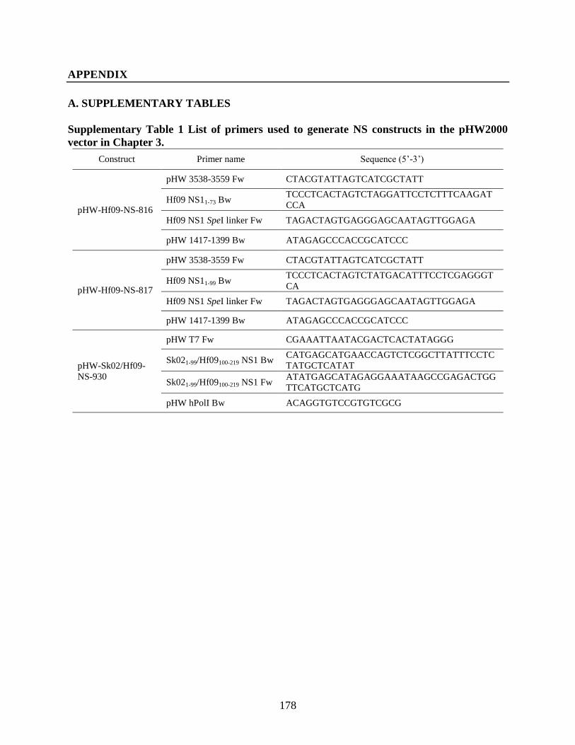
178
APPENDIX
A. SUPPLEMENTARY TABLES
Supplementary Table 1 List of primers used to generate NS constructs in the pHW2000
vector in Chapter 3.
Construct Primer name Sequence (5’-3’)
pHW-Hf09-NS-816
pHW 3538-3559 Fw CTACGTATTAGTCATCGCTATT
Hf09 NS11-73 Bw TCCCTCACTAGTCTAGGATTCCTCTTTCAAGAT
CCA
Hf09 NS1 SpeI linker Fw TAGACTAGTGAGGGAGCAATAGTTGGAGA
pHW 1417-1399 Bw ATAGAGCCCACCGCATCCC
pHW-Hf09-NS-817
pHW 3538-3559 Fw CTACGTATTAGTCATCGCTATT
Hf09 NS11-99 Bw TCCCTCACTAGTCTATGACATTTCCTCGAGGGT
CA
Hf09 NS1 SpeI linker Fw TAGACTAGTGAGGGAGCAATAGTTGGAGA
pHW 1417-1399 Bw ATAGAGCCCACCGCATCCC
pHW-Sk02/Hf09-
NS-930
pHW T7 Fw CGAAATTAATACGACTCACTATAGGG
Sk021-99/Hf09100-219 NS1 Bw CATGAGCATGAACCAGTCTCGGCTTATTTCCTC
TATGCTCATAT
Sk021-99/Hf09100-219 NS1 Fw ATATGAGCATAGAGGAAATAAGCCGAGACTGG
TTCATGCTCATG
pHW hPolI Bw ACAGGTGTCCGTGTCGCG
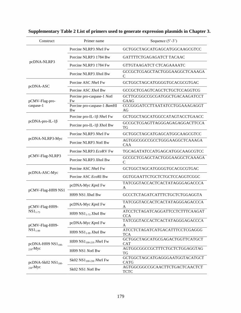
179
Supplementary Table 2 List of primers used to generate expression plasmids in Chapter 3.
Construct Primer name Sequence (5’-3’)
pcDNA-NLRP3
Porcine NLRP3 NheI Fw GCTGGCTAGCATGAGCATGGCAAGCGTCC
Porcine NLRP3 1784 Bw GATTTTCTGAGAGATCT TACAAC
Porcine NLRP3 1784 Fw GTTGTAAGATCT CTCAGAAAATC
Porcine NLRP3 XhoI Bw GCCGCTCGAGCTACTGGGAAGGCTCAAAGA
C
pcDNA-ASC Porcine ASC NheI Fw GCTGGCTAGCATGGGGTGCACGCGTGAC
Porcine ASC XhoI Bw GCCGCTCGAGTCAGCTCTGCTCCAGGTCG
pCMV-Flag-pro-
caspase-1
Porcine pro-caspase-1 NotI
Fw
GCTTGCGGCCGCGATGGCTGACAAGATCCT
GAAG
Porcine pro-caspase-1 BamHI
Bw
CCCGGGATCCTTAATATCCTGGAAAGAGGT
AG
pcDNA-pro-IL-1β
Porcine pro-IL-1β NheI Fw GCTGGCTAGCATGGCCATAGTACCTGAACC
Porcine pro-IL-1β XhoI Bw GCCGCTCGAGTTAGGGAGAGAGGACTTCCA
TG
pcDNA-NLRP3-Myc
Porcine NLRP3 NheI Fw GCTGGCTAGCATGAGCATGGCAAGCGTCC
Porcine NLRP3 NotI Bw AGTGGCGGCCGCCTGGGAAGGCTCAAAGA
CAA
pCMV-Flag-NLRP3
Porcine NLRP3 EcoRV Fw TGCAGATATCCATGAGCATGGCAAGCGTCC
Porcine NLRP3 XhoI Bw GCCGCTCGAGCTACTGGGAAGGCTCAAAGA
C
pcDNA-ASC-Myc Porcine ASC NheI Fw GCTGGCTAGCATGGGGTGCACGCGTGAC
Porcine ASC EcoRI Bw GGTGGAATTCTGCTCTGCTCCAGGTCGGC
pCMV-Flag-Hf09 NS1
pcDNA-Myc KpnI Fw TATCGGTACCACTCACTATAGGGAGACCCA
A
Hf09 NS1 XbaI Bw GCCCTCTAGATCATTTCTGCTCTGGAGGTA
pCMV-Flag-Hf09-
NS11-73
pcDNA-Myc KpnI Fw TATCGGTACCACTCACTATAGGGAGACCCA
A
Hf09 NS11-73 XbaI Bw ATCCTCTAGATCAGGATTCCTCTTTCAAGAT
CCA
pCMV-Flag-Hf09-
NS11-99
pcDNA-Myc KpnI Fw TATCGGTACCACTCACTATAGGGAGACCCA
A
Hf09 NS11-99 XbaI Bw ATCCTCTAGATCATGACATTTCCTCGAGGG
TCA
pcDNA-Hf09 NS1100-
219-Myc
Hf09 NS1100-219 NheI Fw GCTGGCTAGCATGCGAGACTGGTTCATGCT
CAT
Hf09 NS1 NotI Bw AGTGGCGGCCGCTTTCTGCTCTGGAGGTAG
TG
pcDNA-Sk02 NS1100-
230-Myc
Sk02 NS1100-230 NheI Fw GCTGGCTAGCATGAGGGAATGGTACATGCT
CATG
Sk02 NS1 NotI Bw AGTGGCGGCCGCAACTTCTGACTCAACTCT
TCTC

180
Supplementary Table 3 List of primers used to generate expression plasmids in Chapter 4.
Construct Primer name Sequence (5’-3’)
pCMV-Flag-ASC Porcine ASC NotI Fw GCTTGCGGCCGCGATGGGGTGCACGCGTGAC
Porcine ASC BamHI Bw CCCGGGATCCTCAGCTCTGCTCCAGGTCG
pcDNA-Hf09-NS1-
Myc
Hf09 NS1 NheI Fw GCTGGCTAGCACCATGGACTCCAACACCATGTC
Hf09 NS1 NotI Bw AGTGGCGGCCGCTTTCTGCTCTGGAGGTAGTG
pcDNA-Hf09-NS11-99-
Myc
Hf09 NS1 NheI Fw GCTGGCTAGCACCATGGACTCCAACACCATGTC
Hf09 NS11-99 NotI Bw AGTGGCGGCCGCTGACATTTCCTCGAGGGTCA
pCMV-Flag-huASC
Human ASC KpnI Fw TATCGGTACCAATGGGGCGCGCGCGCGAC
Human ASC XbaI Bw ATCCTCTAGATCAGCTCCGCTCCAGGTCC
pCMV-Flag-ASC
(K21R/K22R/K24R/K
26R)
Porcine ASC
K21/22/24/26R Fw AGGATGAGGCTGCTCTCAGTGCCACTG
Porcine ASC
K21/22/24/26R Bw AAACCTCCTGAGCTCGTCGGCTGTCA
pCMV-Flag-ASC
(K55R)
Porcine ASC K55R Fw CTCACTGACAGGCTCGTCAACTAC
Porcine ASC K55R Bw GTCGATGGCATCCAGGGG
pCMV-Flag-ASC
(K79R)
Porcine ASC K79R Fw CTGCGTGACATCGGCATGCGGGAGGTGGCGGA
GCAGC
Porcine ASC K79R Bw GCTGCTCCGCCACCTCCCGCATGCCGATGTCAC
GCAG
pCMV-Flag-ASC
(K87R)
Porcine ASC K87R Fw GGAGGTGGCGGAGCAGCTCCAACGGACCCTGC
ACAAGGGCCC
Porcine ASC K87R Bw GGGCCCTTGTGCAGGGTCCGTTGGAGCTGCTCC
GCCACCTCC
pCMV-Flag-ASC
(K91R)
Porcine ASC K91R Fw GCAGCTGCAGAAGACCCTCCACAGGGGCCCTG
GAGCCAAGC
Porcine ASC K91R Bw GCTTGGCTCCAGGGCCCCTGTGGAGGGTCTTCT
GCAGCTGC
pCMV-Flag-ASC
(K96R)
Porcine ASC K96R Fw CAAGGGCCCTGGAGCCAGGCCTGCTGGAATCA
AAGCCCTTC
Porcine ASC K96R Bw GAAGGGCTTTGATTCCAGCAGGCCTGGCTCCA
GGGCCCTTG
pCMV-Flag-ASC
(K101R)
Porcine ASC K101R Fw GCCAAGCCAGCTGGAATCAGAGCTCTTCCCCTG
AAAGCAGAC
Porcine ASC K101R Bw GTCTGCTTTCAGGGGAAGAGCTCTGATTCCAGC
TGGCTTGGC
pCMV-Flag-ASC
(K106R)
Porcine ASC K106R Fw CTGGAATCAAAGCCCTTCCGCTGAGGGCAGAC
AACAAACCAGCAC
Porcine ASC K106R Bw GTGCTGGTTTGTTGTCTGCCCTCAGCGGAAGGG
CTTTGATTCCAG
pCMV-Flag-ASC
(K110R)
Porcine ASC K110R Fw GCAGACAACAGACCAGCACTG
Porcine ASC K110R Bw TTTCAGGGGAAGGGCTTT
pCMV-Flag-ASC Porcine ASC K140R Fw CTGTACGGGAGGGTGCTGACG

181
(K140R) Porcine ASC K140R Bw GGCATCCAGCAGCCCGTC
pCMV-Flag-ASC
(K159R)
Porcine ASC K159R Fw AACCCCACCAGAATGAGGAGGCTCTTCAG
Porcine ASC K159R Bw GGTGTGCTCCGCCCGCAC
pCMV-Flag-ASC
(K175R)
Porcine ASC K175R Fw CTGACCTGCAGGGACCTGCTC
Porcine ASC K175R Bw GTTCCAGGCCGGAGTGAA
pCMV-Flag-ASC
(K183R)
Porcine ASC K183R Fw CTGCTCCTTCAGGCCCTGAGGGACACCCAGCCC
TACCTG
Porcine ASC K183R Bw CAGGTAGGGCTGGGTGTCCCTCAGGGCCTGAA
GGAGCAG
pCMV-Flag-ASC1-109
Porcine ASC NotI Fw GCTTGCGGCCGCGATGGGGTGCACGCGTGAC
Porcine ASC1-109 BamHI
Bw
CCCGGGATCCTCAGTTGTCTGCTTTCAGGGGAA
G
pCMV-Flag-ASC1-158
Porcine ASC NotI Fw GCTTGCGGCCGCGATGGGGTGCACGCGTGAC
Porcine ASC1-158 BamHI
Bw CCCGGGATCCTCAGGTGGGGTTGGTGTGCTC
pcDNA-HA-ubiquitin
(K48R)
HA-Ubi K48R Fw TTTGCTGGCAGGCAGCTGGAA
HA-Ubi K48R Bw GATCAGTCTCTGCTGATCAGGAGG
pcDNA-HA-ubiquitin
(K63R)
HA-Ubi K63R Fw AATATTCAAAGGGAGTCTACTCTTC
HA-Ubi K63R Bw GTAGTCAGACAAAGTACG

182
Supplementary Table 4 List of primers used to generate expression plasmids in Chapter 5.
Construct Primer name Sequence (5’-3’)
pcDNA-DRP1-Myc Porcine DRP1 XhoI Fw
TAGACTCGAGATGGAGGCACTAATTCCTGT
T
Porcine DRP1 BamHI Bw GTTCGGATCCCCAAAGATGAGTCTCTCGGA
pcDNA-DRP1 (S579D)-
Myc
Porcine DRP1 S579D Fw CATGCCCGCCGATCCACAAAAAG
Porcine DRP1 S579D Bw ATTGGAATTGGTTTTGATTTC
pcDNA-DRP1 (S579A)-
Myc
Porcine DRP1 S579A Fw CATGCCCGCCGCTCCACAAAAAG
Porcine DRP1 S579A Bw ATTGGAATTGGTTTTGATTTC
pcDNA-huRIPK1
(K45A)-Myc
Human RIPK1 NheI Fw GCTGGCTAGCATGCAACCAGACATGTCCTT
G
Human RIPK1 NotI Bw AGTGGCGGCCGCGTTCTGGCTGACGTAAAT
CAA
pcDNA-RIPK1-Myc
Porcine RIPK1 NheI Fw GCTGGCTAGCATGTCTCTGGATGACATTAA
GATG
Porcine RIPK1 NotI Bw AGTGGCGGCCGCGTTCTGGCTGCTGCGGAT
C
pCMV-Flag-RIPK1
Porcine RIPK1 HindIII Fw CGACAAGCTTATGTCTCTGGATGACATTAA
GATG
Porcine RIPK1 NotI Bw ATTCGCGGCCGCCTAGTTCTGGCTGCTGCG
GAT
pCMV-Flag-RIPK1
(K41A/K42A)
Porcine RIPK1
K41A/K42A Fw TGTGATTCTGGCGGCGGTGTACACGGGCCC
Porcine RIPK1
K41A/K42A Bw AGCCCGTGGGATTTGTGA

183
B. ACHIEVEMENTS DURING THE STUDY
B.1 Publications
A Gaba, F Xu, Y Lu, HS Park, G Liu and Y Zhou. (2018) The NS1 Protein of Influenza A
Virus Participates in Necroptosis by Interacting With MLKL and Increasing Its Oligomerization
and Membrane Translocation. Journal of Virology, http://doi.org/10.1128/JVI.01835-18 (I
contributed by examining the effects of necroptosis proteins on the NLRP3 inflammasome
activation and conducting IL-1β ELISA.)
HS Park, G Liu, Q Liu and Y Zhou. (2018) Swine Influenza Virus Induces RIPK1/DRP1-
Mediated Interleukin-1 Beta Production. Viruses, 10(8), 419 https://doi.org/10.3390/v10080419
(I designed most of the experiments, generated most of the tools, performed all the experiments,
analyzed the data and wrote the manuscript.)
HS Park, G Liu, SN Thulasi Raman, SL Landreth, Q Liu and Y Zhou. (2018) NS1 Protein of
2009 Pandemic Influenza A Virus Inhibits Porcine NLRP3 Inflammasome-Mediated Interleukin-
1 Beta Production by Suppressing ASC Ubiquitination. Journal of Virology, 92(8), e00022-18
http://doi.org/10.1128/JVI.00022-18 (I designed some of the experiments, generated most of the
tools, performed all the experiments, analyzed the data and wrote the manuscript.)
G Liu, HS Park, HM Pyo, Q Liu and Y Zhou. (2015) Influenza A Virus Panhandle Structure
Is Directly Involved in RIG-I Activation and Interferon Induction. Journal of Virology, 89(11),
6067-6079 http://doi.org/10.1128/JVI.00232-15 (I contributed by isolating primary PAMs and
conducting porcine IFN-α and IL-1β ELISA.)
B.2 Presentations
HS Park, G Liu, SN Thulasi Raman, SL Landreth, Q Liu and Y Zhou. NS1 Protein of 2009
Pandemic Influenza A Virus Inhibits Porcine NLRP3 Inflammasome-Mediated Interleukin-1
Beta Production by Suppressing ASC Ubiquitination. Poster presentation at the 37th American
Society for Virology Meeting, University of Maryland, College Park, MD, United States, July
14-18, 2018.
HS Park, G Liu, SN Thulasi Raman, Q Liu and Y Zhou. RIP1/RIP3 Signaling Contributes to
the Porcine NLRP3 Inflammasome-Mediated Interleukin-1 Beta Production in Response to
Swine Influenza Virus Infection. Poster presentation at the 36th American Society for Virology
Meeting, University of Wisconsin-Madison, WI, United States, June 24-28, 2017.

184
HS Park, G Liu, SN Thulasi Raman and Y Zhou. NS1 Protein of Human Pandemic Influenza
Virus Contributed to the Down-Regulation of NLRP3 Inflammasome-Mediated Interleukin-1
Beta in Porcine Alveolar Macrophages. Poster presentation at the ImmuNet Research Day 2016,
Edmonton, AB, Canada, June 9, 2016.
HS Park, G Liu and Y Zhou. Swine Influenza Virus Induced NLRP3 Inflammasome-
Mediated Interleukin-1 Beta Production in Porcine Alveolar Macrophages. Poster presentation at
the 34th
American Society for Virology Meeting, Western University, London, ON, Canada. July
10-15, 2015.
HS Park and Y Zhou. Characterization of Interleukin-1 Beta Production in Porcine Alveolar
Macrophages Infected With Swine Influenza Virus. Poster presentation at the 2nd Li Ka Shing
Institute of Virology Symposium, Edmonton, AB, Canada. May 30-31, 2013.
B.3 Scholarships and fellowships
-Graduate Teaching Fellowship ($8,500) from Western College of Veterinary Medicine,
2015.
-Graduate Research Fellowship ($8,000) from Western College of Veterinary Medicine, 2014.
-Graduate Research Fellowship ($8,000) from Western College of Veterinary Medicine, 2013.
-Saskatchewan Innovation and Opportunity Graduate Scholarship ($10,000) from University
of Saskatchewan, 2012.
B.4 Awards
-Dr. John Allen Graduate Student Travel Award ($300) from Department of Veterinary
Microbiology, Western College of Veterinary Medicine, 2018.
-Graduate Student Travel Award ($200) from Western College of Veterinary Medicine, 2018.
-Graduate Student Travel Award ($450) from University of Saskatchewan, 2018.
-Dr. John Allen Graduate Student Travel Award ($300) from Department of Veterinary
Microbiology, Western College of Veterinary Medicine, 2017.
-Graduate Student Travel Award ($200) from Western College of Veterinary Medicine, 2017.
-Graduate Student Travel Award ($450) from University of Saskatchewan, 2017.
-Student Travel Award (US$500) from American Society for Virology, 2017.
185
-Dr. John Allen Graduate Student Travel Award ($200) from Department of Veterinary
Microbiology, Western College of Veterinary Medicine, 2016.
-Dr. John Allen Graduate Student Travel Award ($300) from Department of Veterinary
Microbiology, Western College of Veterinary Medicine, 2015.
-Graduate Student Travel Award ($350) from University of Saskatchewan, 2015.
-Student Travel Award (US$500) from American Society for Virology, 2015.
-Graduate Student Travel Award ($250) from Western College of Veterinary Medicine, 2013.