Peri-Pubertal Emergence of UNC-5 Homologue Expression by Dopamine Neurons in Rodents
14
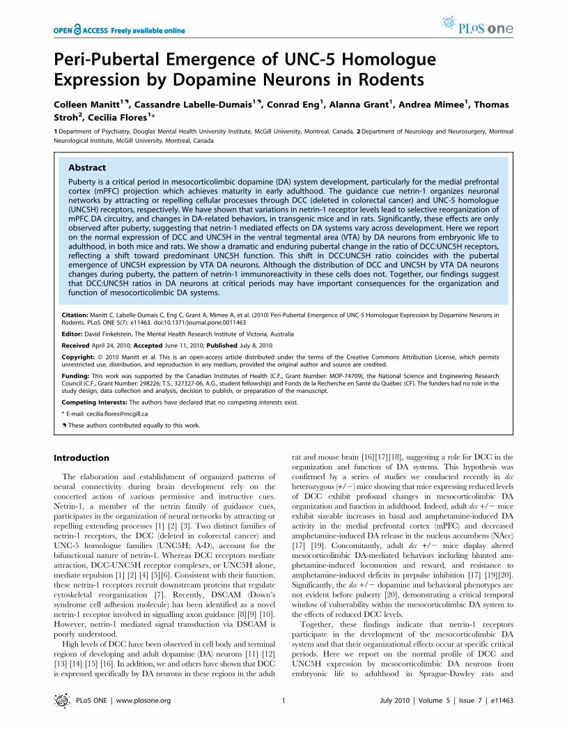
Peri-Pubertal Emergence of UNC-5 Homologue Expression by Dopamine Neurons in Rodents Colleen Manitt 1. , Cassandre Labelle-Dumais 1. , Conrad Eng 1 , Alanna Grant 1 , Andrea Mimee 1 , Thomas Stroh 2 , Cecilia Flores 1 * 1 Department of Psychiatry, Douglas Mental Health University Institute, McGill University, Montreal, Canada, 2 Department of Neurology and Neurosurgery, Montreal Neurological Institute, McGill University, Montreal, Canada Abstract Puberty is a critical period in mesocorticolimbic dopamine (DA) system development, particularly for the medial prefrontal cortex (mPFC) projection which achieves maturity in early adulthood. The guidance cue netrin-1 organizes neuronal networks by attracting or repelling cellular processes through DCC (deleted in colorectal cancer) and UNC-5 homologue (UNC5H) receptors, respectively. We have shown that variations in netrin-1 receptor levels lead to selective reorganization of mPFC DA circuitry, and changes in DA-related behaviors, in transgenic mice and in rats. Significantly, these effects are only observed after puberty, suggesting that netrin-1 mediated effects on DA systems vary across development. Here we report on the normal expression of DCC and UNC5H in the ventral tegmental area (VTA) by DA neurons from embryonic life to adulthood, in both mice and rats. We show a dramatic and enduring pubertal change in the ratio of DCC:UNC5H receptors, reflecting a shift toward predominant UNC5H function. This shift in DCC:UNC5H ratio coincides with the pubertal emergence of UNC5H expression by VTA DA neurons. Although the distribution of DCC and UNC5H by VTA DA neurons changes during puberty, the pattern of netrin-1 immunoreactivity in these cells does not. Together, our findings suggest that DCC:UNC5H ratios in DA neurons at critical periods may have important consequences for the organization and function of mesocorticolimbic DA systems. Citation: Manitt C, Labelle-Dumais C, Eng C, Grant A, Mimee A, et al. (2010) Peri-Pubertal Emergence of UNC-5 Homologue Expression by Dopamine Neurons in Rodents. PLoS ONE 5(7): e11463. doi:10.1371/journal.pone.0011463 Editor: David Finkelstein, The Mental Health Research Institute of Victoria, Australia Received April 24, 2010; Accepted June 11, 2010; Published July 8, 2010 Copyright: ß 2010 Manitt et al. This is an open-access article distributed under the terms of the Creative Commons Attribution License, which permits unrestricted use, distribution, and reproduction in any medium, provided the original author and source are credited. Funding: This work was supported by the Canadian Institutes of Health (C.F., Grant Number: MOP-74709), the National Science and Engineering Research Council (C.F., Grant Number: 298226; T.S., 327327-06, A.G., student fellowship) and Fonds de la Recherche en Sante ´ du Que ´ bec (CF). The funders had no role in the study design, data collection and analysis, decision to publish, or preparation of the manuscript. Competing Interests: The authors have declared that no competing interests exist. * E-mail: [email protected] . These authors contributed equally to this work. Introduction The elaboration and establishment of organized patterns of neural connectivity during brain development rely on the concerted action of various permissive and instructive cues. Netrin-1, a member of the netrin family of guidance cues, participates in the organization of neural networks by attracting or repelling extending processes [1] [2] [3]. Two distinct families of netrin-1 receptors, the DCC (deleted in colorectal cancer) and UNC-5 homologue families (UNC5H; A-D), account for the bifunctional nature of netrin-1. Whereas DCC receptors mediate attraction, DCC-UNC5H receptor complexes, or UNC5H alone, mediate repulsion [1] [2] [4] [5][6]. Consistent with their function, these netrin-1 receptors recruit downstream proteins that regulate cytoskeletal reorganization [7]. Recently, DSCAM (Down’s syndrome cell adhesion molecule) has been identified as a novel netrin-1 receptor involved in signalling axon guidance [8][9] [10]. However, netrin-1 mediated signal transduction via DSCAM is poorly understood. High levels of DCC have been observed in cell body and terminal regions of developing and adult dopamine (DA) neurons [11] [12] [13] [14] [15] [16]. In addition, we and others have shown that DCC is expressed specifically by DA neurons in these regions in the adult rat and mouse brain [16][17][18], suggesting a role for DCC in the organization and function of DA systems. This hypothesis was confirmed by a series of studies we conducted recently in dcc heterozygous (+/2) mice showing that mice expressing reduced levels of DCC exhibit profound changes in mesocorticolimbic DA organization and function in adulthood. Indeed, adult dcc +/2 mice exhibit sizeable increases in basal and amphetamine-induced DA activity in the medial prefrontal cortex (mPFC) and decreased amphetamine-induced DA release in the nucleus accumbens (NAcc) [17] [19]. Concomitantly, adult dcc +/2 mice display altered mesocorticolimbic DA-mediated behaviors including blunted am- phetamine-induced locomotion and reward, and resistance to amphetamine-induced deficits in prepulse inhibition [17] [19][20]. Significantly, the dcc +/2 dopamine and behavioral phenotypes are not evident before puberty [20], demonstrating a critical temporal window of vulnerability within the mesocorticolimbic DA system to the effects of reduced DCC levels. Together, these findings indicate that netrin-1 receptors participate in the development of the mesocorticolimbic DA system and that their organizational effects occur at specific critical periods. Here we report on the normal profile of DCC and UNC5H expression by mesocorticolimbic DA neurons from embryonic life to adulthood in Sprague-Dawley rats and PLoS ONE | www.plosone.org 1 July 2010 | Volume 5 | Issue 7 | e11463
-
Upload
independent -
Category
Documents
-
view
2 -
download
0
Transcript of Peri-Pubertal Emergence of UNC-5 Homologue Expression by Dopamine Neurons in Rodents
Peri-Pubertal Emergence of UNC-5 HomologueExpression by Dopamine Neurons in RodentsColleen Manitt1., Cassandre Labelle-Dumais1., Conrad Eng1, Alanna Grant1, Andrea Mimee1, Thomas
Stroh2, Cecilia Flores1*
1 Department of Psychiatry, Douglas Mental Health University Institute, McGill University, Montreal, Canada, 2 Department of Neurology and Neurosurgery, Montreal
Neurological Institute, McGill University, Montreal, Canada
Abstract
Puberty is a critical period in mesocorticolimbic dopamine (DA) system development, particularly for the medial prefrontalcortex (mPFC) projection which achieves maturity in early adulthood. The guidance cue netrin-1 organizes neuronalnetworks by attracting or repelling cellular processes through DCC (deleted in colorectal cancer) and UNC-5 homologue(UNC5H) receptors, respectively. We have shown that variations in netrin-1 receptor levels lead to selective reorganization ofmPFC DA circuitry, and changes in DA-related behaviors, in transgenic mice and in rats. Significantly, these effects are onlyobserved after puberty, suggesting that netrin-1 mediated effects on DA systems vary across development. Here we reporton the normal expression of DCC and UNC5H in the ventral tegmental area (VTA) by DA neurons from embryonic life toadulthood, in both mice and rats. We show a dramatic and enduring pubertal change in the ratio of DCC:UNC5H receptors,reflecting a shift toward predominant UNC5H function. This shift in DCC:UNC5H ratio coincides with the pubertalemergence of UNC5H expression by VTA DA neurons. Although the distribution of DCC and UNC5H by VTA DA neuronschanges during puberty, the pattern of netrin-1 immunoreactivity in these cells does not. Together, our findings suggestthat DCC:UNC5H ratios in DA neurons at critical periods may have important consequences for the organization andfunction of mesocorticolimbic DA systems.
Citation: Manitt C, Labelle-Dumais C, Eng C, Grant A, Mimee A, et al. (2010) Peri-Pubertal Emergence of UNC-5 Homologue Expression by Dopamine Neurons inRodents. PLoS ONE 5(7): e11463. doi:10.1371/journal.pone.0011463
Editor: David Finkelstein, The Mental Health Research Institute of Victoria, Australia
Received April 24, 2010; Accepted June 11, 2010; Published July 8, 2010
Copyright: � 2010 Manitt et al. This is an open-access article distributed under the terms of the Creative Commons Attribution License, which permitsunrestricted use, distribution, and reproduction in any medium, provided the original author and source are credited.
Funding: This work was supported by the Canadian Institutes of Health (C.F., Grant Number: MOP-74709), the National Science and Engineering ResearchCouncil (C.F., Grant Number: 298226; T.S., 327327-06, A.G., student fellowship) and Fonds de la Recherche en Sante du Quebec (CF). The funders had no role in thestudy design, data collection and analysis, decision to publish, or preparation of the manuscript.
Competing Interests: The authors have declared that no competing interests exist.
* E-mail: [email protected]
. These authors contributed equally to this work.
Introduction
The elaboration and establishment of organized patterns of
neural connectivity during brain development rely on the
concerted action of various permissive and instructive cues.
Netrin-1, a member of the netrin family of guidance cues,
participates in the organization of neural networks by attracting or
repelling extending processes [1] [2] [3]. Two distinct families of
netrin-1 receptors, the DCC (deleted in colorectal cancer) and
UNC-5 homologue families (UNC5H; A-D), account for the
bifunctional nature of netrin-1. Whereas DCC receptors mediate
attraction, DCC-UNC5H receptor complexes, or UNC5H alone,
mediate repulsion [1] [2] [4] [5][6]. Consistent with their function,
these netrin-1 receptors recruit downstream proteins that regulate
cytoskeletal reorganization [7]. Recently, DSCAM (Down’s
syndrome cell adhesion molecule) has been identified as a novel
netrin-1 receptor involved in signalling axon guidance [8][9] [10].
However, netrin-1 mediated signal transduction via DSCAM is
poorly understood.
High levels of DCC have been observed in cell body and terminal
regions of developing and adult dopamine (DA) neurons [11] [12]
[13] [14] [15] [16]. In addition, we and others have shown that DCC
is expressed specifically by DA neurons in these regions in the adult
rat and mouse brain [16][17][18], suggesting a role for DCC in the
organization and function of DA systems. This hypothesis was
confirmed by a series of studies we conducted recently in dcc
heterozygous (+/2) mice showing that mice expressing reduced levels
of DCC exhibit profound changes in mesocorticolimbic DA
organization and function in adulthood. Indeed, adult dcc +/2 mice
exhibit sizeable increases in basal and amphetamine-induced DA
activity in the medial prefrontal cortex (mPFC) and decreased
amphetamine-induced DA release in the nucleus accumbens (NAcc)
[17] [19]. Concomitantly, adult dcc +/2 mice display altered
mesocorticolimbic DA-mediated behaviors including blunted am-
phetamine-induced locomotion and reward, and resistance to
amphetamine-induced deficits in prepulse inhibition [17] [19][20].
Significantly, the dcc +/2 dopamine and behavioral phenotypes are
not evident before puberty [20], demonstrating a critical temporal
window of vulnerability within the mesocorticolimbic DA system to
the effects of reduced DCC levels.
Together, these findings indicate that netrin-1 receptors
participate in the development of the mesocorticolimbic DA
system and that their organizational effects occur at specific critical
periods. Here we report on the normal profile of DCC and
UNC5H expression by mesocorticolimbic DA neurons from
embryonic life to adulthood in Sprague-Dawley rats and
PLoS ONE | www.plosone.org 1 July 2010 | Volume 5 | Issue 7 | e11463

C57BL/6 (BL6) mice. First, using Western blot analysis, we
demonstrate that the ratio of DCC to UNC5H expression switches
toward UNC5H predominance during peri-pubertal age in DA
somatodendritic regions. To capture possible changes in DCC and
UNC5H expression by mesocorticolimbic DA neurons specifically,
we conducted dual-immunofluorescence experiments at specific
developmental stages. These stages were selected based on the
selective changes that take place during the elaboration of the
mesocorticolimbic DA circuitry [21][22][23][24][25]. We find
that whereas DA neurons express DCC from embryonic life to
adulthood, UNC5H expression by these cells emerges at puberty.
Materials and Methods
AnimalsAll experiments were preformed in accordance with the guidelines
of the Canadian Council of Animal Care and all animal procedures
were approved by the McGill University/Douglas Hospital Animal
Care Committee (protocol number: 5084). All animals were kept on a
12 h light-dark cycle with ad libitum access to food and water.
Mice. C57BL/6J (BL6) mice were obtained from The Jackson
Laboratory and bred in our animal colony. Mice were used at
embryonic day (E) 13, 15, and 17; at birth (postnatal day (PND) 0),
at post-weaning PND2161; at peri-pubertal period PND3362,
and at adulthood PND75615. For staging of embryos, the day of
fertilization, assessed by the presence of a vaginal plug, was
considered to be E0. For post-weaning to adulthood experiments,
only male mice were used. Dcc 2/2 mice: For the experiments
aimed at testing DCC antibody specificity (see below), dcc 2/2
embryos were generated by mating adult dcc +/2 mice. Adult dcc
heterozygous (+/2) mice, originally obtained from Dr. S.
Ackerman (The Jackson Laboratory), were maintained on a BL6
background and bred in our animal colony. Targeted inactivation
of the dcc gene was performed by disrupting exon 3, which encodes
most of the protein’s second immunoglobulin-like domain, by
insertion of a neomycin resistance cassette using homologous
recombination [26]. Southern and Western blot analyses were
used to confirm proper targeting and complete loss of DCC
protein, respectively [26]. Unc5c 2/2 mice: For the
experiments aimed at testing the specificity of the anti-UNC5C
rabbit antiserum, brains dissected from adult unc5c 2/2 and +/+mice were used (BL6; Cg-Unc5crcmTg(Ucp)1,23KZ/Slac; obtained
from Dr. S. Ackerman, The Jackson Laboratory). The mutant
allele of the unc5c locus was generated by a transgene insertion that
results in complete loss of unc5c transcript [27]. These mice were
from a hybrid BL6/SJL background because 2/2 progeny from
a pure BL6 background do not typically survive to adulthood.
Genotyping. The targeted and wild-type dcc alleles were
amplified using an annealing temperature of 54uC for 30 cycles
with the following oligonucleotides: DCC code: GGT CAT TGA
GGT TCC TTT, DCC rev: AAG ACG ACC ACA CGC GAC,
and DCC Neo: TCC TCG TGC TTT ACG GTA TC. The
targeted and wild-type unc5c alleles were amplified using an
annealing temperature of 54uC for 40 cycles using the following
oligonucleotides: Tipsy head CAG GAG AAG ATA CAT TTA
ACC AC, Tipsy tail GAC AGA AGA GCA TAG CAT TCAC,
Asp-2M13RA CAC TCT ATG GAA ATG GCT GAA T, Asp-
2M13RB GTC CTC CAA TCC AAG AAC TG.
Rats. The development of midbrain DA neurons in mice
precedes that of rats by two days (Bayer et al., 1995). Thus, we
collected embryos at E15, 17, and 19 and at birth (PND0) from timed
pregnant Sprague-Dawley rats obtained from Charles River. Post-
weaning (PND21), peri-pubertal (PND33) and adult (PND90)
Sprague-Dawley rats were also obtained from Charles River.
Rationale for choosing specific developmental agesThe embryonic and postnatal developmental stages used for the
characterization of the expression pattern of DCC and UNC5H by
rodent midbrain DA neurons were selected on the basis of the
development of the DA system in the rat [21][22] [24]. Briefly, in the
rat, the first DA neurons are generated in the ventral midbrain
between E12 and E15 [22] [28]. Shortly after their specification,
midbrain DA neurons start sending projections toward their targets.
At E13/E14 these neurons extend axons rostrally and, by E15, they
start innervating the ganglionic eminence (i.e. developing striatum).
At E16/E17 some DA fibers grow beyond the ganglionic eminence to
reach the cortical subplate. By E19/E20, DA fibers innervate the
cortical plate of the developing medial prefrontal cortex. At birth, the
number of DA fibers in deep layers of the cortex increase
dramatically. Mesolimbic DA innervation reaches adulthood levels
three weeks after birth. In contrast, maturation of mesocortical DA
innervation proceeds until early adulthood and exhibits significant
remodeling during the post-weaning (PND23) and peri-pubertal
period (PND35) [21][22].
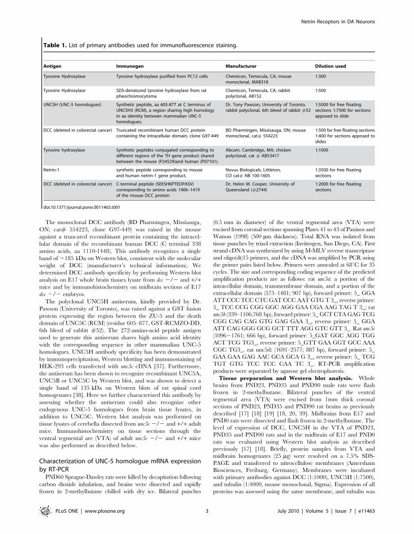
Primary antibody characterizationPlease refer to Table 1 for a list of all primary antibodies used
for immunofluorescence staining. The monoclonal tyrosine
hydroxylase (TH) antibody (Chemicon International, Temecula,
CA; cat # MAB318) was raised in mouse and recognizes
specifically an epitope on the regulatory N-terminus of TH [29].
On Western blot, it recognizes a single band of ,60 kDa, which
corresponds to the predicted molecular weight for TH and does
not react with other closely related catecholamine enzymes
including dopamine-b-hydroxylase, phenylalanine hydroxylase,
tryptophan hydroxylase, dehydropteridine reductase, sepiapterin
reductase and phenethanolamine-M-methyl-transferase (manufac-
turer’s technical information). This antibody was shown to result
in a staining pattern of rat striatal dopaminergic terminals similar
to that described previously [30] and to stain mesencephalic DA
neurons in the adult rat [31]. The rabbit polyclonal anti-TH
antibody (Chemicon International; cat # AB152) was raised in
rabbit against denatured TH from rat pheochromocytoma. On
Western blot, this antibody recognizes a single band of
approximately 60 kDa molecular weight corresponding to the
TH protein (manufacturer’s technical information). This antibody
labels midbrain DA neuron cell bodies and terminals in wild-type
mice, but not in mice that have a non-functional Th gene [32].
The chicken polyclonal anti-TH antibody (Abcam, Cambridge,
MA; cat # AB53417) was raised in chicken against two synthetic
peptides corresponding to different regions of TH with sequences
shared between the mouse (P24529) and human (P01701). This
antibody recognizes a specific band of approximately 59 kDa on
Western blot (manufacturer’s technical information) and results in
a staining pattern of rat midbrain DA neurons identical to that
observed with the two other TH antibodies used in this study. The
chicken anti-mouse netrin-1 antibody (1:7000, Novus Biologicals,
Littleton, CO) was tested by Western blot. Lysates from whole rat
brain were run alongside rat liver, which does not express netrin-1
[33]. A band of ,75 kDa was detected in adult rat brain,
consistent with the predicted molecular weight of rat netrin-1. This
band was not detected in liver (data not shown). The rabbit anti-
DCC polyclonal antiserum (2744, [34] a gift from Dr. H.M.
Cooper) was raised against the C-terminal peptide (SEESHKP-
TEDPASV) corresponding to amino acids 1406–1419 of the
mouse DCC protein [35]. The specificity of the antibody has been
demonstrated previously by preincubating the antiserum with the
peptide antigen prior to incubating sections [36].
Netrin Receptors in DA Neurons
PLoS ONE | www.plosone.org 2 July 2010 | Volume 5 | Issue 7 | e11463
The monoclonal DCC antibody (BD Pharmingen, Missisauga,
ON; cat# 554223, clone G97-449) was raised in the mouse
against a truncated recombinant protein containing the intracel-
lular domain of the recombinant human DCC (C terminal 338
amino acids, aa 1110-1448). This antibody recognizes a single
band of ,185 kDa on Western blot, consistent with the molecular
weight of DCC (manufacturer’s technical information). We
determined DCC antibody specificity by performing Western blot
analysis on E17 whole brain tissues lysate from dcc 2/2 and +/+mice and by immunohistochemistry on midbrain sections of E17
dcc 2/2 embryos.
The polyclonal UNC5H antiserum, kindly provided by Dr.
Pawson (University of Toronto), was raised against a GST fusion
protein expressing the region between the ZU-5 and the death
domain of UNC5C (RCM) (residue 605–877, GST-RCMZO-DD,
6th bleed of rabbit #52). The 272-amino-acid peptide antigen
used to generate this antiserum shares high amino acid identity
with the corresponding sequence in other mammalian UNC-5
homologues. UNC5H antibody specificity has been demonstrated
by immunoprecipitation, Western blotting and immunostaining of
HEK-293 cells transfected with unc5c cDNA [37]. Furthermore,
the antiserum has been shown to recognize recombinant UNC5A,
UNC5B or UNC5C by Western blot, and was shown to detect a
single band of 135 kDa on Western blots of rat spinal cord
homogenates [38]. Here we further characterized this antibody by
assessing whether the antiserum could also recognize other
endogenous UNC-5 homologues from brain tissue lysates, in
addition to UNC5C. Western blot analysis was performed on
tissue lysates of cerebella dissected from unc5c 2/2 and +/+ adult
mice. Immunohistochemistry on tissue sections through the
ventral tegmental are (VTA) of adult unc5c 2/2 and +/+ mice
was also performed as described below.
Characterization of UNC-5 homologue mRNA expressionby RT-PCR
PND60 Sprague-Dawley rats were killed by decapitation following
carbon dioxide inhalation, and brains were dissected and rapidly
frozen in 2-methylbutane chilled with dry ice. Bilateral punches
(0.5 mm in diameter) of the ventral tegmental area (VTA) were
excised from coronal sections spanning Plates 41 to 43 of Paxinos and
Watson (1998) (500 mm thickness). Total RNA was isolated from
tissue punches by trizol extraction (Invitrogen, San Diego, CA). First
strand cDNA was synthesized by using M-MLV reverse transcriptase
and oligo(dt)15 primers, and the cDNA was amplified by PCR using
the primer pairs listed below. Primers were annealed at 68uC for 35
cycles. The size and corresponding coding sequence of the predicted
amplification products are as follows: rat unc5a: a portion of the
intracellular domain, transmembrane domain, and a portion of the
extracellular domain (575–1481; 907 bp), forward primer: 5_ GGA
ATT CCC TCC CTC GAT CCC AAT GTG T 3_, reverse primer:
5_ TCC CCG CGG GGC AGG GAA CGA AAG TAG T 3_; rat
unc5b (339–1106;768 bp), forward primer: 5_ GCT CTA GAG TCG
CGG CAG CAG GTG GAG GAA 3_, reverse primer: 5_ GGA
ATT CAG GGG GCG GCT TTT AGG GTC GTT 3_. Rat unc5c
(109621761; 666 bp), forward primer: 5_GAT GGC AGG TGG
ACT TCG TG3_, reverse primer: 5_GTT GAA GGT GCC AAA
CGC TG3_. rat unc5d: (1691–2577; 887 bp), forward primer: 5_
GAA GAA GAG AAC GCA GCA G 3_, reverse primer: 5_ TCG
TGT GTG TCC TCC CAA TC 3_. RT-PCR amplification
products were separated by agarose gel electrophoresis.
Tissue preparation and Western blot analysis. Whole
brains from PND23, PND35 and PND90 male rats were flash
frozen in 2-methylbutane. Bilateral punches of the ventral
tegmental area (VTA) were excised from 1mm thick coronal
sections of PND23, PND35 and PND90 rat brains as previously
described [17] [18] [19] [18, 20, 39]. Midbrains from E17 and
PND0 rats were dissected and flash frozen in 2-methylbutane. The
level of expression of DCC, UNC5H in the VTA of PND23,
PND35 and PND90 rats and in the midbrain of E17 and PND0
rats was evaluated using Western blot analysis as described
previously [17] [18]. Briefly, protein samples from VTA and
midbrain homogenates (25 mg) were resolved on a 7.5% SDS-
PAGE and transferred to nitrocellulose membranes (Amersham
Biosciences, Freiburg, Germany). Membranes were incubated
with primary antibodies against DCC (1:1000), UNC5H (1:7500),
and tubulin (1:4000, mouse monoclonal, Sigma). Expression of all
proteins was assessed using the same membrane, and tubulin was
Table 1. List of primary antibodies used for immunofluorescence staining.
Antigen Immunogen Manufacturer Dilution used
Tyrosine Hydroxylase Tyrosine hydroxylase purified from PC12 cells Chemicon, Temecula, CA; mousemonoclonal, MAB318
1:300
Tyrosine Hydroxylase SDS-denatured tyrosine hydroxylase from ratpheochromocytoma
Chemicon, Temecula, CA; rabbitpolyclonal, AB152
1:500
UNC5H (UNC-5 homologues) Synthetic peptide, aa 605-877 at C terminus ofUNC5H3 (RCM), a region sharing high homologyin aa identity between mammalian UNC-5homologues.
Dr. Tony Pawson, University of Toronto,rabbit polyclonal, 6th bleed of rabbit #52
1:5000 for free floatingsections 1:7500 for sectionsapposed to slide
DCC (deleted in colorectal cancer) Truncated recombinant human DCC proteincontaining the intracellular domain, clone G97-449
BD Pharmingen, Missisauga, ON; mousemonoclonal, cat# 554223
1:500 for free floating sections1:400 for sections apposed toslides
Tyrosine hydroxylase Synthetic peptides conjugated corresponding todifferent regions of the TH gene product sharedbetween the mouse (P24529)and human (P07101).
Abcam, Cambridge, MA; chickenpolyclonal, cat # AB53417
1:1000
Netrin-1 synthetic peptide corresponding to mouseand human netrin-1 gene product,
Novus Biologicals, Littleton,CO cat# NB 100-1605
1:3500 for free floatingsections
DCC (deleted in colorectal cancer) C-terminal peptide (SEESHKPTEDPASV)corresponding to amino acids 1406–1419of the mouse DCC protein
Dr. Helen M. Cooper, University ofQueensland (#2744)
1:2000 for free floatingsections
doi:10.1371/journal.pone.0011463.t001
Netrin Receptors in DA Neurons
PLoS ONE | www.plosone.org 3 July 2010 | Volume 5 | Issue 7 | e11463
used as a loading control. Importantly, the primary antibodies
against DCC and UNC5H used in this procedure were the same
as the ones described in the immunohistochemistry section.
Immunoreactivity was visualized using peroxidase-conjugated
secondary antibodies (Vector Laboratories) and chemilumine-
scence (Perkin Elmer, Waltham, MA, USA).
ImmunohistochemistryTo collect embryonic brains, pregnant females were killed by
decapitation, embryos were surgically removed from the uterus,
decapitated and their brains dissected in PBS and fixed by
immersion in 4%PFA and 0.2% picric acid in 0.1 M phosphate
buffer, overnight at 4uC. Fixed embryonic brains were then
cryoprotected overnight at 4uC in 30% sucrose in PBS.
Cryoprotected embryonic brains were embedded in Tissue-Tek
O.C.T. compound (Sakura) and frozen in 2-methylbutane chilled
with dry ice. PND0 brains were processed as described for
embryonic brains. 20 mm coronal sections of embryonic and
PND0 brains were cut using a cryostat. Sections were mounted
directly on superfrost slides (Fisher Scientific), air-dried and
stored at 280uC until use. Juvenile, post-weaning and adult
animals were anesthetized with an overdose of sodium pento-
barbital (.75 mg/kg i.p.) and were perfused intracardially with
0.9% saline followed by a fixative solution (4% paraformaldehyde
(PFA) and 0.2% picric acid in 0.1 M phosphate buffer). Brains
were dissected from the skull, post-fixed in the same fixative for
45 minutes at 4uC and cryoprotected in sucrose (30% sucrose in
phosphate buffered saline (PBS)) overnight at 4uC. The following
morning, tissue was rapidly frozen by immersion in 2-methylbu-
tane (Fisher Scientific, Hampton, NH) chilled with dry ice.
Frozen brains were immediately sectioned at 40 mm using a Leica
SM2000-R sliding microtome and collected in PBS. Free-floating
sections were used immediately for immunohistochemical pro-
cessing.
Tissue sections were processed for immunofluorescence as
described previously [17] [18]. Briefly, rat brain sections were
collected and rinsed in PBS and incubated in blocking solution
(rat: 2% bovine serum albumin, 0.2% Tween-20 in PBS; mouse;
M.O.M. kit, Vector Laboratories, Burlingame, CA, USA) for
2 hours at room temperature (RT). Sections were incubated
overnight at 4uC with combinations of primary antibodies diluted
in blocking solution. The combinations of following primary
antibodies were used: mouse monoclonal anti-DCC (1:500,
Pharmigen, Mississsauga, Ontario, Canada, Cat# 554223), rabbit
polyclonal anti-TH (1:500, Chemicon, Temecula, CA, USA,
Cat# AB152), rabbit UNC5H antiserum (1:5000, provided by Dr.
Tony Pawson, University of Toronto), and mouse monoclonal
anti-TH (1:300, Chemicon, Cat# MAB318), polyclonal chicken
anti-mouse netrin-1 (1:3500, Novus Biologicals, Littleton, CO), or
polyclonal rabbit anti-DCC (2744; 1:2000, a gift from Dr. H.M.
Cooper, University of Queensland). Immunostaining was visual-
ized with either Alexa Fluor 350, Alexa Fluor 488 or Alexa Fluor
555-conjugated secondary antibodies raised in goat (1:500,
Molecular Probes, Eugene, OR, USA).
As a negative control, adjacent sections were processed as
described above except that they were incubated overnight at 4uCin blocking solution without primary antibodies. To test for
secondary antibody specificity, sections were incubated with either
only DCC, UNC5H, or netrin-1 primary antibody and,
subsequently, with combinations of anti-rabbit, anti-mouse and
anti-chicken Alexa Fluor-conjugated antibodies. Immunoreactivity
was only observed for the secondary antibody raised against the
species in which the primary antibody was generated.
Microscopy and image analysisImmunofluorescence was visualized using either a 1) Leica
DM4000B microscope equipped with a Ludl XYZ motorized
stage and filter cubes appropriate for detection of Alexa Fluor
350, 488 and 555. Images were captured using a digital
Microfire camera and PictureFrame software (Microbrightfield,
VT, USA) or 2) a Nikon PCM2000 laser-scanning confocal
microscope equipped with argon (488 nm excitation; 10%
neutral density filter) and HeNe (543 nm excitation) lasers.
Confocal images of Alexa Fluor 488 and 555 were obtained
simultaneously, below saturation levels, with minimal gain and
contrast enhancement.
The presence of immunostained cells in the VTA from post-
weaning to adulthood was examined on coronal sections corre-
sponding to sections spanning Plates 55 to 63 of the mouse brain atlas
[40] and sections spanning Plates 40 to 46 of the rat brain atlas [41].
For embryonic ages and for PND0, coronal sections containing
midbrain DA neurons were examined according to the atlas of the
developing mouse [23], and rat [42] brain.
For all fluorescence pictures, adjustment for brightness and
contrast in addition to adjustment of tonal range for each
individual RGB channel were performed using Adobe Photoshop
Creative Suite edition (Adobe System, San Jose, CA).
Results
Peri-pubertal switch in the relative ratio of DCC toUNC5H netrin-1 receptor expression in somatodendriticDA regions
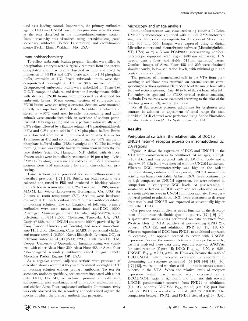
Figure 1A shows the expression of DCC and UNC5H in the
VTA from embryogenesis to adulthood. At all ages, a single
,185 kDa band was observed with the DCC antibody and a
single ,135 kDa band was detected with the UNC5H antiserum.
Whereas DCC immunoreactivity was high in the rodent
midbrain during embryonic development, UNC5H immunore-
activity was barely detectable. At birth, DCC levels continued to
be high compared to UNC5H expression, but were reduced in
comparison to embryonic DCC levels. At post-weaning, a
substantial reduction in DCC expression was observed as well
as a noticeable increase in UNC5H immunoreactivity. From peri-
pubertal period to adulthood, DCC levels continued to decrease
dramatically and UNC5H was expressed at substantially higher
levels than DCC.
Our previous work implicates netrin function in the develop-
ment of the mesocorticolimbic system at puberty [17] [19] [20].
A quantitative analysis was performed on data obtained from
Western blots of VTA punches at post-weaning (PND 21),
puberty (PND 35), and adulthood (PND 90) (Fig. 1B, C).
Whereas expression of DCC from PND21 to adulthood appeared
to decrease, the opposite seemed to occur with UNC5H
expression. Because the immunoblots were developed separately,
we first analyzed these data using separate one-way ANOVAs
for each receptor (Figure 1B; DCC: F (2, 14) = 3.50, p = 0.06;
UNC5H: F (2, 20) = 2.54, p = 0.10). However, because the ratio of
DCC:UNC5H netrin receptor expression is important in
determining the response to netrin-1 [5] [43] [44] [45] [46]
[47] [48], we examined whether a sift in the ratio occurs around
puberty in the VTA. When the relative levels of receptor
expression within each sample were expressed as a
DCC:UNC5H ratio, a significant and dramatic shift toward
UNC5H predominance occurred from PND21 to adulthood
(Fig. 1C; one-way ANOVA: F(2,13) = 4.42; p = 0.03; post hoc
Tukey’s HSD tests revealed a critical q = 3.73, a= 0.05. The
comparison between PND21 and PND35 yielded a q(13) = 3.47,
Netrin Receptors in DA Neurons
PLoS ONE | www.plosone.org 4 July 2010 | Volume 5 | Issue 7 | e11463
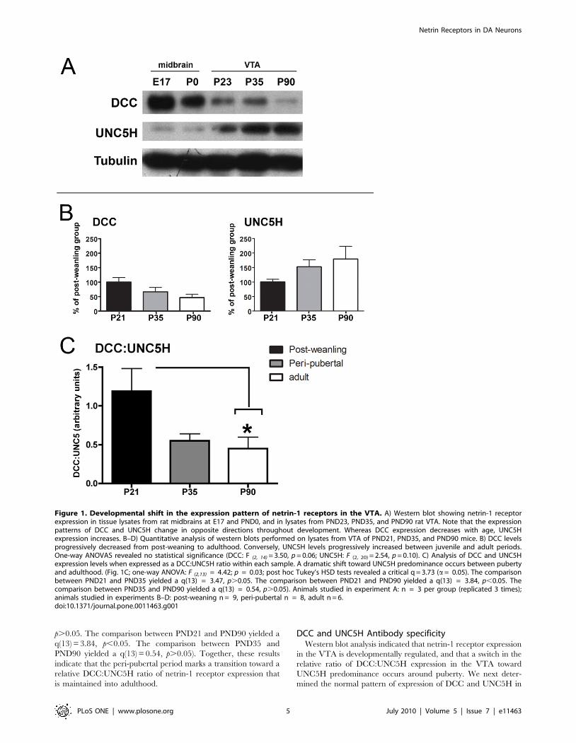
p.0.05. The comparison between PND21 and PND90 yielded a
q(13) = 3.84, p,0.05. The comparison between PND35 and
PND90 yielded a q(13) = 0.54, p.0.05). Together, these results
indicate that the peri-pubertal period marks a transition toward a
relative DCC:UNC5H ratio of netrin-1 receptor expression that
is maintained into adulthood.
DCC and UNC5H Antibody specificityWestern blot analysis indicated that netrin-1 receptor expression
in the VTA is developmentally regulated, and that a switch in the
relative ratio of DCC:UNC5H expression in the VTA toward
UNC5H predominance occurs around puberty. We next deter-
mined the normal pattern of expression of DCC and UNC5H in
Figure 1. Developmental shift in the expression pattern of netrin-1 receptors in the VTA. A) Western blot showing netrin-1 receptorexpression in tissue lysates from rat midbrains at E17 and PND0, and in lysates from PND23, PND35, and PND90 rat VTA. Note that the expressionpatterns of DCC and UNC5H change in opposite directions throughout development. Whereas DCC expression decreases with age, UNC5Hexpression increases. B–D) Quantitative analysis of western blots performed on lysates from VTA of PND21, PND35, and PND90 mice. B) DCC levelsprogressively decreased from post-weaning to adulthood. Conversely, UNC5H levels progressively increased between juvenile and adult periods.One-way ANOVAS revealed no statistical significance (DCC: F (2, 14) = 3.50, p = 0.06; UNC5H: F (2, 20) = 2.54, p = 0.10). C) Analysis of DCC and UNC5Hexpression levels when expressed as a DCC:UNC5H ratio within each sample. A dramatic shift toward UNC5H predominance occurs between pubertyand adulthood. (Fig. 1C; one-way ANOVA: F (2,13) = 4.42; p = 0.03; post hoc Tukey’s HSD tests revealed a critical q = 3.73 (a= 0.05). The comparisonbetween PND21 and PND35 yielded a q(13) = 3.47, p.0.05. The comparison between PND21 and PND90 yielded a q(13) = 3.84, p,0.05. Thecomparison between PND35 and PND90 yielded a q(13) = 0.54, p.0.05). Animals studied in experiment A: n = 3 per group (replicated 3 times);animals studied in experiments B–D: post-weaning n = 9, peri-pubertal n = 8, adult n = 6.doi:10.1371/journal.pone.0011463.g001
Netrin Receptors in DA Neurons
PLoS ONE | www.plosone.org 5 July 2010 | Volume 5 | Issue 7 | e11463
VTA DA neurons from embryogenesis to adulthood in both mice
and rats.
The specificity of the DCC antibody was confirmed by Western
blot performed on whole brain homogenates of E17 dcc 2/2 mice
and +/+ controls (Fig. S1A). As we previously showed in rat and
mouse brains [19] [17] [18], the DCC antibody detected a single
,185 kDa band in brains of E17 +/+ mice. However, no
immunoreactive band was detected in brains of dcc 2/2 embryos.
DCC immunofluorescence on midbrain sections of E17 dcc 2/2
embryos revealed no immunoreactive signal (Fig. S1B), whereas
high levels of DCC have previously been reported in wild type
mice [11] [12].
It is important to mention that similar to what we observed in
+/+ mouse embryos, TH-immunopositive neurons were also
present in the ventral midbrain region of dcc 2/2 embryos (Fig.
S1B), indicating that DCC-mediated netrin-1 signalling is not
required for the formation and differentiation of midbrain DA
neurons.
The specificity of the UNC5H antiserum has been demonstrated
previously [37] [38]. This antiserum was raised against a peptide
sequence in the mouse UNC5C cytoplasmic domain, and its
specificity for UNC5C was confirmed by immunoprecipitation,
Western blot analysis, and immunostaining of HEK-293 cells
expressing unc5c cDNA [37]. This antiserum was also shown to bind
recombinant UNC5A and UNC5B by Western blot analysis [38].
We now further characterized the specificity of this antiserum
by Western blot using whole cerebella from adult unc5c 2/2 and
+/+ mice ([27], Fig. S1C). Adult mouse cerebellum expresses all
the identified mammalian UNC-5 homologues (unc5a & unc5b,
Leonardo et al., 1997; unc5c, Ackerman et al., 1997; unc5d,
unpublished observations, RT-PCR). The antiserum detected a
,135 kDa band in both unc5c 2/2 and +/+ mice, indicating that
it recognizes other endogenous UNC-5 homologues, in addition to
UNC5C. Immunohistochemistry conducted in cerebellar tissue
from unc5c 2/2 and +/+ mice revealed no differences in the
labelling pattern (Fig. S1D).
We also performed RT-PCR experiments on discrete tissue
punches taken from the VTA of adult wild-type mice (Fig. S1E).
unc5c and unc5d were found to be expressed in the VTA. Because
of this finding, we examined the pattern of immunofluorescence
produced by the antiserum in the VTA of adult unc5c 2/2 and
+/+ mice. However, again there was no difference in the pattern
of immunoreactivity between genotypes, confirming that the
antiserum recognizes the two UNC-5 homologues expressed in the
adult VTA, namely UNC5C and UNC5D (Fig. S1F). Further-
more, the similar pattern of UNC5H immunoreactivity between
the two genotypes suggests that UNC5C and UNC5D are co-
expressed. The potential functional significance of UNC5D, or
UNC5C and UNC5D co-expression, by DA neurons is presently
unknown.
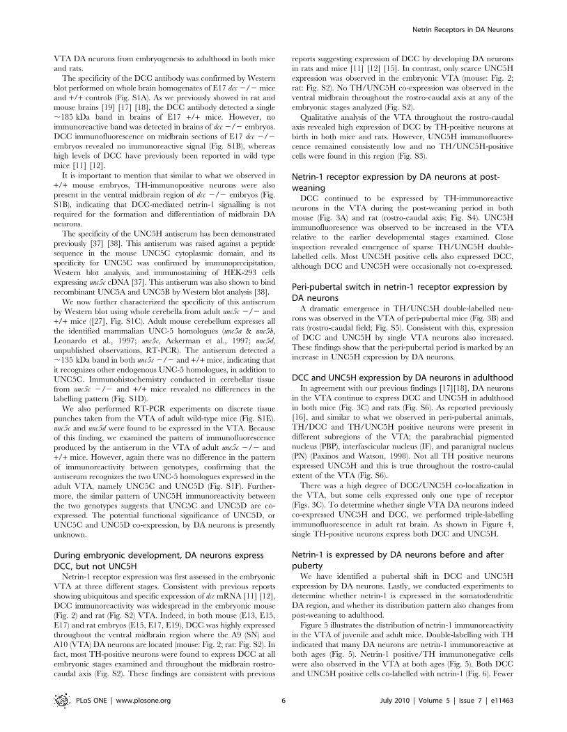
During embryonic development, DA neurons expressDCC, but not UNC5H
Netrin-1 receptor expression was first assessed in the embryonic
VTA at three different stages. Consistent with previous reports
showing ubiquitous and specific expression of dcc mRNA [11] [12],
DCC immunoreactivity was widespread in the embryonic mouse
(Fig. 2) and rat (Fig. S2) VTA. Indeed, in both mouse (E13, E15,
E17) and rat embryos (E15, E17, E19), DCC was highly expressed
throughout the ventral midbrain region where the A9 (SN) and
A10 (VTA) DA neurons are located (mouse: Fig. 2; rat: Fig. S2). In
fact, most TH-positive neurons were found to express DCC at all
embryonic stages examined and throughout the midbrain rostro-
caudal axis (Fig. S2). These findings are consistent with previous
reports suggesting expression of DCC by developing DA neurons
in rats and mice [11] [12] [15]. In contrast, only scarce UNC5H
expression was observed in the embryonic VTA (mouse: Fig. 2;
rat: Fig. S2). No TH/UNC5H co-expression was observed in the
ventral midbrain throughout the rostro-caudal axis at any of the
embryonic stages analyzed (Fig. S2).
Qualitative analysis of the VTA throughout the rostro-caudal
axis revealed high expression of DCC by TH-positive neurons at
birth in both mice and rats. However, UNC5H immunofluores-
cence remained consistently low and no TH/UNC5H-positive
cells were found in this region (Fig. S3).
Netrin-1 receptor expression by DA neurons at post-weaning
DCC continued to be expressed by TH-immunoreactive
neurons in the VTA during the post-weaning period in both
mouse (Fig. 3A) and rat (rostro-caudal axis; Fig. S4). UNC5H
immunofluoresence was observed to be increased in the VTA
relative to the earlier developmental stages examined. Close
inspection revealed emergence of sparse TH/UNC5H double-
labelled cells. Most UNC5H positive cells also expressed DCC,
although DCC and UNC5H were occasionally not co-expressed.
Peri-pubertal switch in netrin-1 receptor expression byDA neurons
A dramatic emergence in TH/UNC5H double-labelled neu-
rons was observed in the VTA of peri-pubertal mice (Fig. 3B) and
rats (rostro-caudal field; Fig. S5). Consistent with this, expression
of DCC and UNC5H by single VTA neurons also increased.
These findings show that the peri-pubertal period is marked by an
increase in UNC5H expression by DA neurons.
DCC and UNC5H expression by DA neurons in adulthoodIn agreement with our previous findings [17][18], DA neurons
in the VTA continue to express DCC and UNC5H in adulthood
in both mice (Fig. 3C) and rats (Fig. S6). As reported previously
[16], and similar to what we observed in peri-pubertal animals,
TH/DCC and TH/UNC5H positive neurons were present in
different subregions of the VTA; the parabrachial pigmented
nucleus (PBP), interfascicular nucleus (IF), and paranigral nucleus
(PN) (Paxinos and Watson, 1998). Not all TH positive neurons
expressed UNC5H and this is true throughout the rostro-caulal
extent of the VTA (Fig. S6).
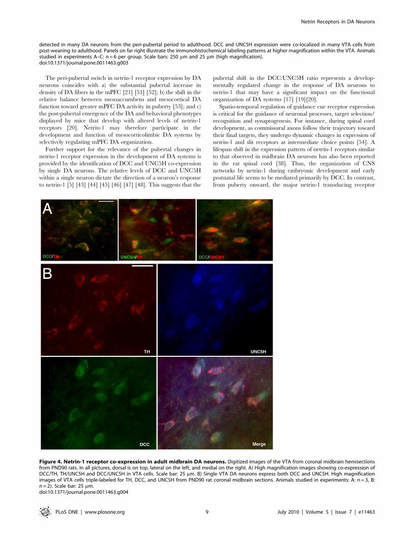
There was a high degree of DCC/UNC5H co-localization in
the VTA, but some cells expressed only one type of receptor
(Figs. 3C). To determine whether single VTA DA neurons indeed
co-expressed UNC5H and DCC, we performed triple-labelling
immunofluorescence in adult rat brain. As shown in Figure 4,
single TH-positive neurons express both DCC and UNC5H.
Netrin-1 is expressed by DA neurons before and afterpuberty
We have identified a pubertal shift in DCC and UNC5H
expression by DA neurons. Lastly, we conducted experiments to
determine whether netrin-1 is expressed in the somatodendritic
DA region, and whether its distribution pattern also changes from
post-weaning to adulthood.
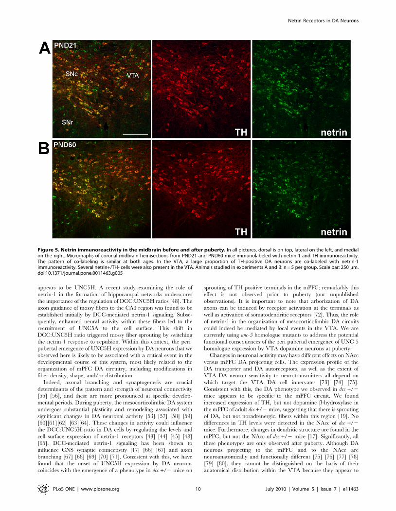
Figure 5 illustrates the distribution of netrin-1 immunoreactivity
in the VTA of juvenile and adult mice. Double-labelling with TH
indicated that many DA neurons are netrin-1 immunoreactive at
both ages (Fig. 5). Netrin-1 positive/TH immunonegative cells
were also observed in the VTA at both ages (Fig. 5). Both DCC
and UNC5H positive cells co-labelled with netrin-1 (Fig. 6). Fewer
Netrin Receptors in DA Neurons
PLoS ONE | www.plosone.org 6 July 2010 | Volume 5 | Issue 7 | e11463
netrin-1/DCC positive cells were detected in adulthood relative to
post-weaning. In contrast, the number of netrin-1 positive cells
that co-labelled with UNC5H appeared to increase from post-
weaning to adulthood. Although netrin-1 is a secreted protein, it
has been shown that soon after secretion it becomes associated
with cellular membranes and extracellular matrix [1] [49] [50].
Thus, the pattern of netrin-1 immunoreactivity that we observed
suggests that DA neurons express netrin-1.
Discussion
The major finding of this study is that the expression pattern of the
netrin-1 receptors DCC and UNC5H by VTA DA neurons shifts
dramatically at the peri-pubertal period in both rats and mice. DCC
is expressed by TH-positive neurons in the VTA from embryonic life
to adulthood. Conversely, UNC5H expression by DA neurons only
emerges at the peri-pubertal age and remains elevated throughout
adulthood. Western blot analysis confirmed that the relative ratio of
DCC to UNC5H (DCC:UNC5H) expression in the VTA shifts
significantly toward UNC5H predominance around puberty. The
level and pattern of netrin-1 immunoreactivity, however, was very
similar between the juvenile and adult period. Together, these results
suggest that netrin-1 function may contribute to the plasticity,
function and peri-pubertal reorganization of mesocorticolimbic DA
systems and that these effects are likely to be controlled by variations
in the levels of netrin-1 receptor expression by DA neurons.
Figure 2. Netrin-1 receptor expression in embryonic VTA dopamine neurons. Low magnification micrographs in the first panel of Athrough D illustrate coronal ventral midbrain sections from E13 wild-type (A,B) and E17 wild-type (C,D) mouse embryos immunolabelled with DCC(A,C; green) or UNC5H (B,D; green) and TH (red). Regions surrounded by a box represent the location within the embryonic VTA (The Altlas of theDeveloping brain, [23]) that is presented in neighboring higher magnification panels. A) Widespread expression of DCC is detected in the embryonicmidbrain, with DCC and TH co-localizing in the ventral midbrain. B) Only scarce expression of UNC5H was observed in the embryonic ventral midbrainand no co-localization of UNC5H and TH was detected. In all pictures, the dorsal aspect of coronal sections is on top. Scale bars: 250 mm (lowmagnification panels) and 25 mm (high magnification panels). C–D) Netrin-1 receptor expression in E17 mouse midbrain dopamine neurons. Digitizedimages of coronal ventral midbrain hemisections from E17 wild-type mouse embryos indicate that DCC (C), but not UNC5H (D), is expressed in THimmunopositive neurons of the VTA in E17 mouse embryos. Dorsal is on top, lateral on the right, and medial is on the left. Similar results wereobtained at E15 (data not shown). Animals studied in experiments A–D: n = 7 per group, Scale bars: 250 mm (low magnification panels) and 25 mm(high magnification panels).doi:10.1371/journal.pone.0011463.g002
Netrin Receptors in DA Neurons
PLoS ONE | www.plosone.org 7 July 2010 | Volume 5 | Issue 7 | e11463
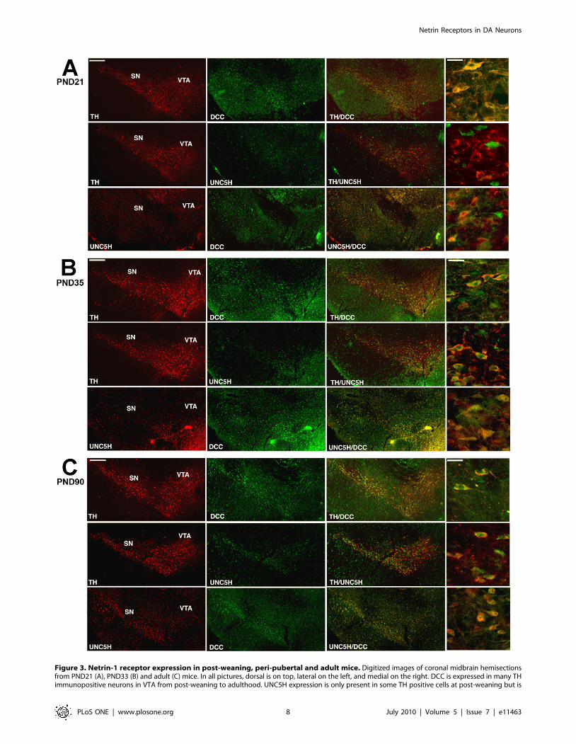
Figure 3. Netrin-1 receptor expression in post-weaning, peri-pubertal and adult mice. Digitized images of coronal midbrain hemisectionsfrom PND21 (A), PND33 (B) and adult (C) mice. In all pictures, dorsal is on top, lateral on the left, and medial on the right. DCC is expressed in many THimmunopositive neurons in VTA from post-weaning to adulthood. UNC5H expression is only present in some TH positive cells at post-weaning but is
Netrin Receptors in DA Neurons
PLoS ONE | www.plosone.org 8 July 2010 | Volume 5 | Issue 7 | e11463
The peri-pubertal switch in netrin-1 receptor expression by DA
neurons coincides with a) the substantial pubertal increase in
density of DA fibers in the mPFC [21] [51] [52]; b) the shift in the
relative balance between mesoaccumbens and mesocortical DA
function toward greater mPFC DA activity in puberty [53]; and c)
the post-pubertal emergence of the DA and behavioral phenotypes
displayed by mice that develop with altered levels of netrin-1
receptors [20]. Netrin-1 may therefore participate in the
development and function of mesocorticolimbic DA systems by
selectively regulating mPFC DA organization.
Further support for the relevance of the pubertal changes in
netrin-1 receptor expression in the development of DA systems is
provided by the identification of DCC and UNC5H co-expression
by single DA neurons. The relative levels of DCC and UNC5H
within a single neuron dictate the direction of a neuron’s response
to netrin-1 [5] [43] [44] [45] [46] [47] [48]. This suggests that the
pubertal shift in the DCC:UNC5H ratio represents a develop-
mentally regulated change in the response of DA neurons to
netrin-1 that may have a significant impact on the functional
organization of DA systems [17] [19][20].
Spatio-temporal regulation of guidance cue receptor expression
is critical for the guidance of neuronal processes, target selection/
recognition and synaptogenesis. For instance, during spinal cord
development, as commissural axons follow their trajectory toward
their final targets, they undergo dynamic changes in expression of
netrin-1 and slit receptors at intermediate choice points [54]. A
lifespan shift in the expression pattern of netrin-1 receptors similar
to that observed in midbrain DA neurons has also been reported
in the rat spinal cord [38]. Thus, the organization of CNS
networks by netrin-1 during embryonic development and early
postnatal life seems to be mediated primarily by DCC. In contrast,
from puberty onward, the major netrin-1 transducing receptor
Figure 4. Netrin-1 receptor co-expression in adult midbrain DA neurons. Digitized images of the VTA from coronal midbrain hemisectionsfrom PND90 rats. In all pictures, dorsal is on top, lateral on the left, and medial on the right. A) High magnification images showing co-expression ofDCC/TH, TH/UNC5H and DCC/UNC5H in VTA cells. Scale bar: 25 mm. B) Single VTA DA neurons express both DCC and UNC5H. High magnificationimages of VTA cells triple-labeled for TH, DCC, and UNC5H from PND90 rat coronal midbrain sections. Animals studied in experiments: A: n = 3, B:n = 2). Scale bar: 25 mm.doi:10.1371/journal.pone.0011463.g004
detected in many DA neurons from the peri-pubertal period to adulthood. DCC and UNC5H expression were co-localized in many VTA cells frompost-weaning to adulthood. Panels on far right illustrate the immunohistochemical labeling patterns at higher magnification within the VTA. Animalsstudied in experiments A–C: n = 6 per group. Scale bars: 250 mm and 25 mm (high magnification).doi:10.1371/journal.pone.0011463.g003
Netrin Receptors in DA Neurons
PLoS ONE | www.plosone.org 9 July 2010 | Volume 5 | Issue 7 | e11463
appears to be UNC5H. A recent study examining the role of
netrin-1 in the formation of hippocampal networks underscores
the importance of the regulation of DCC:UNC5H ratios [48]. The
axon guidance of mossy fibers to the CA3 region was found to be
established initially by DCC-mediated netrin-1 signaling. Subse-
quently, enhanced neural activity within these fibers led to the
recruitment of UNC5A to the cell surface. This shift in
DCC:UNC5H ratio triggered mossy fiber sprouting by switching
the netrin-1 response to repulsion. Within this context, the peri-
pubertal emergence of UNC5H expression by DA neurons that we
observed here is likely to be associated with a critical event in the
developmental course of this system, most likely related to the
organization of mPFC DA circuitry, including modifications in
fiber density, shape, and/or distribution.
Indeed, axonal branching and synaptogenesis are crucial
determinants of the pattern and strength of neuronal connectivity
[55] [56], and these are more pronounced at specific develop-
mental periods. During puberty, the mesocorticolimbic DA system
undergoes substantial plasticity and remodeling associated with
significant changes in DA neuronal activity [53] [57] [58] [59]
[60][61][62] [63][64]. These changes in activity could influence
the DCC:UNC5H ratio in DA cells by regulating the levels and
cell surface expression of netrin-1 receptors [43] [44] [45] [48]
[65]. DCC-mediated netrin-1 signaling has been shown to
influence CNS synaptic connectivity [17] [66] [67] and axon
branching [67] [68] [69] [70] [71]. Consistent with this, we have
found that the onset of UNC5H expression by DA neurons
coincides with the emergence of a phenotype in dcc +/2 mice on
sprouting of TH positive terminals in the mPFC; remarkably this
effect is not observed prior to puberty (our unpublished
observations). It is important to note that arborization of DA
axons can be induced by receptor activation at the terminals as
well as activation of somatodendritic receptors [72]. Thus, the role
of netrin-1 in the organization of mesocorticolimbic DA circuits
could indeed be mediated by local events in the VTA. We are
currently using unc-5 homologue mutants to address the potential
functional consequences of the peri-pubertal emergence of UNC-5
homologue expression by VTA dopamine neurons at puberty.
Changes in neuronal activity may have different effects on NAcc
versus mPFC DA projecting cells. The expression profile of the
DA transporter and DA autoreceptors, as well as the extent of
VTA DA neuron sensitivity to neurotransmitters all depend on
which target the VTA DA cell innervates [73] [74] [75].
Consistent with this, the DA phenotype we observed in dcc +/2
mice appears to be specific to the mPFC circuit. We found
increased expression of TH, but not dopamine b-hydroxylase in
the mPFC of adult dcc +/2 mice, suggesting that there is sprouting
of DA, but not noradrenergic, fibers within this region [19]. No
differences in TH levels were detected in the NAcc of dcc +/2
mice. Furthermore, changes in dendritic structure are found in the
mPFC, but not the NAcc of dcc +/2 mice [17]. Significantly, all
these phenotypes are only observed after puberty. Although DA
neurons projecting to the mPFC and to the NAcc are
neuroanatomically and functionally different [75] [76] [77] [78]
[79] [80], they cannot be distinguished on the basis of their
anatomical distribution within the VTA because they appear to
Figure 5. Netrin immunoreactivity in the midbrain before and after puberty. In all pictures, dorsal is on top, lateral on the left, and medialon the right. Micrographs of coronal midbrain hemisections from PND21 and PND60 mice immunolabeled with netrin-1 and TH immunoreactivity.The pattern of co-labeling is similar at both ages. In the VTA, a large proportion of TH-positive DA neurons are co-labeled with netrin-1immunoreactivity. Several netrin+/TH- cells were also present in the VTA. Animals studied in experiments A and B: n = 5 per group. Scale bar: 250 mm.doi:10.1371/journal.pone.0011463.g005
Netrin Receptors in DA Neurons
PLoS ONE | www.plosone.org 10 July 2010 | Volume 5 | Issue 7 | e11463

Figure 6. Netrin-1 and netrin-1 receptor co-labeling. Micrographs of coronal midbrain hemisections from PND21 and PND60 miceimmunolabeled for netrin-1 and the netrin-1 receptors. Dorsal is on top, lateral on the left, and medial on the right. A) Micrographs of VTA cells co-labeled with netrin-1 and DCC immunoreactivity at PND21 and PND60. The number of netrin-1 positive cells that co-express DCC decreases afterpuberty. B) Micrographs of VTA cells co-labeled with netrin-1 and UNC5H immunoreactivity at PND21 and PND60. The number of netrin-1 positivecells that co-express UNC5H increases after puberty. Animals studied in experiment A: n = 7 per group. Scale bar: 250 mm.doi:10.1371/journal.pone.0011463.g006
Netrin Receptors in DA Neurons
PLoS ONE | www.plosone.org 11 July 2010 | Volume 5 | Issue 7 | e11463
exist as an intermingled population in the PBP, PN and midline
nuclei [75] [81]. The sudden and delayed expression of UNC5H
by DA neurons, which, in contrast to DCC, appears to be
observed within a subset of VTA DA neurons, may be occurring
specifically in mPFC projecting cells. This possibility is being
currently tested in our laboratory using retrograde tracers.
Finally, changes in DCC and UNC5H expression may directly
affect the somatodendritic structure of VTA DA cells and in turn
their activity. Recent work from our group shows robust up-
regulation of DCC and UNC5 expression in the VTA, but not
mPFC or NAcc, following repeated exposure to the stimulant drug
amphetamine, using a regimen known to induced structural
changes in VTA DA cells [18] [82]. Moreover, blockade of DCC
signaling directly into the VTA prevents amphetamine-induced
structural and behavioral plasticity [39]. Remarkably, this
amphetamine-induced netrin-1/dopamine interaction is reversed
when animals are treated before puberty (L. Yetnikoff and C.
Flores, unpublished observations).
In conclusion, the present findings demonstrate that netrin-1
receptors are expressed by DA neurons throughout life. While
DCC expression predominates until the post-weaning period,
UNC5H predominates from puberty onwards. This switch in the
relative levels of DCC and UNC5H takes place during a critical
developmental period marked by substantial reorganization and
vulnerability of the mPFC DA system, which coincides with the
onset of symptoms of many psychiatric disorders, including
schizophrenia. dcc +/2 mice exhibit selective alterations in the
organization and function of the mesocortical DA circuit and
appear to be protected against schizophrenia-like symptoms.
Significantly, this phenotype emerges after puberty. Determining
the precise role of netrin-1 receptors in the establishment of
mesocorticolimbic circuitry may, therefore, help in understanding
the mechanisms underlying the development of individual
differences in vulnerability to psychopathology.
Supporting Information
Figure S1 Netrin-1 receptor antibody specificity. A, B) DCC
antibody specificity. A) The DCC antibody used in this study
detected a single ,185 kDa band corresponding to DCC in wild-
type (+/+) E17 mouse brain, but not in dcc 2/2 E17 mouse
brain. B) Micrographs of coronal midbrain sections from E17 dcc
2/2 mouse embryos double-labeled with DCC and TH
immunoreactivity. No DCC immunoreactivity was observed in
dcc 2/2 embryos, further demonstrating the specificity of the
DCC antibody. Note that TH immunopositive neurons are
present in the ventral midbrain region of E17 dcc 2/2 mouse
embryos, indicating that DCC is not required for the formation
and differentiation of midbrain dopaminergic neurons. Similar
results were obtained at earlier embryonic stages (E15, data
not shown). The dorsal aspect of coronal sections is on top. Scale
bar: 250 mm. C, D) UNC5C antibody specificity. C) UNC5C
antiserum specificity. A single band at ,135 kDa, was detected by
Western blot in lysates from both wild-type (+/+) and unc5c 2/2
cerebella, suggesting that the antiserum can recognize other
endogenous UNC-5 homologues in whole cerebellar homogenates
in addition to UNC5C. D) Micrographs of coronal sections of
cerebella dissected from +/+ and unc5c 2/2 mice. The pattern of
DAB immunolabeling is similar between genotypes, indicating
that the UNC5C antiserum also recognizes additional UNC-5
homologues immunohistochemically. Scale bar: 25 mm. E)
Schematic representation of the locations of bilateral tissue
punches of the VTA for RT-PCR experiments examining the
expression of unc-5 homologue mRNAs in this adult mouse
somatodendritic DA region (PND60; [41]). unc5c and unc5d
homologues are detected in the VTA. The oligonucleotide
sequences used are listed in the Materials and Methods section.
F) UNC5H immunoreactivity in the VTA of adult wild-type +/+and unc5c 2/2 mice. The patterns of immunoreactivity are
similar between the two genotypes, indicating that the antiserum
also recognizes UNC5D using immunohistochemistry. Animals
studied in experiment A: n = 3, B; n = 4, C: n = 5, D: n = 4. Scale
bar: 25 mm.
Found at: doi:10.1371/journal.pone.0011463.s001 (4.47 MB
TIF)
Figure S2 Netrin-1 receptor expression in E17 rat midbrain
dopamine neurons. Digitized images of coronal midbrain sections
from E17 rat embryos (Similar results were obtained in E15 and
E19 rat embryos). Panels on the left hand side show TH expression
at different rostro-caudal levels of the E17 rat midbrain. Panels
adjacent to the low magnification images of TH immunostaining
show co-localization of TH and DCC (A) and absence of co-
localization of TH and UNC5H (B) in the ventral midbrain region
at the corresponding rostro-caudal levels. In all pictures, the dorsal
aspect of coronal sections is on top. Similar results were obtained
in the mouse at the corresponding embryonic age (E15, data not
shown). Animals studied in experiment: n = 3. Scale bars: 250 mm
(images on the extreme left) and 25 mm for other images.
Found at: doi:10.1371/journal.pone.0011463.s002 (4.75 MB
TIF)
Figure S3 Netrin-1 receptor expression in midbrain dopamine
neurons at birth. Digitized images of coronal midbrain hemisec-
tions from PND0 rat embryos. In all pictures, dorsal is on top,
lateral on the left, and medial on the right. Expression of DCC (A),
but not UNC5H (B), was detected in TH immunopositive neurons
in the ventral midbrain. Similar results were obtained in PND0
mouse embryos (data not shown). Animals studied in experiment:
n = 4. Scale bar: 25 mm.
Found at: doi:10.1371/journal.pone.0011463.s003 (8.18 MB TIF)
Figure S4 Netrin-1 receptor expression at post-weaning. Digi-
tized images of coronal midbrain hemisections from PND23 rats at
different rostro-caudal levels. In all pictures, dorsal is on top,
lateral on the left, and medial on the right. Expression of DCC (A)
was detected in TH immunopositive neurons in the VTA
throughout the rostro-caudal axis. At this developmental stage,
UNC5H expression begins to be detected in some TH positive
neurons of the VTA (B). Animals studied in experiment: n = 3.
Scale bar: 250 mm.
Found at: doi:10.1371/journal.pone.0011463.s004 (6.02 MB TIF)
Figure S5 Netrin-1 receptor expression during the peri-pubertal
period. Digitized images of coronal midbrain hemisections from
PND35 rats at different rostro-caudal levels. In all pictures, dorsal
is on top, lateral on the left, and medial on the right. DCC is
expressed in many TH immunopositive neurons in the VTA at all
rostro-caudal levels examined (A). At this developmental stage,
there is a robust up-regulation of UNC5H expression in both TH
negative and TH positive cells in the VTA throughout the rostro-
caudal axis (B). Animals studied in experiment: n = 3. Scale bar:
250 mm.
Found at: doi:10.1371/journal.pone.0011463.s005 (6.22 MB TIF)
Figure S6 Netrin-1 receptor expression in adulthood. Digitized
images of coronal midbrain hemisections from PND90 rats at
different rostro-caudal levels. In all pictures, dorsal is on top,
lateral on the left, and medial on the right. DCC is expressed in
many TH immunopositive neurons in the VTA throughout the
rostro-caudal axis (A). At this developmental stage, UNC5H
Netrin Receptors in DA Neurons
PLoS ONE | www.plosone.org 12 July 2010 | Volume 5 | Issue 7 | e11463
expression is highly expressed in both TH negative and TH
positive cells in the VTA at all rostro-caudal levels examined (B).
Animals studied in experiment: n = 3. Scale bar: 250 mm.
Found at: doi:10.1371/journal.pone.0011463.s006 (6.13 MB TIF)
Acknowledgments
We thank Jane Stewart for critical reading of the manuscript, Tony Pawson
(University of Toronto) for the UNC-5 antiserum and Susan Ackerman
(The Jackson Laboratory) for the unc5c 2/2 mouse brains and the original
dcc heterozygous breeders, and Helen Cooper for the DCC antiserum
(Queensland Brain Institute).
Author Contributions
Conceived and designed the experiments: CM CLD TS CF. Performed the
experiments: CM CLD CE AG AM. Analyzed the data: CM CLD CE AG
AM TS CF. Wrote the paper: CM CLD CE AG AM TS CF.
References
1. Manitt C, Kennedy TE (2002) Where the rubber meets the road: netrin
expression and function in developing and adult nervous systems. Prog BrainRes 137: 425–442.
2. Barallobre MJ, Pascual M, Del Rio JA, Soriano E (2005) The Netrin family ofguidance factors: emphasis on Netrin-1 signalling. Brain Res Brain Res Rev 49:
22–47.
3. Baker KA, Moore SW, Jarjour AA, Kennedy TE (2006) When a diffusible axon
guidance cue stops diffusing: roles for netrins in adhesion and morphogenesis.Curr Opin Neurobiol 16: 529–534.
4. Keino-Masu K, Masu M, Hinck L, Leonardo ED, Chan SS, et al. (1996)Deleted in Colorectal Cancer (DCC) encodes a netrin receptor. Cell 87:
175–185.
5. Hong K, Hinck L, Nishiyama M, Poo MM, Tessier-Lavigne M, et al. (1999) A
ligand-gated association between cytoplasmic domains of UNC5 and DCC
family receptors converts netrin-induced growth cone attraction to repulsion.Cell 97: 927–941.
6. Keleman K, Dickson BJ (2001) Short- and long-range repulsion by theDrosophila Unc5 netrin receptor. Neuron 32: 605–617.
7. Rajasekharan S, Kennedy TE (2009) The netrin protein family. Genome Biol10: 239.
8. Ly A, Nikolaev A, Suresh G, Zheng Y, Tessier-Lavigne M, et al. (2008) DSCAMis a netrin receptor that collaborates with DCC in mediating turning responses to
netrin-1. Cell 133: 1241–1254.
9. Andrews GL, Tanglao S, Farmer WT, Morin S, Brotman S, et al. (2008) Dscam
guides embryonic axons by Netrin-dependent and -independent functions.Development 135: 3839–3848.
10. Liu G, Li W, Wang L, Kar A, Guan KL, et al. (2009) DSCAM functions as anetrin receptor in commissural axon pathfinding. Proc Natl Acad Sci U S A 106:
2951–2956.
11. Gad JM, Keeling SL, Wilks AF, Tan SS, Cooper HM (1997) The expression
patterns of guidance receptors, DCC and Neogenin, are spatially and temporally
distinct throughout mouse embryogenesis. Dev Biol 192: 258–273.
12. Livesey FJ, Hunt SP (1997) Netrin and netrin receptor expression in the
embryonic mammalian nervous system suggests roles in retinal, striatal, nigral,and cerebellar development. Mol and Cell Neurosci 8: 417–429.
13. Volenec A, Zetterstrom TS, Flanigan TP (1998) 6-OHDA denervationsubstantially decreases DCC mRNA levels in rat substantia nigra compacta.
Neuroreport 9: 3553–3556.
14. Shu T, Valentino KM, Seaman C, Cooper HM, Richards LJ (2000) Expression
of the netrin-1 receptor, deleted in colorectal cancer (DCC), is largely confinedto projecting neurons in the developing forebrain. J Comp Neurol 416: 201–212.
15. Lin L, Rao Y, Isacson O (2005) Netrin-1 and slit-2 regulate and direct neuritegrowth of ventral midbrain dopaminergic neurons. Mol Cell Neurosci 28:
547–555.
16. Osborne PB, Halliday GM, Cooper HM, Keast JR (2005) Localization of
immunoreactivity for deleted in colorectal cancer (DCC), the receptor for the
guidance factor netrin-1, in ventral tier dopamine projection pathways in adultrodents. Neuroscience 131: 671–681.
17. Grant A, Hoops D, Labelle-Dumais C, Prevost M, Rajabi H, et al. (2007)Netrin-1 receptor-deficient mice show enhanced mesocortical dopamine
transmission and blunted behavioural responses to amphetamine.Eur J Neurosci 26: 3215–3228.
18. Yetnikoff L, Labelle-Dumais C, Flores C (2007) Regulation of netrin-1 receptorsby amphetamine in the adult brain. Neuroscience 150: 764–773.
19. Flores C, Manitt C, Rodaros D, Thompson KM, Rajabi H, et al. (2005) Netrinreceptor deficient mice exhibit functional reorganization of dopaminergic
systems and do not sensitize to amphetamine. Mol Psychiatry 10: 606–612.
20. Grant A, Speed Z, Labelle-Dumais C, Flores C (2009) Post-pubertal emergence
of a dopamine phenotype in netrin-1 receptor-deficient mice. Eur J Neurosci 30:
1318–1328.
21. Kalsbeek A, Voorn P, Buijs RM, Pool CW, Uylings HB (1988) Development of
the dopaminergic innervation in the prefrontal cortex of the rat. J Comp Neurol269: 58–72.
22. Voorn P, Kalsbeek A, Jorritsma-Byham B, Groenewegen HJ (1988) The pre-and postnatal development of the dopaminergic cell groups in the ventral
mesencephalon and the dopaminergic innervation of the striatum of the rat.Neuroscience 25: 857–887.
23. Jacobowitz DM, Abbott LC (1998) Chemoarchitectonic atlas of the developingmouse brain. Boca Raton. Florida: CRC Press LLC.
24. Riddle R, Pollock JD (2003) Making connections: the development of
mesencephalic dopaminergic neurons. Brain Res Dev Brain Res 147: 3–21.
25. Van den Heuvel DM, Pasterkamp RJ (2008) Getting connected in the dopamine
system. Prog Neurobiol 85: 75–93.
26. Fazeli A, Dickinson SL, Hermiston ML, Tighe RV, Steen RG, et al. (1997)
Phenotype of mice lacking functional Deleted in colorectal cancer (Dcc) gene.
Nature 386: 796–804.
27. Ackerman SL, Kozak LP, Przyborski SA, Rund LA, Boyer BB, et al. (1997) The
mouse rostral cerebellar malformation gene encodes an UNC-5-like protein.
Nature 386: 838–842.
28. Smidt MP, Burbach JP (2007) How to make a mesodiencephalic dopaminergic
neuron. Nat Rev Neurosci 8: 21–32.
29. Wolf ME, G. K (1989) Flow cytometric analysis and isolation of permeabilized
dopamine nerve terminals from rat striatum. J Neurosci 9: 106–114.
30. Yang Z, You Y, Levison SW (2008) Neonatal hypoxic/ischemic brain injury
induces production of calretinin-expressing interneurons in the striatum. J Comp
Neurol 511: 19–33.
31. Lu W, Monteggia LM, Wolf ME (2002) Repeated administration of
amphetamine or cocaine does not alter AMPA receptor subunit expression in
the rat midbrain. Neuropsychopharmacology 26: 1–13.
32. Hnasko TS, Perez FA, Scouras AD, Stoll EA, Gale SD, et al. (2006) Cre
recombinase-mediated restoration of nigrostriatal dopamine in dopamine-
deficient mice reverses hypophagia and bradykinesia. Proc Natl Acad Sci U S A
103: 8858–8863.
33. Kennedy TE, Serafini T, de la Torre JR, Tessier-Lavigne M (1994) Netrins are
diffusible chemotropic factors for commissural axons in the embryonic spinal
cord. Cell 78: 425–435.
34. Gad JM, Keeling SL, Shu T, Richards LJ, Cooper HM (2000) The spatial and
temporal expression patterns of netrin receptors, DCC and neogenin, in the
developing mouse retina. Exp Eye Res 70: 711–722.
35. Cooper HM, Armes P, Britto J, Gad J, Wilks AF (1995) Cloning of the mouse
homologue of the deleted in colorectal cancer gene (mDCC) and its expression in
the developing mouse embryo. Oncogene 11: 2243–2254.
36. Seaman C, Anderson R, Emery B, Cooper HM (2001) Localization of the netrin
guidance receptor, DCC, in the developing peripheral and enteric nervous
systems. Mech Dev 103: 173–175.
37. Tong J, Killeen M, Steven R, Binns KL, Culotti J, et al. (2001) Netrin stimulates
tyrosine phosphorylation of the UNC-5 family of netrin receptors and induces
Shp2 binding to the RCM cytodomain. J Biol Chem 276: 40917–40925.
38. Manitt C, Thompson KM, Kennedy TE (2004) Developmental shift in
expression of netrin receptors in the rat spinal cord: predominance of unc-5
homologues in adulthood. J Neurosci Res 77: 690–700.
39. Yetnikoff L, Eng C, Benning S, Flores C. Netrin-1 receptor in the ventral
tegmental area is required for sensitization to amphetamine. Eur J Neurosci 31:
1292–1302.
40. Paxinos G, Franklin KBJ (2001) The mouse brain in stereotaxic coordinates. San
Diego: Academic Press.
41. Paxinos G, Watson C (1998) The rat brain in stereotaxic coordinates. New York:
Academic Press.
42. Paxinos G, Tork I, Tecott LH, Valentino KL (1991) Atlas of the developing rat
brain. San Diego: Academic Press.
43. Williams ME, Wu SC, McKenna WL, Hinck L (2003) Surface expression of the
netrin receptor UNC5H1 is regulated through a protein kinase C-interacting
protein/protein kinase-dependent mechanism. J Neurosci 23: 11279–11288.
44. Bouchard JF, Moore SW, Tritsch NX, Roux PP, Shekarabi M, et al. (2004)
Protein kinase A activation promotes plasma membrane insertion of DCC from
an intracellular pool: A novel mechanism regulating commissural axon
extension. J Neurosci 24: 3040–3050.
45. Bouchard JF, Horn KE, Stroh T, Kennedy TE (2008) Depolarization recruits
DCC to the plasma membrane of embryonic cortical neurons and enhances
axon extension in response to netrin-1. J Neurochem.
46. Bartoe JL, McKenna WL, Quan TK, Stafford BK, Moore JA, et al. (2006)
Protein interacting with C-kinase 1/protein kinase Calpha-mediated endocytosis
converts netrin-1-mediated repulsion to attraction. J Neurosci 26: 3192–3205.
47. McKenna WL, Wong-Staal C, Kim GC, Macias H, Hinck L, et al. (2008)
Netrin-1-independent adenosine A2b receptor activation regulates the response
of axons to netrin-1 by controlling cell surface levels of UNC5A receptors.
J Neurochem 104: 1081–1090.
Netrin Receptors in DA Neurons
PLoS ONE | www.plosone.org 13 July 2010 | Volume 5 | Issue 7 | e11463
48. Muramatsu R, Nakahara S, Ichikawa J, Watanabe K, Matsuki N, et al. The
ratio of ’deleted in colorectal cancer’ to ’uncoordinated-5A’ netrin-1 receptors on
the growth cone regulates mossy fibre directionality. Brain 133: 60–75.
49. Serafini T, Kennedy TE, Galko MJ, Mirzayan C, Jessell TM, et al. (1994) The
netrins define a family of axon outgrowth-promoting proteins homologous to C.
elegans UNC-6. Cell 78: 409–424.
50. Manitt C, Colicos MA, Thompson KM, Rousselle E, Peterson AC, et al. (2001)
Widespread expression of netrin-1 by neurons and oligodendrocytes in the adult
mammalian spinal cord. J Neurosci 21: 3911–3922.
51. Benes FM, Taylor JB, Cunningham MC (2000) Convergence and plasticity of
monoaminergic systems in the medial prefrontal cortex during the postnatal
period: implications for the development of psychopathology. Cereb Cortex 10:
1014–1027.
52. Juarez I, Gratton A, Flores G (2008) Ontogeny of altered dendritic morphology
in the rat prefrontal cortex, hippocampus, and nucleus accumbens following
Cesarean delivery and birth anoxia. J Comp Neurol 507: 1734–1747.
53. Spear LP (2000) The adolescent brain and age-related behavioral manifesta-
tions. Neurosci Biobehav Rev 24: 417–463.
54. Sabatier C, Plump AS, Le M, Brose K, Tamada A, et al. (2004) The divergent
Robo family protein rig-1/Robo3 is a negative regulator of slit responsiveness
required for midline crossing by commissural axons. Cell 117: 157–169.
55. Uesaka N, Ruthazer ES, Yamamoto N (2006) The role of neural activity in
cortical axon branching. Neuroscientist 12: 102–106.
56. Gogolla N, Galimberti I, Caroni P (2007) Structural plasticity of axon terminals
in the adult. Curr Opin Neurobiol 17: 516–524.
57. Teicher MH, Andersen SL, Hostetter JC, Jr. (1995) Evidence for dopamine
receptor pruning between adolescence and adulthood in striatum but not
nucleus accumbens. Brain Res Dev Brain Res 89: 167–172.
58. Andersen SL, Thompson AT, Rutstein M, Hostetter JC, Teicher MH (2000)
Dopamine receptor pruning in prefrontal cortex during the periadolescent
period in rats. Synapse 37: 167–169.
59. Tarazi FI, Baldessarini RJ (2000) Comparative postnatal development of
dopamine D(1), D(2) and D(4) receptors in rat forebrain. Int J Dev Neurosci 18:
29–37.
60. Andersen SL (2003) Trajectories of brain development: point of vulnerability or
window of opportunity? Neurosci Biobehav Rev 27: 3–18.
61. Adriani W, Laviola G (2004) Windows of vulnerability to psychopathology and
therapeutic strategy in the adolescent rodent model. Behav Pharmacol 15:
341–352.
62. Cao J, Lotfipour S, Loughlin SE, Leslie FM (2007) Adolescent maturation of
cocaine-sensitive neural mechanisms. Neuropsychopharmacology 32:
2279–2289.
63. Tseng KY, O’Donnell P (2007) D2 dopamine receptors recruit a GABA
component for their attenuation of excitatory synaptic transmission in the adult
rat prefrontal cortex. Synapse 61: 843–850.
64. Tseng KY, O’Donnell P (2007) Dopamine modulation of prefrontal cortical
interneurons changes during adolescence. Cereb Cortex 17: 1235–1240.
65. Hanson MG, Landmesser LT (2004) Normal patterns of spontaneous activity
are required for correct motor axon guidance and the expression of specificguidance molecules. Neuron 43: 687–701.
66. Colon-Ramos DA, Margeta MA, Shen K (2007) Glia promote local
synaptogenesis through UNC-6 (netrin) signaling in C. elegans. Science 318:103–106.
67. Manitt C, Nikolakopoulou AM, Almario DR, Nguyen SA, Cohen-Cory S (2009)Netrin participates in the development of retinotectal synaptic connectivity by
modulating axon arborization and synapse formation in the developing brain.
J Neurosci 29: 11065–11077.68. Lim YS, Mallapur S, Kao G, Ren XC, Wadsworth WG (1999) Netrin UNC-6
and the regulation of branching and extension of motoneuron axons from theventral nerve cord of Caenorhabditis elegans. J Neurosci 19: 7048–7056.
69. Dent EW, Barnes AM, Tang F, Kalil K (2004) Netrin-1 and semaphorin 3Apromote or inhibit cortical axon branching, respectively, by reorganization of
the cytoskeleton. J Neurosci 24: 3002–3012.
70. Tang F, Kalil K (2005) Netrin-1 induces axon branching in developing corticalneurons by frequency-dependent calcium signaling pathways. J Neurosci 25:
6702–6715.71. Hutchins BI, Kalil K (2008) Differential outgrowth of axons and their branches
is regulated by localized calcium transients. J Neurosci 28: 143–153.
72. Schmitz Y, Luccarelli J, Kim M, Wang M, Sulzer D (2009) Glutamate controlsgrowth rate and branching of dopaminergic axons. J Neurosci 29: 11973–11981.
73. Ford CP, Mark GP, Williams JT (2006) Properties and opioid inhibition ofmesolimbic dopamine neurons vary according to target location. J Neurosci 26:
2788–2797.74. Margolis EB, Mitchell JM, Ishikawa J, Hjelmstad GO, Fields HL (2008)
Midbrain dopamine neurons: projection target determines action potential
duration and dopamine D(2) receptor inhibition. J Neurosci 28: 8908–8913.75. Lammel S, Hetzel A, Hackel O, Jones I, Liss B, et al. (2008) Unique properties of
mesoprefrontal neurons within a dual mesocorticolimbic dopamine system.Neuron 57: 760–773.
76. Le Moal M, Simon H (1991) Mesocorticolimbic dopaminergic network:
functional and regulatory roles. Physiol Rev 71: 155–234.77. Knable MB, Weinberger DR (1997) Dopamine, the prefrontal cortex and
schizophrenia. J Psychopharmacol 11: 123–131.78. Tam SY, Roth RH (1997) Mesoprefrontal dopaminergic neurons: can tyrosine
availability influence their functions? Biochem Pharmacol 53: 441–453.79. Sesack SR, Hawrylak VA, Guido MA, Levey AI (1998) Cellular and subcellular
localization of the dopamine transporter in rat cortex. Adv Pharmacol 42:
171–174.80. Carr DB, Sesack SR (2000) Projections from the rat prefrontal cortex to the
ventral tegmental area: target specificity in the synaptic associations withmesoaccumbens and mesocortical neurons. J Neurosci 15: 3864–3873.
81. Bjorklund A, Dunnett SB (2007) Dopamine neuron systems in the brain: an
update. Trends Neurosci 30: 194–202.82. Mueller D, Chapman CA, Stewart J (2006) Amphetamine induces dendritic
growth in ventral tegmental area dopaminergic neurons in vivo via basicfibroblast growth factor. Neuroscience 137: 727–735.
Netrin Receptors in DA Neurons
PLoS ONE | www.plosone.org 14 July 2010 | Volume 5 | Issue 7 | e11463