
The pro-apoptotic activity of Drosophila Rbf1 involves dE2F2-dependent downregulation of diap1 and...
10
OPEN The pro-apoptotic activity of Drosophila Rbf1 involves dE2F2-dependent downregulation of diap1 and buffy mRNA A Clavier 1 , A Baillet 1 , A Rincheval-Arnold 1 , A Cole ´ no-Costes 2,3,4 , C Lasbleiz 1,5 , B Mignotte 1 and I Gue ´nal* ,1 The retinoblastoma gene, rb, ensures at least its tumor suppressor function by inhibiting cell proliferation. Its role in apoptosis is more complex and less described than its role in cell cycle regulation. Rbf1, the Drosophila homolog of Rb, has been found to be pro-apoptotic in proliferative tissue. However, the way it induces apoptosis at the molecular level is still unknown. To decipher this mechanism, we induced rbf1 expression in wing proliferative tissue. We found that Rbf1-induced apoptosis depends on dE2F2/dDP heterodimer, whereas dE2F1 transcriptional activity is not required. Furthermore, we highlight that Rbf1 and dE2F2 downregulate two major anti-apoptotic genes in Drosophila: buffy, an anti-apoptotic member of Bcl-2 family and diap1, a gene encoding a caspase inhibitor. On the one hand, Rbf1/dE2F2 repress buffy at the transcriptional level, which contributes to cell death. On the other hand, Rbf1 and dE2F2 upregulate how expression. How is a RNA binding protein involved in diap1 mRNA degradation. By this way, Rbf1 downregulates diap1 at a post-transcriptional level. Moreover, we show that the dREAM complex has a part in these transcriptional regulations. Taken together, these data show that Rbf1, in cooperation with dE2F2 and some members of the dREAM complex, can downregulate the anti-apoptotic genes buffy and diap1, and thus promote cell death in a proliferative tissue. Cell Death and Disease (2014) 5, e1405; doi:10.1038/cddis.2014.372; published online 4 September 2014 The retinoblastoma gene (rb) is the first tumor suppressor identified in human cells. Its product, pRb, inhibits cell proliferation by controlling the G1/S transition. 1 The best characterized partners of pRB in cell cycle regulation belong to the E2F family of transcription factors. 2 In contrast to its tumor suppressor role, pRb is surprisingly often described as an anti-apoptotic protein. 3–10 However, a growing number of studies show a pro-apoptotic role for pRb. 11–14 Drosophila, which presents a lesser genetic complexity than mammals, offers the possibility to decipher the roles of Rb and E2F family proteins in apoptosis in vivo. Indeed, the Drosophila genome contains two E2F genes 15–17 (de2f1 and de2f2), one DP gene 15 (dDp) that encodes an E2F cofactor and two Rb genes 18,19 (rbf1 and rbf2). dE2F1 acts mostly as a transcrip- tional activator, 20 whereas dE2F2 represses transcription. 21 As pRb protein in mammals, Rbf1 can bind both activator and repressor E2F members. 21 Furthermore, it fulfills the same function as pRb in the cell cycle. Rbf1 is thus considered as a pRb homolog. rbf1 loss-of-function is lethal at early larval stage 20,22 that attests its essential role. Homozygous rbf1- mutant embryos have many apoptotic cells 22 and it is admitted that Rbf1 protects cells by inhibiting the transcriptional activity of dE2F1 that is considered as a pro-apoptotic factor. Indeed, de2f1 and dDp co-expression induces apoptosis in the eye imaginal discs 23 and the pro-apoptotic genes reaper and dark are dE2F1-transcriptional targets. 24,25 In contrast to these data, we have previously shown that rbf1 can also have a pro- apoptotic function. Indeed, rbf1 overexpression in proliferating cells of wing imaginal discs leads to apoptosis and loss of tissue in adult wings. 26 This cell death is caspase dependent and can be inhibited by de2f1 expression. However, the precise mechanism underlying rbf1-induced apoptosis is still unknown. Rbf1 being mainly described as a transcriptional regulator, one may wonder whether this activity is involved in its pro-apoptotic effect. Recent reports have clarified the role of Rbf1 in transcription. Rbf1 binding to chromatin requires dE2F/dDP complexes. 27 This binding is mainly observed near transcription start sites (TSS). When associated with dE2F2, Rbf1 belongs to a transcription regulator complex named dREAM 28–30 (drosophila RBF, E2F and Myb-interacting proteins). This complex maintains the transcriptional repres- sion of certain E2F target genes in the proliferating tissues by at least two distinct mechanisms: histone deacetylation of nucleosomes near TSSs and dimethylation of histone H3 Lys27 at nucleosomes located downstream of TSSs. 31 Although the first reports described the dREAM complex as 1 Laboratoire de Ge ´ne ´ tique et Biologie Cellulaire, EA4589, Universite ´ de Versailles Saint-Quentin-en-Yvelines, Ecole Pratique des Hautes Etudes, 2 avenue de la Source de la Bie ` vre, Montigny-le-Bretonneux, France; 2 Laboratoire de Biologie du De ´ veloppement—Institut de Biologie Paris Seine, Sorbonne Universite ´s, UPMC Univ Paris 06, UMR7622, Paris, France and 3 CNRS, UMR7622, Laboratoire de Biologie du De ´ veloppement—Institut de Biologie Paris Seine, Paris, France *Corresponding author: I Gue ´ nal, Laboratoire de Ge ´ne ´ tique et Biologie Cellulaire, EA4589, Universite ´ de Versailles Saint-Quentin-en-Yvelines, Ecole Pratique des Hautes Etudes, 2 avenue de la Source de la Bie `vre 78180 Montigny-le-Bretonneux, France. Tel: +33 0 1 70 42 94 36; Fax: +33 0 1 70 42 95 03; E-mail: [email protected] 4 Present address: IGH—CNRS, 141 rue de la Cardonille, 34396 Montpellier, France. 5 Present address: UMR Inserm U710-UM2-EPHE, Universite ´ Montpellier 2, place Eugene Bataillon-CC105, 34095 Montpellier, France. Received 07.3.14; revised 23.7.14; accepted 28.7.14; Edited by E Baehrecke Abbreviations: ChIP, chromatin immunoprecipitation; dIAP1, Drosophila inhibitor of apoptosis; dREAM, drosophila RBF, E2F and Myb-interacting proteins; en, engrailed; how, held out wings; rb, retinoblastoma gene; TSS, transcription start sites; vg, vestigial Citation: Cell Death and Disease (2014) 5, e1405; doi:10.1038/cddis.2014.372 & 2014 Macmillan Publishers Limited All rights reserved 2041-4889/14 www.nature.com/cddis
Transcript of The pro-apoptotic activity of Drosophila Rbf1 involves dE2F2-dependent downregulation of diap1 and...

OPEN
The pro-apoptotic activity of Drosophila Rbf1 involvesdE2F2-dependent downregulation of diap1 and buffymRNA
A Clavier1, A Baillet1, A Rincheval-Arnold1, A Coleno-Costes2,3,4, C Lasbleiz1,5, B Mignotte1 and I Guenal*,1
The retinoblastoma gene, rb, ensures at least its tumor suppressor function by inhibiting cell proliferation. Its role in apoptosis ismore complex and less described than its role in cell cycle regulation. Rbf1, the Drosophila homolog of Rb, has been found to bepro-apoptotic in proliferative tissue. However, the way it induces apoptosis at the molecular level is still unknown. To decipherthis mechanism, we induced rbf1 expression in wing proliferative tissue. We found that Rbf1-induced apoptosis depends ondE2F2/dDP heterodimer, whereas dE2F1 transcriptional activity is not required. Furthermore, we highlight that Rbf1 and dE2F2downregulate two major anti-apoptotic genes in Drosophila: buffy, an anti-apoptotic member of Bcl-2 family and diap1, a geneencoding a caspase inhibitor. On the one hand, Rbf1/dE2F2 repress buffy at the transcriptional level, which contributes to celldeath. On the other hand, Rbf1 and dE2F2 upregulate how expression. How is a RNA binding protein involved in diap1 mRNAdegradation. By this way, Rbf1 downregulates diap1 at a post-transcriptional level. Moreover, we show that the dREAM complexhas a part in these transcriptional regulations. Taken together, these data show that Rbf1, in cooperation with dE2F2 and somemembers of the dREAM complex, can downregulate the anti-apoptotic genes buffy and diap1, and thus promote cell death in aproliferative tissue.Cell Death and Disease (2014) 5, e1405; doi:10.1038/cddis.2014.372; published online 4 September 2014
The retinoblastoma gene (rb) is the first tumor suppressoridentified in human cells. Its product, pRb, inhibits cellproliferation by controlling the G1/S transition.1 The bestcharacterized partners of pRB in cell cycle regulation belongto the E2F family of transcription factors.2 In contrast to itstumor suppressor role, pRb is surprisingly often described asan anti-apoptotic protein.3–10 However, a growing number ofstudies show a pro-apoptotic role for pRb.11–14 Drosophila,which presents a lesser genetic complexity than mammals,offers the possibility to decipher the roles of Rb and E2F familyproteins in apoptosis in vivo. Indeed, the Drosophila genomecontains two E2F genes15–17 (de2f1 and de2f2), one DPgene15 (dDp) that encodes an E2F cofactor and two Rbgenes18,19 (rbf1 and rbf2). dE2F1 acts mostly as a transcrip-tional activator,20 whereas dE2F2 represses transcription.21
As pRb protein in mammals, Rbf1 can bind both activator andrepressor E2F members.21 Furthermore, it fulfills the samefunction as pRb in the cell cycle. Rbf1 is thus considered as apRb homolog. rbf1 loss-of-function is lethal at early larvalstage20,22 that attests its essential role. Homozygous rbf1-mutant embryos have many apoptotic cells22 and it is admittedthat Rbf1 protects cells by inhibiting the transcriptional activityof dE2F1 that is considered as a pro-apoptotic factor. Indeed,
de2f1 and dDp co-expression induces apoptosis in the eyeimaginal discs23 and the pro-apoptotic genes reaper and darkare dE2F1-transcriptional targets.24,25 In contrast to thesedata, we have previously shown that rbf1 can also have a pro-apoptotic function. Indeed, rbf1 overexpression in proliferatingcells of wing imaginal discs leads to apoptosis and loss oftissue in adult wings.26 This cell death is caspase dependentand can be inhibited by de2f1 expression. However, theprecise mechanism underlying rbf1-induced apoptosis is stillunknown. Rbf1 being mainly described as a transcriptionalregulator, one may wonder whether this activity is involved inits pro-apoptotic effect. Recent reports have clarified the roleof Rbf1 in transcription. Rbf1 binding to chromatin requiresdE2F/dDP complexes.27 This binding is mainly observed neartranscription start sites (TSS). When associated with dE2F2,Rbf1 belongs to a transcription regulator complex nameddREAM28–30 (drosophila RBF, E2F and Myb-interactingproteins). This complex maintains the transcriptional repres-sion of certain E2F target genes in the proliferating tissues byat least two distinct mechanisms: histone deacetylation ofnucleosomes near TSSs and dimethylation of histone H3Lys27 at nucleosomes located downstream of TSSs.31
Although the first reports described the dREAM complex as
1Laboratoire de Genetique et Biologie Cellulaire, EA4589, Universite de Versailles Saint-Quentin-en-Yvelines, Ecole Pratique des Hautes Etudes, 2 avenue de la Sourcede la Bievre, Montigny-le-Bretonneux, France; 2Laboratoire de Biologie du Developpement—Institut de Biologie Paris Seine, Sorbonne Universites, UPMC Univ Paris06, UMR7622, Paris, France and 3CNRS, UMR7622, Laboratoire de Biologie du Developpement—Institut de Biologie Paris Seine, Paris, France*Corresponding author: I Guenal, Laboratoire de Genetique et Biologie Cellulaire, EA4589, Universite de Versailles Saint-Quentin-en-Yvelines, Ecole Pratique desHautes Etudes, 2 avenue de la Source de la Bievre 78180 Montigny-le-Bretonneux, France. Tel: +33 0 1 70 42 94 36; Fax: +33 0 1 70 42 95 03;E-mail: [email protected] address: IGH—CNRS, 141 rue de la Cardonille, 34396 Montpellier, France.5Present address: UMR Inserm U710-UM2-EPHE, Universite Montpellier 2, place Eugene Bataillon-CC105, 34095 Montpellier, France.
Received 07.3.14; revised 23.7.14; accepted 28.7.14; Edited by E Baehrecke
Abbreviations: ChIP, chromatin immunoprecipitation; dIAP1, Drosophila inhibitor of apoptosis; dREAM, drosophila RBF, E2F and Myb-interacting proteins;en, engrailed; how, held out wings; rb, retinoblastoma gene; TSS, transcription start sites; vg, vestigial
Citation: Cell Death and Disease (2014) 5, e1405; doi:10.1038/cddis.2014.372& 2014 Macmillan Publishers Limited All rights reserved 2041-4889/14
www.nature.com/cddis

an exclusive transcriptional repressor, the recent onesshowed that this complex is also required to maintain theexpression of some genes, highlighting that it can alsoparticipate in transcriptional activation.32 Several screenshave identified Rbf1 target genes.27,32,33 However, how theregulation of these genes is related to the different functions ofRbf1 remains to be explained. Notably, the targets of Rbf1 inapoptosis are not known.
Here we show that Rbf1-induced apoptosis results fromtranscriptional regulation of at least two genes by Rbf1 anddE2F2. First, Rbf1 and dE2F2 repress the expression of buffy,the anti-apoptotic member of the Bcl-2 family in Drosophila.Second, Rbf1 and dE2F2 activate the expression of how (heldout wings), which encodes an RNA-binding protein thatdestabilizes diap1 (Drosophila inhibitor of apoptosis).
Result
dE2F2 and dDP cooperate with Rbf1 to induce apoptosis.As previously described, overexpression of rbf1 in the dorsalregion of wing imaginal discs using the UAS-Gal4 systemwith the ‘vestigial’ (vg) Gal4 driver induced notches along thewing margin. The number of notches correlated with the
amount of apoptosis in wing imaginal discs of third instarlarvae.26 To determine the relative importance of the twodE2F factors in Rbf1-induced apoptosis, we performedgenetic interaction tests. For each gene studied, we verifiedthat the alteration of this gene expression level by itself(overexpression, RNAi or mutant) did not induce any wingphenotype, nor apoptosis at larval stage. Wing phenotypeswere classified into four categories according to the numberof notches: wild type (no notch), weak, intermediate andstrong (Figure 1a). Notches were counted in the wings of fliesoverexpressing rbf1 in a heterozygous dE2F2-mutant back-ground (vg-Gal44UAS-Rbf1; dE2F276Q1/þ ) and in fliessimultaneously overexpressing rbf1 and de2f2 (vg-Gal44UAS-rbf1; UAS-dE2F2) (Figure 1b). When rbf1 wasoverexpressed in dE2F276Q1 heterozygous context, a sig-nificant shift of the distribution toward weaker phenotypeswas observed as compared with overexpression of rbf1alone (Figure 1b). On the contrary, when rbf1 and dE2F2were co-overexpressed, the distribution significantly shiftedtoward stronger phenotypes. Thus, these results show thatdE2F2 is necessary for Rbf1-induced notched wing pheno-type. Previous data have shown that de2f1 heterozygousloss-of-function mutant context enhances rbf1-induced
Figure 1 Rbf1-induced apoptosis involves dE2F2 and dDP. (a) Wing phenotypes were grouped in four categories (wild type, weak, intermediate and strong) according tothe number of notches observed on the wing margin (asterisks). (b–d) Distribution of notch wing phenotypes in vg-Gal44UAS-rbf1, vg-Gal44UAS-rbf1; de2f276Q1 andvg-Gal44UAS-rbf1, UAS-de2f2 (b), in vg-Gal44UAS-rbf1 and vg-Gal44UAS-rbf1; dDpa1 (c), and in vg-Gal44UAS-rbf1 and vg-Gal44UAS-rbf1; de2f1i2 (d). Statisticalanalysis was performed using Wilcoxon tests. Each experiment was independently performed three times; as the results were similar, only one experiment is presented here.(e–j) Apoptotic cells were visualized by TUNEL staining (white dots) of wing imaginal discs of the genotype indicated at the top of the image. All the pictures are at the samescale, scale bar: 100 mm. (k–m) Quantification of TUNEL-positive cells in the wing pouch. Asterisks indicate a statistically significant difference between two genotypes(Student’s t-test, Po0.05)
Rbf1/dE2F2 induces diap1 and buffy downregulationA Clavier et al
2
Cell Death and Disease

notched wing phenotype, showing that de2f1 antagonizesrbf1-induced phenotypes.26 These results suggest thatdE2F1 and dE2F2 have antagonistic roles in rbf1-inducednotched wing phenotype. dDP is the cofactor shared bydE2F1 and dE2F2. In the absence of dDP, both dE2F1 anddE2F2 transcriptional activity is abolished.34 In dDPa1
heterozygote context, the rbf1-induced notched wing pheno-types significantly shifted toward weaker phenotypes(Figure 1c) as observed in dE2F2-mutant context. Thisresult indicates that the reduction of the net transcriptionalactivity of dE2F factors due to dDp-mutant context rescuesrbf1-induced loss of tissue. Thus, dE2F’s net transcriptionalactivity promotes rbf1-induced notched wing phenotype.
We used de2f1i2 mutant to determine whether dE2F1inhibitory role in rbf1-induced loss of tissue involved itstransactivation domain. This dE2F1 mutant lacks both thetransactivation domain and its ability to bind Rbf1.20 However,it retains the DNA binding domain. By this way, it is able to binddE2F consensus site and could exclude some complexes,such as dE2F2/Rbf1, from these genomic sites.35 When rbf1was overexpressed in a de2f1i2 heterozygous context, thedistribution of the phenotypes shifted toward weaker pheno-types as compared with the overexpression of rbf1 alone(Figure 1d). Thus, dE2F1i2 suppresses rbf1-induced loss oftissue and the transcriptional activation mediated by dE2F1does not seem to be required to inhibit rbf1-induced loss oftissue.
To verify that the variation of the phenotypic distributionbetween these different genetic contexts correspond to avariation of the amount of apoptosis in larvae, we performedTUNEL staining of third instar larval wing imaginal discs.Few apoptotic cells were detected in vg-Gal4/þ control(Figure 1e). On the contrary, many cells were TUNEL labeledin vg-Gal4/þ ; UAS-rbf1/þ wing discs (Figure 1f). When rbf1was overexpressed in a de2f276Q1, dDpa1 or de2f1i2 context(Figures 1g–j), we observed a significant decrease of TUNEL-labeled cells as compared with the overexpression of rbf1alone (Figures 1k–m). On the contrary, when rbf1 and de2f2were co-overexpressed, the number of apoptotic cells wassignificantly increased (Figures 1h and k).
These data show that dE2F1 might inhibit Rbf1-inducedapoptosis independently of its transactivation activity,whereas dE2F2 and dDP cooperate with Rbf1 to induceapoptosis in the wing imaginal disc, a proliferating tissue.
Rbf1 and dE2F2 induce a reduction of buffy and diap1mRNA levels. dE2F2 is a well-known transcriptional repressor.To explain its role in rbf1-induced apoptosis, we hypothesizedthat an Rbf1/dE2F2 complex could repress anti-apoptoticgenes, leading to cell death. We focused on the two best-described anti-apoptotic factors in Drosophila, diap1 and buffy,which encode a caspase inhibitor and a member of the Bcl-2family, respectively. When rbf1 was overexpressed under vgcontrol, buffy mRNA were significantly decreased as comparedwith the control vg-Gal4/þ (Figure 2a). In contrast, when rbf1was inactivated by RNAi (vg-Gal44UAS-RNAi-rbf1), buffymRNA was significantly increased as compared with the control(Figure 2b). The same result was obtained in dDP heterozygousloss-of-function context (data not shown). In dE2F276Q1 hetero-zygous larvae, the amount of buffy mRNA was similar to the one
of the control vg-Gal4/þ . When rbf1 was overexpressed in adE2F276Q1 heterozygous context, the amount of buffy mRNAwas similar to the one of the vg-Gal4/þ control (Figure 2a),indicating that dE2F2 is necessary for rbf1-induced buffy mRNAdecrease. These data suggest that Rbf1 represses buffy in wingimaginal discs in a de2f2-dependent manner. Similarly, rbf1overexpression induced a decrease of diap1 mRNA leveldependent of dE2F2 (Figure 2c). We did not observe anymodification of diap1 mRNA levels when rbf1 was inactivated byRNAi (data not shown).
To verify whether the amount of buffy and diap1 mRNAcorrelated with rbf1-induced apoptosis, we performed geneticinteraction tests. When rbf1 was overexpressed in a buffyH37
heterozygous context, distribution of the wing phenotypesshifted toward stronger phenotypes as compared with theoverexpression of rbf1 alone (Figure 2d). On the contrary,when rbf1 and buffy were co-overexpressed, distribution ofthe wing phenotypes shifted toward weaker phenotypes. Thevariation of the phenotypic distribution between these differentgenetic contexts correlated with a variation of the amount ofapoptosis in wing imaginal discs (Figure 2f). These resultssuggest that the decrease of buffy mRNA is a part of the celldeath mechanism induced by Rbf1.
When rbf1 was overexpressed and diap1 was simulta-neously inactivated by RNAi, we observed an increase ofnotches in the wings (Figure 2e) as well as an increase in theamount of apoptosis in wing imaginal discs (Figure 2g) ascompared with the overexpression of rbf1 alone. On thecontrary, we detected a phenotypic rescue when rbf1 anddiap1 were co-overexpressed. This suggests that diap1mRNA level reduction contributes to Rbf1-induced apoptosis.
Rbf1 and dE2F2 increase how mRNA leading to diap1mRNA destabilization. Our data suggest that Rbf1 anddE2F2 could directly repress the transcription of diap1 andbuffy. A putative E2F binding site is present in buffy 50UTRbut absent in diap1 (data not shown). To confirm that diap1mRNA reduction was due to transcriptional regulation, weused a diap1-LacZ reporter transgene in en-Gal4/þ controlwing imaginal discs and en-Gal4; UAS-rbf1 wing imaginaldiscs. In posterior compartment, rbf1 overexpression slightlyalter the b-Gal staining aspect as compared with the en-gal4/þcontrol (Figures 3a and b), probably due to the presence ofapoptotic cells, but we could not observe a real stainingdecrease. Nevertheless, Diap1 protein had decreased in theposterior compartment upon rbf1 overexpression (Figure 3d).These data suggest that rbf1 overexpression would induce apost-transcriptional reduction of diap1 mRNA.
Interestingly, it has been reported that diap1 is a target ofHow, an RNA-binding protein that belongs to the STARfamily.36 Two How isoforms have been described. The shortisoform, How(S), is involved in mRNA stability and splicing.37
The long isoform, How(L), binds the 30UTR of target mRNAs,leading to their destabilization and their rapid degradation.diap1 is a target of How(L).36 We used howstru loss-of-functionmutant to determine the implication of How in Rbf1-inducedapoptosis. When rbf1 was overexpressed in a howstru
heterozygous context, distribution of the phenotypes signifi-cantly shifted toward weaker phenotypes as compared withthe expression of rbf1 alone. Consistently, the number of
Rbf1/dE2F2 induces diap1 and buffy downregulationA Clavier et al
3
Cell Death and Disease

apoptotic cells decreased in wing imaginal discs of the samegenotype (Figures 3e and f). Thus, How is, necessary forRbf1-induced apoptosis. To confirm that How is involved indiap1 regulation, we performed RT-qPCR. In howstru hetero-zygotes, diap1 mRNA were slightly but significantlydecreased indicating that How is required to maintain a basallevel of diap1 mRNA (Figure 3g). When rbf1 was over-expressed in a howstru heterozygous context, diap1 mRNAincreased as compared with the rbf1 overexpression aloneand even exceeded the level observed in vg-Gal4/þ control.Therefore, these data suggest that Rbf1-induced apoptosisleads to diap1 mRNA destabilization by How.
To confirm whether diap1 downregulation by How in Rbf1-induced apoptosis involved diap1 30UTR, we performed agenetic interaction test using a UAS-diap1UY1835 transgenicline. This line bears a P element in the 50UTR sequence ofthe diap1 gene, which allows overexpressing diap1 with its30UTR sequence.38 Contrary to the phenotypic rescue
observed when rbf1 was co-overexpressed with diap1devoid of its 30UTR sequence (Figure 2e), rbf1co-over-expression with diap1UY1835 did not lead to a rescue of notchphenotypes (Figure 3h). Interestingly, diap1UY1835 allowedan effective increase in the Diap1 protein level as attested byDiap1 immunostaining (Figure 3j); however, this protein levelwas significantly lower when rbf1 was overexpressed(Figure 3k). This suggests that Diap1 cannot accumulate inan rbf1-overexpressing context. When rbf1 and diap1UY1835
were co-overexpressed in a howstru heterozygous context,the Diap1 protein accumulated (Figure 3l) and consistently,we observed a significant rescue as compared with theexpression of rbf1 in a howstru mutant context (Figure 3h,Wilcoxon test: n¼ 308, ao10-30, Ws¼ � 8.95). The varia-tion of the phenotypic distribution between these differentgenetic contexts correlated with a variation of the amount ofapoptosis in wing imaginal discs (Figure 3m). Thus, thehowstru heterozygote context prevents diap1 repression by
Figure 2 rbf1 overexpression induces downregulation of buffy and diap1 mRNA. (a–c) Quantification of buffy (a, b) and diap1 (c) mRNA by RT-qPCR in wing imaginaldiscs. Data are normalized against rp49 and correspond to the mean of three independent experiments. Error bars are the S.E.M. Asterisks indicate statistical significantdifference between two genotypes (Student’s t-test, Po0,05). (d, e) Distribution of notches in wings of vg-Gal44UAS-rbf1, vg-Gal44UAS-rbf1; buffyH37 andvg-Gal44UAS-rbf1; UAS-buffy flies (d), vg-Gal44UAS-rbf1, vg-Gal44UAS-rbf1; UAS-RNAi-diap1 and vg-Gal44UAS-rbf1; UAS-diap1 flies (e). Wing phenotypes weregrouped in four categories according to the number of notches (wild type, weak, intermediate, strong). Statistical analysis was performed using Wilcoxon tests. Eachexperiment was independently performed three times; as the results were similar, only one experiment is presented here. (f, g) Quantification of TUNEL-positive cells in thewing pouch. Asterisks indicate a statistically significant difference between two genotypes (Student’s t-test, Po0.05)
Rbf1/dE2F2 induces diap1 and buffy downregulationA Clavier et al
4
Cell Death and Disease
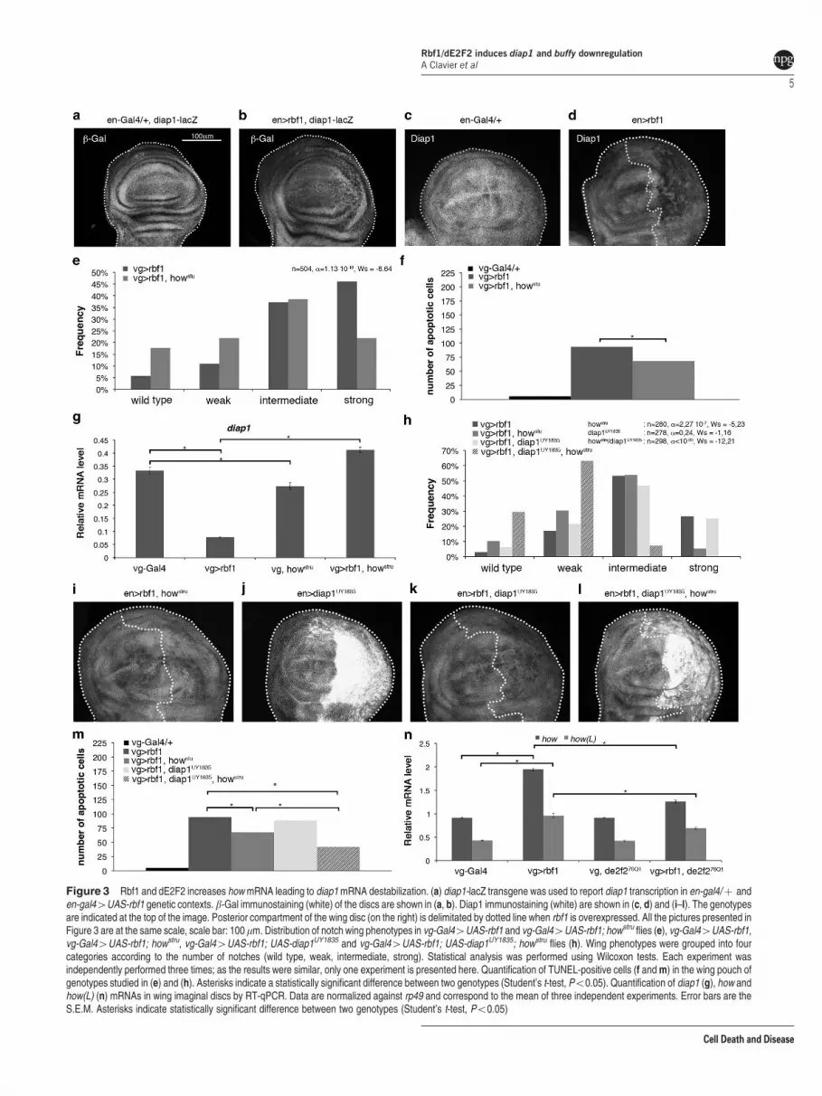
Figure 3 Rbf1 and dE2F2 increases how mRNA leading to diap1 mRNA destabilization. (a) diap1-lacZ transgene was used to report diap1 transcription in en-gal4/þ anden-gal44UAS-rbf1 genetic contexts. b-Gal immunostaining (white) of the discs are shown in (a, b). Diap1 immunostaining (white) are shown in (c, d) and (i–l). The genotypesare indicated at the top of the image. Posterior compartment of the wing disc (on the right) is delimitated by dotted line when rbf1 is overexpressed. All the pictures presented inFigure 3 are at the same scale, scale bar: 100mm. Distribution of notch wing phenotypes in vg-Gal44UAS-rbf1 and vg-Gal44UAS-rbf1; howstru flies (e), vg-Gal44UAS-rbf1,vg-Gal44UAS-rbf1; howstru, vg-Gal44UAS-rbf1; UAS-diap1UY1835 and vg-Gal44UAS-rbf1; UAS-diap1UY1835; howstru flies (h). Wing phenotypes were grouped into fourcategories according to the number of notches (wild type, weak, intermediate, strong). Statistical analysis was performed using Wilcoxon tests. Each experiment wasindependently performed three times; as the results were similar, only one experiment is presented here. Quantification of TUNEL-positive cells (f and m) in the wing pouch ofgenotypes studied in (e) and (h). Asterisks indicate a statistically significant difference between two genotypes (Student’s t-test, Po0.05). Quantification of diap1 (g), how andhow(L) (n) mRNAs in wing imaginal discs by RT-qPCR. Data are normalized against rp49 and correspond to the mean of three independent experiments. Error bars are theS.E.M. Asterisks indicate statistically significant difference between two genotypes (Student’s t-test, Po0.05)
Rbf1/dE2F2 induces diap1 and buffy downregulationA Clavier et al
5
Cell Death and Disease
Rbf1. These results indicate that diap1 downregulation byRbf1 requires both the diap1 30UTR sequence and How. Wenext asked whether rbf1 overexpression could affect theexpression of how(L). Indeed, how(L) mRNA increased onrbf1 overexpression (Figure 3n). Surprisingly, this raisedepended on dE2F2 as it was reduced in a de2f276Q1
heterozygous context. This could be explained by an indirecteffect of dE2F2, or by an unusual transcriptional activity ofdE2F2. Thus, these data suggest that Rbf1 and dE2F2increase of how(L) mRNA leads to destabilization of diap1mRNA that induces apoptosis.
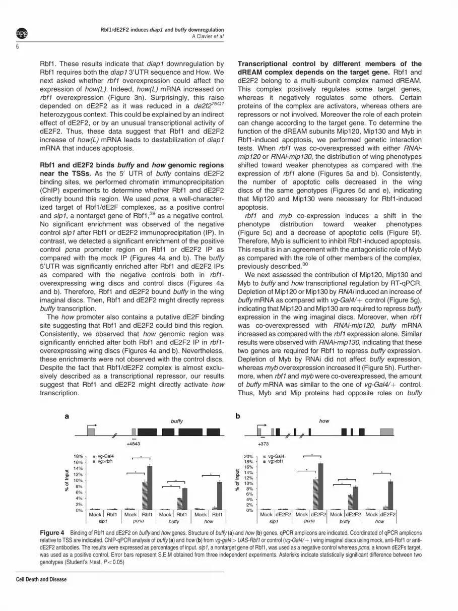
Rbf1 and dE2F2 binds buffy and how genomic regionsnear the TSSs. As the 50 UTR of buffy contains dE2F2binding sites, we performed chromatin immunoprecipitation(ChIP) experiments to determine whether Rbf1 and dE2F2directly bound this region. We used pcna, a well-character-ized target of Rbf1/dE2F complexes, as a positive controland slp1, a nontarget gene of Rbf1,39 as a negative control.No significant enrichment was observed of the negativecontrol slp1 after Rbf1 or dE2F2 immunoprecipitation (IP). Incontrast, we detected a significant enrichment of the positivecontrol pcna promoter region on Rbf1 or dE2F2 IP ascompared with the mock IP (Figures 4a and b). The buffy50UTR was significantly enriched after Rbf1 and dE2F2 IPsas compared with the negative controls both in rbf1-overexpressing wing discs and control discs (Figures 4aand b). Therefore, Rbf1 and dE2F2 bound buffy in the wingimaginal discs. Then, Rbf1 and dE2F2 might directly repressbuffy transcription.
The how promoter also contains a putative dE2F bindingsite suggesting that Rbf1 and dE2F2 could bind this region.Consistently, we observed that how genomic region wassignificantly enriched after both Rbf1 and dE2F2 IP in rbf1-overexpressing wing discs (Figures 4a and b). Nevertheless,these enrichments were not observed with the control discs.Despite the fact that Rbf1/dE2F2 complex is almost exclu-sively described as a transcriptional repressor, our resultssuggest that Rbf1 and dE2F2 might directly activate howtranscription.
Transcriptional control by different members of thedREAM complex depends on the target gene. Rbf1 anddE2F2 belong to a multi-subunit complex named dREAM.This complex positively regulates some target genes,whereas it negatively regulates some others. Certainproteins of the complex are activators, whereas others arerepressors or not involved. Moreover the role of each proteincan change according to the target gene. To determine thefunction of the dREAM subunits Mip120, Mip130 and Myb inRbf1-induced apoptosis, we performed genetic interactiontests. When rbf1 was co-overexpressed with either RNAi-mip120 or RNAi-mip130, the distribution of wing phenotypesshifted toward weaker phenotypes as compared with theexpression of rbf1 alone (Figures 5a and b). Consistently,the number of apoptotic cells decreased in the wingdiscs of the same genotypes (Figures 5d and e), indicatingthat Mip120 and Mip130 were necessary for Rbf1-inducedapoptosis.
rbf1 and myb co-expression induces a shift in thephenotype distribution toward weaker phenotypes(Figure 5c) and a decrease of apoptotic cells (Figure 5f).Therefore, Myb is sufficient to inhibit Rbf1-induced apoptosis.This result is in an agreement with the antagonistic role of Mybas compared with the role of other members of the complex,previously described.30
We next assessed the contribution of Mip120, Mip130 andMyb to buffy and how transcriptional regulation by RT-qPCR.Depletion of Mip120 or Mip130 by RNAi induced an increase ofbuffy mRNA as compared with vg-Gal4/þ control (Figure 5g),indicating that Mip120 and Mip130 are required to repress buffyexpression in the wing imaginal discs. Moreover, when rbf1was co-overexpressed with RNAi-mip120, buffy mRNAincreased as compared with the rbf1 expression alone. Similarresults were observed with RNAi-mip130, indicating that thesetwo genes are required for Rbf1 to repress buffy expression.Depletion of Myb by RNAi did not affect buffy expression,whereas myb overexpression increased it (Figure 5h). Further-more, when rbf1 and myb were co-overexpressed, the amountof buffy mRNA was similar to the one of vg-Gal4/þ control.Thus, Myb and Mip proteins had opposite roles on buffy
Figure 4 Binding of Rbf1 and dE2F2 on buffy and how genes. Structure of buffy (a) and how (b) genes. qPCR amplicons are indicated. Coordinated of qPCR ampliconsrelative to TSS are indicated. ChIP-qPCR analysis of buffy (a) and how (b) from vg-gal44UAS-Rbf1 or control (vg-Gal4/þ ) wing imaginal discs using mock, anti-Rbf1 or anti-dE2F2 antibodies. The results were expressed as percentages of input. slp1, a nontarget gene of Rbf1, was used as a negative control whereas pcna, a known dE2Fs target,was used as a positive control. Error bars represent S.E.M obtained from three independent experiments. Asterisks indicate statistically significant difference between twogenotypes (Student’s t-test, Po0.05)
Rbf1/dE2F2 induces diap1 and buffy downregulationA Clavier et al
6
Cell Death and Disease

transcriptional regulation: whereas Myb activated buffy tran-scription, Mip factors repressed it.
Inactivation of Mip120 or Mip130 by RNAi did not affect thebasal level of how mRNA (Figure 5i). Moreover, when rbf1 wasco-overexpressed with either RNAi-mip120 or RNAi-mip130,the amount of how mRNA was similar to the one observed forrbf1 overexpression alone, suggesting that these Mip factorsare not involved in the transcriptional regulation of how by Rbf1.
Inactivation or overexpression of myb did not alter the basalamount of how mRNA (Figure 5j). However, rbf1 and myb co-overexpression decreased the level of how mRNA ascompared with the rbf1 overexpression alone. These datasuggest that, contrary to dE2F2, Myb antagonizes Rbf1-induced how transcriptional activation.
Discussion
E2F transcription factors are the main partners of Rbf1. dE2F1
is widely described for its important role in the control of cell
proliferation by Rbf1, whereas the best-described role of dE2F2
is the repression of replication during oogenesis.35,40 Here, we
show that dE2F2 and dDP are required for Rbf1-induced
apoptosis suggesting that they are pro-apoptotic factors,
contrary to dE2F1, which inhibits this apoptosis. Until now,
dE2F1 was mainly described as a pro-apoptotic factor,
whereas dE2F2 was most of the time described as an anti-
apoptotic factor. Indeed, de2f1 and dDp co-expression induces
apoptosis in the eye imaginal discs23 and dE2F1 promotes
irradiation-induced Dp53-independent apoptosis in wing
Figure 5 Mip proteins and Myb are differentially involved in Rbf1-induced apoptosis. (a–c) Distribution of notch wing phenotypes in vg-Gal44UAS-rbf1,UAS-cherry andvg-Gal44UAS-rbf1,UAS-RNAi-mip120 (a), in vg-Gal44UAS-rbf1,UAS-cherry and vg-Gal44UAS-rbf1; UAS-RNAi-mip130 (b) and in vg-Gal44UAS-rbf1 andvg-Gal44UAS-rbf1; UAS-myb (c). Wing phenotypes were grouped in four categories according to the number of notches (wild type, weak, intermediate, strong). Statisticalanalysis was performed using Wilcoxon tests. Each experiment was independently performed three times; as the results were similar, only one experiment is presented here.(d–f) Quantification of TUNEL-positive cells in the wing pouch of genotypes studied in (a–c). Asterisks indicate a statistically significant difference between two genotypes(Student’s t-test, Po0.05). (g–j) Quantification of buffy (g, h) and how (i, j) mRNA in wing imaginal discs by RT-qPCR. Data are normalized against rp49 and correspond to themean of three independent experiments. Error bars are the S.E.M. Asterisks indicate statistically significant difference between two genotypes (Student’s t-test, Po0.05)
Rbf1/dE2F2 induces diap1 and buffy downregulationA Clavier et al
7
Cell Death and Disease

imaginal discs, whereas dE2F2 inhibits apoptosis in the samemodel.41 Recently, Rovani et al.42 showed that the dREAMcomplex, which includes dE2F2, cooperates with the pro-apoptotic factor Grim to induce cell death in the peripheralnervous system. This result suggests a pro-apoptotic role fordE2F2, which is consistent with our results. Furthermore, therole of dE2F1 in apoptosis might depend on the cellular context.Indeed, dE2F1 is important for DNA damage-induced apopto-sis in the wing imaginal discs, but its role varies depending onthe position of the cell within the disc.43 Together, this result andours indicate that the role of both dE2F1 and dE2F2 inapoptosis control depends on the cellular context. Similarly,depending on the cells or tissues, Rbf1 has a pro- or anti-apoptotic effect. Indeed, RBF expression induces apoptosis indifferent proliferative tissues, whereas this effect was notobserved in differentiated post-mitotic cells.26
RNAi-based studies suggested a requirement of buffy for cellsurvival during embryonic development.44 Another studysuggests that Buffy is not involved in developmental cell deathbut modulates the response to irradiation-induced cell death.45
Here, we show that buffy is involved in apoptosis induced byoverexpression of the tumor suppressor gene rbf1. Further-more, our data reveal for the first time that Rbf1 regulates buffytranscription. It would thus be interesting to determine whetherRbf1 also regulates buffy in response to irradiation.
At least in some cancers, pRb oncosuppressor activityrelies on its apoptosis-inducing activity. It has been suggestedthat RB mutations can affect the sensitivity to mitomycin/anthracycline treatment in breast cancer.46 Several reportsunderline the importance of pRb in the apoptotic response ofprostate cancer cells to radiotherapy or chemotherapeuticdrugs.11,12,47 How pRb mediates apoptosis in these casesremains unclear. Other data show that DNA damagepromotes the formation of a pRB/E2F1 complex involved inthe activation of pro-apoptotic genes such as Caspase 7 and
p73,13 and that RB/E2F-1 is a major contributor of Noxainduction in response to ABT-737 treatment, a Bcl-2inhibitor.48 Thus, pRb/E2F proapoptotic signaling(s) can beactivated in response to oncogenic stress, DNA damage orBcl-2 inhibition. Similarly, our data show that in Drosophila,Rbf1/dE2F2 can regulate apoptosis by upregulating how(Figure 6). Although this regulation seems non-essential tomaintain the how mRNA basal level, we cannot exclude that itoccurs in response to some stresses and/or therapeutictreatments. Indeed, Quaking, the homolog of how, has beenshown to be a tumor suppressor.49–51 It would be interestingto study whether a pRb/E2F complex can regulate amammalian homolog of the dIAP1 gene via Quaking.
Rbf1 and dE2F2 belong to a large complex called dREAM.Inactivation of members of dREAM by RNAi in Kc cells leadsto variations of how and buffy expression,32 which suggeststhat these two genes could be direct transcriptional targets ofdREAM. Such as described by Georlette et al.,32 we foundthat the involvement of dREAM complex members are notequivalent: some members can be activators for a specifictarget, whereas others are repressors or are not involved intranscriptional regulation of this target. Indeed dE2F2, Mip120and Mip130 are required for Rbf1-induced transcriptionalrepression of buffy, whereas Myb has an opposite effect.Moreover, contrary to dE2F2 and Myb, Mip120 and Mip130are not involved in how regulation.
dE2F2 and Myb have opposite effects on Rbf1-induced buffyand how transcription. Our results are in agreement withtranscriptomic data indicating that there are no genes negativelyco-regulated by Myb and dE2F2 but many genes are regulatedboth positively by Myb and negatively by dE2F2.32
Our results confirm a previous report32 indicating that Rbf1and the dREAM complex can act both as a transcriptionalactivator and as a transcriptional repressor (Figure 6). Themolecular mechanisms for transcriptional repression have
dDP
Rbf1
dE2F2
dREAM
complex
buffy dDP
Rbf1
dE2F2
dREAM
complex
how
buffy mRNA how mRNA
diap1 mRNA
APOPTOSIS
How
AAAA AAAA
AAAA
AAAA
dIAP1
Buffy
Figure 6 Rbf1-induced apoptosis involved transcriptional regulation of buffy and how genes. Rbf1 with dE2F2 binds buffy genomic site near to TSS and represses itstranscription. The decrease of buffy mRNA contributes to Rbf1-induced apoptosis. Rbf1 can also bind how promoter with dE2F2 but this time it induces a transcriptionalupregulation. As a consequence, How level increases allowing diap1 mRNA degradation which promotes Rbf1-induced cell death. Some members of the dREAM complex areinvolved in these transcriptional regulations
Rbf1/dE2F2 induces diap1 and buffy downregulationA Clavier et al
8
Cell Death and Disease

been deciphered.31 It would be interesting to determine whichepigenetic mechanisms are involved in the transcriptionalactivation of dREAM complex target genes.
Using an overexpression system, we have identified thebcl-2 family gene buffy, as a transcriptional target of Rbf1. Inan rbf1 loss-of-function mutant, buffy transcription increasesshow that in wild-type cells not committed to apoptosis, Rbf1 isnecessary to limit buffy expression. Thus, rbf1 loss of functioncould render cells more resistant to apoptosis. Identification ofbuffy as an Rbf1/dE2F pathway target gene is consistent withthe role of tumor suppressor described for the humancounterpart Rb. Transcriptional regulation of bcl-2 familygenes by Rb/E2F complexes has also been characterized inmammals. Indeed, pRb/E2F1 directly regulates noxa,48 bim52
and puma.53 Regulation by Rb of bcl-2 family gene expressionmay have a major impact on cell death and can thus contributeto its tumor suppressor action.
Materials and MethodsFly stocks. Flies were raised at 25 1C on a standard medium. The UAS-Rbf1and vg-Gal4 strains were generous gifts from J Silber. The en-Gal4 strain waskindly provided by L Theodore. The UAS-diap1 strain was a generous gift fromA-M Pret. In this strain, a transgene containing diap1 cDNA under the control of aUAS sequence is inserted on the second chromosome and allows the expressionof diap1 without its 30UTR sequence. The UAS-diap1UY1835 was a kind gift from SNetter.38 In this transgenic line, a P element is inserted in the 50UTR sequence ofdiap1 gene in the correct orientation to allow the expression of diap1 with its30UTR sequence. The following strains were obtained from the Bloomington StockCenter (Bloomington, IN, USA): dE2F276Q1 (7436), UAS-dE2F2 (17314), dDpa1
(7277), dE2F1i2 (7274), buffyH37 (27340), UAS-buffy (32059), diap1-lacZ (12093),howstru (2301), UAS-RNAi-mip120 (32461), UAS-RNAi-mip130 (32462), UAS-RNAi-myb (35053), UAS-myb (32044). The UAS-RNAi-diap1 strain was from NIGcollection (12284R-2). The UAS-RNAi-rbf1 strain was from VDRC collection (10696).
Test of phenotype suppression in the wing. To test the implication ofseveral genes (dE2F1, dE2F2, dDP, Buffy, Diap1, How, Mip120, Mip130 and Myb)in rbf1-induced apoptosis, the severity of the notched wing phenotype induced byUAS-Rbf1 overexpression led by vg-Gal4 driver was assayed in different geneticcontexts. For each gene, we verified that the alteration of this gene expressionlevel (overexpression, RNAi or mutant) did not induce any wing phenotype. vg-Gal4 4UAS-Rbf1 Drosophila females were crossed with males bearing a loss-of-function mutation for the different genes or allowing their overexpression. Theprogenies of all crosses were classified according to the number of notches on thewing margin. Wilcoxon tests were performed as described previously.54
TUNEL staining of imaginal discs. Third instar larvae were dissected inPBS pH 7.6, fixed in PBS/formaldehyde 3.7%, washed three times for 10 min in PBT(1� PBS, 0.5% Triton). Discs were then dissected and TUNEL staining wasperformed according to manufacturer’s instructions (ApopTag Red in situ apoptosisdetection kit, Millipore, Temecula, CA, USA). Discs were mounted in CitifluorTM(Biovalley, Marne-La-Vallee, France) and observed with a Leica SPE uprightconfocal microscope (Leica, Wetzlar, Germany). White patches in the wing pouchwere counted for at least 30 wing imaginal discs per genotype. Student’s t-testswere performed and results were considered to be significant when ao 5%.
Immunochemistry. The following antibodies were used: anti-b-Gal (mousemonoclonal antibody, 1/200, 40-1a, DSHB) and anti-Diap1 (mouse monoclonalantibody, 1/200, generous gift from B Hay). Third instar larvae were dissected inPBS pH 7.6, fixed in PBS-3.7% formaldehyde, washed three times for 10 min eachin PBT (PBS, 0,3% Triton) and incubated with primary antibody overnight at 4 1Cin PBT-FCS (PBS, 0,3% Triton, 10% FCS). Incubation with anti-mouse secondaryantibody (Alexa Fluor 488 Goat Anti-Mouse IgG (Hþ L) Antibody, MolecularProbes, Thermo Fisher Scientific, Waltham, MA, USA) was carried in PBT-FCS for2 h at room temperature. Larvae were then washed thrice in PBT. Finally, wingdiscs were mounted in CitifluorTM (Biovalley) and observed with a Leica SPEupright confocal microscope.
Chromatin immunoprecipitation. ChIPs were performed as previouslydescribed,55 with minor modification. Briefly, 50 vg-Gal44UAS-rbf1 wing imaginaldiscs of third instar larvae were dissected on ice in serum-free Schneider medium.They were fixed in 500ml of formaldehyde (1.8% in PBS) for 10 min at roomtemperature under gentle agitation. Cross-linking was stopped by adding 50ml ofglycine 1.25 M. Fixed wing discs were washed 3 times with PBS, dried, flash-freezedin liquid nitrogen and stored at � 80 1C. Cell lysis was performed by adding 100mlof lysis buffer (140 mM NaCl, 10 mM Tris-HCl pH 8.0, 1 mM EDTA, 1% Triton X-100,0.1% sodium deoxycholate, Roche complete EDTA-free protease inhibitor cocktail)complemented with 1% SDS and sonicated in a Bioruptor sonifier (Diagenode,Seraing, Belgium). Conditions were established to obtain chromatin fragments from200 to 1000 bp in length (30 s ON 30 s OFF, high power, 10 cycles). Pooledchromatin was centrifuged for 20 min at 14 000 g at 4 1C. The supernatant (solublechromatin) was recovered and 10ml were kept as input sample. For each IP, 10mlof protein A-coated paramagnetic beads (Diagenode) were washed once in lysisbuffer, 1mg of antibody was added and beads were incubated for 2 h at 4 1C on arotating wheel. After washing in lysis buffer, antibody coated beads wereresuspended in 300ml of lysis buffer and 100ml of chromatin were added. Afterincubation on a rotating wheel overnight at 4 1C, beads were washed at 4 1C fivetimes for 10 min each in lysis buffer, once in LiCl buffer (Tris-HCl 10 mM pH8.0, LiCl0.25 M, 0.5% NP-40, 0.5% sodium deoxycholate, 1 mM EDTA) and twice in TE(10 mM Tris-HCl, pH 8.0, 1 mM EDTA). Immunoprecipitated as well as input DNAswere purified with the IPure kit following the manufacturer’s instructions (Diagenode).Elution was performed twice with 35ml of water. 5ml of DNA were used per PCR.Real-time PCR data were normalized against the input sample and depicted aspercentage of input (see Supplementary Table S1 for primers). pcna, a wellcharacterized target of Rbf1/dE2F complexes, was used as a positive control andslp1, a nontarget gene of Rbf1,39 as a negative control.
Antibodies used for chromatin immunoprecipitation were anti-Rbf1 (rabbitpolyclonal, Custom antibody against amino acids 250-845 of Rbf1 protein,Proteogenix, Schiltigheim, France), anti-dE2F2 (rabbit polyclonal, Custom antibodyagainst the whole protein, Proteogenix). Rabbit pre-immune sera were used asnegative controls (mocks).
RNAs extraction and RT-qPCR. Fifty wing imaginal discs per genotypewere dissected on ice in serum-free Schneider medium. Total RNAs were extractedfrom each sample using the RNeasy Mini kit (Qiagen, Venlo, Netherlands), byfollowing the manufacturer’s instructions. RT was performed on each sample using4.8mg of RNA incubated with random primer oligonucleotides (Invitrogen, LifeTechnologies, Carlsbad, CA, USA) with Recombinant Taq DNA Polymerase(Invitrogen), according to the manufacturer’s instructions.
Real-time PCR analysis was performed using the ABI Prism 7700 HT apparatus(Applied Biosystems, Life Technologies). Briefly, PCR was performed with theABsolute blue QPCR SYBR Green ROX mix (Abgene, Thermo Fisher Scientific),using 11 ng of cDNA per RT. The primers used for real-time PCR are presented inSupplementary Table S2. Data were normalized against rp49. Three independentRT experiments were performed and the S.E.M was calculated from these threeindependent samples.
Conflict of InterestThe authors declare no conflict of interest.
Acknowledgements. We are grateful to S Szuplewski, S Gaumer andF Peronnet for their critical reading of the manuscript. We thank B Hay for agenerous gift of dIAP1 antibody. Confocal microscopy was performed onCYMAGES imaging facility. qPCR experiments were done in the UMR 1198‘Biologie du Developpement et Reproduction’ (INRA, Jouy-en-Josas). This workwas supported by the ‘Universite de Versailles Saint-Quentin-en-Yvelines’ (UVSQ)and by grants from the ‘Ligue Nationale Contre le Cancer’. Amandine Clavier wasthe recipient of a doctoral contract from UVSQ. Adrienne Baillet was supported bythe UVSQ and the Ecole Pratique des Hautes Etudes.
1. van den Heuvel S, Dyson NJ. Conserved functions of the pRB and E2F families. Nat RevMol Cell Biol 2008; 9: 713–724.
2. Frolov MV, Dyson NJ. Molecular mechanisms of E2F-dependent activation andpRB-mediated repression. J Cell Sci 2004; 117: 2173–2181.
Rbf1/dE2F2 induces diap1 and buffy downregulationA Clavier et al
9
Cell Death and Disease

3. Almasan A, Yin Y, Kelly RE, Lee EY, Bradley A, Li W et al. Deficiency of retinoblastomaprotein leads to inappropriate S-phase entry, activation of E2F-responsive genes, andapoptosis. Proc Natl Acad Sci USA 1995; 92: 5436–5440.
4. Knudsen KE, Booth D, Naderi S, Sever-Chroneos Z, Fribourg AF, Hunton IC et al.RB-dependent S-phase response to DNA damage. Mol Cell Biol 2000; 20: 7751–7763.
5. Clarke AR, Maandag ER, van Roon M, van der Lugt NM, van der Valk M, Hooper ML et al.Requirement for a functional Rb-1 gene in murine development. Nature 1992; 359: 328–330.
6. Jacks T, Fazeli A, Schmitt EM, Bronson RT, Goodell MA, Weinberg RA. Effects of an Rbmutation in the mouse. Nature 1992; 359: 295–300.
7. Lee EY, Chang CY, Hu N, Wang YC, Lai CC, Herrup K et al. Mice deficient for Rb arenonviable and show defects in neurogenesis and haematopoiesis. Nature 1992; 359: 288–294.
8. Tsai KY, Hu Y, Macleod KF, Crowley D, Yamasaki L, Jacks T. Mutation of E2f-1suppresses apoptosis and inappropriate S phase entry and extends survival of Rb-deficientmouse embryos. Mol Cell 1998; 2: 293–304.
9. Huh MS, Parker MH, Scime A, Parks R, Rudnicki MA. Rb is required for progressionthrough myogenic differentiation but not maintenance of terminal differentiation. J Cell Biol2004; 166: 865–876.
10. Biasoli D, Kahn SA, Cornelio TA, Furtado M, Campanati L, Chneiweiss H et al.Retinoblastoma protein regulates the crosstalk between autophagy and apoptosis, andfavors glioblastoma resistance to etoposide. Cell Death Dis 2013; 4: e767.
11. Zhao X, Day ML. RB activation and repression of C-MYC transcription precede apoptosisof human prostate epithelial cells. Urology 2001; 57: 860–865.
12. Bowen C, Spiegel S, Gelmann EP. Radiation-induced apoptosis mediated byretinoblastoma protein. Cancer Res 1998; 58: 3275–3281.
13. Ianari A, Natale T, Calo E, Ferretti E, Alesse E, Screpanti I et al. Proapoptotic function ofthe retinoblastoma tumor suppressor protein. Cancer Cell 2009; 15: 184–194.
14. Hilgendorf KI, Leshchiner ES, Nedelcu S, Maynard MA, Calo E, Ianari A et al. The retinoblastomaprotein induces apoptosis directly at the mitochondria. Genes Dev 2013; 27: 1003–1015.
15. Dynlacht BD, Brook A, Dembski M, Yenush L, Dyson N. DNA-binding and trans-activationproperties of Drosophila E2F and DP proteins. Proc Natl Acad Sci USA 1994; 91: 6359–6363.
16. Ohtani K, Nevins JR. Functional properties of a Drosophila homolog of the E2F1 gene. MolCell Biol 1994; 14: 1603–1612.
17. Sawado T, Yamaguchi M, Nishimoto Y, Ohno K, Sakaguchi K, Matsukage A. dE2F2, anovel E2F-family transcription factor in Drosophila melanogaster. Biochem Biophys ResCommun 1998; 251: 409–415.
18. Du W, Vidal M, Xie JE, Dyson N. RBF, a novel RB-related gene that regulates E2F activityand interacts with cyclin E in Drosophila. Genes Dev 1996; 10: 1206–1218.
19. Stevaux O, Dimova D, Frolov MV, Taylor-Harding B, Morris E, Dyson N. Distinct mechanismsof E2F regulation by Drosophila RBF1 and RBF2. Embo J 2002; 21: 4927–4937.
20. Du W. Suppression of the rbf null mutants by a de2f1 allele that lacks transactivationdomain. Development 2000; 127: 367–379.
21. Frolov MV, Huen DS, Stevaux O, Dimova D, Balczarek-Strang K, Elsdon M et al.Functional antagonism between E2F family members. Genes Dev 2001; 15: 2146–2160.
22. Du W, Dyson N. The role of RBF in the introduction of G1 regulation during Drosophilaembryogenesis. Embo J 1999; 18: 916–925.
23. Du W, Xie JE, Dyson N. Ectopic expression of dE2F and dDP induces cell proliferation anddeath in the Drosophila eye. Embo J 1996; 15: 3684–3692.
24. Asano M, Nevins JR, Wharton RP. Ectopic E2F expression induces S phase and apoptosisin Drosophila imaginal discs. Genes Dev 1996; 10: 1422–1432.
25. Zhou L, Steller H. Distinct pathways mediate UV-induced apoptosis in Drosophila embryos.Dev Cell 2003; 4: 599–605.
26. Milet C, Rincheval-Arnold A, Mignotte B, Guenal I. The Drosophila retinoblastoma protein
induces apoptosis in proliferating but not in post-mitotic cells. Cell Cycle 2010; 9: 97–103.27. Korenjak M, Anderssen E, Ramaswamy S, Whetstine JR, Dyson NJ. RBF binding to both
canonical E2F targets and noncanonical targets depends on functional dE2F/dDPcomplexes. Mol Cell Biol 2012; 32: 4375–4387.
28. Korenjak M, Taylor-Harding B, Binne UK, Satterlee JS, Stevaux O, Aasland R et al. NativeE2F/RBF complexes contain Myb-interacting proteins and repress transcription ofdevelopmentally controlled E2F target genes. Cell 2004; 119: 181–193.
29. Taylor-Harding B, Binne UK, Korenjak M, Brehm A, Dyson NJ. p55, the Drosophila orthologof RbAp46/RbAp48, is required for the repression of dE2F2/RBF-regulated genes. Mol CellBiol 2004; 24: 9124–9136.
30. Lewis PW, Beall EL, Fleischer TC, Georlette D, Link AJ, Botchan MR. Identification of aDrosophila Myb-E2F2/RBF transcriptional repressor complex. Genes Dev 2004; 18: 2929–2940.
31. Lee H, Ohno K, Voskoboynik Y, Ragusano L, Martinez A, Dimova DK. Drosophila RBproteins repress differentiation-specific genes via two different mechanisms. Mol Cell Biol2010; 30: 2563–2577.
32. Georlette D, Ahn S, MacAlpine DM, Cheung E, Lewis PW, Beall EL et al. Genomic profilingand expression studies reveal both positive and negative activities for the Drosophila MybMuvB/dREAM complex in proliferating cells. Genes Dev 2007; 21: 2880–2896.
33. Acharya P, Negre N, Johnston J, Wei Y, White KP, Henry RW et al. Evidence forautoregulation and cell signaling pathway regulation from genome-wide binding of theDrosophila retinoblastoma protein. G3 (Bethesda) 2012; 2: 1459–1472.
34. Frolov MV, Moon NS, Dyson NJ. dDP is needed for normal cell proliferation. Mol Cell Biol2005; 25: 3027–3039.
35. Bosco G, Du W, Orr-Weaver TL. DNA replication control through interaction of E2F-RB andthe origin recognition complex. Nat Cell Biol 2001; 3: 289–295.
36. Reuveny A, Elhanany H, Volk T. Enhanced sensitivity of midline glial cells to apoptosis isachieved by HOW(L)-dependent repression of Diap1. Mech Dev 2009; 126: 30–41.
37. Volk T, Israeli D, Nir R, Toledano-Katchalski H. Tissue development and RNA control:‘HOW’ is it coordinated? Trends Genet 2008; 24: 94–101.
38. Marchal C, Vinatier G, Sanial M, Plessis A, Pret AM, Limbourg-Bouchon B et al. The HIV-1Vpu protein induces apoptosis in Drosophila via activation of JNK signaling. PLoS One2012; 7: e34310.
39. Acharya P, Raj N, Buckley MS, Zhang L, Duperon S, Williams G et al. Paradoxicalinstability-activity relationship defines a novel regulatory pathway for retinoblastomaproteins. Mol Biol Cell 2010; 21: 3890–3901.
40. Tower J. Developmental gene amplification and origin regulation. Annu Rev Genet 2004;38: 273–304.
41. Wichmann A, Uyetake L, Su TT. E2F1 and E2F2 have opposite effects on radiation-induced p53-independent apoptosis in Drosophila. Dev Biol 2010; 346: 80–89.
42. Rovani MK, Brachmann CB, Ramsay G, Katzen AL. The dREAM/Myb-MuvB complex andGrim are key regulators of the programmed death of neural precursor cells at theDrosophila posterior wing margin. Dev Biol 2012; 372: 88–102.
43. Moon NS, Frolov MV, Kwon EJ, Di Stefano L, Dimova DK, Morris EJ et al. Drosophila E2F1has context-specific pro- and antiapoptotic properties during development. Dev Cell 2005;9: 463–475.
44. Quinn L, Coombe M, Mills K, Daish T, Colussi P, Kumar S et al. Buffy, a Drosophila Bcl-2protein, has anti-apoptotic and cell cycle inhibitory functions. EMBO J 2003; 22: 3568–3579.
45. Sevrioukov EA, Burr J, Huang EW, Assi HH, Monserrate JP, Purves DC et al. DrosophilaBcl-2 proteins participate in stress-induced apoptosis, but are not required for normaldevelopment. Genesis 2007; 45: 184–193.
46. Berge EO, Knappskog S, Geisler S, Staalesen V, Pacal M, Borresen-Dale AL et al.Identification and characterization of retinoblastoma gene mutations disturbing apoptosis inhuman breast cancers. Mol Cancer 2010; 9: 173.
47. Sharma A, Comstock CE, Knudsen ES, Cao KH, Hess-Wilson JK, Morey LM et al.Retinoblastoma tumor suppressor status is a critical determinant of therapeutic response inprostate cancer cells. Cancer Res 2007; 67: 6192–6203.
48. Bertin-Ciftci J, Barre B, Le Pen J, Maillet L, Couriaud C, Juin P et al. pRb/E2F-1-mediatedcaspase-dependent induction of Noxa amplifies the apoptotic effects of the Bcl-2/Bcl-xLinhibitor ABT-737. Cell Death Differ 2013; 20: 755–764.
49. Yin D, Ogawa S, Kawamata N, Tunici P, Finocchiaro G, Eoli M et al. High-resolutiongenomic copy number profiling of glioblastoma multiforme by single nucleotidepolymorphism DNA microarray. Mol Cancer Res 2009; 7: 665–677.
50. Bian Y, Wang L, Lu H, Yang G, Zhang Z, Fu H et al. Downregulation of tumor suppressorQKI in gastric cancer and its implication in cancer prognosis. Biochem Biophys ResCommun 2012; 422: 187–193.
51. Zhao Y, Zhang G, Wei M, Lu X, Fu H, Feng F et al. The tumor suppressing effects of QKI-5 inprostate cancer: a novel diagnostic and prognostic protein. Cancer Biol Ther 2014; 15: 108–118.
52. Zhao Y, Tan J, Zhuang L, Jiang X, Liu ET, Yu Q. Inhibitors of histone deacetylases targetthe Rb-E2F1 pathway for apoptosis induction through activation of proapoptotic proteinBim. Proc Natl Acad Sci USA 2005; 102: 16090–16095.
53. Hao H, Dong Y, Bowling MT, Gomez-Gutierrez JG, Zhou HS, McMasters KM. E2F-1induces melanoma cell apoptosis via PUMA up-regulation and Bax translocation. BMCCancer 2007; 7: 24.
54. Brun S, Rincheval V, Gaumer S, Mignotte B, Guenal I. reaper and bax initiate two differentapoptotic pathways affecting mitochondria and antagonized by bcl-2 in Drosophila.Oncogene 2002; 21: 6458–6470.
55. Coleno-Costes A, Jang SM, de Vanssay A, Rougeot J, Bouceba T, Randsholt NB et al.New partners in regulation of gene expression: the enhancer of Trithorax and PolycombCorto interacts with methylated ribosomal protein l12 via its chromodomain. PLoS Genet2012; 8: e1003006.
Cell Death and Disease is an open-access journalpublished by Nature Publishing Group. This work is
licensed under a Creative Commons Attribution-NonCommercial-ShareAlike 3.0 Unported License. The images or other third partymaterial in this article are included in the article’s Creative Commonslicense, unless indicated otherwise in the credit line; if the material isnot included under the Creative Commons license, users will need toobtain permission from the license holder to reproduce the material. Toview a copy of this license, visit http://creativecommons.org/licenses/by-nc-sa/3.0/
Supplementary Information accompanies this paper on Cell Death and Disease website (http://www.nature.com/cddis)
Rbf1/dE2F2 induces diap1 and buffy downregulationA Clavier et al
10
Cell Death and Disease