The Pattern Of Ankle Fractures In Kenyatta National Hospital
50
1 THE PATTERN OF ANKLE FRACTURES IN KENYATTA NATIONAL HOSPITAL BY DR MUSTAFA MUZAHIR KHANBHAI MBChB(NAIROBI) H58/64274/2010 2015 A DISSERATION SUBMITTED IN PART FULFILMENT FOR THE DEGREE OF MASTER OF MEDICINE IN ORTHOPAEDIC SURGERY
-
Upload
khangminh22 -
Category
Documents
-
view
0 -
download
0
Transcript of The Pattern Of Ankle Fractures In Kenyatta National Hospital
1
THE PATTERN OF ANKLE FRACTURES IN KENYATTA NATIONAL HOSPITAL
BY
DR MUSTAFA MUZAHIR KHANBHAI
MBChB(NAIROBI)
H58/64274/2010
2015
A DISSERATION SUBMITTED IN PART FULFILMENT FOR THE DEGREE OF MASTER OF MEDICINE IN ORTHOPAEDIC SURGERY
2
DECLARATION
This dissertation is my original work and has never been presented by me or any other person for a degree in any other university
Signed _______________________ date _______________
DR MUSTAFA.MUZAHIR KHANBHAI, Principal Investigator.
MBChB (Nbi)
REG. NO. H58/64274/10
SUPERVISORS
Submitted with our approval as the university supervisors
1. Professor J.O. Mulimba
MBChB,M.Med,FRCS, FCS(ECSA),FRSM,
Professor of orthopaedics
University of Nairobi
Signature .......................................... date...........................
2. Dr. E. Oburu
MBChB, M.Med, MRCSEd, FRCSEd, (Tr.&ortho)
Lecturer University of Nairobi
Signature .................................. date...........................
3
CERTIFICATE OF AUTHENTICITY
This to to certify that this dissertation is the original work of Dr. Mustafa. M.
Khanbhai, Mmed student , Registration number H58/64274/2010, department of
orthopaedic surgery, university of Nairobi
This research will be carried out at the Kenyatta National Hospital, department of
orthopaedic surgery
Signature:-
Proffesor. J.E O. ATINGA .............................. date
..................................
Chairman, department of orthopaedic surgery,
University od Nairobi, Kenya

4
DEDICATIONS
This thesis is dedicated to my beloved parents who have always been a source of support and encouragement and also my lovely wife Sakina and my daughters Batul and Husseina for the patience and endurance throughout the study.

5
ACKNOWLEDGEMENTS
I would like to express my sincere gratitude and appreciation to my supervisors
namely Prof. Mulimba and Dr. Oburu for their invaluable assistance, guidance
and constructive criticism offered to me during preparation of this manuscript.
I also would like to thank members of staff in the orthopedic wards, clinics and
emergency department of Kenyatta National Hospital for their assistance in
obtaining data. Not forgetting all the lecturers and my colleagues in the
department of orthopedics whose wise advice was sort.
Lastly would like to express my sincere gratitude to the patients who gave
consent and were patient enough during the process of data collection.

6
TABLE OF CONTENTS
1. TITLE------------------------------------------------------------1
2. DECLARATION-----------------------------------------------2
3. CERTIFICATE OF AUTHENTICITY----------------------3
4. DEDICATION--------------------------------------------------4
5. ACKNOWLEDGEMENTS-----------------------------------5
6. TABLE OF CONTENTS--------------------------------------6
7. LIST OF TABLES---------------------------------------------8
8. LIST OF FIGURES--------------------------------------------9
9. ABBREVIATIONS-------------------------------------------10
10. ABSTRACT---------------------------------------------------11
11. INTRODUCTION-------------------------------------------13
12. LITERATURE REVIEW----------------------------------15
13. JUSTIFICATION-------------------------------------------27
14. STUDY QUESTION-----------------------------------------27
15. OBJECTIVES ------------------------------------------------27
16. HYPOTHESIS -------------------------------------------------28
17. SELECTION CRITERIA--------------------------------------28
18. MATERIALS AND METHOD-------------------------------28
19. ETHICAL CONSIDERATIONS------------------------------30
20. DESSEMINATION ----------------------------------------------31

7
21. METHOD OF COLLECTION------------------------------31
22. METHODOLOGY--------------------------------------------32
23. RESULTS-------------------------------------------------------33
24. DISCUSSION--------------------------------------------------38
25 LIMITATIONS ------------------------------------------------40
26. CONCLUSION------------------------------------------------40
27. RECOMMENDATIONS-------------------------------------40
28. REFERENCES-------------------------------------------------41
24APPENDIX1-----------------------------------------------------45
25APPENDIX2------------------------------------------------------49
26. APPENDIX 3 (IMPLEMENTATION SCHEDULE &
BUDGET ---------------------------------50
8
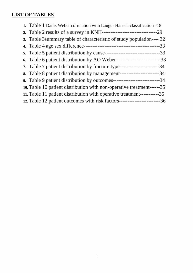
LIST OF TABLES
1. Table 1 Danis Weber correlation with Lauge- Hansen classification--18 2. Table 2 results of a survey in KNH--------------------------------29 3. Table 3summary table of characteristic of study population---- 32 4. Table 4 age sex difference--------------------------------------------33 5. Table 5 patient distribution by cause--------------------------------33 6. Table 6 patient distribution by AO Weber--------------------------33 7. Table 7 patient distribution by fracture type-----------------------34 8. Table 8 patient distribution by management-----------------------34 9. Table 9 patient distribution by outcomes---------------------------34 10. Table 10 patient distribution with non-operative treatment------35 11. Table 11 patient distribution with operative treatment-----------35 12. Table 12 patient outcomes with risk factors------------------------36

9
LIST OF FUGURES
1. Figure 1 AO Weber classification------------------------------17
10
ABBREVIATIONS
1. Mm---------------------------millimeters 2. GRF-------------------------- ground force reaction 3. US---------------------------- United States 4. AO/ASIF---------------------Arbeitsgemeinscaft fur
Osteosynthesefragen/Association for the Study of Internal Fixation 5. OMA-----------------Olerud Molander Ankle Score 6. OA---------------------Osteoarthritis 7. DM--------------------- Diabetes Mellitus 8. KNH---------------------Kenyatta National Hospital 9. BMI----------------------Body Mass Index
10. MVA-------------------motor vehicle accidents
11. MCA--------------------motor cycle accidents
11
ABSTRACT
Background
Ankle fractures are among the most common injuries treated by orthopeadic surgeons.The
management is dependent on the extent of bony involement, soft tissue and ligamentous
injury.
Epidemiology of these fractures is changing with rise in incidence among elderly population
as seen in the Western world and the young population in Africa. The commonest aetiology
of these fracturesin Africa is road traffic accidents. The treatment of ankle fractures is
challenging despite several studies and the advancement of treatment modalities. Kenyatta
National Hospital deals with this fractures on daily basis. This study looked at the pattern of
ankle fractures in Kenyatta National Hospital.
Objectives
To determine the pattern and modalities of treatment of adult ankle fractures among patients
presenting at Kenyatta National Hospital .
Study design
A cross sectional prospective analytical study.
Setting
Kenyata National Hospital orthopedic unit
Patients and method
Patients with ankle fractures were recruited by the investigator and the assistant. The patients
who gave consent and met the inclusion criteria were recruitedin the study. A questionaire
was used to determine the age, gender, socioeconomic status, mechanism and type of
injuries.
12
Results
A total of 100 patients recruited over the period of 3months period. The mean age was
40years with range of 20-80 years. The sample had 70 males(70%)and 30 females(30%).
Most fractures were as a result of motorcycle accidents accounting for 34% and least being
assault 2.%. 73% had closed injuries 27% open injuries. B2 type being the most common
type(35%) of injury.Non-operative treatment of fractures wascommonly employedwith a
proportion of 68% while operativetreatment had a proportion of 28%. During the period of
study the 54.2% cases healed with no complications while 45.8% cases were associated with
complications.Two of the cases were lost to follow up.
Conclusion
The study showed a high rate of fractures due to road traffic accidents secondary to
motorcycle injury. The majority were males in their third decade and older women. Weber
type B was the commonest type of ankle fractures and most of them treated non operatively..
13
INTRODUCTION
Ankle fractures are among the most common injuries treated by orthopeadic surgeons and
their management depends on the extent of of bone, soft tissue and ligamentous injury(1).
Ankle fracture are usually as a result of low energy and rotational forces(twisting
mechanism)(1,2).
Observational studies have shown that the incidence and severity of ankle fractures have
risen and mostly occur in elderly populations (3). The incidence of ankle fractures in the
United Kingdom is estimated at about 400,000 injuries every year, while in United States of
America it was found to be 492 000 per year (3). In Africa the incidence is expected to be
higher due to road carnage. In Nigeria, 46.3% of ankle fractures were due to road traffic
accidents (4) and in Kenya 69% of patients involved in road traffic accidents had
fractures.(5)
Kenyatta National Hospital deals with ankle fracture on daily basis. This injury leads to loss
of productive man hours of patients making them unproductive. This study looked at pattern
of ankle fractures seen at Kenyatta National hospital and treatment modalities used for these
fractures.
Anatomy
The ankle joint is a modified hinge joint which consists of three bones (tibia,fibula and talus)
and the ligaments which bind these bones together as a unit(6). The stability of this joint is
achieved by both osseous congruity and ligaments around the joint.. The lateral collateral
ligament consists of three components: anterior talofibular, calcaneofibular and posterior
talofibular ligament. The medial deltoid ligament consists of a superficial and deep portions,
the deep being the main ankle stabilizer.
14
The distal end of the fibula lies on the tibial groove held by strong syndesmosis which has
both anterior and posterior tibiofibular components and the thickest part of the interosseous
membrane.
There are no muscles attached around the ankle.Eleven tendons and neurovascular structures
cross it.Stabilityof this joint is purely relied on bony configuration and capsuloligamentous
structures.
Biomechanical consideration
The ankle joint is subjected to enormous force on a small surface area of contact about 1.5
times body weight on normal gait and 5.5 times body weight during strenuous
excercises(1).The three planes of motion in the foot and ankle are defined as sagittal plane,
frontal plane and transverse plane .From these axis movements of the foot and ankle can be
identified: (plantar) flexion/(dorsi) extension (sagittal plane), inversion/eversion (frontal
plane) and abduction/adduction (transverse plane). Supination is a combined movement of
adduction, inversion and flexion, and pronation the opposite with abduction, eversion and
extension(7) .As for kinetics, the foot and ankle must both absorb and transmit forces, both
internal and external(8). Ground reaction force (GRF) is usually studied and magnitudes of
vertical GRF of between 1.1 to 1.3 times body weight have been reported depending on
walking speed(9).
While the ankle was previously known as a simple hinge joint many studies have now shown
more complex biomechanics of the ankle joint(10)
Ankle movements range from 5-20 degrees dorsiflexion and 10-40 degrees planter flexion.
For normal gait only 10 degrees dorsiflexion and 20 degrees planterflexion are reqiured(2
15
,6). The joint is most unstable in planter flexion due to the talus being narrower posteriorly
and most of the injuries occur in this position(11).
The role of different ankle structures have been widely studied and the conclusion is that the
primary stabilizer of the ankle joint is the lateral fibular complex with the talus(6,12). The
tibiofibular dysfunction results in marked talar displacement which correlates with
degerative changes (6,8).
In a cadavaric study it was shown that about 1 mm translation of talus reduces the surface
area of contact by 42 % and 2mm translation reduces contact by 64%. Decreased area of
contact may lead to abnormal distribution of joint forces and hence post traumatic
arthritis(2).
LITERATURE REVIEW
Ankle fractures incidence has varied between various studies but most have shown a trend
of increase incidence over time(3,14,15). Most of these studies, either had limited number of
patients, limited areas or use of a single hospital. They too demonstrate a pronounced rise in
eldely women(3,14,15,16). Several studies have shown a switch in sex by age with a greater
incidence in younger men and older women(16).Currently in US, ankle fractures reported in
as many as 8.3 per 1000, similarly in the Finish population showed a rise in ankle fractures
in elderly patients. In Africa most ankle fracture were seen due to road traffic accidents(17).
In a Nigerian study 46.3% of all fracture due to road traffic accident had ankle fractures
,while 88% of ankle fractures were due to road carnage in a Ghanian experience(4,61). In
Kenya this incidence of ankle fractures is lacking, however 69% of cases seen at Kenyatta
National Hospital who were involved in road traffic accidents had fractures.(5)
16
A classification system should be easy to use in daily practice. It should be based on
information easily obtained such as patients history,clinical examination and radiological
findings.It should also define severity and serve as a basis for treament. The number of
malleoli involved can describe ankle fractures:uni-,bi- and trimalleolar. During the last
century, a number of classification system have been developed.Ashhurst and Bromer made
the first classification in 1922(18). They divided the fractures according to the vector of
trauma in 300 patients: external rotation, abduction and adduction included about 95% of all
ankle fractures. The rest were mainly caused by compression in the long axis of the limb(19).
The system was further developed by Lauge-Hansen who developed a classification in 1942
after cadaver experiments(20). He named each type by a double name, where the first part
defines the position of the foot at the moment of trauma and the second part specified the
direction of the dislocating force at the moment of trauma. Lauge-Hansen identified four
groups of fractures, each with a number of subgroups: supination-eversion fractures,
supination-adduction fractures, pronation-eversion fractures and pronation-abduction
fractures(21). The most common fractures are in the group of supination-eversion fractures.
Suppination external rotation(eversion) accounts 40-75% of all ankle fractures. While others
like suppination adduction accounts for 10-20%, pronation abduction 5-15% pronation
external rotation 7-19%.(6,22 ,23).Lauge-Hansen also described pronation-dorsiflexion
injuries. This injury was combined with compression of the joint and is thus not a true ankle
fracture. This classification could be used as a guide for closed reduction(24). The Lauge-
Hansen classification has been recommended by several authors.(24) However, studies have
17
shown high interobserver variation and the system described thus became difficult to
apply(10,26).
In 1949, Danis described a classification which was more pathological-anatomical and
designed for application to operative treatment. This classification is based on the lateral
malleolar fracture, syndesmotic disruption and talar instability. This system has later been
further developed by Weber and the AO-group founded in 1958(27). The fractures are
divided into three fracture types: A, B and C with further subgroups. This division is based
on the level of the lateral malleolar fracture in relation to the level of the syndesmosis
(Figure 1).The AO (Danis-Weber) classification system does not take the direction or force
of injury into account but has been considered easier to use. The interobserver correlation
has previously been shown to be good(28). The frequencies of the different fractures have
been found to be more stable between studies than for the Lauge-Hansen classification.(29)
18
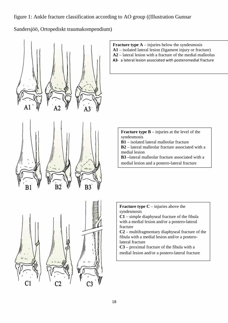
figure 1: Ankle fracture classification according to AO group ((Illustration Gunnar
Sandersjöö, Ortopediskt traumakompendium)
Fracture type A – injuries below the syndesmosis A1 – isolated lateral lesion (ligament injury or fracture) A2 – lateral lesion with a fracture of the medial malleolus A3- a lateral lesion associated with posteromedial fracture
Fracture type B – injuries at the level of the syndesmosis B1 – isolated lateral malleolar fracture B2 – lateral malleolar fracture associated with a medial lesion B3 –lateral malleolar fracture associated with a medial lesion and a postero-lateral fracture
Fracture type C – injuries above the syndesmosis C1 – simple diaphyseal fracture of the fibula with a medial lesion and/or a postero-lateral fracture C2 – multifragmentary diaphyseal fracture of the fibula with a medial lesion and/or a postero-lateral fracture C3 – proximal fracture of the fibula with a medial lesion and/or a postero-lateral fracture
19
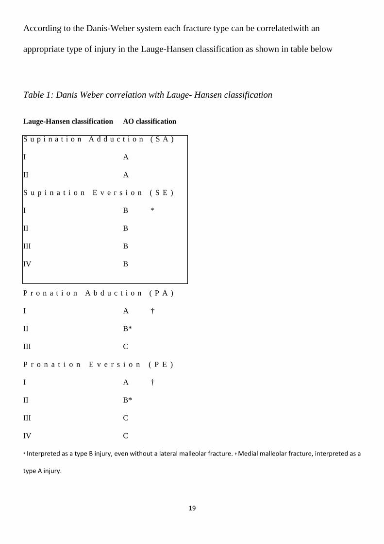
According to the Danis-Weber system each fracture type can be correlatedwith an
appropriate type of injury in the Lauge-Hansen classification as shown in table below
Table 1: Danis Weber correlation with Lauge- Hansen classification
Lauge-Hansen classification AO classification
S u p i n a t i o n A d d u c t i o n ( S A )
I
II
A
A
S u p i n a t i o n E v e r s i o n ( S E )
I
II
III
IV
B *
B
B
B
P r o n a t i o n A b d u c t i o n ( P A )
I
II
III
A †
B*
C
P r o n a t i o n E v e r s i o n ( P E )
I
II
III
IV
A †
B*
C
C
* Interpreted as a type B injury, even without a lateral malleolar fracture. † Medial malleolar fracture, interpreted as a
type A injury.
20
To know the mechanism of injury is of great importance, especially when assessing the
soft tissue around the fracture. The magnitude and the direction of force are important factors
in determining resultant of injury. The mechanism of injury affects the displacement of the
fracture at the injury and therefore the surrounding soft tissue.
Ankle fractures are most commonly caused by low-energy accidents such as fall in the same
level which is a resultant of a twisting mechnism(30). High-energy trauma such road traffic
accidents, crush injuries or a fall > 5m have been found to be risk factors for soft tissue
complications(31). Sports injuries to the ankle are also common and accounts for 20-25%.
Road traffic accident was the commonest mechanism of ankle fractures in Africa with higher
percentage having open fractures (4,5,32).
The aim when treating ankle fracture is to re-establish the function of the injured joint. This
includes reduction of fracture, retention of the reduced position and rehabilitation. This can
be achieved either operatively or non- operatively. When deciding which method to use, the
fracture, state of the surrounding tissues and patient characteristics and timing have to be
considered. “A good closed reduction followed by cast imbolization certainly give a better
outcome than a poorly planned and executed open reduction and internal fixation(33). The
most common method of non operative treatment is plaster cast imbolization and this is
frequently used(10,14,34,35). It is however, essential to master the art of “plastercraft”.
Charnley attributed the failure of non-operative treatment to inadequate plaster technique.A-
O group recommendations for non operative management includes typeA isolated fracture
with no medial lesion as well as a non –displaced type B fracture with no medial lesion. The
favourable outcome of closed treatment of suppination external rotation type of injury(type
21
B) has been supported by many studies .(35,36,37). Closed treatment for displaced fractures
has become a lost art, but there are some consideration for this form of treatment in imature
skeleton,patient unfit for surgery, fracture through osteoporotic bone and where patient’s
skin precludes open surgery(35).
Fracture dislocation injuries with ankle mortise incongruity are treated with open reduction
and internal fixation to restore joint congruity(26,33,38,39). Contraindication to internal
fixation includes infection, paraplegic patient, elderlysedentary patients and patients with
multiple life threatening injuries (3). There are varied methods of internal fixation. Palmer,
Wiberg and Cedell described non-rigid, adaptive internal fixation techniques with cerclage
wiring, staples, pins and small-threaded screws, enough to keep the malleolar fragments
adapted, combined with a protective plaster cast during bone healing(39,40).This method is
widely used in Sweden. However it is shown that the non rigid technique is not adequate for
maintaining congruency in bi maleollar and trimalleolar fractures.Danis developed the
principle of absolute fixation which restores bone congriuty, allows immediate return of joint
movement and allows direct union. This concept was taken up by AO group and spread
world wide where the fracture is treated with metal plates and screws(41). Rigid fixation was
found to be superior to non rigid fixation in retention of congruency.(13)
The timing of surgery vis a vis time of injury has been debated. Time is not always easy to
determine, since time of injury to presentation at hospital may be difficult to specify.
Majority of authors prefered slight delay on the timing of surgery so as to allow the phase of
acute inflammation to subside, however delay of more than 3 weeks made anatomic
reduction difficult and led to poorer outcomes(1,6,11,42)
22
Open ankle fractures are considered severe injuries and the principles used in treatment of
these fractures is still the same as developed by Gustillo and Anderson. They classified open
fractures according to degree of soft tissue damage and contamination.
Type 1 clean wound laceration of less than 1 cm (size of the wound) without evidence of
deep muscle crushing and contamination is minimal. Fracture typical of a low energy type.
Type 2 laceration more than 1 cm and less then 10 cm without extensive soft tissue demage
fracture due to high energy trauma. Type 3 skin laceration is more then 10 cm and usually
divided into three types as follows:-
Type 3a extensive soft tissue laceration flaps adequate to cover bone.
Type 3b extensive soft tissue laceration with exposed bone.
Type 3c open fracture with vascular injury reqiuring repair. (43)
Gustillo and Anderson classification has been found to have high interobserver variation,
despite this limitation still remains the prefered system of classification system.
Some authors advocate for early internal fixation, however, it is advised that for grade 2 and
3 that external fixation should be used to allow soft tissue to heal. Usual sequence in the
managemnt of open fracture include Irrigation and debribement of the wound with internal
or external fixation of the fracture. Lastly covering of the wound can be achieved primarily
or secondary, asissted by skin grafting or by use of flaps(6,44). Irrigation is a key
component in open fractures management as it serves to decrease bacterial load and remove
foreign bodies. Many guidelines recommend the use copious amount of irrigation however
there is no data which suggests the volume of fluid required for adequate lavage. Early open
reduction and internal fixation of open ankle fractures is advocated and leads to good
functional outcome, reduced hospital stay and less joint stiffnes as compared to conservative
23
or delayed fixation. Only when there is inadequate soft tissue coverage of osteosynthesis
material should external fixators be used(44). Most orthopedic surgeons are now moving
towards early closure of open wounds with local flaps and skin grafting after adequate
debribement. Grade 1 and 2 types of wound are stitched primarly or left for secondary
healing, while grade 3 wounds are closed with local flaps or split thickness skin grafts.(44).
Unstable ankle fractures usually have syndesmotic disruption which leads to more pain and
poorer functional outcome. A syndesmotic screw is used to fix the disruption of the
syndesmosis. There is no consensus to whether use of one screw or two screws and
tricortices ir tetracortices. However there is no change in patients whose syndesmotic
screw/s are removed on weight bearing and non removal of transyndesmotic screw on the
outcome.(1,6,35).
The word “complication” commonly used in both clinical setting and scientific publication
may not be clearly defined. It could be further explained by defination adverse events,
which is any untoward medical occurrence, unintended disease, injury or clinical signs in a
subject(46). When discussing local complication following ankle fractures both adverse
events following course of healing( soft tissue complication,technical failure) as well as
conditions that might be considered as sequelae(gait deviation, osteoarthritis) are usually
mentioned. Early soft tissue complication following open reduction internal fixation of ankle
fractures may include wound dehiscence,necrosis and infection. Rates of this different types
of complication vary between 5-27% according to following studies(29, 47,48).
24
Reports on technical failure are infrequent in literature. Sometimes different terms such as
failed reduction and loss of reduction are used, the latter indicating primary post operative
radiograph have been good and failure of osteosynthesis occured later during post operative
period. Technical failure and failed reduction have been reported between3-6%(29,47,48).
Technical failure was found most commonly in unstable fractures treated with non operative
measures leading to malunoin and non union(34, 49)
Osteoarthritis (OA) is an irreversible condition resulting from mechanical and biological
events affecting the articular cartilage and subchondral bone in a joint. The condition is
frequently accompanied by pain and stiffness in the affected joint. OA can be divided into
primary and secondary types. Primary OA usually develops after the age of 50 and the cause
is largely unknown. In secondary OA the cause is identified: infection, ligamentous
instability, congenital anomaly, physeal separation, haemophilia and fractures are risk factors
for developing secondary OA(50).OA in the ankle joint is most frequently of post-traumatic
origin, 80% as compared to post-traumatic OAof the hip (2%) or knee (10%)(51). Of the
patients presenting with post-traumatic ankle OA, malleolar fractures have been identified as
the most common cause (37- 53%)(52).
When evaluating indication for surgery many risk factors must be taken into account. Many
studies have evaluated risk factors and ankle surgery, however these have been moslty
retrospective.
25
Age was considered a risk factor for soft tissue complication following ankle surgery, this
has been defined by several studies(52). These studies have defined elderly as aged
from 50-80 years(52). However these studies found no significant difference in soft tissue
complications as compared to controls Significant difference was found in another study
when the cut off age was increased to 65 years(53 ).When studying age as a continuous
variable, mean age of infected patients has been found to be higher, but this was not
statistically significant(54). Age alone was not seen as an independent risk factor by Höiness
et al. for sustaininga superficial wound infection(47)
These studies described several factors including advanced age beyond 50 years Fracture
type and fracture mechanism .High energy trauma have found to be a risk factor for soft
tissue complication(48). Preoperative soft tissue affection after ankle injury is of great
importance. Höiness et al. Found about 14% soft tissue affection after ankle injury and also
noted that Type B2,TypeB3 had higher soft tissue complication rates.(28).
Diabetes mellitus is a chronic disease leading to related comorbidities e.g. vasculopathy,
neuropathy and neuroarthropathy of the foot. The burden of diabetes is increasing, especially
in developing countries and therefore is an important risk factor to assess(56). Impact of
diabetes on ankle fracture include :- increase in hospital mortality, increase in hospital post
operative complication and prolonged hospital stay. Several studies conducted showed that
the effect of DM on ankle fractures. These studies showed DM lead to higher rates of
delayed wound healing, malunion, prolonged imobilization of the injured limb and increased
rates wound infection( 55).
26
Patients with a high body mass index(BMI) of more than 25 are said to be over weight, BMI
exceeding 30 are obese. As for soft tissue complication or self reported functional outcome,
no association with increased BMI was found, although these patients had a high risk of
sustaining typeB and C injury. Patients who have higher BMI were found to have displaced
ankle fractures(57). Patients with higher BMI also are also very poor in non weight bearing
hence leading to poor outcomes.(58)
Alcoholism and smoking have also been shown to have impact on complication rates
following surgery. These have been found to increase post operative morbidity especially
due to infection, among patients who are consuming five or more bottles of malt beer, 125
mls of whiskey or more then 5 glasses of wine all this eqiuvalent to 60 g of ethanol in a day
(56,59) . This findings were explained partly by suppressed immune defence by ethanol and
inhibited wound healing associated with alcoholism. Further more may lead to delayed bone
healing secondary to defective osteoblastic function by ethanol.
Smoking ceasation prior to surgery has been shown to reduce post operative wound
infection(59).
27
JUSTIFICATION
Ankle fractures are among the common injuries and most of them are treated conservatively
in our set up. However no data is available on the type of ankle fractures, modalities of
treatment and subsequent outcomes seen at Kenyatta National Hospital. This study is
designed to be able to discover the sequence in which this fractures presents at Kenyatta
National Hospital and modes of management used for this fractures. This study is also aimed
to provide a baseline for future studies of this problem.Results from this research will also
facilitate change of practice among orthopedic surgeons and residents in Kenyatta National
Hospital.
STUDY QUESTION
Is there any difference in the pattern of ankle fractures and their management at the
Kenyatta Natonal Hospital as compared to other geographical areas.
OBJECTIVES
Broad objective
To determine the pattern and modalities of treatment of ankle fractures among patients
presenting at the Kenyatta National hospital .
Specific objectives
1. To determine the type, mechanism and modes of ankle fracture.
2. To determine the treatment measures used for ankle fractures.
3. To assess the complications of the treatment used.
4. To determine the comobidities affecting ankle fractures
28
HYPOTHESIS
There is no difference in the pattern of ankle fracture and management as seen in Kenyatta
National hospital and other geographical areas(alternative hypothesis)
SELECTION CRITERIA
Inclusion criteria
1. All adult patients above 18 years seen in the accident and emergency, orthopaedic
wards, and clinic no. 5 with ankle fractures, who have consented were included in the study.
Exclusion criteria
1. Patients who did not consent .
2. Pediatric age group was excluded
3. Old ankle fractures (more than 3 months )
4. Concomittent fractures on the ipsilateral side.
MATERIALS AND METHOD
Study design
A cross sectional prospective analytical study from 1st May 2014 to 31st Oct 2014
Setting
The patients as seen in accident and emergency,orthopaedic clinic and orthopaedic wards in
Kenyatta National hospital. This is a teaching and refferal hospital located in the capital city
of Nairobi.

29
Sample size
Determination of sample size was based on a Ghanain study in the journal medical and
biomedical science, where ankle fractures seen accounted about 7% of all skeletal fractures
seen(57)
Using Fishers’ formula
n = Z2 PQ
D2
Where
n is the estimated sample size.
Z2 is the score of confidence interval at 95% and is 1.962.
P is the prevalence in this case at 7% and Q is 1 – P.
D2 is the degree of error which is 0.052
Therefore
n = 1.962 x 0.07 (1 – 0.07)
0.052
N= 100 patients
30
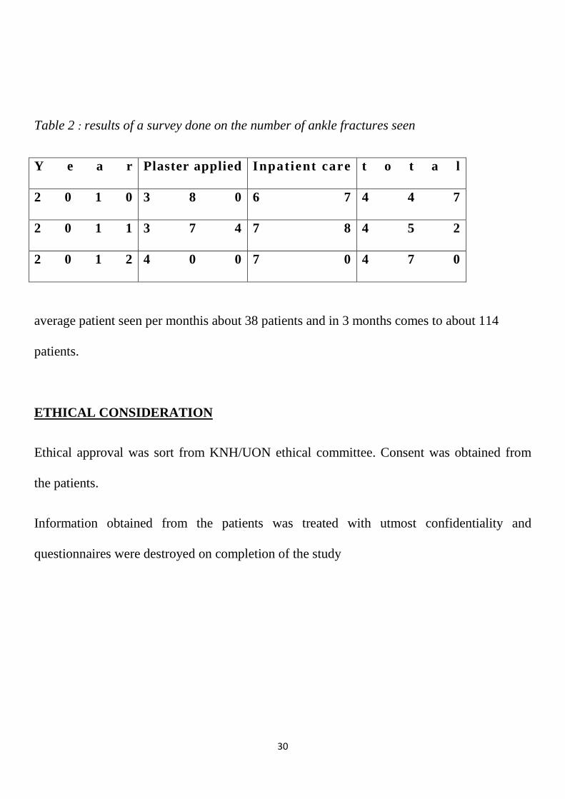
Table 2 : results of a survey done on the number of ankle fractures seen
Y e a r Plaster applied Inpat ient care t o t a l
2 0 1 0 3 8 0 6 7 4 4 7
2 0 1 1 3 7 4 7 8 4 5 2
2 0 1 2 4 0 0 7 0 4 7 0
average patient seen per monthis about 38 patients and in 3 months comes to about 114
patients.
ETHICAL CONSIDERATION
Ethical approval was sort from KNH/UON ethical committee. Consent was obtained from
the patients.
Information obtained from the patients was treated with utmost confidentiality and
questionnaires were destroyed on completion of the study
31
DISSEMINATION
Information acquired will be distributed as follows:-
A copy to department of orthopaedics, copy to the school library and excerpts extracted and published in peer review journals
METHOD OF COLLECTIONS
Patients with ankle fractures as they came in to Kenyatta National Hospital were seen by the
medical officcers, plasters technicians and orthopedic residents. The above group was alerted
and through the research comittee for KNH posters were printed with contacts of the primary
investigator. The assistant was trained by the principal investigator on collection of data
from patients, so that in the event the primary investigator was not able to capture patients
then it was done by the assistant. All patient follow up was done by the principal
investigator.
32
METHODOLOGY
Patients with ankle fractures as evidenced by radiographs were selected from accident and
emergency, orthopaedic clinic and orthopaedic wards in Kenyatta National Hospital.
Collection of data was case based as patients presented to Kenyatta National hospital. The
patient who gave consent the following data was extracted by use of a questionnairei.e. the
patient’s age, sex, body mass index (BMI), diabetes status and other risk factors such as
alcohol use and smoking. Additionally, the fractures mechanism was also examined in terms
of a fall from same level, fall from height, road traffic accident and assault. Ankle fracture of
each individual was then classified according to AO Weber classification. The treatment
modality used was indicated at the first meeting of the patient. Patients were then followed
up and came for review after 6 weeks and 12 weeks. Two Radiographs (AP and lateral)
were done at 6th and 12 weeks of the follow up for all patients. During the follow up period
patient were assessed in terms of healing and maintenance of reduction for the non-operative
method, and wound complications healing and maintenance of reduction for the operative
group Most of the data collected by the chief investigator and a trained assistant. During the
descriptive analysis, the frequency and percentages were obtained. The association between
the outcomes and the independent variables including risk factures type of ankle fractures
and treatment modalities was obtained through the use of chi-square test.
33
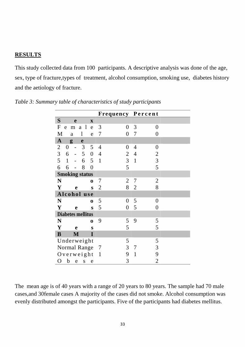
RESULTS
This study collected data from 100 participants. A descriptive analysis was done of the age,
sex, type of fracture,types of treatment, alcohol consumption, smoking use, diabetes history
and the aetiology of fracture.
Table 3: Summary table of characteristics of study participants
The mean age is of 40 years with a range of 20 years to 80 years. The sample had 70 male cases,and 30female cases A majority of the cases did not smoke. Alcohol consumption was evenly distributed amongst the participants. Five of the participants had diabetes mellitus.
Frequency P e r c e n t S e x F e m a l e 3 0 3 0 M a l e 7 0 7 0 A g e 2 0 - 3 5 4 0 4 0 3 6 - 5 0 4 2 4 2 5 1 - 6 5 1 3 1 3 6 6 - 8 0 5 5 Smoking status N o 7 2 7 2 Y e s 2 8 2 8 Alcoho l use N o 5 0 5 0 Y e s 5 0 5 0 Diabetes mellitus N o 9 5 9 5 Y e s 5 5 B M I Underweight 5 5 Normal Range 7 3 7 3 O v e r w e i g h t 1 9 1 9 O b e s e 3 2
34
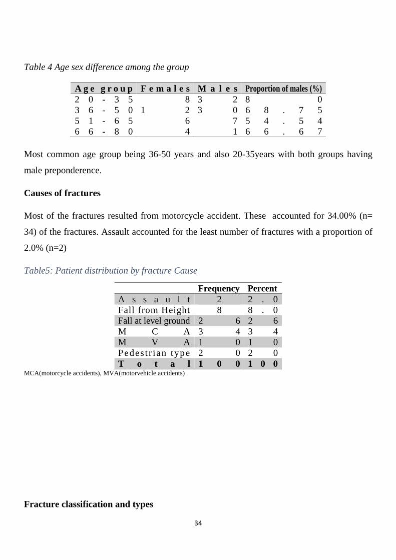
Table 4 Age sex difference among the group
Most common age group being 36-50 years and also 20-35years with both groups having
male preponderence.
Causes of fractures
Most of the fractures resulted from motorcycle accident. These accounted for 34.00% (n=
34) of the fractures. Assault accounted for the least number of fractures with a proportion of
2.0% (n=2)
Table5: Patient distribution by fracture Cause
MCA(motorcycle accidents), MVA(motorvehicle accidents)
Fracture classification and types
A g e g r o u p F e m a l e s M a l e s Proportion of males (%) 2 0 - 3 5 8 3 2 8 0 3 6 - 5 0 1 2 3 0 6 8 . 7 5 5 1 - 6 5 6 7 5 4 . 5 4 6 6 - 8 0 4 1 6 6 . 6 7
Frequency Percent A s s a u l t 2 2 . 0 Fall from Height 8 8 . 0 Fall at level ground 2 6 2 6 M C A 3 4 3 4 M V A 1 0 1 0 Pedest r ian type 2 0 2 0 T o t a l 1 0 0 1 0 0
35
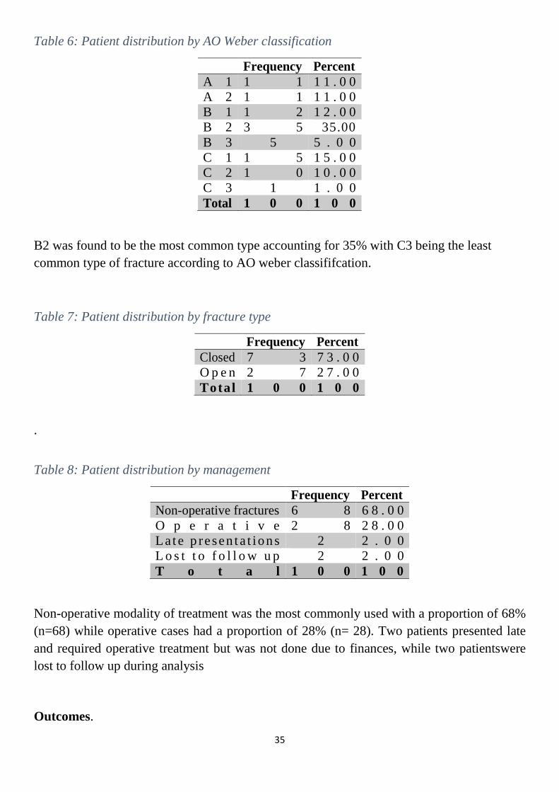
Table 6: Patient distribution by AO Weber classification
Frequency Percent A 1 1 1 1 1 . 0 0 A 2 1 1 1 1 . 0 0 B 1 1 2 1 2 . 0 0 B 2 3 5 35.00 B 3 5 5 . 0 0 C 1 1 5 1 5 . 0 0 C 2 1 0 1 0 . 0 0 C 3 1 1 . 0 0 Total 1 0 0 1 0 0
B2 was found to be the most common type accounting for 35% with C3 being the least common type of fracture according to AO weber classififcation.
Table 7: Patient distribution by fracture type
Frequency Percent Closed 7 3 7 3 . 0 0 O p e n 2 7 2 7 . 0 0 Total 1 0 0 1 0 0
.
Table 8: Patient distribution by management
Frequency Percent Non-operative fractures 6 8 6 8 . 0 0 O p e r a t i v e 2 8 2 8 . 0 0 La te p resen ta t ions 2 2 . 0 0 L o s t t o f o l l o w u p 2 2 . 0 0 T o t a l 1 0 0 1 0 0
Non-operative modality of treatment was the most commonly used with a proportion of 68% (n=68) while operative cases had a proportion of 28% (n= 28). Two patients presented late and required operative treatment but was not done due to finances, while two patientswere lost to follow up during analysis
Outcomes.
36
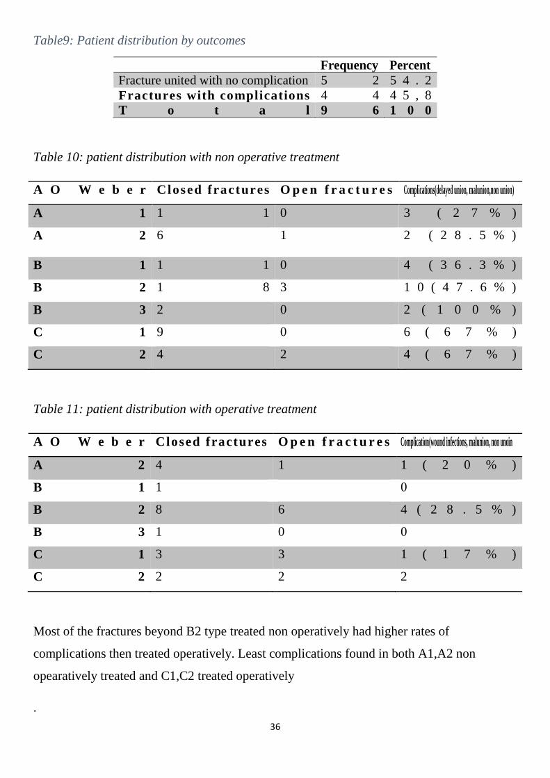
Table9: Patient distribution by outcomes
Frequency Percent Fracture united with no complication 5 2 5 4 . 2 Fractures with complications 4 4 4 5 , 8 T o t a l 9 6 1 0 0
Table 10: patient distribution with non operative treatment
A O W e b e r Closed f ractures O p e n f r a c t u r e s Complications(delayed union, malunion,non union)
A 1 1 1 0 3 ( 2 7 % )
A 2 6 1 2 ( 2 8 . 5 % )
B 1 1 1 0 4 ( 3 6 . 3 % )
B 2 1 8 3 1 0 ( 4 7 . 6 % )
B 3 2 0 2 ( 1 0 0 % )
C 1 9 0 6 ( 6 7 % )
C 2 4 2 4 ( 6 7 % )
Table 11: patient distribution with operative treatment
A O W e b e r Closed f ractures O p e n f r a c t u r e s Complication(wound infections, malunion, non unoin
A 2 4 1 1 ( 2 0 % )
B 1 1 0
B 2 8 6 4 ( 2 8 . 5 % )
B 3 1 0 0
C 1 3 3 1 ( 1 7 % )
C 2 2 2 2
Most of the fractures beyond B2 type treated non operatively had higher rates of
complications then treated operatively. Least complications found in both A1,A2 non
opearatively treated and C1,C2 treated operatively
.
37
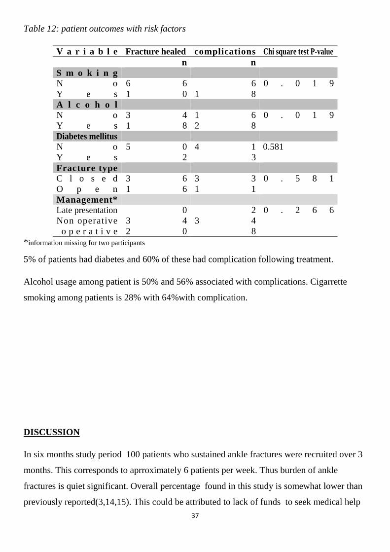
Table 12: patient outcomes with risk factors
V a r i a b l e Fracture healed complications Chi square test P-value n n S m o k i n g N o 6 6 6 0 . 0 1 9 Y e s 1 0 1 8 A l c o h o l N o 3 4 1 6 0 . 0 1 9 Y e s 1 8 2 8 Diabetes mellitus N o 5 0 4 1 0.581 Y e s 2 3 Fracture type C l o s e d 3 6 3 3 0 . 5 8 1 O p e n 1 6 1 1 Management* Late presentation 0 2 0 . 2 6 6 Non operative 3 4 3 4 o p e r a t i v e 2 0 8
* information missing for two participants
5% of patients had diabetes and 60% of these had complication following treatment.
Alcohol usage among patient is 50% and 56% associated with complications. Cigarrette
smoking among patients is 28% with 64%with complication.
DISCUSSION
In six months study period 100 patients who sustained ankle fractures were recruited over 3
months. This corresponds to aprroximately 6 patients per week. Thus burden of ankle
fractures is quiet significant. Overall percentage found in this study is somewhat lower than
previously reported(3,14,15). This could be attributed to lack of funds to seek medical help
38
and also use of traditional bone setters in management of fractures. In many developing
countries traditional care of diseases remain popular despite civilization and availability of
modernised health care. Study done in Nigeria showed a higher numbers of traditional bone
setters as compared to other care givers(60). There are also bone setters in kenya of which
information regarding them is scanty in literature.
Studies have shown fairly equal sex distribution, However this study had male
preponderence(14,29).This was also different from a local study in Kenya which had a
female prepondance and it could be due to lower numbers seen in the study(49).The mean
age also differed among men and women. In the study Men had their peak incidence in third
decade of life while women in their third and fifth decade which was consistent with other
studies(49). The bimodal distribution of this fracture is as a result of younger men with
higher incidence of road traffic accidents while older women most commonly sustaining
injuries secondary to trivial falls.
As in previous studies, fall in the same level was dominating mechanism of injury. In this
study injuries relating to motorcycle accidents accounting for 34% (9,15,62). This was s
consistant to study by Gichuhi, where motorcycle injury incidence is much higher then
pedestrian(4,61).This high rate could be attributed to rise in motorcycle as means of transport
in the region around Nairobi. According to the regional traffic unit majority of motorcycle
drivers are without proper driving licence leading to careless and reckless driving
culminating to high rates of road traffic accidents. High energy trauma was more common in
males and most of them were open injuries. This findings emphasizes the pattern of ankle
fractures with younger men with higher rates of high energy fractures.
Fracture classification according to AO weber, type B2(35%) is shown to be the most
common type of fracture which is consistent with most of the studies(9,27,30). Besides bone
classification it is necessary to evaluate surrounding soft tissues. Most of the fractures found
to be closed injuries accounting to 73%. Open ankle fractures are rare with reported rates of
2-5%(11). However this study found a rate 27.0 % attributed to increase in high energy
injuries due to motorcycle and motor vehicle accidents.
39
Most common mode of treament of ankle fractures was non operative at 68.00% while
28.00% of ankle fractures were operated. Some studies suggest that non operative
management should be reserved for undisplaced, stable ankle fractures and for patient who
are medically unfit for operation. While unstable fractures are better operated.(11).In a recent
cochrane review, surgical treatment was compared with non surgical treatment with long
term functional outcome. This review included four studies and were unable to sufficiently
conclude whether to operate or not(62). High rate of non operative treatment was thought to
be due overcrowding of orthopaedic wards, lack of funds and patient choice for the non
operative management.
The study found high rate of complication for type B and C treated non operatively than
treated operatively. This was simillar to Pugh et at who also showed high complication and
failure rates for type B and C which were treated non operatively.(63). Type B and C are
consider to be unstable fractures and similar study done by Makana showed better outcome
in patients who were operated than the non operative group in terms of range of motion,
pains and swelling and instability of the ankle joint(34).
Risk factors including alcohol, cigarette smoking, diabetes mellitus and high body mass
index were assessed. In this study only 5% patients were found to have diabetes and 60% of
these had complications and fracture did not heal. These results are consistant with many
studies which demonstrated that patients with diabetes have higher rate of complications and
more severe complications then other patients(55). However specific outcomes were difficult
to compare in this study due to the small number of patients.There is a positive correlation
between alcohol and cigerette smoking to poor fracture healing, failure rates and wound
complications following surgery this was consistent with other studies reporting high rates of
non union wound complications infection in patients who consumed alcohol.
LIMITATIONS
1. Risk factors was assessed clinically using patient’s history and no special test was done to evaluate the risk factors.
2. Follow up was short because of student time constriants
40
3. Inadequate funds.
CONCLUSIONS
In conclusion the study showed high rate of fractures due to motorcycle accidents. Majority
were males in their twenties and older women. Type B being the commonest type of ankle
fractures. Non operative treatment was the commonest mode of treatment offered even for
fractures which needed operations the lead to higher complication rates among the mode of
treatment offered.
RECOMMENDATIONS
From this study accident and fall were shown to be the most common cause of ankle
fractures. Accidents were seen in three categories pedestrian, motorcycle and motor vehicle.
A significant number was due to motorcycle injuries. There is need for good road safety
measures concerning especially motorcycle to reduce the trauma burden.
Most of ankle injuries were treated non operatively, there is need for a protocol to be
developed to ensure appropriate management of ankle fractures. This will reduce the
number of fractures inappropraitly managed .
There is need for a larger, longer and multicentre study of ankle fracture management to
make more appropriate recommendations..
Quantitative study for risk factors is proposed as there is a positive corelation with risk
factors and fracture also subsequent healing.
REFERENCES
1. Micheal. P. Clare MD. Rational approach to ankle fractures.Foot Ankle Clin N Am 13 2008 593-610 doi; 10.1016/j.fcl.2008.09.003 foot.theclinics.com
2. Ramsey PL, Hamilton W. Changes in tibiotalar area of contact caused by lateral talar shift, journal Bone Joint Surgery (Am). 1976;58:356.
3. Chalse M,Court-Brown, McBirinie. J and Wilson G. Adult ankle fractures – an increasing problem? Acta Orthop Scand 1998; 69(1): 43-47.
41
4. Ifesanya O. A. And Alonge O. T.operative stabilization of open long bone fractures: A tropical tertiary hospital experience. Nigeria Medical Journal 2012; 53: 16-20
5. Gichuhi.K. Injury pattern among non- fatal road traffic crash victimsEAOJ 2007 ;1: 23-25.
6. RW Westerman and K Porter. Ankle fractures in adult: an overview, trauma 2007;9: 267-272. DOI: 10.1177/1460408607088292.
7. Abboud RJ. Relevant foot biomechanics. Current Orthopaedics. 2002;16:165-179. 8. Czerniecki JM. Foot and ankle biomechanics in walking and running. A review. Am J
Phys Med Rehabil. Dec 1988;67(6):246-252. 9. Rodgers MM. Dynamic foot biomechanics. J Orthop Sports Phys Ther. Jun
1995;21(6):306-316. 10. Lindsjö U. Operative treatment of ankle fractures. Uppsala: Department pf Orthopaedic
Surgery, Uppsala University Hosptial; 1980. 11. Aleksander L , Bumbasirevic M. Ankle fractures, Current Orthopaedics (2004) 18,
232–244. 12. Winter DA. Biomechanics and Motor Control of Human Movement. 2nd ed. New
York: John Wiley & Sons Inc; 1990 13. Olerud C, Molander H. A scoring scale for symptom evaluation after ankle fracture.
Arch Orthop Trauma Surg. 1984;103(3):190-194. 14. Daly PJ, Fitzgerald RH Jr., Melton LJ, Ilstrup DM. Epidemiology of ankle fractures in
Rochester, Minnesota. Acta Orthop Scand. Oct 1987;58(5):539-544 15. Jensen SL, Andresen BK, Mencke S, Nielsen PT. Epidemiology of ankle fractures. A
prospective population-based study of 212 cases in Aalborg, Denmark. Acta Orthop Scand. Feb 1998;69(1):48-50.
16. Singer BR, McLauchlan GJ et al. Epidemiology of fractures in 15,000 adults: the influence of age and gender. J Bone Joint Surg Br. Mar 1998;80(2):243-248.
17. Kannus P, Pehenem M, Niemi S. Increasing number and incidence of low trauma ankle fracture in elderly, Finnish statistics during 1970-2000and projection for future. Bone 2002;31:430-433.
18. Ashhurst AP, Bromer RS. Classification and mechanism of fractures of the leg bones involving the ankle. Arch Surg 1922;4(1):51-129.
19. Yde J. The Lauge Hansen classification of malleolar fractures. Acta Orthop Scand. Feb 1980;51(1):181-192
20. Lauge-Hansen N. Ankelbrud I. Genetisk diagnose og reposition (Fractures of the ankle I. Genetic diagnosis and treatment). Köpenhavn: Munksgaard1942
21. Robert W G, Michelson J D, Larry B B. Fracture of ankle and distal part of tibia. The journal of bone and joint surgery: 1996 vol 78-A; 11:1772-1783
22. Schmolke S and Nikolaus wuelker. Ankle fractures, current opinion in orthopedics 2000, 11;99-102
23. Lauge-Hansen N. Fractures of the ankle. IV. Clinical use of genetic roentgen diagnosis and genetic reduction. AMA Arch Surg. Apr 1952;64(4):488-500.
42
24. Burwell HN, Charnley AD. The treatment of displaced fractures at the ankle by rigid internal fixation and early joint movement. J Bone Joint Surg Br. Nov 1965;47(4):634-660.
25. Nielsen JO, Dons-Jensen H, Sorensen HT. Lauge-Hansen classification of malleolar fractures. An assessment of the reproducibility in 118 cases. Acta Orthop Scand. Oct 1990;61(5):385-387.
26. Müller ME, Nazarian S,et al The Comprehensive Classification of Fractures of Long Bones.1990. Berlin, Heidelberg, New York: Springer-Verlag.
27. Hoiness P, Stromsoe K. Early complications of surgically managed ankle fractures related to the AO classification.A review of 118 ankle fractures treated with open reduction and internal fixation. Arch Orthop Trauma Surg. 1999;119(5-6):276-279.
28. Lindsjo U. Classification of ankle fractures: the Lauge-Hansen or AO system? Clin Orthop Relat Res. Oct 1985(199):12-16.
29. Court-Brown CM, Ceaser D. Epidemiology of adult fractures: A review. Injury aug 2006;37(8):691-697.
30. Hoiness P, Engebretsen L, Stromsoe K. Soft tissue problems in ankle fractures treated surgically. A prospective study of 154 consecutive closed ankle fractures. Injury. Dec 2003;34(12):928-931.
31. Phillips WA, Schwartz HS,et al. A prospective, randomized study of the management of severe ankle fractures. J Bone Joint Surg Am. Jan 1985;67(1):67
32. Odero W . Road traffic accidents in kenya; An epidemiological appraisal. East african medical journal 1995;72(5):299-305.
33. Cooper J.Management of of simple and complex ankle fractures, Trauma 2000;2: 199-210.
34. Makwana N.K, Bhowal. B, W. Harper M, HuiA. W. Conservative versus operative treatment for displaced ankle fracture in patient over 55 years of age. J Bone Joint surg Br. May 2001;83-B(4):525-529.
35. Kristensen KD, Hansen T. Closed treatment of ankle fractures. Stage II supination-eversion fractures followed for 20 years. Acta Orthop Scand. Apr 1985;56(2):107-109.
36. Yde J, Kristensen KD. Ankle fractures. Supination-eversion fractures stage II. Primary and late results of operative and non-operative treatment. Acta Orthop Scand. Aug 1980;51(4):695-702.
37. Vasli S. Operative treatment of ankle fractures. Acta Chir Scand Suppl. 1957;226:1-74. 38. Perren SM. The classic. The aims of internal fixation Robert Danis (1880-1962). Clin
Orthop Relat Res. Jan-Feb 1979(138):23-25. 39. Cedell CA. Supination-outward rotation injuries of the ankle. A clinical and
roentgenological study with special reference to the operative treatment. Acta Orthop Scand. 1967:Suppl 110.
40. Rüedi TP, Murphy WM. ed AO Principles of Fracture Management. Stuttgart, New York: Thieme 2000.
41. Olerud C, Molander H. [Surgical treatment of ankle fractures. A comparison of 2 osteosynthesis technics]. Läkartidningen. Oct 2 1985;82(40):3378-3379.
42. Nelson F. SooHoo, Lucie Krenek,Michael J. Eagan, Barkha Gurbani, Clifford Y. Ko and David S Zingmond, MD, PhD. Complication rates following open reduction and internal fixation of ankle fractures, J Bone Joint Surg Am.2009;91:1042-9.
43
43. Gustillo RB, Anderson JT. Prevention of infection in treatment of 1025 fracture of long bones :retrospective and prospective analysis.J Bone Joint surgery Am.1976;58(4).453-458.
44. Egol. K.A, R. Dolan, K.J. Koval. Functional outcome of surgery for fractures of the ankle.J Bone and Joint Surg [Br]2000;82-B:246-9
45. Ponzer, S. Nasell, H. Bergman, B. Tӧrnkvist, H. Functional outcome and quality of life in patients with type B ankle fractures: a two year follow up study. Journal of Orthopaedic Trauma july 1999 ;13(5):363-368
46. Audige L, Goldhahn S, Daigl M, Goldhahn J, Blauth M, Hanson B. How to document and report orthopedic complications in clinical studies? A proposal for standardization. Arch Orthop Trauma Surg. Sep 8 2011
47. Hoiness P, Stromsoe K. The influence of the timing of surgery on soft tissue complications and hospital stay. A review of 84 closed ankle fractures. Ann Chir Gynaecol. 2000;89(1):6-9.
48. Carragee EJ, Csongradi JJ, Bleck EE. Early complications in the operative treatment of ankle fractures. Influence of delay before operation. J Bone Joint Surg Br. Jan 1991;73(1):79-82
49. Kilonzi N.Mwangi H.R,Lelei L.K. et al. Treatment and outcome of ankle fractures at Moi Teaching and Referralhospital.Annals of African surgery 11:1 2014
50. Canale ST, ed Campbell's Operative Orthopaedics. 10 ed. Philadelphia: Mosby; 2003. Dabov G, Perez, E.A., ed. Miscellaneous Nontraumatic Disorders; No. 1.
51. Brown TD, Johnston RC, Saltzman CL, Marsh JL, Buckwalter JA. Posttraumatic osteoarthritis: a first estimate of incidence, prevalence, and burden of disease. J Orthop Trauma. Nov-Dec 2006;20(10):739-744.
52. Horisberger M, Valderrabano V, Hintermann B. Posttraumatic ankle osteoarthritis after ankle-related fractures. J Orthop Trauma. Jan 2009;23(1):60-67.
53. Anderson S.A, Li. X, Franklin P Ankle fractures in elderly :Initial and long term outcome. Foot ankle Int. Dec 2008;29(12):1184-1188
54. Saithna A. Moody W. Jenkenson E et al. The influence of timing of surgery on soft tissue complications in closed ankle fractues. Eur J. Orthop Surg Trauma vol 2009(19):481-484.
55. Shanti P.G, Peitroban R. Et al. Impart of diabetes on patient outcomeafter ankle fractures. J Bone Joint Surg vol 87.A Aug 2005.
56. Hanne Tonnesen, Anne Pedersen, et al. Ankle fractures and alcoholism. J Bone Joint Surg [Br] 1991 ; 73-B :51 1-3.
57. Strauss EJ, Frank JB,et al. Does obesity influence the outcome after the operative treatment of ankle fractures? J Bone Joint Surg Br. Jun 2007;89(6):794-798.
58. King CM, Hamilton GA, et al. Association between Ankle Fractures and Obesity. J Foot Ankle Surg. Jul 11 2012.
59. Nåsell H, Adami J, et al.. Effect of smoking cessation intervention on results of acute fracture surgery: a randomized controlled trial. J Bone Joint Surg Am. Jun 2010;92(6):1335-1342.
60. Dada A. A, Yinusa. W, Giwa S.O. Review of the practice of traditional bone setting in Nigeria. African Health Sciences.vol 11 No.2 June 2011.
61. Kuubiere C.B, A. Alhassan and S. F. Majeed Management of complex ankle fracture: A Ghanain experience, journal of medical biomedical science 2012;1(4):1-6.
44
62. Donken CC,Al-Khateeb H, et al. Surgical versus conservative intervention for treating adult fractures in adults. Cochrane Database syst Rev.2012;8:CD008470.
63. Pugh K.J, Fitzgerald R.H Kauger H.ed Fractures and soft tissue injuries about ankle fractures in orthopedics. Philadelphia Mosby 2002 419-434.
APPENDIX
CONSENT BY THE PARTICIPATING PATIENT/ GUARDIAN Study No………………. HospitalNo…………….. Principal investigator: Dr Mustafa. Khanbhai Authorized by : Kenyatta National Hospital Ethics & Research Committee
45
Introduction Ankle fractures are among the commonest injuries that occur in the society at large. Most of this fractures are managed conservatively. Patterns and modality of management are poorly studied.this study aims to fill this gap. You are invited to participate in this study whic will look at patterns of ankle fracture and modalities used in management.kindly read this form and understand it well before agreeing to this study. Any questions you have will be answered. Purpose of the study The purpose of this study is to determine the patterns of ankle fractures in Kenyatta National Hospital and modalities used in their management. Lastly information obtained will be used for purposes of obtaining a Master degree in Orthopedic Surgery for the principal investigator. Study procedure If you agree to participate in this study, your particulars will be recorded in the data collection sheet. Patients who meet the criteria history of injury will be taken in terms of mechanism of injury, history of alcohol , cigarette smoking and diabetes mellitus.the type of fracture will be noted and management used. Post management check xray and achievement of reduction. Routine follow up at 6 and 12 weeks. The radiographs taken after 6 and 12 weeks on routine follow up wil be reviewed. Complication following all modalities of treatment will be accessed clinically. Risks and benefits
There is no harm or risk anticipated in participating in this study.There is no added radiation risk associated with taking of x-rays. Participation in this study will result in better management of patients with fractures of the ankle.
Study costs
If you accept to take part in this study, there will be no payment expected from you or to you.
No added investigations will be required and x-rays done will be as per routine post-operative management
and follow up of these fractures.
Confidentiality
The data collection sheet is strictly confidential. Your name will not appear in it and your telephone number
is strictly for follow up purposes. If you so wish you will be given a copy of this consent form.
Participant information
Your participation in this study is voluntary and failure to participate or withdrawal from the study will not
affect your management in any way at any stage.
Participant consent form
46
I have understood the above information which has been fully explained to me by the investigator and I
voluntarily consent to participate.
Signature…………………………………………………………………..
Or participants thumb print.
For any enquiries or further clarification, please contact the following people
1. PRINCIPAL INVESTIGATOR :- DR MUSTAFA. KHANBHAI TEL : - 0733 248147/ 0721444369
For any conformation of authority and complains please contact
1. CHAIRMAN, KENYATTA NATIONAL HOSPITAL ETHICS & RESEARCH COMMITTEE – Tel 0722 70880
KIBALI CHA RUHUSA YA KUHUSIKA
Numbariyahospitali_____ Utafiti ya matokeo ya wagonjwa waliovunjika mguu karibu na kisigino. Ninafanya utafiti kuhusu wagonjwa waliovunjika mfupa wa mguu karibu na kisigino. Namna walivunjika, tumizi ya pombe na sigara, ugonjwa wa
47
sukari.Tafadhali soma fomu hii na kuielewa vizuri kabla ya kukubali utafiti. Maswali yoyote utakayokuwa nayo yatajibiwa.
Sababu ya utafiti
Lengo ni kupata taarifa juu ya wagonjwa waliovunjika mifupa ya mguu karibu na kisigino.Huu utafiti utasaidia pia katika mabadiliko ya usimamizi wa sera ya majeraha hayo katika hospitali na nchi kwa ujumla. Taarifa itakayopatikana ni muhimu pia kwa kufikia shahada ya uzamili katika upasuaji wa mifupa (orthopaedic surgery) kwa mpelelezi mkuu. Utaratibu wa utafiti
Habari kuu inayohitajika kutoka kwako ni maelezo yako kama katika karatasi ukusanyaji. Namna ya majiraha, utumizi ya sigara na pombe, matibabu kama plaster ama upasuaji ilitumika kwa hiyo frakchari ya mguu.Picha ambazo zitapigwa kama mandelezo ya matibabu yako zitafanywa baada ya wiki sita na mwezi tatu. Utafuatiliwa kwa muda wa mwezi tatu. Hatari na manufaa
Hakuna hatari zozote zinaweza kutokea kwa kushiriki katika utafiti huu.Hakuna hatari zaidi itatokana na
kupigwa picha ya mkono na pia hakuna gharama zaidi zitatozwa kwa kushiriki katika utafiti huu.
Usiri
Ukusanyaji wa karatasi takwimu ni madhubuti ya siri. Jina lako hatilitaandikwa na nambari yako ya simu ni
madhubuti kwa ajili ya kufuatilia makusudi. Kama unataka utapewa nakala ya fomu hii ya ridhaa.
Habari kwa mshiriki
Ushiriki wako katika utafiti huu ni hiari yako na kushindwa kushiriki au kujiondoa kutoka utafiti huu,
hautaadhiri usimamizi wako na matibabu yako katika njia yoyote katika hatua yoyote.
Fomu ya mshiriki wa ridhaa.
Mimi nimeshaelewa maelezo nimeyoambiwa kikamilifu na mpelelezi na nitashiriki kwa hiari yangu kwa
kutia sahihi kwa ridhaa.

48
Sahihi .............................................................................
Au kidole gumba cha mshiriki.
Kama una maswali yoyote au wasiwasi kuhusu utafiti huu unaweza kuwasiliana na mtafiti anayefanya utafiti
huu Dkt mustafa numbari ya simu 0721444369, barua pepe kwa [email protected] au
Mkurugenzi,KNH / Chuo Kikuu cha Nairobi - Maadili Kamati ya UchunguziSimu:- 726300 – 9 or (254 -
020) 2726300 Ext 44102

49
APPENDIX 2 Data collection sheet Study no __________ patients contact ____________________ Age : Sex : male female weight___________ height ______ BMI ____________
Cormodities History of smoking, alcohol abuse, if having diabetes If alcohol usage how much either social drinker or everyday consumption.____________ Smoking how many pack years ____________ Mechanism of injury_______________________________ Classification of injury according to radiographic views AO weber classification type _______________________ Type of fracture Open closed Mode of treatment used nonoperative operative If non operative was fracture well reduced, how many times was it maniulated Joint congruity in post reduction plus paster xrays achieved If not was there remanipultion yes or no ______________ If above is operative timing of surgery _______________________________ days Rigid or non rigid fixation ___________________ Hospital stay ____________________ If open fracture mode of treatment used ________________________ Infection accessed clincally present absent during hosspital stay Restoration of joint congruity on post operative xrays Reoperation Fusion Follow up
6 w e e k s 1 2 w e e k s Type of complication Wound status Well healed dehiscence Necrosis Infection if presence of pus from woound site
P a i n S w e l l i n g Healing assesed using radiographic evidence of callus, Function of the joint Dorsiflexion (degrees) Planterflexion (measured using a goniometer)
I m p l a n t f a i l u r e Plaster failure Neglected ankle

50
Appendix 3.
IMPLEMENTATION SCHEDULE
P r o p o s a l w r i t i n g O C T 2 0 1 3 - A P R I L 2 0 1 4
P a t i e n t r e c r u i t m e n t M A Y 2 0 1 4 - J U L Y 2 0 1 4
F o l l o w u p A U G 2 0 1 4 - O C T 2 0 1 4
Data analys is and d issertat ion wri t ing N O V - D E C 2 0 1 4
R e s u l t s p r e s e n t a t i o n J A N 2 0 1 5
BUDGET
R e s e a r c h f e e s ( K N H / E R C ) 3 , 0 0 0
S t a t i o n e r y 1 0 , 0 0 0
S t a t i s t i c i a n 4 0 , 0 0 0
R e s e a r c h a s s i s t a n t s 4 0 , 0 0 0
C o n t i n g e n c i e s 1 0 , 0 0 0
T o t a l ( K S h s ) 1 0 3 , 0 0 0