The Parkinson's disease associated LRRK2 exhibits weaker in vitro phosphorylation of 4E-BP compared...
10
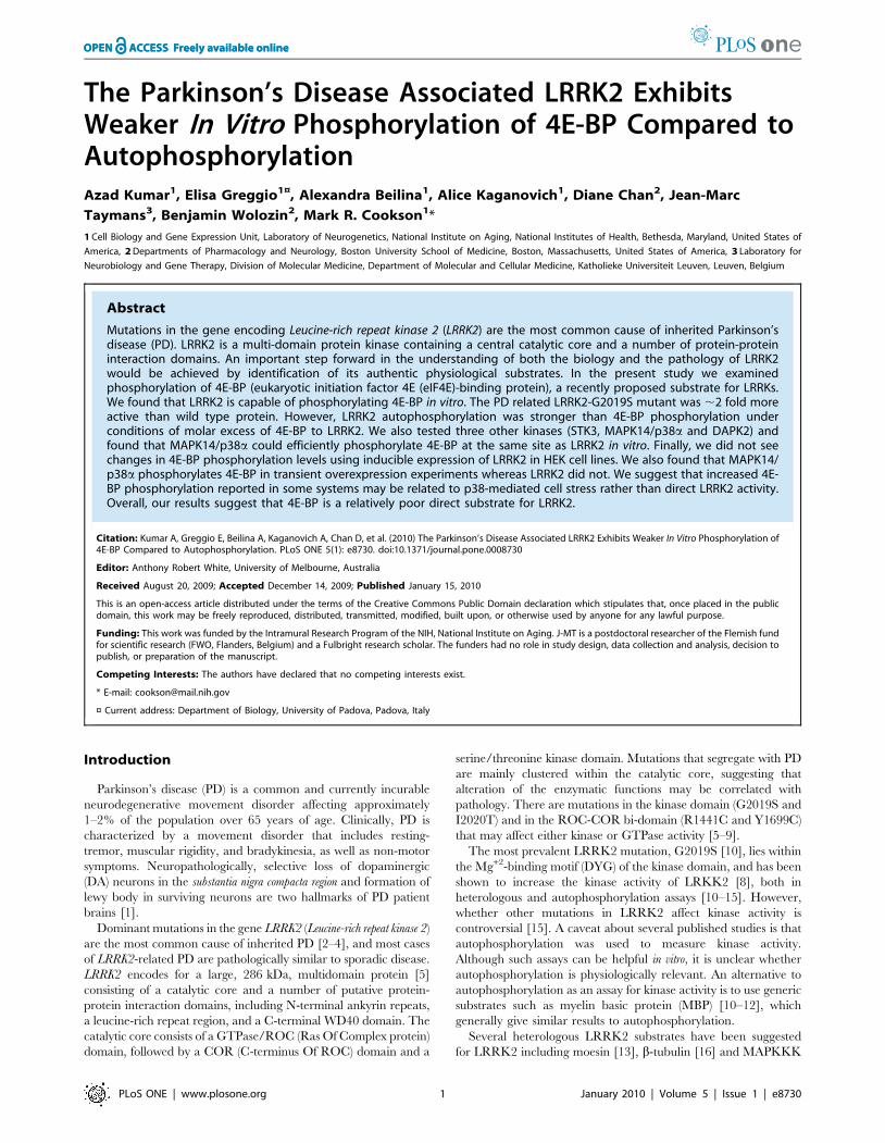
The Parkinson’s Disease Associated LRRK2 Exhibits Weaker In Vitro Phosphorylation of 4E-BP Compared to Autophosphorylation Azad Kumar 1 , Elisa Greggio 1¤ , Alexandra Beilina 1 , Alice Kaganovich 1 , Diane Chan 2 , Jean-Marc Taymans 3 , Benjamin Wolozin 2 , Mark R. Cookson 1 * 1 Cell Biology and Gene Expression Unit, Laboratory of Neurogenetics, National Institute on Aging, National Institutes of Health, Bethesda, Maryland, United States of America, 2 Departments of Pharmacology and Neurology, Boston University School of Medicine, Boston, Massachusetts, United States of America, 3 Laboratory for Neurobiology and Gene Therapy, Division of Molecular Medicine, Department of Molecular and Cellular Medicine, Katholieke Universiteit Leuven, Leuven, Belgium Abstract Mutations in the gene encoding Leucine-rich repeat kinase 2 (LRRK2) are the most common cause of inherited Parkinson’s disease (PD). LRRK2 is a multi-domain protein kinase containing a central catalytic core and a number of protein-protein interaction domains. An important step forward in the understanding of both the biology and the pathology of LRRK2 would be achieved by identification of its authentic physiological substrates. In the present study we examined phosphorylation of 4E-BP (eukaryotic initiation factor 4E (eIF4E)-binding protein), a recently proposed substrate for LRRKs. We found that LRRK2 is capable of phosphorylating 4E-BP in vitro. The PD related LRRK2-G2019S mutant was ,2 fold more active than wild type protein. However, LRRK2 autophosphorylation was stronger than 4E-BP phosphorylation under conditions of molar excess of 4E-BP to LRRK2. We also tested three other kinases (STK3, MAPK14/p38a and DAPK2) and found that MAPK14/p38a could efficiently phosphorylate 4E-BP at the same site as LRRK2 in vitro. Finally, we did not see changes in 4E-BP phosphorylation levels using inducible expression of LRRK2 in HEK cell lines. We also found that MAPK14/ p38a phosphorylates 4E-BP in transient overexpression experiments whereas LRRK2 did not. We suggest that increased 4E- BP phosphorylation reported in some systems may be related to p38-mediated cell stress rather than direct LRRK2 activity. Overall, our results suggest that 4E-BP is a relatively poor direct substrate for LRRK2. Citation: Kumar A, Greggio E, Beilina A, Kaganovich A, Chan D, et al. (2010) The Parkinson’s Disease Associated LRRK2 Exhibits Weaker In Vitro Phosphorylation of 4E-BP Compared to Autophosphorylation. PLoS ONE 5(1): e8730. doi:10.1371/journal.pone.0008730 Editor: Anthony Robert White, University of Melbourne, Australia Received August 20, 2009; Accepted December 14, 2009; Published January 15, 2010 This is an open-access article distributed under the terms of the Creative Commons Public Domain declaration which stipulates that, once placed in the public domain, this work may be freely reproduced, distributed, transmitted, modified, built upon, or otherwise used by anyone for any lawful purpose. Funding: This work was funded by the Intramural Research Program of the NIH, National Institute on Aging. J-MT is a postdoctoral researcher of the Flemish fund for scientific research (FWO, Flanders, Belgium) and a Fulbright research scholar. The funders had no role in study design, data collection and analysis, decision to publish, or preparation of the manuscript. Competing Interests: The authors have declared that no competing interests exist. * E-mail: [email protected] ¤ Current address: Department of Biology, University of Padova, Padova, Italy Introduction Parkinson’s disease (PD) is a common and currently incurable neurodegenerative movement disorder affecting approximately 1–2% of the population over 65 years of age. Clinically, PD is characterized by a movement disorder that includes resting- tremor, muscular rigidity, and bradykinesia, as well as non-motor symptoms. Neuropathologically, selective loss of dopaminergic (DA) neurons in the substantia nigra compacta region and formation of lewy body in surviving neurons are two hallmarks of PD patient brains [1]. Dominant mutations in the gene LRRK2 (Leucine-rich repeat kinase 2) are the most common cause of inherited PD [2–4], and most cases of LRRK2-related PD are pathologically similar to sporadic disease. LRRK2 encodes for a large, 286 kDa, multidomain protein [5] consisting of a catalytic core and a number of putative protein- protein interaction domains, including N-terminal ankyrin repeats, a leucine-rich repeat region, and a C-terminal WD40 domain. The catalytic core consists of a GTPase/ROC (Ras Of Complex protein) domain, followed by a COR (C-terminus Of ROC) domain and a serine/threonine kinase domain. Mutations that segregate with PD are mainly clustered within the catalytic core, suggesting that alteration of the enzymatic functions may be correlated with pathology. There are mutations in the kinase domain (G2019S and I2020T) and in the ROC-COR bi-domain (R1441C and Y1699C) that may affect either kinase or GTPase activity [5–9]. The most prevalent LRRK2 mutation, G2019S [10], lies within the Mg +2 -binding motif (DYG) of the kinase domain, and has been shown to increase the kinase activity of LRKK2 [8], both in heterologous and autophosphorylation assays [10–15]. However, whether other mutations in LRRK2 affect kinase activity is controversial [15]. A caveat about several published studies is that autophosphorylation was used to measure kinase activity. Although such assays can be helpful in vitro, it is unclear whether autophosphorylation is physiologically relevant. An alternative to autophosphorylation as an assay for kinase activity is to use generic substrates such as myelin basic protein (MBP) [10–12], which generally give similar results to autophosphorylation. Several heterologous LRRK2 substrates have been suggested for LRRK2 including moesin [13], b-tubulin [16] and MAPKKK PLoS ONE | www.plosone.org 1 January 2010 | Volume 5 | Issue 1 | e8730
-
Upload
independent -
Category
Documents
-
view
0 -
download
0
Transcript of The Parkinson's disease associated LRRK2 exhibits weaker in vitro phosphorylation of 4E-BP compared...
The Parkinson’s Disease Associated LRRK2 ExhibitsWeaker In Vitro Phosphorylation of 4E-BP Compared toAutophosphorylationAzad Kumar1, Elisa Greggio1¤, Alexandra Beilina1, Alice Kaganovich1, Diane Chan2, Jean-Marc
Taymans3, Benjamin Wolozin2, Mark R. Cookson1*
1 Cell Biology and Gene Expression Unit, Laboratory of Neurogenetics, National Institute on Aging, National Institutes of Health, Bethesda, Maryland, United States of
America, 2 Departments of Pharmacology and Neurology, Boston University School of Medicine, Boston, Massachusetts, United States of America, 3 Laboratory for
Neurobiology and Gene Therapy, Division of Molecular Medicine, Department of Molecular and Cellular Medicine, Katholieke Universiteit Leuven, Leuven, Belgium
Abstract
Mutations in the gene encoding Leucine-rich repeat kinase 2 (LRRK2) are the most common cause of inherited Parkinson’sdisease (PD). LRRK2 is a multi-domain protein kinase containing a central catalytic core and a number of protein-proteininteraction domains. An important step forward in the understanding of both the biology and the pathology of LRRK2would be achieved by identification of its authentic physiological substrates. In the present study we examinedphosphorylation of 4E-BP (eukaryotic initiation factor 4E (eIF4E)-binding protein), a recently proposed substrate for LRRKs.We found that LRRK2 is capable of phosphorylating 4E-BP in vitro. The PD related LRRK2-G2019S mutant was ,2 fold moreactive than wild type protein. However, LRRK2 autophosphorylation was stronger than 4E-BP phosphorylation underconditions of molar excess of 4E-BP to LRRK2. We also tested three other kinases (STK3, MAPK14/p38a and DAPK2) andfound that MAPK14/p38a could efficiently phosphorylate 4E-BP at the same site as LRRK2 in vitro. Finally, we did not seechanges in 4E-BP phosphorylation levels using inducible expression of LRRK2 in HEK cell lines. We also found that MAPK14/p38a phosphorylates 4E-BP in transient overexpression experiments whereas LRRK2 did not. We suggest that increased 4E-BP phosphorylation reported in some systems may be related to p38-mediated cell stress rather than direct LRRK2 activity.Overall, our results suggest that 4E-BP is a relatively poor direct substrate for LRRK2.
Citation: Kumar A, Greggio E, Beilina A, Kaganovich A, Chan D, et al. (2010) The Parkinson’s Disease Associated LRRK2 Exhibits Weaker In Vitro Phosphorylation of4E-BP Compared to Autophosphorylation. PLoS ONE 5(1): e8730. doi:10.1371/journal.pone.0008730
Editor: Anthony Robert White, University of Melbourne, Australia
Received August 20, 2009; Accepted December 14, 2009; Published January 15, 2010
This is an open-access article distributed under the terms of the Creative Commons Public Domain declaration which stipulates that, once placed in the publicdomain, this work may be freely reproduced, distributed, transmitted, modified, built upon, or otherwise used by anyone for any lawful purpose.
Funding: This work was funded by the Intramural Research Program of the NIH, National Institute on Aging. J-MT is a postdoctoral researcher of the Flemish fundfor scientific research (FWO, Flanders, Belgium) and a Fulbright research scholar. The funders had no role in study design, data collection and analysis, decision topublish, or preparation of the manuscript.
Competing Interests: The authors have declared that no competing interests exist.
* E-mail: [email protected]
¤ Current address: Department of Biology, University of Padova, Padova, Italy
Introduction
Parkinson’s disease (PD) is a common and currently incurable
neurodegenerative movement disorder affecting approximately
1–2% of the population over 65 years of age. Clinically, PD is
characterized by a movement disorder that includes resting-
tremor, muscular rigidity, and bradykinesia, as well as non-motor
symptoms. Neuropathologically, selective loss of dopaminergic
(DA) neurons in the substantia nigra compacta region and formation of
lewy body in surviving neurons are two hallmarks of PD patient
brains [1].
Dominant mutations in the gene LRRK2 (Leucine-rich repeat kinase 2)
are the most common cause of inherited PD [2–4], and most cases
of LRRK2-related PD are pathologically similar to sporadic disease.
LRRK2 encodes for a large, 286 kDa, multidomain protein [5]
consisting of a catalytic core and a number of putative protein-
protein interaction domains, including N-terminal ankyrin repeats,
a leucine-rich repeat region, and a C-terminal WD40 domain. The
catalytic core consists of a GTPase/ROC (Ras Of Complex protein)
domain, followed by a COR (C-terminus Of ROC) domain and a
serine/threonine kinase domain. Mutations that segregate with PD
are mainly clustered within the catalytic core, suggesting that
alteration of the enzymatic functions may be correlated with
pathology. There are mutations in the kinase domain (G2019S and
I2020T) and in the ROC-COR bi-domain (R1441C and Y1699C)
that may affect either kinase or GTPase activity [5–9].
The most prevalent LRRK2 mutation, G2019S [10], lies within
the Mg+2-binding motif (DYG) of the kinase domain, and has been
shown to increase the kinase activity of LRKK2 [8], both in
heterologous and autophosphorylation assays [10–15]. However,
whether other mutations in LRRK2 affect kinase activity is
controversial [15]. A caveat about several published studies is that
autophosphorylation was used to measure kinase activity.
Although such assays can be helpful in vitro, it is unclear whether
autophosphorylation is physiologically relevant. An alternative to
autophosphorylation as an assay for kinase activity is to use generic
substrates such as myelin basic protein (MBP) [10–12], which
generally give similar results to autophosphorylation.
Several heterologous LRRK2 substrates have been suggested
for LRRK2 including moesin [13], b-tubulin [16] and MAPKKK
PLoS ONE | www.plosone.org 1 January 2010 | Volume 5 | Issue 1 | e8730
substrates MKK3/6 or MKK4/7 [17]. Whether any of these
substrates are physiologically relevant is currently unclear.
Recently Imai et al. reported 4E-BP as a potential substrate of
LRRK2 [18]. 4E-BP is an interactor of the eukaryotic protein
translation initiation factor eIF4E, which in turn binds to capped
mRNA species, promoting their translation. Binding of 4E-BP to
eIF4E prevents the latter being active and, therefore, 4E-BP is a
repressor of protein translation. Oxidative stress and other stimuli
that impact protein translation affect phosphorylation of 4E-BP.
Imai et al. proposed that LRRK2 modulates this system by
phosphorylating 4E-BP at a specific site (T37/T46), which then
acts as a stimulus for further phosphorylation by other kinases at
secondary sites including S65/S70. There was a modest decrease
in phosphorylation of 4E-BP T37/T46 and S65 when LRRK2
levels were knocked down with RNAi. Overexpression of 4E-BP
rescued the effects of LRRK mutants in vivo using Drosophila
models. Similarly, Tain et al have shown that phospho-4E-BP
levels are decreased in a homozygous knockout model of drosophila
LRRK [19]. Collectively these data are supportive of 4E-BP being
a substrate for LRRK2 or its Drosophila homologue, dLRRK.
However, the kinetics of the phosphorylation reaction have not yet
been reported. In the present study we investigated the kinetic
properties of 4E-BP phosphorylation by LRRK2 compared to
autophosphorylation activity and we used a LRRK2 inducible
cellular model as well as transient expression in HEK cells to
measure the effect of LRRK2 expression on 4E-BP activation at
T37/T46.
Results
In Vitro Kinase AssayWe first examined 4E-BP phosphorylation by LRRK2-wild type
and LRRK2-G2019S in vitro (Figure 1). We used recombinant
protein expressed in insect cells containing amino acids 970–2527
of human LRRK2 that has been previously shown to be highly
active [14] rather than immunoprecipitated LRRK2 from
mammalian cells as the latter may contain contaminant kinases.
We confirmed [18] that LRRK2 variants were able to phosphor-
ylate 4E-BP and that the LRRK2-G2019S mutant increased 4E-
BP phosphorylation compared to wild-type proteins. As a negative
control, a kinase dead LRRK2 (D1994A) mutant had a negligible
activity towards 4E-BP phosphorylation under the same conditions
(data not shown).
After confirming that 4E-BP is an in vitro substrate for LRRK2, we
assessed the kinetic catalytic activity of LRRK2-wild type, LRRK2-
kinase dead and LRRK2-G2019S toward 4E-BP. Increasing
concentrations of 4E-BP (1.5, 3, 6 and 12 mM) were incubated with
12 nM of LRRK2, and the incorporation of 33P over the time-
course of reaction was measured (Figure 1A, C and D). The
efficiency of 4E-BP phosphorylation was almost two times higher for
LRRK2-G2019S (11.960.66, arbitrary phosphorescence units)
compared to LRRK2-wild type (5.160.45, arbitrary phosphores-
cence units), and again kinase dead LRRK2 was inactive (Table 1).
However, Km for 4E-BP was similar for both wild type and LRRK2-
G2019S (2.6960.64 mM and 2.8760.45 mM, respectively).
Interestingly, under the same conditions incorporation of
radioactivity into LRRK2 was substantially higher than 4E-BP
phosphorylation, despite LRRK2 being present at a much lower
concentration (,12 nM LRRK2 versus 1.5 to 12 mM 4E-BP),
which is apparent on examination of a single exposure with both
reactions (Figure 1A). Quantitation demonstrated that for 12 nM
LRRK2 and 12 mM 4E-BP, almost three times as much
radioactivity is incorporated into LRRK2 (Figure 1B and C). As
for 4E-BP phosphorylation, G2019S increased autophosphoryla-
tion activity by about two-fold (Figure 1B) and D1994A abolished
activity (data not shown).
Prior studies have suggested that phosphorylation of 4E-BP can
be affected by the presence of its binding partner eIF4E [20]. We
compared kinase activity of wild type LRRK2 towards 4E-BP
alone or in an equimolar complex with eIF4E (Figure 2A and B).
We found that the presence of eIF4E partially blocked 4E-BP
phosphorylation but did not prevent phosphorylation of 4E-BP
entirely. No evidence of phosphorylation of eIF4E was found.
These results suggest that LRRK2 can phosphorylate 4E-BP
weakly even in the presence of eIF4E.
We next considered whether kinases other than LRRK2 could
mediate the same reaction picking three kinases from different
families in the kinome (Figure 3). Under conditions similar to the
LRRK2 reactions, MAPK14/p38a lacks autokinase activity but
STK3 and DAPK2 could autophosphorylate. We found that
MAPK14/p38a phosphorylated 4E-BP at a much higher rate
(Figure 3A and B) compared to STK3 whereas DAPK2 showed no
reactivity towards 4E-BP. These results suggest that 4E-BP can be
phosphorylated by several different kinases in vitro. We also noted
that the putative LRRK2 site recognized by antibodies against
T37/T46 were also phosphorylated by MAPK14/p38a. The
efficiency of 4E-BP phosphorylation was almost twenty times
higher for MAPK14/p38a (10268.14, arbitrary phosphorescence
units) compared to LRRK2-wild type (5.160.45, arbitrary
phosphorescence units). However, Km for 4E-BP was similar for
both wild type LRRK2 and MAPK14/p38a (2.6960.64 mM and
2.5660.59 mM, respectively) (Table 1).
Stoichiometry of LRRK2 Autophosphorylation and 4E-BPPhosphorylation
To determine the stoichiometry of LRRK2 autophosphoryla-
tion compared to 4E-BP phosphorylation, 12 mM of 4E-BP was
incubated with 12 nM of LRRK2, and the incorporation of 33P
was measured by scintillation counting. We observed that 1 mole
of ATP was incorporated into 70 moles of LRRK2 whereas 1 mole
of ATP was incorporated into 1.586103 moles of 4E-BP after
60 minutes of reaction suggesting that LRRK2 autophosphoryla-
tion is approximately 20 times more efficient than 4E-BP
phosphorylation under the same conditions. Table 2 shows the
percentage ratio between the number of inorganic phosphates (Pi)
incorporated by LRRK2 or 4E-BP and the total number of
LRRK2 or 4E-BP molecules at different time points.
Phosphorylation of 4E-BP by LRRK2 in CellsNext we assessed the effect of expression of LRRK2 in inducible
stable HEK 293FT clones. Although we were able to readily
detect the phospho-4E-BP T37/T46, S65 and T70 epitope in
these cells (Figure 4A), we did not see increased LRRK2 4E-BP
phosphorylation upon LRRK2 expression, even when the
pathological mutations G2019S or R1441C were introduced
(Figure 4B). LRRK2 protein extracted and immunoprecipitated
from these cells was active in kinase assays (Figure 4C).
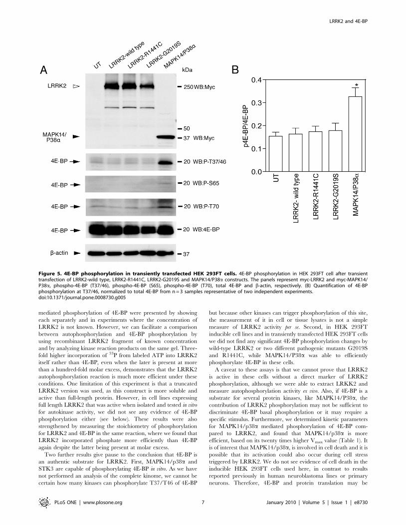
Finally we transiently overexpressed LRRK2-wild type,
LRKK2-G2019S, LRRK2-R1441C and MAPK14/P38a in
HEK 293FT cells. We did not detect any alteration in 4E-BP
phosphorylation either by wild type or mutant LRRK2, although
we found that MAPK14/P38a could phosphorylate 4E-BP under
the same conditions (Figure 5A and B).
Discussion
Although mutations in the LRRK2 gene represent the most
common cause of PD [2,3,21], little is known about LRRK2-linked
LRRK2 and 4E-BP
PLoS ONE | www.plosone.org 2 January 2010 | Volume 5 | Issue 1 | e8730

Figure 1. Phosphorylation of 4E-BP by LRRK2 in vitro. (A) Time-course catalytic assays were performed for GST-LRRK2 wild-type (upper panel)or GST-LRRK2-G2019S (lower panels) by measuring the incorporation of [c33P] ATP at different time points (0, 5, 15, 30 and 60 mins) and at differentconcentrations of 4E-BP (1.5, 3, 6 and 12 mM), respectively, followed by SDS-PAGE and autoradiography. (B) Autophosphorylation of LRRK2-wild type(left graph) and G2019S (right graph) were plotted as a function of time. (C) Phosphorylation of 4E-BP by LRRK2-wild type (left graph) and G2019S(right graph) were plotted as a function of time. (D) Results of the catalytic assays were derived by calculating the reaction rate of each assay atdifferent 4E-BP concentrations (1.5, 3, 6 and 12 mM). For all experiments in A–D, each point is the average of three independent experiments and errorbars indicate the SEM.doi:10.1371/journal.pone.0008730.g001
LRRK2 and 4E-BP
PLoS ONE | www.plosone.org 3 January 2010 | Volume 5 | Issue 1 | e8730

molecular pathogenesis. Therefore, in the present study we have
investigated the kinase activity of LRRK2 with respect to its
autophosphorylation as well as phosphorylation of 4E-BP, a novel
reported LRRK2 substrate. We were able to replicate some
previous experiments showing that 4E-BP can be phosphorylated
by LRRK2 in vitro but we found that this activity is weak compared
to autophosphorylation. Moreover, using inducible LRRK2 cells
or transient transfections, we could not detect any appreciable
increased in 4E-BP phosphorylation after LRRK2 expression.
However, we observed that MAPK14/p38a increases 4E-BP
phosphorylation in HEK 293FT cells after transient expression,
suggesting that MAPK14/P38a is a robust kinase for 4E-BP.
Table 1. Vmax and Km for LRRK2 and MAPK14/P38aphosphorylation of 4E-BP in vitro.
Kinase WT LRRK2 G2019S LRRK2 MAPK14/P38a
Vmax (arbitraryphosphorescence unit)
5.160.45 11.960.66 10268.14
Km(mM) 2.6960.641 2.8760.452 2.5660.592
Each data point is calculated Vmax and Km values (6SEM) for assays carried outin triplicate.doi:10.1371/journal.pone.0008730.t001
Figure 2. Effect of 4E-BP and eIF4E complex on phosphorylation of 4E-BP by LRRK2 in vitro. (A) Time-course assays were performed forLRRK2 wild-type (upper panel) by measuring the incorporation of [c33P] ATP at different time points (0, 5, 15, 30 and 60 mins) and at 3 mMconcentrations of 4E-BP (upper left panel), 3 mM concentrations of eIF4E (upper middle panel) and 3 mM concentrations of 4E-BP and 3 mMconcentrations of eIF4E (upper right panel), respectively, followed by SDS-PAGE and autoradiography. (B) Phosphorylation of 4E-BP by LRRK2-wildtype were plotted as a function of time.doi:10.1371/journal.pone.0008730.g002
LRRK2 and 4E-BP
PLoS ONE | www.plosone.org 4 January 2010 | Volume 5 | Issue 1 | e8730

Figure 3. Kinases other than LRRK2 can also phosphorylate 4E-BP. (A) Kinase assays were performed for LRRK2, STK3, MAPK14//P38a andDAPK2 by measuring the incorporation of [c33P] ATP at different time points (0, 5, 15, 30 and 60 mins) and at 12 mM concentrations of 4E-BP,followed by SDS-PAGE and autoradiography. (B) Time-course catalytic assays were performed for MAPK14/P38a by measuring the incorporation of[c33P] ATP at different time points (0, 5, 15, 30 and 60 mins) and at different concentrations of 4E-BP (1.5, 3, 6 and 12 mM) respectively, followed bySDS-PAGE and autoradiography. (C) 4E-BP(T37/46) phosphorylation is plotted as a function of time.doi:10.1371/journal.pone.0008730.g003
LRRK2 and 4E-BP
PLoS ONE | www.plosone.org 5 January 2010 | Volume 5 | Issue 1 | e8730

Prior experiments suggested that LRRK2 can phosphorylate
4E-BP in vitro and that I2020T, a pathogenic mutation found in
the original PARK8-linked family [22], showed increased activity
towards 4E-BP. We originally wished to establish whether all
mutations in LRRK2 had enhanced activity towards 4E-BP, as
this has been controversial for other measures of LRRK2 kinase
activity. We were able to confirm that 4E-BP acts as a LRRK2
substrate in vitro and that 4E-BP phosphorylation is enhanced
when incubated with LRKK2-G2019S, in line with multiple
studies consistently showing an enhanced kinase activity for this
mutation. Our kinetic analysis with 4E-BP also indicates that the
G2019S mutation stimulates LRRK2 activity by increasing the
catalytic Vmax constant rather than enhancing substrate-kinase
binding Km affinity. This supports prior suggestions that G2019S
alters processivity for LRRK2 rather than substrate binding and
would be consistent with the relative ease by which enhanced
activity of G2019S has been demonstrated. The G2019S mutation
occurs within a highly conserved activation segment of the protein
and is postulated to result in a gain of function of its kinase activity,
compatible with its autosomal dominant mode of transmission
[23,24]. Conformational changes in the activation loop, which in
many kinases are induced by phosphorylation, are needed to
switch between the inactive and the active state of a kinase [25].
However, we also found that while LRRK2 is able to
phosphorylate 4E-BP, autophosphorylation is more efficient under
the conditions in which we performed these assays. This was
perhaps not immediately appreciated from prior studies where the
relative activities of LRRK2 autophosphorylation and LRRK2
Table 2. Stoichiometry of 4E-BP phosphorylation and LRRK2 autophosphorylation.
Time (min) 0 5 15 30 60
4E-BP 0.000260.00003% 0.000760.00033% 0.001260.00015% 0.001560.00012% 0.001760.00026%
LRRK2 0.0860.03% 0.4460.06% 0.9060.08% 1.2460.20% 1.3960.23%
Ratio between the number of inorganic phosphates (Pi) incorporated by LRRK2 or 4E-BP and the total number of LRRK2 or 4E-BP molecules at different time points.Each figure is the mean +/2 for assays carried out in triplicate.doi:10.1371/journal.pone.0008730.t002
Figure 4. 4E-BP phosphorylation in inducible HEK 293FT cells. (A) 4E-BP phosphorylation before and after induction of LRRK2 expression. Thepanels represent phospho-4E-BP (T37/46), phospho-4E-BP (S65), phospho-4E-BP (T70), total 4E-BP and V5-LRRK2, respectively. (B) Quantificationanalysis of 4E-BP phosphorylation of n = 14 independent experiments. (C) Autophosphorylation assays of LRRK2 immunoprecipitated from HEK 293FTcells. Lanes 1 and 2 are immunoprecipitated V5-tagged LRRK2 proteins transiently transfected and lanes 3–7 represent V5-LRRK2 immunoprecipitatedfrom inducible cells.doi:10.1371/journal.pone.0008730.g004
LRRK2 and 4E-BP
PLoS ONE | www.plosone.org 6 January 2010 | Volume 5 | Issue 1 | e8730
mediated phosphorylation of 4E-BP were presented by showing
each separately and in experiments where the concentration of
LRRK2 is not known. However, we can facilitate a comparison
between autophosphorylation and 4E-BP phosphorylation by
using recombinant LRRK2 fragment of known concentration
and by analyzing kinase reaction products on the same gel. Three-
fold higher incorporation of 33P from labeled ATP into LRRK2
itself rather than 4E-BP, even when the later is present at more
than a hundred-fold molar excess, demonstrates that the LRRK2
autophosphorylation reaction is much more efficient under these
conditions. One limitation of this experiment is that a truncated
LRRK2 version was used, as this construct is more soluble and
active than full-length protein. However, in cell lines expressing
full length LRRK2 that was active when isolated and tested in vitro
for autokinase activity, we did not see any evidence of 4E-BP
phosphorylation either (see below). These results were also
strengthened by measuring the stoichiometry of phosphorylation
for LRRK2 and 4E-BP in the same reaction, where we found that
LRRK2 incorporated phosphate more efficiently than 4E-BP
again despite the latter being present at molar excess.
Two further results give pause to the conclusion that 4E-BP is
an authentic substrate for LRRK2. First, MAPK14/p38a and
STK3 are capable of phosphorylating 4E-BP in vitro. As we have
not performed an analysis of the complete kinome, we cannot be
certain how many kinases can phosphorylate T37/T46 of 4E-BP
but because other kinases can trigger phosphorylation of this site,
the measurement of it in cell or tissue lysates is not a simple
measure of LRRK2 activity per se. Second, in HEK 293FT
inducible cell lines and in transiently transfected HEK 293FT cells
we did not find any significant 4E-BP phosphorylation changes by
wild-type LRRK2 or two different pathogenic mutants G2019S
and R1441C, while MAPK14/P38a was able to efficiently
phosphorylate 4E-BP in these cells.
A caveat to these assays is that we cannot prove that LRRK2
is active in these cells without a direct marker of LRRK2
phosphorylation, although we were able to extract LRKK2 and
measure autophosphorylation activity ex vivo. Also, if 4E-BP is a
substrate for several protein kinases, like MAPK14/P38a, the
contribution of LRRK2 phosphorylation may not be sufficient to
discriminate 4E-BP basal phosphorylation or it may require a
specific stimulus. Furthermore, we determined kinetic parameters
for MAPK14/p38a mediated phosphorylation of 4E-BP com-
pared to LRRK2, and found that MAPK14/p38a is more
efficient, based on its twenty times higher Vmax value (Table 1). It
is of interest that MAPK14/p38a, is involved in cell death and it is
possible that its activation could also occur during cell stress
triggered by LRRK2. We do not see evidence of cell death in the
inducible HEK 293FT cells used here, in contrast to results
reported previously in human neuroblastoma lines or primary
neurons. Therefore, 4E-BP and protein translation may be
Figure 5. 4E-BP phosphorylation in transiently transfected HEK 293FT cells. 4E-BP phosphorylation in HEK 293FT cell after transienttransfection of LRRK2-wild type, LRRK2-R1441C, LRRK2-G2019S and MAPK14/P38a constructs. The panels represent myc-LRRK2 and myc-MAPK14/P38a, phospho-4E-BP (T37/46), phospho-4E-BP (S65), phospho-4E-BP (T70), total 4E-BP and b-actin, respectively. (B) Quantification of 4E-BPphosphorylation at T37/46, normalized to total 4E-BP from n = 3 samples representative of two independent experiments.doi:10.1371/journal.pone.0008730.g005
LRRK2 and 4E-BP
PLoS ONE | www.plosone.org 7 January 2010 | Volume 5 | Issue 1 | e8730
relevant to PD models, including those of Imai et al [18] and Tain
et al [19] even if not directly through LRRK2 but rather through
other stress induced protein kinases.
Of interest, Imai et al reported that 4E-BP could be co-
immunoprecipitated with LRRK2, suggesting that the two
proteins may form a complex rather than being a conventional
kinase-substrate pair with high processivity. Perhaps supporting
this idea, we did note that addition of 4E-BP to LRRK2 increased
autophosphorylation of the kinase, which might feasibly occur via
direct binding. In the case of LRRK2, 4E-BP phosphorylation can
occur when complexed to eIF4E although at a slightly decreased
rate. Previous studies [25] have shown that the phosphorylation of
4E-BP by MAP kinase is restricted when 4E-BP is bound to eIF4E.
Phosphorylation of 4E-BP at Ser64 by an mTOR-associated
kinase can result in dissociation of the 4E-BP/eIF4E complex [26].
Therefore, kinase activity towards 4E-BP, even at a single site can
be influenced by, and influences, 4E-BP and eIF4E binding.
It is also important to note that these experiments do not prove
that autophosphorylation of LRRK2 is an authentic physiological
reaction. We and others, have recently mapped the autopho-
sphorylation sites of LRRK2 to the ROC domain [27,28]
although these sites have not yet been proven to exist in vivo. It
is equally likely that an as yet uncharacterized LRRK2 substrate
may be more efficient than 4E-BP.
In summary, our data shows that in vitro LRRK2 mediated
4E-BP phosphorylation is weak compared to LRRK2 autopho-
sphorylation. Additionally, the 4E-BP phosphorylation state in
mammalian cells is unaffected by LRRK2 overexpression using a
stable, inducible cell line approach or in transient transfections.
These results allow us to conclude that 4E-BP is not a major
substrate of LRRK2, and that there remains an urgent need to
identify LRRK2 physiological substrates. Whether 4E-BP is a
direct, authentic substrate of LRRK2 is unclear and should be
considered provisional.
Materials and Methods
Chemicals and ReagentsTruncated recombinant LRRK2 wild-type and variants
(LRRK2-G2019S and LRRK2-D1994A kinase dead) (aa 970–
2527) were purchased from Invitrogen (Madison, WI, USA).
Recombinant eIF4E protein was purchased from Origene (Rock-
ville, MD, USA). Monoclonal anti-GST LRRK2 antibody and
monoclonal anti-V5 LRRK2 antibody were purchased from
Amersham (Piscataway, NJ, USA) and Invitrogen (Madison, WI,
USA) respectively, and used at 1:5000 for chemiluminescent
immunoblotting. Antibodies against phospho 4E-BP (T37/46,
T70 & S65) and non-phospho 4E-BP and eIF4E were purchased
from Cell Signaling Technology (Danvers, MA, USA). [c33P]ATP
(10 mCi/ml) was purchased from Perkin Elmer (Waltham, MA,
USA).
SDS/PAGE and Western BlottingFor western blots, proteins were separated on 4–20% Tris-HCl
gels (BioRad, Hercules, CA, USA) and were transferred onto
PVDF Membrane (Millipore, Bedford, MA, USA). Membranes
were blocked in 5% bovine serum albumin (BSA) in 16TBS-0.1%
Tween 20 buffer and incubated with the indicated primary
antibodies overnight at 4uC or 2hrs at room temperature. Blots
were washed and incubated with horseradish peroxidase conju-
gated secondary antibodies (Jackson ImmunoResearch, Iowa city,
IA, USA) and developed by ECL (GE Healthcare, Piscataway, NJ,
USA) and captured on BioMax film (Kodak, Rochester, NY,
USA).
Cell LinesThe tetracycline inducible HEK 293FT LRRK2 cell lines were
generated using the Flp-In T-Rex system (Invitrogen). Flp-In T-
Rex 293 host cells containing the tetracycline binding protein were
purchased from Invitrogen and grown in medium containing
DMEM, 10% (v/v) FBS, 2 mM L-glutamine, 1% (v/v) Pen-Strep,
100 mg mL21 Zeocin. To introduce LRRK2 (wild-type, G2019S,
R1441C) we modified pcDNA5/FRT/TO expression vector to
accept Gateway cloning by ligating a attR lambda recombination
signal using the topoisomerase cloning site, and confirmed by
sequencing. LRRK2 (wild-type, G2019S, R1441C) was then
introduced into the vector using the Gateway clonase (Invitrogen).
The resulting plasmids were co-transfected with a p0G44 plasmid
carrying the Flp-recombinase into the Flp-In T-Rex 293 host cells,
and stable integrants were selected by resistance to 200 mg mL21
hygromycin and 15 mg mL21 blasticidin. The cells were then
maintained in medium containing 100 mg mL21 hygromycin and
15 mg mL21 blasticidin.
4E-BP Expression and Purification4E-BP1 cDNA (NM_004095.2) cloned in pReceiver-B01 vector
was purchased from GeneCopoeia (Germantown, MD, USA).
Recombinant protein were expressed in BL21(DE3) Escherichia coli
(Novagen) in fusion with a non-cleavable N-terminal His6 tag
(vector-derived sequence LEHHHHHH) for purification by metal
affinity Ni2+-nitrilotriacetic acid chromatography. Bacteria were
grown in Luria Bertani (LB) medium until reached an optical
density (OD) of ,0.6 and recombinant protein expression was
induced by the addition of 0.5 mM of IPTG (isopropyl-
thiogalactopyranoside). Cell pellets were stored at 280uC until
needed. Cells were sonicated in lysis buffer (50 mM Tris-HCl,
50 mM NaCl, 0.5 mM EDTA, 5% glycerol, 0.1% Triton-X,
1 mM DTT and 1 mM protease inhibitor) and recombinant His6-
tagged proteins purified using Ni2+-nitrilotriacetic acid His-Select
resin (Sigma) and eluted with elution buffer (20 mM Tris, 200 mM
NaCl and 200 mM imidazole). Purified 4E-BP ran as a single
band of ,20 kDa on Coomassie-stained SDS-PAGE and identity
was confirmed by western blot with anti-4E-BP antibodies.
In Vitro Kinase AssayNon-radioactive in vitro kinase assays were set up in a total
volume of 20 ml of kinase buffer [20 mM Tris/HCl (pH 7.5),
10 mM MgCl2, 1 mM EGTA, 1mM Na3VO4, 5 mM b-
glycerophosphate, 0.02% Triton X-100, and 2 mM fresh
dithiothreitol] containing ,12 nM of truncated recombinant
LRRK2 wild-type, LRRK2-G2019S or LRKK2 kinase dead with
different concentrations of 4E-BP (1.5, 3, 6 and 12 mM) and ATP
(100 mM). Reactions were stopped by the addition of SDS sample
buffer and western blots were performed after electrophoresis of
samples on 4–20% Tris-HCl gels. Blots were incubated with anti
GST, phospho 4E-BP (T37/T46, S65 and T70) or total 4E-BP
antibodies.
For kinetic studies and LRRK2 autophosphorylation experi-
ments, truncated recombinant LRRK2 wild-type, LRRK2 kinase
dead and LRRK2-G2019S were incubated separately with
[c33P]ATP (5 mCi/reaction) in kinase reaction buffer consisting
of 20 mM Tris/HCl (pH 7.5), 10 mM MgCl2, 1 mM EGTA,
1mM Na3VO4, 5 mM b-glycerophosphate, 0.02% Triton X-100,
and 2 mM fresh dithiothreitol at 30uC in a final volume of 20 mL
at five different time intervals (0, 5, 15, 30 and 60 minutes) with
four different concentrations of 4E-BP (1.5, 3, 6 and 12 mM).
Reactions were terminated by the addition of SDS sample buffer
(Invitrogen). LRRK2 autophosphorylation and 4E-BP phosphor-
ylation were detected by autoradiography using a phosphor
LRRK2 and 4E-BP
PLoS ONE | www.plosone.org 8 January 2010 | Volume 5 | Issue 1 | e8730

screen, scanned on a STORM 840 scanner (GE Healthcare,
Piscataway, NJ, USA), and quantitated using IMAGE J software
(NIH). The kinetic parameter Km and Vmax were determined by
fitting the rate of reaction versus substrate concentration curves to
the Michaelis-Menten equation, n/Vmax = [S]/([S]+Km) using a
nonlinear regression analysis in Graphpad Prism software version
4.0 (GraphPad software, La Jolla, CA, USA). All fitted curves had
R.0.9.
To estimate the effect of eIF4E on 4E-BP phosphorylation,
LRRK2 wild-type (12 nM) were incubated separately with 4E-BP
(3 mM), eIF4E (3 mM), 4E-BP and eIF4E complex (3 mM each) at
five different time intervals. 4E-BP phosphorylation were detected
by autoradiography using a phosphor screen. Blots were incubated
with total 4E-BP and total eIF4E antibodies.
We also performed similar reactions with three other kinases.
STK3 (serine/threonine kinase 3), MAPK14/p38a (mitogen-
activated protein kinase 14) and DAPK2 (death associated protein
kinase 2) were incubated separately with [c33P]ATP (5 mCi/
reaction) in kinase reaction buffer at 30uC in a final volume of
20 mL at five different intervals of time (0, 5, 15, 30 and
60 minutes) with 12 mM 4E-BP.
Stochiometery of LRRK2 Autophosphorylation and 4E-BPPhosphorylation
In vitro phosphorylation assays performed as previously de-
scribed, were run on SDS-PAGE and transferred onto PVDF
membranes. After ponceau staining, bands corresponding to
LRRK2 or 4E-BP were excised according to their molecular
weight and incorporated radioactivity was assessed by LS 6500
mulitipurpose scinitillation counter (Beckman coulter, Sykesville,
MD). A calibration curve was built with known amounts of
radiolabeled 33P-ATP (equation: y = 2.49E-076; R2 = 9.97E-01;
where y = pmoles and x = cpm) and the number of moles of Pi
(inorganic phosphates) incorporated per mole of protein was
calculated by linear interpolation of the cpm (count per minute)
data collected from the phosphorylated proteins.
Mammalian Cell Expression ConstructsFull-length LRRK2, LRRK2-G2019S and LRRK2-R1441C
were cloned to generate N-terminal 26-myc as described
previously [29,30]. Human MAPK14/P38a gene was amplified
from whole brain cDNA using primers 59-ATGTCTCAGGA-
GAGCCCACGTTCTAC-39 and 59-TCAGGACTCCATCCT-
TCTTGGTCAAG-39. MAPK14/P38a cDNA was subsequently
cloned by gateway recombination from the donor vector pCR-
GW8 (Invitrogen) into pCMV-26Myc destination vector de-
scribed by Greggio et al (29).
Phosphorylation of 4E-BP in Inducible Cells and TransientTransfections
LRRK2 wild-type, LRRK2-R1441C and LRRK2-G2019S
HEK 293FT inducible cells were grown in Dulbecco’s modified
Eagle’s medium (Gibco, Carlsbad, CA) supplemented with 10%
fetal bovine serum (Interon Inc., New York, NY) and penicillin
(100 units/mL) and streptomycin (100 units/mL) at 37uC and 5%
CO2. Cells were induced by 1 mg/ml doxycycline after one day in
culture. Cells were harvested 24hrs after induction. 4E-BP
phosphorylation was determined by western blot after electropho-
resis of samples on 4–20% Tris-HCl gels. Blots were incubated
with anti V5, phospho 4E-BP (T37/T46, S65 and T70) or total
4E-BP antibodies.
For transient transfection HEK 293FT cells were cultured as
above and transfected using 40 ug of PEI (polyethyeleneimine)
(Polysciences, Warrington, PA) for 20 ug of plasmid DNA. Cells
were harvested 48hrs after transfection. 4E-BP phosphorylation
was determined by western blot after electrophoresis of samples
on 4–20% Tris-HCl gels. Blots were incubated with anti myc,
phospho 4E-BP (T37/T46, S65 and T70) or total 4E-BP
antibodies.
Author Contributions
Conceived and designed the experiments: AK EG BW MRC. Performed
the experiments: AK EG AB AK DC JMT. Analyzed the data: AK EG
JMT MRC. Contributed reagents/materials/analysis tools: DC BW.
Wrote the paper: AK EG MRC.
References
1. Lang AE, Lozano AM (1998) Parkinson’s disease. Second two parts. N Engl J Med
339: 1130–1143.
2. Paisan-Ruiz C, Jain S, Evans EW, Gilks WP, Simon J, et al. (2004) Cloning of
the gene containing mutations that cause PARK8-linked Parkinson’s disease.
Neuron 44: 595–600.
3. Zimprich A, Biskup S, Leitner P, Lichtner P, Farrer M, et al. (2004) Mutations in
LRRK2 cause autosomal-dominant parkinsonism with pleomorphic pathology.
Neuron 44: 601–607.
4. Bonifati V (2007) LRRK2 low-penetrance mutations (Gly2019Ser) and risk
alleles (Gly2385Arg)-linking familial and sporadic Parkinson’s disease. Neuro-
chem Res 32: 1700–1708.
5. Mata IF, Wedemeyer WJ, Taylor JP, Gallo KA (2006) LRRK2 in Parkinson’s
disease: protein domains and functional insights. Trends Neurosci 29: 286–
293.
6. Kachergus J, Mata IF, Hulihan M, Taylor JP, Lincoln S, et al. (2005)
Identification of a novel LRRK2 mutation linked to autosomal dominant
parkinsonism: evidence of a common founder across European populations.
Am J Hum Genet 76: 672–680.
7. Gilks WP, Abou-Sleiman PM, Gandhi S, Jain S, Singleton A, et al. (2005) A
common LRRK2 mutation in idiopathic Parkinson’s disease. Lancet 365:
415–416.
8. Nichols WC, Pankratz N, Hernandez D, Paisan-Ruız C, Jain S, et al. (2005)
Genetic screening for a single common LRRK2 mutation in familial Parkinson’s
disease. Lancet 365: 410–412.
9. Di Fonzo A, Rohe CF, Ferreira J, Chien HF, Vacca L, et al. (2005) A frequent
LRRK2 gene mutation associated with autosomal dominant Parkinson’s disease.
Lancet 365: 412–415.
10. West AB, Moore DJ, Biskup S, Bugayenko A, Smith WW, et al. (2005)
Parkinson’s disease-associated mutations in leucine-rich repeat kinase 2 augment
kinase activity. Proc Natl Acad Sci USA 102: 16842–16847.
11. Iaccarino C, Crosio C, Vitale C, Sanna G, Carrı MT, et al. (2007) Apoptotic
mechanisms in mutant LRRK2-mediated cell death. Hum Mol Genet 16:
1319–1326.
12. Luzon-Toro B, Rubio de la Torre E, Delgado A, Perez-Tur J, Hilfiker S (2007)
Mechanistic insight into the dominant mode of the Parkinson’s disease-
associated G2019S LRRK2 mutation. Hum Mol Genet 16: 2031–2039.
13. Jaleel M, Nichols RJ, Deak M, Campbell DG, Gillardon F, et al. (2007) LRRK2
phosphorylates moesin at threonine-558: characterization of how Parkinson’s
disease mutants affect kinase activity. Biochem J 405: 307–317.
14. Anand VS, Reichling LJ, Lipinski K, Stochaj W, Duan W, et al. (2009)
Investigation of leucine-rich repeat kinase 2 : enzymological properties and novel
assays. FEBS J 276: 466–478.
15. Greggio E, Cookson MR (2009) Leucine-rich repeat kinase 2 mutations and
Parkinson’s disease: three questions. ASN Neuro 1: 13–24.
16. Gillardon F (2009) Leucine-rich repeat kinase 2 phosphorylates brain tubulin-
beta isoforms and modulates microtubule stability - a point of convergence in
Parkinsonian neurodegeneration? J Neurochem 110: 1514–1522.
17. Gloeckner CJ, Schumacher A, Boldt K, Ueffing M (2009) The Parkinson
disease-associated protein kinase LRRK2 exhibits MAPKKK activity and
phosphorylates MKK3/6 and MKK4/7, in vitro. J Neurochem 109: 959–968.
18. Imai Y, Gehrke S, Wang HQ, Takahashi R, Hasegawa K, et al. (2008)
Phosphorylation of 4E-BP by LRRK2 affects the maintenance of dopaminergic
neurons in Drosophila. EMBO J 27: 2432–2443.
19. Tain LS, Mortiboys H, Tao RN, Ziviani E, Bandmann O, et al. (2009)
Rapamycin activation of 4E-BP prevents parkinsonian dopaminergic neuron
loss. Nat Neurosci 12: 129–135.
20. Diggle TA, Moule SK, Avison MB, Flynn A, Foulstone EJ, et al. (1996) Both
rapamycin-sensitive and –insensitive pathways are involved in the phosphory-
lation of the initiation factor-4E-binding protein (4E-BP1) in response to insulin
in rat epididymal fat-cells. Biochem J 316: 447–453.
LRRK2 and 4E-BP
PLoS ONE | www.plosone.org 9 January 2010 | Volume 5 | Issue 1 | e8730
21. Hardy J, Cai H, Cookson MR, Gwinn-Hardy K, Singleton A (2006) Genetics of
Parkinson’s disease and parkinsonism. Ann Neurol 60: 389–398.
22. Funayama M, Hasegawa K, Ohta E, Kawashima N, Komiyama M, et al. (2005)
An LRRK2 mutation as a cause for the parkinsonism in the original PARK8
family. Ann Neurol 57: 918–921.
23. Ross OA, Farrer MJ (2005) Pathophysiology, pleiotrophy and paradigm shifts:
genetic lessons from Parkinson’s disease. Biochem Soc Trans 33: 586–590.
24. William-Gray CH, Goris A, Foltynie T, Brown J, Maranian M, et al. (2006)
Prevalence of the LRRK2 G2019S mutation in a UK community based
idiopathic Parkinson disease cohort. J Neurol Neurosurg Psychiatry 77:
665–667.
25. Nolen B, Taylor S, Ghosh G (2004) Regulation of protein kinases; controlling
activity through activation segment conformation. Mol Cell 15: 661–675.
26. Heesom KJ, Denton RM (1999) Dissociation of the eukaryotic initiation factor-
4E/4E-BP1 complex involves phosphorylation of 4E-BP1 by an mTOR-associated kinase. FEBS Letters 457: 489–493.
27. Greggio E, Tayman JM, Zhen EY, Ryder J, Vancraenenbroeck R, et al. (2009)
The Parkinson’s disease kinase LRRK2 autophosphorylates its GTPase domainat multiple sites. Biochem Biophys Res Commun 389: 449–454.
28. Kamikawaji S, Ito G, Iwatsubo T (2009) Identification of the autopho-sphorylation sites of LRRK2. Biochemistry 48: 10963–10975.
29. Greggio E, Jain S, Kingsbury A, Bandopadhyay R, Lewis P, et al. (2006) Kinase
activity is required for the toxic effects of mutant LRRK2/dardarin. NeurobiolDis 23: 329–41.
30. Greggio E, Lewis PA, van der Brug MP, Ahmad R, Kaganovich A, et al. (2007)Mutations in LRRK2/dardarin associated with Parkinson disease are more toxic
than equivalent mutations in the homologous kinase LRRK1. J Neurochem 102:93–102.
LRRK2 and 4E-BP
PLoS ONE | www.plosone.org 10 January 2010 | Volume 5 | Issue 1 | e8730