Protein kinase inhibition: natural and synthetic variations on a theme
1999, 19(6):4525. Mol. Cell. Biol.
Burgering, Brian A. Hemmings and George ThomasAlmut Dufner, Mirjana Andjelkovic, Boudewijn M. T. PhosphorylationInitiation Factor 4E-Binding Protein 1 Activity and Eukaryotic TranslationActivation Differentially Affect S6 Kinase 1 Protein Kinase B Localization and
http://mcb.asm.org/content/19/6/4525Updated information and services can be found at:
These include:
REFERENCEShttp://mcb.asm.org/content/19/6/4525#ref-list-1at:
This article cites 57 articles, 28 of which can be accessed free
CONTENT ALERTS more»articles cite this article),
Receive: RSS Feeds, eTOCs, free email alerts (when new
http://journals.asm.org/site/misc/reprints.xhtmlInformation about commercial reprint orders: http://journals.asm.org/site/subscriptions/To subscribe to to another ASM Journal go to:
on October 6, 2014 by guest
http://mcb.asm
.org/D
ownloaded from
on O
ctober 6, 2014 by guesthttp://m
cb.asm.org/
Dow
nloaded from
MOLECULAR AND CELLULAR BIOLOGY,0270-7306/99/$04.0010
June 1999, p. 4525–4534 Vol. 19, No. 6
Copyright © 1999, American Society for Microbiology. All Rights Reserved.
Protein Kinase B Localization and Activation DifferentiallyAffect S6 Kinase 1 Activity and Eukaryotic TranslationInitiation Factor 4E-Binding Protein 1 Phosphorylation
ALMUT DUFNER,1 MIRJANA ANDJELKOVIC,1 BOUDEWIJN M. T. BURGERING,2
BRIAN A. HEMMINGS,1 AND GEORGE THOMAS1*
Friedrich Miescher Institute, CH-4058 Basel, Switzerland,1 and Laboratory of Physiological Chemistry,Utrecht University, 3584 CG Utrecht, The Netherlands2
Received 21 December 1998/Returned for modification 25 January 1999/Accepted 23 March 1999
Recent studies indicate that phosphatidylinositide-3OH kinase (PI3K)-induced S6 kinase (S6K1) activationis mediated by protein kinase B (PKB). Support for this hypothesis has largely relied on results obtained withhighly active, constitutively membrane-localized alleles of wild-type PKB, whose activity is independent ofPI3K. Here we set out to examine the importance of PKB signaling in S6K1 activation. In parallel, glycogensynthase kinase 3b (GSK-3b) inactivation and eukaryotic translation initiation factor 4E-binding protein 1(4E-BP1) phosphorylation were monitored as markers of the rapamycin-insensitive and -sensitive branches ofthe PI3K signaling pathway, respectively. The results demonstrate that two activated PKBa mutants, whosebasal activity is equivalent to that of insulin-induced wild-type PKB, inhibit GSK-3b to the same extent as ahighly active, constitutively membrane-targeted wild-type PKB allele. However, of these two mutants, only theconstitutively membrane-targeted allele of PKB induces S6K1 activation. Furthermore, an interfering mutantof PKB, which blocks insulin-induced PKB activation and GSK-3b inactivation, has no effect on S6K1activation. Surprisingly, all the activated PKB mutants, regardless of constitutive membrane localization,induce 4E-BP1 phosphorylation and the interfering PKB mutant blocks insulin-induced 4E-BP1 phosphory-lation. The results demonstrate that PKB mediates S6K1 activation only as a function of constitutive mem-brane localization, whereas the activation of PKB appears both necessary and sufficient to induce 4E-BP1phosphorylation independently of its intracellular location.
Mitogens induce the coordinated activation of a number ofanabolic events which culminate in cell growth and division(41). Recent studies have defined the distinction betweengrowth and proliferation, demonstrating the dominance ofgrowth in this process (42, 58). An important component of thegrowth response is the generation of new translational machin-ery, required to accommodate the increased demand for addi-tional proteins (56). The enhanced expression of protein syn-thetic components, most notably ribosomal proteins andelongation factors, is largely controlled at the translationallevel (5, 39). The transcripts for ribosomal proteins and elon-gation factors are characterized by an oligopyrimidine tract,termed 59TOP, at their translational start site (5, 39). Moreimportantly, it has been shown that the translational upregu-lation of these transcripts is mediated, in part, by the activationof the 40S ribosomal protein S6 kinase (S6K1) (32), presum-ably through the increased phosphorylation of S6 (33, 34). Theimportance of S6K1 in cell growth was initially inferred fromthe microinjection of neutralizing antibodies into cells (38, 50)and the use of the immunosuppressant rapamycin, each inhib-iting mitogen-induced S6K1 activation and impeding cellgrowth (33, 34). Recently, the significance of S6K1 in cellgrowth has been emphasized by the generation of S6K1-defi-cient mice, which are significantly reduced in size (55), and bythe discovery of a new, highly homologous S6 kinase, S6K2 (28,55).
The signal transduction pathway which mediates S6K1 acti-vation has received considerable attention because of its im-
plied importance in the growth response (25, 47). Early studiesdemonstrated that mitogen-induced S6K1 activation is initi-ated at a specific growth factor receptor docking site distinctfrom that utilized by the mitogen-activated protein kinase-Rassignaling pathway (40). The use of the inhibitory fungal me-tabolite wortmannin and platelet-derived growth factor(PDGF) receptor mutants led to the identification of phos-phatidylinositide-3OH kinase (PI3K) as the effector which ini-tiates downstream signaling from the receptor (17). The acti-vation of S6K1 appears to be mediated in a hierarchicalmanner, initiated by the phosphorylation of a set of sites in itsautoinhibitory domain (24) that facilitate subsequent phos-phorylation at T389 in the adjacent linker domain. These twosets of initial phosphorylation events act in a synergistic man-ner to regulate T229 phosphorylation in the catalytic domainand thus kinase activation (24). Except for the recent identifi-cation of phosphoinositide-dependent protein kinase 1(PDK1) as the S6K1-T229 kinase (4, 49), little is known con-cerning the identity of the other kinases which regulate theadditional phosphorylation of S6K1 (49). The key step in theactivation process is T389 phosphorylation, which unlike thephosphorylation of T229, appears to be positively regulated bya wortmannin-sensitive PI3K-dependent input (23). Althoughthe S6K1-T389 kinase has yet to be identified, recent studieshave suggested that this step is mediated by PI3K through theactivation of protein kinase B (PKB) (15). Like S6K1, PKBactivation is mediated by PI3K through the increased phos-phorylation of T308 and S473, the sites homologous to T229and T389 in S6K1, respectively (1). The dependence of PKBactivation on PI3K as well as its wortmannin sensitivity can becircumvented by constitutively anchoring PKB to the mem-brane (6). This leads to increased levels of T308 and S473
* Corresponding author. Mailing address: Friedrich Miescher Insti-tute, Maulbeerstrasse 66, CH-4058 Basel, Switzerland. Phone: 41-61-6973012. Fax: 41-61-6976681. E-mail: [email protected].
4525
on October 6, 2014 by guest
http://mcb.asm
.org/D
ownloaded from
phosphorylation, highly activates PKB (6), and induces S6K1activation (15). These findings have led to the hypothesis thatPKB mediates PI3K-induced S6K1 activation.
Membrane-targeted alleles of PKB also induce the phos-phorylation of two additional signaling components which reg-ulate the translational machinery. One is glycogen synthasekinase 3 (GSK-3), whose direct phosphorylation by PKB leadsto its inactivation (21). A key substrate of GSK-3 is the εsubunit of protein synthesis initiation factor eIF2B (65), whose
phosphorylation suppresses eIF2B GDP-GTP exchange activ-ity, inhibiting translation (65). The inhibition of GSK-3 activityby PKB increases eIF2B GDP-GTP exchange activity, raisingthe amount of active eIF2-GTP and leading to increased ratesof global translation (19). The second downstream target ofPKB is eukaryotic translation initiation factor 4E-binding pro-tein 1 (4E-BP1) (27, 36, 53, 60). The phosphorylation of 4E-BP1 disrupts its interaction with the mRNA m7G cap-bindingprotein eIF4E, allowing eIF4E to form a productive initiation
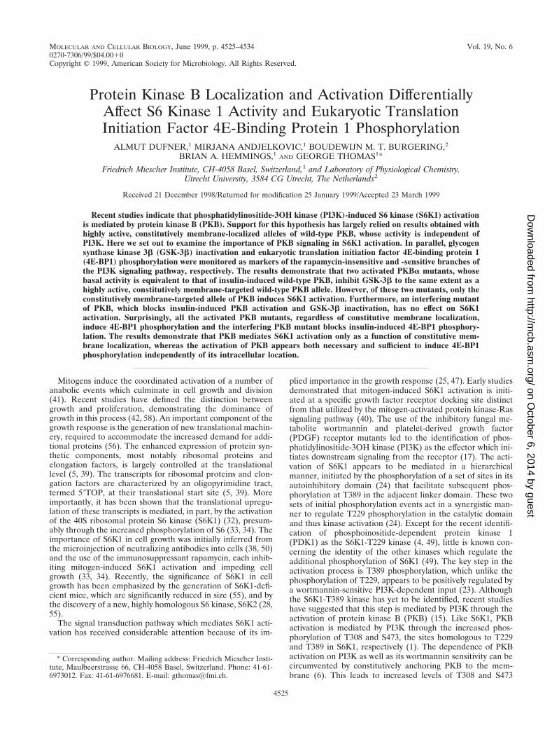
FIG. 1. Activation of PKB mutants. (A) Schematic presentation of the PKBa mutants employed. Membrane-targeted forms of PKBa contain either the lckmyristylation and palmitylation signal (m/p) at the N terminus or the CaaX box from Ki-Ras at the C terminus. PH, pleckstrin homology domain. All the PKBaconstructs are HA epitope tagged at the N terminus, except for PKBa-CaaX. (B) Ectopically expressed PKB variants from extracts of human 293 cells were assayedas described in Materials and Methods. The extracts are derived from the cotransfection experiment with S6K1 described in the legend to Fig. 2A. The cells were serumstarved for 24 h and either extracted immediately, stimulated for 30 min with 1 mM insulin, or pretreated with 250 nM wortmannin for 1 h prior to stimulation withinsulin, as indicated. Error bars indicate standard deviations.
4526 DUFNER ET AL. MOL. CELL. BIOL.
on October 6, 2014 by guest
http://mcb.asm
.org/D
ownloaded from
complex (44). Unlike GSK-3, the effects of PKB on increased4E-BP1 phosphorylation are indirect and may be mediated bya second kinase, possibly mTOR, the target of rapamycin (14,53), or a kinase tightly bound to mTOR (43). Indeed, 4E-BP1is thought to reside on the same rapamycin-sensitive PI3K-dependent signaling pathway as S6K1 (64).
Although highly activated alleles of PKB lead to S6K1 acti-vation, the same is also true for activated oncogenic alleles ofRas (10). However, the activation of wild-type Ras by mitogensis neither sufficient nor necessary to bring about S6K1 activa-tion (40). This raised the possibility that highly activated allelesof PKB or constitutive membrane localization may not reflectwild-type PKB signaling. To test the role of PKB signaling inS6K1 activation we have compared the abilities of specificvariants of PKB to activate and confer wortmannin resistanceon S6K1, as well as the ability of interfering mutants to blockthis response. In parallel, we have used GSK-3b inactivationand 4E-BP1 phosphorylation to independently monitor theeffects of the different PKB alleles on rapamycin-sensitive and-insensitive PKB downstream signaling. The results demon-strate that constitutive membrane localization of active PKB is
essential for its ability to signal S6K1 and that the pathwayleading to 4E-BP1 phosphorylation, but surprisingly not S6K1activation, is dominantly regulated by PKB.
MATERIALS AND METHODS
Construction of expression vectors. The S6K1 construct was tagged by theinsertion of the myc 9E10 epitope immediately following the S6K1 initiator ATGcodon as described previously (40). Myc-S6K1-GST (63) and the pCMV con-structs encoding human hemagglutinin (HA)-PKBa, T308D-S473D PKBa, myri-stylated and palmitylated PKBa, and kinase-dead PKB have been described (1,6, 7). GSK-3b cDNA was amplified from a template construct and subcloned intoa prk5 expression vector. The PCR primers served to add a myc tag at the Cterminus of GSK-3 and provide the suitable XbaI/NotI restriction sites for sub-cloning. The construct was verified by DNA sequencing. Myc-PKB was generatedby PCR with HA-PKBa (7) as a template. The resulting product was subclonedas a BglII-EcoRI fragment into the pECE vector and then transferred as aBglII-XbaI fragment into a pCMV5 vector. The construction of untagged PKB-CaaX (61) and HA–4E-BP1 (63) have been described previously. PKB-CaaX wassubcloned into a pCMV5 expression vector.
Cell culture and transfection. Human embryonic kidney 293 cells were main-tained in Dulbecco’s modified Eagle medium containing 10% fetal calf serum asdescribed previously (40) and seeded at 106 cells per 10-cm-diameter plate 24 hprior to transfection. Transient transfection was performed overnight by using amodified calcium phosphate procedure with 10 mg of plasmid DNA (45). The
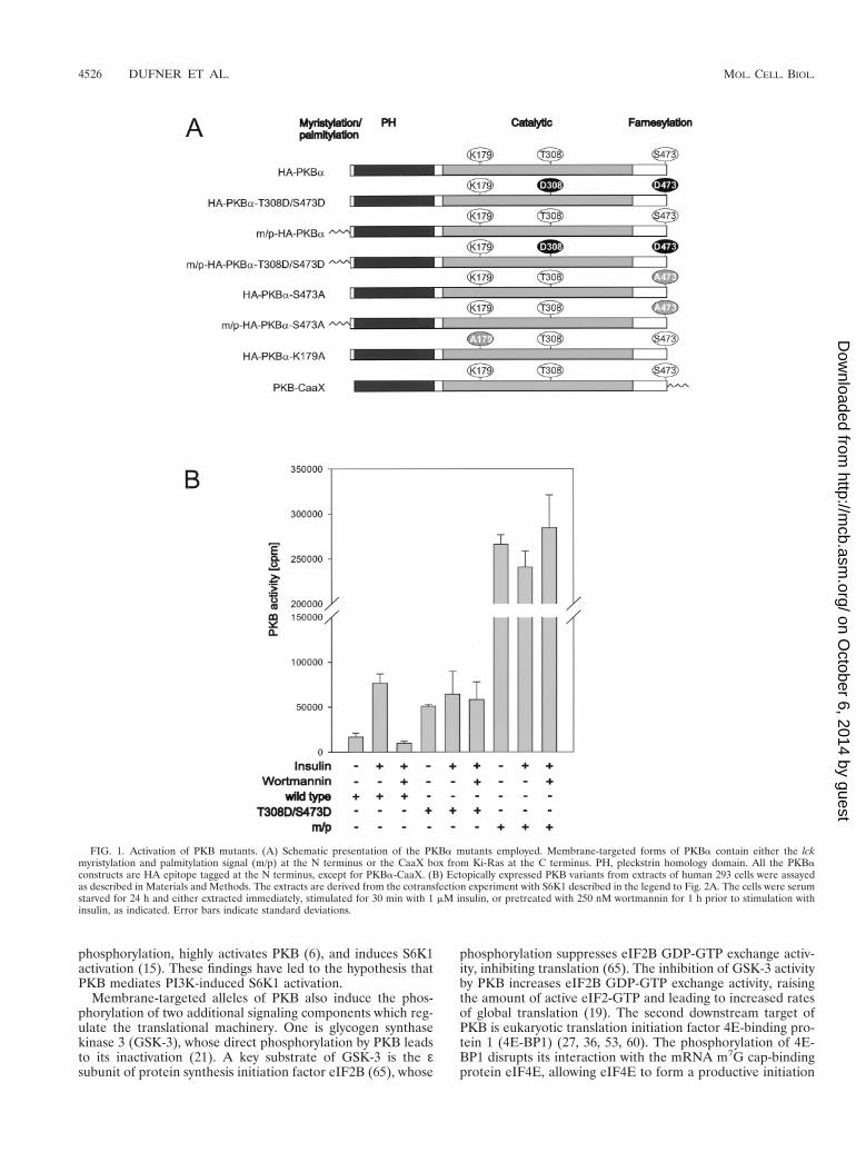
FIG. 2. Effects of PKB variants on S6K1 activation. (A) The activity of ectopically expressed S6K1 from extracts of human 293 cells cotransfected with the emptyvector or indicated PKB variants, as labeled in Fig. 1, was assayed. S6K1 activity is presented as 32P incorporation into 40S ribosomal protein S6 (lower panel). Cellswere deprived of serum for 24 h and either extracted immediately, stimulated for 30 min with 1 mM insulin, or pretreated with 250 nM wortmannin for 1 h prior tostimulation with insulin, as indicated. Protein extracts were analyzed by Western blotting for PKB expression with the 12CA5 antibody (upper panel) and for S6K1 withthe 9E10 antibody (middle panel) (see Materials and Methods). m/p, myristylation and palmitylation signal. (B) Phosphopeptide analysis of myc-tagged S6K1-GSTcoexpressed with the empty vector or the indicated PKB variant, as labeled in Fig. 1, in 293 cells. Prior to lysis cells were incubated for 3 h with 0.2 mCi of 32Pi perml. In the case of the empty vector control 1 mM insulin was added 30 min before lysis. The Myc-S6K1-GST phosphopeptides were analyzed by two-dimensionalthin-layer electrophoresis and chromatography as described in Materials and Methods. m/p, m/p-HA-PKBa.
VOL. 19, 1999 PKB SIGNALING TO S6K1 AND 4E-BP1 4527
on October 6, 2014 by guest
http://mcb.asm
.org/D
ownloaded from
next day, cells were washed twice with Dulbecco’s modified Eagle medium toremove the serum and then made quiescent by incubation in the same mediumfor 24 h. Prior to lysis the cells were stimulated with specific agonists or inhibitorsas described in the text. Insulin was employed at a concentration of 1 mM (stock,10 mg/ml in 1% acetic acid). In the case of the wortmannin pretreatment, eitherthe vehicle dimethyl sulfoxide or 250 nM wortmannin was added.
Immunoprecipitation and in vitro kinase assays. Human 293 cell extracts wereprepared by lysing cells in a buffer containing 50 mM Tris-HCl (pH 7.5), 1%Nonidet P-40, 120 mM NaCl, 1 mM EDTA, 6 mM EGTA, 1 mM benzamidine,15 mM sodium diphosphate, 30 mM 4-nitrophenyl phosphate (disodium salt),and 0.2 mM phenylmethylsulfonyl fluoride. Lysates were centrifuged for 15 minat 12,000 3 g at 4°C. PKB was immunoprecipitated with either monoclonal12CA5 or 9E10 antibody, and S6K1 was immunoprecipitated with a monoclonal9E10 antibody. Immunocomplexes were collected with protein G-Sepharosebeads. PKBa activity was assayed with the peptide GRPRTSSFAEG as a sub-strate, as described previously (21). The kinase assay for S6K1 has been reported(30). Cells transfected with Myc–GSK-3b were lysed in buffer A, which contains50 mM Tris-HCl (pH 7.5), 1 mM EDTA, 1 mM EGTA, 1% (by volume) TritonX-100, 1 mM sodium orthovanadate, 10 mM sodium glycerophosphate, 50 mMNaF, 5 mM sodium pyrophosphate, 1 mM Microcystin-LR, 0.27 M sucrose, 1 mMbenzamidine, 0.2 mM phenylmethylsulfonyl fluoride, 10 mg of leupeptin per ml,and 0.1% (vol/vol) 2-mercaptoethanol. The lysate was centrifuged as describedabove, and an aliquot of the supernatant was incubated for 1 h on a shakingplatform with 5 ml of protein G-Sepharose coupled to 2 mg of 9E10 monoclonalantibody. The protein G–Sepharose–antibody–Myc–GSK-3b complex waswashed twice with buffer A containing 0.5 M NaCl and twice with buffer B (50mM Tris-HCl [pH 7.5], 0.1 mM EGTA, 0.1% 2-mercaptoethanol), and theimmunoprecipitate was assayed for GSK-3b activity after incubation with eitherprotein phosphatase 2A (PP2A) or microcystin in an assay buffer containing 50mM Tris-HCl, 0.1 mM EGTA, 10 mM MgCl, 2.5 mM PKI, 100 mM ATP, and 30mM phospho-glycogen synthase peptide 2 with the sequence YRRAAVPPSPSLSRHSSPHQpSEDEEE. For the PP2A reaction the beads were incubatedfor 30 min at 30°C in buffer B containing 1 mg of bovine serum albumin per mlwith the catalytic subunit of PP2A (final concentration, 25 mU/ml), and thereaction was stopped by adding 5 mM microcystin to the mixture.
Phosphorylation of 4E-BP1. Human 293 cells transfected with HA–4E-BP1were lysed in a buffer containing 50 mM Tris-HCl (pH 7.5), 1% Nonidet P-40,120 mM NaCl, 25 mM NaF, 40 mM b-glycerophosphate, 0.1 mM sodium van-adate, 1 mM benzamidine, and 0.2 mM phenylmethylsulfonyl fluoride. Westernblot analysis of 4E-BP1 was carried out as previously described (64), except thatthe 12CA5 antibody was used for the detection and the N,N9-methylenebis-acrylamide content of the gel was lowered to 0.25%.
Western blot analysis. The protein concentration in samples was determinedwith the Bio-Rad D/C protein assay. Protein samples (40 mg) were subjected tosodium dodecyl sulfate-polyacrylamide gel electrophoresis and then electro-phoretically transferred to Immobilon P membranes (Millipore). Construct ex-pression was quantified by Western blotting with monoclonal antibody 9E10 or12CA5, rabbit anti-mouse immunoglobulin G (DAKO), and finally with fluores-cein isothiocyanate (FITC)-labelled swine anti-rabbit immunoglobulin G(DAKO) or goat anti-mouse immunoglobulin G coupled to horseradish perox-idase (DAKO) as a secondary antibody. Endogenous PKB was quantified with apolyclonal anti-PKB antibody raised against the C terminus of PKB (6) andFITC-labelled swine anti-rabbit immunoglobulin G (DAKO) or mouse anti-rabbit immunoglobulin G coupled to horseradish peroxidase (DAKO) as a sec-ondary antibody. Enhanced chemiluminescence or storage phosphorimagery andfluorimetry (Molecular Dynamics) were used to visualize expression levels andS6K1 activity as phosphate incorporation into 40S ribosomal protein S6.
Two-dimensional phosphopeptide mapping. Human 293 cells were transientlytransfected with the indicated construct and labelled with 0.2 mCi of 32Pi per ml,3 h prior to extraction. Myc-S6K1-GST was precipitated from labelled extractswith glutathione-Sepharose (Sigma) and purified by electroelution (63). Trichlo-roacetic acid precipitation, performic acid oxidation, trypsin-chymotrypsin digestion,and two-dimensional phosphopeptide mapping were carried out as described pre-viously (45). Phosphopeptides were visualized with a PhosphorImager.
Immunofluorescence. Human 293 cells were transfected on coverslips with theindicated PKB construct, fixed, permeabilized, and incubated with a polyclonalanti-PKB antibody before being stained with an FITC-conjugated secondaryantibody, as described previously (6).
RESULTS
Activation of PKB variants. To address the importance ofPKB activation in S6K1 signaling, we compared the activity ofwild-type PKB and its sensitivity to wortmannin with those oftwo activated alleles of PKB, one of which was constitutivelymembrane targeted. The latter allele contains the myristylationand palmitylation signal of the proto-oncogene lck attached toits amino terminus (Fig. 1A), which constitutively localizes thePKB variant to the membrane (see reference 6 and below).The second variant of PKB contains acidic residues at theprimary sites of phosphorylation associated with PKB activa-tion, T308, in the activation loop, and S473, near the carboxyterminus (Fig. 1A) (1). Wild-type PKB and each of the variantswere HA epitope tagged, transiently transfected into human293 cells, and analyzed for basal and insulin-stimulated activity,with Crosstide (21) as a peptide substrate (Fig. 1B). In contrastto the HA epitope-tagged wild-type PKB, HA-PKBa, bothvariants had high basal kinase activity which was not furtheraugmented by insulin stimulation (Fig. 1B). The activity of thedouble acidic variant, HA-PKBa-T308D/S473D, was equiva-lent to that of insulin-stimulated HA-PKBa, whereas the mem-brane-targeted variant, m/p-HA-PKBa, was approximatelyfourfold more active than insulin-stimulated HA-PKBa (Fig.1B). Furthermore, in contrast to HA-PKBa, both HA-PKBa-T308D/S473D and m/p-HA-PKBa were resistant to wortman-nin (Fig. 1B). Thus, the two activated alleles of PKB, thoughdisplaying distinct levels of activity, were unresponsive to mi-togen stimulation and the effects of wortmannin.
Differential regulation of S6K1 by PKB. To determine theeffect of PKB on S6K1 activation, each of the HA epitope-tagged PKB constructs was cotransfected with a Myc epitope-tagged S6K1 reporter, Myc-S6K1. Consistent with the resultsof earlier studies the wild-type construct had little effect onS6K1 activation (4), whereas m/p-HA-PKBa led to full S6K1activation in the absence of insulin treatment (Fig. 2A) (15). Incontrast to wild-type PKB, m/p-HA-PKBa also conferredwortmannin resistance on the Myc-S6K1 reporter construct
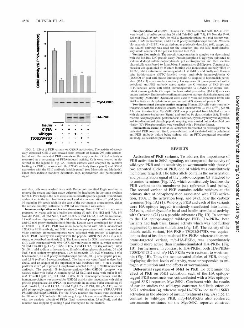
FIG. 3. Effect of PKB variants on GSK-3 inactivation. The activity of ectopi-cally expressed GSK-3 was assayed from extracts of human 293 cells cotrans-fected with the indicated PKB variants or the empty vector. GSK-3 activity ismeasured as a percentage of PP2A-induced activity. Cells were treated as de-scribed in the legend to Fig. 2A. Protein extracts were analyzed by Westernblotting for PKB expression with the 12CA5 antibody (lower panel) and GSK-3expression with the 9E10 antibody (middle panel) (see Materials and Methods).Error bars indicate standard deviations. m/p, myristylation and palmitylationsignal.
4528 DUFNER ET AL. MOL. CELL. BIOL.
on October 6, 2014 by guest
http://mcb.asm
.org/D
ownloaded from
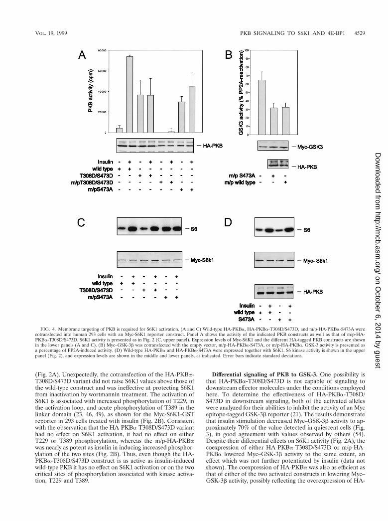
(Fig. 2A). Unexpectedly, the cotransfection of the HA-PKBa-T308D/S473D variant did not raise S6K1 values above those ofthe wild-type construct and was ineffective at protecting S6K1from inactivation by wortmannin treatment. The activation ofS6K1 is associated with increased phosphorylation of T229, inthe activation loop, and acute phosphorylation of T389 in thelinker domain (23, 46, 49), as shown for the Myc-S6K1-GSTreporter in 293 cells treated with insulin (Fig. 2B). Consistentwith the observation that the HA-PKBa-T308D/S473D varianthad no effect on S6K1 activation, it had no effect on eitherT229 or T389 phosphorylation, whereas the m/p-HA-PKBawas nearly as potent as insulin in inducing increased phosphor-ylation of the two sites (Fig. 2B). Thus, even though the HA-PKBa-T308D/S473D construct is as active as insulin-inducedwild-type PKB it has no effect on S6K1 activation or on the twocritical sites of phosphorylation associated with kinase activa-tion, T229 and T389.
Differential signaling of PKB to GSK-3. One possibility isthat HA-PKBa-T308D/S473D is not capable of signaling todownstream effector molecules under the conditions employedhere. To determine the effectiveness of HA-PKBa-T308D/S473D in downstream signaling, both of the activated alleleswere analyzed for their abilities to inhibit the activity of an Mycepitope-tagged GSK-3b reporter (21). The results demonstratethat insulin stimulation decreased Myc–GSK-3b activity to ap-proximately 70% of the value detected in quiescent cells (Fig.3), in good agreement with values observed by others (54).Despite their differential effects on S6K1 activity (Fig. 2A), thecoexpression of either HA-PKBa-T308D/S473D or m/p-HA-PKBa lowered Myc–GSK-3b activity to the same extent, aneffect which was not further potentiated by insulin (data notshown). The coexpression of HA-PKBa was also as efficient asthat of either of the two activated constructs in lowering Myc–GSK-3b activity, possibly reflecting the overexpression of HA-
FIG. 4. Membrane targeting of PKB is required for S6K1 activation. (A and C) Wild-type HA-PKBa, HA-PKBa-T308D/S473D, and m/p-HA-PKBa-S473A werecotransfected into human 293 cells with an Myc-S6K1 reporter construct. Panel A shows the activity of the indicated PKB constructs as well as that of m/p-HA-PKBa-T308D/S473D. S6K1 activity is presented as in Fig. 2 (C, upper panel). Expression levels of Myc-S6K1 and the different HA-tagged PKB constructs are shownin the lower panels (A and C). (B) Myc–GSK-3b was cotransfected with the empty vector, m/p-HA-PKBa-S473A, or m/p-HA-PKBa. GSK-3 activity is presented asa percentage of PP2A-induced activity. (D) Wild-type HA-PKBa and HA-PKBa-S473A were expressed together with S6K1. S6 kinase activity is shown in the upperpanel (Fig. 2), and expression levels are shown in the middle and lower panels, as indicated. Error bars indicate standard deviations.
VOL. 19, 1999 PKB SIGNALING TO S6K1 AND 4E-BP1 4529
on October 6, 2014 by guest
http://mcb.asm
.org/D
ownloaded from
PKBa and the high affinity of the two kinases for one another(61). However, the kinase-inactive form of PKBa, in whichK179 in the ATP binding site had been replaced by an alanine(Fig. 1A), had no effect on Myc–GSK-3b activity (Fig. 3). Thus,the overexpression of wild-type PKB is sufficient to induceGSK-3b inactivation, but not S6K1 activation, and althoughthe double acidic mutant has no effect on S6K1 activity (Fig.2A), it is unimpaired in its ability to signal GSK-3b (Fig. 3).
Membrane targeting of PKB. A potential explanation forthe inability of the double acidic PKB to activate S6K1, versusm/p-HA-PKBa, is that such activation requires the constitutivemembrane targeting of PKB. To test this possibility the doubleacidic mutant was constitutively targeted to the membrane byvirtue of the myristylation and palmitylation signal. However,this form of the kinase was inactive, even in the presence ofinsulin (Fig. 4A). Therefore, to resolve this issue we exploitedan m/p-HA-PKBa variant in which S473 was mutated to ala-nine, m/p-HA-PKBa-S473A (6). This mutant has basal activityequal to that of HA-PKBa-T308D/S473D and is significantlyless active than the m/p-HA-PKBa parent construct (Fig. 4Aand 1B). The addition of insulin slightly raises the kinase ac-tivity of m/p-HA-PKBa-S473A, although the level of activationis lower than that of the wild-type enzyme in the presence ofinsulin (Fig. 4A). Furthermore, this construct was as potent asthe m/p-HA-PKBa parent construct in lowering basal Myc–GSK-3b activity (Fig. 4B). Despite the significantly lower basalactivity of m/p-HA-PKBa-S473A compared with that of m/p-HA-PKBa (Fig. 4A), the constitutive targeting of this con-struct to the membrane led to the full activation of coexpressedMyc-S6K1, an effect not further augmented by insulin (Fig.4C). To ensure that this effect was due to membrane targeting,the same variant lacking the myristylation and palmitylationsignal, HA-PKBa-S473A, was coexpressed with Myc-S6K1.
Like the double acidic mutant, HA-PKBa-S473A did not po-tentiate S6K1 activation beyond that of wild-type HA-PKBa(Fig. 4D). As with the double acidic mutant, we have alsofound that the m/p-HA-PKBa construct harboring an alanineat T308 is inactive and fails to induce S6K1 activation (data notshown). Taken together the data demonstrate that full S6K1activation is a function of the constitutive membrane localiza-tion of active PKB rather than its specific activity.
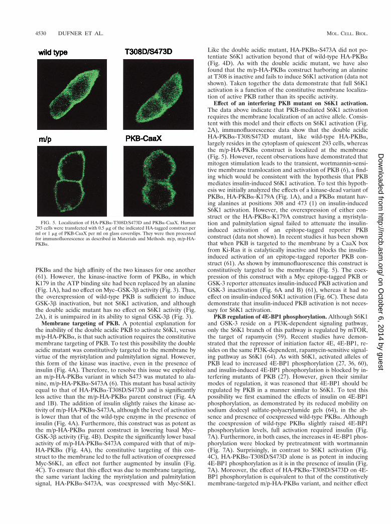
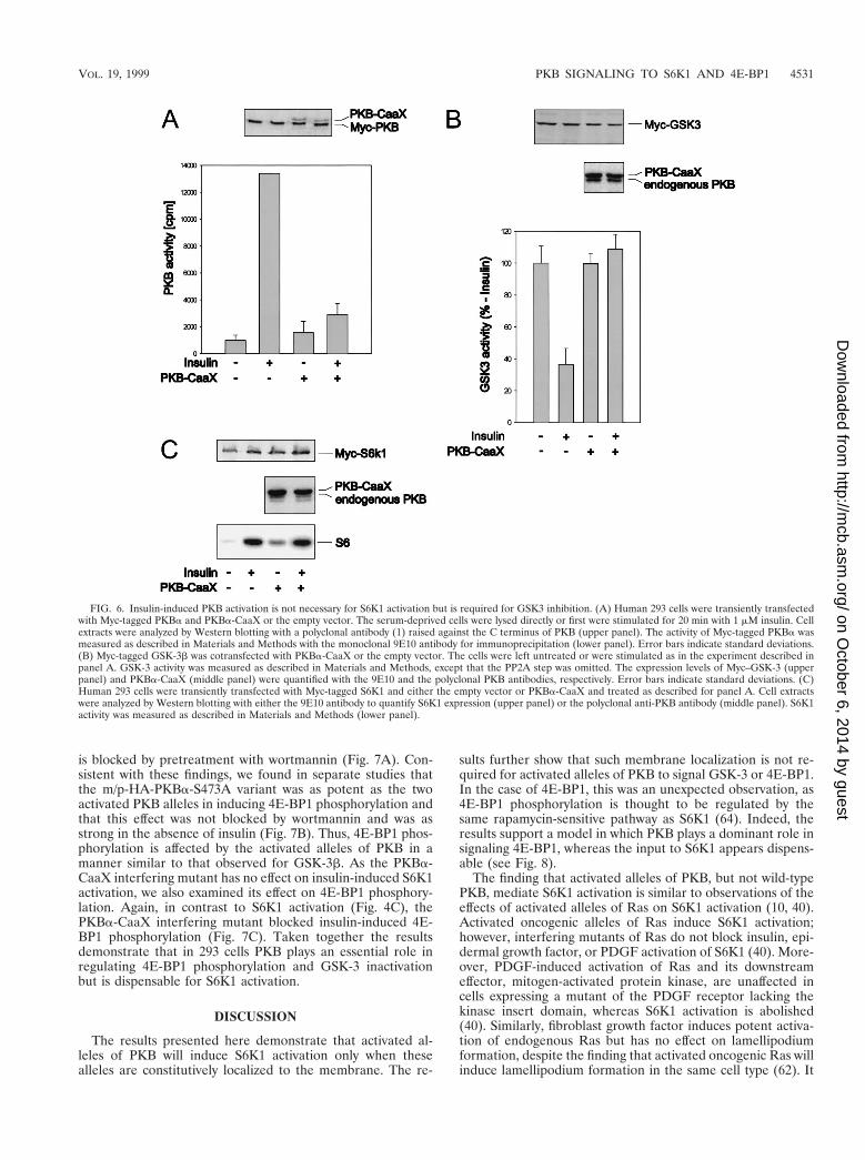
Effect of an interfering PKB mutant on S6K1 activation.The data above indicate that PKB-mediated S6K1 activationrequires the membrane localization of an active allele. Consis-tent with this model and their effects on S6K1 activation (Fig.2A), immunofluorescence data show that the double acidicHA-PKBa-T308/S473D mutant, like wild-type HA-PKBa,largely resides in the cytoplasm of quiescent 293 cells, whereasthe m/p-HA-PKBa construct is localized at the membrane(Fig. 5). However, recent observations have demonstrated thatmitogen stimulation leads to the transient, wortmannin-sensi-tive membrane translocation and activation of PKB (6), a find-ing which would be consistent with the hypothesis that PKBmediates insulin-induced S6K1 activation. To test this hypoth-esis we initially analyzed the effects of a kinase-dead variant ofPKBa, HA-PKBa-K179A (Fig. 1A), and a PKBa mutant hav-ing alanines at positions 308 and 473 (1) on insulin-inducedS6K1 activation. However, the overexpression of either con-struct or the HA-PKBa-K179A construct having a myristyla-tion and palmitylation signal failed to attenuate the insulin-induced activation of an epitope-tagged reporter PKBconstruct (data not shown). In recent studies it has been shownthat when PKB is targeted to the membrane by a CaaX boxfrom Ki-Ras it is catalytically inactive and blocks the insulin-induced activation of an epitope-tagged reporter PKB con-struct (61). As shown by immunofluorescence this construct isconstitutively targeted to the membrane (Fig. 5). The coex-pression of this construct with a Myc epitope-tagged PKB orGSK-3 reporter attenuates insulin-induced PKB activation andGSK-3 inactivation (Fig. 6A and B) (61), whereas it had noeffect on insulin-induced S6K1 activation (Fig. 6C). These datademonstrate that insulin-induced PKB activation is not neces-sary for S6K1 activation.
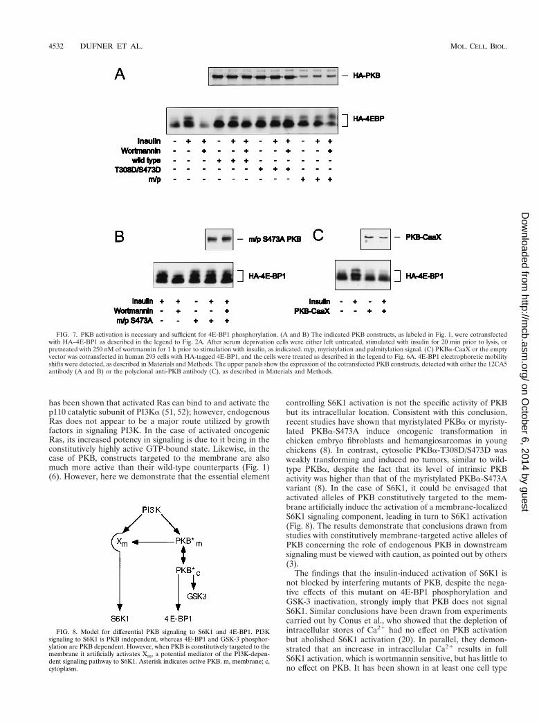
PKB regulation of 4E-BP1 phosphorylation. Although S6K1and GSK-3 reside on a PI3K-dependent signaling pathway,only the S6K1 branch of this pathway is regulated by mTOR,the target of rapamycin (59). Recent studies have demon-strated that the repressor of initiation factor 4E, 4E-BP1, re-sides on the same PI3K-dependent, rapamycin-sensitive signal-ing pathway as S6K1 (64). As with S6K1, activated alleles ofPKB lead to increased 4E-BP1 phosphorylation (27, 36, 60),and insulin-induced 4E-BP1 phosphorylation is blocked by in-terfering mutants of PKB (27). However, given their similarmodes of regulation, it was reasoned that 4E-BP1 should beregulated by PKB in a manner similar to S6K1. To test thispossibility we first examined the effects of insulin on 4E-BP1phosphorylation, as demonstrated by its reduced mobility onsodium dodecyl sulfate-polyacrylamide gels (64), in the ab-sence and presence of coexpressed wild-type PKBa. Althoughthe coexpression of wild-type PKBa slightly raised 4E-BP1phosphorylation levels, full activation required insulin (Fig.7A). Furthermore, in both cases, the increases in 4E-BP1 phos-phorylation were blocked by pretreatment with wortmannin(Fig. 7A). Surprisingly, in contrast to S6K1 activation (Fig.4C), HA-PKBa-T308D/S473D alone is as potent in inducing4E-BP1 phosphorylation as it is in the presence of insulin (Fig.7A). Moreover, the effect of HA-PKBa-T308D/S473D on 4E-BP1 phosphorylation is equivalent to that of the constitutivelymembrane-targeted m/p-HA-PKBa variant, and neither effect
FIG. 5. Localization of HA-PKBa-T308D/S473D and PKBa-CaaX. Human293 cells were transfected with 0.5 mg of the indicated HA-tagged construct perml or 1 mg of PKB-CaaX per ml on glass coverslips. They were then processedfor immunofluorescence as described in Materials and Methods. m/p, m/p-HA-PKBa.
4530 DUFNER ET AL. MOL. CELL. BIOL.
on October 6, 2014 by guest
http://mcb.asm
.org/D
ownloaded from
is blocked by pretreatment with wortmannin (Fig. 7A). Con-sistent with these findings, we found in separate studies thatthe m/p-HA-PKBa-S473A variant was as potent as the twoactivated PKB alleles in inducing 4E-BP1 phosphorylation andthat this effect was not blocked by wortmannin and was asstrong in the absence of insulin (Fig. 7B). Thus, 4E-BP1 phos-phorylation is affected by the activated alleles of PKB in amanner similar to that observed for GSK-3b. As the PKBa-CaaX interfering mutant has no effect on insulin-induced S6K1activation, we also examined its effect on 4E-BP1 phosphory-lation. Again, in contrast to S6K1 activation (Fig. 4C), thePKBa-CaaX interfering mutant blocked insulin-induced 4E-BP1 phosphorylation (Fig. 7C). Taken together the resultsdemonstrate that in 293 cells PKB plays an essential role inregulating 4E-BP1 phosphorylation and GSK-3 inactivationbut is dispensable for S6K1 activation.
DISCUSSION
The results presented here demonstrate that activated al-leles of PKB will induce S6K1 activation only when thesealleles are constitutively localized to the membrane. The re-
sults further show that such membrane localization is not re-quired for activated alleles of PKB to signal GSK-3 or 4E-BP1.In the case of 4E-BP1, this was an unexpected observation, as4E-BP1 phosphorylation is thought to be regulated by thesame rapamycin-sensitive pathway as S6K1 (64). Indeed, theresults support a model in which PKB plays a dominant role insignaling 4E-BP1, whereas the input to S6K1 appears dispens-able (see Fig. 8).
The finding that activated alleles of PKB, but not wild-typePKB, mediate S6K1 activation is similar to observations of theeffects of activated alleles of Ras on S6K1 activation (10, 40).Activated oncogenic alleles of Ras induce S6K1 activation;however, interfering mutants of Ras do not block insulin, epi-dermal growth factor, or PDGF activation of S6K1 (40). More-over, PDGF-induced activation of Ras and its downstreameffector, mitogen-activated protein kinase, are unaffected incells expressing a mutant of the PDGF receptor lacking thekinase insert domain, whereas S6K1 activation is abolished(40). Similarly, fibroblast growth factor induces potent activa-tion of endogenous Ras but has no effect on lamellipodiumformation, despite the finding that activated oncogenic Ras willinduce lamellipodium formation in the same cell type (62). It
FIG. 6. Insulin-induced PKB activation is not necessary for S6K1 activation but is required for GSK3 inhibition. (A) Human 293 cells were transiently transfectedwith Myc-tagged PKBa and PKBa-CaaX or the empty vector. The serum-deprived cells were lysed directly or first were stimulated for 20 min with 1 mM insulin. Cellextracts were analyzed by Western blotting with a polyclonal antibody (1) raised against the C terminus of PKB (upper panel). The activity of Myc-tagged PKBa wasmeasured as described in Materials and Methods with the monoclonal 9E10 antibody for immunoprecipitation (lower panel). Error bars indicate standard deviations.(B) Myc-tagged GSK-3b was cotransfected with PKBa-CaaX or the empty vector. The cells were left untreated or were stimulated as in the experiment described inpanel A. GSK-3 activity was measured as described in Materials and Methods, except that the PP2A step was omitted. The expression levels of Myc–GSK-3 (upperpanel) and PKBa-CaaX (middle panel) were quantified with the 9E10 and the polyclonal PKB antibodies, respectively. Error bars indicate standard deviations. (C)Human 293 cells were transiently transfected with Myc-tagged S6K1 and either the empty vector or PKBa-CaaX and treated as described for panel A. Cell extractswere analyzed by Western blotting with either the 9E10 antibody to quantify S6K1 expression (upper panel) or the polyclonal anti-PKB antibody (middle panel). S6K1activity was measured as described in Materials and Methods (lower panel).
VOL. 19, 1999 PKB SIGNALING TO S6K1 AND 4E-BP1 4531
on October 6, 2014 by guest
http://mcb.asm
.org/D
ownloaded from
has been shown that activated Ras can bind to and activate thep110 catalytic subunit of PI3Ka (51, 52); however, endogenousRas does not appear to be a major route utilized by growthfactors in signaling PI3K. In the case of activated oncogenicRas, its increased potency in signaling is due to it being in theconstitutively highly active GTP-bound state. Likewise, in thecase of PKB, constructs targeted to the membrane are alsomuch more active than their wild-type counterparts (Fig. 1)(6). However, here we demonstrate that the essential element
controlling S6K1 activation is not the specific activity of PKBbut its intracellular location. Consistent with this conclusion,recent studies have shown that myristylated PKBa or myristy-lated PKBa-S473A induce oncogenic transformation inchicken embryo fibroblasts and hemangiosarcomas in youngchickens (8). In contrast, cytosolic PKBa-T308D/S473D wasweakly transforming and induced no tumors, similar to wild-type PKBa, despite the fact that its level of intrinsic PKBactivity was higher than that of the myristylated PKBa-S473Avariant (8). In the case of S6K1, it could be envisaged thatactivated alleles of PKB constitutively targeted to the mem-brane artificially induce the activation of a membrane-localizedS6K1 signaling component, leading in turn to S6K1 activation(Fig. 8). The results demonstrate that conclusions drawn fromstudies with constitutively membrane-targeted active alleles ofPKB concerning the role of endogenous PKB in downstreamsignaling must be viewed with caution, as pointed out by others(3).
The findings that the insulin-induced activation of S6K1 isnot blocked by interfering mutants of PKB, despite the nega-tive effects of this mutant on 4E-BP1 phosphorylation andGSK-3 inactivation, strongly imply that PKB does not signalS6K1. Similar conclusions have been drawn from experimentscarried out by Conus et al., who showed that the depletion ofintracellular stores of Ca21 had no effect on PKB activationbut abolished S6K1 activation (20). In parallel, they demon-strated that an increase in intracellular Ca21 results in fullS6K1 activation, which is wortmannin sensitive, but has little tono effect on PKB. It has been shown in at least one cell type
FIG. 7. PKB activation is necessary and sufficient for 4E-BP1 phosphorylation. (A and B) The indicated PKB constructs, as labeled in Fig. 1, were cotransfectedwith HA–4E-BP1 as described in the legend to Fig. 2A. After serum deprivation cells were either left untreated, stimulated with insulin for 20 min prior to lysis, orpretreated with 250 nM of wortmannin for 1 h prior to stimulation with insulin, as indicated. m/p, myristylation and palmitylation signal. (C) PKBa-CaaX or the emptyvector was cotransfected in human 293 cells with HA-tagged 4E-BP1, and the cells were treated as described in the legend to Fig. 6A. 4E-BP1 electrophoretic mobilityshifts were detected, as described in Materials and Methods. The upper panels show the expression of the cotransfected PKB constructs, detected with either the 12CA5antibody (A and B) or the polyclonal anti-PKB antibody (C), as described in Materials and Methods.
FIG. 8. Model for differential PKB signaling to S6K1 and 4E-BP1. PI3Ksignaling to S6K1 is PKB independent, whereas 4E-BP1 and GSK-3 phosphor-ylation are PKB dependent. However, when PKB is constitutively targeted to themembrane it artificially activates Xm, a potential mediator of the PI3K-depen-dent signaling pathway to S6K1. Asterisk indicates active PKB. m, membrane; c,cytoplasm.
4532 DUFNER ET AL. MOL. CELL. BIOL.
on October 6, 2014 by guest
http://mcb.asm
.org/D
ownloaded from
that a dominant interfering mutant of PKB inhibits S6K1 ac-tivation at very similar doses to those that block PKB activation(35). In this case, the dominant interfering mutant employedcontained alanines at T308 and S473 and was delivered to thecell by utilizing an adenovirus infection strategy. In parallel,these authors observed little to no effect of the same mutant oninsulin-induced S6K1 activation in a second cell type (35). Asphosphoinositide-dependent protein kinase 1 is the activationloop kinase for both S6K1 and PKB (2, 4, 49, 57), it may be thatthe effect on S6K1 activation is accomplished not through theinhibition of PKB but through the inhibition of a parallelpathway. Indeed, recent results from this laboratory haveclearly demonstrated that interfering mutants of S6K1 canaffect parallel pathways (see reference 63 and below). Al-though the results presented here do not exclude a role forPKB in S6K1 activation, they strongly support a modelwhereby PKB resides on a pathway parallel to that of S6K1(Fig. 8).
Rapamycin inhibits mitogen-induced 4E-BP1 and S6K1phosphorylation (9, 18, 37, 48, 64), whereas rapamycin-resis-tant mutants of mTOR protect both S6K1 (12) and 4E-BP1(14, 27, 31) phosphorylation from the effects of rapamycin.Although mTOR has significant homology with lipid kinases ofthe PI3K family (11), in vitro it autophosphorylates and canphosphorylate the rapamycin-sensitive sites in S6K1 (16) and4E-BP1 (13). However, not all the data are consistent with therole of mTOR as the 4E-BP1 and S6K1 kinase. First, the sitesof phosphorylation in 4E-BP1 contain a proline in the 11position (14, 26), whereas in S6K1 the critical residue, T389, isflanked by large aromatic residues (45). More importantly, amutant of S6K1 lacking both the amino and carboxy termini,although still sensitive to wortmannin inhibition, is resistant torapamycin-induced T389 dephosphorylation (23). These latterstudies led to the hypothesis that rapamycin may block S6K1activation by activating a phosphatase which is negatively reg-ulated by mTOR (22, 23). In the case of 4E-BP1, five phos-phorylation sites were initially identified as the targets ofmTOR in vitro and in vivo (26). More recent studies suggestthat only two of these sites may serve as in vitro targets andthat they may be highly phosphorylated in quiescent cells,acting as priming sites for an unidentified mitogen-activatedkinase (see references 16 and 22). mTOR contains 16 36- to47-amino-acid repeats, termed HEAT domains, which arecharacterized by a series of spaced hydrophobic amino acids(22), which have been shown in the A subunit of PP2A to serveas a protein docking site (29). Consistent with the existence ofHEAT domains, recent studies have shown that other kinasesmay be tightly associated with mTOR (43). Obviously it will beimportant to identify mTOR-associated kinases and to deter-mine whether they serve as either 4E-BP1 or S6K1 kinases.
The differential effects of PKB signaling on 4E-BP1 andS6K1 were unexpected. As stated above, 4E-BP1 and S6K1reside on the same PI3K-dependent signaling pathway andboth are sensitive to rapamycin. Furthermore, the overexpres-sion of S6K1 blocks 4E-BP1 phosphorylation at the same sitesas rapamycin, suggesting that the 4E-BP1 and S6K1 signalingpathway bifurcates just upstream of S6K1, with the most ob-vious point of bifurcation being mTOR (63). One possibleexplanation that reconciles these seemingly disparate observa-tions is that mTOR acts as both a kinase and a scaffold forother interacting proteins, as we have recently suggested (22).In this case, PKB signaling to 4E-BP1 would be accomplishedthrough mTOR or an associated kinase, as indicated by recentfindings (43, 53), but this event would not act on the S6K1signaling pathway. In this model, S6K1 may still require mTORfor activation but in a PKB-independent manner. The overex-
pression of S6K1 would sequester mTOR or a molecule whichinteracts with mTOR and block 4E-BP1 phosphorylation. Thevalidation of this model will require more knowledge concern-ing mTOR structure and function.
ACKNOWLEDGMENTS
We thank P. B. Dennis, D. Evans, R. Meier, and N. Pullen for theircritical reading of the manuscript. In addition we are grateful to D.Alessi, M. Shaw, and P. Cohen for their help in establishing the GSK-3assays.
Part of these studies was supported by a short-term EMBO fellow-ship to A.D. This work was partially supported by grants from theHuman Frontier Science Program and European Economic Commu-nities to G.T.
REFERENCES
1. Alessi, D. R., M. Andjelkovic, B. Caudwell, P. Cron, N. Morrice, P. Cohen,and B. A. Hemmings. 1996. Mechanism of activation of protein kinase B byinsulin and IGF-1. EMBO J. 23:6541–6551.
2. Alessi, D. R., M. Deak, A. Casamayor, F. B. Caudwell, N. Morrice, D. G.Norman, P. Gaffney, C. B. Reese, C. N. MacDougall, D. Harbison, A. Ash-worth, and M. Bownes. 1997. 3-Phosphoinositide-dependent protein ki-nase-1 (PDK1): structural and functional homology with the DrosophilaDSTPK61 kinase. Curr. Biol. 7:776–789.
3. Alessi, D. R., and C. P. Downes. 1998. The role of PI 3-kinase in insulinaction. Biochim. Biophys. Acta 1436:151–164.
4. Alessi, D. R., M. T. Kozlowski, Q. P. Weng, N. Morrice, and J. Avruch. 1998.3-Phosphoinositide-dependent protein kinase 1 (PDK1) phosphorylates andactivates the p70S6 kinase in vivo and in vitro. Curr. Biol. 8:69–81.
5. Amaldi, F., and P. Pierandrei-Amaldi. 1997. TOP genes: a translationallycontrolled class of genes including those coding for ribosomal proteins, vol.18. Springer-Verlag, Berlin, Germany.
6. Andjelkovic, M., D. R. Alessi, R. Meier, A. Fernandez, N. J. Lamb, M. Frech,P. Cron, P. Cohen, J. M. Lucocq, and B. A. Hemmings. 1997. Role oftranslocation in the activation and function of protein kinase B. J. Biol.Chem. 272:31515–31524.
7. Andjelkovic, M., T. Jakubowicz, P. Cron, X.-F. Ming, J.-W. Han, and B. A.Hemmings. 1996. Activation and phosphorylation of a pleckstrin homologydomain containing protein kinase (RAC-PK/PKB) promoted by serum andprotein phosphatase inhibitors. Proc. Natl. Acad. Sci. USA 93:5699–5704.
8. Aoki, M., O. Batista, A. Bellacosa, P. Tsichlis, and P. K. Vogt. 1998. The aktkinase: molecular determinants of oncogenicity. Proc. Natl. Acad. Sci. USA95:14950–14955.
9. Beretta, L., A.-C. Gingras, Y. V. Svitkin, M. N. Hall, and N. Sonenberg. 1996.Rapamycin blocks the phosphorylation of 4E-BP1 and inhibits cap-depen-dent initiation of translation. EMBO J. 15:658–664.
10. Blenis, J., and R. L. Erikson. 1984. Phosphorylation of the ribosomal proteinS6 is elevated in cells transformed by a variety of tumor viruses. J. Virol.50:966–969.
11. Brown, E. J., M. A. Albers, T. B. Shin, K. Ichikawa, C. T. Keith, W. S. Lane,and S. L. Schreiber. 1994. A mammalian protein targeted by G1-arrestingrapamycin-receptor complex. Nature 369:756–758.
12. Brown, E. J., P. A. Beal, C. T. Keith, J. Chen, T. B. Shin, and S. L. Schreiber.1995. Control of p70 S6 kinase by kinase activity of FRAP in vivo. Nature377:441–446.
13. Brunn, G. J., P. Fadden, T. A. J. Haystead, and J. C. Lawrence, Jr. 1997. Themammalian target of rapamycin phosphorylates sites having a (Ser/Thr)-Promotif and is activated by antibodies to a region near its COOH terminus.J. Biol. Chem. 272:32547–32550.
14. Brunn, G. J., C. C. Hudson, A. Sekulic, J. M. Williams, H. Hosoi, P. J.Houghton, J. C. Lawrence, Jr., and R. T. Abraham. 1997. Phosphorylation ofthe translational repressor PHAS-I by the mammalian target of rapamycin.Science 277:99–101.
15. Burgering, B. M. T., and P. J. Coffer. 1995. Protein kinase B (c-Akt) inphosphatidylinositol-3-OH kinase signal transduction. Nature 376:599–602.
16. Burnett, P. E., R. K. Barrow, N. A. Cohen, S. H. Snyder, and D. M. Sabatini.1998. RAFT1 phosphorylation of the translational regulators p70 S6 kinaseand 4E-BP1. Proc. Natl. Acad. Sci. USA 95:1432–1437.
17. Chung, J., T. C. Grammer, K. P. Lemon, A. Kazlauskas, and J. Blenis. 1994.PDGF- and insulin-dependent pp70s6k activation mediated by phosphatidyl-inositol-3-OH kinase. Nature 370:71–75.
18. Chung, J., C. J. Kuo, G. R. Crabtree, and J. Blenis. 1992. Rapamycin-FKBPspecifically blocks growth-dependent activation of and signaling by the 70 kdS6 protein kinases. Cell 69:1227–1236.
19. Clemens, M. G. 1996. Protein kinases that phosphorylate eIF2 and eIF2B,and their role in eukaryotic cell translational control. Cold Spring HarborLaboratory Press, Cold Spring Harbor, N.Y.
20. Conus, N. M., B. A. Hemmings, and R. B. Pearson. 1998. Differential reg-ulation by calcium reveals distinct signaling requirements for the activation
VOL. 19, 1999 PKB SIGNALING TO S6K1 AND 4E-BP1 4533
on October 6, 2014 by guest
http://mcb.asm
.org/D
ownloaded from
of Akt and p70S6k. J. Biol. Chem. 273:4776–4782.21. Cross, D. A. E., D. R. Alessi, P. Cohen, M. Andjelkovic, and B. A. Hemmings.
1995. Inhibition of glycogen synthase kinase-3 by insulin mediated by proteinkinase B. Nature 378:785–789.
22. Dennis, P. B., S. Fumagalli, and G. Thomas. 1999. Target of rapamycin(TOR); balancing the opposing forces of protein synthesis and degradation.Curr. Opin. Genet. Dev. 9:49–54.
23. Dennis, P. B., N. Pullen, S. C. Kozma, and G. Thomas. 1996. The principalrapamycin-sensitive p70s6k phosphorylation sites, T-229 and T-389, are dif-ferentially regulated by rapamycin-insensitive kinase kinases. Mol. Cell. Biol.16:6242–6251.
24. Dennis, P. B., N. Pullen, R. B. Pearson, S. C. Kozma, and G. Thomas. 1998.Phosphorylation sites in the autoinhibitory domain participate in p70(s6k)activation loop phosphorylation. J. Biol. Chem. 273:14845–14852.
25. Downward, J. 1998. Lipid-regulated kinases: some common themes at last.Science 279:673–674.
26. Fadden, P., T. A. J. Haystead, and J. C. Lawrence, Jr. 1997. Identification ofphosphorylation sites in the translational regulator, PHAS-I, that are con-trolled by insulin and rapamycin in rat adipocytes. J. Biol. Chem. 272:10240–10247.
27. Gingras, A.-C., S. G. Kennedy, M. A. O’Leary, N. Sonenberg, and N. Hay.1998. 4E-BP1, a repressor of mRNA translation, is phosphorylated andactivated by the Akt (PKB) signaling pathway. Genes Dev. 12:502–513.
28. Gout, I., T. Minami, K. Hara, Y. Tsujishita, V. Filonenko, M. D. Waterfield,and K. Yonezawa. 1998. Molecular cloning and characterization of a novelp70 S6 kinase, p70 S6 kinase b containing a proline-rich region. J. Biol.Chem. 273:30061–30064.
29. Groves, M. R., N. Hanlon, P. Turowski, B. A. Hemmings, and D. Barford.1999. The structure of the PR65/A-subunit of protein phosphatase 2A re-veals the conformation of a scaffolding molecule composed of 15 tandemlyrepeated HEAT motifs. Cell 96:99–110.
30. Han, J. W., R. B. Pearson, P. B. Dennis, and G. Thomas. 1995. Rapamycin,wortmannin, and the methylxanthine SQ20006 inactivate p70s6k by inducingdephosphorylation of the same subset of sites. J. Biol. Chem. 36:21396–21403.
31. Hara, K., K. Yonezawa, Q.-P. Weng, M. T. Kozlowski, C. Belham, and J.Avruch. 1998. Amino acid sufficiency and mTOR regulate p70 S6 kinase andeIF-4E BP1 through a common effector mechanism. J. Biol. Chem. 273:14484–14494.
32. Jefferies, H. B. J., S. Fumagalli, P. B. Dennis, C. Reinhard, R. B. Pearson,and G. Thomas. 1997. Rapamycin suppresses 59TOP mRNA translationthrough inhibition of p70s6k. EMBO J. 12:3693–3704.
33. Jefferies, H. B. J., C. Reinhard, S. C. Kozma, and G. Thomas. 1994. Rapa-mycin selectively represses translation of the “polypyrimidine tract” mRNAfamily. Proc. Natl. Acad. Sci. USA 91:4441–4445.
34. Jefferies, H. B. J., and G. Thomas. 1996. Ribosomal protein S6 phosphory-lation and signal transduction, p. 389–409. In J. W. B. Hershey, M. B.Mathews, and N. Sonenberg (ed.), Translational control. Cold Spring Har-bor Laboratory Press, Cold Spring Harbor, N.Y.
35. Kitamura, T., W. Ogawa, H. Sakaue, Y. Hino, S. Kuroda, M. Takata, M.Matsumoto, T. Maeda, H. Konishi, U. Kikkawa, and M. Kasuga. 1998.Requirement for activation of the serine-threonine kinase Akt (protein ki-nase B) in insulin stimulation of protein synthesis but not of glucose trans-port. Mol. Cell. Biol. 18:3708–3717.
36. Kohn, A. D., A. Barthel, K. S. Kovacina, A. Boge, B. Wallach, S. A. Summers,M. J. Birnbaum, P. H. Scott, J. C. Lawrence, and R. A. Roth. 1998. Con-struction and characterization of a conditionally active version of the serine/threonine kinase Akt. J. Biol. Chem. 273:11937–11943.
37. Kuo, C. J., J. Chung, D. F. Fiorentino, W. M. Flanagan, J. Blenis, and G. R.Crabtree. 1992. Rapamycin selectively inhibits interleukin-2 activation of p70S6 kinase. Nature 358:70–73.
38. Lane, H. A., A. Fernandez, N. J. C. Lamb, and G. Thomas. 1993. p70s6k
function is essential for G1 progression. Nature 363:170–172.39. Meyuhas, O., D. Avni, and S. Shama. 1996. Translational control of ribo-
somal protein mRNAs in eukaryotes. In J. W. B. Hershey, M. B. Mathews,and N. Sonenberg (ed.), Translational control. Cold Spring Harbor Labora-tory Press, Cold Spring Harbor, N.Y.
40. Ming, X. F., B. M. T. Burgering, S. Wennstrom, L. Claesson-Welsh, C. H.Heldin, J. L. Bos, S. C. Kozma, and G. Thomas. 1994. Activation of p70/p85S6 kinase by a pathway independent of p21ras. Nature 371:426–429.
41. Nasmyth, K. 1996. Another role rolls in. Nature 382:28–29.42. Neufeld, T. P., A. F. de la Cruz, L. A. Johnston, and B. A. Edgar. 1998.
Coordination of growth and cell division in the Drosophila wing. Cell 93:1183–1193.
43. Nishiuma, T., K. Hara, Y. Tsujishita, K. Kaneko, K. Shii, and K. Yonezawa.1998. Characterization of the phosphoproteins and protein kinase activity inmTOR immunoprecipitates. Biochem. Biophys. Res. Commun. 252:440–444.
44. Pause, A., G. J. Belsham, A.-C. Gingras, O. Donze, T. A. Lin, J. C. Lawrence,Jr., and N. Sonenberg. 1994. Insulin-dependent stimulation of protein syn-thesis by phosphorylation of a regulator of 59-cap function. Nature 371:762–767.
45. Pearson, R. B., P. B. Dennis, J. W. Han, N. A. Williamson, S. C. Kozma,R. E. H. Wettenhall, and G. Thomas. 1995. The principal target of rapa-mycin-induced p70s6k inactivation is a novel phosphorylation site within aconserved hydrophobic domain. EMBO J. 21:5279–5287.
46. Pearson, R. B., and G. Thomas. 1995. Regulation of p70s6k/p85s6k and itsrole in the cell cycle, p. 21–32. In L. Meijer, S. Guidet, and H. Y. L. Tung(ed.), Progress in cell cycle research, vol. 1. Plenum Press, New York, N.Y.
47. Peterson, R. T., and S. L. Schreiber. 1998. Translation control: connectingmitogens and the ribosome. Curr. Biol. 8:R248–R250.
48. Price, D. J., J. R. Grove, V. Calvo, J. Avruch, and B. E. Bierer. 1992.Rapamycin-induced inhibition of the 70-kilodalton S6 protein kinase. Sci-ence 257:973–977.
49. Pullen, N., P. B. Dennis, M. Andjelkovic, A. Dufner, S. Kozma, B. A. Hem-mings, and G. Thomas. 1998. Phosphorylation and activation of p70s6k byPDK1. Science 279:707–710.
50. Reinhard, C., A. Fernandez, N. J. C. Lamb, and G. Thomas. 1994. Nuclearlocalization of p85s6k: functional requirement for entry into S phase. EMBOJ. 13:1557–1565.
51. Rodriguez-Viciana, P., P. H. Warne, R. Dhand, B. Vanhaesebroeck, I. Gout,M. J. Fry, M. D. Waterfield, and J. Downward. 1994. Phosphatidylinositol-3-OH kinase as a direct target of Ras. Nature 370:527–532.
52. Rodriguez-Viciana, P., P. H. Warne, A. Khwaja, B. M. Marte, D. Pappin, P.Das, M. D. Waterfield, A. Ridley, and J. Downward. 1997. Role of phospho-inositide 3-OH kinase in cell transformation and control of the actin cy-toskeleton by Ras. Cell 89:457–467.
53. Scott, P. H., G. J. Brunn, A. D. Kohn, R. A. Roth, and J. C. Lawrence, Jr.1998. Evidence of insulin-stimulated phosphorylation and activation of themammalian target of papamycin mediated by a protein kinase B signalingpathway. Proc. Natl. Acad. Sci. USA 95:7772–7777.
54. Shaw, M., P. Cohen, and D. R. Alessi. 1997. Further evidence that theinhibition of glycogen synthase kinase-3beta by IGF-1 is mediated by PDK1/PKB-induced phosphorylation of Ser-9 and not by dephosphorylation ofTyr-216. FEBS Lett. 416:3097–311.
55. Shima, H., M. Pende, Y. Chen, S. Fumagalli, G. Thomas, and S. C. Kozma.1998. Disruption of the p70(s6k)/p85(s6k) gene reveals a small mouse phe-notype and a new functional S6 kinase. EMBO J. 17:6649–6659.
56. Sonenberg, N. 1996. Translational control. Cold Spring Harbor LaboratoryPress, Cold Spring Harbor, N.Y.
57. Stephens, L., K. Anderson, D. Stokoe, H. Erdjument-Bromage, G. F. Painter,A. B. Holmes, P. R. Gaffney, C. B. Reese, F. McCormick, P. Tempst, J.Coadwell, and P. T. Hawkins. 1998. Protein kinase B kinases that mediatephosphatidylinositol 3,4,5-trisphosphate-dependent activation of protein ki-nase B. Science 279:710–714.
58. Su, T. T., and P. H. O’Farrell. 1998. Size control: cell proliferation does notequal growth. Curr. Biol. 8:R687–R689.
59. Thomas, G., and M. N. Hall. 1997. Tor signalling and control of cell growth.Curr. Biol. 9:782–787.
60. Ueki, K., R. Yamamoto-Honda, Y. Kaburagi, T. Yamauchi, K. Tobe, B. M.Burgering, P. J. Coffer, I. Komuro, Y. Akanuma, Y. Yazaki, and T. Ka-dowaki. 1998. Potential role of protein kinase B in insulin-induced glucosetransport, glycogen synthesis, and protein synthesis. J. Biol. Chem. 273:5315–5322.
61. van Weeren, P. C., K. M. de Bruyn, A. M. de Vries-Smits, J. van Lint, andB. M. Burgering. 1998. Essential role for protein kinase B (PKB) in insulin-induced glycogen synthase kinase 3 inactivation. Characterization of domi-nant-negative mutant of PKB. J. Biol. Chem. 273:13150–13156.
62. van Weering, D. H. J., J. de Rooij, B. Marte, J. Downward, J. L. Bos, andB. M. T. Burgering. 1998. Protein kinase B activation and lamellipodiumformation are independent phosphoinositide 3-kinase-mediated events dif-ferentially regulated by endogenous Ras. Mol. Cell. Biol. 18:1802–1811.
63. Von Manteuffel, S. R., P. B. Dennis, N. Pullen, A.-C. Gingras, N. Sonenberg,and G. Thomas. 1997. The insulin-induced signalling pathway leading to S6and initiation factor 4E binding protein 1 phosphorylation bifurcates at arapamycin-sensitive point immediately upstream of p70s6k. Mol. Cell. Biol.17:5426–5436.
64. Von Manteuffel, S. R., A.-C. Gingras, X.-F. Ming, N. Sonenberg, and G.Thomas. 1996. 4E-BP1 phosphorylation is mediated by the FRAP-p70s6k
pathway and is independent of mitogen-activated protein kinase. Proc. Natl.Acad. Sci. USA 93:4076–4080.
65. Welsh, G. I., C. M. Miller, A. J. Loughlin, N. T. Price, and C. G. Proud. 1998.Regulation of eukaryotic initiation factor eIF2B: glycogen synthase kinase-3phosphorylates a conserved serine which undergoes dephosphorylation inresponse to insulin. FEBS Lett. 421:125–130.
4534 DUFNER ET AL. MOL. CELL. BIOL.
on October 6, 2014 by guest
http://mcb.asm
.org/D
ownloaded from
Copyright © 2022 FDOKUMEN