Protein kinase inhibition: natural and synthetic variations on a theme
8
219 Protein kinase inhibition: natural and synthetic variations on a theme Susan S Taylor* and Elzbieta Radzio-Andzelm How a protein kinase is turned off is as critical for its physiological function as is its catalytic activity. Examination of solved crystal structures representing different protein kinase subfamilies reveals a variety of strategies that are utilized by nature to lock protein kinases into inactive conformations. Pseudosubstrate and adenine mimetic mechanisms as well as complementarity to surfaces other than the active site are effective. Although most synthetic or natural product inhibitors target the active site, specifically the ATP binding site, a remarkably high degree of specificity can be achieved which is due to the extended surface of the protein that these inhibitors occupy. Although targeting of the ATP binding site is proving to be very successful, there is also wide latitude for designing inhibitors that target other surfaces of the kinases. Addresses Department of Chemistry and Biochemistry, University of California at San Diego, La Jolla, California 92093-0654, USA *e-mail: [email protected] Current Opinion in Chemical Biology 1997, 1:219-226 http://biomednet.com/elecref/1367593100100219 0 Current Biology Ltd ISSN 1367-5931 Abbreviations C catalytic cdk cyclin-dependent kinase MAP mitogen-activated protein MLCK myosin light chain kinase PKA CAMP-dependent protein kinase PKG cGMP-dependent protein kinase PKI protein kinase inhibitor PRS2 peripheral recognition site 2 R regulatory Introduction Fischer and Krebs [l] were the first to demonstrate, in 1955, that the reversible equilibrium between active and inactive conformations of a protein could be regulated by the post-translational addition of a phosphate moiety. Not only are there thousands of proteins that utilize this regulatory mechanism, but the number of protein kinases that catalyze these phosphoryl transfer reactions also number in the hundreds (reviewed in [Z]). It is predicted that the human genome alone encodes over 2000 protein kinases. Since these enzymes regulate almost every process in the eukaryotic cell by serving as on/off switches, it is critical that every kinase be tightly regulated physiologically. They are turned off and on by very precise signals; thus if one could develop strategies for selectively regulating a specific kinase by therapeutic intervention, one could control an entire signaling pathway. The mitogen-activated protein (MAP) kinases, which are activated through complex but tightly controlled kinase cascades in response to a number of factors, including stress and cytokines, represent only one set of targets where selective inhibitors are actively being sought. Although the catalytic core of protein kinases has been evolutionarily conserved in all eukaryotic protein kinases that phosphorylate serine, threonine, and tyrosine (re- viewed in [Z]), the mechanisms by which the inhibition of each kinase is achieved vary considerably. Although the details differ, a continuing theme is the utilization of modular domains for multiple functions. As the crystal structures of more protein kinases have been solved, not only of the catalytic cores but also of the cores plus their regulatory domains, a variety of motifs for regulation have been identified. These examples, summarized below, not only provide a foundation for thinking more broadly about the molecular features that regulate each kinase, but also provide wide latitude for developing inhibitors which extend beyond those obtainable using the conventional approach of designing molecules that bind to the enzyme’s active site. The purpose of this review is to summarize some of the mechanisms used physiologically to achieve protein kinase inhibition and compare them to the mech- anism that is used by most known synthetic and natural product inhibitors. Three specific inhibition mechanisms will be discussed. Twitchin and CAMP-dependent protein kinase (PKA) are examples of protein kinases that are regulated by a pseudosubstrate mechanism; cdk2 and the inactive insulin receptor provide examples of inhibition that is achieved by an adenine mimetic mechanism; Hck and Src provide examples of inhibition that is achieved by locking the enzyme into an inactive conformation using surfaces other than the active site. Inhibition mechanisms are discussed in the context of the active enzyme and the requirements for activity at the active site cleft. The inhibitor domains can be part of the same polypeptide chain, as in twitchin and Hck, or part of a separate subunit, as in PKA and cdk2. In many cases these inhibitory domains serve dual functions by inhibiting the kinase in its inactive state and binding to other ligands in the active conformation. Structural features of the protein kinase core All known eukaryotic protein kinases share a conserved catalytic core, in spite of considerable diversity in size, mechanism of regulation and activation, and subcellular localization. Since the structural features of this core in its active conformation are best understood in functional terms for PKA, one of the simplest protein kinases, we shall use it as a prototype for the entire family. The
Transcript of Protein kinase inhibition: natural and synthetic variations on a theme

219
Protein kinase inhibition: natural and synthetic variations on a theme Susan S Taylor* and Elzbieta Radzio-Andzelm
How a protein kinase is turned off is as critical for its
physiological function as is its catalytic activity. Examination of
solved crystal structures representing different protein kinase
subfamilies reveals a variety of strategies that are utilized by
nature to lock protein kinases into inactive conformations.
Pseudosubstrate and adenine mimetic mechanisms as well
as complementarity to surfaces other than the active site
are effective. Although most synthetic or natural product
inhibitors target the active site, specifically the ATP binding
site, a remarkably high degree of specificity can be achieved
which is due to the extended surface of the protein that these
inhibitors occupy. Although targeting of the ATP binding site
is proving to be very successful, there is also wide latitude for
designing inhibitors that target other surfaces of the kinases.
Addresses Department of Chemistry and Biochemistry, University of California at San Diego, La Jolla, California 92093-0654, USA *e-mail: [email protected]
Current Opinion in Chemical Biology 1997, 1:219-226
http://biomednet.com/elecref/1367593100100219
0 Current Biology Ltd ISSN 1367-5931
Abbreviations
C catalytic cdk cyclin-dependent kinase MAP mitogen-activated protein MLCK myosin light chain kinase PKA CAMP-dependent protein kinase PKG cGMP-dependent protein kinase PKI protein kinase inhibitor PRS2 peripheral recognition site 2 R regulatory
Introduction Fischer and Krebs [l] were the first to demonstrate, in
1955, that the reversible equilibrium between active and
inactive conformations of a protein could be regulated
by the post-translational addition of a phosphate moiety.
Not only are there thousands of proteins that utilize
this regulatory mechanism, but the number of protein
kinases that catalyze these phosphoryl transfer reactions
also number in the hundreds (reviewed in [Z]). It is
predicted that the human genome alone encodes over
2000 protein kinases. Since these enzymes regulate
almost every process in the eukaryotic cell by serving
as on/off switches, it is critical that every kinase be
tightly regulated physiologically. They are turned off and
on by very precise signals; thus if one could develop
strategies for selectively regulating a specific kinase by
therapeutic intervention, one could control an entire
signaling pathway. The mitogen-activated protein (MAP)
kinases, which are activated through complex but tightly
controlled kinase cascades in response to a number of
factors, including stress and cytokines, represent only one
set of targets where selective inhibitors are actively being
sought.
Although the catalytic core of protein kinases has been
evolutionarily conserved in all eukaryotic protein kinases
that phosphorylate serine, threonine, and tyrosine (re-
viewed in [Z]), the mechanisms by which the inhibition
of each kinase is achieved vary considerably. Although
the details differ, a continuing theme is the utilization
of modular domains for multiple functions. As the crystal
structures of more protein kinases have been solved, not
only of the catalytic cores but also of the cores plus
their regulatory domains, a variety of motifs for regulation
have been identified. These examples, summarized below,
not only provide a foundation for thinking more broadly
about the molecular features that regulate each kinase, but
also provide wide latitude for developing inhibitors which
extend beyond those obtainable using the conventional
approach of designing molecules that bind to the enzyme’s
active site. The purpose of this review is to summarize
some of the mechanisms used physiologically to achieve
protein kinase inhibition and compare them to the mech-
anism that is used by most known synthetic and natural
product inhibitors. Three specific inhibition mechanisms
will be discussed. Twitchin and CAMP-dependent protein
kinase (PKA) are examples of protein kinases that are
regulated by a pseudosubstrate mechanism; cdk2 and the
inactive insulin receptor provide examples of inhibition
that is achieved by an adenine mimetic mechanism; Hck
and Src provide examples of inhibition that is achieved by
locking the enzyme into an inactive conformation using
surfaces other than the active site. Inhibition mechanisms
are discussed in the context of the active enzyme and
the requirements for activity at the active site cleft. The
inhibitor domains can be part of the same polypeptide
chain, as in twitchin and Hck, or part of a separate subunit,
as in PKA and cdk2. In many cases these inhibitory
domains serve dual functions by inhibiting the kinase in
its inactive state and binding to other ligands in the active
conformation.
Structural features of the protein kinase core All known eukaryotic protein kinases share a conserved
catalytic core, in spite of considerable diversity in size,
mechanism of regulation and activation, and subcellular
localization. Since the structural features of this core in
its active conformation are best understood in functional
terms for PKA, one of the simplest protein kinases, we
shall use it as a prototype for the entire family. The
220 Next generation therapeutics
Modes of inhibition Pseudosubstrate inhibition
The mechanism of inhibition whereby the active site
of the kinase is occupied by an inhibitor peptide that
resembles a substrate was first suggested as the mechanism
for inhibition of the catalytic subunit of PKA by its
regulatory subunits (discussed in [14]). This strategy as
a general inhibition mechanism was more fully explored
by Kemp and co-workers [15] with myosin light chain
kinase (hILCK). The crystal structures of PKA [4] and
twitchin [16**] support this mechanism of inhibition. The
crystal structure of the C subunit of PKA w;1s first solved
as a binary complex with a peptide derived from the heat
stable protein kinase inhibitor (PKI) (residues 5-24). The
PKI sequence resembles the consensus sequence for PKA
substrates. As predicted by the binding of peptide analogs
to PKA [17], this pseudosubstrate occupies the active site
cleft [18]. In addition to this consensus sequence, which
alone is not sufficient to convey high affinity binding, PKI
has an essential amphipathic helix that lies amino-terminal
to the consensus site. This helix binds to a site peripheral
to the active site cleft. This peptide, PKI (residues 5-2-l),
binds with high affinity (Ki =0.2 nhl) in the presence of
ATP [17,19].
The regulatory (R) subunits of PKA contain a similar
consensus sequence, which is thought to occupy the
active site cleft in a similar manner. To achieve high
affinity binding, however, the R subunit interacts with
the surface on the C subunit that lies carboxy-terminal
to the consensus site. This region includes the essential
phosphorylation site, Thr197, and the basic residues that
surround this phosphate [ZO]. This site is designated as
peripheral recognition site 2 (PRSZ), to distinguish it from
the surface that is required by PKI [21]. Binding of these
inhibitors is thus bipartite. One segment that resembles
a substrate binds to the active site cleft. while a second
segment. contiguous in PKI but not contiguous in R, binds
to an additional site peripheral to the active site cleft.
Both PKI and R are competitive inhibitors with respect
to peptide substrates.
The structure of twitchin (Figure 2) provides probably
the most dramatic example to date of pseudosubstrate
inhibition [ 16”]. In this case, the conserved kinase core is
followed by an extended pseudosubstrate sequence, which
occupies the active site cleft as was predicted earlier. This
is followed by an IgG domain. The structure of the kinase
domain and the autoinhibitor segment alone define the
core and confirmed the pseudosubstrate mechanism of
kinase inhibition [ZZ], while the subsequent structure of
the kinase core plus the IgG domain shows the active
site completely occluded by the adjacent domain [ 16”].
The pseudosubstrate is docked into the active site cleft
while the IgG domain securely anchors itself on the
Protein kinase inhibition: natural and synthetic variations on a theme Taylor and Radzio-Andzelm 221
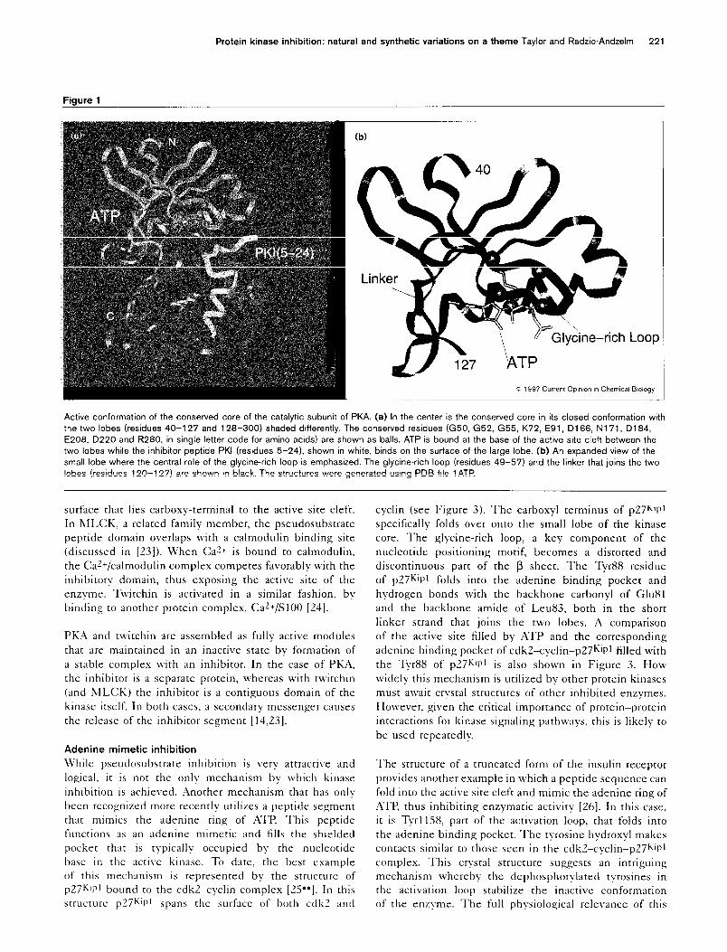
Figure 1
(b)
.oop
c 1997 Current Opmvx I” Chemical Biology
h Active conformation of the conserved core of the catalytic subunit of PKA. (a) In the center is the conserved core in its closed conformation wit
the two lobes (residues 40-l 27 and 126-300) shaded differently. The conserved residues (G50, G52, G55, K72, E91, Dl66, N171, D164,
E206, D220 and R260, in single letter code for amino acids) are shown as balls. ATP is bound at the base of the active site cleft between the
two lobes while the inhibitor peptide PKI (residues 5-241, shown in white, binds on the surface of the large lobe. (b) An expanded view of the
small lobe where the central role of the glycine-rich loop is emphasized. The glycine-rich loop (residues 49-57) and the linker that joins the two
lobes (residues 120-l 27) are shown in black. The structures were generated using PDB file 1 ATP.
surface that lies carboxy-terminal to the active site cleft.
In hlLCK. a related family member, the pseudosubstrate
peptide domain overlaps with a calmodulin binding site
(discussed in [23]). When Car+ is bound to calmodulin,
the CaZ+/calmodulin complex competes favorably with the
inhibitory domain, thus exposing the active site of the
enzyme. Twitchin is activated in a similar fashion, by
binding to another protein complex, CaZ+/SlOO [El].
PKA and twitchin are assembled as fully active modules
that are maintained in an inactive state by formation of
a stable complex with an inhibitor. In the case of PKA,
the inhibitor is a separate protein, whereas with twitchin
(and IZILCK) the inhibitor is a contiguous domain of the
kinase itself. In both cases, a secondary messenger causes
the release of the inhibitor segment [14,23].
Adenine mimetic inhibition
While pseudosubstrate inhibition is very attractive and
logical, it is not the only mechanism by which kinase
inhibition is achieved. Another mechanism that has onlv
been recognized more recently utilizes a peptide segment
that mimics the adenine ring of ATP. This pepcide
functions as an adenine mimetic and fills the shielded
pocket that is typically occupied by the nucleotide
base in the active kinase. To date, the best example
of this mechanism is represented by the structure of
p27Kllll bound to the cdk2-cyclin complex [ZS”]. In this
structure p27KiPl spans the surface of both cdk2 and
cyclin (see Figure 3). The carboxyl terminus of p27Q~l
specifically folds over onto the small lobe of the kinase
core. The glycine-rich loop, a key component of the
nucleotide positioning motif, becomes a distorted and
discontinuous part of the p sheet. The Tyr88 residue
of p27Kipl folds into the adenine binding pocket and
hydrogen bonds with the backbone carbonyl of Glu81
and the backbone amide of Leu83, both in the short
linker strand that joins the two lobes. A comparison
of the active site filled by ATP and the corresponding
adenine binding pocket of cdkZ-cyclin-p27Kil’l filled with
the Tyr88 of p27Kipl is also shown in Figure 3. How
widely this mechanism is utilized by other protein kinases
must await crystal structures of other inhibited enzymes.
However, given the critical importance of protein-protein
interactions for kinase signaling pathways. this is likely to
be used repeatedly.
The structure of a truncated form of the insulin receptor
provides another example in which a peptide sequence can
fold into the active site cleft and mimic the adenine ring of
ATP, thus inhibiting enzymatic activity [26]. In this case,
it is Tyrl158, part of the activation loop, that folds into
the adenine binding pocket. The tyrosine hydroxyl makes
contacts similar to those seen in the cdk2-cyclin-p27W~~
complex. This crystal structure suggests an intriguing
mechanism whereby the dephosphorylated tyrosines in
the activation loop stabilize the inactive conformation
of the enzyme. The full physiological relevance of this
222 Next generation therapeutics
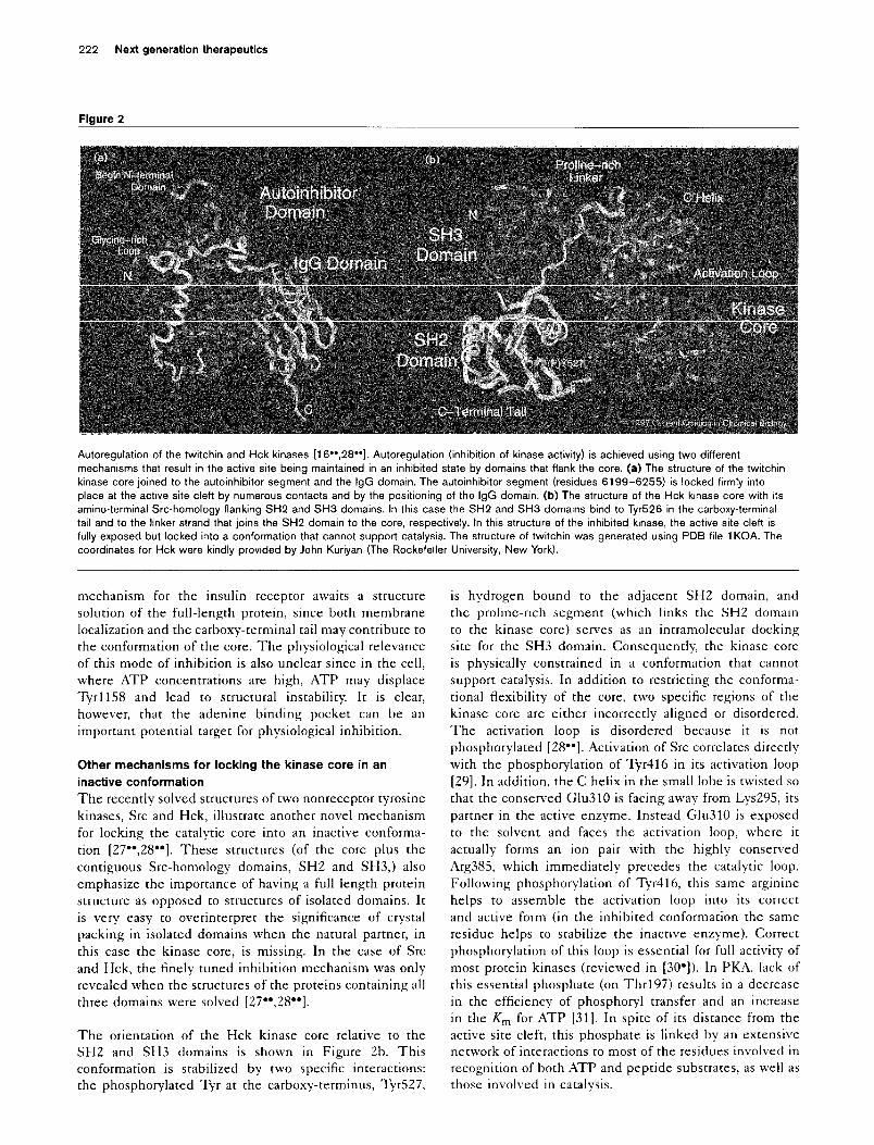
Figure 2
Autoregulation of the twitchin and Hck kinases [16”,26”]. Autoregulation (inhibition of kinase activity) is achieved using two different
mechanisms that result in the active site being maintained in an inhibited state by domains that flank the core. (a) The structure of the twitchin
kinase core joined to the autoinhibitor segment and the IgG domain. The autoinhibitor segment (residues 6199-6255) is locked firmly into
place at the active site cleft by numerous contacts and by the positioning of the IgG domain. (b) The structure of the Hck kinase core with its
amino-terminal &c-homology flanking SH2 and SH3 domains. In this case the SH2 and SH3 domains bind to Tyr526 in the carboxy-terminal
tail and to the linker strand that joins the SH2 domain to the core, respectively. In this structure of the inhibited kinase, the active site cleft is
fully exposed but locked into a conformation that cannot support catalysis. The structure of twitchin was generated using PDB file 1 KOA. The
coordinates for Hck were kindly provided by John Kuriyan (The Rockefeller University, New York).
mechanism for the insulin receptor awaits a structure
solution of the full-length protein, since both membrane
localization and the carboxy-terminal tail may contribute to
the conformation of the core. The physiological relevance
of this mode of inhibition is also unclear since in the cell,
where ATP concentrations are high, ATP may displace
Tyr1158 and lead to structural instability. It is clear,
however, that the adenine binding pocket can be an
important potential target for physiological inhibition.
Other mechanisms for locking the kinase core in an
inactive conformation
The recently solved structures of two nonreceptor tyrosine
kinases, Src and Hck, illustrate another novel mechanism
for locking the catalytic core into an inactive conforma-
tion [27”,28**]. These structures (of the core plus the
contiguous Src-homology domains, SH2 and SH3,) also
emphasize the importance of having a full length protein
structure as opposed to structures of isolated domains. It
is very easy to overinterpret the significance of crystal
packing in isolated domains when the natural partner, in
this case the kinase core, is missing. In the case of Src
and Hck, the finely tuned inhibition mechanism was only
revealed when the structures of the proteins containing all
three domains were solved [27**,28”].
The orientation of the Hck kinase core relative to the
SH2 and SH3 domains is shown in Figure 2b. This
conformation is stabilized by two specific interactions:
the phosphorylated Tyr at the carboxy-terminus, Tyr527,
is hydrogen bound to the adjacent SH2 domain, and
the proline-rich segment (which links the SH2 domain
to the kinase core) serves as an intramolecular docking
site for the SH3 domain. Consequently, the kinase core
is physically constrained in a conformation that cannot
support catalysis. In addition to restricting the conforma-
tional flexibility of the core, two specific regions of the
kinase core are either incorrectly aligned or disordered.
The activation loop is disordered because it is not
phosphorylated [B”]. Activation of Src correlates directly
with the phosphorylation of Tyr416 in its activation loop
[29]. In addition, the C helix in the small lobe is twisted so
that the conserved Glu310 is facing away from Lys295, its
partner in the active enzyme. Instead Glu310 is exposed
to the solvent and faces the activation loop, where it
actually forms an ion pair with the highly conserved
Arg385, which immediately precedes the catalytic loop.
Following phosphorylation of Tyr416, this same arginine
helps to assemble the activation loop into its correct
and active form (in the inhibited conformation the same
residue helps to stabilize the inactive enzyme). Correct
phosphorylation of this loop is essential for full activity of
most protein kinases (reviewed in [30’]). In PKA, lack of
this essential phosphate (on Thr197) results in a decrease
in the efficiency of phosphoryl transfer and an increase
in the Km for ATP [31]. In spite of its distance from the
active site cleft, this phosphate is linked by an extensive
network of interactions to most of the residues involved in
recognition of both ATP and peptide substrates, as well as
those involved in catalysis.
Protein kinase inhibition: natural and synthetic variations on a theme Taylor and Radzio-Andzelm 223
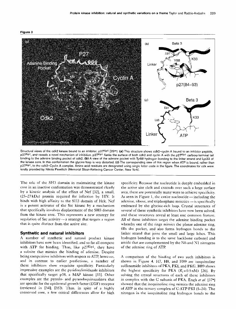
Figure 3
fb) Beta 3
Linker
p27(84-93)
Linker ’ ATP
Structural views of the cdk2 kinase bound to an inhibitor, p2i’W [25”1. (a) This structure shows cdk2-cyclin A bound to an inhibitor peptide,
p2Wpt, and reveals a novel mechanism of inhibition. p27W flanks the surface of both cdk2 and cyclin A with the p27W carboxy-terminal tail
binding to the adenine binding pocket of cdk2. (b) A view of the adenine pocket with Tyr88 hydrogen bonding to the linker strand and Lys33 of
the kinase core. In this conformation the glycine loop is very distorted. (c) The corresponding view of this region when ATP is bound, rather than
p27W. to the cdk2-Cyclin A complex. Amino acid residues are designated using single letter code in the figure. The coordinates for cdk were
kindly provided by Nikola Paveltich (Memorial Sloan-Kettering Cancer Center, New York).
The role of the SH3 domain in maintaining the kinase
core in an inactive conformation was demonstrated clearly
by a kinetic analysis of the effect of Nef [32], a small,
(25-27 kDa) protein required for infection by HIV. It
binds with high affinity to the SH3 domain of Hck. Nef
is a potent activator of the Src kinase by a mechanism
that specifically involves displacement of the SH3 domain
from the kinase core. This represents a new strategy for
regulation of Src activity-a strategy that targets a region
that is quite distinct from the active site.
Synthetic and natural inhibitors A number of synthetic and natural product kinase
inhibitors have now been identified, and so far all compete
with ATP for binding. Thus, like p27Kip1, they have
a subsite that mimics the binding of adenine. Despite
being competitive inhibitors with respect to ATP, however,
and in contrast to earlier predictions, a number of
these inhibitors show exquisite specificity. Particularly
impressive examples are the pyridinylimidazole inhibitors
that specifically target ~38, a MAP kinase [33]. Other
examples are the pyrrolo- and pyrazoloquinazolines that
are specific for the epidermal growth factor (EGF) receptor
(reviewed in [34]; [35]). Thus, in spite of a highly
conserved core, a few critical differences allow for high
specificity. Because the nucleotide is deeply embedded in
the active site cleft and extends over such a large surface
area, there are potentially many ways to achieve specificity.
As seen in Figure 1, the entire nucleotide-including the
adenine, ribose, and triphosphate moieties -is specifically
embraced by the glycine-rich loop. Crystal structures of
several of these synthetic inhibitors have now been solved,
and these structures reveal at least one common feature.
All of these inhibitors target the adenine binding pocket
whereby one of the rings mimics the planar adenine ring,
fills the pocket, and also forms hydrogen bonds to the
linker strand that joins the small and large lobes. This
hydrogen bonding is to the same backbone carbonyl and
amide that are complemented by the N6 and Nl nitrogens
of the adenine ring of ATP.
A comparison of the binding of two such inhibitors is
shown in Figure 4. H7, H8, and H89 are isoquinoline
sulfonamide inhibitors of PKA, PKI, and PKG. H89 shows
the highest specificity for PKA (Ki =O.SnM) [36]. By
solving the crystal structures of each of these inhibitors
in complex with the C subunit of PKA, Engh et a/. [37-l
showed that the isoquinoline ring mimics the adenine ring
of ATP in the ternary complex of CATP.PKI (5-24). The
nitrogen in the isoquinoline ring hydrogen bonds to the
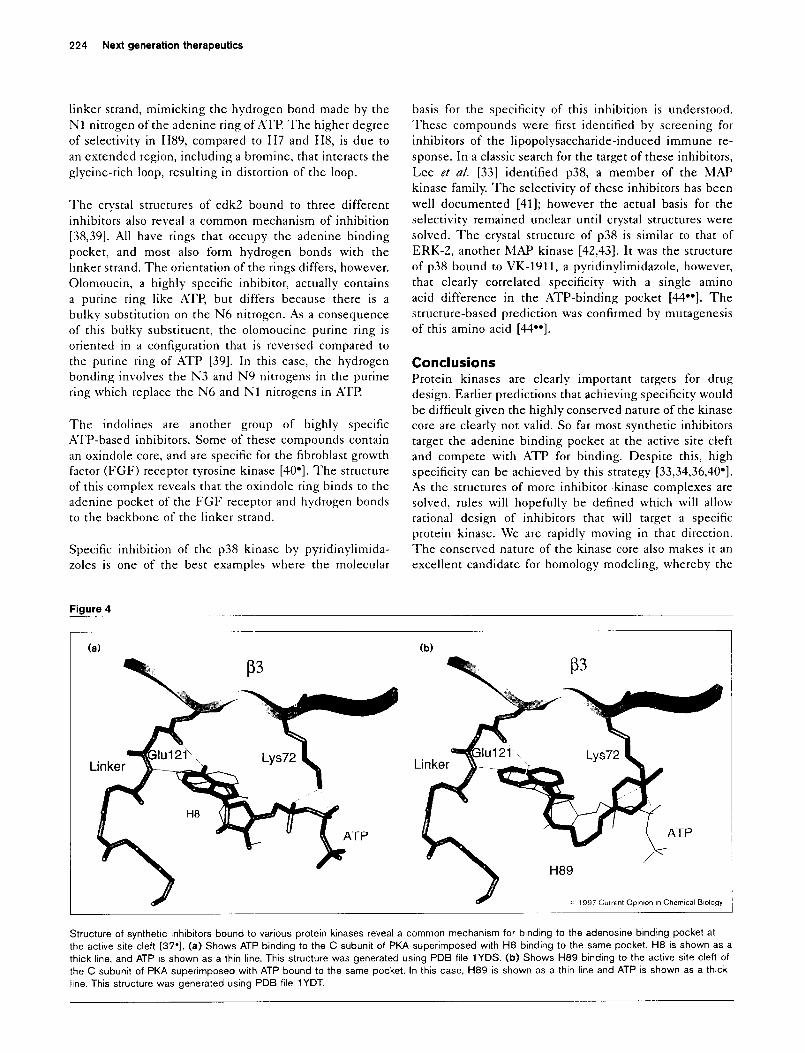
224 Next generation therapeutics
linker strand, mimicking the hydrogen bond made by the
Nl nitrogen of the adenine ring of ATI? The higher degree
of selectivity in H89, compared to H7 and H8, is due to
an extended region, including a bromine, that interacts the
glycine-rich loop, resulting in distortion of the loop.
The crystal structures of cdk2 bound to three different
inhibitors also reveal a common mechanism of inhibition
[38,39]. All have rings that occupy the adenine binding
pocket, and most also form hydrogen bonds with the
linker strand. The orientation of the rings differs, however.
Olomoucin, a highly specific inhibitor, actually contains
a purine ring like ATP, but differs because there is a
bulky substitution on the N6 nitrogen. As a consequence
of this bulky substituent, the olomoucine purine ring is
oriented in a configuration that is reversed compared to
the purine ring of ATP [39]. In this case, the hydrogen
bonding involves the N3 and N9 nitrogens in the purine
ring which replace the N6 and Nl nitrogens in ATI?
The indolines are another group of highly specific
ATP-based inhibitors. Some of these compounds contain
an oxindole core, and are specific for the fibroblast growth
factor (FGF) receptor tyrosine kinase [40*]. The structure
of this complex reveals that the oxindole ring binds to the
adenine pocket of the FGF receptor and hydrogen bonds
to the backbone of the linker strand.
Specific inhibition of the ~38 kinase by pyridinylimida-
zoles is one of the best examples where the molecular
Figure 4
basis for the specificity of this inhibition is understood.
These compounds were first identified by screening for
inhibitors of the lipopolysaccharide-induced immune re-
sponse. In a classic search for the target of these inhibitors,
Lee et al. [33] identified ~38, a member of the MAP
kinase family. The selectivity of these inhibitors has been
well documented [41]; however the actual basis for the
selectivity remained unclear until crystal structures were
solved. The crystal structure of ~38 is similar to that of
ERK-2, another MAP kinase [42,43]. It was the structure
of ~38 bound to VK-1911, a pyridinylimidazole, however,
that clearly correlated specificity with a single amino
acid difference in the ATP-binding pocket [44*-l. The
structure-based prediction was confirmed by mutagenesis
of this amino acid [44**].
Conclusions Protein kinases are clearly important targets for drug
design. Earlier predictions that achieving specificity would
be difficult given the highly conserved nature of the kinase
core are clearly not valid. So far most synthetic inhibitors
target the adenine binding pocket at the active site cleft
and compete with ATP for binding. Despite this, high
specificity can be achieved by this strategy [33,34,36,40*].
As the structures of more inhibitor-kinase complexes are
solved, rules will hopefully be defined which will allow
rational design of inhibitors that will target a specific
protein kinase. We are rapidly moving in that direction.
The conserved nature of the kinase core also makes it an
excellent candidate for homology modeling, whereby the
(a) (b)
Structure of synthetic inhibitors bound to various protein kinases reveal a common mechanism for binding to the adenosine binding pocket at
the active site cleft [37-l. (a) Shows ATP binding to the C subunit of PKA superimposed with H8 binding to the same pocket. H8 is shown as a
thick line. and ATP is shown as a thin line. This structure was generated using PDB file 1YDS. (b) Shows H89 binding to the active site clefi of
the C subunit of PKA superimposed with ATP bound to the same pocket. In this case, H89 is shown as a thin line and ATP is shown as a thtck
line. This structure was generated using PDB file 1 YDT.
Protein kinase inhibition: natural and synthetic variations on a theme Taylor and Radzio-Andzelm 225
sequence of the conserved core of any protein kinase can
be modeled onto a three-dimensional structural template
[15]. Once again, as more structures are solved, the
accuracy of the modeling also will improve. It is also
clear, however, that the design of specific inhibitors need
not target only the active site cleft. The C helix, the
activation loop or the segments that flank the kinase core
are equally valid targets for drug design, as is targeting the
ligand binding sites at the active site. Although designing
inhibitors that bind to the active kinase is one strategy,
designing or screening compound libraries for inhibitors
that prevent kinase activation is an equally plausible
strategy - one that has been explored by nature but yet
to be exploited by combinatorial chemistry.
Acknowledgements SW is supported by grants from the National Institutes of Health and the American Cancer Society. ER-A was supported in part by National Institutes of Health ‘liaining Grant I’HS ‘1‘32 DK07233. The authors wsh to thank N Narayana and T Diller for helpful comments and discussions.
References and recommended reading Papers of particular interest, published within the annual period of review, have been highlighted as:
. of special interest
. . of outstanding intt,est
1. Fischer EH, Krebs EG: Conversion of phosphorylase b to phosphorylase a in muscle extracts. J Biol Chem 1955, 216:121-132.
2. Hanks SK, Hunter T: Protein kinases 6. The eukaryotic protein kinase superfamily: kinase (catalytic) domain structure and classification. FASEB J 1995, 9576-596.
3. Zheng J, Knighton DR, Xuong N-h, Taylor SS, Sowadski JM, Ten Eyck LF: Crystal structures of the myristylated catalytic subunit of CAMP-dependent protein kinase reveal open and closed conformations. Protein Sci 1993, 2:1559-l 573.
4. Knighton DR, Zheng J, Ten Eyck LF, Ashford VA, Xuong N-h, Taylor SS, Sowadski JM: Crystal structure of the catalytic subunit of CAMP-dependent protein kinase. Science i991( 253:407-414.
5. Grant BD, Adams JA: Pre-steady-state analysis of CAMP- dependent protein kinase using rapid quench-flow techniques. Biochemistry 1996, 35:2022-2029.
6. Lew J, Taylor SS, Adams JA: Identification of a partially rate- determining step in the catalytic mechanism of CAMP- dependent protein kinase: a transient kinetic study using stopped-flow fluorescence spectroscopy. Biochemistry 1997, 36:6717-6724.
z .
Narayana N, Cox S, Shumel S, Taylor SS, Xuong N: Crystal structure of the poly-histidine tagged recombinant catalytic subunit of CAMP-dependent protein kinase complexed with the peptide inhibitor, PKl(5-24) and adenosine. Biochemistry 1997, 36:4438-4448.
This paper gives an in-depth description of the nucleotide binding site in the catalytic subunit of CAMP-dependent protein kinase (PKA).
8. Hon W-C, McKay GA, Thompson PR, Sweet RM, Yang DSC, Wright GD, Berghuis AM: Structure of an enzyme required for aminoglycoside antibiotic resistance reveal hom&ogy to eukaryotic protein kinases. Cell 1997, 89:887-895.
9. Bossemeyer D, Engh RA, Kinzel V, Ponstingl H, Huber R: Phosphotransferase and substrate binding mechanism of the CAMP-dependent protein kinas? catalytic subunit from porcine heart as deduced from the 2.0A structure of the complex with Mnz+ adenyl imidodiphosphate and inhibitor peptide PKl(5-24). EM50 J 1993, 12:649-859.
10. Zheng J, Knighton DR, Ten Eyck LF, Karlsson R, Xuong N-h, Taylor SS, Sowadski JM: Crystal structure of the catalytic
11.
12.
13.
14.
15.
16. . .
subunit of CAMP-dependent protein kinase complexed with MgATP and peptide inhibitor. Biochemistry 1993, 32:2154-2161.
Hemmer W, McGlone ML, Taylor SS: The role of the glycine triad in the ATP-binding site of CAMP-dependent protein kinase. J B/o/ Chem 1997, 272:16946-l 6954.
Bossemeyer D: The glycine-rich sequence of protein kinases: a multifunctional element Trends Biochem Sci 1994, 19:201-205.
Herberg F, Zimmermann B, McGlone M, Taylor SS: Importance of the A-helix of the cat&tic subunit of CAMP-deoendent orotein inase for stability and f& orienting subdomain; at the ileft interface. Protein Sci 1997, 6:569-579.
Taylor SS, Buechler JA, Yonemoto W: CAMP-dependent protein kinase: framework for a diverse family of regulatory enzymes. Annu Rev Biochem 1990, 59:971-l 005.
Kniahton DR. Pearson RB. Sowadski JM. Means AR. Ten Evck LF. Taylor SS, Kemp BE: Structural basis for intrasteric regulation’ of myosin light chain kinase. Science 1992, 258:130-135.
Kobe B. Heierhorst J. Feil SC. Parker MW. Benian GM. Klaudiusz RW, Kemp BE: Giant protein kinases: domain interactions and structural basis of autoregulation. EMBO J 1996, 15:6810-6821.
Describes the structure of the klnase domam of twttchin plus the pseudosub- strate region and the IgG domain that lie carboxy-terminal to the conserved core.
1 7.
18.
19.
20.
21.
22.
23.
24.
25. . .
Walsh DA, Angelos KL, Van Patten SM, Glass DB, Garetto LP: The inhibitor protein of the CAMP-dependent protein kinase. In Peptides and Protein Phosphorylation. Edited by BE Kemp. Boca Raton: CRC Press, Inc.; 1990:43-84.
Knighton DR, Zheng J, Ten Eyck LF, Xuong N-h, Taylor SS, Sowadski JM: Structure of a peptide inhibitor bound to the catalytic subunit of cyclic adenosine monophosphate- dependent protein kinase. Science 1991, 253:414-420.
Herberg FW, Taylor SS: Physiological inhibitors of the catalytic subunit of CAMP-dependent protein kinase: effect of MgATP on protein/protein interaction. Biochemisrry 1993, 32:14015- 14022.
Gibbs CS, Knighton DR, Sowadski JM, Taylor SS, Zoller MJ: Systematic mutational analysis of CAMP-dependent protein kinase identifies unregulated catalytic subunits and defines regions important for the recognition of the regulatory subunit J Biol Chem 1992, 267:4806-4614.
Gibson RM, Ji-Buechler Y, Taylor SS: Identification of electrostatic interaction sites between the regulatory and catalytic subunits of cyclic AMP-dependent protein kinase. Protein Sci 1997, in press.
Hu S-H, Parker MW, Lei JY, Wilce MCJ, Benian GM, Kemp BE: Insights into autoregulation from the crystal structure of twitchin kinase. Nature 1994, 369:578-581.
Kemp BE, Faux MC, Means AR, House C, Tiganis T, Hu S-H, Mitchelhill KI: Structural aspects: pseudosubstrate and substrate interactions. In Protein Kinase. Edited by JR Woodgett. Oxford: IRL Press at Oxford University Press; 1994:30-67.
Heierhorst J, Kobe B, Feil SC, Parker MW, Benian GM, Weiss KR, Kemp BE: Ca2+/SlOO regulation of giant protein kinases. Nature 1996, 380:636-639.
Russo AA, Jeffrey PD, Patten AK, Massague J, Pavletich NP: Crystal Structure of the p27Kipl cyclin-dependent-kinase inhibitor bound to the cyclin A-CdkP complex. Nature 1996, 382:325-331,
The structure of ~27~lP’ bound to cdk2-cyclin A reveals an adenine mimetic mechanism of kinase inhibition whereby the carboxyl terminus of p27KlPl occupies the adenine binding pocket of cdk2.
26. Hubbard SR, Wei L, Ellis L, Hendrickson WA: Crystal structure of the tyrosine kinase domain of the human insulin receptor?. Nature 1994, 372:746-754.
27. Xu W, Harrison SC, Eck MJ: Three-dimensional structure of the tyrosine kinase c-Src. Nature 1997, 385:595-599.
Aase see annotation [28**].
28. Sicheri F, Moarefi I, Kuriyan J: Crystal structure of the Src family . . tyrosine kinase Hck Nature 1997, 385:602-609. These two papers [27”,28”1 describe the structures of two nonreceptor tyrosine kinases. Both include the contiguous SH2 and SH3 domains and provide excellent examples of an inhibitory mechanism that is distinct from a pseudosubstrate-based mechanism.
226 Next generation therapeutics
29. Boemer RJ, Kassel DB, Barker SC, Ellis B, DeLacy P, Knight WB: Correlation of the phosphorylation states of pp60c-src with tyrosine kinase activity: the intramolecular pY530-SH2 complex retains significant activity if Y419 is phosphorylated. Biochemistry 1996, 35:9519-9525.
30. Johnson LN, Noble ME, Owen DJ: Active and inactive protein . kinases: structural basis for regulation. Cell 1996, 85:149-l 56. An excellent review of activation loops in protein kinases that explains why phosphorylation of this region is typically essential for activity.
31.
32.
33.
34
35.
36.
37. .
Adams JA, McGlone ML, Gibson R, Taylor SS: Phosphorylation modulates catalytic function and regulation in CAMP- dependent protein kinase. Biochemistry 1995, 34:2447-2454.
Moarefi I. LaFevre-Bernt M. Sicheri F. Huse M. Lee C-H. Kurivan J. Miller k: Activation of tGe~Src-family tyrosine kinas; Hck by SH3 domain displacement. Nature 1997, 365:650-653.
Lee JC, Laydon JT, McDonnel PC, Gallagher TF, Kumar S, Green D, McNulty D, Blumenthal MJ, Heys JR, Landvatter SW: A protein kinase involved in the regulation of inflammatory cytokine biosynthesis. Nature 1994, 372:739-746.
Palmer BD, Trumpp-Kallmeyer S, Fry DW, Nelson JM, Showalter HDH, Denny WA: Tyrosine kinase inhibitors. 11. Soluble analogues of pyrrol& and pyrazoloquinazolines as epidermal growth factor receptor inhibitors: synthesis, biological evaluation, and modeling of the mode of binding. I Med Chem 1997, 40:1519-l 529.
Fry DW, Kraker Al, McMichael A, Ambroso LA, Nelson JM, Leopold WR, Connors RW, Bridges AJ: A specific inhibitor of the.epidermal growth factor receptor tyrosine kinase. Science 1994, 265:1093-l 095.
Chijiwa T, Michima A, Hagiwara M, Hayashi K, lnoue T, Naito K, Toshioka T, Hidaka H: Inhibition of forskolin-induced neurite outgrowth and protein phosphorylation by a newly synthesized selective inhibitor of cyclic AMP-dependent protein kinase. J Biol Chem 1990, 265:5267-5272.
Engh RA, Girod A, Kinzel V, Huber R, Bossemeyer D: Crystal structures of catalytic subunit of CAMP-dependent protein kinase in complexes with isoquinolinesulfonyl protein kinase inhibitors H7, H8, H89. J Biol Chem 1996, 271:26157-26164.
This set of structures shows the H-series inhibitors bound to the catalytic subunit of CAMP-dependent protein kinase.
36.
39.
40. .
Filgueira de Azevedo W Jr, Mueller-Dieckmann H-J, Schulze- Gahmen U, Worland PJ, Sausville E, Kim S-H: Structural basis for soecificitv and ootencv of a flavonoid inhibitor of human CDKi a cell-cycle ‘kinase: Proc Nat/ Acad Sci USA 1996, 93:2735-2740.
Schulze-Gahmen U. Brandsen J. Jones HD. Moraan DO. Meiier L. , Vesely J, Kim S-H: Multiple modes of ligand reiognition: crystal structures of cyclin-dependent protein kinase 2 in complex with ATP and two inhibitors, olomoucine and isopentenyladenine. Proteins: Strut Func Genef 1995, 22:376-391.
Mohammadi M, McMahon G, Sun L, Tang C, Hirth P, Yeh BK, Hubbard SR, Schlessinger J: Structures of the tyrosine kinase domain of fibroblast growth factor receptor in complex with inhibitors. Science 1997, 276:955-960.
An example of specific kinase inhibitor that docks to the active site of the fibroblast growth factor receptor.
41. Cuenda A, Cohen P, Buee-Scherrer V, Goedert M: Activation of stress-activated protein kinase 3 (SAPKS) by cytokines and cellular stresses is mediated via SAPKKS (MKKG); comparison of the specificities of SAPKB and SAPK2 (RK/p38). EMBO J 1997, 16:295-305.
42. Wang 2, Harkins PC, Ulevitch RI, Han J, Cobb MH, Goldsmith El.: The structure of mitogen-activated protein kinase ~38 at 2.1 A resolution. Proc Nat/ Acad Sci USA 1997, 94:2327-2332.
43. Wilson KP, Fitzgibbon MJ, Caron PR, Griiith JP, Chen W, McCaffrey PG, Chambers SP, Su MS-S: Crystal structure of ~38 mitogen-activated protein kinase. J Biol Chem 1996, 271:27696-27700.
44. Wilson KP, McCaffrey PG, Hsiao K, Pazhanisamy S, Galullo V, . . Bemis GW, Fitzgibbon MJ, Caron PR, Murcko MA, Su MSS:
Structural basis for specificity of pyridinylimidazole inhibitors of p38 MAP Kinase. Chem Biochem 1997, 4:423-431.
This structure of a p36-specific inhibitor bound to ~36 provides an excellent example of specificity. Mutagenesis of a single residue functionally confirms the structural basis for specificity.