Upregulation of protein synthesis initiation factor eIF-4E is an early event during colon...
11
Upregulation of protein synthesis initiation factor eIF-4E is an early event during colon carcinogenesis Igor B Rosenwald* ,1,2,3 , Jane-Jane Chen 2 , Songtao Wang 1 , Lou Savas 1 , Irving M London 2 and James Pullman* ,1 1 Department of Pathology, University of Massachusetts Medical Center, 55 Lake Avenue North, Worcester, Massachusetts 01655, USA; 2 Harvard University-Massachusetts Institute of Technology, Division of Health Sciences and Technology, Room E25-545, 77 Massachusetts Avenue, Cambridge, Massachusetts 02139, USA A general increase in protein synthesis and a specific increase in the synthesis of growth-promoting proteins are necessary for mitogenesis. Regulation of protein synthesis, as well as preferential translation of some mRNAs coding for growth promoting proteins (e.g. cyclin D1), involves the essential protein synthesis initiation factor eIF-4E. This factor is induced by various oncoproteins, and, when overexpressed, it can transform cultured cells. In this report we explore the roles of eIF-4E in human neoplastic disorders of the colon and in the regulation of general and specific protein synthesis. We find that eIF-4E is increased in colon adenomas and carcinomas, and this increase is accompanied in most but not all cases by elevation of cyclin D1 levels. While general protein synthesis is increased by eIF-4E overexpression in cultured cells, only a small proportion of proteins is preferentially up- regulated by eIF-4E, as revealed by two-dimensional gel electrophoresis. These results are consistent with the view that eIF-4E plays a role in carcinogenesis by increasing general protein synthesis and by preferentially upregulat- ing a subset of putative growth promoting proteins. Our results, taken together with the recent findings that c- myc transcription is negatively regulated by APC and our earlier data on transcriptional activation of eIF-4E expression by c-Myc suggest that eIF-4E is a down- stream target of the APC/b-catenin/Tcf-4 pathway, and is strongly involved in colon tumorigenesis. Keywords: eIF-4E; translation factors; cyclins; protein synthesis; transformation; colon cancer Introduction Mitogenic stimulation of resting cells leads to both a general increase in net protein synthesis and prefer- ential upregulation of cell cycle promoting proteins (Epifanova, 1977; Pardee et al., 1978; Sherr, 1996; Weinberg, 1995; Bartek et al., 1996). The general increase in protein synthesis is necessary to allow the cell to double its protein content and size before mitosis so that an average cell size is maintained during the proliferative response. When growth-stimulated cells are treated with protein synthesis inhibitors to decrease their protein synthesis by only 50% before the restriction point (R-point) in mid-late G1, they withdraw into G0 resting state (Zetterberg and Larsson, 1991; Zetterberg et al., 1995). However, if protein synthesis is decreased after the R-point, the cell is able to complete one round of replication before arresting in G0. Normal cells are able to down regulate protein synthesis and withdraw into quiescence (G0) when growth factors are limiting (Hershko et al., 1971; for review see Rosenwald, 1996a). The upregulation of net protein synthesis after mitogenic stimulation is due to increased function and expression of translation factors (Sonenberg, 1996; Morris, 1995). It is generally accepted now that the critical event that facilitates transition through the R-point is inactivation of the retinoblastoma tumor-suppressor protein (pRb) as a result of its hyperphosphorylation by cyclin D-dependent kinase (CDK). Hyperphosphor- ylation of pRb causes the release of E2F transcription factors and other proteins from their complexes with pRb which leads to activation of genes coding for positive regulators or direct executors of cell prolifera- tion (Weinberg, 1995; Bartek et al., 1996; Zetterberg et al., 1995). We have found earlier that overexpression of translation initiation factor eIF-4E in NIH3T3 cells leads to a specific increase in the level of cyclin D1 (Rosenwald et al., 1993a, 1995). In turn, cyclin D1 activates CDK-4/CDK-6 to phosphorylate pRb, thus contributing to the transition of cells through the restriction point (Sherr, 1996; Weinberg, 1995; Bartek et al., 1996). Translation initiation factor eIF-4E is the least abundant subunit of the eIF-4F complex. The two other subunits are eIF-4A, which is a helicase responsible for unwinding secondary structures in mRNA (in collaboration with another initiation factor eIF-4B); and eIF-4G which holds the complex together and is responsible for ribosome binding (Sonenberg, 1996; Rhoads et al., 1994). The most well established function of eIF-4E is binding of the 5’ cap structure that is present in virtually all eukaryotic mRNA, thus providing for the association of mRNAs with the eIF-4F complex. Growth factors increase eIF-4E activity by two principal mechanisms. First, treatment of resting cells with growth factors leads to activation of eIF-4E gene expression and causes an increase in both eIF- 4E mRNA and protein concentrations (Rosenwald et al., 1993a, 1995; for reviews see Rosenwald, 1996a; Sonenberg, 1996; Morris, 1995). Second, phosphorylation of eIF-4E increases its cap- *Correspondence: IB Rosenwald and J Pullman 3 Igor B Rosenwald address after July 1 1999: Beth Israel-Deaconess Medical Center, Harvard Medical School, Clinical Pathology Division of Hematopathology, YA-309, 330 Brookline Ave., Boston MA 02215 Received 15 June 1998; revised 26 October 1998; accepted 3 November 1998 Oncogene (1999) 18, 2507 – 2517 ª 1999 Stockton Press All rights reserved 0950 – 9232/99 $12.00 http://www.stockton-press.co.uk/onc
-
Upload
independent -
Category
Documents
-
view
3 -
download
0
Transcript of Upregulation of protein synthesis initiation factor eIF-4E is an early event during colon...

Upregulation of protein synthesis initiation factor eIF-4E is an early eventduring colon carcinogenesis
Igor B Rosenwald*,1,2,3, Jane-Jane Chen2, Songtao Wang1, Lou Savas1, Irving M London2 andJames Pullman*,1
1Department of Pathology, University of Massachusetts Medical Center, 55 Lake Avenue North, Worcester, Massachusetts 01655,USA; 2Harvard University-Massachusetts Institute of Technology, Division of Health Sciences and Technology, Room E25-545, 77Massachusetts Avenue, Cambridge, Massachusetts 02139, USA
A general increase in protein synthesis and a speci®cincrease in the synthesis of growth-promoting proteinsare necessary for mitogenesis. Regulation of proteinsynthesis, as well as preferential translation of somemRNAs coding for growth promoting proteins (e.g.cyclin D1), involves the essential protein synthesisinitiation factor eIF-4E. This factor is induced byvarious oncoproteins, and, when overexpressed, it cantransform cultured cells. In this report we explore theroles of eIF-4E in human neoplastic disorders of thecolon and in the regulation of general and speci®cprotein synthesis. We ®nd that eIF-4E is increased incolon adenomas and carcinomas, and this increase isaccompanied in most but not all cases by elevation ofcyclin D1 levels. While general protein synthesis isincreased by eIF-4E overexpression in cultured cells,only a small proportion of proteins is preferentially up-regulated by eIF-4E, as revealed by two-dimensional gelelectrophoresis. These results are consistent with the viewthat eIF-4E plays a role in carcinogenesis by increasinggeneral protein synthesis and by preferentially upregulat-ing a subset of putative growth promoting proteins. Ourresults, taken together with the recent ®ndings that c-myc transcription is negatively regulated by APC andour earlier data on transcriptional activation of eIF-4Eexpression by c-Myc suggest that eIF-4E is a down-stream target of the APC/b-catenin/Tcf-4 pathway, andis strongly involved in colon tumorigenesis.
Keywords: eIF-4E; translation factors; cyclins; proteinsynthesis; transformation; colon cancer
Introduction
Mitogenic stimulation of resting cells leads to both ageneral increase in net protein synthesis and prefer-ential upregulation of cell cycle promoting proteins(Epifanova, 1977; Pardee et al., 1978; Sherr, 1996;Weinberg, 1995; Bartek et al., 1996). The generalincrease in protein synthesis is necessary to allow thecell to double its protein content and size beforemitosis so that an average cell size is maintained during
the proliferative response. When growth-stimulatedcells are treated with protein synthesis inhibitors todecrease their protein synthesis by only 50% before therestriction point (R-point) in mid-late G1, theywithdraw into G0 resting state (Zetterberg andLarsson, 1991; Zetterberg et al., 1995). However, ifprotein synthesis is decreased after the R-point, the cellis able to complete one round of replication beforearresting in G0. Normal cells are able to down regulateprotein synthesis and withdraw into quiescence (G0)when growth factors are limiting (Hershko et al., 1971;for review see Rosenwald, 1996a). The upregulation ofnet protein synthesis after mitogenic stimulation is dueto increased function and expression of translationfactors (Sonenberg, 1996; Morris, 1995).
It is generally accepted now that the critical eventthat facilitates transition through the R-point isinactivation of the retinoblastoma tumor-suppressorprotein (pRb) as a result of its hyperphosphorylationby cyclin D-dependent kinase (CDK). Hyperphosphor-ylation of pRb causes the release of E2F transcriptionfactors and other proteins from their complexes withpRb which leads to activation of genes coding forpositive regulators or direct executors of cell prolifera-tion (Weinberg, 1995; Bartek et al., 1996; Zetterberg etal., 1995). We have found earlier that overexpression oftranslation initiation factor eIF-4E in NIH3T3 cellsleads to a speci®c increase in the level of cyclin D1(Rosenwald et al., 1993a, 1995). In turn, cyclin D1activates CDK-4/CDK-6 to phosphorylate pRb, thuscontributing to the transition of cells through therestriction point (Sherr, 1996; Weinberg, 1995; Barteket al., 1996).
Translation initiation factor eIF-4E is the leastabundant subunit of the eIF-4F complex. The twoother subunits are eIF-4A, which is a helicase responsiblefor unwinding secondary structures in mRNA (incollaboration with another initiation factor eIF-4B);and eIF-4G which holds the complex together and isresponsible for ribosome binding (Sonenberg, 1996;Rhoads et al., 1994). The most well established functionof eIF-4E is binding of the 5' cap structure that is presentin virtually all eukaryotic mRNA, thus providing for theassociation of mRNAs with the eIF-4F complex.
Growth factors increase eIF-4E activity by twoprincipal mechanisms. First, treatment of resting cellswith growth factors leads to activation of eIF-4Egene expression and causes an increase in both eIF-4E mRNA and protein concentrations (Rosenwaldet al., 1993a, 1995; for reviews see Rosenwald,1996a; Sonenberg, 1996; Morris, 1995). Second,phosphorylation of eIF-4E increases its cap-
*Correspondence: IB Rosenwald and J Pullman3 Igor B Rosenwald address after July 1 1999: Beth Israel-DeaconessMedical Center, Harvard Medical School, Clinical PathologyDivision of Hematopathology, YA-309, 330 Brookline Ave., BostonMA 02215Received 15 June 1998; revised 26 October 1998; accepted 3November 1998
Oncogene (1999) 18, 2507 ± 2517ã 1999 Stockton Press All rights reserved 0950 ± 9232/99 $12.00
http://www.stockton-press.co.uk/onc

binding activity and phosphorylation of eIF-4E-binding proteins (4E-BPs) leads to release of eIF-4E from inactive complexes with 4E-BPs (Sonenberg,1996; Rhoads et al., 1994).
The key role of eIF-4E in cell growth is emphasizedby several observations. First, overexpression of eIF-4E converts rodent cells to a tumorigenic phenotype(Lazaris-Karatzas et al., 1990; Lazaris Karatzas andSonenberg, 1992) and drastically ampli®es the neoplas-tic morphology of human HeLa cells (De Benedettiand Rhoads, 1990). Second, inhibition of eIF-4Efunction by expressing antisense mRNA or 4E-BPslows down cell growth and reverses the malignantphenotype of ras-transformed cells (De Benedetti et al.,1991; Gra� et al., 1995; Rousseau et al., 1996a). Third,expression of eIF-4E becomes constitutive, i.e.independent of extracellular growth factors in culturedtumor cells (Rosenwald, 1995, 1996a,b). Fourth, anumber of oncogenes, including ras, src, abl and mycincrease the level and/or activity of eIF-4E (reviewed inRosenwald, 1996a). All of these ®ndings suggest thatincreasing the level and activity of eIF-4E may be oneof the key e�ects of oncogenes leading to neoplastictransformation (Rhoads, 1991; Rosenwald, 1996a,b).
To examine further the role of eIF-4E in neoplastictransformation suggested by experiments with culturedcells, we initiated an analysis of various malignantneoplasms in humans. In this report we describe our®ndings on the elevated expression of eIF-4E in colonadenomas and adenocarcinomas and suggest itspotential role in initiating tumor progression via bothan increase in general protein synthesis and preferentialsynthesis of speci®c growth-promoting proteins.
Results
Immunohistochemical analysis of eIF-4E expression incultured cells
Before analysing expression of eIF-4E in humanneoplasms, it was imperative to demonstrate theability of the anti-eIF-4E antibodies to detect thedi�erent levels of eIF-4E expression. In order to doso, eIF-4E-overexpressing and control NIH3T3 cellswere cultured to obtain cell pellets which were ®xedand processed for para�n embedded sections in amanner identical to preparation of surgical samplesfor pathological analysis. We have found that botha�nity puri®ed polyclonal antibody (data not shown)and a monoclonal antibody to eIF-4E detect anincreased level of this protein in eIF-4E-overexpres-sing cells (Figure 1, top). This result corresponds to a5 ± 10-fold increase in eIF-4E level as demonstrated byquantitation of Western blots (Rosenwald et al.,1993b). Also, in agreement with Western blotting,our current immunohistochemical study shows thateIF-4E overexpressing cells display elevated levels ofcyclin D1. Nearly 100% of them express cyclin D1 inthe nucleus (as compared to about 50% in controlcells) and demonstrate stronger cytoplasmic stainingas well. This increase in cyclin D1 expressiondemonstrated by immunohistochemical analysis (pre-sented in Figure 1) corresponds to a 4 ± 5-fold increaseas determined by quantitation of the Western blots. Ithas been shown that whereas cyclin D1 is barely
detectable in the cytoplasm of resting (G0) ®broblasts,it accumulates in the nucleus during the G1 periodand is released into the cytoplasm as the cells progressinto S phase and mitosis (Baldin et al., 1993). As canbe seen in Figure 1, both control and eIF-4Eoverexpressing NIH3T3 cells express cyclin D1 in thenucleus. However, mitotic cells (examples indicated byan arrow) display only cytoplasmic staining, which isconsistent with G1-speci®c expression of cyclin D1 inthe nucleus.
We have previously found by Western blotting thattranslation initiation factor eIF-2a is not increased inresponse to eIF-4E overexpression (Rosenwald et al.,1993b), and our immunohistochemical analysis of eIF-2a also failed to detect an appreciable di�erence incontrol and eIF-4E-overexpressing cells (Figure 1).There is no speci®c e�ect of eIF-4E overexpression onthe level of pRb (Rosenwald et al., 1993b) and p53tumor suppressors or p27 CDK inhibitor (data notshown).
Expression of eIF-4E and cyclin D1 in colonadenocarcinomas
To determine if the expression of eIF-4E is increased incolon adenocarcinomas, tissue blocks from 34 repre-sentative cases, each containing both normal mucosaand tumor areas in close proximity were selected sothat both areas could be seen in one microscopic ®eld.Figure 2a demonstrates routine hematoxylin & eosin(H&E) staining (top) and immunohistochemical stain-ing for eIF-4E (bottom) of one of the cases analysed.Invading tumor front (area marked by letter T inFigure 2a) displays a higher eIF-4E level (browncolour) than neighboring normal mucous glands (areamarked by letter G in Figure 2a) which have barelydetectable eIF-4E expression. However, we have oftendetected some eIF-4E expression in the proliferativeareas of the normal glands (crypts). When the areas ofthe most intense expression of eIF-4E in the normalcolonic glands (Figure 2b, top right) are compared withareas of the tumor (top left), the di�erence is still quitestriking. Overall, eIF-4E was found to be increased inall (34) cases analysed. Noteworthy, tumor cells in themargins display higher levels of eIF-4E than core ofthe tumor (see Discussion). Importantly, the expressionof eIF-4E in the stromal component of the tumors(marked by letter S in Figure 2b, top left) and in thesubepithelial connective tissue (marked by letter C inFigure 2b, top right) is not detectable. This indicatesthat the levels of eIF-4E display tissue speci®cvariations and thus any study addressing its expressionin the tumors should rely on comparing its level in thenormal tissue of origin and not in any surroundingnormal tissue.
Furthermore, in the majority of the cases (22 out of34), cyclin D1 expression in the tumor cells located inthe margins is found to be increased as well. In Figure2b, nests of the tumor cells (bottom, left) demonstratecyclin D1 expression in the nucleus, while cells in thenormal glands in the crypts display barely detectablecytoplasmic expression, and rare cells show weaknuclear staining (bottom, right). The lack of nuclearexpression of cyclin D1 in normal glands indicates thatmost of the cells are not advancing through the cellcycle.
Translation initiation factor 4E and colon carcinogenesisIB Rosenwald et al
2508

Expression of eIF-4E and cyclin D1 in benign colonneoplasms
To determine whether elevation of eIF-4E and cyclinD1 expression are early or late events in coloncarcinogenesis we analysed their expression in villous,tubular and tubulovillous adenomas (total 36 cases). Arepresentative case of villous adenoma is shown inFigure 3 (a, H&E; b, eIF-4E low power; c, eIF-4E highpower and d, cyclin D1). Adenomatous areas displayhigh expression of eIF-4E (Figure 3b, left part of thephotograph) while in normal mucosa eIF-4E is weaklyexpressed only at the bottom of normal glands(proliferative areas). The high magni®cation view(Figure 3c) shows an area where normal mucosaabruptly changes into adenoma. This transition(indicated by an arrow) is accompanied by a dramaticincrease in eIF-4E expression. Furthermore, adenoma-tous epithelium displays elevated cyclin D1 expressionin the nuclei of the cells usually located in the margins(Figure 3d) as compared to normal glands (where onlyrare cells express cyclin D1, Figure 2b). In summary,
all 36 cases examined reveal increased expression ofeIF-4E in colon adenomas. Cyclin D1 expression in themarginal areas is elevated in 33 out of 36 cases.
Western blot analysis of eIF-4E expression
To quantitate the expression of eIF-4E in somerepresentative cases included in this study, wecollected both tumor tissue and normal mucosa fromfour cases of colon carcinoma and one case ofadenoma, demonstrated in a Western blot (Figure4b). We have performed the collection of normalmucosa in a manner that would minimize contamina-tion by connective tissue elements (see Materials andmethods) which have virtually undetectable expressionof eIF-4E. This procedure is critical in the quantitationof eIF-4E in colonic glands since the underlyingconnective tissue has low expression of eIF-4E andwould `dilute' eIF-4E coming from normal epithelium.To further minimize the chance of erroneous inter-pretation of Western blot results we examined theexpression of actin species by two di�erent anti-actin
Figure 1 Expression of eIF-4E and cyclin D1 in NIH3T3 cells arti®cially overexpressing eIF-4E and parental cells. Left: eIF-4Eoverexpresing cells. Right: control cells. Imunohistochemical analysis performed with monoclonal anti eIF-4E antibodies, anti-cyclinD1 antibodies and monoclonal anti-eIF-2a antibody is indicated at the left side of the Figure (original magni®cation6100)
Translation initiation factor 4E and colon carcinogenesisIB Rosenwald et al
2509

antibodies in four cases of adenocarcinoma and onecase of adenoma (cases included in Western blotstudy). The results are the same in all cases studied(tumor in the case 4 of Western blot is analysed bypan-actin antibody and is shown in Figure 4a, left). Wehave found that the actin is expressed predominantly inthe stromal component of the tumors (Figure 4a, left)and in subepithelial connective tissue (not shown) whileit is virtually undetectable or expressed at very lowlevels in tumor cells (Figure 4a, left) and in normalglands as well (data not shown). These results indicatethat using actin as the normalization control forWestern blot analysis is likely to lead to misinterpreta-tion of results as it would preferentially re¯ect theportion of total protein coming from stromal elementsof the tumor and subepithelial connective tissues.
In order to ®nd an appropriate marker for normal-ization of the neoplasm and normal epithelium-derivedprotein we employed a monoclonal antibody to lowmolecular weight cytokeratins (CK-LMW) which havebeen shown to be speci®cally expressed in epithelialtissues (Moll et al., 1982). The monoclonal antibody toCK-LMW is used routinely to discriminate colonicneoplasms from those originating in non-epithelialtissues (Makin et al., 1984). We have analysed theexpression of CK-LMW in para�n-embedded tissuesfrom all cases shown in the Western blot. The results inall cases are similar and case 4 is shown in the Figure 4a,right. As can be seen, the expression of CK-LMW isdetected in both tumor and normal epithelial cells, andlevels of expression are comparable. It should be notedthat while the relative expression of CK-LMW species ofa particular molecular weight varies from case to case(see Western blot in the Figure 4b), there is no di�erencein the total amount of CK-LMW expression between thecolonic neoplasm and normal epithelium in eachindividual case, as assessed by immunohistochemistry.Importantly, there is no detectable expression of CK-
LMW in the stromal elements of the tumors and inconnective tissue underlying normal epithelium. Wetherefore used CK-LMW to normalize for the portionof total protein extracted from tumor cells and normalcolonic epithelium.
Western blot analysis of eIF-4E and CK-LMW incolonic neoplasms and corresponding normal mucosa ispresented in the Figure 4b. In agreement with originalanalysis of CK-LMW in colonic epithelial cells, thisantibody recognizes several species of CK-LMW aboveand below 46 kd size marker (Makin et al., 1984). Weused densitometric analysis to quantite the amount ofCK-LMW (as a sum of all bands revealed by CK-LMWantibody in each lane). This analysis indicates that thereis little variability in the loading of total protein extractedfrom epithelial tissues with the exception of the lysatefrom tumor tissue in case 4. This particular case ischaracterized by a relatively abundant stromal compo-nent (Figure 4a), as compared to other cases (data notshown). Therefore, the lower amount of CK-LMW inthis case is due to relative underloading of epithelium-derived proteins since a relatively large portion of totalprotein is derived from the stroma in this case.
An analysis of eIF-4E in each case shows that itsexpression is signi®cantly higher in the tumor than innormal colonic epithelium of the same patient, cases 1 to4 (when data are normalized to CK-LMW). Densito-metric data show that the increase is variable and rangesfrom two to sixfold. The expression of eIF-4E is alsohigher in colonic adenoma than in the correspondingnormal tissue (case 5), as would be predicted from ourimmunohistochemical analysis.
Preferential regulation of speci®c proteins versus generalrole of eIF-4E in translation
As discussed in the Introduction, overexpression ofeIF-4E leads to malignant transformation of rodent
a
Translation initiation factor 4E and colon carcinogenesisIB Rosenwald et al
2510

cells. The mechanism of this e�ect is not fullyunderstood. It has been found that translation ofmRNAs containing a high degree of secondarystructure is strongly dependent on eIF-4E as com-pared to other mRNAs that do not have encumbered5' UTRs (Sonenberg, 1996; Rhoads et al., 1994;Kozak, 1994). Among these 5' encumbered mRNAsare those coding for regulatory proteins such astranscription factors, signal transducing molecules,and growth factors (Kozak, 1994). Thus, it has beenproposed that translation of a subset of mRNAscoding for growth promoting proteins is increased byeIF-4E and is responsible for eIF-4E-induced transfor-
mation (Sonenberg, 1996; Rhoads et al., 1994; Lazaris-Karatzas et al., 1990; De Benedetti and Rhoads, 1990).In this study we analysed the e�ect of eIF-4E upon theexpression pattern of speci®c cellular proteins anddetermined the role of eIF-4E in the regulation ofgeneral protein synthesis. To analyse the relativeexpression of speci®c proteins, we performed two-dimensional (2D) gel electrophoresis of cellularproteins prepared from wild type (wt) eIF-4E over-expressing or control cells labeled with 35S-methionine.We used both parental NIH3T3 cells and cellsoverexpressing mutant eIF-4E (alanine substitutionfor serine in position 53) as control cells. This mutant
b
Figure 2 Immunohistochemical analysis of eIF-4E and cyclin D1 expression in a representative case of colon adenocarcinoma. (a)Top: hematoxylin and eosin staining. Bottom: Analysis for eIF-4E indicates positively staining tumor invading negative or barelystaining areas of normal mucosa (625 original magni®cation). (b) High power view (6100) of tumor area and normal (control)colonic glands analysed for eIF-4E and cyclin D1. T-tumor cells; S, stromal component of the tumor; G, normal colonic glands; C,subepithelial connective tissue
Translation initiation factor 4E and colon carcinogenesisIB Rosenwald et al
2511

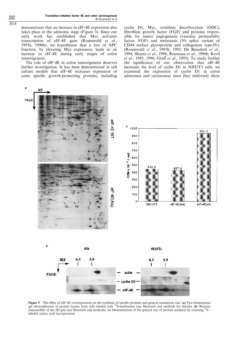
has been shown to be non-functional in that it isunable to transform NIH3T3 cells (Lazaris-Karatzaset al., 1990) and does not increase the cyclin D1 level(Rosenwald et al., 1993b; 1995). Two-dimensional gelelectrophoresis of equal amounts of newly synthesizedtotal protein (normalized by loading of lysatescontaining the same amount of TCA-precipitable 35S-labeled protein) are shown in Figure 5a. ParentalNIH3T3 cells (not shown) and mutant (Ala) eIF-4Eoverexpressing cells showed the same pattern ofprotein expression. Among about a thousand proteinsseparated by 2D gel electrophoresis, we identi®edsigni®cant di�erences in expression of a few proteinsin eIF-4E-overexpressing versus control cells (Ala).The synthesis of proteins represented by spots 1, 2, 3and 4 is higher in wt eIF-4E-overexpressing cells thanin control cells, whereas the synthesis of proteins
indicated by spots 5 and 6 is decreased in wt eIF-4E-overexpressing cells as compared to control cells.These ®ndings are consistent with some preferentiale�ect of eIF-4E on the expression of particularproteins (see Discussion). We could not identify thespot corresponding to cyclin D1 on the autoradio-graphs from the 2D gels, probably because of a slowrate of synthesis of this crucial cell cycle regulator.However, using Western blot analysis of the 2D gels,we were able to demonstrate a strong speci®c increasein cyclin D1 (Figure 5b), which agrees well with ourearlier studies employing 1D gel electrophoresis andWestern blotting (Rosenwald et al., 1993b; 1995) andcon®rms that 2D gel analysis is able to demonstratespeci®c increases of particular proteins in eIF-4Eoverexpressing cells, provided that detection methodis su�ciently sensitive.
Figure 3 Immunohistochemical analysis of eIF-4E and cyclin D1 expression in a representative case of colon villous adenoma. (a)H & E staining (625 original magni®cation); (b) eIF-4E (625); (c) eIF-4E (6100); (d) cyclin D1 (6100)
Translation initiation factor 4E and colon carcinogenesisIB Rosenwald et al
2512

Finally, as demonstrated in Figure 5c, overexpres-sion of wt eIF-4E increases general protein synthesisabout twofold. Taken together, these ®ndings suggestthat elevated activity of eIF-4E can lead to both apreferential increase in the synthesis of some cellularproteins and to a general increase in the synthesis ofmost cellular proteins.
Discussion
Our immunohistochemical study indicates that colonadenocarcinomas display a strong increase in expres-sion of translation initiation factor 4E when comparedto its expression in normal colonic epithelium (Figure2). The Western blot analysis of representative casescon®rms these ®ndings (Figure 4). Quantitativeanalysis shows that bulk of the tumor may have 2 ±6-fold increase in eIF-4E expression as compared tonormal epithelium. Local increase in the margins oftumors may be even higher, as we have noted byimmunohistochemistry that areas of tumors close to orinvading into normal mucosa frequently display higher
levels of eIF-4E than the main bulk of the tumor. Thisis consistent with the fact that margins are theproliferative areas of the tumor, while the cells in thetumor core frequently lack nutrients, do not proliferateand display extensive cell death.
The transformation of normal colonic epithelium toa malignant tumor involves several steps, includinghyperplastic change, adenoma, and carcinoma in situ(Kinzler and Vogelstein, 1996). Many di�erent geneticchanges occur during tumorigenesis. One of the mostfrequent events is inactivation of the adenomatouspolyposis coli (APC) gene found in more than 80% ofboth sporadic and familial cases. The functionalsigni®cance of this gene product is not completelyunderstood, but it appears to be related to transcrip-tional regulation of gene expression (Kinzler andVogelstein, 1996; Morin et al., 1997). It has beenreported very recently that loss of wild-type APC leadsto transcriptional activation of c-myc expressionthrough b-catenin/Tcf-4 complex (He et al., 1998).The loss of APC function takes place early since theinitial pathologic manifestation is multiple adenoma-tous lesions. Our survey of colonic neoplasms
Figure 4 Western blot analysis of eIF-4E expression in representative cases of colon neoplasms. (a) Comparison of actin and CK-LMW as potential normalization standards. Actin expression (as determined by anti-pan actin monoclonal antibody, left picture) inthe stroma and its undetectable expression in the epithelial component precludes the use of this marker for normalization ofWestern blot. In contrast, CK-LMW (right picture) is expressed speci®cally in normal epithelium and in neoplastic components atcomparable levels, while the stroma of the tumors (marked by letter S) and subepithelial connective tissue (marked by letter C) donot express this protein at all. Therefore, CK-LMW is suitable as a marker for normalization of the portion of total protein comingfrom colonic epithelial tissues, while actin is not. (b) Western immunoblot: N, normal mucosa, T, adenocarcinoma, A, adenoma.Cases 1 to 4: tissues were isolated from patients with carcinomas and case 5 from a patient with villous adenoma. Fold increase ofeIF-4E in each neoplasm versus normal colonic epithelium from the same patient (normalized to the sum of CK-LMW species ineach lane) is shown under corresponding lanes
Translation initiation factor 4E and colon carcinogenesisIB Rosenwald et al
2513
demonstrates that an increase in eIF-4E expression alsotakes place at the adenoma stage (Figure 3). Since ourearly work has established that Myc activatestranscription of eIF-4E gene (Rosenwald et al.,1993a, 1996b), we hypothesize that a loss of APCfunction, by elevating Myc expression, leads to anincrease in eIF-4E during early stages of colontumorigenesis.
The role of eIF-4E in colon tumorigenesis deservesfurther investigation. It has been demonstrated in cellculture models that eIF-4E increases expression ofsome speci®c growth-promoting proteins, including
cyclin D1, Myc, ornithine decarboxylase (ODC),®broblast growth factor (FGF) and proteins respon-sible for tumor angiogenesis (vascular permeabilityfactor, FGF) and metastasis (V6 splice variant ofCD44 surface glycoprotein and collagenase type-IV),(Rosenwald et al., 1993b, 1995; De Benedetti et al.,1994; Shantz et al., 1996; Rousseau et al., 1996b; Kevilet al., 1995, 1996; Gra� et al., 1995). To study furtherthe signi®cance of our observation that eIF-4Eincreases the level of cyclin D1 in NIH3T3 cells, weexamined the expression of cyclin D1 in colonadenomas and carcinomas since they uniformly show
Figure 5 The e�ect of eIF-4E overexpression on the synthesis of speci®c proteins and general translation rate. (a) Two-dimensionalgel electrophoresis of protein lysates from cells labeled with 35S-methionine (see Materials and methods for details). (b) Westernimmunoblot of the 2D gels (see Materials and methods). (c) Determination of the general rate of protein synthesis by counting 14C-labeled amino acid incorporation
b
a
c
Translation initiation factor 4E and colon carcinogenesisIB Rosenwald et al
2514

an increase in eIF-4E expression. In 22 out of 34 casesof colon adenocarcinomas cyclin D1 expression wasobviously increased in tumor margins. Our ®ndingsagree well with the results of others demonstrating thatcyclin D1 is often overexpressed in colorectaladenocarcinomas (Maeda et al., 1997). However, sincein 12 cases with eIF-4E elevation only occasional cellswere expressing cyclin D1, we conclude that eIF-4E isnot the only factor that regulates the level of cyclin D1in colon epithelium and that cyclin D1 is not the onlytarget mediating the e�ects of eIF-4E on cellproliferation and transformation. Analysis of cyclinD1 expression in adenomas shows that its expression inthe margins is upregulated in 34 out of 36 cases. Thesedata are in agreement with our results from the studyof other tumors in that eIF-4E is not the sole factordetermining cyclin D1 expression (manuscript inpreparation).
As a strategy for determining the potential of eIF-4Eto increase the expression of speci®c proteins weemployed two-dimensional protein electrophoresis oflysates from control non-functional eIF-4E-overexpres-sing cells (Ala) or parental NIH3T3 cells and wild-typeeIF-4E overexpressing cells (Figure 5a and b). Whileoverexpression of eIF-4E results in a general increaseof protein synthesis (Figure 5c), some proteins aremore markedly increased in their expression (spots 1, 2,3 and 4 in Figure 5a), whereas, the relative synthesis oftwo proteins is decreased (spots 5 and 6). This may beexplained by the fact that some mRNAs can betransferred to ribosomes and translated in a cap-independent manner (Rhoads and Lamphear, 1995).When cap-dependent recruitment of mRNAs intoribosomes becomes more e�cient due to eIF-4Eoverexpression, those cap-independent mRNAs arecompeted out (Rhoads and Lamphear, 1995). Wesuppose that eIF-4E preferentially a�ects synthesis oflow-abundance proteins, like cyclin D1, which was notdetectable in our two-dimensional study when lysatesof 35S-labeled cells were analysed. However, by Westernblotting of duplicate 2D-gels it has been readily foundthat cyclin D1 is strongly increased in wild-type eIF-4Eoverexpressing cells (Figure 5b). Importantly, thegeneral rate of protein synthesis is increased in wild-type eIF-4E overexpressing cells (Figure 5c). Theapparent lack of speci®c changes in the relativesynthesis rates of the overwhelming majority ofproteins resolved by 2D gels (Figure 5a) and generalincrease in protein synthesis rate (Figure 5c) indicatethat eIF-4E equally increases translation of mostcellular mRNAs and only a minor portion of mRNAspecies is preferentially dependent on eIF-4E. Both apreferential increase in the synthesis of speci®c proteinsand a non speci®c increase in net protein synthesis cancontribute to accelerated growth and metastasis. Theproteins speci®cally upregulated by eIF-4E (e.g. cyclinD1, ODC, FGF, Myc, vascular permeability factor, V6splice variant of CD44 surface glycoprotein andcollagenase type-IV, for references see Rosenwald etal., 1993b, 1995; De Benedetti et al., 1994; Shantz etal., 1996; Rousseau et al., 1996b; Kevil et al., 1995,1996; Gra� et al., 1995) would activate proliferativereactions and facilitate metastasis. Increased produc-tion of growth factors and activation of oncoproteinsinvolved in signal transduction lead to increasedexpression and augmented function of translation
initiation factors, creating a positive feedback loop(discussed in Rosenwald, 1996a). Earlier investigationshave demonstrated that Myc expression is increased in100% cases of colon adenocarcinomas and benignadenomas (Melhem et al., 1992). It has beendemonstrated that Myc activates transcription of eIF-4E gene (Rosenwald et al., 1993a; Jones et al., 1996)and, in turn, eIF-4E increases the level of Myc protein(De Benedetti et al., 1994), providing a possibility for apositive feedback loop inside the cell. At the same time,a general increase in protein synthesis would acceleratethe rate of doubling cellular protein content, thusfacilitating cell growth and replication. Finally, over-expression of eIF-4E in cultured cells preventsapoptotic death when concentration of extracellulargrowth factors is low (Polunovsky et al., 1996). Inconclusion, we propose a working model for the role ofeIF-4E in colon tumorigenesis. An initial increase ineIF-4E leads to an outgrowth of benign neoplasm dueto a general increase in protein synthesis, preferentialsynthesis of cell cycle-promoting proteins and diminu-tion of cell death rate. Additional mutations includingthose which cause transcriptional upregulation ofinvasion and metastasis-facilitating proteins and theirtranslational ampli®cation lead to a formation of afull-blown malignancy.
Some recent publications show that eIF-4E expres-sion may be frequently elevated in breast as well as inhead and neck carcinomas (Li et al., 1997; Nathan etal., 1997). Interestingly, cyclin D1 protein expression isincreased in 50% of breast carcinomas, while its gene isampli®ed in approximately 13% of cases (Hall andPeters, 1996). This raises the possibility that increasedeIF-4E expression may be one of the factors leading tocyclin D1 upregulation without gene ampli®cation. Anextensive analysis of di�erent tumors, including coloncancers will further elucidate the role of translationinitiation factor 4E and other translation factors inhuman neoplastic disorders.
Materials and methods
Cell culture and sample preparation
NIH3T3 cells and eIF-4E overexpressing NIH3T3 cellswere cultured in DMEM supplemented with 10% fetal calfserum (FCS), penicillin and streptomycin until grown to75% con¯uence in culture ¯asks (Lazaris-Karatzas et al.,1990). Then cells were collected by brief trypsinization,which was blocked by addition of 10% FCS. After washingonce with PBS, cells were pelleted by centrifugation at 1000r.p.m. in 50 ml falcon tubes. Supernatant was removed and10% formalin was carefully layered on cell pellets. After3 ± 4 days of ®xation, cell pellets were processed forpara�n embedded sections in the same manner as anyother tissue material is handled for pathological analysis.
Immunohistochemical analysis of protein expression
Parental and eIF-4E overexpressing NIH3T3 cells wereprocessed as above. Surgical material and cell pellets werefurther treated identically. The material was ®xed in 10%bu�ered formalin and routinely processed through a VIPTissue Tek processor. Sections were cut at 4 microns,heated at 608C for 30 min, then depara�nized andhydrated through a series of xylenes and alcohol. Thesections for cyclin D1 analysis were antigen retrieved byputting the slides in 1 mM EDTA (pH 8.0) for 5 min in an
Translation initiation factor 4E and colon carcinogenesisIB Rosenwald et al
2515

800 W microwave oven. Following the replenishment ofthis solution the slides were microwaved again for anadditional 5 min and then allowed to cool for 20 min. Thesections for eIF-4E, eIF-2a, actin and low molecularweight cytokeratin (CK-LMW) analysis were antigenretrieved in the same way except the solution used was0.01 M citrate bu�er (pH 6). The histological sections werestained on a TechMate 1000 (Ventana Medical Systems,Tucson, AZ, USA) automated immunostainer using anavidin/biotin complex (ABC) staining procedure. Follow-ing a hydrogen peroxide block of endogenous peroxide anda serum blocking step, the slides were incubated with oneof the primary antibodies: anti-eIF-4E monoclonal anti-body (1 : 400 dilution, Transduction Laboratories, Lexing-ton, KY, USA), anti-eIF-4E polyclonal antibody (1 : 500,provided by N Sonenberg, McGill University, Montreal),anti-eIF-2a monoclonal antibody (1 : 2000 dilution, ob-tained from E Henshaw, Rochester University), anti-CK-LMW antibody (1 : 2, CAM 5.2, Becton Dickinson, No7650), anti-pan actin antibody (1 : 100, DAKO, MO 635)and monoclonal anti-cyclin D1 antibodies (1 : 40, Novocas-tra Laboratories, Newcastle upon Tyne, UK) for 45 min.This was followed by brief bu�er washes and incubation ina cocktail of biotinylated anti-mouse IgG/IgM and anti-rabbit IgG (Ventana) for 30 min. The sections were washedafter that, incubated in avidin/biotin complex (Ventana)for 30 min, washed and reacted with diaminobenzidine andhydrogen peroxide. The sections were counterstained withhematoxylin. For a technical control a duplicate set ofslides was stained by normal mouse serum (preimmune)instead of primary mouse monoclonal antibody andshowed no reactivity. Rabbit preimmune serum was usedas a control for rabbit polyclonal antibody and showed nospeci®c reactivity as well.
We have compared polyclonal and monoclonal anti-eIF-4E antibodies by analysing various tumor samples and founda perfect agreement in results obtained with these twoantibodies (data not shown). Thus, we proceeded to useanti-eIF-4E monoclonal antibody for all studies reported inthis paper.
Western blot protein analysis
Western blot analysis was performed as describedpreviously (Rosenwald et al., 1993b, 1995) except thatprotein lysates were obtained from tissues rather thancultured cells. Tumor tissues were taken immediately afterresection and included areas away from margins of thetumors because we had to make sure that tumor samplesdo not include normal mucosa; also most of the marginalareas and areas close to margins were not available forcollection as most of it was required for diagnosticevaluation. Normal colonic epithelium was obtained fromthe areas of normal mucosa away from tumor by raisingepithelium with forceps, making an initial incision withsmall scissors and dissecting it from the underlying layers.This technique allows to maximally avoid contamination ofepithelium by underlying tissues. Two or three samplesfrom 0.5 ± 1 cm3 were collected from both tumors andnormal mucosa. The collected tissues were immediatelyplaced in a small plastic cup, tops closed, put in liquidnitrogen and transferred for storage in 7708C. In order toobtain protein lysates, the frozen tissues were wrapped byseveral layers of liquid nitrogen-frozen aluminum foil, keptin liquid nitrogen brie¯y, then disrupted by a hummerseveral times on a dry ice-precooled bench surface. Thenaluminum foil containing disrupted tissue was put intoliquid nitrogen again and the cycle of disruption wasrepeated two ± three times. After that, aluminum foil wasunwrapped, and frozen tissue powder was transferred into15 ml Falcon tubes kept on ice. The ice-cold lysis bu�ercontaining 0.5% Nonidet P-40, 420 mM NaCl, 20 mM Tris
pH 7.5, 2 mM PMSF, 0.02 mM leupeptin was layered ontop of disrupted tissue and pipetted several times by atransfer pipette to obtain a suspension. The volume ofbu�er was 4 ± 5-fold that of tissue to be lysed. Eachsuspension was forcefully passed ten times through a 18G1gauge needle to disrupt cellular material following whichthe lysates were kept on ice for 15 min, aliquoted and thanfrozen at 7208C. Before running on the gel, 1 ml of eachlysate was centrifuged in epindorf tubes at 106103 r.p.m.at 48C for 15 min, supernatants collected in a manner toavoid the pellet on the bottom and some ¯oating debri aswell. If supernatant still contained some debri the samplewas centrifuged a second time. Protein content wasanalysed by the Bradford Assay (BioRad). Forty mg oftotal protein from each lysate were run on a 8% SDS-polyacrylamide gel and blotted for 18 h at 20 V in a coldroom onto Immobilon PVDF membrane (Millipore) usinga liquid transfer solution containing 25 mM Tris pH 7.5,192 mM glycin, 10% methanol and 0.01% SDS. Themonoclonal mouse anti-eIF-4E antibody (1 : 1000 dilution,Transduction Laboratories, Lexington, KY, USA), andmonoclonal mouse anti CK-LMW (1 : 50 dilution ofantibody CAM 5.2, Becton Dickinson, No 7650) wereused sequentially, followed by a second HRP-conjugatedanti-mouse IgG (1 : 3000, Promega) to detect correspondingproteins using a ECL developing system (Amersham).
Two-dimensional protein electrophoresis and immunoblotting
Two dimensional gel electrophoresis were performed asdescribed by O'Farrel (1975). Brie¯y, cells were grown inDMEM with 10% FCS until 2/3 con¯uent in 10 cm culturedishes. Cells were washed once with methionine-free mediaand further incubated in a 9 : 1 mixture of methionine-freemedia with normal media (supplemented with 10% FCS)for 1 h. At this time, media was changed for the samemedia containing [35S]methionine, New England Nuclear(330 mCi per 2.5 ml of incubation media). Cells werelabeled for 1 h, washed three times with PBS and lysedby a bu�er specially prepared for running on 2D gel (9.5 M
urea, 2% (w/v) NP-40, 2% ampholines [0.4% pH range 3 ±10 and 1.6% pH range 5 ± 7] and 5% b-mercaptoethanol).A sample of lysate from each cell culture was used todetermine TCA-precipitable counts for normalization ofthe amounts of total newly synthesized protein in eachlysate. Duplicate 2D gels for each culture were loaded withthe lysates adjusted by volume to contain equal amounts ofnewly synthesized proteins (2.56106 c.p.m. per gel).Isoelectrofocusing was followed by SDS ± PAGE (36).After that one gel of each culture was dried and exposedto Kodak ®lm with subsequent analysis of developed ®lmsby Fluor-F MultiImager System (Biorad). Westernimmunoblots of duplicated gels were performed. Poly-clonal rabbit anti-human eIF-4E antibodies (1 : 2500dilution, provided by Dr Sonenberg, McGill University),polyclonal rabbit anti-cyclin D1 antibodies (1 : 1500,Rosenwald et al., 1993b) and monoclonal anti-actinantibody (1 : 5000, N350, Amersham) were used sequen-tially to detect corresponding proteins using ECL devel-oping system (Amersham).
Determination of general protein synthesis
Net protein synthesis was determined as described before(Rosenwald, 1996b). Brie¯y, all cell lines were plated(206103 cell per well) in 24 well culture plates in DMEMwith 10% FCS and antibiotics. Next day, media wasreplaced with the same fresh media supplemented with0.5 mCi/ml of 14C-labeled amino acid mixture (NewEngland Nuclear) in triplicate and cultured for 6 h. Afterlabeling, cells were washed once with PBS precipitated with10% TCA, transferred onto ®ber glass ®lters; ®lters were
Translation initiation factor 4E and colon carcinogenesisIB Rosenwald et al
2516

washed by passing PBS through them under the vacuum,then washed in ethanol and air-dryed. 14C incorporationinto cellular protein was determined. To evaluate the rateof incorporation per cell, parallel unlabeled cultures intriplicate were evaluated for cell numbers and results areexpressed as c.p.m. per cell (Figure 5c).
AcknowledgmentsThis work was supported in part by Johnson & Johnsongrant to IBR, UMMC Pathology Department TeachingGrant to JP and IBR, and NIH grant DK 16272 to J-J C.We thank Dr Kenneth L Rock (Department of Pathology,University of Massachusetts Medical Center) for hissupport and advice during this work. We express ourgratitude to Dr Nahum Sonenberg and Dr Anne-Claude
Gingras (McGill University, Montreal) for providing antieIF-4E polyclonal antibodies. We are grateful to DrGerman Pihan and Jane Wallace (Pathology Department,University of Massachusetts Medical Center) for their helpin processing of frozen tissues into protein lysates andKaren Graham and Kristine Israelian (same department)for their assistance in performing immunohistochemicalanalysis. We also thank Maryam Ra®e-Kolpin (Harvard-MIT Division of Health Sciences and Technology) andRobin Ganesan (University of Massachusetts, Departmentof Molecular Genetics and Microbiology) for their help inscanning and densitometric evaluation of 2D gels andWestern blots. We wish to thank Dr James Michaelson(MGH Cancer Center, Harvard Medical School), Dr RogerKaspar (Brigham Young University, Provo) and DrMichael Hatzler (Department of Pathology, University ofMassachusetts Medical Center) for critically reading thismanuscript.
References
Baldin V, Lukas J, Marcote MJ, Pagano M and Draetta G.(1993). Genes Dev., 7, 812 ± 821.
Bartek J, Bartkova J and Lukas J. (1996). Cur. Opin. CellBiol., 8, 805 ± 814.
De Benedetti A, Joshi B, Gra� JR and Zimmer SG. (1994).Mol. Cell. Di�er., 2, 347 ± 371.
De Benedetti A, Joshi-barve S, Rinker-Scha�er C andRhoads RE. (1991). Mol. Cell Biol., 11, 5435 ± 5445.
De Benedetti A and Rhoads RE. (1990). Proc. Natl. Acad.Sci. USA, 87, 8212 ± 8216.
Epifanova OI. (1977). Intern. Rev. Cytol., 5 (suppl.), 303 ±335.
Gra� JR, Boghaert ER, De Benedetti A, Tudor DL, ZimmerCC, Chan SK and Zimmer SG. (1995). Int. J. Cancer, 60,255 ± 263.
Hall M and Peters G. (1996). Adv. Cancer Res., 68, 67 ± 108.He T-C, Sparks AB, Rago C, Hermeking H, Zawel L, da
Costa LT, Morin PJ, Vogelstein B and Kinzler KW.(1998). Science, 281, 1509 ± 1512.
Hershko A, Mamont P, Shields R and Tomkins GM. (1971).Nature New Biol. (Lond.), 232, 206 ± 211.
Jones RM, Branda J, Jonston KA, Polymenis M, Gadd M,Rustgi A, Callanan L and Schmidt EV. (1996). Mol. Cell.Biol., 16, 4754 ± 4764.
Kevil K, Carter P, Hu B and De Benedetti A. (1995).Oncogene, 11, 2339 ± 2348.
Kevil CG, De Benedetti A, Payne DK, Coe LL, Laroux FSand Alexander JS. (1996). Int. J. Cancer, 65, 785 ± 790.
Kinzler KW and Vogelstein B. (1996). Cell, 87, 159 ± 170.Kozak M. (1994). Biochimie, 76, 815 ± 821.Lazaris-Karatzas A, Montine KS and Sonenberg N. (1990).
Nature (Lond.), 345, 544 ± 546.Lazaris-Karatzas A and Sonenberg N. (1992). Mol. Cell.
Biol., 12, 1234 ± 1238.Li BDL, Liu L, Dawson M and De Benedetti A. (1997).
Cancer, 79, 2385 ± 2390.Maeda K, Chung Y-S, Kang S-M, Ogawa M, Onoda N,
Nakata B, Nishiguchi Y, Ikehara T, Okuno M and SowaM. (1997). Int. J. Cancer, 74, 310 ± 315.
Makin CA, Bobrow LG and Bodmer WF. (1984). J. Clin.Pathol., 37, 975 ± 983.
Melhem MF, Meisler AI, Finley GG, Bryce WH, Jones MO,Tribby II, Pipas JM and Koski RA. (1992). Cancer Res.,52, 5853 ± 5864.
Moll R, Franke WW and Schiller DL. (1982). Cell, 31, 11 ±24.
Morin PJ, Sparks AB, Korinek V, Barker N, Clevers H,Vogelstein B and Kinzler KW. (1997). Science, 275, 1787 ±1790.
Morris DR. (1995). Progr. Nucl. Acid Res. Mol. Biol., 51,339 ± 363.
Nathan C-AO, Liu L, Li BDL, Abreo FW, Nandy I and DeBenedetti A. (1997). Oncogene, 5, 579 ± 584.
O'Farrel PH. (1975). J. Biol. Chem., 250, 4007 ± 4021.Pardee AB, Dubrow R, Hamlin JL and Kletzien RF. (1978).
Ann. Rev. Biochem., 47, 715 ± 750.Polunovsky VA, Rosenwald IB, Thurik AT, White J, Chiang
L, Sonenberg N and Bitterman PB. (1996). Mol. Cell.Biol., 16, 6573 ± 6581.
Rhoads RE. (1991). Curr. Opin. Cell Biol., 3, 1019 ± 1024.Rhoads RE and Lamphear BJ. (1995). Curr. Top. Microbiol.
Immunol., 203, 131 ± 153.Rhoads RE, Joshi B and Minich WB. (1994). Biochimie, 76,
831 ± 838.Rosenwald IB, Rhoads DB, Callanan LD, Isselbacher KJ
and Schmidt EV. (1993a). Proc. Natl. Acad. Sci. USA, 90,6175 ± 6178.
Rosenwald IB, Lazaris-Karatzas A, Sonenberg N andSchmidt EV. (1993b). Mol. Cell. Biol., 13, 7358 ± 7363.
Rosenwald IB, Kaspar R, Rousseau D, Gehrke L, LeBoulchP, Chen J-J, Schmidt EV, Sonenberg N and London IM.(1995). J. Biol. Chem., 270, 21176 ± 21180.
Rosenwald IB. (1995). Cancer Letts, 98, 77 ± 82.Rosenwald IB. (1996a). BioEssays, 18, 243 ± 249.Rosenwald IB. (1996b). Cancer Letts, 102, 113 ± 123.Rousseau D, Gingras A-C, Pause A and Sonenberg N.
(1996a). Oncogene, 13, 2415 ± 2420.Rousseau D, Kaspar R, Rosenwald I, Gehrke L and
Sonenberg N. (1996b). Proc. Natl. Acad. Sci. USA, 93,1065 ± 1070.
Shantz LM, Hu R-H and Pegg AE. (1996). Cancer Res., 56,3265 ± 3269.
Sherr CJ. (1996). Science, 274, 1672 ± 1677.Sonenberg N. (1996). Translational Control. In: Hershey
JWB, Mathews MB and Sonenberg N. (eds). Cold SpringHarbor Laboratory Press: Cold Spring Harbor, pp. 245 ±269.
Weinberg RA. (1995). Cell, 81, 323 ± 330.Zetterberg A and Larsson O. (1991). Cold Spring Harbor
Symposia on Quantitative Biology, 56, 137 ± 147.Zetterberg A, Larsson O andWiman KG. (1995). Curr. Opin.
Cell Biol., 7, 835 ± 842.
Translation initiation factor 4E and colon carcinogenesisIB Rosenwald et al
2517