The Painful Neuroma and the Use of Conduits
10
The Painful Neuroma and the Use of Conduits Emilio Wagner, MD a,b,c, *, Cristian Ortiz, MD a,c After nerve injury, a reparative response inevitably takes place, where neurotrophic factors aid axons to regenerate from the proximal stump, and a wallerian degeneration occurs in the distal axon. Degeneration occurs also for a variable distance on the prox- imal axon. After the period of degeneration, myelinated and unmyelinated fibers grow from the proximal stump, trying to reinnervate the distal stump. 1 If a minimal amount of structures is left of the distal axon, namely, its endoneurial basement membrane, the regenerating fibers reach the end organ. When the complete connective tissue framework is damaged, as in a Sunderland grade IV injury, there is an exaggerated inflammatory response, with extensive deposition of collagen tissue, together with inflammatory cells, and the regenerating fibers do not reach their destination, prolifer- ating in a chaotic manner. The resulting bulb-shaped tissue is known as a neuroma, where an unorganized network of connective tissue is intermingled with nerve fibers, Schwann cells, macrophages, fibroblasts, and myofibroblasts, the latter thought to contribute to pain causing the collagen matrix to contract around nerve fibers. 2 There is a paucity of studies dealing with neuromas in the foot and ankle, and most knowl- edge comes from level 4 studies, with small numbers of patients involved, and most of them come from hand surgery. According to the anatomy of peripheral nerves, each nerve is encircled by an external epineurium. Nerve fascicles are group of nerve fibers embedded in endoneu- rium, correspondingly encircled by perineurium. This structure is the smallest one capable of accepting sutures. 1 Many fascicles are grouped together, surrounded by epineurium, thus forming a classic peripheral nerve. Nerve fibers occupy 25% to The authors have nothing to disclose. a Universidad del Desarrollo, Escuela de Medicina, Avda Las Condes 12438, Las Condes, Santiago, Chile 7550000 b Foot and Ankle Service, Hospital Padre Hurtado, Calle Esperanza 2150, San Ramon, Santiago, Chile 8880465 c Foot and Ankle Service, Clinica Alemana, Avda Vitacura 5951, Vitacura, Santiago, Chile 7650568 * Corresponding author. Foot and Ankle Service, Clinica Alemana, Avda Vitacura 5951, Vitacura, Santiago, Chile 7650568. E-mail address: [email protected] KEYWORDS Neuroma Conduit Graft Transposition Foot Ankle Clin N Am 16 (2011) 295–304 doi:10.1016/j.fcl.2011.01.004 foot.theclinics.com 1083-7515/11/$ – see front matter Ó 2011 Elsevier Inc. All rights reserved.
-
Upload
independent -
Category
Documents
-
view
5 -
download
0
Transcript of The Painful Neuroma and the Use of Conduits

The Painful Neuromaand the Use ofConduits
Emilio Wagner, MDa,b,c,*, Cristian Ortiz, MDa,c
KEYWORDS
� Neuroma � Conduit � Graft � Transposition
After nerve injury, a reparative response inevitably takes place, where neurotrophicfactors aid axons to regenerate from the proximal stump, and a wallerian degenerationoccurs in the distal axon. Degeneration occurs also for a variable distance on the prox-imal axon. After the period of degeneration, myelinated and unmyelinated fibers growfrom the proximal stump, trying to reinnervate the distal stump.1 If a minimal amountof structures is left of the distal axon, namely, its endoneurial basement membrane,the regenerating fibers reach the end organ. When the complete connective tissueframework is damaged, as in a Sunderland grade IV injury, there is an exaggeratedinflammatory response, with extensive deposition of collagen tissue, together withinflammatory cells, and the regenerating fibers do not reach their destination, prolifer-ating in a chaotic manner. The resulting bulb-shaped tissue is known as a neuroma,where an unorganized network of connective tissue is intermingled with nerve fibers,Schwann cells, macrophages, fibroblasts, and myofibroblasts, the latter thought tocontribute to pain causing the collagen matrix to contract around nerve fibers.2 Thereis a paucity of studies dealing with neuromas in the foot and ankle, and most knowl-edge comes from level 4 studies, with small numbers of patients involved, and most ofthem come from hand surgery.According to the anatomy of peripheral nerves, each nerve is encircled by an
external epineurium. Nerve fascicles are group of nerve fibers embedded in endoneu-rium, correspondingly encircled by perineurium. This structure is the smallest onecapable of accepting sutures.1 Many fascicles are grouped together, surrounded byepineurium, thus forming a classic peripheral nerve. Nerve fibers occupy 25% to
The authors have nothing to disclose.a Universidad del Desarrollo, Escuela de Medicina, Avda Las Condes 12438, Las Condes,Santiago, Chile 7550000b Foot and Ankle Service, Hospital Padre Hurtado, Calle Esperanza 2150, San Ramon, Santiago,Chile 8880465c Foot and Ankle Service, Clinica Alemana, Avda Vitacura 5951, Vitacura, Santiago, Chile7650568* Corresponding author. Foot and Ankle Service, Clinica Alemana, Avda Vitacura5951, Vitacura, Santiago, Chile 7650568.E-mail address: [email protected]
Foot Ankle Clin N Am 16 (2011) 295–304doi:10.1016/j.fcl.2011.01.004 foot.theclinics.com1083-7515/11/$ – see front matter � 2011 Elsevier Inc. All rights reserved.

Wagner & Ortiz296
75% of the cross-sectional area of a nerve, compared with the cross-sectional area ofa neuroma, which is occupied by 80% of connective tissue.3 The most commonmechanism of nerve injuries are stretch and contusive forces. Common causesinclude gunshot wounds, fractures, iatrogenic causes, lacerations, and so forth. Clas-sically, nerve lesions have been classified according to Sunderland, where mildlesions with no apparent structural damage are grade I lesions, and grade IV lesionsaffect all component of the nerve architecture.1 Every nerve injury, as contusion, avul-sion, or direct injury, may produce a neuroma. Neuromas are classified in 3 types:neuromas in continuity, neuromas in completely severed nerves, and amputationneuromas.2 This article focuses on neuromas in continuity and after completelysevered nerves.
PATHOPHYSIOLOGY
A neuroma alters normal signal conduction through the nerves, and it affects neigh-boring nerves too. Normal nociceptive responses are augmented, and small stimuligenerate abnormal responses, creating, for example, hyperesthesia. This hyperexcit-ability may be explained by an abnormal accumulation of potassium ion channels andsodium ion channels on the axons in the neuroma.3 This hyperexcited state explainswhat has been called ectopic neuralgia, where a patient suffers spontaneous paindischarges without any external stimulus.4 Locally, the disorganized connective andneural tissue, where the nerve fibers innervate scar tissue and skin, yield neuromassensitive to mechanical stimulation.5 This has been called nociceptive neuralgia.4
PATIENT EVALUATION
A complete understanding of the neural anatomy in the foot and ankle is needed tounderstand and interpret symptoms and clinical signs. A review of the pertinentanatomy is beyond the scope of this article, but it is recommended for understandinghow to proceed with neuroma treatment. The mechanism of injury is important,because it is different when dealing with a gunshot wound or an open fracture, wherenerve scarring is expected, a gradual decrease in nerve function may be observed,and a longer waiting time before intervention is warranted, compared with a clean,sharp injury where immediately afterwards a deficit in nerve function appears anda sooner operative intervention is recommended.Pain in relation to a surgical scar which also radiates along the course of a nerve is
a typical clinical sign of a neuroma. There may be a Tinel sign on top of the scar andaltered sensation in the territory corresponding to that specific nerve. The authorsagree with Mackinnon in that patients who are willing to palpate and massage thearea of pain are not good surgical candidates. The area of discomfort should be painfulto light touch, and pain proximal to the suspected area of neuroma is a frequent finding(sometimes called “Mackinnon sign”) and should be looked for.3 The complete areaand its surroundings should be inspected and palpated to find additional territoriesinnervated by nerves which may be involved in the neuropathic pain. These additionalnerves have to be addressed when performing a surgical intervention.Imaging studies generally are not necessary, except when dealing with previous
open multiple fractures or gunshot wounds, where the exact location of the injury isnot completely clear. Ultrasound imaging may assist in being certain where the lesionis. An MRI can also be obtained, although its utility is best when dealing with bone orsoft tissue tumors, if no history of injury is clear. A history of chronic symptoms mayindicate the need for an electrodiagnostic study. This study is also useful whenfollowing the recovery of an injured nerve. Diabetes and hypothyroidism have to be

The Painful Neuroma and the Use of Conduits 297
ruled out, because they are common systemic disorders that may present with neuro-pathic pain of unknown origin.
TREATMENT
When dealing with a severed nerve, the ideal treatment is a primary direct repair. If thatis not possible, a reconstruction using some sort of nerve conduit or nerve grafting isthe procedure of choice. A nerve repair provides an adequate environment for theproximal regenerating axons to find their way into the distal stump and reach their finalorgan destination. The use of grafts or absorbable or artificial nerve conduits alsoprovide an environment for regenerating axons. For this regeneration process tohappen, we have to achieve ideal local conditions for which a clear knowledge ofthe regional anatomy and magnification loupes are needed. To help decrease localscarring a minimum amount of sutures should be used besides obtaining a tension-free repair.1
If there already is a neuroma, either in a severed nerve or a neuroma in continuity,how critical the function is of the nerve that is worked with and what the consequencesare if the nerve is deleted need to be known. The authors consider nerve function to becritical if its loss will hinder in some way the normal foot function: losing motor functionimportant for normal daily activities (losing intrinsic muscle function in the foot, whichmay create hammer or claw toe deformities, or losing extrinsic muscle function, as ina common peroneal nerve injury) or losing protective sensation in the sole of the footthat renders an insensate foot with the known risks of ulcerations. After analyzing howcritical the function of the injured nerve is, we can plan the necessary intervention.Before surgery, a trial of conservative treatment is warranted, even though the
benefits of it are inconsistent.6,7 Available alternatives include cryotherapy, desen-sitization therapies, therapeutic nerve blocks, transcutaneous electrical nervestimulation, local use of capsaicin-derived gels, oral medications that decrease neuro-pathic pain as pregabalin or gabapentin, acupuncture, and so forth. If these treat-ments fail, a surgical intervention is planned.Simple resection has been used by many surgeons, but there is evidence in the liter-
ature that neuroma resection is not routinely successful.6 The reported reoperationrates have been as high as 65%, which explains the use of additional maneuvers,such as burying the proximal stump into some other tissue.Generally speaking, if dealing with critical nerves, such as the tibial or common
peroneal nerve, the treatment of a neuroma is nerve or conduit grafting. If the nerveis not critical, then a neuroma resection and transfer to a muscular bed is the methodof choice (Figs. 1 and 2).3,5–8
APPROACH TO INJURED NERVES WITH CRITICAL FUNCTION
Critical nerves are the main tibial nerve and the common peroneal nerve. If the entiretibial nerve is injured, there is pain at the injury site with lack of sensation and motorfunction on the distribution of the tibial nerve, posterior tibial muscle, hallux and toeflexors, and intrinsic muscles, as well as loss of sensibility on the plantar aspect ofthe foot. This lack of sensation is badly tolerated and may result in skin breakdownand ulcerations. This skin breakdown is more likely to occur if there are associatedfoot deformities. For the peroneal nerve, lack of sensation in the dorsum of the footand lack of motor function corresponding to the lateral and anterolateral compart-ments of the leg are seen. A nerve reconstruction should be attempted, extendingthe previous surgical scar proximally to find a healthy nerve and distally past theneuroma. A resection of the neuroma and a nerve graft should be used. Most
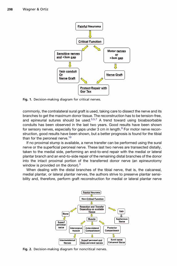
Fig. 1. Decision-making diagram for critical nerves.
Wagner & Ortiz298
commonly, the contralateral sural graft is used, taking care to dissect the nerve and itsbranches to get the maximum donor tissue. The reconstruction has to be tension-free,and epineurial sutures should be used.3,5,7 A trend toward using bioabsorbableconduits has been observed in the last two years. Good results have been shownfor sensory nerves, especially for gaps under 3 cm in length.9 For motor nerve recon-struction, good results have been shown, but a better prognosis is found for the tibialthan for the peroneal nerve.10
If no proximal stump is available, a nerve transfer can be performed using the suralnerve or the superficial peroneal nerve. These last two nerves are transected distally,taken to the medial side, performing an end-to-end repair with the medial or lateralplantar branch and an end-to-side repair of the remaining distal branches of the donorinto the intact proximal portion of the transferred donor nerve (an epineurotomywindow is provided on the donor).3
When dealing with the distal branches of the tibial nerve, that is, the calcaneal,medial plantar, or lateral plantar nerves, the authors strive to preserve plantar sensi-bility and, therefore, perform graft reconstruction for medial or lateral plantar nerve
Fig. 2. Decision-making diagram for noncritical nerves.

The Painful Neuroma and the Use of Conduits 299
injuries. After reconstruction, the graft is protected and the neighboring nerve endswith a nonabsorbable conduit, expanded polytetrafluoroethylene (Preclude SpinalMembrane [Gore & Associates, Flagstaff, AZ, USA]), which has decreased the rateof postsurgical scarring around the reconstruction (Cristian Ortiz, MD, Emilio Wagner,MD, unpublished data, 2010) (Fig. 3). If the calcaneal branch is damaged, it is consid-ered a noncritical function nerve, and thus, it is dissected and transposed proximallyinto a different tissue bed (discussed later). If the grafting fails for the medial or lateralplantar nerves, the authors recommend resecting the neuroma, dissecting it proxi-mally from the main posterior tibial trunk, and transposing it into the soleus muscle.3
APPROACH TO INJURED NERVES WITHOUT CRITICAL FUNCTION
For these type of nerves, because the resulting deficit in function is mild and the dener-vation if a nerve is resected is well tolerated, no reconstruction is attempted. A recon-struction will have a longer recovery time and the surgical effort won’t be rewardedwith an equal return in function. As discussed previously, simple resection ofa neuroma does not suffice in the majority of cases, and, therefore, most investigatorsrecommend adding a transfer of the nerve to a tissue bed, which hopefully willdecrease mechanical and neurotrophic stimuli to avoid painful neuroma formation,provided that this tissue bed is nonmobile or has little mobility and gives adequatemechanical cushioning. Most articles refer to transposing nerves to musclebeds,3–7,11–14 with reported success rates of 80%. In animal models, nerve ends inmuscle beds form rounded smooth bulbs with well-organized nerve fibers nonadher-ent to surrounding tissue.6
Other options have been studied as tissue beds for injured nerves. Translocatingnerves into veins has been reported, with 87% satisfaction, although in a small caseseries.15 In this series, the nerve stump was transposed into a vein in an end-to-endfashion, or in an end-to-side fashion, or into a vein which was then tied distally.6
The article suggests that this option could be better done for smaller sensory nerves.Its advantage is that it uses local freely available tissue, obviating extending the inci-sion in search for muscular or bone tissue as alternatives. Another option using localveins is called the extended autologous venous nerve conduit, referencing the actionof transposing a nerve into a vein and the vein burying into a muscle.6
Translocating the nerve ends into bone is another alternative studied. Resultsranging from 70% to 90% success rates are reported in the literature, all small case
Fig. 3. Preclude Spinal Membrane protecting reconstruction of the posterior tibial branches(plantar medial and lateral).

Wagner & Ortiz300
series.6 Chiodo and Miller16 showed a 75% satisfaction rate for superficial peronealneuromas translocated into the fibula compared with a 46% rate for relief of symptomswhen transposed into the peroneus brevis muscle.Finally, nerve-to-nerve anastomosis has been reported, either end-to-end or end-
to-side anastomosis. The results reported are good, although in very small series.6,17
The authors’ surgical alternative depends on the nerve and the surrounding tissue(see Fig. 1). Generally speaking, when dealing with the superficial peroneal nerve,the authors try to resect the neuroma and translocate it into bone or lateral compart-ment musculature. For the deep peroneal nerve, calcaneal branch of the posteriortibial nerve, intermetatarsal nerve, saphenous nerve, and sural nerve, the authors tryto bury into the nearest muscle, which provides a good resting bed. If the muscle isconsidered too far away from the neuroma, or, if after resection the proximal stumpof the nerve is considered too short, a venous conduit is used, using the vein to routethe nerve into a muscular area.
APPROACH TO INDIVIDUAL NERVESCalcaneal Nerve Neuroma
A neuroma of this nerve can be seen after plantar fasciotomy, calcaneal spur removal,or tarsal tunnel decompression,12 the latter of these the most common in the authors’practice. The surgical approach should be an extension of the previous surgical inci-sion, generally the tarsal tunnel incision after the posterior tibial nerve. The incision hasto be extended proximally and distally if necessary. After identification of the neuroma,it is resected and translocated proximally into the nearest muscle belly, typically theflexor hallucis longus (Fig. 4). Although this approach has good reported results, theflexor hallucis longus may not provide adequate protection because it is a very mobilemuscle. The authors try, if possible, to additionally protect the nerve with a veinconduit.
Intermetatarsal Nerve Neuromas
Intermetatarsal nerve neuromas result from an inadequate initial approach to an inter-metatarsal neuritis, or Morton neuroma. After initial surgery for a true neuroma of thesenerves, the authors favor a plantar longitudinal approach, starting at the level of themetatarsal head distally, which provides good exposure and the possibility of extend-ing the incision proximally if needed (Fig. 5). A dorsal approach could be used if clearsigns of a totally inadequate previous dissection are present as a short nerve reportedin the previous biopsy or an extremely short dorsal incision. After dissecting through
Fig. 4. Transposition of a calcaneal nerve neuroma into the flexor hallucis longus muscle.

Fig. 5. Revision of an intermetatarsal nerve neuroma, plantar longitudinal approach.
The Painful Neuroma and the Use of Conduits 301
the plantar fascia, the corresponding branches of the medial or lateral plantar nervesare identified, the neuroma is resected, and the proximal stump is tagged with absorb-able suture and passed through the interosseus muscles to the dorsum of the foot(Fig. 6). The suture is attached to the subcutaneous layer of the dorsum of the foot,allowing the nerve to rest approximately 1 cm below the skin level. If left with a smallproximal stump after resecting the neuroma, specifically when too near to a proximaldivision of the main trunk of the nerve (medial plantar or lateral plantar nerve), theauthors use a vein conduit to protect the nerve stump, suture it through the epineuriumto the vein, and pass a suture through the distal end of the vein and pass it to thedorsum, suturing it to the dorsal subcutaneous layer, taking care to leave the freeend of the vein 1 cm below the level of the skin.Although it has not been the authors’ experience, it has been suggested that many
of these cases may have a concomitant tarsal tunnel syndrome, which should be ruledout before deciding the specific plan for surgery.14 This entity may be suspected ifmultiple sites of neuropathic pain are present with just one surgery done previouslyor if the pain is present in an atypical intermetatarsal space. In these cases, othercauses should be ruled out as systemic causes of neuropathic pain (discussed previ-ously). A neuroma in the second intermetatarsal space may cause tenderness or havesome referral of pain to the first intermetatarsal space; the third intermetatarsal spaceneuroma may cause symptoms in the fourth intermetatarsal space. Because primaryneuromas are unusual in these spaces (first or fourth intermetatarsal space), imaging,including MRI and ultrasound, may be indicated to confirm the lack of pathology inthese spaces. In addition, patients with tarsal tunnel syndrome complain of pain inthe heel and arch but may be sensitive in all the intermetatarsal spaces. Furthermore,patients with intermetatarsal neuromas complain of pain in the metatarsal region both

Fig. 6. Tagging of the proximal stump of an intermetatarsal nerve after neuroma resectionbefore passing it to the dorsum of the foot.
Wagner & Ortiz302
dorsally and plantarly while also exhibiting sensitivity on testing over the tibial nerveposteromedially at the heel. These observations may lead to confusion if the historyis not carefully taken or these facts are not appreciated.
Sural Nerve Neuroma
Sural nerve neuroma may occur after sural nerve biopsies, which is the most frequentcause seen by the authors. The previous surgical scar should be extended longitudi-nally following the course of the nerve. An additional incision may be used proximallyto recover the nerve on the posterior aspect of the calf, to translocate the nerve intoa muscular bed, generally between the gastrocnemius or soleus muscle.5 The authors’preferred method currently is to transpose the nerve into a local vein in an end-to-endfashion, as suggested by Koch and colleagues,15 because it allows decreasing thesize of the surgical incision.
Saphenous Nerve Neuromas
Injury to the saphenous nerve may occur after saphenous vein harvest for cardiacsurgery or, more frequently in the authors’ center, after orthopedic procedures,namely, ankle fracture treatment. The incision should be placed longitudinally followingthe nerve direction. The neuroma has to be resected and transposed into the leg asproximal as possible. The authors have tried translocating the nerve into thetibia with success but only in few cases (EmilioWagner, MD, Cristian Ortiz, MD, unpub-lished data, 2009). If the neuroma is near the ankle, in young women or patients withhigh cosmetic expectations, the authors do not extend the incision more proximal totranspose it to muscle and use a vein conduit to protect it and hopefully reach

Fig. 7. Anterior aspect of the ankle joint showing scar result of previous distal tibia fracture,with superficial peroneal neuroma symptoms.
The Painful Neuroma and the Use of Conduits 303
a muscular bed.15 If the neuroma is proximal to the ankle, a transfer of the proximalstump to the posterior musculature is possible.
Superficial Peroneal Nerve
The superficial peroneal nerve may be injured in the lower leg when fasciotomies areperformed at the level of the ankle when fractures are addressed or in the anterior partof the ankle when ankle arthroscopies are done (Fig. 7). A Tinel sign is present, andpain and numbness in relation to the territory innervated by the corresponding branchare present. When the neuroma is present proximal to the ankle, a resection of theneuroma and a translocation of the nerve to the lateral muscular compartment isthe rule.3 Transposing it into the fibula is another alternative with good reportedresults.16 When present at the level of the ankle, an extended longitudinal incisionshould be done to translocate the proximal stump proximally into the anteriormuscular compartment. When using either muscular compartment, a fasciotomy ofthe corresponding compartment has to be done, because it prevents recurrence ofpain decreasing the pressure of the new nerve environment.13
SUMMARY
Treatment of neuromas in the foot and ankle is evolving. There is a paucity of studiesdealing with neuromas in this anatomic region and most knowledge comes from handsurgery. A trend toward reconstructive surgery using nerve grafts and conduits fornerves with critical function is being seen, including the use of artificial conduits formotor nerves. For noncritical nerves, the most generally accepted treatment isneuroma resection and burial into a tissue bed, commonly muscle, which protectsthe proximal stump and avoids the generation of a painful neuroma. A clear knowledgeof the neural anatomy is paramount together with correct identification of all the nervesinvolved in the pain generation process. More studies dealing with neuromas in thisarea are needed to provide evidence-based information.
REFERENCES
1. Midha R, Mackay M. Principles of nerve regeneration and surgical repair. SeminNeurosurg 2001;12(1):81–92.
2. Mavrogenis A, Pavlakis K, Satamatoukou A, et al. Current treatment concepts forneuromas-in-continuity. Injury 2008;39:43–8.
Wagner & Ortiz304
3. Vernadakis A, Koch H, Mackinnon S. Management of neuromas. Clin Plast Surg2003;30:247–68.
4. Vora A, Schon L. Revision peripheral nerve surgery. Foot Ankle Clin 2004;9(2):305–18.
5. Mackinnon S. Neuromas. Foot Ankle Clin 1998;3(3):385–404.6. Wu J, Chiu D. Painful neuromas: a review of treatment modalities. Ann Plast Surg
1999;43(6):661–7.7. Glazebrook M, Paletz J. Treatment of posttraumatic injuries to the nerves in the
foot and ankle. Foot Ankle Clin 2006;11:183–90.8. Lewin-Kowalik J, Marcol W, Kotulska K, et al. Prevention and management of
painful neuroma. Neurol Med Chir (Tokyo) 2006;46:62–8.9. Moore A, Kasukurthi R, Magill C, et al. Limitations of conduits in peripheral nerve
repairs. Hand (N Y) 2009;4:180–6.10. Rosson G, Williams E, Dellon A. Motor nerve regeneration across a conduit.
Microsurgery 2009;29:107–14.11. Nahabedian M, Johnson C. Operative management of neuromatous knee pain:
patient selection and outcome. Ann Plast Surg 2001;46:15–22.12. Kim J, Dellon A. Neuromas of the calcaneal nerves. Foot Ankle Int 2001;22(11):
890–4.13. Dellon A, Aszmann O. Treatment of superficial and deep peroneal neuromas by
resection and translocation of the nerves into the anterolateral compartment. FootAnkle Int 1998;19:300–3.
14. Wolfort S, Dellon A. Treatment of recurrent neuroma of the interdigital nerve byimplantation of the proximal nerve into muscle in the arch of the foot. J Foot AnkleSurg 2001;40(6):404–10.
15. Koch H, Hubmer M, Welkerling H, et al. The treatment of painful neuroma on thelower extremity by resection and nerve stump transplantation into a vein. FootAnkle Int 2004;25(7):476–81.
16. Chiodo C, Miller S. Surgical treatment of superficial peroneal neuroma. Foot AnkleInt 2004;25(10):689–94.
17. Lidor C, Hall R, Nunley J. Centrocentral anastomosis with autologous nerve grafttreatment of foot and ankle neuromas. Foot Ankle Int 1996;17(2):85–8.