Effects of painful stimulation and acupuncture on attention networks in healthy subjects
11
RESEARCH Open Access Effects of painful stimulation and acupuncture on attention networks in healthy subjects Gang Liu 1 , Hui-juan Ma 1 , Pan-pan Hu 1 , Yang-hua Tian 1 , Shen Hu 1 , Jin Fan 2,3 and Kai Wang 1* Abstract Pain is a subjective sensory and emotional experience, and it has been reported that many different brain regions are regulated by pain, and that pain can impact attention. Acupuncture is an important treatment component of Chinese traditional medicine, and has been used for thousands of years to treat a wide variety of conditions. Although several studies have shown that acupuncture improves consciousness, the precise impact of both acupuncture and painful stimulation on attention is unclear. Are all of the attention networks modulated, or do these stimuli act on a specific network? Is the effect of painful stimulation similar to that of acupuncture? We administered the attention network test to 30 participants (15 males) to investigate the relative efficiencies of three independent attention networks (alerting, orienting, and executive control networks) under three conditions: baseline, after painful stimulation, and after acupuncture. The degree of pain experienced was assessed on a horizontally oriented visual analogue scale. The results showed that painful stimulation and acupuncture had similar effects on the orienting and executive control networks; however, there was a significantly different effect between the three conditions on the alerting network. In conclusion, (1) painful stimulation can selectively impact attention; (2) acupuncture can also selectively impact attention; i.e., both have selective influences on the alerting and executive control networks, but not on the orienting network; (3) the effects of acupuncture and painful stimulation are not identical. The mechanisms by which painful stimulation and acupuncture influence attention warrant further research. Keywords: Painful stimulation, Acupuncture, Attention networks, Alerting network, Orienting network, Executive control network Introduction Pain Pain is one of the most studied topics in neuroscience, and an almost ubiquitous symptom in a clinical setting. Pain has been defined as “an unpleasant sensory and emotional experience associated with actual or potential tissue damage, or described in terms of such damage” by the International Association for the Study of Pain [1]. It signals danger, and urges action to avoid further damage. When the body is hurt, for example pricked, cut, squeezed, burned, or frozen, nociceptors in the periphery respond, and the central nervous system acts to avoid or alleviate injuries. The properties of pain are related to the underlying pathological process, with its location generally indicates where the damage has occurred. Research has demonstrated the involvement of both the peripheral and central nervous systems in pain processing. Pain involves a multifactorial, multipathway system, and each component is thought to be regulated by different brain regions. Human brain imaging studies have identi- fied nociceptive nerve fibers passing through the nuclei of the thalamus, and projecting to higher cortical areas via two main pathways—the lateral and the medial. The lateral pain pathway travels through the ventral postero- lateral nucleus and the ventral posteromedial nucleus of the thalamus, and then projects to the primary and secondary somatosensory cortices. The medial pain path- way travels through more medial cortical areas, including the anterior cingulate cortex (ACC), insula, amygdala, and hypothalamus. These two pathways are highly integrated, and both are indispensable for normal pain perception. * Correspondence: [email protected] 1 Department of Neurology, The First Hospital of Anhui Medical University, Hefei, Anhui Province, P. R. China Full list of author information is available at the end of the article © 2013 Liu et al.; licensee BioMed Central Ltd. This is an Open Access article distributed under the terms of the Creative Commons Attribution License (http://creativecommons.org/licenses/by/2.0), which permits unrestricted use, distribution, and reproduction in any medium, provided the original work is properly cited. Liu et al. Behavioral and Brain Functions 2013, 9:23 http://www.behavioralandbrainfunctions.com/content/9/1/23
Transcript of Effects of painful stimulation and acupuncture on attention networks in healthy subjects

RESEARCH Open Access
Effects of painful stimulation and acupuncture onattention networks in healthy subjectsGang Liu1, Hui-juan Ma1, Pan-pan Hu1, Yang-hua Tian1, Shen Hu1, Jin Fan2,3 and Kai Wang1*
Abstract
Pain is a subjective sensory and emotional experience, and it has been reported that many different brain regionsare regulated by pain, and that pain can impact attention. Acupuncture is an important treatment component ofChinese traditional medicine, and has been used for thousands of years to treat a wide variety of conditions.Although several studies have shown that acupuncture improves consciousness, the precise impact of bothacupuncture and painful stimulation on attention is unclear. Are all of the attention networks modulated, or dothese stimuli act on a specific network? Is the effect of painful stimulation similar to that of acupuncture? Weadministered the attention network test to 30 participants (15 males) to investigate the relative efficiencies of threeindependent attention networks (alerting, orienting, and executive control networks) under three conditions:baseline, after painful stimulation, and after acupuncture. The degree of pain experienced was assessed on ahorizontally oriented visual analogue scale. The results showed that painful stimulation and acupuncture had similareffects on the orienting and executive control networks; however, there was a significantly different effect betweenthe three conditions on the alerting network. In conclusion, (1) painful stimulation can selectively impact attention;(2) acupuncture can also selectively impact attention; i.e., both have selective influences on the alerting andexecutive control networks, but not on the orienting network; (3) the effects of acupuncture and painful stimulationare not identical. The mechanisms by which painful stimulation and acupuncture influence attention warrantfurther research.
Keywords: Painful stimulation, Acupuncture, Attention networks, Alerting network, Orienting network, Executivecontrol network
IntroductionPainPain is one of the most studied topics in neuroscience,and an almost ubiquitous symptom in a clinical setting.Pain has been defined as “an unpleasant sensory andemotional experience associated with actual or potentialtissue damage, or described in terms of such damage” bythe International Association for the Study of Pain [1]. Itsignals danger, and urges action to avoid further damage.When the body is hurt, for example pricked, cut, squeezed,burned, or frozen, nociceptors in the periphery respond,and the central nervous system acts to avoid or alleviateinjuries. The properties of pain are related to the underlying
pathological process, with its location generally indicateswhere the damage has occurred.Research has demonstrated the involvement of both the
peripheral and central nervous systems in pain processing.Pain involves a multifactorial, multipathway system, andeach component is thought to be regulated by differentbrain regions. Human brain imaging studies have identi-fied nociceptive nerve fibers passing through the nucleiof the thalamus, and projecting to higher cortical areasvia two main pathways—the lateral and the medial. Thelateral pain pathway travels through the ventral postero-lateral nucleus and the ventral posteromedial nucleusof the thalamus, and then projects to the primary andsecondary somatosensory cortices. The medial pain path-way travels through more medial cortical areas, includingthe anterior cingulate cortex (ACC), insula, amygdala, andhypothalamus. These two pathways are highly integrated,and both are indispensable for normal pain perception.
* Correspondence: [email protected] of Neurology, The First Hospital of Anhui Medical University,Hefei, Anhui Province, P. R. ChinaFull list of author information is available at the end of the article
© 2013 Liu et al.; licensee BioMed Central Ltd. This is an Open Access article distributed under the terms of the CreativeCommons Attribution License (http://creativecommons.org/licenses/by/2.0), which permits unrestricted use, distribution, andreproduction in any medium, provided the original work is properly cited.
Liu et al. Behavioral and Brain Functions 2013, 9:23http://www.behavioralandbrainfunctions.com/content/9/1/23

Moreover, these areas are thought to be involved in sen-sory, motor, emotional, memory, and attentional processes.
Pain and attentionIt is widely known that pain serves as a warning mechan-ism of danger to the individual: it interrupts, distracts, anddemands attention. Moreover, attention deficit is one ofthe most frequently reported accompanying symptoms inpeople experiencing either acute or chronic pain.Many clinical studies have found that pain impacts
attentional processes [2], and the brain regions whereattention is modulated by pain have been identified byfunctional magnetic resonance imaging [3]; the ACCand thalamus are known to play key roles in both painperception and attentional tasks, and pain and attentioncan both activate the same specific brain areas [4]. How-ever, it remains to be determined whether people experi-encing pain have deficits in global attention, or in aspecific attention network.
Attention networksOn the basis of many neuroanatomical and neuropsycho-logical studies, Posner divided the human attention systeminto three independent networks: the alerting, orienting,and executive control networks. These networks havebeen distinguished at both the biochemical and cognitivelevels, and have distinct neuroanatomical correlates [5-8].The alerting network is involved in the individual’s abilityto tonically maintain an alert state, and in producing aphasic response to a warning signal. Its function is criticalfor optimal performance. It is localized to the thalamusand frontal and parietal areas of the right hemisphere,and primarily utilizes the norepinephrine system. Theorienting network is associated with selectively focusingon one, or a few items, out of many candidates. Previousstudies have revealed that the orienting network consistsof the temporal–parietal junction, superior and inferiorparietal lobe, frontal eye fields, and the pulvinar and re-ticular nuclei of the thalamus, and is modulated by thecholinergic system. Finally, the executive control networkrefers to the ability to monitor and resolve conflicts inthe presence of competing information. Neuroimagingand neuropathological studies have revealed that the ACCand lateral prefrontal cortex are involved in executivecontrol [9-16].The attention network test (ANT) can simultaneously
measure the activity of all three networks, and evaluatetheir interrelationships [17], and all test results can beobtained within 20 min. Therefore, the ANT has beenwidely used to investigate attentional function both inhealthy subjects, and in patients with disorders such asschizophrenia, Alzheimer’s disease, and borderline person-ality disorder, among others [18-25].
Acupuncture and attentionAcupuncture is an ancient eastern healing methodology,which is effective in the treatment of various brain disor-ders, such as psychiatric diseases [26,27], insomnia [28],addiction [29,30], and stroke [31,32]. A review of the lit-erature suggests that acupuncture can indeed influencebrain activity [33-35], and modulation of several corticaland subcortical regions, including the insula, amygdala,ACC, and thalamus has been reported [36]. These regionsare also highly involved in the limbic system, which playsa significant role in regulating and integrating cognition,emotion, sensory perception, and behavior. However,the neural mechanisms activated by acupuncture are ex-tremely complicated and incompletely understood; ingeneral, acupuncture modulates endogenous regulatorysystems, including the autonomic nervous system, theendocrine system, and the neuroendocrine system, andexerts its effects not only through local reflexes but alsothrough the central nervous system [37].The impact of acupuncture on attention, and its three
independent networks, is also unclear. Acupuncture issometimes accompanied by the sensation of pain; how-ever, compared to painful stimulation, acupuncture ap-pears to have a more complex neural mechanism. Is theeffect of painful stimulation similar to that of acupunc-ture? Do painful stimulation and acupuncture modulateall of the three attention networks, or only one or twospecific networks? The present study aimed to investi-gate and compare the impact of painful stimulationand acupuncture on attention using the ANT. Wehypothesize that: (1) painful stimulation can impact at-tention; (2) acupuncture can impact attention; (3) theeffects of acupuncture and painful stimulation on at-tention are similar.
MethodsSubjectsIn order to reduce intersubject variability, participantsconsisted of a homogeneous group of 30 college students[15 male, 15 females, aged 23.5 ± 1.2 (mean ± SD) years].In order to exclude any effects of practice, we recruitedanother 30 college students [15 male, 15 female, aged 22.3 ±1.4 (mean ± SD) years] as a control group to perform theANT three times in a similar environment and on a similarschedule. All subjects were recruited from Anhui MedicalUniversity located in Hefei City, Anhui Province, China.All were right handed, and acupuncture naive. They allhad normal speaking, writing, language expression, andcognitive skills. None had any history of serious physicaldisorders or of mental illness. Our study protocol wasapproved by the Anhui Medical University Ethics Com-mittee, and written informed consent was obtained fromall participants before the study.
Liu et al. Behavioral and Brain Functions 2013, 9:23 Page 2 of 11http://www.behavioralandbrainfunctions.com/content/9/1/23
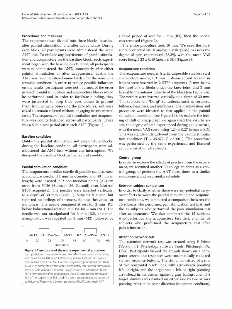
Procedures and measuresThe experiment was divided into three blocks: baseline,after painful stimulation, and after acupuncture. Duringeach block, all participants were administered the sameANT task. To exclude any interference of painful stimula-tion and acupuncture on the baseline block, each experi-ment began with the baseline block. Then, all participantswere re-administered the ANT, immediately after eitherpainful stimulation or after acupuncture. Lastly, theANT was re-administered immediately after the remainingstimulus condition. In order to reduce possible influenceson the results, participants were not informed of the orderin which painful stimulation and acupuncture blocks wouldbe performed, and in order to facilitate blinding, theywere instructed to keep their eyes closed to preventthem from actually observing the procedures, and wereasked to remain relaxed without engaging in any mentaltasks. The sequence of painful stimulation and acupunc-ture was counterbalanced across all participants. Therewas a 5-min rest period after each ANT (Figure 1).
Baseline conditionUnlike the painful stimulation and acupuncture blocks,during the baseline condition, all participants were ad-ministered the ANT task without any interruption. Wedesigned the baseline block as the control condition.
Painful stimulation conditionThe acupuncture needles (sterile disposable stainless steelacupuncture needle, 0.3 mm in diameter and 40 mm inlength) were inserted in 2 non-meridian points (2–3 cmaway from ST36 [Stomach 36, Zusanli] near bilateralST36 acupoints). The needles were inserted vertically,to a depth of 30 mm (Table 1). Subjects felt pain, butreported no feelings of soreness, fullness, heaviness ornumbness. The needle remained at rest for 2 min (R1)before bidirectional rotation at 1 Hz for 2 min (M1). Theneedle was not manipulated for 3 min (R2), and then,manipulation was repeated for 2 min (M2), followed by
a third period of rest for 1 min (R3), then the needlewas removed (Figure 2).The entire procedure took 10 min. We used the hori-
zontally oriented visual analogue scale (VAS) to assess thedegree of pain experienced [38,39], with the mean VASscore being 5.23 ± 0.49 (mean ± SD) (Figure 3).
Acupuncture conditionThe acupuncture needles (sterile disposable stainless steelacupuncture needle, 0.3 mm in diameter and 40 mm inlength) were inserted in 2 ST36 acupoints (5 mm belowthe head of the fibula under the knee joint, and 2 mmlateral to the anterior tubercle of the tibia) (see Figure 2A).The needles were inserted vertically, to a depth of 30 mm.The subjects felt “De-qi” sensations, such as soreness,fullness, heaviness, and numbness. The manipulation andprocedure were identical to that applied in the painfulstimulation condition (see Figure 2B). To exclude the feel-ing of dull or sharp pain, we again used the VAS to as-sess the degree of pain experienced during acupuncture,with the mean VAS score being 1.24 ± 0.27 (mean ± SD).This was significantly different from the painful stimula-tion condition (T = 35.877, P < 0.001). The procedurewas performed by the same experienced and licensedacupuncturist on all subjects.
Control groupIn order to exclude the effects of practice from the experi-ment, we recruited another 30 college students as a con-trol group, to perform the ANT three times in a similarenvironment and on a similar schedule.
Between-subject comparisonIn order to clarify whether there were any potential carry-over effects between the painful stimulation and acupunc-ture conditions, we conducted a comparison between the15 subjects who performed pain stimulation test first, andthe 15 subjects who performed the pain stimulation testafter acupuncture. We also compared the 15 subjectswho performed the acupuncture test first, and the 15subjects who performed the acupuncture test afterpain stimulation.
Attention network testThe attention network test was created using E-Prime(Version 1.1, Psychology Software Tools, Pittsburgh, PA,USA). Participants viewed the stimuli shown on a com-puter screen, and responses were automatically collectedvia two response buttons. The stimuli consisted of a rowof five horizontal black lines, with arrowheads pointingleft or right, and the target was a left or right pointingarrowhead in the center, against a gray background. Thetarget stimulus was flanked on either side by two arrowspointing either in the same direction (congruent condition),
ANT1 R1 Pain/Acu ANT2 R2 Acu/Pain ANT3
0 20 25 35 55 60 70 90Time (min)
Figure 1 Time course of the entire experimental procedure.Each participant was administered the ANT three times: at baseline,after painful stimulation, and after acupuncture. First, all participantswere administered the ANT1 without any interruption (Baseline). Then,all were re-administered the ANT2 immediately after painful stimulation(Pain) or after acupuncture (Acu). Lastly, all were re-administered theANT3 immediately after acupuncture (Acu) or after painful stimulation(Pain). The sequence of Pain and Acu were counterbalanced across allparticipants. There was a 5-min rest period (R1, R2) after each ANT.
Liu et al. Behavioral and Brain Functions 2013, 9:23 Page 3 of 11http://www.behavioralandbrainfunctions.com/content/9/1/23

or in the opposite direction (incongruent condition), or bynothing (neutral condition). Participants were instructed tofocus on a centrally located stationary cross throughout thetask, and to respond as quickly and accurately as possible.The participant’s task was to identify the direction of thecenter arrow by pressing one button for the left directionwith the index finger of their left hand, or a second buttonfor the right direction with the index finger of their righthand. The target stimulus remained on the screen untilthe participant responded, but the maximum responsetime was cut off at 1700 ms. Cues consisted of an asteriskappearing for 100 ms, presented 400 ms before the pres-entation of the target. There were four cue conditions inthe process: (1) no cue, the participant was shown across at the same location as the first stationary crossfor 100 ms; (2) a center cue, an asterisk was presentedon the central point; (3) a double cue, an asterisk waspresented at two target locations simultaneously, aboveand below the central point; and (4) a spatial cue, anasterisk was presented at a target location either aboveor below the central point. The whole process of theANT consisted of a 24-trial practice block, and threeexperimental blocks of trials. The presentation of trials
was in a random order for each participant. Each experi-mental block consisted of 96 trials (48 different conditions:4 cue types × 2 target locations × 2 target directions × 3congruencies, with 2 repetitions). The entire ANT wascompleted in 20 min (Figure 4).
Calculation of attention network efficienciesThe ANT makes use of differences in reaction times (RTs)derived from the different experimental conditions to meas-ure the efficiency of the alerting, orienting, and executivecontrol networks [17]. Alerting efficiency was calculated bysubtracting the mean RTs of the conditions with doublecues from those of the conditions with no cue, as neitherof these conditions provided any information on thespatial location of the target. Similarly, orienting effi-ciency was calculated by subtracting the mean RTs ofthe conditions with spatial cues from those of the condi-tions with center cues. In both conditions, the subjectwas alert, but only the spatial cue provided orientationinformation. Likewise, executive control efficiency was cal-culated by subtracting the mean RTs of congruent targetconditions from those of incongruent target conditions.
Table 1 Procedure used for the painful stimulation and acupuncture conditions
Painful stimulation Acupuncture
Location non-meridian points (2–3 cm away from ST36) ST36
Needle gauge 0.3 mm in diameter and 40 mm in length 0.3 mm in diameter and 40 mm in length
Needle insertion depth under the skin 30 mm under the skin 30 mm
Insert direction vertically vertically
Bneedle in needle out
0 2 4 7 9 10Time (min)
R1 M1 R2 M2 R3
A
Figure 2 Painful stimulation and acupuncture conditions: A ST36: 5 mm below the head of the fibula under the knee joint, and 2 mmlateral to the anterior tubercle of the tibia. B shows the paradigm for painful stimulation and acupuncture: The needle remained at rest for 2 min(R1) before bidirectional rotation at 1 Hz for 2 min (M1). The needle was not manipulated for 3 min (R2), and then, manipulation was repeated for 2 min(M2), followed by a third period of rest for 1 min (R3). Then the needle was removed.
Liu et al. Behavioral and Brain Functions 2013, 9:23 Page 4 of 11http://www.behavioralandbrainfunctions.com/content/9/1/23

Data analysisStatistical analysis was performed using SPSS (Version13.0, Statistical Program for Social Sciences, SPSS Inc.,Chicago, IL, USA). The statistical significance of differ-ences between the baseline, after painful stimulation,and after acupuncture conditions was evaluated by a
one-way analysis of variance (ANOVA); the between-subject comparison was evaluated using an independentsamples t-test, and the threshold for statistical signifi-cance was set at P < 0.05. To assess differences betweenindividual conditions, a Student–Newman–Keuls (SNK)test was also used.
Figure 3 Visual Analogue Scale of pain (VAS). VAS is presented as a ruler with a movable cursor, the length being approximately 10 cm.“0” represents “No pain,” while “10” represents “Agonizing.” When the test began, subjects moved the cursor to the position on the ruler thatbest represented the degree of pain they were currently experiencing.
(b) Target conditions
(c) Presentation time course
(a) Cue conditions
Figure 4 Experimental paradigm for the attention network test. (a) The four cue conditions. (b) The six stimuli used in the presentexperiment. (c) An example of the procedure and the time course of a trial using a spatial cue with incongruent flankers.
Liu et al. Behavioral and Brain Functions 2013, 9:23 Page 5 of 11http://www.behavioralandbrainfunctions.com/content/9/1/23

ResultsReaction times and accuracyThe mean RTs and accuracy rates for each of the threeconditions are summarized in Table 2. During the experi-ment, any incorrect or missed responses were excludedfrom the data set. We conducted a 4 (cue condition: centercue, double cue, no cue, spatial cue) × 3 (flanker type: con-gruent, incongruent, neutral) repeated measures ANOVAon the RT data listed in Table 2. There was a signifi-cant main effect for both cue condition and flanker type([i] baseline: cue condition, F (3, 87) = 147.225, P < 0.001;flanker type, F (2, 58) = 345.498, P < 0.001; interactionof cue conditions × flanker types, F(6, 174) = 8.626,P < 0.001; [ii] after painful stimulation: cue condition,F (3, 87) = 167.751, P < 0.001; flanker type, F (2, 58) =382.327, P < 0.001; interaction of cue condition × flankertype, F(6, 174) = 18.639, P < 0.001; and [iii] after acupunc-ture: cue condition, F (3, 87) = 250.158, P < 0.001; flankertype, F (2, 58) = 415.649, P < 0.001; interaction of cuecondition × flanker type, F(6, 174) = 20.530, P < 0.001).We conducted an analysis of variance with repeated
measures for accuracy as well, but there were no signifi-cant main effects of cue conditions or flanker type in thethree conditions (Table 2).
Effects of painful stimulation and acupuncture on thethree attention networksThe effects of the three attention networks in three con-ditions are summarized in Table 3. There was a signifi-cant main effect of the three conditions on the alertingnetwork tasks (F (2, 87) = 9.200, P < 0.001). Our resultsshow that participants were significantly more vigilantfollowing painful stimulation or acupuncture than in thebaseline condition (SNK, P < 0.05), and the comparisonbetween painful stimulation and acupuncture conditionsrevealed a significant difference (SNK, P < 0.05). Therewas a significant main effect of the three conditions onthe executive control network tasks (F(2, 87) = 8.811,P < 0.01). Participants had less difficulties in resolvingconflict after either painful stimulation or acupuncturethan in the baseline condition (SNK, P < 0.05), whilethere was no significant difference between the painfulstimulation and acupuncture conditions (SNK, P > 0.05).However, there were no significant differences betweenthe three conditions on the orienting network tasks(F(2, 87) = 2.398, P > 0.05). Thus, participants exhibitspecific improvements in the performance of the alertingand executive control networks after either painful stimu-lation or acupuncture, but there was no impact on thefunction of the orienting network. There also was a sig-nificant main effect of the three conditions on the overallmean RTs (F(2, 87) = 23.238, P < 0.01). Participants tooksignificantly less time to finish the test following eitherpainful stimulation or acupuncture than they did in the
baseline condition (SNK, P < 0.05). There was no sig-nificant difference between overall mean RTs after eitherof the two interferences (SNK, P > 0.05). Additionally,there was no significant difference observed in responseaccuracy between the three conditions (F (2, 87) = 0.811,P > 0.05) (Figure 5).
Correlation between the VAS and the attention networksWe examined the relationship between the VAS and thescores of the three attentional network tests. No signifi-cant correlations were identified between the VAS andthe three network scores for either the painful stimula-tion or the acupuncture condition ([i] the painful stimu-lation: VAS and alerting, r = 0.218, P > 0.05; VAS andorienting, r = 0.097, P > 0.05; VAS and executive con-trol, r = 0.273, P > 0.05; [ii] the acupuncture condition:VAS and alerting, r = 0.065, P > 0.05; VAS and orienting,r = 0.071, P > 0.05; VAS and executive control, r =−0.125, P > 0.05).
Control group resultsThere were no significant differences observed in alerting,orienting, and executive control networks between thethree trials performed by the control group ([i] Alerting:F (2, 87) = 0.070, P > 0.05; [ii] Orienting: F (2, 87) =0.361, P > 0.05; [iii] Executive control: F (2, 87) = 0.168,P > 0.05).
Between-subject comparison resultsThere were no significant differences observed in thealerting, orienting, or executive control networks in abetween-subject comparison of the pain stimulation andthe acupuncture conditions ([i] pain stimulation: Alerting,T = 0.364, P > 0.05; Orienting, T = 0.348, P > 0.05; Execu-tive control, T = 0.071, P > 0.05); [ii] acupuncture condi-tion: Alerting, T = 0.006, P > 0.05; Orienting, T = 0.921,P > 0.05; Executive control, T = 0.795, P > 0.05).
DiscussionOur study used the ANT to measure participant’s per-formance under baseline conditions, after painful stimula-tion, and after acupuncture, in order to investigate howpainful stimulation and acupuncture might impact thethree distinct networks of attention. Painful stimulationwas used as an independent variable, and the subjectiveexperience of pain as measured by VAS was significantlydifferent between the painful stimulation and acupunctureconditions. No significant correlations were identifiedbetween VAS and the three network scores for eitherthe painful stimulation or the acupuncture conditions.Our experimental results exclude any significant effectsof practice on the outcome, and confirmed there wereno carryover effects between the painful stimulationand acupuncture conditions. We found that there were
Liu et al. Behavioral and Brain Functions 2013, 9:23 Page 6 of 11http://www.behavioralandbrainfunctions.com/content/9/1/23

Table 2 Mean reaction times and accuracy under each cue condition for baseline, after painful stimulation, and after acupuncture
Condition No cue Double cue Center cue Spatial cue
Congruent Incongruent Neutral Congruent Incongruent Neutral Congruent Incongruent Neutral Congruent Incongruent Neutral
Mean RTs (ms) and standard deviations
Baseline 609 (82) 715 (93) 563 (69) 570 (74) 697 (87) 521 (66) 575 (77) 706 (90) 531 (69) 543 (72) 640 (86) 493 (59)
After painful stimulation 593 (75) 686 (87) 557 (61) 554 (69) 664 (86) 497 (63) 5560 (75) 673 (85) 500 (64) 524 (74) 608 (93) 476 (62)
After acupuncture 592 (82) 686 (84) 553 (72) 545 (76) 656 (89) 486 (64) 547 (76) 664 (91) 493 (71) 516 (77) 597 (91) 469 (62)
ACC and standard deviations
Baseline 0.99 (0.01) 0.97 (0.04) 0.99 (0.01) 0.99 (0.01) 0.98 (0.03) 1.00(0.00) 0.99 (0.01) 0.97 (0.05) 0.99 (0.02) 1.00 (0.00) 0.98 (0.03) 0.99 (0.01)
After painful stimulation 0.99 (0.01) 0.97 (0.05) 0.99 (0.01) 0.99 (0.01) 0.98 (0.03) 1.00 (0.00) 1.00 (0.00) 0.97 (0.06) 0.99 (0.01) 0.99 (0.01) 0.99 (0.02) 0.99 (0.01)
After acupuncture 0.99 (0.01) 0.96 (0.06) 0.99 (0.01) 0.99 (0.01) 0.98 (0.03) 0.99 (0.00) 1.00 (0.00) 0.96 (0.07) 0.99 (0.01) 0.99 (0.01) 0.99 (0.02) 1.00 (0.00)
Liuet
al.BehavioralandBrain
Functions2013,9:23
Page7of
11http://w
ww.behavioralandbrainfunctions.com
/content/9/1/23

significant effects of both pain and acupuncture stimulion the alerting network, as well as on the executive con-trol network. In contrast, no effect of either was foundon the orienting network. Moreover, a participant’sperformance demonstrated that acupuncture increasedalertness to a greater extent than did painful stimula-tion. These results suggest that painful stimulation andacupuncture both exert effects on the alerting and execu-tive control attention networks, but not on the orientingnetwork; they also indicate that, in the alerting network,the effect of acupuncture is quantitatively different fromthat of painful stimulation.Several previous studies have shown that pain influ-
ences cognitive abilities [40-42]. As expected, the present
study showed that alerting functions increased after theadministration of either painful stimulation or acupunc-ture, with participants becoming more vigilant than theywere under baseline condition. These stimuli may placethem in a vigilant state, thereby ensuring that they arecapable of reacting to any warning signal that may requirean immediate response. Some patients with chronic painhave indeed exhibited an attentional bias, or hypervigilancefor painful stimuli, which is believed to play a role in thedevelopment and maintenance of chronic pain states[43,44]. Several authors have found that an associationexists between pain and hypervigilance [45,46]. One suchstudy found that subjects showed an increased level ofattention in a semantic task, which consisted of eitherword generation (category fluency) or word repetition,while they were receiving a painful stimulus [47].Neural activity in several brain regions is increased in
painful conditions as compared to pain-free conditions,and many imaging studies have demonstrated a minimalinvolvement of the prefrontal cortex, and particularlythe orbitofrontal cortex and thalamus [48]. As we know,the alerting network is localized to the thalamus, frontaland parietal areas of the right hemisphere, involves thecortical projections of the norepinephrine system [49],and is responsible for activating and maintaining a vigi-lant state. When faced with pain, the body comes into astate of emergency, with an increase in norepinephrine se-cretion, which might lead to excessive norepinephrine levelsin the prefrontal cortex. This might induce hypervigilanceafter pain. Alternatively, one study suggested that peoplehave a limited capacity for attention, with pain being anextremely powerful noxious stimulus that demands atten-tion, decreasing the resources available to perform othercognitive functions [50].In our study, the performance of the executive control
network was significantly decreased after both the pain-ful stimulation and acupuncture conditions. Consistentwith our first and second hypotheses, our results indi-cate that participants resolve conflicts more rapidly aftereither painful stimulation and acupuncture than they do
Table 3 Attention network scores (in RT and ratio score) of baseline, after painful stimulation, and after acupuncture
Baseline After painful stimulation After acupuncture
Mean SE Mean SE Mean SE
Alerting (ms) RT 29.43 3.73 37.27 3.66 45.47 3.54
Ratio 0.05 0.01 0.07 0.01 0.08 0.01
Orienting (ms) RT 45.8 3.59 38.83 3.34 40.6 2.46
Ratio 0.08 0.01 0.07 0.01 0.07 0.00
Executive control (ms) RT 116.17 6.57 100.8 5.93 99.73 5.02
Ratio 0.2 0.01 0.18 0.01 0.18 0.01
Accuracy (%) 98.63 0.2 98.8 0.24 98.6 0.26
Overall mean (ms) RT 594.8 13.36 572.37 13.16 565.1 13.58
0
20
40
60
80
100
120
140
Alerting Orienting Executive control
Mea
n re
actio
n tim
e (m
s ±
S.E
.M.)
BaselineAfter painful stimulationAfter acupuncture
P < 0.05
P < 0.05
P < 0.05
P < 0.05
P < 0.05
Figure 5 Selective modulation of the three attention networksafter the painful stimulation and acupuncture conditions. Bar chartshows means and standard errors. The results show that withregards to alerting, participants were significantly more vigilant aftereither the painful stimulation or acupuncture conditions than in thebaseline condition (SNK, P < 0.05), and the comparison between thepainful stimulation and acupuncture conditions also showed asignificant difference (SNK, P < 0.05). With regards to executivecontrol, participants had less difficulties in resolving conflict aftereither painful stimulation or acupuncture than under baselineconditions (SNK, P < 0.05), and there was no significant differencebetween the painful stimulation and acupuncture conditions (SNK,P > 0.05). With regards to orienting, there was no significant differencein performance between any of the three conditions (SNK, P > 0.05).
Liu et al. Behavioral and Brain Functions 2013, 9:23 Page 8 of 11http://www.behavioralandbrainfunctions.com/content/9/1/23

under baseline conditions. As part of attentional processes,the executive control network is responsible for monitoringand resolving conflicts. On the basis of neuroanatomicaland neurotransmitter research, the executive control net-work has been localized to the midline frontal areas, suchas the ACC and prefrontal cortex, is modulated by dopa-mine (DA), and acts to monitor and resolve conflictsbetween competing information. Previous studies onanimals and humans have shown that the medial frontalcortex, and particularly the ACC, can greatly reinforceacute nociceptive responses; for example, formalin injec-tions enhanced behavior with disgust experience [51,52].Furthermore, patients with frontal lesions or cingulateresections often cannot feel pain [53]. Electroencephalo-graphic and neuroimaging studies have revealed thatthere are specific nociceptive neurons in the ACC thatrespond to noxious stimuli [54-59]. Additionally, lesionsto dopaminergic neurons [60] located in the prefrontalcortex [61] result in the impairment of the executivecontrol network. One study suggested that an age-relatedimprovement in the performance of executive controltasks is paralleled by changing expression levels of DAand its receptor or gene [62].In our study, the orienting network was not affected
by either painful stimulation or acupuncture. This is inagreement with the findings of several other studies. Forexample, no difference was found in the function of theorienting network from 6 years of age to adulthood, thefindings indicating that the orienting network as assessedby the ANT remains stable during brain development[21], and children with idiopathic generalized epilepsy hadno deficit in the orienting network either [63]. However,some authors have suggested that exogenous orientingwould be enhanced when the cue was painful [64]. Incontrast to this, it has been shown that neuronal responsesto nociceptive stimuli are weaker and less effective thanresponses to anti-nociceptive stimuli in orienting atten-tion [65]. In our present study, the orienting networkwas not affected by either painful stimulation or acu-puncture, but its mechanisms are not well understood,and warrant further research in the future.In our present study, the results confirmed that acu-
puncture can impact selective attention networks, enhan-cing the efficiency of the alerting and executive controlnetworks; notably, when compared to painful stimulation,acupuncture had a significantly greater effect on thealerting network. This is contrary to our third hypoth-esis; however, the mechanisms underlying this differencehave yet to be elucidated. While the neural mechanisms ofacupuncture are unknown, it has been suggested that itmay induce an increased release of endorphins, serotonin,norepinephrine, or γ-aminobutyric acid [66]. Acupuncturemight improve cognitive function by influencing neuro-transmitter levels. The exact interaction between painful
stimulation and acupuncture warrant further researchin the future.
LimitationsThere are a number of potential limitations to the currentstudy. We included a 5-min rest period after each ANT;it is possible that this period was not long enough forsubjects to rest well. Future studies could include a lon-ger rest period between trials.In conclusion, the results of the current investigation
support the existence of selective effects on the alertingand executive control attentional networks followingeither painful stimulation or acupuncture. To the bestof our knowledge, this is the first study to concurrentlyassess the effects of both painful stimulation and acu-puncture on the three attention networks, using theANT. From a clinical/functional point of view, we con-firmed that: (1) painful stimulation can impact attention;(2) acupuncture can impact attention; (3) the effects of acu-puncture and painful stimulation are not identical. Furtherstudies could combine the ANT with functional magneticresonance imaging and positron emission computed tom-ography in order to more precisely investigate the neuralmechanisms underlying the influence of painful stimulationand acupuncture on functional attention networks.
Competing interestThe authors have declared that they have no competing interest.
Authors’ contributionsConceived and designed the experiments: GL, JF, KW. Performed theexperiments: GL, HJM, SH, YHT. Analyzed the data: GL, PPH. Wrote the paper:GL. All authors read and approved the final manuscript.
Financial disclosureThis work was supported by a grant from the National Natural ScienceFoundation of China (Grant # 81171273), and the National Basic ResearchProgram of China (973 Program) (Grant #2011CB707805).The funding organizations had no role in the study design, data collection,data analysis, data interpretation, or writing of the report. The correspondingauthor had full access to all of the data in the study and takes responsibilityfor the integrity of the data and the accuracy of the data analysis.
Author details1Department of Neurology, The First Hospital of Anhui Medical University,Hefei, Anhui Province, P. R. China. 2Department of Psychology, QueensCollege, City University of New York, New York, NY 10029, USA.3Departments of Psychiatry and Neuroscience, Mount Sinai School ofMedicine, New York, NY 10029, USA.
Received: 2 October 2012 Accepted: 2 June 2013Published: 7 June 2013
References1. Eccleston C: Chronic pain and attention: a cognitive approach. Br J Clin
Psychol 1994, 33(Pt 4):535–547.2. Dick BD, Rashiq S: Disruption of attention and working memory traces
in individuals with chronic pain. Anesth Analg 2007, 104:1223–1229.tables of contents.
3. Buffington AL, Hanlon CA, McKeown MJ: Acute and persistent painmodulation of attention-related anterior cingulate fMRI activations.Pain 2005, 113:172–184.
Liu et al. Behavioral and Brain Functions 2013, 9:23 Page 9 of 11http://www.behavioralandbrainfunctions.com/content/9/1/23

4. Bantick SJ, Wise RG, Ploghaus A, Clare S, Smith SM, Tracey I: Imaging howattention modulates pain in humans using functional MRI. Brain 2002,125(Pt 2):310–319.
5. Posner MI, Petersen SE: The attention system of the human brain. Annu RevNeurosci 1990, 13:25–42.
6. Posner MI, DiGirolamo GJ, Fernandez-Duque D: Brain Mechanisms ofCognitive Skills. Conscious Cogn 1997, 6:267–290.
7. Posner MI, Gilbert CD: Attention and primary visual cortex. Proc Natl AcadSci USA 1999, 96:2585–2587.
8. Fossella J, Posner MI, Fan J, Swanson JM, Pfaff DW: Attentional phenotypesfor the analysis of higher mental function. ScientificWorldJournal 2002,2:217–223.
9. Marrocco RT, Davidson MC: Neurochemistry of attention. In The AttentiveBrain. Edited by Parasuraman R. Cambridge, Mass: MIT; 1998:35–50.
10. Corbetta M, Shulman GL: Control of goal-directed and stimulus-drivenattention in the brain. Nat Rev Neurosci 2002, 3:201–215.
11. Benes FM: Emerging principles of altered neural circuitry inschizophrenia. Brain Res Brain Res Rev 2000, 31(2–3):251–269.
12. Fossella J, Sommer T, Fan J, Wu Y, Swanson JM, Pfaff DW, Posner MI:Assessing the molecular genetics of attention networks. BMC Neurosci2002, 3:14.
13. Fan J, Fossella J, Sommer T, Wu Y, Posner MI: Mapping the geneticvariation of executive attention onto brain activity. Proc Natl Acad Sci USA2003, 100:7406–7411.
14. Diamond A, Briand L, Fossella J, Gehlbach L: Genetic and neurochemicalmodulation of prefrontal cognitive functions in children. Am J Psychiatry2004, 161:125–132.
15. Thienel R, Kellermann T, Schall U, Voss B, Reske M, Halfter S, Sheldrick AJ,Radenbach K, Habel U, Shah NJ, Kircher T: Muscarinic antagonist effectson executive control of attention. Int J Neuropsychopharmacol 2009,12:1307–1317.
16. Thienel R, Voss B, Kellermann T, Reske M, Halfter S, Sheldrick AJ, Radenbach K,Habel U, Shah NJ, Schall U, Kircher T: Nicotinic antagonist effects onfunctional attention networks. Int J Neuropsychopharmacol 2009,12:1295–1305.
17. Fan J, McCandliss BD, Sommer T, Raz A, Posner MI: Testing the efficiency andindependence of attentional networks. J Cogn Neurosci 2002, 14:340–347.
18. Posner MI, Rothbart MK, Vizueta N, Levy KN, Evans DE, Thomas KM, Clarkin JF:Attentional mechanisms of borderline personality disorder. Proc Natl AcadSci USA 2002, 99:16366–16370.
19. Klein RM: Chronometric explorations of disordered minds. Trends Cogn Sci2003, 7:190–192.
20. Mezzacappa E: Alerting, orienting, and executive attention:developmental properties and sociodemographic correlates in anepidemiological sample of young, urban children. Child Dev 2004,75:1373–1386.
21. Rueda MR, Fan J, McCandliss BD, Halparin JD, Gruber DB, Lercari LP, Posner MI:Development of attentional networks in childhood. Neuropsychologia 2004,42:1029–1040.
22. Sobin C, Kiley-Brabeck K, Daniels S, Blundell M, Anyane-Yeboa K,Karayiorgou M: Networks of attention in children with the 22q11 deletionsyndrome. Dev Neuropsychol 2004, 26:611–626.
23. Wang K, Fan J, Dong Y, Wang CQ, Lee TM, Posner MI: Selective impairmentof attentional networks of orienting and executive control inschizophrenia. Schizophr Res 2005, 78:235–241.
24. Fernandez-Duque D, Black SE: Attentional networks in normal aging andAlzheimer’s disease. Neuropsychology 2006, 20:133–143.
25. Gu X, Liu X, Guise KG, Fossella J, Wang K, Fan J: Alexithymic trait andvoluntary control in healthy adults. PLoS One 2008, 3:e3702.
26. Wang SM, Peloquin C, Kain ZN: The use of auricular acupuncture toreduce preoperative anxiety. Anesth Analg 2001, 93:1178–1180.
27. Wu J, Yeung AS, Schnyer R, Wang Y, Mischoulon D: Acupuncture fordepression: a review of clinical applications. Can J Psychiatry 2012,57:397–405.
28. Yeung WF, Chung KF, Leung YK, Zhang SP, Law AC: Traditional needleacupuncture treatment for insomnia: a systematic review of randomizedcontrolled trials. Sleep Med 2009, 10:694–704.
29. Mayer DJ: Acupuncture: an evidence-based review of the clinicalliterature. Annu Rev Med 2000, 51:49–63.
30. Ulett GA, Han J, Han S: Traditional and evidence-based acupuncture:history, mechanisms, and present status. South Med J 1998, 91:1115–1120.
31. Zhao XF, Du Y, Liu PG, Wang S: Acupuncture for stroke: evidence ofeffectiveness, safety, and cost from systematic reviews. Top Stroke Rehabil2012, 19:226–233.
32. Inoue I, Chen L, Zhou L, Zeng X, Wang H: Reproduction of scalpacupuncture therapy on strokes in the model rats, spontaneoushypertensive rats-stroke prone (SHR-SP). Neurosci Lett 2002, 333:191–194.
33. Esch T, Guarna M, Bianchi E, Zhu W, Stefano GB: Commonalities in thecentral nervous system’s involvement with complementary medicaltherapies: limbic morphinergic processes. Med Sci Monit 2004, 10:MS6–MS17.
34. Hui KK, Liu J, Marina O, Napadow V, Haselgrove C, Kwong KK, Kennedy DN,Makris N: The integrated response of the human cerebro-cerebellar andlimbic systems to acupuncture stimulation at ST 36 as evidenced byfMRI. Neuroimage 2005, 27:479–496.
35. Samuels N, Gropp C, Singer SR, Oberbaum M: Acupuncture for psychiatricillness: a literature review. Behav Med 2008, 34:55–64.
36. Feng Y, Bai L, Ren Y, Wang H, Liu Z, Zhang W, Tian J: Investigation of thelarge-scale functional brain networks modulated by acupuncture.Magn Reson Imaging 2011, 29:958–965.
37. Hori E, Takamoto K, Urakawa S, Ono T, Nishijo H: Effects of acupuncture onthe brain hemodynamics. Auton Neurosci 2010, 157:74–80.
38. Price DD, McGrath PA, Rafii A, Buckingham B: The validation of visualanalogue scales as ratio scale measures for chronic and experimentalpain. Pain 1983, 17:45–56.
39. Ogon M, Krismer M, Söllner W, Kantner-Rumplmair W, Lampe A: Chroniclow back pain measurement with visual analogue scales in differentsettings. Pain 1996, 64:425–428.
40. Eccleston C, Crombez G, Aldrich S, Stannard C: Attention and somaticawareness in chronic pain. Pain 1997, 72:209–215.
41. Dick B, Eccleston C, Crombez G: Attentional functioning in fibromyalgia,rheumatoid arthritis, and musculoskeletal pain patients. Arthritis Rheum2002, 47:639–644.
42. Harman K, Ruyak P: Working through the pain: a controlled study of theimpact of persistent pain on performing a computer task. Clin J Pain2005, 21:216–222.
43. Beck JG, Freeman JB, Shipherd JC, Hamblen JL, Lackner JM: Specificity ofStroop interference in patients with pain and PTSD. J Abnorm Psychol2001, 110:536–543.
44. Keogh E, Ellery D, Hunt C, Hannent I: Selective attentional bias for pain-related stimuli amongst pain fearful individuals. Pain 2001, 91:91–100.
45. Roelofs J, Peters ML, McCracken L, Vlaeyen JM: The pain vigilance andawareness questionnaire (PVAQ): further psychometric evaluation infibromyalgia and other chronic pain syndromes. Pain 2003, 101:299–306.
46. Asmundson GJ, Wright KD, Hadjistavropoulos HD: Hypervigilance andattentional fixedness in chronic musculoskeletal pain: consistency offindings across modified stroop and dot-probe tasks. J Pain 2005, 6:497–506.
47. Rémy F, Frankenstein UN, Mincic A, Tomanek B, Stroman PW: Painmodulates cerebral activity during cognitive performance. Neuroimage2003, 19:655–664.
48. Apkarian AV, Bushnell MC, Treede RD, Zubieta JK: Human brainmechanisms of pain perception and regulation in health and disease.Eur J Pain 2005, 9:463–484.
49. Coull JT, Nobre AC, Frith CD: The noradrenergic alpha2 agonist clonidinemodulates behavioural and neuroanatomical correlates of humanattentional orienting and alerting. Cereb Cortex 2001, 11:73–84.
50. Kuhajda MC, Thorn BE, Klinger MR, Rubin NJ: The effect of headache painon attention (encoding) and memory (recognition). Pain 2002, 97:213–221.
51. Lee DE, Kim SJ, Zhuo M: Comparison of behavioral responses to noxiouscold and heat in mice. Brain Res 1999, 845:117–121.
52. Johansen JP, Fields HL, Manning BH: The affective component of pain inrodents: direct evidence for a contribution of the anterior cingulatecortex. Proc Natl Acad Sci USA 2001, 98:8077–8082.
53. Zhuo M: Glutamate receptors and persistent pain: targeting forebrainNR2B subunits. Drug Discov Today 2002, 7:259–267.
54. Talbot JD, Marrett S, Evans AC, Mever E, Bushnell MC, Duncan GH: Multiplerepresentations of pain in human cerebral cortex. Science 1991,251:1355–1358.
55. Sikes RW, Vogt BA: Nociceptive neurons in area 24 of rabbit cingulatecortex. J Neurophysiol 1992, 68:1720–1732.
56. Rainville P, Duncan GH, Price DD, Carrier B, Bushnell MC: Pain affectencoded in human anterior cingulate but not somatosensory cortex.Science 1997, 277:968–971.
Liu et al. Behavioral and Brain Functions 2013, 9:23 Page 10 of 11http://www.behavioralandbrainfunctions.com/content/9/1/23

57. Casey KL: Forebrain mechanisms of nociception and pain: analysisthrough imaging. Proc Natl Acad Sci USA 1999, 96:7668–7674.
58. Hutchison WD, Davis KD, Lozano AM, Tasker RR, Dostrovsky JO: Pain-relatedneurons in the human cingulate cortex. Nat Neurosci 1999, 2:403–405.
59. Rainville P, Bushnell MC, Duncan GH: Representation of acute andpersistent pain in the human CNS: potential implications for chemicalintolerance. Ann N Y Acad Sci 2001, 933:130–141.
60. Simon H, Scatton B, Moal ML: Dopaminergic A10 neurones are involvedin cognitive functions. Nature 1980, 286:150–151.
61. Brozoski TJ, Brown RM, Rosvold HE, Goldman PS: Cognitive deficit causedby regional depletion of dopamine in prefrontal cortex of rhesusmonkey. Science 1979, 205:929–932.
62. Lidow MS, Goldman-Rakic PS, Gallager DW, Rakic P: Distribution ofdopaminergic receptors in the primate cerebral cortex: quantitativeautoradiographic analysis using [3H]raclopride, [3H]spiperone and [3H]SCH23390. Neuroscience 1991, 40:657–671.
63. Tian Y, Dong B, Ma J, Zhou S, Zhou N, Wang K: Attention networks inchildren with idiopathic generalized epilepsy. Epilepsy Behav 2010,19:513–517.
64. Van Damme S, Crombez G, Lorenz J: Pain draws visual attention to itslocation: experimental evidence for a threat-related bias. J Pain 2007,8:976–982.
65. Legrain V, Damme SV, Eccleston C, Davis KD, Seminowicz DA, Crombez G:A neurocognitive model of attention to pain: behavioral andneuroimaging evidence. Pain 2009, 144:230–232.
66. Sun Y, Gan TJ, Dubose JW, Habib AS: Acupuncture and related techniquesfor postoperative pain: a systematic review of randomized controlledtrials. Br J Anaesth 2008, 101:151–160.
doi:10.1186/1744-9081-9-23Cite this article as: Liu et al.: Effects of painful stimulation andacupuncture on attention networks in healthy subjects. Behavioral andBrain Functions 2013 9:23.
Submit your next manuscript to BioMed Centraland take full advantage of:
• Convenient online submission
• Thorough peer review
• No space constraints or color figure charges
• Immediate publication on acceptance
• Inclusion in PubMed, CAS, Scopus and Google Scholar
• Research which is freely available for redistribution
Submit your manuscript at www.biomedcentral.com/submit
Liu et al. Behavioral and Brain Functions 2013, 9:23 Page 11 of 11http://www.behavioralandbrainfunctions.com/content/9/1/23