Microtubule stabilizing effect of notch activation in primary cortical neurons
Upload
khangminh22Category
view
0download
0
Research ArticleEffects of Acupuncture on Cortical Activation in Patientswith Disorders of Consciousness: A Functional Near-InfraredSpectroscopy Study
Wen Xin ,1,2 Zicai Liu ,2 Yuchun Shao ,1 Yang Peng ,1 Huiyu Liu ,1
Minghong Wang ,1 and Junbin Chen 3
1Department of Rehabilitation Medicine, Yue Bei People’s Hospital, Shaoguan, Guangdong, China2Gannan Medical University, Ganzhou, Jiangxi, China3Department of Neurology Medicine, Yue Bei People’s Hospital, Shaoguan, Guangdong, China
Correspondence should be addressed to Huiyu Liu; [email protected], Minghong Wang; [email protected],and Junbin Chen; [email protected]
Received 24 February 2022; Revised 12 June 2022; Accepted 28 June 2022; Published 14 July 2022
Academic Editor: Zhiqian Zhang
Copyright © 2022 Wen Xin et al. �is is an open access article distributed under the Creative Commons Attribution License,which permits unrestricted use, distribution, and reproduction in any medium, provided the original work is properly cited.
Background. Disorder of consciousness (DoC) is a clinical condition caused by severe brain damage. Some studies have reportedthat acupuncture, a traditional Chinese treatment, could facilitate the recovery of the patient’s consciousness. �e therapeutice�ects of acupuncture may be due to its modulation of facilitating cortex (PFC) activity, but it has not been greatly demonstrated.Objectives. We intended to observe the e�ects of acupuncture on prefrontal cortical activity, explore the potential correlationbetween cortical activation and the severity of DoC, and analyze the functional brain network connectivity to provide a theoreticalbasis for its application in clinical practice. Methods. Participants diagnosed with DoC were included in the study. Before theintervention, we assessed the patient’s state of consciousness using relevant scales, such as the Glasgow coma scale (GCS) and thecoma recovery scale-revised (CRS-R). All patients received acupuncture manipulation with the functional near-infraredspectroscopy (fNIRS) system monitored. Result. A total of 16 subjects participated in our study. We observed that the con-centration of oxygenated hemoglobin (HbO) in the PFC was increased during the acupuncture manipulation and declined duringthe resting state. �en, the connection strength of the left cerebral cortex was generally higher than that of the right. Finally, weobserved only a weak di�erence in hemodynamic responses of PFC between the vegetative state (VS) and minimally consciousstate (MCS) groups. However, the di�erence was not statistically signi¡cant. Conclusion. Our results indicated that acupuncturecan increase the concentration of HbO in the PFC and strengthen the connection strength of the left cerebral cortex. However, ourpresent study did not ¡nd a signi¡cant correlation between the cortical hemodynamic response and the severity of DoC.
1. Introduction
A disorder of consciousness (DoC) is a state in which there isa disturbance in the way people perceive themselves andtheir environment or in the mental activities by which theyperceive their environment. Many diseases can lead toimpaired consciousness, such as cerebral infarction, cerebralhemorrhage, and traumatic brain injury (TBI) [1]. DoCcontains coma, vegetative state (VS), and minimally con-scious state (MCS) [2, 3]. Coma is the most serious DoCwhere the patient cannot be awakened and is without a
normal sleep-wake cycle [4]. VS, also named unresponsivewakefulness syndrome (UWS), is when the patient can opentheir eyes but is unresponsive (i.e., only shows re§exivemovements and does not respond to commands) [5]. Pa-tients with MCS can perceive their environment consciouslyand respond repetitively to environmental stimuli [6–8].�emain mechanisms for the occurrence of consciousnessdisorders are cerebral ischemia, hypoxia, inadequate glucosesupply, and abnormal enzyme metabolism, which can causemetabolic disorders in brain cells, thus leading to impair-ment of reticular structure function and brain hypofunction
HindawiEvidence-Based Complementary and Alternative MedicineVolume 2022, Article ID 5711961, 10 pageshttps://doi.org/10.1155/2022/5711961

[9]. Patients with consciousness disorders are bedridden forlong periods, unable to care for themselves, and expensive totreat. 2is is a huge burden and has an impact on the familyand society, so it is very necessary to facilitate the recovery ofthe patient’s conscious state.
Some studies have shown that acupuncture could im-prove blood circulation to the brain, promote the recoveryand regeneration of brain nerve cells, stimulate nerve cells ina “dormant” state, and relieve cortical inhibition, thus fa-cilitating the recovery of the patient’s state of consciousness[10]. Renzhong point (GV26) is one of the common acu-puncture points to improve consciousness disorders. It islocated at the junction of the upper 1/3 andmiddle 1/3 of theRenzhong sulcus [11] and has the effect of tranquilizing andcalming the mind, awakening the brain, and opening theorifices [12]. 2e acupuncture technology mainly includesneedle liftings and needle twists. A study by Li et al. [13]found that acupuncture of the Renzhong acupoint signifi-cantly increased cerebral blood flow and dilated microvas-cular diameter, which in turn improved impairedconsciousness.
Functional near-infrared spectroscopy (fNIRS) is asubstitutable tool for fMRI with features such as non-invasiveness and portability, and it can instantly respond tothe activity of the cerebral cortex by detecting the con-centration changes of oxygenated hemoglobin (HbO) anddeoxygenated hemoglobin (HbR) in the target cortical re-gion [14, 15]. Laureys and Schiff [16] proposed a model ofconsciousness as a collective behavior of extensive prefrontalnetwork connections regulated by specific brain circuitmechanisms. Compared with other technologies (fMRI andEEG), fNIRS is safe, portable, and relatively inexpensive,making it convenient to implement [17, 18].
2erefore, based on the above background, the purposeof this experiment is to use fNIRS to detect the concentrationchanges of HbO in the prefrontal cortex (PFC), to compareconcentrations of HbO during acupuncture and resting statein patients with DoC, to explore the potential correlationbetween the prefrontal cortical hemodynamic response andthe severity of patients’ impaired consciousness, and toanalyze the functional brain network connectivity to providea theoretical basis for its application in clinical practice.
2. Materials and Methods
2.1. Participants. All patients were recruited from the De-partment of Rehabilitation Medicine and Neurology Med-icine, Yuebei People’s Hospital, between June 2021 andSeptember 2021. Subjects included in the trial met thefollowing inclusion criteria: aged >18 years; all patients metthe diagnostic criteria for DoC, referring to the EuropeanAcademy of Neurology guidelines for the diagnosis of comaand other disorders of consciousness in 2020 [4]; vital signswere stable; and family members voluntarily signed an in-formed consent form. 2e exclusion criteria were as follows:(1) intracranial retention of metal objects, cranial debride-ment, or presence of cranial defects and any clinically sig-nificant or unstable medical disorders. All subjects signed aninformed consent form before enrollment in the study. 2e
experiment was supported by the Human Research EthicsCommittee of Yuebei People’s Hospital (number: KY-2021-095), and our clinical trial was conducted and reported inaccordance with the Consolidated Standards for ReportingTrials (CONSORT) guidelines [19]. 2e protocol was reg-istered with the China Clinical Trials Registry under theregistration number: ChiCTR2100051970.
2.2. Methods. 2e traditional Chinese acupuncture tech-niques were used in this experiment, and all procedures werecompleted by a professional acupuncturist. 2e experimentcomprised 3 tasks: needle insertion, needle twirls, and needleremoval. Methods and procedures were followed accordingto the 2019 study by Fernandez Rojas et al. [18].
2e first task is a 5s insertion needle after a 5s prepa-ration. 2e second task is a 30s needle twirl after resting for30S and then repeated twice again. 2e third task is a 5Sneedle removal.2e entire manipulation process is shown inFigure 1.2e acupoint utilized in our trial was the Renzhongpoint. We chose the Renzhong point because it is the mostfrequently used point to improve consciousness disordersand promote wakefulness. All patients were detected withthe fNIRS system in the supine position.
2.3. Assessment. All subjects were assessed by the fNIRSsystem and scales related to the impairment of conscious-ness, including the Glasgow coma scale (GCS) and the comarecovery scale-revised (CRS-R).
2.3.1. Glasgow Coma Scale (GCS). 2e GCS is extensivelyapplied to objectively describe the severity of DoC [20]. 2escale consists of three aspects: eye-opening response (E),verbal response (V), and motor response (E), with a max-imum score of 15 and a minimum score of 3 [21]. Standardsof evaluation: 15 points indicate clear consciousness; 12–14points indicate mildly impaired consciousness; 9–11 pointsindicate moderately impaired consciousness; and less than 9points indicate coma. 2e lower the score, the more severethe DoC.
2.3.2. Coma Recovery Scale-Revised (CRS-R). 2e CRS-R isan established tool and is widely acknowledged as the in-ternational gold standard for the behavioral assessment ofdiagnoses of DoC [22–24]. 2e CRS-R is composed of sixsubscales for auditory, verbal, visual, communication, mo-tor, and arousal levels [25]. 2e scale score ranges from 0 to23 [26], with higher scores resulting in lower levels of im-paired consciousness. 2e scale was developed specifically todistinguish between MCS and VS [23, 27], and the specificevaluation criteria [27] are given in Table1.
2e GCS and CRS-R assessments were implemented bytwo professional physicians in rehabilitation. Any differ-ences in the evaluation process were resolved throughdiscussion. Subjects were grouped into VS and MCS groupsaccording to their CRS-R scores. Based on the GCS score,subjects were divided into moderate DoC and severe DoC
2 Evidence-Based Complementary and Alternative Medicine

groups.2e patient subgroups and their basic characteristicsare given in Tables 2 and 3.
2.3.3. Functional Near-Infrared Spectroscopy. fNIRS is apractical technique that is frequently employed to estimateactivation of the cortex through near-infrared probes[28, 29]. A continuous-wave NIRS instrument (Nirsmart,Danyang Huichuang Medical Equipment Co., Ltd., China)was utilized in this study. 2is system was equipped with 2wavelengths of near-infrared light (730 nm and 850 nm) todetect changes in hemoglobin concentration in the cerebralcortex. 2is equipment has a total of 48 channels, including23 emitters and 16 receivers. 2e instrument can measuretotal hemoglobin (HbT), HbO, and HbR concentrations.
A study [16] showed that there were extensive brainnetwork connections between the prefrontal lobes and thethalamus and that the brain areas where conscious arousalwas associated with. HbO was used as an evaluation indi-cator rather than HbR because the former is more sensitivein assessing cerebral blood flow [30, 31]. 2erefore, in ourstudy, HbO in the PFC was observed. We defined a PFC areacontaining 19 channels (channels 29/30/31/32/33/34/35/36/37/38/39/40/41/42/43/44/45/46/47). Figure 2 shows the lo-cation of the channels corresponding to the PFC. In addi-tion, the locations corresponding to channels 29, 30, 35, 36,42, and 43 were artificially defined as right prefrontal cortex(R-PFC), the locations corresponding to channels 31, 32, 37,38, 39, 44, and 45 as middle prefrontal cortex (M-PFC), andthe locations corresponding to channels 33, 34, 39, 40, 41, 46,and 47 as left prefrontal cortex (L-PFC).
2.4. Data Processing and Analysis
2.4.1. fNIRS Data Processing. As the signal collected infNIRS is susceptible to interference from external noise, we
preprocessed the initial data with NirSprak. Data processingincludes the following steps: selected the channels, we wereinterested in: prefrontal cortex; automatically eliminated themotional artefacts not relevant to this experiment; lightintensity signal was converted into optical density (OD); thecomponents with the frequency between 0.01Hz and 0.2Hzwere filtered by using bandpass filtering [32, 33]; OD wastransmitted into the concentration changes of HbO usingmodified Beer–Lambert law [34, 35]; and a hemodynamicresponse function (HRF) was obtained. 2e HbO valueswere applied to each of the analyses in this study.
2.4.2. Analysis of Hemodynamic Response and PrefrontalCortex Activation. 2e general linear model (GLM) is usedin many fNIRS studies [36, 37]. After preprocessing, weapplied GLM to implement an individual-level analysischannel by channel [38]. Afterward, a one-sample t-test wasused on each channel to compare the HbO concentrationbetween the resting state and acupuncture. A significancethreshold (P< 0.05) was set and a false discovery rate (FDR)correction [39, 40] was performed. P value under 0.05 for achannel indicated a significant difference in HbO concen-tration between the resting state and during acupuncture, inwhich case the cortical area corresponding to that channelwas considered to be activated. 2en, the activation of thechannels and their corresponding patterns of changes in theconcentration of HbO in the cerebral cortex were observed.
2.4.3. Cortical Functional Connectivity Analysis. In order toexplore the mechanism of action of acupuncture to improvethe impairment of consciousness, we used functional con-nectivity analysis to observe the effect of acupuncture on thefunctional connectivity of the cortical network. 2e changesin HbO concentrations at each time point measured by thesubject during acupuncture manipulation and resting statewere extracted in the network module of the NirSparksoftware, and the Pearson correlation coefficients [41] of theHbO and HbR contents of each channel were analyzed on atime series. 2e Fisher-Z transformation [42] was then
NeedleInsertion
NeedleRemovalTwirl 3Twirl 2Twirl 1
preparation rest rest rest rest end
5s 5s5s 30s 30s 30s 30s 30s 30s 30s
Figure 1: Block sequence of three tasks.
Table 1: CRS-R criteria for VS and MCS.
CRS-R subscales VS MCS
Auditory ≤2 3–4And Or
Visual ≤1 2–5And Or
Motor ≤2 3–6And Or
Verbal ≤2 3And Or
Communication 0 1–3CRS-R, coma recovery scale-revised; VS, vegetative state; MCS, minimallyconscious state.
Table 2: Characteristics of VS and MCS groups.
Features VS group (n� 10) MCS group (n� 6) P valueAge (years) 56.2± 19.89 68.33± 18.62 0.356Gender (M/F) 5M/5F 2M/4F 0.633CRS-R (score) 3.1± 1.29 7.3± 1.37 0.001CRS-R, coma recovery scale revise; VS, vegetative state; MCS, minimallyconscious state; M, male; F, female.
Evidence-Based Complementary and Alternative Medicine 3

performed, and the transformed values were de¡ned as thefunctional connection strengths between the channels.
2.4.4. Analysis of the Correlation between HemodynamicResponse and Severity of DoC. After grouping according toGCS scores, it was found that the sample sizes of themoderate DoC and severe DoC groups di�ered signi¡cantly,and there was a signi¡cant di�erence (P � 0.031) in age, sono further statistical analysis was done on this. �erefore, inthis study, the hemodynamic responses of patients in theMCS and VS groups were analyzed and compared and thenfurther investigated the potential relationship between thehemodynamic responses and the severity of DoC.
To explore the potential relationship between the se-verity of impaired consciousness and the hemodynamicresponse of the PFC, the data were processed as follows:select the data for the activated channels (channels 32, 34, 45,and 46) in the PFC; calculate the mean HbO concentrationof the activated channels in the resting and acupuncturemanipulation states and compare them to calculate thedi�erence between the two; and two-sample t-test forchanges in HbO concentration for each activated channel.
3. Results
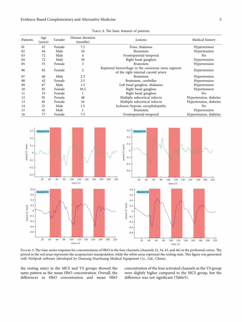
In total, 16 patients (age: 60.75± 19.74 years; 7 males and 9females) with DoC were enrolled in our study. �e basicfeatures of the subjects are given in Table 4.
3.1. Hemodynamic Response and Cortical Activation. �ehemodynamic response of all patients was measured fromthe PFC area. In the present study, the change in HbO
concentration was used as the main observational indicatorrepresenting cortical cerebral blood §ow. Compared tobaseline, all observed channels induced typical event-relatedchanges in hemodynamic responses during needle twirl. Itwas found that the concentration of HbO in the PFC showeda tendency to increase during acupuncture manipulation,while the concentration of HbO decreased at resting statecompared to baseline, suggesting that acupuncture of theRenzhong point could increase cerebral blood §ow to PFC.Figure 3 shows an example of the relative concentrationchanges of HbO of one subject during the block tasks.
Unfortunately, the study found that not all patients hadthe same activation channels, but several of all channels inthe PFC were activated more frequently, which may berelated to the patient’s degree of arousal. In other words, itwas found that changes in HbO concentration in somechannels (channels 32, 34, 45, and 46) of the PFC weresigni¡cantly increased during needle manipulation com-pared to baseline among more people (Figure 3). Besides, itwas said that the four activated channels correspond to theL-PFC (channels 34 and 46) and M-PFC (channels 32 and45). �e speci¡c location in the PFC corresponding to theactivated channels is shown in Figure 4.
3.2. Acupuncture Manipulation-Related FunctionalConnectivity. �e functional connectivity diagram based onHbO concentration is shown in Figure 5. Almost all networkconnectivity diagrams showed a pattern that the connectionstrength of the L-PFC was generally higher than that of theright during acupuncture manipulation compared with theresting state. Importantly, the diagrams also clearly dem-onstrated that the L-PFC has distinctly increased functionalconnections with the M-PFC and left occipital lobe (LOL).Moreover, compared with the R-PFC, L-PFC and M-PFCwere found to have slightly higher functional connectivitystrength during acupuncture manipulation.
3.3. Correlation between the Changes in HbO Concentrationand Severity of DoC. �e HbO concentrations of di�erentactivated channels between the VS group and the MCSgroup were compared. �e results showed that the meanHbO concentration of the 32 and 46 channels at the time ofneedle twirling in the VS group was higher than the MCSgroup, and there was no statistically signi¡cant di�erence(P � 0.740; P � 0.465). Meanwhile, the mean HbO con-centration of the 34 and 45 channels in the VS group waslower than the MCS group, and there was also no signi¡cantdi�erence (P � 0.532; P � 0.795). In addition, it was foundthat the changes in HbO concentration (the di�erence inHbO concentration between the acupuncture twirl state and
Table 3: Characteristics of moderate and severe groups.
Features Severe DoC group (n� 13) Moderate DoC group (n� 3) P valueAge (years) 55.62± 18.06 83.00± 7.21 0.031Gender (M/F) 7M/6F 3F 0.213GCS (score) 5.67± 1.50 9.25± 0.96 0.07GCS, Glasgow coma scale; DoC, disorders of consciousness; M, male; F, female.
44373635
29 30 31 3238 39
454342
33 34
4140
46 47
282715
26252423 13222019
1817
Figure 2: �e cortical locations correspond to the channels. �earea circled in red is the location of the prefrontal cortex (PFC).
4 Evidence-Based Complementary and Alternative Medicine
the resting state) in the MCS and VS groups showed thesame pattern as the mean HbO concentration. Overall, thedifferences in HbO concentration and mean HbO
concentration of the four activated channels in the VS groupwere slightly higher compared to the MCS group, but thedifference was not significant (Table5).
Table 4: 2e basic features of patients.
Patients Age(years) Gender Disease duration
(months) Lesions Medical history
01 47 Female 7.5 Pons, thalamus Hypertension02 44 Male 24 Brainstem Hypertension03 72 Male 4 Frontoparietal-temporal No04 72 Male 36 Right basal ganglion Hypertension05 55 Female 3 Brainstem Hypertension
06 82 Female 2 Ruptured hemorrhage in the cavernous sinus segmentof the right internal carotid artery Hypertension
07 46 Male 2.5 Brainstem Hypertension08 42 Female 2.5 Brainstem, cerebellar Hypertension09 67 Male 1.5 Left basal ganglion, thalamus Hypertension10 81 Female 36.5 Right basal ganglion Hypertension11 53 Female 1 Right basal ganglion No12 91 Female 48 Multiple subcortical infarcts Hypertension, diabetes13 81 Female 16 Multiple subcortical infarcts Hypertension, diabetes14 21 Male 1.5 Ischemic-hypoxic encephalopathy No15 41 Male 1 Brainstem Hypertension16 77 Female 7.5 Frontoparietal-temporal Hypertension, diabetes
mm
ol (L
*mm
)
channel 320.3
0.2
0.1
0
-0.1
-0.2
-0.320 40 60 80 100 120 140 160 180 200 220
time (s)
mm
ol (L
*mm
)
mm
ol (L
*mm
)
mm
ol (L
*mm
)
channel 34
0.4
0.2
0
-0.6
20 40 60 80 100 120 140 160 180 200 220time (s)
0.6
-0.4
-0.2
channel 450.8
0.6
0.4
0.2
0
-0.2
-0.4
-0.6
-0.8
20 40 60 80 100 120 140 160 180 200 220time (s)
channel 460.8
0.6
0.4
0.2
0
-0.2
-0.4
-0.6
-0.8
20 40 60 80 100 120 140 160 180 200 220time (s)
Figure 3:2e time-series response for concentrations of HbO in the four channels (channels 32, 34, 45, and 46) in the prefrontal cortex.2eperiod in the red areas represents the acupuncture manipulation, while the white areas represent the resting state. 2is figure was generatedwith NirSprak software (developed by Danyang Huichuang Medical Equipment Co., Ltd., China).
Evidence-Based Complementary and Alternative Medicine 5

4. Discussion
Consciousness disorder is a common condition after braininjury. Although several studies [43–48] have suggested thatacupuncture can effectively improve consciousness states,the therapeutic mechanism is not well understood. A pre-vious study [49] showed that fNIRS could be applied tounderstand brain function in patients with DoC. However,
no relevant literature involving patients with DoCwas foundon the effect of acupuncture on the hemodynamic responseof the PFC with the monitor of the fNIRS system. 2e PFCmay be a key site for regulating awareness and can be used tosignal the emergence of consciousness [50, 51]. Further-more, a study by Hong et al. [52] demonstrated that acu-puncture could increase the activated area and enhance theconnectivity in the PFC.2erefore, this study was the first to
32 34
45 46
Figure 4: 2e activation maps with cortically projected channel positions.
R-OL R-OLL-OL L-OL
PLR-TA L-TA
L-PFC
M-PFCFrontalFrontal
Back Back
R-PFC
PLR-TA L-TA
L-PFC
M-PFC
R-PFCconn
ectio
n str
engt
h
conn
ectio
n str
engt
h
0.6
0.5
0.4
0.3
0.2
0.1
0
0.6
0.5
0.4
0.3
0.2
0.1
0
Figure 5: Analysis of functional connectivity in two subjects with consciousness disorders during acupuncture manipulation. R-PFC, rightprefrontal cortex; L-PFC, left prefrontal cortex; M-PFC, middle prefrontal cortex; PL, parietal lobe; R-TA, right temporoparietal association;L-TA, left temporoparietal association; ROL, right occipital lobe; LOL, left occipital lobe.
Table 5: Comparison of HbO concentrations in different channels of the two groups (VS and MCS groups).
ChannelMean HbO concentration at stimulation (mmol/L) Difference HbO concentration (mmol/L)VS group MCS group P value VS group MCS group Pvalue
32 0.061 0.043 0.740 0.086 0.044 0.43034 0.062 0.083 0.532 0.071 0.096 0.50145 0.062 0.072 0.795 0.077 0.078 0.97246 0.049 0.026 0.465 0.057 0.048 0.775Total 0.059 0.056 0.875 0.073 0.067 0.759HbO, oxyhemoglobin; VS, vegetative state; MCS, minimally conscious state.
6 Evidence-Based Complementary and Alternative Medicine

utilize fNIRS to measure the hemodynamic response of thePFC to investigate the effect of acupuncture on the hemo-dynamic response of the PFC in patients with impairedconsciousness, to analyze the brain network functionalconnectivity, and to further explore the potential relation-ship between the hemodynamic response and the severity ofDoC. For this purpose, 16 participants (6 MCS patients and10 VS patients) received acupuncture treatment in a rela-tively quiet environment with fNIRS recording.
Our results demonstrated that the prefrontal corticalconcentration of HbO was visibly increased during theacupuncture manipulation phase and declined during theresting phase, compared with baseline. 2is phenomenonwas somewhat consistent with the study by Zhang et al. [53],whose results suggested that HbT concentration in the PFCincreased when patients received stimulation. Importantly,the effects of three acupuncture manipulation blocks on theconcentration of HbO were fairly similar. Moreover, withineach resting state, the concentration of HbO decreased fromthe end of the acupuncture manipulation to the next be-ginning of stimulation. 2ese findings might imply thatacupuncture may contribute to the restoration of the level ofconsciousness by participating in the neuromodulation ofthe prefrontal regions, especially the PFC locations corre-sponding to channels 32, 34, 45, and 46. It is highly likelythat the excitability of the cortex corresponding to these fouractivated channels serves as a signal marker for the emer-gence of consciousness, and further studies need to be donein the future to confirm this idea. Furthermore, we observedmore significant changes in the concentration of HbO in theL-PFC and M-PFC than in the R–PFC. 2at means corticalexcitability in the left prefrontal and middle prefrontal maybe important for the emergence of awareness.
In this study, we found that the connection strength ofthe L-PFC was generally higher than that of the right duringacupuncture manipulation compared with the resting state.However, there were significant differences in the strength offunctional connectivity between individuals. One possiblereason for this is the wide variation in the age of the subjectsincluded in this experiment, with the oldest being 91 yearsold and the youngest being 21 years old. 2e strength offunctional connectivity may be largely related to the subject’sage. A study by Chenyu et al. [54] proposed that the strengthof functional brain connections is weaker in older adultscompared to young adults. Interestingly, compared with theR-PFC, M-PFC and L-PFC were found to have slightlyhigher functional connectivity strength during acupuncturemanipulation. 2is finding differs from the previous studyby Leon-Dominguez et al. [55] who indicated that only theright dorsomedial PFC was significantly activated during theemergence phase. Further studies need to be done to dis-tinguish the specific functions of the L-PFC as well as theright dorsomedial PFC.
Our study found only a weak difference in hemodynamicresponses of prefrontal areas between the VS and MCSgroups. In other words, no significant correlation was foundbetween the cortical hemodynamic response and the severityof DoC. Some previous articles have also attempted to in-vestigate the hemodynamic differences between the MCS and
VS groups, but no statistically significant differences werefound. A review by Liberati et al. [56] also illustrated that halfof the researchers reported statistically significant differencesbetween VS and MCS patients. One reason for this may bethat the data measured by fNIRS is influenced by the patient’ssystemic physiological state (such as heart rate, blood pres-sure, and respiratory rhythm) [57], resulting in a less thantypical fNIRS response, increasing the incomparability of thepatients' fNIRS response [58]. Another reason could be thatthe nociceptive stimuli produced during acupuncture ma-nipulation influenced the fluctuations in patient wakefulnessand masked the differences between the two groups.
2ere were three limitations in our study. 2e firstlimitation of this study was the technical challenge of usingan fNIRS device. First, the number of optical channels usedin this study is limited and does not completely cover allregions of the PFC; again, although the measured data havebeen subjected to a series of preprocessing, for example,automatic removal of motion artifacts and filtering algo-rithms, there are still effects that cannot be completelyeliminated, which in turn affects the experimental results;finally, poor contact between the probe and the scalp mayresult in weak signal acquisition.2erefore, in future studies,a more advanced fNIRS instrument will need to be used toobtain more accurate results. Furthermore, greater pro-cessing methods will need to be utilized in further studies.
2e second limitation of our study was the small samplesize and the wide variation in the age of the subjects included inthis experiment. It has been shown that the hemodynamicresponse of the cortex decreases with age, even in healthy adults[58]. 2us, in future studies, the hemodynamic response ofpatients with DoC at different ages can be analyzed separately,thus reducing the effects due to neurodegenerative changes.
2e third limitation of our experiment was that thehemodynamic response of patients in the resting state wasnot collected separately. 2e study was a block-designedexperiment in which acupuncture stimulation was given foran extended time, followed by a period of resting state. Withthis approach, there may be a temporal lag effect of acu-puncture on the hemodynamic response of the patient’scortex, resulting in the hemodynamic response duringresting state not returning to the starting level and thusmasking part of the effect of acupuncture on changes in thehemodynamic response. 2e hemodynamic response ofpatients in the resting state needs to be collected separately insubsequent studies.
Despite some limitations of our study, it is still in-structive for clinical practice. 2is study may provide areference for the mechanisms of improving awarenessdisorders by acupuncture, but its specific effects requirefurther investigation. Furthermore, the potential of using thefNIRS device to detect the impact of acupuncture in patientswith consciousness disorders is significant.
5. Conclusion
Our results showed that the concentration of HbO in PFC(especially in the L-PFC and M-PFC) was increased duringthe acupuncture manipulation phase and declined during
Evidence-Based Complementary and Alternative Medicine 7

the resting phase. In addition, we also observed the con-nection strength of the left cerebral cortex was generallyhigher than that of the right during acupuncture manipu-lation compared with the resting state. Finally, only a weakdifference in hemodynamic responses of prefrontal areasbetween the VS and MCS groups was found, but no sta-tistically significant differences. 2ese results indicate thatacupuncture can increase the concentration of HbO in thePFC (especially in the L-PFC and M-PFC) and strengthenthe connection strength of the left cerebral cortex. Ourfindings suggest that there is no significant correlation be-tween the cortical hemodynamic response and the severity ofDoC.
Additional Points
2e concentration of HbO in PFC was increased during theacupuncture manipulation and declined during the restingstate.
Disclosure
Wen Xin and Zicai Liu are the co-first authors.
Conflicts of Interest
2e authors declare that they have no conflicts of interest.
Authors’ Contributions
Huiyu Liu conceptualized the study and wrote and reviewedthe article. Junbin Chen wrote and reviewed the article. XinWen wrote the original draft, collected, analyzed, andassessed data. Zicai Liu collected and assessed data and wroteand reviewed the article. Yuchun Shao collected data andperformed acupuncture manipulation. Yang Peng collected,analyzed, and assessed data. Wen Xin and Zicai Liu con-tributed equally to this study.
References
[1] B. L. Edlow, J. Claassen, N. D. Schiff, and D. M. Greer,“Recovery from disorders of consciousness: mechanisms,prognosis, and emerging therapies,” Nature Reviews Neu-rology, vol. 17, pp. 135–156, 2021.
[2] A. 2ibaut, N. Schiff, J. Giacino, S. Laureys, and O. Gosseries,“2erapeutic interventions in patients with prolonged dis-orders of consciousness,” 6e Lancet Neurology, vol. 18,pp. 600–614, 2019.
[3] J. J. Fins and J. L. Bernat, “Ethical, palliative, and policyconsiderations in disorders of consciousness,” Archives ofPhysical Medicine and Rehabilitation, vol. 99, no. 9,pp. 1927–1931, 2018.
[4] D. Kondziella, A. Bender, K. Diserens et al., “EuropeanAcademy of Neurology guideline on the diagnosis of comaand other disorders of consciousness,” European Journal ofNeurology, vol. 27, no. 5, pp. 741–756, 2020.
[5] S. Laureys, G. G. Celesia, F. Cohadon et al., “Unresponsivewakefulness syndrome: a new name for the vegetative state orapallic syndrome,” BMC Medicine, vol. 8, no. 1, p. 68, 2010.
[6] A. Bender, R. J. Jox, E. Grill, A. Straube, and D. Lule, “Per-sistent vegetative state and minimally conscious state: a
systematic review and meta-analysis of diagnostic proce-dures,” Deutsches Arzteblatt International, vol. 112, no. 14,pp. 235–242, 2015.
[7] J. T. Giacino, S. Ashwal, N. Childs et al., “2e minimallyconscious state: definition and diagnostic criteria,” Neurology,vol. 58, no. 3, pp. 349–353, 2002.
[8] L. Naccache, “Minimally conscious state or cortically medi-ated state,” Brain, vol. 141, no. 4, pp. 949–960, 2018.
[9] R. Hirschberg and J. T. Giacino, “2e vegetative and mini-mally conscious states: diagnosis, prognosis and treatment,”Neurologic Clinics, vol. 29, no. 4, pp. 773–786, 2011.
[10] W. Cao and Z. Xiong, “Clinical observation on electro-acupuncture for treatment of 26 cases of persistent vegetativestate,” Zhongguo Zhen Jiu, vol. 23, pp. 511-512, 2003.
[11] J. J. Pan, X. Z. Hou, P. Wang, W. Li, Q. Zhang, and T. Dong,“Effect of acupuncture method of removing-stasisand re-suscitating on the consciousness of patients with severetraumatic brain injury: a randomized clinical trial,” Medicine(Baltimore), vol. 99, no. 36, Article ID e22056, 2020.
[12] Y. Huang, X. Lai, and C. Tang, “Study on specification ofshuigou (DU26) with positron emission tomography andsingle photon emission computed tomography,” Journal ofChengdu University of Traditional Chinese Medicine, vol. 31,pp. 5–8, 2008.
[13] Y. Li, X. Fan, and S. Wang, “Study on the parameter opti-mization of acupuncture at shuigou (GV 26) for the treatmentof brain inf arction,” Journal of Traditional Chinese Medicine,vol. 50, pp. 428–431, 2009.
[14] M. Rupawala, H. Dehghani, S. J. E. Lucas, P. Tino, andD. Cruse, “Shining a light on awareness: a review of functionalnear-infrared spectroscopy for prolonged disorders of con-sciousness,” Frontiers in Neurology, vol. 9, p. 350, 2018.
[15] W. Struckmann, J. Persson, W. Weigl, M. Gingnell, andR. Boden, “Modulation of the prefrontal blood oxygenationresponse to intermittent theta-burst stimulation in depres-sion: a sham-controlled study with functional near-infraredspectroscopy,” World Journal of Biological Psychiatry, vol. 22,no. 4, pp. 247–256, 2021.
[16] S. Laureys and N. D. Schiff, “Coma and consciousness:paradigms (re) framed by neuroimaging,” NeuroImage,vol. 61, no. 2, pp. 478–491, 2012.
[17] A. Abdalmalak, D. Milej, L. Norton, D. B. Debicki,A. M. Owen, and K. S. Lawrence, “2e potential role of fNIRSin evaluating levels of consciousness,” Frontiers in HumanNeuroscience, vol. 15, Article ID 703405, 2021.
[18] R. Fernandez Rojas, M. Liao, J. Romero, X. Huang, andK. L. Ou, “Cortical network response to acupuncture and theeffect of the hegu point: an fNIRS study,” Sensors, vol. 19,no. 2, p. 394, 2019.
[19] X. Yao, I. D. Florez, P. Zhang et al., “Clinical researchmethodsfor treatment, diagnosis, prognosis, etiology, screening, andprevention: a narrative review,” Journal of Evidence-BasedMedicine, vol. 13, no. 2, pp. 130–136, 2020.
[20] S. Jain and L. M. Iverson, Glasgow Coma Scale, StatPearls,StatPearls Publishing Copyright ©, Treasure Island, FL, USA,2021.
[21] G. Teasdale and B. Jennett, “Assessment of coma and impairedconsciousness. A practical scale,” 6e Lancet, vol. 304,no. 7872, pp. 81–84, 1974.
[22] R. Formisano, M. Contrada, M. Iosa, G. Ferri, S. Schiattone,andM. Aloisi, “Coma recovery scale-revised with and withoutthe emotional stimulation of caregivers,” 6e CanadianJournal of Neurological Sciences, vol. 46, no. 5, pp. 607–609,2019.
8 Evidence-Based Complementary and Alternative Medicine

[23] J. T. Giacino, K. Kalmar, and J. Whyte, “2e JFK ComaRecovery Scale-Revised: measurement characteristics anddiagnostic utility11No commercial party having a direct fi-nancial interest in the results of the research supporting thisarticle has or will confer a benefit upon the authors or uponany organization with which the authors are associated,”Archives of Physical Medicine and Rehabilitation, vol. 85,no. 12, pp. 2020–2029, 2004.
[24] M. D. Cortese, F. Riganello, F. Arcuri et al., “Coma recoveryscale-r: variability in the disorder of consciousness,” BMCNeurology, vol. 15, no. 1, p. 186, 2015.
[25] L. F. Lucca, D. Lofaro, L. Pignolo et al., “Outcome predictionin disorders of consciousness: the role of coma recovery scalerevised,” BMC Neurology, vol. 19, no. 1, p. 68, 2019.
[26] P. Gerrard, R. Zafonte, and J. T. Giacino, “Coma RecoveryScale-Revised: evidentiary support for hierarchical grading oflevel of consciousness,” Archives of Physical Medicine andRehabilitation, vol. 95, no. 12, pp. 2335–2341, 2014.
[27] C. Schnakers, A. Vanhaudenhuyse, J. Giacino et al., “Diag-nostic accuracy of the vegetative and minimally consciousstate: clinical consensus versus standardized neurobehavioralassessment,” BMC Neurology, vol. 9, no. 1, p. 35, 2009.
[28] P. Pinti, I. Tachtsidis, A. Hamilton et al., “2e present andfuture use of functional near-infrared spectroscopy (fNIRS)for cognitive neuroscience,” Annals of the New York Academyof Sciences, vol. 1464, no. 1, pp. 5–29, 2020.
[29] F. Scholkmann, S. Kleiser, A. J. Metz et al., “A review oncontinuous wave functional near-infrared spectroscopy andimaging instrumentation and methodology,” NeuroImage,vol. 85, pp. 6–27, 2014.
[30] Y. Hoshi, N. Kobayashi, and M. Tamura, “Interpretation ofnear-infrared spectroscopy signals: a study with a newlydeveloped perfused rat brain model,” Journal of AppliedPhysiology, vol. 90, no. 5, pp. 1657–1662, 2001.
[31] G. Strangman, J. P. Culver, J. H. 2ompson, and D. A. Boas,“A quantitative comparison of simultaneous BOLD fMRI andNIRS recordings during functional brain activation,” Neu-roImage, vol. 17, no. 2, pp. 719–731, 2002.
[32] N. Zhang, X. Yuan, Q. Li et al., “2e effects of age on braincortical activation and functional connectivity during videogame-based finger-to-thumb opposition movement: a func-tional near-infrared spectroscopy study,” Neuroscience Let-ters, vol. 746, Article ID 135668, 2021.
[33] L. Almulla, I. Al-Naib, and M. Althobaiti, “Hemodynamicresponses during standing and sitting activities: a study to-ward fNIRS-BCI,” Biomed Physics and Engineering Express,vol. 6, no. 5, Article ID 055005, 2020.
[34] M. Cope and D. T. Delpy, “System for long-term measure-ment of cerebral blood and tissue oxygenation on newborninfants by near infra-red transillumination,” Medical, andBiological Engineering and Computing, vol. 26, no. 3,pp. 289–294, 1988.
[35] K. Kawabata Duncan, T. Tokuda, C. Sato, K. Tagai, and I. Dan,“Willingness-to-Pay-Associated right prefrontal activationduring a single, real use of cosmetics as revealed by functionalnear-infrared spectroscopy,” Frontiers in Human Neurosci-ence, vol. 13, p. 16, 2019.
[36] X. A. Lin, C. Wang, J. Zhou, L. Sai, and G. Fu, “Neuralcorrelates of spontaneous deception in a non-competitiveinterpersonal scenario: a functional near-infrared spectros-copy (fNIRS) study,” Brain and Cognition, vol. 150, Article ID105704, 2021.
[37] P. Pinti, A. Merla, C. Aichelburg et al., “A novel GLM-basedmethod for the Automatic IDentification of functional Events
(AIDE) in fNIRS data recorded in naturalistic environments,”NeuroImage, vol. 155, pp. 291–304, 2017.
[38] Y. Zhang, W. Shi, H. Wang, M. Liu, and D. Tang, “2e impactof acute exercise on implicit cognitive reappraisal in associ-ation with left dorsolateral prefronta activation: a fNIRSstudy,” Behavioural Brain Research, vol. 406, Article ID113233, 2021.
[39] X. Chen, “False discovery rate control for multiple testingbased on discrete p-values,” Biometrical Journal, vol. 62, no. 4,pp. 1060–1079, 2020.
[40] A. K. Singh and I. Dan, “Exploring the false discovery rate inmultichannel NIRS,”Neuro Image, vol. 33, no. 2, pp. 542–549,2006.
[41] S. Y. Dong, J. Choi, Y. Park et al., “Prefrontal functionalconnectivity during the verbal fluency task in patients withmajor depressive disorder: a functional near-infrared spec-troscopy study,” Frontiers in Psychiatry, vol. 12, Article ID659814, 2021.
[42] U. Ghafoor, J. H. Lee, K. S. Hong, S. S. Park, J. Kim, andH. R. Yoo, “Effects of acupuncture therapy on MCI patientsusing functional near-infrared spectroscopy,” Frontiers inAging Neuroscience, vol. 11, p. 237, 2019.
[43] Y. C. Bao, F. Zhang, Q. Li et al., “Midnight-noon ebb-flowacupuncture combined with rehabilitation therapy for severecraniocerebral trauma patients with vegetative state: a ran-domized controlled trial,” Zhongguo Zhen Jiu, vol. 40,pp. 234–238, 2020.
[44] B. F. Cao, C. Zhang,W.H. Liang, and L. Chen, “Effects of earlyacupuncture combined with exercise therapy on the con-sciousness-regaining treatment of coma patients in neuro-surgery,” Zhongguo Zhen Jiu, vol. 31, pp. 121–123, 2011.
[45] Y. L. Gao, G. J. Xu, J. G. Zhu, G. Peng, J. Xi, and M. X. Xue,“Xiao’s “xingnaofusu” needling for regaining consciousnessand the influence on width of the third ventricle in patientswith persistent vegetative state,” Zhen Ci Yan Jiu, vol. 45,pp. 233–236, 2020.
[46] J. Matsumoto-Miyazaki, Y. Asano, S. Yonezawa et al.,“Acupuncture increases the excitability of the cortico-spinalsystem in patients with chronic disorders of consciousnessfollowing traumatic brain injury,” Journal of Alternative andComplementary Medicine, vol. 22, no. 11, pp. 887–894, 2016.
[47] H. Song, X. Chen, Y. Yu, and L. Zhang, “Xingnao Kaiqiaoacupuncture combined with Angong Niuhuang Wan for apatient under persistent vegetative state: a case report,”Frontiers of Medicine, vol. 12, no. 3, pp. 334–339, 2018.
[48] L. Zheng, “Acupuncture treatment for persistent vegetativestate after operation of acute subdural hematoma,” ZhongguoZhen Jiu, vol. 25, pp. 82–84, 2005.
[49] A. M. Kempny, L. James, K. Yelden et al., “Functional nearinfrared spectroscopy as a probe of brain function in peoplewith prolonged disorders of consciousness,” NeuroImage:Clinic, vol. 12, pp. 312–319, 2016.
[50] A. Del Cul, S. Dehaene, P. Reyes, E. Bravo, and A. Slachevsky,“Causal role of prefrontal cortex in the threshold for access toconsciousness,” Brain, vol. 132, no. 9, pp. 2531–2540, 2009.
[51] J. T. Giacino, J. J. Fins, S. Laureys, and N. D. Schiff, “Disordersof consciousness after acquired brain injury: the state of thescience,” Nature Reviews Neurology, vol. 10, no. 2, pp. 99–114,2014.
[52] K. S. Hong, M. A. Khan, U. Ghafoor, and H. R. Yoo,“Acupuncture enhances brain function in patients with mildcognitive impairment: evidence from a functional-near in-frared spectroscopy study,” Neural Regeneration Research,vol. 17, no. 8, p. 1850, 2022.
Evidence-Based Complementary and Alternative Medicine 9

[53] Y. Zhang, Y. Yang, J. Si, X. Xia, J. He, and T. Jiang, “Influenceof inter-stimulus interval of spinal cord stimulation in pa-tients with disorders of consciousness: a preliminary func-tional near-infrared spectroscopy study,” NeuroImage: Clinic,vol. 17, pp. 1–9, 2018.
[54] F. H. Chenyu, Li. Hongyu, and H. Xie, “Healthy young andold adults functional connectivity among cortical networks: Aresting-state functional near-infrared spectroscopy study,”Chinese Journal of Rehabilitation Medicine, vol. 36, pp. 931–942, 2021.
[55] U. Leon-Dominguez, M. Izzetoglu, J. Leon-Carrion et al.,“Molecular concentration of deoxyHb in human prefrontalcortex predicts the emergence and suppression of con-sciousness,” NeuroImage, vol. 85, pp. 616–625, 2014.
[56] G. Liberati, T. Hunefeldt, and M. Olivetti Belardinelli,“Questioning the dichotomy between vegetative state andminimally conscious state: a review of the statistical evidence,”Frontiers in Human Neuroscience, vol. 8, p. 865, 2014.
[57] G. Bauernfeind, S. C. Wriessnegger, I. Daly, and G. R. Muller-Putz, “Separating heart and brain: on the reduction ofphysiological noise from multichannel functional near-in-frared spectroscopy (fNIRS) signals,” Journal of Neural En-gineering, vol. 11, no. 5, Article ID 056010, 2014.
[58] E. M. Kurz, G. Wood, S. E. Kober et al., “Towards using fNIRSrecordings of mental arithmetic for the detection of residualcognitive activity in patients with disorders of consciousness(DOC),” Brain and Cognition, vol. 125, pp. 78–87, 2018.
10 Evidence-Based Complementary and Alternative Medicine
Copyright © 2022 FDOKUMEN


![[Posterior cortical atrophy]](https://static.fdokumen.com/doc/165x107/6331b9d14e01430403005392/posterior-cortical-atrophy.jpg)