Inhibition by inorganic ions of a sustained calcium signal evoked by activation of mGlu5 receptors...
11
Inhibition by inorganic ions of a sustained calcium signal evoked by activation of mGlu5 receptors in rat cortical neurons and glia 1 Larissa S. Prothero, 1 Christopher D. Richards & 2,3 Alistair Mathie 1 Department of Physiology, University College London, Royal Free Campus, Rowland Hill Street, London NW3 2PF; 2 Department of Pharmacology, Medawar Building, University College London, Gower Street, London WC1E 6BT 1 The eect of mGlu receptor agonists on intracellular calcium (Ca 2+ ) in rat cortical neurons and glial cells was studied. The responses evoked consisted of two phases; an initial transient response followed by a sustained plateau. In both cell types the order of potency of group I mGlu receptor agonists was DHPG41S,3R ACPD43-HPG. 2 The selective mGlu5 agonist CHPG elicited responses in both cell types as did S4C3-HPG which is thought to be an mGlu5 agonist at high concentrations. S4-CPG had no eect on intracellular Ca 2+ levels nor did it inhibit the action of 1S,3R ACPD. These results suggest that the responses in both cell types are mediated by mGlu5 receptors. 3 In the absence of extracellular Ca 2+ ions, 1S,3R ACPD (100 mM) induced only a transient Ca 2+ response which decayed to baseline with a time constant of approximately 20 s in both cell types. Subsequent readdition of Ca 2+ (2 mM) to the external solution in the continued presence of 1S,3R ACPD induced a sustained Ca 2+ plateau. 4 The sustained Ca 2+ plateau could be blocked by a number of inorganic cations, with an order of potency of Zn 2+ 5La 3+ 4Cd 2+ 5Co 2+ 4Ni 2+ 4Mg 2+ . Similar concentrations of Zn 2+ had little eect on Ca 2+ -influx evoked by 25 mM K + . 5 It is concluded that the Ca 2+ -entry pathway activated by mGlu5 receptors resembles store-operated Ca 2+ -entry pathways that have been described in other cell types. Keywords: Metabotropic glutamate receptor; calcium; store-operated calcium entry; zinc; lanthanum; mGlu5; CHPG Introduction Metabotropic glutamate (mGlu) receptors are a family of G protein coupled receptors that share sequence homology with GABA B receptors and Ca 2+ sensing proteins (Conn & Pin, 1997; Brown et al., 1993; Kaupmann et al., 1997). They have much less homology with other classes of G protein coupled receptors (see Pin & Duvoisin, 1995). They are involved in many aspects of neuronal function within the CNS including development and synaptic plasticity (e.g. Bashir et al., 1993; Lu et al., 1997). Eight dierent mGlu receptors have been cloned (mGlu1 – 8) and these have been classified into three groups according to their sequence similarities, the signal transduction pathways to which they can couple and the selectivity of certain receptor agonists (Nakanishi & Masu, 1994; Pin & Duvoisin, 1995). Group I receptors (mGlu 1 and 5) activate phospholipase C while activation of Group II (mGlu 2 and 3) and Group III (mGlu 4, 6, 7 and 8) receptors inhibits adenylyl cyclase activity (Pin & Duvoisin, 1995; Conn & Pin, 1997). In this study, we have investigated the role of mGlu receptors in stimulating an increase in intracellular calcium concentration in neurons and glial cells isolated from rat cerebral cortex. Firstly, we have characterized the subtype of mGlu receptor underlying the response with the aid of a number of selective pharmacological agents. The rise in calcium following mGlu receptor activation consists of two phases – an initial rapid transient rise in calcium followed by a secondary sustained rise which is dependent on the presence of extracellular calcium (see Mathie & Richards, 1997). Such a response profile has been seen following activation of a number of other G protein coupled receptors in other cell types (e.g. Grudt et al., 1996). For other receptors, the initial transient response has been attributed to mobilization of calcium from intracellular stores, while the sustained phase may be caused by activation of a store-operated calcium entry pathway (SOC) (Berridge, 1995; Clapham, 1995). Secondly, we have characterized some of the properties of this second phase of the response, in particular its sensitivity to block by inorganic cations, since this property has been used previously to aid the characterization of such SOC pathways in other cell types (e.g. Aussel et al., 1996; Hoth & Penner, 1993; Grudt et al., 1996; Wayman et al., 1996). Preliminary accounts of some of these results have been published (Mathie et al., 1996; Prothero et al., 1998). Methods Cell culture Neonatal Sprague-Dawley rats (2 – 5 days old) were anaes- thetized with isoflurane and then decapitated. The neocortex was aseptically dissected and placed in HEPES buered minimum essential medium (M.E.M.). The tissue was mechanically dissociated by passing the tissue through fire- polished pasteur pipettes of decreasing bore. The cell debris was allowed to settle and the supernatant containing disaggregated cells was then spun at 1506g for 3 min to separate the intact cells. The supernatant was removed and the pellet resuspended in HEPES buered MEM. The cells were then plated at a density of 0.3 – 1610 6 cells per 50 mm culture dishes containing 5 ml MEM supplemented with 2.5% Foetal Bovine Serum. Each dish contained coverslips that had been coated with poly-L-lysine (0.1 mg ml 71 ). The cultures were maintained in a humidified 5% CO 2 atmosphere at 378C for 3 Author for correspondence. British Journal of Pharmacology (1998) 125, 1551 – 1561 ª 1998 Stockton Press All rights reserved 0007 – 1188/98 $12.00 http://www.stockton-press.co.uk/bjp
Transcript of Inhibition by inorganic ions of a sustained calcium signal evoked by activation of mGlu5 receptors...

Inhibition by inorganic ions of a sustained calcium signal evoked byactivation of mGlu5 receptors in rat cortical neurons and glia
1Larissa S. Prothero, 1Christopher D. Richards & 2,3Alistair Mathie
1Department of Physiology, University College London, Royal Free Campus, Rowland Hill Street, London NW3 2PF;2Department of Pharmacology, Medawar Building, University College London, Gower Street, London WC1E 6BT
1 The e�ect of mGlu receptor agonists on intracellular calcium (Ca2+) in rat cortical neurons and glialcells was studied. The responses evoked consisted of two phases; an initial transient response followed bya sustained plateau. In both cell types the order of potency of group I mGlu receptor agonists wasDHPG41S,3R ACPD43-HPG.
2 The selective mGlu5 agonist CHPG elicited responses in both cell types as did S4C3-HPG which isthought to be an mGlu5 agonist at high concentrations. S4-CPG had no e�ect on intracellular Ca2+
levels nor did it inhibit the action of 1S,3R ACPD. These results suggest that the responses in both celltypes are mediated by mGlu5 receptors.
3 In the absence of extracellular Ca2+ ions, 1S,3R ACPD (100 mM) induced only a transient Ca2+
response which decayed to baseline with a time constant of approximately 20 s in both cell types.Subsequent readdition of Ca2+ (2 mM) to the external solution in the continued presence of 1S,3RACPD induced a sustained Ca2+ plateau.
4 The sustained Ca2+ plateau could be blocked by a number of inorganic cations, with an order ofpotency of Zn2+5La3+4Cd2+5Co2+4Ni2+4Mg2+. Similar concentrations of Zn2+ had little e�ecton Ca2+-in¯ux evoked by 25 mM K+.
5 It is concluded that the Ca2+-entry pathway activated by mGlu5 receptors resembles store-operatedCa2+-entry pathways that have been described in other cell types.
Keywords: Metabotropic glutamate receptor; calcium; store-operated calcium entry; zinc; lanthanum; mGlu5; CHPG
Introduction
Metabotropic glutamate (mGlu) receptors are a family of G
protein coupled receptors that share sequence homology withGABAB receptors and Ca2+ sensing proteins (Conn & Pin,1997; Brown et al., 1993; Kaupmann et al., 1997). They havemuch less homology with other classes of G protein coupled
receptors (see Pin & Duvoisin, 1995). They are involved inmany aspects of neuronal function within the CNS includingdevelopment and synaptic plasticity (e.g. Bashir et al., 1993; Lu
et al., 1997). Eight di�erent mGlu receptors have been cloned(mGlu1 ± 8) and these have been classi®ed into three groupsaccording to their sequence similarities, the signal transduction
pathways to which they can couple and the selectivity ofcertain receptor agonists (Nakanishi & Masu, 1994; Pin &Duvoisin, 1995). Group I receptors (mGlu 1 and 5) activate
phospholipase C while activation of Group II (mGlu 2 and 3)and Group III (mGlu 4, 6, 7 and 8) receptors inhibits adenylylcyclase activity (Pin & Duvoisin, 1995; Conn & Pin, 1997).
In this study, we have investigated the role of mGlu
receptors in stimulating an increase in intracellular calciumconcentration in neurons and glial cells isolated from ratcerebral cortex. Firstly, we have characterized the subtype of
mGlu receptor underlying the response with the aid of anumber of selective pharmacological agents. The rise incalcium following mGlu receptor activation consists of two
phases ± an initial rapid transient rise in calcium followed by asecondary sustained rise which is dependent on the presence ofextracellular calcium (see Mathie & Richards, 1997). Such a
response pro®le has been seen following activation of anumber of other G protein coupled receptors in other celltypes (e.g. Grudt et al., 1996). For other receptors, the initial
transient response has been attributed to mobilization of
calcium from intracellular stores, while the sustained phasemay be caused by activation of a store-operated calcium entrypathway (SOC) (Berridge, 1995; Clapham, 1995). Secondly, wehave characterized some of the properties of this second phase
of the response, in particular its sensitivity to block byinorganic cations, since this property has been used previouslyto aid the characterization of such SOC pathways in other cell
types (e.g. Aussel et al., 1996; Hoth & Penner, 1993; Grudt etal., 1996; Wayman et al., 1996). Preliminary accounts of someof these results have been published (Mathie et al., 1996;
Prothero et al., 1998).
Methods
Cell culture
Neonatal Sprague-Dawley rats (2 ± 5 days old) were anaes-thetized with iso¯urane and then decapitated. The neocortexwas aseptically dissected and placed in HEPES bu�ered
minimum essential medium (M.E.M.). The tissue wasmechanically dissociated by passing the tissue through ®re-polished pasteur pipettes of decreasing bore. The cell debris
was allowed to settle and the supernatant containingdisaggregated cells was then spun at 1506g for 3 min toseparate the intact cells. The supernatant was removed and the
pellet resuspended in HEPES bu�ered MEM. The cells werethen plated at a density of 0.3 ± 16106 cells per 50 mm culturedishes containing 5 ml MEM supplemented with 2.5% FoetalBovine Serum. Each dish contained coverslips that had been
coated with poly-L-lysine (0.1 mg ml71). The cultures weremaintained in a humidi®ed 5% CO2 atmosphere at 378C for3Author for correspondence.
British Journal of Pharmacology (1998) 125, 1551 ± 1561 ã 1998 Stockton Press All rights reserved 0007 ± 1188/98 $12.00
http://www.stockton-press.co.uk/bjp

5 ± 7 days before use. Occasionally cultures of 10 ± 14 days wereused but no di�erences in the response to agonists wereevident. Neurons were identi®ed by their size and morphology
(phase bright cell bodies possessing long neurites with fewrecurrent branches) and astrocytes were identi®ed by their¯attened phase-dark appearance (see Figure 1).
Intracellular Ca2+ measurements
Ca2+ measurements were made with Fura-2 (Molecular
Probes, Eugene, OR, U.S.A.) which was loaded into the cellsas its AM-ester. Measurements were made using dualexcitation (340 and 380 nm) with emission above 420 nm. All
experiments were conducted at room temperature (20 ± 278C).For ¯uorescence imaging, light was collected via a cooled CCDcamera (supplied by Digital Pixel, Brighton, U.K.) and
analysed using software supplied by Kinetic Imaging (Liver-pool, U.K.). To improve the signal-to-noise ratio, thecollection times were adjusted to increase the total numberof photons accumulated at the lowest intensity of emission.
Thus the collection period for 340 nm excitation was threetimes that for 380 nm excitation (the collection period wasgenerally 600 ms compared to 200 ms). The ratio values in the
Figures are not corrected for these di�erent collection times.In each experiment, ratio values were calculated after
subtraction of the background emission; auto¯uorescence was
less than the background signal. Background emission wasdetermined at the end of every experiment by treating the cellswith 0.1% Triton. For determination of the local Ca2+ from
the ratio images, speci®c areas of interest was chosen. Theaverage ratio value for the designated areas was thencalculated and plotted as a function of time. For eachexperimental procedure, the `n' values refer to the number of
cells. In each case these cells were taken from a minimum ofthree coverslips from at least two separate cortical culturepreparations. Throughout the text values are given as
mean+s.e.mean.Calibration of the ratios in the terms of [Ca2+] was carried
out in situ in several experiments using the ionophore 4Br-
A23187. Cells were exposed to a `zero' calcium solutioncontaining 20 mM EGTA and 0 mM Ca2+ to obtain aminimum ratio and a `high' calcium solution containing 2 mM
Ca2+ to obtain a maximum ratio, both in the presence of 5 mM4Br-A23187. Cells were exposed to these zero and high calciumsolutions alternately until consistent ratio values wereobtained. The average values from these calibrations has been
used to quantify the changes in intracellular Ca2+ where thiswas considered appropriate using the following equation(Grynkiewicz et al., 1985):
�Ca2�� � KdRÿRmin
Rmax ÿR
� �Sf2Sb2
� �where R is the measured ratio of interest, Rmin and Rmax theratios recorded with zero and high extracellular calcium, Sf2and Sb2 the signals at 380 nm in zero and high calcium and KD
is the apparent dissociation constant for Fura-2.For 28 cells in six separate calibration experiments the
resting calcium concentration was calculated to be 53+14 nM.
Solutions
Cells were bathed in Locke's solution bu�ered with 16 or
30 mM HEPES. The unbu�ered Locke's solution had thefollowing composition (mM) NaCl 140, KCl 3 or 5, MgCl2 1.8or 1, CaCl2 2 and glucose 5.5. The pH was adjusted to 7.3 using
1 M NaOH. The mGlu receptor agonists were diluted from
concentrated stocks into the appropriate bathing solutionimmediately before use. Agonists and inorganic ions wereadded to the bath for the durations indicated by the bars in the
Figures.Routine chemicals and tissue culture materials were
supplied by Sigma and the speci®c mGlu receptor agonistsand antagonists described below were all obtained from Tocris
Cookson: the non-selective mGlu receptor agonist, 1S,3RACPD ((1S,3R)-1-aminocyclopentane-1,3-dicarboxylic acid);two Group I mGlu receptor agonists, 3-HPG ((S)-3-
hydroxyphenylglycine) and DHPG (3,5-dihydroxyphenylgly-cine); the mGlu5 receptor agonist, CHPG ((RS)-2 chloro-5-hydroxyphenylglycine); the putative mGlu5 receptor agonist,
TADA (trans-azetidine-2,4-dicarboxylic acid); the mGlu1receptor antagonist/mGlu2 receptor agonist, S4-CPG ((S)-4-carboxyphenylglycine), the mGlu1 receptor antagonist/mGlu5
receptor agonist; S4C3-HPG ((S)-4-carboxy-3-hydroxyphenyl-glycine) and the group III mGlu receptor agonist, L-AP4(L(+)-2-amino-4-phosphonobutyric acid).
Results
1S,3R ACPD induced calcium responses in corticalneurons and glial cells
The mGlu receptor agonist, 1S,3R ACPD induced a rise inintracellular calcium concentration in cortical neurons and glial
cells. This is illustrated in Figure 1 for both cell types. Typicalresponses in both cells consisted of an initial transient increasein intracellular calcium followed by a maintained plateauresponse. The sustained plateau response was maintained for as
long as the cells were exposed to the agonist (see Figure 1,neuron 3) and, indeed, could often outlast application of theagonist by several minutes. In some cells, particularly at
intermediate agonist concentrations (10 ± 25 mM), the plateauresponses were oscillatory. This is illustrated in Figure 1 byneuron number 2. Cells could be divided, broadly, into three
groups: those that gave no response, those that gave a sustainedplateau response and those that gave an oscillatory response.However, 1S,3R ACPD at 100 mM produced plateau responsesin the large majority (around 80%) of either cell type.
A series of experiments were carried out to determine theconcentration-response relationships for 1S,3R ACPD (overthe concentration range 0.25 ± 250 mM) for both cell types.
Data from these experiments are shown in Table 1. It can beseen that the amplitude of the evoked responses increases withagonist concentration for both cell types. 1S,3R ACPD was
more potent in eliciting a response in glial cells compared toneurons. 1S,3R ACPD had an EC50 of approximately 5 mM forglial cells and 20 mM for neurons. 100 mM 1S,3R ACPD was
found to be a maximally e�ective concentration of 1S,3RACPD in both cell types, capable of evoking peak calciumratio signals of up to seven (see Figures 1 and 8) correspondingto a peak calcium concentration of around 1.6 mM. For theparticular series of experiments shown in Table 1 all cells areincluded regardless of whether they produced a detectableresponse. 1S,3R ACPD (100 mM) induced responses in 80% of
neurons and 83% of glial cells respectively; the mean peakcalcium signal was 2.37+0.11 (corresponding to a calciumconcentration of 241 nM) in glial cells and 2.09+0.19 (183 nM)
in neurons. All responses are from cells exposed to the agonistfor the ®rst time to avoid problems caused by receptordesensitization at high concentrations or priming at lowconcentrations.
mGlu calcium signals1552 L.S. Prothero et al

Pharmacology of mGlu receptor-mediated responses
Although, 1S,3R ACPD is not a selective agonist at any one
group of mGlu receptors, the response to 1S,3R ACPD couldbe mimicked by the Group I selective agonists DHPG (1 ±300 mM) and 3-HPG (10 mM± 1 mM) (Hayashi et al., 1994;
Brabet et al., 1995) as shown in Figures 2 and 3 and Table 1.At maximally e�ective concentrations, both agonists evokedcalcium responses with the same pro®le as those evoked by
1S,3R ACPD and which consisted of an initial transient phasefollowed by a sustained plateau. DHPG was much morepotent than 3-HPG (compare Figures 2 and 3) and, indeed,
Figure 1 1S,3R ACPD (100 mM) increases the intracellular calcium concentration of cortical neurons and glial cells. (A) Shows aphase-contrast image of a ®eld of cells while (B) shows a ¯uorescent image following excitation at 380 nm after loading of the cellswith Fura-2 showing the neurons and glial cells more clearly. The time course of the e�ect of 1S,3R ACPD on the 340/380 ratio ofthe identi®ed neurons and glial cells is shown in (C) and (D) respectively. 1S,3R ACPD was applied at the point indicated by thearrows and was then present for the remainder of the experiment. Images were taken 3.3 s apart. (E) Shows a series of pseudocolourimages with changes in Fura-2 ratios for neurons and glial cells at the image numbers indicated. A rise in the Fura-2 ratio isindicated by an increased brightness as shown by the calibration wedge on the far right. Note the typical sustained increase inintracellular calcium concentration following addition of 1S,3R ACPD in some cells exempli®ed by neuron 3 and the oscillatorybehaviour of others shown by neuron 2.
Table 1 Concentration-response relationships for 1S,3R ACPD, 3-HPG and DHPG in neurons and glial cells
Neurons Glia
AgonistConcentration
(mM) (n)Mean peakcalcium ratio (n)
Mean peakcalcium ratio
1S,3RACPD
3-HPG
DHPG
0.2512.5102510025010501005001000
1330100300
(9)(22)(21)(50)(19)(20)(22)(33)(49)(30)(57)(38)(8)(23)(30)(16)(25)
no responses1.02+0.060.85+0.040.95+0.031.13+0.072.09+0.191.56+0.100.88+0.061.14+0.070.97+0.091.21+0.091.78+0.151.34+0.072.83+0.593.11+0.413.28+0.492.67+0.43
(12)(8)(11)(22)(10)(12)(9)(12)(19)(14)(39)(19)(17)(22)(22)(12)(15)
no responses0.93+0.090.85+0.051.18+0.082.14+0.722.37+0.112.19+0.311.56+0.331.39+0.111.79+0.291.83+0.102.36+0.251.94+0.322.49+0.573.61+0.653.37+0.604.13+0.82
mGlu calcium signals 1553L.S. Prothero et al
more potent than 1S,3R ACPD, with 30 mM DHPG appearingto be close to a maximally e�ective concentration in bothneurons and glial cells (Figure 2, Table 1). This concentration
evoked a mean peak response of 3.11+0.41 (416 nM) in
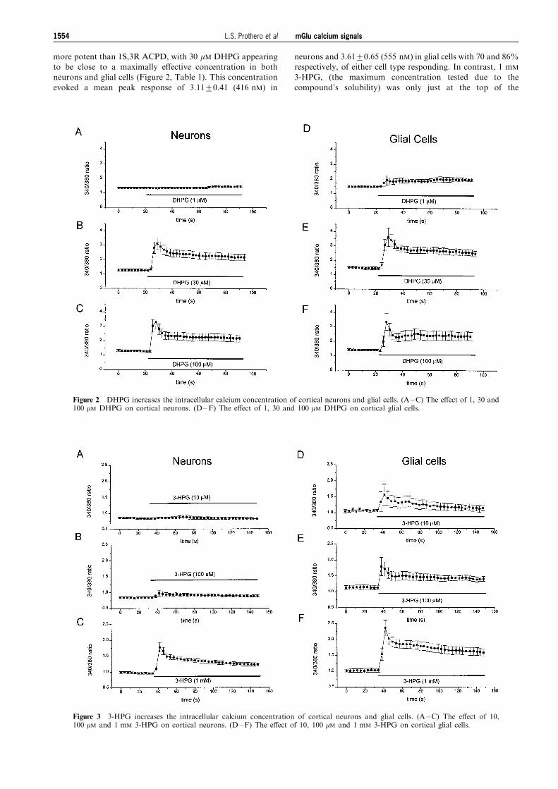
neurons and 3.61+0.65 (555 nM) in glial cells with 70 and 86%respectively, of either cell type responding. In contrast, 1 mM
3-HPG, (the maximum concentration tested due to the
compound's solubility) was only just at the top of the
Figure 2 DHPG increases the intracellular calcium concentration of cortical neurons and glial cells. (A ±C) The e�ect of 1, 30 and100 mM DHPG on cortical neurons. (D±F) The e�ect of 1, 30 and 100 mM DHPG on cortical glial cells.
Figure 3 3-HPG increases the intracellular calcium concentration of cortical neurons and glial cells. (A ±C) The e�ect of 10,100 mM and 1 mM 3-HPG on cortical neurons. (D±F) The e�ect of 10, 100 mM and 1 mM 3-HPG on cortical glial cells.
mGlu calcium signals1554 L.S. Prothero et al

concentration-response relationship for neurons inducingmean peak calcium signals of 1.78+0.15 (123 nM) in 82% ofneurons (Figure 3, Table 1). At the same concentration, 3-
HPG evoked a calcium signal of 2.36+0.25 (239 nM) in glialcells with 89% of cells tested giving a response. For both celltypes, the relative e�ectiveness of these three agonists wasDHPG41S,3R ACPD43-HPG. The Group III agonist L-
AP4 (100 mM) had little e�ect on intracellular calcium (datanot shown).
Recently, a number of compounds have been developed
which may distinguish between e�ects mediated by mGlu1 andmGlu5 receptors. In order to determine the nature of thereceptor underlying these responses further, four additional
pharmacological agents were tested. The ®rst of these was S4-CPG, which has been reported to be a competitive antagonistat mGlu1 receptors but an agonist at mGlu2 receptors. It has
no action on mGlu5 receptors (Brabet et al., 1995). The secondwas S4C3-HPG which is reported to be antagonist at mGlu1receptors but an agonist at mGlu5 receptors at highconcentrations (Brabet et al., 1995 but see Kingston et al.,
1995). Thirdly, we used TADA a putative agonist at mGlu5receptors (Favaron et al., 1993; Opitz et al., 1995) and fourthlyCHPG, a recently developed selective agonist at mGlu5
receptors with no action at mGlu1 receptors (Doherty et al.,1997).
Figures 4 and 5 summarize the results obtained with these
four compounds. Figure 4A and B shows that the mGlu1receptor antagonist/mGlu2 receptor agonist, S4-CPG(500 mM) does not evoke any change in calcium in either
neurons or glia and does not inhibit the response of the cellsto subsequent application of an intermediate concentration of1S,3R ACPD (25 mM). This would suggest that neither
mGlu1 nor mGlu2 receptors underlie the responses seen.Figure 4C and D shows that S4C3-HPG (500 mM) evokes acalcium response in both cell types which would suggest thatan mGlu5 receptor underlies the responses. The recently
developed, selective, mGlu5 receptor agonist CHPG(100 mM±1 mM) is also e�ective at inducing changes inintracellular calcium in both cell types. Figure 5A and B
shows that 1 mM CHPG evokes the characteristic transientincrease followed by sustained elevation in [Ca2+] in both celltypes, with mean peak calcium signals of 3.55+0.19 (537 nM)
in 16 out of 28 neurons (57%) and 2.90+0.18 (363 nM) in 18out of 20 glial cells (90%). Both S4C3-HPG and CHPGevoked responses with the same pro®le as that evoked by
1S,3R ACPD, 3-HPG and DHPG, namely an initial transientcalcium increase followed by a sustained plateau. Theputative mGlu5 receptor agonist TADA (500 mM) was unableto evoke changes in intracellular calcium in neurons and only
induced a slow rise in intracellular calcium in glial cells.Furthermore, this e�ect was observed only after a consider-able delay following agonist application when compared to
the responses seen for the ®ve other e�ective agonistsdescribed above (Figure 5C and D). Thus our data suggestthat the rise in calcium following activation of mGlu
receptors in both cortical neurons and glial cells is broughtabout through activation of the mGlu5 receptor subtype (Abeet al., 1992).
Figure 4 (A and B) The e�ect of S4-CPG (500 mM) on intracellular calcium levels in cortical neurons (A; n=35) and glial cells (B;n=24) before and after the addition of 1S,3R ACPD (25 mM). (C and D) The e�ect of S4C3-HPG (500 mM) on intracellular calciumlevels in cortical neurons (C; n=17) and glial cells (D; n=24).
mGlu calcium signals 1555L.S. Prothero et al

Dependence of the sustained calcium entry onextracellular calcium
The second part of this study considers the nature of theresponse evoked by activation of mGlu receptors in corticalneurons and glial cells. 1S,3R ACPD at a maximally e�ective
concentration of 100 mM, evoked responses which, in themajority of cells, consisted of a transient response followed bya plateau phase (see, for example, Figures 1, 7 and 8). When
extracellular calcium was removed, 1S,3R ACPD still evokedan increase in intracellular calcium which consisted only ofthe initial transient phase, the response decaying exponen-tially to baseline in both cell types with a time constant of
20 s in neurons and 19 s in glial cells (Figure 6). Thesustained calcium response could be induced by readdition ofcalcium (2 mM) to the external solution in the continued
presence of 1S,3R ACPD (Figure 6). Thus the initial calciumresponse depends on the mobilization of calcium frominternal stores, while the sustained phase depends on
extracellular calcium.
Block of sustained calcium entry by inorganic cations
The activation of this sustained calcium entry resembles theactivation of store-operated calcium entry pathways (SOCs)which have been described in a number of other cell types, (see
Discussion). The magnitude of the sustained calcium signalevoked by 1S,3R ACPD (100 mM) was 2.15+0.02 (n=64) inneurons and 2.22+0.03 (n=44) in glial cells in this series of
experiments corresponding to a sustained intracellular calcium
concentration of 195 and 210 nM, respectively. The peakcalcium signals in these cells were 4.44+0.26 (n=64) forneurons and 4.90+0.36 (n=44) for glial cells. One feature of
SOCs is their susceptibility to block by a number of inorganiccations. Figure 7 and Table 2 shows the e�ect of Zn2+ (1 ±100 mM) on the sustained calcium response to 1S,3R ACPD in
neurons. At 100 mM, Zn2+ virtually abolished the sustainedresponse in both cell types. Zn2+ was more e�ective in neuronsthan in glial cells with an estimated IC50 of 6.5 mM in neurons
but 15 mM in glial cells (see Figure 7 and Table 2). Zinc appliedbefore the addition of 1S,3R ACPD blocked the sustainedplateau response but did not a�ect the transient increaseinduced by the agonist (data not shown).
A number of other inorganic ions were examined on thesustained calcium in¯ux evoked by 1S,3R ACPD and theirrelative e�ectiveness at 100 mM is shown in Figure 8 (see also
Table 2). Zn2+ and La3+ were the most e�ective ions tested, atthis concentration producing 101+6% and 78+7% block,respectively, in neurons followed by Cd2+ (52+7%), Co2+
(47+8%) and Ni2+ (20+3%). Ni2+ when given at aconcentration of 400 mM, produced 60+9% inhibition of thecalcium entry pathway in neurons and 36+5% in glial cells,
however, a 4 fold increase in concentration to 1.6 mM
produced no further increase in the degree of inhibition ineither cell type. Mg2+, even at concentration of 10 mM, onlyproduced a partial inhibition of this calcium entry pathway
(47+11%). For all ions, block was more e�ective in neuronscompared with glial cells (see Table 2) however the relativepotency sequence was the same in either cell type and was
Zn2+5La3+4Cd2+5Co2+4Ni2+4Mg2+.
Figure 5 (A and B) The e�ect of CHPG (1 mM) on intracellular calcium levels in cortical neurons (A; n=10) and glial cells (B;n=11). (C and D) The e�ect of TADA (500 mM) on intracellular calcium levels in cortical neurons (C; n=29) and glial cells (D;n=12).
mGlu calcium signals1556 L.S. Prothero et al

In contrast, Zn2+ (100 mM) had little detectable e�ect oncalcium entry evoked by depolarization of the neurons with
25 mM K+ which evokes calcium entry through voltage-gatedcalcium channels (Figure 9). This K+-induced calcium entrycould, however, be inhibited by both 100 mM Cd2+ and 10 mMLa3+ (Figure 9).
Discussion
Activation of group I mGlu receptors of rat cortical neuronsand glial cells induced a rise in the intracellular calcium
concentration which consisted of two phases. The ®rst, initialtransient phase re¯ects release of calcium from intracellularstores while the second sustained phase is due to calcium in¯ux
across the plasma membrane, possibly via a store-operatedcalcium entry pathway (Berridge, 1995). We have previouslyshown that responses to 1S,3R ACPD could be inhibited by
pretreatment of the cells with the Ca-ATPase inhibitor andstore depletor, thapsigargin (0.1 ± 1 mM; Mathie & Richards,1997). Similar calcium response pro®les have now been
observed following activation of a number of di�erent Gprotein coupled receptors in many cell types (see for example,Irving et al., 1992; Grudt et al., 1996).
We have characterized the nature of the mGlu receptors
which underlie these responses using a number of di�erentmGlu receptor agonists. The most convincing evidence wasobtained using the recently developed selective mGlu5 receptor
agonist, CHPG (Doherty et al., 1997). This compound
produced clear responses in both cell types at concentrationsranging from 100 mM to 1 mM. The pro®le of the responseswere very similar to those evoked by 1S,3R ACPD. This
agonist is the best compound currently available to distinguishbetween the two types of Group I mGlu receptor as itselectively activates recombinant mGlu5 receptors but notmGlu1 receptors even at concentrations as high as 4 mM
(Doherty et al., 1997). Our data with two other phenylglycinederivatives (S4-CPG and S4C3-HPG) are also consistent withthe idea that mGlu5 receptors underlie the calcium responses
(see Brabet et al., 1995): S4-CPG has no agonist action nordoes it antagonize the e�ect of 1S,3R ACPD while S4C3-HPGacts as an agonist.
Human recombinant mGlu5 receptors, however, appear tohave a pharmacological pro®le that di�ers from that of rat asS4-CPG and S4C-HPG act as antagonists at this receptor
(Kingston et al., 1995). This suggests that there are majorspecies di�erences between these receptors. It is however,possible that these two compounds are not as reliable asCHPG as indicators of the subtype of mGlu receptors
underlying the responses. TADA is the least well characterizedof the four compounds tested and it has been suggested to beselective for mGlu5 receptors on the basis of some rather
indirect evidence from experiments on native receptors(Favaron et al., 1993; Opitz et al., 1995) together with a lackof e�ect on expressed mGlu1 receptors (Favaron et al., 1993).
We found that this compound had little action on neurons,although it did produce a delayed slow rise in intracellularcalcium in glial cells. While this response is very di�erent in
pro®le to that evoked by the other e�ective agonists, it couldlead to a net rise in intracellular calcium of a similar magnitudeto that induced by the other agonists.
Immunocytochemical studies with a speci®c mGlu5
receptor antibody suggests that this receptor is widelyexpressed throughout the brain but that the cortex is amongthe areas of the brain with the highest expression levels
(Romano et al., 1995). Levels of mGlu5 receptor are thoughtto be at their highest in the neonatal rat brain with around a 2fold reduction during development (Romano et al., 1996).
Recent RT±PCR experiments have suggested that corticalglial cells only express the mGlu5 subtype of Group I receptors(e.g. Ciccarelli et al., 1997).
Studies with recombinant Group I mGlu receptors have
suggested that the pro®le of the calcium response seenfollowing receptor activation, di�ers between mGlu1 andmGlu5 receptors (Kawabata et al., 1996). Activation of mGlu5
receptors gives rise to oscillatory calcium signals, whileactivation of mGlu1 leads to a quite di�erent response pro®lecomprising a single peaked calcium response with no
maintained plateau. This di�erence has been attributed to asingle amino acid residue (threonine at position 840) present inmGlu5 receptors which can be phosphorylated by protein
kinase C (Kawabata et al., 1996). The oscillatory responsesfollowing mGlu5 receptor activation resemble the responsesseen in a proportion of the cells in this study at intermediateagonist concentrations. However, unlike the sustained
responses seen in this study at high agonist concentrations,calcium oscillations were seen even in the absence ofextracellular calcium. Similar oscillatory responses were
observed for native mGlu5 receptors in cortical astrocytes butonly when the expression of these receptors was selectivelyupregulated by basic ®broblast growth factor (bFGF),
epidermal growth factor (EGF) and transforming growthfactor-alpha (TGF-a) (Nakahara et al., 1997; see also Miller etal., 1995; Balazs et al., 1997). Responses seen in untreated cellsresembled those seen in this study at high agonist concentra-
Figure 6 The response to 1S,3R ACPD (100 mM) in the absence ofextracellular calcium in neurons (A; n=7) and glial cells (B; n=4).The response decays exponentially back to baseline in both cell types.Readdition of extracellular calcium (2 mM) induces a sustained rise incalcium in both cell types in the continued presence of 1S,3R ACPD.
mGlu calcium signals 1557L.S. Prothero et al

Figure 7 The e�ect of increasing concentrations of zinc on the sustained plateau evoked by 1S,3R ACPD (100 mM) in corticalneurons. (A ±C) Show the e�ects of 1, 10 and 100 mM zinc in neurons. (D) Shows the percentage inhibition produced by theseconcentrations of zinc in both cell types measured 37 s after the addition of the ion.
Table 2 Concentration-response relationships for block by inorganic cations of the sustained calcium responsefollowing activation of mGluR5 receptors by 1S,3R ACPD (100 mM)
Neurons GliaIonspecies
Concentration(mM) (n)
% inhibition of plateauphase+s.e.mean} (n)
% inhibition of plateauphase+s.e.mean}
Zn2+
La3+
Ni2+
Co2+
Cd2+
Mg2+
11030501001025100501002004008001600551002224448001008001000
(11)(8)(9)(17)(14)(8)(11)(12)(11)(17)(7)(8)(10)(14)(8)(8)(4)(10)(12)(7)(8)(6)
25+474+1396+775+5101+694+4102+778+734+720+371+1660+947+854+939+647+469+766+781+452+782+547+11
(14)(5)(7)(12)(6)(11)(12)(12)(7)(10)(4)(13)(18)(19)(4)(4)(7)(7)(12)(4)(8)(8)
28+450+847+759+7111+371+861+859+629+717+338+1336+532+436+317+523+740+435+757+436+678+1441+6
}As determined 37 s after ion application.
mGlu calcium signals1558 L.S. Prothero et al

tions comprising of an initial transient response followed by asustained plateau (Nakahara et al., 1997). Thus the pro®le ofthe calcium response seen following mGlu5 receptor activation
may depend on the agonist concentration, the density of thereceptors and their phosphorylation state.
The sustained plateau response seen in this study could beabolished by the removal of extracellular calcium. This
response resembles SOCs that have been described in a
number of other cell types which are activated followingdepletion of intracellular calcium stores (Berridge, 1995). Thelink between store depletion and SOC channel activation
remains to be determined (Clapham, 1995), the two most likelymechanisms being either a di�usible second messenger(Randriamampita & Tsien, 1993) or a direct protein/proteininteraction between the SOC channel and the IP3 receptor (see
Putney, 1997). The molecular identity of the channels
Figure 8 The e�ect of various inorganic cations on the sustained plateau evoked by 1S,3R ACPD (100 mM) in cortical neurons (A)Zn2+ (100 mM); (B) La3+ (100 mM); (C) Ni2+ (100 mM); (D) Co2+ (100 mM); (E) Cd2+ (100 mM); (F) Mg2+ (10 mM). Inhibitionproduced by these ions was measured 37 s after the addition of the ion.
Figure 9 The e�ect of (A) Cd2+ (100 mM; n=14); (B) La3+ (10 mM; n=9) and (C) Zn2+ (100 mM; n=14) on the calcium responseevoked by depolarizing cortical neurons with 25 mM K+.
mGlu calcium signals 1559L.S. Prothero et al

underlying SOCs also remains to be established. It has beensuggested that the Drosophila protein, trp, might be thefunctional analogue of mammalian SOCs and a number of
homologous mammalian genes have recently been cloned (seeClapham, 1996).
The sustained calcium in¯ux evoked by activation of mGlu5receptors in this study could be blocked by a number of
inorganic cations of which Zn2+ and La3+ were the moste�ective of the ions tested. This is in contrast to Ca2+ in¯uxevoked by depolarization of the cells with 25 mM K+ in
neurons which was blocked by 100 mM La3+ and Cd2+ butvirtually una�ected by the same concentration of Zn2+,consistent with Ca2+ entry through voltage-gated Ca2+
channels (Wakamori et al., 1998). Glial cells gave little or noresponse to high K+ treatment in contrast to theirresponsiveness to mGlu receptor agonists suggesting that
depolarization of the cells does not lead to signi®cantglutamate release. The most comprehensive study on thee�ects of inorganic ions on calcium entry pathways is that ofHoth & Penner (1993) studying Icrac currents in mast cells.
These authors studied a range of inorganic ions and found apotency series similar but not identical to what we have foundin cortical neurons and glial cells. For Icrac in mast cells, the
order of potency was: Zn2+5La3+4Cd2+4Be2+5Co2+4Mn2+4Ni2+4Sr2+4Ba2+ (Hoth & Penner, 1993),the only di�erence from this study being a detectably larger
inhibitory e�ect of Cd2+ compared to Co2+ for Icrac in mastcells. Additionally, however, all of the ions seemed to be about10 fold less potent at inhibiting Icrac than they were at
inhibiting the plateau phase of the response to mGlu receptoragonists. Other studies have found quite variable e�ects ofcertain inorganic ions on Ca2+ in¯ux pathways whether thepathways have been activated by store depletion following G
protein stimulation of IP3 production or whether the storeshave been depleted directly with thapsigargin or cyclopiazonicacid. For example, La3+ has been shown to block Ca2+ entry
through the SOC channel in cells treated with thapsigargin atconcentrations of 1 mM in salivary cells (Foskett & Wong,1994), 200 nM in C6-2B glioma cells (Chiono et al., 1995),
around 20 nM Jurkat T cells (Aussel et al., 1996) and 75 nM inSH-SY5Y neuroblastoma cells (Grudt et al., 1996). In C6-glioma cells, Wu et al. (1997) have found that depletion ofintracellular stores with thapsigargin leads to activation of a
Ca2+ in¯ux pathway which was markedly inhibited by La3+
and Ni2+. However, in mouse smooth muscle cells La3+ hadno e�ect on Ca2+ in¯ux through the SOC pathway at
concentrations up to 400 mM (Wayman et al., 1996). This
suggests that a number of di�erent channels may underlie SOCCa2+ entry in di�erent tissues each with rather di�erentpharmacological characteristics.
Although the response of the glial cells and neurons werebroadly similar in all aspects of this study, there were someobvious potency di�erences. For example, all the mGlureceptor agonists were more potent on glial cells than on
neurons producing, at submaximal concentrations, both largerCa2+ signals and a larger proportion of responsive cells. Incontrast, the blocking action of divalent cations on SOC Ca2+
entry was invariably more pronounced in neurons than in glialcells. The larger agonist-evoked signals in glial cells may re¯ectsubtle di�erences in the nature of the proteins involved in Ca2+
mobilization in the two cell types but more likely, it re¯ectsdi�erences in receptor number or a greater signal ampli®cationat some step in the transduction pathway leading to a greater
mobilization of Ca2+ in glial cells for a given agonistconcentration. In view of the heterologous nature of SOCpathways in di�erent cells (see above), di�erences in theblocking potency of the di�erent ions may truly re¯ect
di�erent proteins in neurons compared with glial cellsunderlying Ca2+ entry.
The sustained rises in intracellular calcium following mGlu5
receptor activation in rat cortical neurons and glial cells areaccompanied by sustained changes in intracellular pH with theneurons acidifying and the glial cells alkalizing (Amos &
Chesler, 1998; Amos et al., 1998). These changes will haveprofound consequences for cell function both physiologically,and in pathological situations such as cerebral ischaemia where
there is a prolonged rise in extracellular glutamate. Thesustained rises in intracellular calcium will stimulate calcium-dependent enzyme activity and Ca2+ dependent gene expres-sion (see, for example, Clapham, 1995) which may underlie the
plasticity changes seen following stimulation of mGlureceptors (e.g. Bashir et al., 1993) and which are impaired inmice lacking mGlu5 receptors (Lu et al., 1997). Following
cerebral trauma, there is a rise in extracellular glutamate andastrocytes proliferate as part of the repair process. Stimulationof astrocyte mGlu5 receptors enhances glial cell proliferation
(Ciccarelli et al., 1997) and it is probable that either thesustained rise in Ca2+ and/or the subsequent Ca2+-dependentglial cell alkalinization (Amos et al., 1998) may serve as thesignal for astrocytes to proliferate.
Thanks to the Wellcome Trust for ®nancial support and to JamesWood and Gladys Onambe le for participating in some experiments.
References
ABE, T., SUGIHARA, H., NAWA, H., SHIGEMOTO, R., MIZUNO, N. &
NAKANISHI, S. (1992). Molecular characterisation of a novelmetabotropic glutamate receptor mGluR5 coupled to inositolphosphate/Ca2+ signal transduction. J. Biol. Chem., 267.13361 ± 13368.
AMOS, B.J. & CHESLER, M. (1998). Characterization of anintracellular alkaline shift in rat astrocytes triggered bymetabotropic glutamate receptors. J. Neurophysiol., 79, 695 ±703.
AMOS, B.J., MATHIE, A. & RICHARDS, C.D. (1998). Activation ofgroup I metabotropic glutamate receptors elicits pH changes incultured rat cortical glia and neurones.Neurosci., 86, 1109 ± 1120.
AUSSEL, C., MARHABA, R., PELASSY, C. & BREITTMAYER, J.
(1996). Submicromolar La3+ concentrations block the calcium-release activated channel and impair CD69 and CD26 expressionin CD3- or thapsigargin-activated Jurkat cells. Biochem. J., 313,909 ± 913.
BALAZS, R., MILLER, S., ROMANO, C., DE VRIES, A., CHUN, Y. &
COTMAN, C.W. (1997). Metabotropic glutamate receptormGluR5 in astrocytes: pharmacological properties and agonistregulation. J. Neurochem., 69, 151 ± 163.
BASHIR, Z.I., BORTOLOTTO, Z.A., DAVIES, C.H., BERRETTA, N.,
IRVING, A.J., SEAL, A.J., HENLEY, J.M., JANE, D.E., WATKINS,
J.C. & COLLINGRIDGE, G.L. (1993). Induction of LTP in thehippocampus needs synaptic activation of glutamate metabo-tropic receptors. Nature, 363, 347 ± 350.
BERRIDGE, M.J. (1995). Capacitative calcium entry. Biochem. J.,312, 1 ± 11.
BRABET, I., MARY, S., BOCKAERT, J. & PIN, J.P. (1995).Phenylglycine derivatives discriminate between mGluR1-mediated and mGlu5-mediated responses. Neuropharmacol., 34,895 ± 903.
mGlu calcium signals1560 L.S. Prothero et al

BROWN, E.M., GAMBA, G., RICCARDI, D., LOMBARDI, M.,
BUTTERS, R., KIFOR, O., SUN, A., HEDIGER, M.A., LYTTON, J.
& HEBERT, S.C. (1993). Cloning and characterization of anextracellular Ca2+-sensing receptor from bovine parathyroid.Nature, 366, 575 ± 580.
CHIONO, M., MAHEY, R., TATE, G. & COOPER, D. (1995).Capacitative Ca2+ entry exclusively inhibits cAMP synthesis inC6-2B glioma cells. J. Biol. Chem., 270, 1149 ± 1155.
CICCARELLI, R., SUREDA, F.X., CASABONA, G., DI IORIO, P.,
CARUSO, A., SPINELLA, F., CONDORELLI, D.F., NICOLETTI, F.
& CACIAGLI, F. (1997). Opposite in¯uence of the metabotropicglutamate receptor subtypes mGlu3 and -5 on astrocyteproliferation in culture. Glia, 21, 390 ± 398.
CLAPHAM, D.E., (1995). Calcium Signaling. Cell, 80, 259 ± 268.CLAPHAM, D.E. (1996). TRP is cracked but is CRAC TRP? Neuron,
16, 1069 ± 1072.CONN, P.J. & PIN, J.P. (1997). Pharmacology and functions of
metabotropic glutamate receptors. Ann. Rev. Pharm. Toxicol.,37, 205 ± 237.
DOHERTY, A.J., PALMER, M.J., HENLEY, J.M., COLLINGRIDGE,
G.L. & JANE, D.E. (1997). (RS)-2-chloro-5-hydroxyphenylglycine(CHPG) activates mGlu5, but not mGlu1, receptors expressed inCHO cells and potentiates NMDA responses in the hippocam-pus. Neuropharmacol., 36, 265 ± 267.
FAVARON, M., MANEV, R.M., CANDEO, P., ARBAN, R., GABELLINI,
N., KOZIKOWSKI, A.P. & MANEY, H. (1993). Trans-azetidine-2,4-dicarboxylic acid activates neuronal metabotropic receptors.Neuroreport, 4, 967 ± 970.
FOSKETT, J.K. & WONG, D.C.P. (1994). [Ca2+]i inhibition of Ca2+
release-activated Ca2+ in¯ux underlies agonist- and thapsigar-gin-induced [Ca2+] oscillations in salivary acinar cells. J. Biol.Chem., 269, 31525 ± 31532.
GRUDT, T.J., USOWICZ, M.M. & HENDERSON, G. (1996). Ca2+ entryfollowing store depletion in SH-SY5Y neuroblastoma cells. Mol.Brain Res., 36, 93 ± 100.
GRYNKIEWICZ, G., POENIE, M. & TSIEN, R.Y. (1985). A newgeneration of Ca2+ indicators with greatly improved ¯uorescenceproperties. J. Biol. Chem., 260, 3440 ± 3450.
HAYASHI, Y., SEKIYAMA, N., NAKANISHI, S., JANE, D.E., SUNTER,
D.C., BIRSE, E.F., UDVARHELYI, P.M. & WATKINS, J.C. (1994).Analysis of agonist and antagonist activities of phenylglycinederivatives for di�erent cloned metabotropic glutamate receptorsubtypes. J. Neurosci., 14, 3370 ± 3377.
HOTH, M. & PENNER, R. (1993). Calcium release-activated calciumcurrent in rat mast cells. J. Physiol., 465, 359 ± 386.
IRVING, A.J., COLLINGRIDGE, G.L. & SCHOFIELD, J.G. (1992).Interactions between Ca2+ mobilising mechanisms in culturedrat cerebellar granule cells. J. Physiol., 456, 667 ± 680.
KAUPMANN, K., HUGGEL, K., HEID, J., FLOR, P.J., BISCHOFF, S.,
MICKEL, S.J., MCMASTER, G., ANGST, C., BITTIGER, H.,
FROESTL, W. & BETTLER, B. (1997). Expression cloning ofGABAB receptors uncovers similarity to metabotropic glutamatereceptors. Nature, 386, 239 ± 246.
KAWABATA, S., TSUTSUMI, R., KOHARA, A., YAMAGUCHI, T.,
NAKANISHI, S. & OKADA, M. (1996). Control of calciumoscillations by phosphorylation of metabotropic glutamatereceptors. Nature, 383, 89 ± 92.
KINGSTON, A.E., BURNETT, J.P., MAYNE, N.G. & LODGE, D. (1995).Pharmacological analysis of 4-carboxyphenylglycine deriva-tives ± comparison of e�ects on mGluR1a and mGluR5asubtypes. Neuropharmacol., 34, 887 ± 894.
LU, Y.M., JIA, Z., JANUS, C., HENDERSON, J.T., GERLAI, R.,
WOJTOWICZ, J.M. & RODER, J.C. (1997). Mice lacking metabo-tropic glutamate receptor 5 show impaired learning and reducedCA1 long-term potentiation (LTP) but normal CA3 LTP. J.Neurosci., 17, 5196 ± 5206.
MATHIE, A., AMOS, B.J. & RICHARDS, C.D. (1996). Pharmacologicalcharacterisation of metabotropic glutamate receptor-inducedincreases in intracellular [Ca2+] in cultured rat cortical neuronsand glial cells. Brit. J. Pharmacol., 117, 65P.
MATHIE, A. & RICHARDS, C.D. (1997). Calcium signalling in ratcortical neurones and glia in culture following activation ofgroup I metabotropic glutamate receptors (mGluRs). J. Physiol.,499, 18P.
MILLER, S., ROMANO, C. & COTMAN, C.W. (1995). Growth factorupregulation of a phosphoinositide-coupled metabotropic gluta-mate receptor in cortical astrocytes. J. Neurosci., 15, 6103 ± 6109.
NAKAHARA, K., OKADA, M. & NAKANISHI, S. (1997). Themetabotropic glutamate receptor mGluR5 induces calciumoscillations in cultured astrocytes via protein kinase C phosphor-ylation. J. Neurochem., 69, 1467 ± 1475.
NAKANISHI, S. & MASU, M. (1994). Molecular diversity andfunctions of glutamate receptors. Ann. Rev. Biophys. Biomol.Struct., 23, 319 ± 348.
OPITZ, T., RICHTER, P., CARTE, A.J., KOZIKOWSKI, A.P., SHINO-
ZAKI, H. & REYMANN, K.G. (1995). Metabotropic glutamatereceptor subtypes di�erentially in¯uence neuronal recovery fromin vitro hypoxia/hypoglycemia in rat hippocampal slices.Neurosci., 68, 989 ± 1001.
PIN, J.P. & DUVOISIN, R. (1995). The metabotropic glutamatereceptors ± structure and functions. Neuropharmacol., 34, 1 ± 26.
PROTHERO, L.S., WOOD, J., ONAMBEÂ LEÂ , G.N., MATHIE, A. &
RICHARDS, C.D. (1998). Inorganic ion selectivity of the sustainedcalcium response following activation of metabotropic glutamatereceptors in cultured rat cortical neurones and glia. J. Physiol.,506, 147 ± 148P.
PUTNEY, J.W. (1997). Type 3 IP3 receptor and capacitative calciumentry. Cell Calcium, 21, 257 ± 261.
RANDRIAMAMPITA, C. & TSIEN, R.Y. (1993). Emptying ofintracellular calcium stores release a novel small messenger thatstimulates calcium in¯ux. Nature, 364, 809 ± 814.
ROMANO, C., SESMA, M.A., MCDONALD, C.T., O'MALLEY, K., VAN
DEN POL, A.N. & OLNEY, J.W. (1995). Distribution of themetabotropic glutamate receptor mGluR5 immunoreactivity inrat brain. J. Comp. Neurol., 355, 455 ± 469.
ROMANO, C., VAN DEN POL, A.N. & O'MALLEY, K.L. (1996).Enhanced early developmental expression of the metabotropicglutamate receptor mGluR5 in rat brain: protein, mRNA splicevariants and regional distribution. J. Comp. Neurol., 367, 403 ±412.
WAKAMORI, M., STROBECK, M., NIIDOME, T., TERAMOTO, T.,
IMOTO, K. & MORI, Y. (1998). Functional characterization of ionpermeation pathway in the N-type Ca2+ channel. J. Neurophy-siol., 79, 622 ± 634.
WAYMAN, C., MCFADZEAN, I., GIBSON, A. & TUCKER, J.F. (1996).Two distinct membrane currents activated by cyclopiazonic acid-induced calcium store depletion in single smooth muscle cells ofthe mouse anococcygeus. Brit. J. Pharmacol., 117, 566 ± 572.
WU, M.L., KAO, E.F., LIU, I.H., WANG, B.S. & LINSHIAU, S.Y. (1997).Capacitative Ca2+ in¯ux in glial cells is inhibited by glycolyticinhibitors. Glia, 21, 315 ± 326.
(Received July 6, 1998Revised August 25, 1998
Accepted September 7, 1998)
mGlu calcium signals 1561L.S. Prothero et al