
Attention inhibition of early cortical activation to fearful faces
11
Research Report Attention inhibition of early cortical activation to fearful faces Dimitri J. Bayle a,b,c, ⁎ , Margot J. Taylor c,d a INSERM U821, Bâtiment 452, Centre hospitalier Le Vinatier, 95 boulevard Pinel 69500 BRON, France b Universite Lyon 1, Lyon, F-69000, France c Diagnostic Imaging, Research Institute, Hospital for Sick Children, Toronto, Canada d University of Toronto, Canada ARTICLE INFO ABSTRACT Article history: Accepted 21 November 2009 Available online 11 December 2009 Several lines of evidence demonstrate that processing facial expression can occur in the first 130 ms following a face presentation, but it remains unclear how this is modulated by attention. We presented neutral, fearful and happy faces to subjects who attended either to repeated identity or to repeated emotions. Brain activity was recorded using magnetoencephalography (MEG) and analyzed with event-related beamforming, providing both temporal and spatial information of processing in the brain. The first MEG component, at 90 ms (M90), was sensitive to facial expression, but only when attention was not directed to expression; non-attended fearful faces increased activation in occipital and right middle frontal gyri. Around 150 ms, activity in several brain regions, regardless of the direction of attention, was larger to emotional compared to neutral faces; attention directed to facial expressions increased activity in the right fusiform gyrus and the anterior insula bilaterally. M220 was not modulated by individual facial expressions; however, attention directed to facial expressions enhanced activity in the right inferior parietal lobe and precuneus, while attention directed to identity enhanced posterior cingulate activity.These data demonstrate that facial expression processing involves frontal brain areas as early as 90 ms. Attention directed to emotional expressions obscured this early automatic processing but increased the M170 activity. The M220 sources varied with the direction of attention. Thus, the pattern of neural activation to faces varied with attention to emotions or to identity, demonstrating separate and only partially overlapping networks for these two facets of information contained in faces. © 2009 Elsevier B.V. All rights reserved. Keywords: Emotion Fear Attention MEG Frontal lobe M170 1. Introduction We extract considerable information critical for social inter- actions, rapidly and effectively from faces, including identity and emotions. A recent focus in the neurosciences has been the perception and processing of facial expressions, with studies implicating a large distributed neural network. This affect-sensitive network includes regions in the visual–ventral stream as well as the superior temporal sulcus and dorso- frontal and orbito-frontal regions (Adolphs, 2002; Phan et al., 2002), but the timing of their involvement and their modula- tion by attention are only beginning to emerge. BRAIN RESEARCH 1313 (2010) 113 – 123 ⁎ Corresponding author. INSERM U821, Bâtiment 452, Centre hospitalier Le Vinatier, 95 boulevard Pinel 69500 BRON, France. Fax: +33 47 213 8901. E-mail address: [email protected] (D.J. Bayle). 0006-8993/$ – see front matter © 2009 Elsevier B.V. All rights reserved. doi:10.1016/j.brainres.2009.11.060 available at www.sciencedirect.com www.elsevier.com/locate/brainres
Transcript of Attention inhibition of early cortical activation to fearful faces
B R A I N R E S E A R C H 1 3 1 3 ( 2 0 1 0 ) 1 1 3 – 1 2 3
ava i l ab l e a t www.sc i enced i r ec t . com
www.e l sev i e r . com/ loca te /b ra i n res
Research Report
Attention inhibition of early cortical activation to fearful faces
Dimitri J. Baylea,b,c,⁎, Margot J. Taylorc,d
aINSERM U821, Bâtiment 452, Centre hospitalier Le Vinatier, 95 boulevard Pinel 69500 BRON, FrancebUniversite Lyon 1, Lyon, F-69000, FrancecDiagnostic Imaging, Research Institute, Hospital for Sick Children, Toronto, CanadadUniversity of Toronto, Canada
A R T I C L E I N F O
⁎ Corresponding author. INSERM U821, Bâtime8901.
E-mail address: [email protected] (
0006-8993/$ – see front matter © 2009 Elsevidoi:10.1016/j.brainres.2009.11.060
A B S T R A C T
Article history:Accepted 21 November 2009Available online 11 December 2009
Several lines of evidence demonstrate that processing facial expression can occur in the first130 ms following a face presentation, but it remains unclear how this is modulated byattention. We presented neutral, fearful and happy faces to subjects who attended either torepeated identity or to repeated emotions. Brain activity was recorded usingmagnetoencephalography (MEG) and analyzed with event-related beamforming, providingboth temporal and spatial information of processing in the brain. The first MEG component,at 90 ms (M90), was sensitive to facial expression, but only when attention was not directedto expression; non-attended fearful faces increased activation in occipital and right middlefrontal gyri. Around 150 ms, activity in several brain regions, regardless of the direction ofattention, was larger to emotional compared to neutral faces; attention directed to facialexpressions increased activity in the right fusiform gyrus and the anterior insula bilaterally.M220 was not modulated by individual facial expressions; however, attention directed tofacial expressions enhanced activity in the right inferior parietal lobe and precuneus, whileattention directed to identity enhanced posterior cingulate activity.These data demonstratethat facial expression processing involves frontal brain areas as early as 90 ms. Attentiondirected to emotional expressions obscured this early automatic processing but increasedthe M170 activity. The M220 sources varied with the direction of attention. Thus, the patternof neural activation to faces varied with attention to emotions or to identity, demonstratingseparate and only partially overlapping networks for these two facets of informationcontained in faces.
© 2009 Elsevier B.V. All rights reserved.
Keywords:EmotionFearAttentionMEGFrontal lobeM170
1. Introduction
We extract considerable information critical for social inter-actions, rapidly and effectively from faces, including identityand emotions. A recent focus in the neurosciences has beenthe perception and processing of facial expressions, with
nt 452, Centre hospitalier
D.J. Bayle).
er B.V. All rights reserved
studies implicating a large distributed neural network. Thisaffect-sensitive network includes regions in the visual–ventralstream as well as the superior temporal sulcus and dorso-frontal and orbito-frontal regions (Adolphs, 2002; Phan et al.,2002), but the timing of their involvement and their modula-tion by attention are only beginning to emerge.
Le Vinatier, 95 boulevard Pinel 69500 BRON, France. Fax: +33 47 213
.
114 B R A I N R E S E A R C H 1 3 1 3 ( 2 0 1 0 ) 1 1 3 – 1 2 3
The classic view is that after occipital activity around100 ms, face processing continues along the ventral visualpathways arriving at the fusiform gyrus at around 170 ms(Allison et al., 1994; Bentin et al., 1996), before reachinganterior regions (Haxby et al., 2000). However, event-relatedpotential (ERP/EEG) and event-related field magnetoencepha-lography (ERF/MEG) studies have shown more rapid proces-sing of facial expressions, with components between 90 and130 ms indexing differences between emotional and neutralfaces, seen anteriorly in fronto-medial and orbito-frontalregions (Eimer and Holmes, 2002; Holmes et al., 2003; Streitet al., 2003) and occipitally (Batty et al., 2003; Eger et al., 2003;Halgren et al., 2000; Pizzagalli et al., 1999).
The question of the neural pathways allowing this rapidfrontal activation is still posed but the implication of asubcortical route is increasingly hypothesized. As suggestedfirst by several fMRI and PET studies, this route would bypassthe visual cortex and involve the superior colliculus, pulvinarand amygdala. It would effect a rapid and coarse visualanalysis, notably of aversive stimuli such as fearful or angryfaces (Das et al., 2005; de Gelder et al., 1999; de Marco et al.,2006; Liddell et al., 2005; Morris et al., 2001; Morris et al., 1999;Ohman and Mineka, 2001). However, neither fMRI nor PET candetermine the time course of such pathways. The most directevidence of a subcortical pathway comes from MEG studieswhich have shown very rapid activation of the amygdalafollowing presentation of stimuli including fearful faces(Cornwell et al., 2008; Ioannides, 2006; Liu et al., 1999; Luo et al.,2007, 2009). The role of such a pathway in processing aversivestimuli is reinforced by the fact that the majority of the datashowing frontal activation for facial expression, at latenciesinferior to 130 ms were using fearful faces stimuli (Eimer andHolmes, 2002; Holmes et al., 2003; Kawasaki et al., 2001; Liuet al., 1999; Streit et al., 2003; Williams et al., 2004), and thatamygdala activation has been largely associated with fearperception (Adolphs et al., 2005; Adolphs et al., 1995;Anderson and Phelps, 2000; Broks et al., 1998; Calder et al.,2001; Krolak-Salmon et al., 2004; Morris et al., 1996).
Thus, the very rapid processing of fearful faces has beendemonstrated by a range of electromagnetic studies, yet themodulation of this processing by attention is still not fullycharacterized. Although Pessoa et al. (2002) reported activa-tion to emotional expressions only if subjects attended to theemotions, suggesting that attention is necessary for thisprocess, most authors report that fear processing occurswithout directed attention (Batty et al., 2003; Bradley, 1997;Vuilleumier and Schwartz, 2001) or unconsciously (de Gelderet al., 1999; Morris et al., 2001; Morris et al., 1999; Whalen et al.,1998), and that a fast neural network is activated automati-cally for the unconscious capture of attention by aversivefaces (Bradley, 1997; de Gelder et al., 1999; Pourtois et al., 2004;Pourtois et al., 2005; Vuilleumier and Schwartz, 2001).Whetherattention is or is not necessary for the early fear processing, itcould nevertheless play a modulating role. Williams et al.(2005) compared neutral and fearful faces using fMRI in twoattentional conditions: attend-face and attend-house. Whensubjects attended to houses, amygdala activation for fearfulfaces was greater than when subjects attended to faces; i.e.,amygdala sensitivity to potentially threatening stimuli wasgreater under conditions of inattention. Likewise, Batty et al.,
(2003) found the ERP P1 (at 100 ms) sensitive to facialexpressions, but only in the implicit emotion discriminationtask. In fact, the neurophysiological studies that report asensitivity for facial expression in the first 130msweremostlyimplicit emotional perception tasks (Eger et al., 2003; Eimerand Holmes, 2002; Halgren et al., 2000; Holmes et al., 2003;Kawasaki et al., 2001; Pizzagalli et al., 1999; Streit et al., 2003),while contrarily, the response to attended fearful faces wasrecorded only after 250 ms (Krolak-Salmon et al., 2001, 2003).Taken together, these results suggest that attention directedto the emotional content of faces could inhibit the fastnetwork.
To determine whether the early frontal neural response toemotional faces can be modulated by emotion-specificattention, we used two tasks – an emotion and an identitydiscrimination task – in which subjects attended either tofacial emotion or to facial identity. The use of MEG andbeamformer source analyses provided the means to assessboth the timing of face processing, with superior temporalresolution than fMRI, and spatial localization of face proces-sing, with superior resolution than ERPs. We expected thatevidence of a fast emotion-detection pathway would be moreapparent when emotional expression was processed implic-itly. As the early cortical emotional response has beenreported primarily with fearful faces, we used happy andfearful faces to determine if this activation is triggered byfacial expression more generally or only by threat-relatedstimuli.
2. Results
2.1. Behavioural performance
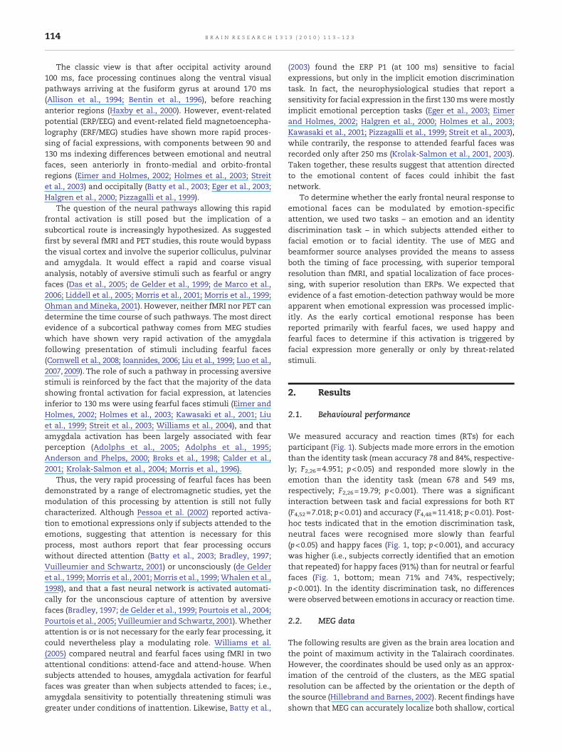
We measured accuracy and reaction times (RTs) for eachparticipant (Fig. 1). Subjects made more errors in the emotionthan the identity task (mean accuracy 78 and 84%, respective-ly; F2,26=4.951; p<0.05) and responded more slowly in theemotion than the identity task (mean 678 and 549 ms,respectively; F2,26=19.79; p<0.001). There was a significantinteraction between task and facial expressions for both RT(F4,52=7.018; p<0.01) and accuracy (F4,48=11.418; p<0.01). Post-hoc tests indicated that in the emotion discrimination task,neutral faces were recognised more slowly than fearful(p<0.05) and happy faces (Fig. 1, top; p<0.001), and accuracywas higher (i.e., subjects correctly identified that an emotionthat repeated) for happy faces (91%) than for neutral or fearfulfaces (Fig. 1, bottom; mean 71% and 74%, respectively;p<0.001). In the identity discrimination task, no differenceswere observed between emotions in accuracy or reaction time.
2.2. MEG data
The following results are given as the brain area location andthe point of maximum activity in the Talairach coordinates.However, the coordinates should be used only as an approx-imation of the centroid of the clusters, as the MEG spatialresolution can be affected by the orientation or the depth ofthe source (Hillebrand and Barnes, 2002). Recent findings haveshown that MEG can accurately localize both shallow, cortical
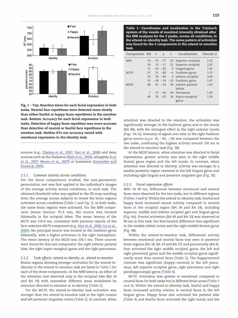
Table 1 – Coordinates and localization in the Talairachsystem of the voxels of maximal intensity obtained afterthe ERB analyses for the 3 peaks, across all conditions, inthe attend-to-identity task. The same pattern of activationwas found for the 3 components in the attend-to-emotiontask.
Component BA x y z Localization Pseudo-Z
M90 19 −15 −77 22 Superior occipital 2.1619 15 −77 22 Superior occipital 2.0217 −10 −83 0 Lingual gyrus 1.45
M170 37 31 −60 −6 Fusiform gyrus 3.1519 39 −69 0 Inferior occipital 3.0419 −28 −74 −10 Fusiform gyrus 1.67
M220 40 45 −42 44 Inferior parietallobule
1.43
7 15 −56 44 Precuneus 1.3648 50 −42 34 Supra-marginal
gyrus1.24
Fig. 1 – Top: Reaction times for each facial expression in bothtasks. Neutral face repetitions were detected more slowlythan either fearful or happy faces repetitions in the emotiontask. Bottom: Accuracy for each facial expression in bothtasks. Detection of happy faces repetition was more accuratethan detection of neutral or fearful face repetitions in theemotion task. Neither RTs nor accuracy varied withemotional expression in the identity task.
115B R A I N R E S E A R C H 1 3 1 3 ( 2 0 1 0 ) 1 1 3 – 1 2 3
sources (e.g., Cheyne et al., 2007; Itier et al., 2006) and deepsources such as the thalamus (Bish et al., 2004), amygdala (Luoet al., 2007; Moses et al., 2007) or brainstem (Ioannides andFenwick, 2005).
2.2.1. Common activity across conditionsFor the three components studied, the non-parametricpermutation test was first applied to the individual's imagesof the average activity across conditions, in each task. Theobtained threshold value was applied to the 3D map resultingfrom the average across subjects to reveal the brain regionsactivated across conditions (Table 1 and Fig. 2). In both tasks,the same brain regions were activated. For the M90 compo-nent (mean latency: 91.6 ms), the source was locatedbilaterally in the occipital lobes. The mean latency of theM170 was 150.4 ms, consistent with previous reports of theface-selective M170 component (e.g., Itier et al., 2006; Liu et al.,2000); the principal source was located in the fusiform gyrusbilaterally, with a higher activation in the right hemisphere.The mean latency of the M220 was 226.1 ms. Three sourceswere found for this last component: the right inferior parietallobe, the right supra-marginal gyrus and the right precuneus.
2.2.2. Task effects: attend-to-identity vs. attend-to-emotionBrains regions showing stronger activation for the attend-to-identity or the attend-to-emotion task are listed in Table 2 foreach of the three components. At the M90 latency, an effect ofthe attention was observed only in the occipital lobe (BA 18and BA 19) with somewhat different areas modulated byattention directed to emotion or to identity (Table 2).
For the M170, the attend-to-identity task activation wasstronger than the attend-to-emotion task in the right cuneusand left posterior cingulate cortex (Table 2). In contrast, when
attention was directed to the emotion, the activation wassignificantly stronger in the fusiform gyrus and in the insula(BA 48), with the strongest effect in the right anterior insula(Figs. 3A–C). Intensity of signal over time in the right fusiformgyrus source (x,y,z: 35, −60, −10) was compared between thetwo tasks, confirming the highest activity around 150 ms inthe attend-to-emotion task (Fig. 3B).
At the M220 latency, when attention was directed to facialexpressions, greater activity was seen in the right middlefrontal gyrus region and the left insula. In contrast, whenattention was directed to identity, activity was stronger in amedial posterior region centered in the left lingual gyrus andincluding right lingual and posterior cingulate gyri (Fig. 3E).
2.2.3. Facial expression effectsM90: At 90 ms, differences between emotional and neutralfaces were observed for the two tasks, but in different regions(Tables 3 and 4).Within the attend-to-identity task, fearful andhappy faces increased neural activity compared to neutralfaces in the occipital region (BA 18 and BA 19), includingsuperior, middle and inferior occipital gyri and lingual gyrus(Fig. 4A). Frontal activation (BA 46 and BA 10) was observed at90 ms in this task; the fear/neutral difference was significantin the medial orbital cortex and the right middle frontal gyrus(Fig. 4B).
Within the attend-to-emotion task, differential activitybetween emotional and neutral faces was seen in posteriorbrain regions (BA 18, BA 19 and BA 37) and precentrally (BA 6).Fear activated the right middle occipital gyrus, the left andright precentral gyrus and the middle occipital gyrus signifi-cantly more than neutral faces (Table 3). The happy/neutralcontrast was significant (happy>neutral) in the left precu-neus, left superior occipital gyrus, right precuneus and rightparahippocampal gyrus (Table 4).
M170: Activation was greater to emotional compared toneutral faces for both tasks but in different brain areas (Table 3and 4). Within the attend-to-identity task, fearful and happyfaces increased activity relative to neutral faces in the leftlingual gyrus. Happy faces also activated the parietal lobe(Table 4) and fearful faces activated the right insula and the
Fig. 2 – Group-averaged ERB images in the Talairach coordinate system, obtained for the M90, M170 and M220 components,across all conditions during the attend-to-identity task. The voxels shown passed the group statistical threshold of p<0.01. Onthe axial images, right is on the right.
116 B R A I N R E S E A R C H 1 3 1 3 ( 2 0 1 0 ) 1 1 3 – 1 2 3
cingulate region (Table 3). Frontal activations were alsoobserved in the attend-to-identity task: fearful faces activatedthe inferior frontal gyrus (Table 3), while happy facesincreased activity the right middle frontal gyrus (Table 4;Fig. 5).
Within the attend-to-emotion task, fearful and happy facesincreased activity in posterior brain regions (BA 18, BA 21, BA23, BA 37): rightmiddle occipital gyrus for happy faces (Table 4)and right middle temporal gyrus, left lingual gyrus and rightposterior cingulate gyrus for fearful faces (Table 3).
M220: Contrasts between emotional and neutral faces didnot reveal any differences in location or power of activity ineither task, showing that the M220 was not modulated byfacial expression per se.
3. Discussion
3.1. M90
Neutral, happy and fearful faces were presented whilesubjects attended to either the facial identity or the facialexpression. Using MEG we demonstrated that fearful facesincreased brain activation at 90 ms in occipital and frontalregions; however, this fast frontal response was inhibited ifattention was directed to the emotional information. In both
Table 2 – Task effects: Coordinates and localization in the Talairemotions, the brain areas that were more active when attentio(right column), at the three latencies measured. Sources are lissignificant at p<0.01.
Component Regions more active in the attend-to-identity ta
BA x y z Localization Pseud
M90 19 −25 −82 18 Middle occipital, cuneus 0.6418 20 −58 3 Lingual gyrus 0.53
M170 18 10 −67 17 Cuneus, precuneus 1.0117 −15 −58 8 Posterior cingulate 0.85
M220 18 −5 −63 −3 Lingual and posterior 0.83Cingulate gyri
⁎ p<.005.⁎⁎ p<.001.
attentional tasks, fearful faces enhanced the activity inoccipital regions, showing that fear is processed in the first100 ms, independent of attention demand, consistent withstudies using various attentional procedures (Batty et al., 2003;Eger et al., 2003; Halgren et al., 2000; Pizzagalli et al., 1999) andshowing an early automatic encoding of emotional facialexpression (Batty et al., 2003; Pizzagalli et al., 1999). In contrastto posterior activity, the frontal activation to fearful faces atM90 was present only in the attend-to-identity task. Thissuggests that two processes were engaged during the first90 ms following a fearful face presentation: one increasingactivity in the posterior regions and not modulated byattention and a second one evident in frontal regions, onlywhen attention was not directed to the facial expression.These data support a two pathway hypothesis that has beenproposed for early emotional processing (Morris et al., 1999;Vuilleumier, 2005), wherein fearful faces are processed inparallel by both cortical and subcortical visual pathways. Theproposed subcortical visual pathway includes the superiorcolliculus, the pulvinar, the amygdalae and the locus ceruleus,before reaching the frontal cortex (Liddell et al., 2005; Morris etal., 1999). The existence of such a visual pathway bypassingthe striate cortex has been demonstrated anatomically in thenon-human primates (Cowey and Stoerig, 1991; Hernandez-Gonzalez et al., 1994; Rodman et al., 1989), and studies withblindsight patients demonstrate its probable implication in
ach system for the task effect. Regardless of the three facialn was directed to the identity (left column) or the emotionted from largest to smallest. All reported activations are
sk Regions more active in the attend-to-emotion task
o-Z BA x y z Localization Pseudo-Z
⁎ 19 −25 −70 26 Superior occipital 0.78 ⁎19 30 −62 17 Occipital sub-gyrus 0.68 ⁎
⁎⁎ 48 45 −24 15 Insula, precentral gyrus 0.57 ⁎48 −38 −16 9 Insula 0.4417 35 −60 −10 Fusiform gyrus 0.43
⁎ 44 35 12 41 Middle frontal 0.4448 −40 −9 10 Insula 0.39
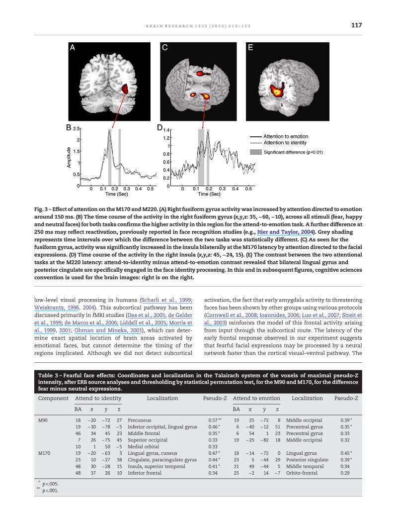
Fig. 3 – Effect of attention on theM170 andM220. (A) Right fusiformgyrus activitywas increased by attention directed to emotionaround 150 ms. (B) The time course of the activity in the right fusiform gyrus (x,y,z: 35, −60, −10), across all stimuli (fear, happyand neutral faces) for both tasks confirms the higher activity in this region for the attend-to-emotion task. A further difference at250 ms may reflect reactivation, previously reported in face recognition studies (e.g., Itier and Taylor, 2004). Grey shadingrepresents time intervals over which the difference between the two tasks was statistically different. (C) As seen for thefusiformgyrus, activitywas significantly increased in the insula bilaterally at theM170 latency by attention directed to the facialexpressions. (D) Time course of the activity in the right insula (x,y,z: 45, −24, 15). (E) The contrast between the two attentionaltasks at the M220 latency: attend-to-identity minus attend-to-emotion contrast revealed that bilateral lingual gyrus andposterior cingulate are specifically engaged in the face identity processing. In this and in subsequent figures, cognitive sciencesconvention is used for the brain images: right is on the right.
117B R A I N R E S E A R C H 1 3 1 3 ( 2 0 1 0 ) 1 1 3 – 1 2 3
low-level visual processing in humans (Scharli et al., 1999;Weiskrantz, 1996, 2004). This subcortical pathway has beendiscussed primarily in fMRI studies (Das et al., 2005; de Gelderet al., 1999; de Marco et al., 2006; Liddell et al., 2005; Morris etal., 1999, 2001; Ohman and Mineka, 2001), which can deter-mine exact spatial location of brain areas activated byemotional faces, but cannot determine the timing of theregions implicated. Although we did not detect subcortical
Table 3 – Fearful face effects: Coordinates and localization inintensity, after ERB source analyses and thresholding by statisticfear minus neutral expressions.
Component Attend to identity Localization
BA x y z
M90 18 −20 −72 27 Precuneus19 −30 −78 −5 Inferior occipital, lingual gyrus46 34 45 23 Middle frontal7 26 −75 45 Superior occipital
10 1 50 −5 Medial orbitalM170 19 −20 −63 3 Lingual gyrus, cuneus
23 10 −27 38 Cingulate, paracingulate gyrus48 30 −28 15 Insula, superior temporal48 37 26 10 Inferior frontal
⁎ p<.005.⁎⁎ p<.001.
activation, the fact that early amygdala activity to threateningfaces has been shown by other groups using various protocols(Cornwell et al., 2008; Ioannides, 2006; Luo et al., 2007; Streit etal., 2003) reinforces the model of this frontal activity arisingfrom input through the subcortical route. The latency of theearly frontal response observed in our experiment suggeststhat fearful facial expressions may be processed by a neuralnetwork faster than the cortical visual–ventral pathway. The
the Talairach system of the voxels of maximal pseudo-Zal permutation test, for theM90 andM170, for the difference
Pseudo-Z Attend to emotion Localization Pseudo-Z
BA x y z
0.57 ⁎⁎ 19 25 −72 8 Middle occipital 0.39 ⁎0.46 ⁎ 6 −40 −12 51 Precentral gyrus 0.35 ⁎0.35 ⁎ 6 54 1 23 Precentral gyrus 0.330.33 19 −25 −82 18 Middle occipital 0.320.330.47 ⁎ 18 −14 −72 0 Lingual gyrus 0.45 ⁎0.44 ⁎ 23 5 −44 29 Posterior cingulate 0.39 ⁎0.41 ⁎ 21 49 −44 5 Middle temporal 0.340.34 25 −2 14 −7 Orbito-frontal 0.29
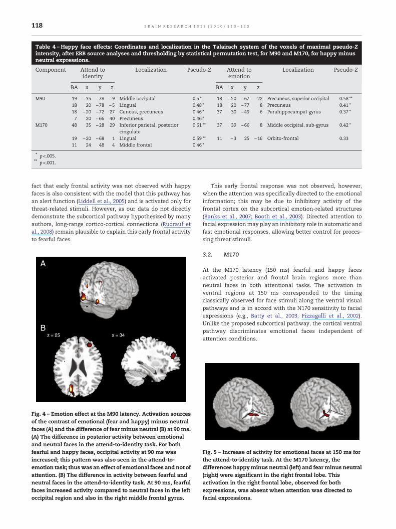
Table 4 – Happy face effects: Coordinates and localization in the Talairach system of the voxels of maximal pseudo-Zintensity, after ERB source analyses and thresholding by statistical permutation test, for M90 and M170, for happy minusneutral expressions.
Component Attend toidentity
Localization Pseudo-Z Attend toemotion
Localization Pseudo-Z
BA x y z BA x y z
M90 19 −35 −78 −9 Middle occipital 0.5 ⁎ 18 −20 −67 22 Precuneus, superior occipital 0.58 ⁎⁎18 20 −78 −5 Lingual 0.48 ⁎ 18 20 −77 8 Precuneus 0.41 ⁎18 −20 −72 27 Cuneus, precuneus 0.46 ⁎ 37 30 −49 6 Parahippocampal gyrus 0.37 ⁎7 20 −66 40 Precuneus 0.46 ⁎
M170 48 35 −28 29 Inferior parietal, posteriorcingulate
0.61 ⁎⁎ 37 39 −66 8 Middle occipital, sub-gyrus 0.42 ⁎
19 −20 −68 1 Lingual 0.59 ⁎⁎ 11 −3 25 −16 Orbito-frontal 0.3311 24 48 4 Middle frontal 0.46 ⁎
⁎ p<.005.⁎⁎ p<.001.
118 B R A I N R E S E A R C H 1 3 1 3 ( 2 0 1 0 ) 1 1 3 – 1 2 3
fact that early frontal activity was not observed with happyfaces is also consistent with the model that this pathway hasan alert function (Liddell et al., 2005) and is activated only forthreat-related stimuli. However, as our data do not directlydemonstrate the subcortical pathway hypothesized by manyauthors, long-range cortico-cortical connections (Rudrauf etal., 2008) remain plausible to explain this early frontal activityto fearful faces.
Fig. 4 – Emotion effect at the M90 latency. Activation sourcesof the contrast of emotional (fear and happy) minus neutralfaces (A) and the difference of fear minus neutral (B) at 90 ms.(A) The difference in posterior activity between emotionaland neutral faces in the attend-to-identity task. For bothfearful and happy faces, occipital activity at 90 ms wasincreased; this pattern was also seen in the attend-to-emotion task; thuswas an effect of emotional faces and not ofattention. (B) The difference in activity between fearful andneutral faces in the attend-to-identity task. At 90 ms, fearfulfaces increased activity compared to neutral faces in the leftoccipital region and also in the right middle frontal gyrus.
This early frontal response was not observed, however,when the attention was specifically directed to the emotionalinformation; this may be due to inhibitory activity of thefrontal cortex on the subcortical emotion-related structures(Banks et al., 2007; Booth et al., 2003). Directed attention tofacial expressionmay play an inhibitory role in automatic andfast emotional responses, allowing better control for proces-sing threat stimuli.
3.2. M170
At the M170 latency (150 ms) fearful and happy facesactivated posterior and frontal brain regions more thanneutral faces in both attentional tasks. The activation inventral regions at 150 ms corresponded to the timingclassically observed for face stimuli along the ventral visualpathways and is in accord with the N170 sensitivity to facialexpressions (e.g., Batty et al., 2003; Pizzagalli et al., 2002).Unlike the proposed subcortical pathway, the cortical ventralpathway discriminates emotional faces independent ofattention conditions.
Fig. 5 – Increase of activity for emotional faces at 150 ms forthe attend-to-identity task. At the M170 latency, thedifferences happyminus neutral (left) and fear minus neutral(right) were significant in the right frontal lobe. Thisactivation in the right frontal lobe, observed for bothexpressions, was absent when attention was directed tofacial expressions.
119B R A I N R E S E A R C H 1 3 1 3 ( 2 0 1 0 ) 1 1 3 – 1 2 3
The activity in the right fusiform gyrus increased when thesubject's attention was directed to the facial expressions. Thefusiform modulation observed by spatial attention (Vuilleu-mier et al., 2001) or by specific attention to faces (Haxby et al.,1994; Wojciulik et al., 1998) indexes increased face processingdepending on the attentional condition. The enhancement ofactivity in the current study, seen as a recruitment ofresources with attention directed to facial expressions, isconsistent with ERP studies showing larger N170s to fearfulfaces (Batty et al., 2003). Attention to emotion inhibits an earlyautomatic cerebral response in anterior regions, but itenhances activity in the ventral visual stream 50–60 mslater. These results suggest that attention directed to emotionmay also influence the structural encoding of faces which hasbeen suggested to occur at the N170 (M170) latency (e.g.,Bentin et al., 1996).
At the same latency, insula activity was seen when theattention was directed to facial affect. The insula is commonlyassociated with the perception of disgust (Calder et al., 2000;Calder et al., 2001; Krolak-Salmon et al., 2003; Phillips et al.,1997; Sprengelmeyer et al., 1998;Wicker et al., 2003), but insulaactivation has also been reported during recognition of facialexpressions of angry and happy faces (Gorno-Tempini et al.,2001) or for painful expressions (Botvinick et al., 2005). Theinsula may play a role in aversive stimulus processing (Pauluset al., 2003; Paulus and Stein, 2006; Simmons et al., 2006). Theinsula activity in the present study was seen when attentionwas directed to the emotional information of faces suggestingthat it is involved in explicit processing of emotions.
3.3. M220
Differences in neural recruitment depending on the attentiondemand were also seen at the M220. At this latency, inferiorparietal cortex activation was present in both attentionaltasks, reflecting memory processing of the stimulus, as hasbeen demonstrated with other n-back studies (Cohen et al.,1997; D'Esposito et al., 1998; Ravizza et al., 2004; Romero et al.,2006); the right-sided only inferior parietal activation waslikely due to the stimuli being exclusively faces. However, theright middle frontal lobe was more active when attention wasdirected to emotion, consistent with the earlier activations offrontal areas for emotional processing, while the lingualgyrus and posterior cingulate were more active with attentiondirected to identity. The posterior cingulate gyrus is implicat-ed in memory processing (Fletcher et al., 1995; Grasby et al.,1993; Maddock et al., 2001; Minoshima et al., 1997) includingmemory for faces (Khader et al., 2005; Kosaka et al., 2003),suggesting its role in the acquisition of facial familiarity(Kosaka et al., 2003). Our data are in accordance with thesestudies, implicating the posterior cingulate gyrus in faceidentity processing. The fact that the posterior cingulategyrus was more active in the attend-to-identity taskindicated that this is not the reflection of mnemonicprocessing but a specific activation driven by face identifi-cation. Thus, the neuronal network recruited in the twoattention tasks differed, in agreement with the Haxby et al.(2000) and Bruce and Young (1986) models of face processing,arguing that emotion and identity are processed by differentbrain regions.
3.4. Conclusions
In summary, we highlight the role played by directed attentionin early facial expression processing. Our findings confirmthat facial emotional information is processed automaticallyin the first 100 ms, if it appears incidentally. The consequenceof attention directed to facial expressions is the disappearanceof the automatic early frontal activity, but an enhancement ofthe fusiform gyrus responses, reflecting a deeper processing ofthe facial components. By controlling attentional direction, wedifferentiated neural activities associated with emotional oridentity processing: insula was involved in emotion recogni-tion at 150 ms while identity recognition involved lingualgyrus and posterior cingulate at 220 ms. Depending onwhether subjects paid attention to identity or to facialexpression, the cerebral responses differed both spatiallyand temporally, contributing further evidence of the complexoverlapping networks (i.e., Barbeau et al., 2008) involved in thiscritical human skill of face recognition.
4. Experimental procedures
4.1. Subjects
The participants were 12 adult volunteers (6 men, 6 women)aged 24 to 40 years (mean age of 32.5 years); they gaveinformed written consent. Participants were right-handed,had normal or corrected-to-normal vision and had no historyof neurological disorders. Seven other volunteers (4 men, 3women, aged 22 to 27 years), who did not participate in theMEG experiment, participated in stimulus validation.
4.2. Stimuli
Black and white photographs of faces of 120 adults (60 femaleand 60 male) were used for this study. Each individual wasrepresented in 3 different expressions: neutral, happy andfear. For most individuals, there were two different photo-graphs for each expression, thus typically 6 photos per model.These photographs were from the MacBrain Face Stimulus Set(Tottenham et al., 2009) – we used only those rated as ≥80%accurately classified – and from our own previously validateddatabase (Batty et al., 2003), where all of the faces wereaccurately classified by emotion at ≥80%.
However, to confirm the emotion validity the facialexpressions for the present study, all the photographs wereshown in random order to seven subjects, who classified theemotions expressed in each photograph; they were given theoptions of the six basic expressions and neutral (Ekman andFriesen, 1971). Photographs incorrectly categorized by two ormore subjects were discarded. The final set consisted of 450different black and white photographs brightness-adjustedwith a 272×350 pixel resolution.
4.3. Task procedure
Participants lay comfortably supine for the MEG study, at aviewing distance of 60 cm from the screen inside themagnetically shielded room. Stimuli were presented on the
120 B R A I N R E S E A R C H 1 3 1 3 ( 2 0 1 0 ) 1 1 3 – 1 2 3
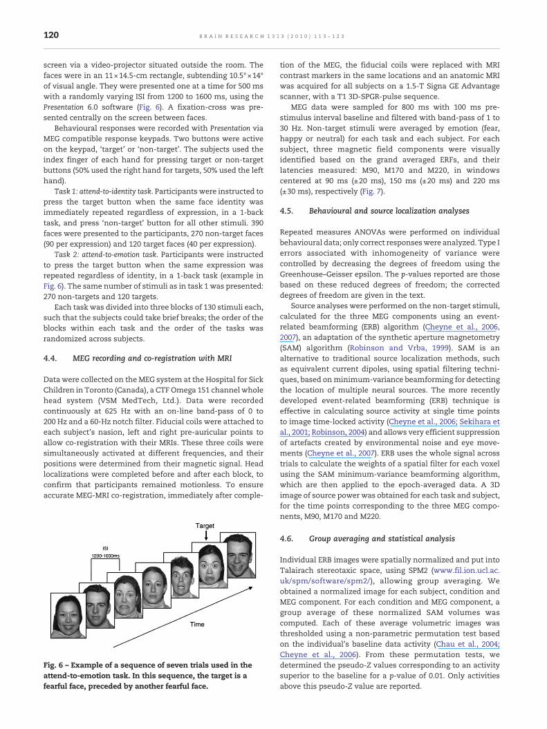
screen via a video-projector situated outside the room. Thefaces were in an 11×14.5-cm rectangle, subtending 10.5°×14°of visual angle. They were presented one at a time for 500 mswith a randomly varying ISI from 1200 to 1600 ms, using thePresentation 6.0 software (Fig. 6). A fixation-cross was pre-sented centrally on the screen between faces.
Behavioural responses were recorded with Presentation viaMEG compatible response keypads. Two buttons were activeon the keypad, ‘target’ or ‘non-target’. The subjects used theindex finger of each hand for pressing target or non-targetbuttons (50% used the right hand for targets, 50% used the lefthand).
Task 1: attend-to-identity task. Participants were instructed topress the target button when the same face identity wasimmediately repeated regardless of expression, in a 1-backtask, and press ‘non-target’ button for all other stimuli. 390faces were presented to the participants, 270 non-target faces(90 per expression) and 120 target faces (40 per expression).
Task 2: attend-to-emotion task. Participants were instructedto press the target button when the same expression wasrepeated regardless of identity, in a 1-back task (example inFig. 6). The same number of stimuli as in task 1 was presented:270 non-targets and 120 targets.
Each task was divided into three blocks of 130 stimuli each,such that the subjects could take brief breaks; the order of theblocks within each task and the order of the tasks wasrandomized across subjects.
4.4. MEG recording and co-registration with MRI
Data were collected on theMEG system at the Hospital for SickChildren in Toronto (Canada), a CTF Omega 151 channel wholehead system (VSM MedTech, Ltd.). Data were recordedcontinuously at 625 Hz with an on-line band-pass of 0 to200 Hz and a 60-Hz notch filter. Fiducial coils were attached toeach subject's nasion, left and right pre-auricular points toallow co-registration with their MRIs. These three coils weresimultaneously activated at different frequencies, and theirpositions were determined from their magnetic signal. Headlocalizations were completed before and after each block, toconfirm that participants remained motionless. To ensureaccurate MEG-MRI co-registration, immediately after comple-
Fig. 6 – Example of a sequence of seven trials used in theattend-to-emotion task. In this sequence, the target is afearful face, preceded by another fearful face.
tion of the MEG, the fiducial coils were replaced with MRIcontrast markers in the same locations and an anatomic MRIwas acquired for all subjects on a 1.5-T Signa GE Advantagescanner, with a T1 3D-SPGR-pulse sequence.
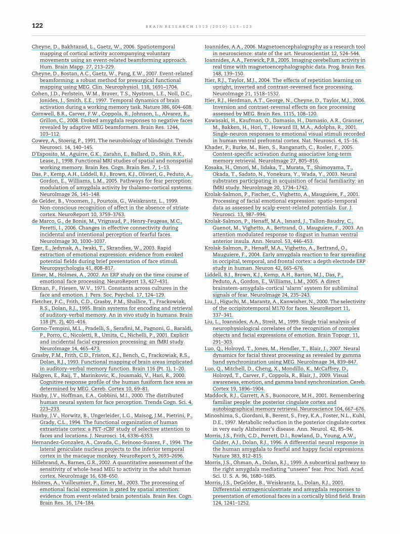
MEG data were sampled for 800 ms with 100 ms pre-stimulus interval baseline and filtered with band-pass of 1 to30 Hz. Non-target stimuli were averaged by emotion (fear,happy or neutral) for each task and each subject. For eachsubject, three magnetic field components were visuallyidentified based on the grand averaged ERFs, and theirlatencies measured: M90, M170 and M220, in windowscentered at 90 ms (±20 ms), 150 ms (±20 ms) and 220 ms(±30 ms), respectively (Fig. 7).
4.5. Behavioural and source localization analyses
Repeated measures ANOVAs were performed on individualbehavioural data; only correct responseswere analyzed. Type Ierrors associated with inhomogeneity of variance werecontrolled by decreasing the degrees of freedom using theGreenhouse–Geisser epsilon. The p-values reported are thosebased on these reduced degrees of freedom; the correcteddegrees of freedom are given in the text.
Source analyses were performed on the non-target stimuli,calculated for the three MEG components using an event-related beamforming (ERB) algorithm (Cheyne et al., 2006,2007), an adaptation of the synthetic aperture magnetometry(SAM) algorithm (Robinson and Vrba, 1999). SAM is analternative to traditional source localization methods, suchas equivalent current dipoles, using spatial filtering techni-ques, based onminimum-variance beamforming for detectingthe location of multiple neural sources. The more recentlydeveloped event-related beamforming (ERB) technique iseffective in calculating source activity at single time pointsto image time-locked activity (Cheyne et al., 2006; Sekihara etal., 2001; Robinson, 2004) and allows very efficient suppressionof artefacts created by environmental noise and eye move-ments (Cheyne et al., 2007). ERB uses the whole signal acrosstrials to calculate the weights of a spatial filter for each voxelusing the SAM minimum-variance beamforming algorithm,which are then applied to the epoch-averaged data. A 3Dimage of source power was obtained for each task and subject,for the time points corresponding to the three MEG compo-nents, M90, M170 and M220.
4.6. Group averaging and statistical analysis
Individual ERB images were spatially normalized and put intoTalairach stereotaxic space, using SPM2 (www.fil.ion.ucl.ac.uk/spm/software/spm2/), allowing group averaging. Weobtained a normalized image for each subject, condition andMEG component. For each condition and MEG component, agroup average of these normalized SAM volumes wascomputed. Each of these average volumetric images wasthresholded using a non-parametric permutation test basedon the individual's baseline data activity (Chau et al., 2004;Cheyne et al., 2006). From these permutation tests, wedetermined the pseudo-Z values corresponding to an activitysuperior to the baseline for a p-value of 0.01. Only activitiesabove this pseudo-Z value are reported.
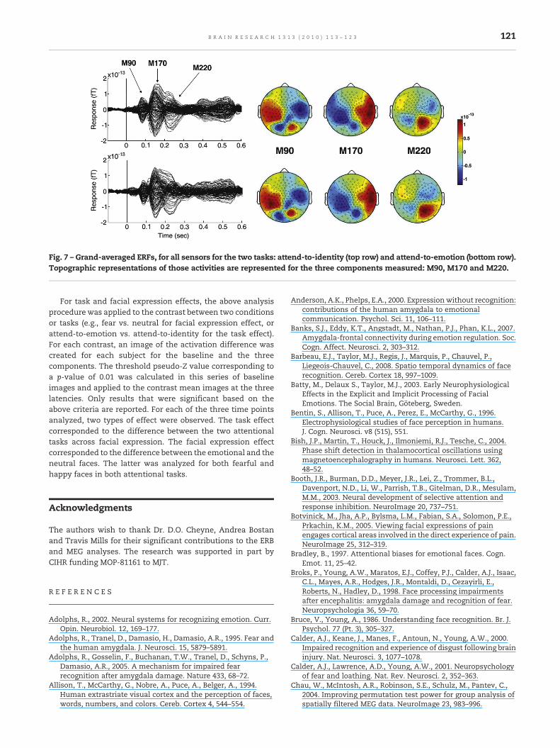
Fig. 7 – Grand-averaged ERFs, for all sensors for the two tasks: attend-to-identity (top row) and attend-to-emotion (bottom row).Topographic representations of those activities are represented for the three components measured: M90, M170 and M220.
121B R A I N R E S E A R C H 1 3 1 3 ( 2 0 1 0 ) 1 1 3 – 1 2 3
For task and facial expression effects, the above analysisprocedure was applied to the contrast between two conditionsor tasks (e.g., fear vs. neutral for facial expression effect, orattend-to-emotion vs. attend-to-identity for the task effect).For each contrast, an image of the activation difference wascreated for each subject for the baseline and the threecomponents. The threshold pseudo-Z value corresponding toa p-value of 0.01 was calculated in this series of baselineimages and applied to the contrast mean images at the threelatencies. Only results that were significant based on theabove criteria are reported. For each of the three time pointsanalyzed, two types of effect were observed. The task effectcorresponded to the difference between the two attentionaltasks across facial expression. The facial expression effectcorresponded to the difference between the emotional and theneutral faces. The latter was analyzed for both fearful andhappy faces in both attentional tasks.
Acknowledgments
The authors wish to thank Dr. D.O. Cheyne, Andrea Bostanand Travis Mills for their significant contributions to the ERBand MEG analyses. The research was supported in part byCIHR funding MOP-81161 to MJT.
R E F E R E N C E S
Adolphs, R., 2002. Neural systems for recognizing emotion. Curr.Opin. Neurobiol. 12, 169–177.
Adolphs, R., Tranel, D., Damasio, H., Damasio, A.R., 1995. Fear andthe human amygdala. J. Neurosci. 15, 5879–5891.
Adolphs, R., Gosselin, F., Buchanan, T.W., Tranel, D., Schyns, P.,Damasio, A.R., 2005. A mechanism for impaired fearrecognition after amygdala damage. Nature 433, 68–72.
Allison, T., McCarthy, G., Nobre, A., Puce, A., Belger, A., 1994.Human extrastriate visual cortex and the perception of faces,words, numbers, and colors. Cereb. Cortex 4, 544–554.
Anderson, A.K., Phelps, E.A., 2000. Expression without recognition:contributions of the human amygdala to emotionalcommunication. Psychol. Sci. 11, 106–111.
Banks, S.J., Eddy, K.T., Angstadt, M., Nathan, P.J., Phan, K.L., 2007.Amygdala-frontal connectivity during emotion regulation. Soc.Cogn. Affect. Neurosci. 2, 303–312.
Barbeau, E.J., Taylor, M.J., Regis, J., Marquis, P., Chauvel, P.,Liegeois-Chauvel, C., 2008. Spatio temporal dynamics of facerecognition. Cereb. Cortex 18, 997–1009.
Batty, M., Delaux S., Taylor, M.J., 2003. Early NeurophysiologicalEffects in the Explicit and Implicit Processing of FacialEmotions. The Social Brain, Göteberg, Sweden.
Bentin, S., Allison, T., Puce, A., Perez, E., McCarthy, G., 1996.Electrophysiological studies of face perception in humans.J. Cogn. Neurosci. v8 (515), 551.
Bish, J.P., Martin, T., Houck, J., Ilmoniemi, R.J., Tesche, C., 2004.Phase shift detection in thalamocortical oscillations usingmagnetoencephalography in humans. Neurosci. Lett. 362,48–52.
Booth, J.R., Burman, D.D., Meyer, J.R., Lei, Z., Trommer, B.L.,Davenport, N.D., Li, W., Parrish, T.B., Gitelman, D.R., Mesulam,M.M., 2003. Neural development of selective attention andresponse inhibition. NeuroImage 20, 737–751.
Botvinick, M., Jha, A.P., Bylsma, L.M., Fabian, S.A., Solomon, P.E.,Prkachin, K.M., 2005. Viewing facial expressions of painengages cortical areas involved in the direct experience of pain.NeuroImage 25, 312–319.
Bradley, B., 1997. Attentional biases for emotional faces. Cogn.Emot. 11, 25–42.
Broks, P., Young, A.W., Maratos, E.J., Coffey, P.J., Calder, A.J., Isaac,C.L., Mayes, A.R., Hodges, J.R., Montaldi, D., Cezayirli, E.,Roberts, N., Hadley, D., 1998. Face processing impairmentsafter encephalitis: amygdala damage and recognition of fear.Neuropsychologia 36, 59–70.
Bruce, V., Young, A., 1986. Understanding face recognition. Br. J.Psychol. 77 (Pt. 3), 305–327.
Calder, A.J., Keane, J., Manes, F., Antoun, N., Young, A.W., 2000.Impaired recognition and experience of disgust following braininjury. Nat. Neurosci. 3, 1077–1078.
Calder, A.J., Lawrence, A.D., Young, A.W., 2001. Neuropsychologyof fear and loathing. Nat. Rev. Neurosci. 2, 352–363.
Chau, W., McIntosh, A.R., Robinson, S.E., Schulz, M., Pantev, C.,2004. Improving permutation test power for group analysis ofspatially filtered MEG data. NeuroImage 23, 983–996.
122 B R A I N R E S E A R C H 1 3 1 3 ( 2 0 1 0 ) 1 1 3 – 1 2 3
Cheyne, D., Bakhtazad, L., Gaetz, W., 2006. Spatiotemporalmapping of cortical activity accompanying voluntarymovements using an event-related beamforming approach.Hum. Brain Mapp. 27, 213–229.
Cheyne, D., Bostan, A.C., Gaetz, W., Pang, E.W., 2007. Event-relatedbeamforming: a robust method for presurgical functionalmapping using MEG. Clin. Neurophysiol. 118, 1691–1704.
Cohen, J.D., Perlstein, W.M., Braver, T.S., Nystrom, L.E., Noll, D.C.,Jonides, J., Smith, E.E., 1997. Temporal dynamics of brainactivation during a working memory task. Nature 386, 604–608.
Cornwell, B.R., Carver, F.W., Coppola, R., Johnson, L., Alvarez, R.,Grillon, C., 2008. Evoked amygdala responses to negative facesrevealed by adaptive MEG beamformers. Brain Res. 1244,103–112.
Cowey, A., Stoerig, P., 1991. The neurobiology of blindsight. TrendsNeurosci. 14, 140–145.
D'Esposito, M., Aguirre, G.K., Zarahn, E., Ballard, D., Shin, R.K.,Lease, J., 1998. Functional MRI studies of spatial and nonspatialworking memory. Brain Res. Cogn. Brain Res. 7, 1–13.
Das, P., Kemp, A.H., Liddell, B.J., Brown, K.J., Olivieri, G., Peduto, A.,Gordon, E., Williams, L.M., 2005. Pathways for fear perception:modulation of amygdala activity by thalamo-cortical systems.NeuroImage 26, 141–148.
de Gelder, B., Vroomen, J., Pourtois, G., Weiskrantz, L., 1999.Non-conscious recognition of affect in the absence of striatecortex. NeuroReport 10, 3759–3763.
de Marco, G., de Bonis, M., Vrignaud, P., Henry-Feugeas, M.C.,Peretti, I., 2006. Changes in effective connectivity duringincidental and intentional perception of fearful faces.NeuroImage 30, 1030–1037.
Eger, E., Jedynak, A., Iwaki, T., Skrandies, W., 2003. Rapidextraction of emotional expression: evidence from evokedpotential fields during brief presentation of face stimuli.Neuropsychologia 41, 808–817.
Eimer, M., Holmes, A., 2002. An ERP study on the time course ofemotional face processing. NeuroReport 13, 427–431.
Ekman, P., Friesen, W.V., 1971. Constants across cultures in theface and emotion. J. Pers. Soc. Psychol. 17, 124–129.
Fletcher, P.C., Frith, C.D., Grasby, P.M., Shallice, T., Frackowiak,R.S., Dolan, R.J., 1995. Brain systems for encoding and retrievalof auditory-verbal memory. An in vivo study in humans. Brain118 (Pt. 2), 401–416.
Gorno-Tempini, M.L., Pradelli, S., Serafini, M., Pagnoni, G., Baraldi,P., Porro, C., Nicoletti, R., Umita, C., Nichelli, P., 2001. Explicitand incidental facial expression processing: an fMRI study.NeuroImage 14, 465–473.
Grasby, P.M., Frith, C.D., Friston, K.J., Bench, C., Frackowiak, R.S.,Dolan, R.J., 1993. Functional mapping of brain areas implicatedin auditory–verbal memory function. Brain 116 (Pt. 1), 1–20.
Halgren, E., Raij, T., Marinkovic, K., Jousmaki, V., Hari, R., 2000.Cognitive response profile of the human fusiform face area asdetermined by MEG. Cereb. Cortex 10, 69–81.
Haxby, J.V., Hoffman, E.A., Gobbini, M.I., 2000. The distributedhuman neural system for face perception. Trends Cogn. Sci. 4,223–233.
Haxby, J.V., Horwitz, B., Ungerleider, L.G., Maisog, J.M., Pietrini, P.,Grady, C.L., 1994. The functional organization of humanextrastriate cortex: a PET-rCBF study of selective attention tofaces and locations. J. Neurosci. 14, 6336–6353.
Hernandez-Gonzalez, A., Cavada, C., Reinoso-Suarez, F., 1994. Thelateral geniculate nucleus projects to the inferior temporalcortex in the macaque monkey. NeuroReport 5, 2693–2696.
Hillebrand, A., Barnes, G.R., 2002. A quantitative assessment of thesensitivity of whole-head MEG to activity in the adult humancortex. NeuroImage 16, 638–650.
Holmes, A., Vuilleumier, P., Eimer, M., 2003. The processing ofemotional facial expression is gated by spatial attention:evidence from event-related brain potentials. Brain Res. Cogn.Brain Res. 16, 174–184.
Ioannides, A.A., 2006. Magnetoencephalography as a research toolin neuroscience: state of the art. Neuroscientist 12, 524–544.
Ioannides, A.A., Fenwick, P.B., 2005. Imaging cerebellum activity inreal time withmagnetoencephalographic data. Prog. Brain Res.148, 139–150.
Itier, R.J., Taylor, M.J., 2004. The effects of repetition learning onupright, inverted and contrast-reversed face processing.NeuroImage 21, 1518–1532.
Itier, R.J., Herdman, A.T., George, N., Cheyne, D., Taylor, M.J., 2006.Inversion and contrast-reversal effects on face processingassessed by MEG. Brain Res. 1115, 108–120.
Kawasaki, H., Kaufman, O., Damasio, H., Damasio, A.R., Granner,M., Bakken, H., Hori, T., Howard III, M.A., Adolphs, R., 2001.Single-neuron responses to emotional visual stimuli recordedin human ventral prefrontal cortex. Nat. Neurosci. 4, 15–16.
Khader, P., Burke, M., Bien, S., Ranganath, C., Rosler, F., 2005.Content-specific activation during associative long-termmemory retrieval. NeuroImage 27, 805–816.
Kosaka, H., Omori, M., Iidaka, T., Murata, T., Shimoyama, T.,Okada, T., Sadato, N., Yonekura, Y., Wada, Y., 2003. Neuralsubstrates participating in acquisition of facial familiarity: anfMRI study. NeuroImage 20, 1734–1742.
Krolak-Salmon, P., Fischer, C., Vighetto, A., Mauguiere, F., 2001.Processing of facial emotional expression: spatio-temporaldata as assessed by scalp event-related potentials. Eur. J.Neurosci. 13, 987–994.
Krolak-Salmon, P., Henaff, M.A., Isnard, J., Tallon-Baudry, C.,Guenot, M., Vighetto, A., Bertrand, O., Mauguiere, F., 2003. Anattention modulated response to disgust in human ventralanterior insula. Ann. Neurol. 53, 446–453.
Krolak-Salmon, P., Henaff, M.A., Vighetto, A., Bertrand, O.,Mauguiere, F., 2004. Early amygdala reaction to fear spreadingin occipital, temporal, and frontal cortex: a depth electrode ERPstudy in human. Neuron 42, 665–676.
Liddell, B.J., Brown, K.J., Kemp, A.H., Barton, M.J., Das, P.,Peduto, A., Gordon, E., Williams, L.M., 2005. A directbrainstem-amygdala-cortical ‘alarm’ system for subliminalsignals of fear. NeuroImage 24, 235–243.
Liu, J., Higuchi, M., Marantz, A., Kanwisher, N., 2000. The selectivityof the occipitotemporal M170 for faces. NeuroReport 11,337–341.
Liu, L., Ioannides, A.A., Streit, M., 1999. Single trial analysis ofneurophysiological correlates of the recognition of complexobjects and facial expressions of emotion. Brain Topogr. 11,291–303.
Luo, Q., Holroyd, T., Jones, M., Hendler, T., Blair, J., 2007. Neuraldynamics for facial threat processing as revealed by gammaband synchronization using MEG. NeuroImage 34, 839–847.
Luo, Q., Mitchell, D., Cheng, X., Mondillo, K., McCaffrey, D.,Holroyd, T., Carver, F., Coppola, R., Blair, J., 2009. Visualawareness, emotion, and gamma band synchronization. Cereb.Cortex 19, 1896–1904.
Maddock, R.J., Garrett, A.S., Buonocore, M.H., 2001. Rememberingfamiliar people: the posterior cingulate cortex andautobiographical memory retrieval. Neuroscience 104, 667–676.
Minoshima, S., Giordani, B., Berent, S., Frey, K.A., Foster, N.L., Kuhl,D.E., 1997. Metabolic reduction in the posterior cingulate cortexin very early Alzheimer's disease. Ann. Neurol. 42, 85–94.
Morris, J.S., Frith, C.D., Perrett, D.I., Rowland, D., Young, A.W.,Calder, A.J., Dolan, R.J., 1996. A differential neural response inthe human amygdala to fearful and happy facial expressions.Nature 383, 812–815.
Morris, J.S., Ohman, A., Dolan, R.J., 1999. A subcortical pathway tothe right amygdala mediating “unseen” fear. Proc. Natl. Acad.Sci. U. S. A. 96, 1680–1685.
Morris, J.S., DeGelder, B., Weiskrantz, L., Dolan, R.J., 2001.Differential extrageniculostriate and amygdala responses topresentation of emotional faces in a cortically blind field. Brain124, 1241–1252.
123B R A I N R E S E A R C H 1 3 1 3 ( 2 0 1 0 ) 1 1 3 – 1 2 3
Moses, S.N., Houck, J.M., Martin, T., Hanlon, F.M., Ryan, J.D.,Thoma, R.J., Weisend, M.P., Jackson, E.M., Pekkonen, E., Tesche,C.D., 2007. Dynamic neural activity recorded from humanamygdala during fear conditioning usingmagnetoencephalography. Brain Res. Bull. 71, 452–460.
Ohman, A., Mineka, S., 2001. Fears, phobias, and preparedness:toward an evolved module of fear and fear learning. Psychol.Rev. 108, 483–522.
Paulus, M.P., Stein, M.B., 2006. An insular view of anxiety. Biol.Psychiatry 60, 383–387.
Paulus, M.P., Rogalsky, C., Simmons, A., Feinstein, J.S., Stein, M.B.,2003. Increased activation in the right insula during risk-takingdecisionmaking is related to harm avoidance and neuroticism.NeuroImage 19, 1439–1448.
Pessoa, L., McKenna, M., Gutierrez, E., Ungerleider, L.G., 2002.Neural processing of emotional faces requires attention. Proc.Natl. Acad. Sci. U. S. A. 99, 11458–11463.
Phan, K.L., Wager, T., Taylor, S.F., Liberzon, I., 2002. Functionalneuroanatomy of emotion: a meta-analysis of emotionactivation studies in PET and fMRI. NeuroImage 16, 331–348.
Phillips, M.L., Young, A.W., Senior, C., Brammer, M., Andrew, C.,Calder, A.J., Bullmore, E.T., Perrett, D.I., Rowland, D., Williams,S.C., Gray, J.A., David, A.S., 1997. A specific neural substratefor perceiving facial expressions of disgust. Nature 389,495–498.
Pizzagalli, D., Regard, M., Lehmann, D., 1999. Rapid emotional faceprocessing in the human right and left brain hemispheres: anERP study. NeuroReport 10, 2691–2698.
Pizzagalli, D.A., Lehmann, D., Hendrick, A.M., Regard, M.,Pascual-Marqui, R.D., Davidson, R.J., 2002. Affective judgmentsof faces modulate early activity (approximately 160 ms) withinthe fusiform gyri. NeuroImage 16, 663–677.
Pourtois, G., Grandjean, D., Sander, D., Vuilleumier, P., 2004.Electrophysiological correlates of rapid spatial orientingtowards fearful faces. Cereb. Cortex 14, 619–633.
Pourtois, G., Thut, G., Grave de Peralta, R., Michel, C., Vuilleumier,P., 2005. Two electrophysiological stages of spatial orientingtowards fearful faces: early temporo-parietal activationpreceding gain control in extrastriate visual cortex.NeuroImage 26, 149–163.
Ravizza, S.M., Delgado, M.R., Chein, J.M., Becker, J.T., Fiez, J.A.,2004. Functional dissociations within the inferior parietalcortex in verbal working memory. NeuroImage 22, 562–573.
Robinson, S.E., 2004. Localization of event-related activity by SAM(erf). Neurol. Clin. Neurophysiol. 30, 109.
Robinson, S.E., Vrba, J., 1999. Functional neuroimaging bysynthetic aperture magnetometry. In: Yoshimoto, T, Kotani, M,Kuriki, S, Karibe, H, Nakasato, N (Eds.), Recent Advances inMagnetometry. Tohoku University Press, Sendai, pp. 302–305.
Rodman, H.R., Gross, C.G., Albright, T.D., 1989. Afferent basis ofvisual response properties in area MT of themacaque. I. Effectsof striate cortex removal. J. Neurosci. 9, 2033–2050.
Romero, L., Walsh, V., Papagno, C., 2006. The neural correlatesof phonological short-term memory: a repetitive transcranialmagnetic stimulation study. J. Cogn. Neurosci. 18,1147–1155.
Rudrauf, D., David, O., Lachaux, J.P., Kovach, C.K., Martinerie, J.,Renault, B., Damasio, A., 2008. Rapid interactions between theventral visual stream and emotion-related structures rely on atwo-pathway architecture. J. Neurosci. 28, 2793–2803.
Scharli, H., Harman, A.M., Hogben, J.H., 1999. Residual vision in asubject with damaged visual cortex. J. Cogn. Neurosci. 11,502–510.
Sekihara, K., Nagarajan, S.S., Poeppel, D., Marantz, A., Miyashita,Y., 2001. Reconstructing spatio-temporal activities of neuralsources using an MEG vector beamformer technique. IEEETrans. Biomed. Eng. 48 (7), 760–771.
Simmons, A., Strigo, I., Matthews, S.C., Paulus, M.P., Stein, M.B.,2006. Anticipation of aversive visual stimuli is associated withincreased insula activation in anxiety-prone subjects. Biol.Psychiatry 60, 402–409.
Sprengelmeyer, R., Rausch, M., Eysel, U.T., Przuntek, H., 1998.Neural structures associated with recognition of facialexpressions of basic emotions. Proc. Biol. Sci. 265, 1927–1931.
Streit, M., Dammers, J., Simsek-Kraues, S., Brinkmeyer, J., Wolwer,W., Ioannides, A., 2003. Time course of regional brainactivations during facial emotion recognition in humans.Neurosci. Lett. 342, 101–104.
Tottenham, N., Tanaka, J.W., Leon, A.C., McCarry, T., Nurse, M.,Hare, T.A., Marcus, D.J., Westerlund, A., Casey, B.J., Nelson, C.,2009. The NimStim set of facial expressions: judgments fromuntrained research participants. Psychiatry Res. 168, 242–249.
Vuilleumier, P., 2005. How brains beware: neural mechanisms ofemotional attention. Trends Cogn. Sci. 9, 585–594.
Vuilleumier, P., Schwartz, S., 2001. Emotional facial expressionscapture attention. Neurology 56, 153–158.
Vuilleumier, P., Armony, J.L., Driver, J., Dolan, R.J., 2001. Effects ofattention and emotion on face processing in the human brain:an event-related fMRI study. Neuron 30, 829–841.
Weiskrantz, L., 1996. Blindsight revisited. Curr. Opin. Neurobiol. 6,215–220.
Weiskrantz, L., 2004. Roots of blindsight. Prog. Brain Res. 144,229–241.
Whalen, P.J., Rauch, S.L., Etcoff, N.L., McInerney, S.C., Lee, M.B.,Jenike, M.A., 1998. Masked presentations of emotional facialexpressions modulate amygdala activity without explicitknowledge. J. Neurosci. 18, 411–418.
Wicker, B., Keysers, C., Plailly, J., Royet, J.P., Gallese, V., Rizzolatti,G., 2003. Both of us disgusted in My insula: the common neuralbasis of seeing and feeling disgust. Neuron 40, 655–664.
Williams, L.M., Liddell, B.J., Rathjen, J., Brown, K.J., Gray, J., Phillips,M., Young, A., Gordon, E., 2004. Mapping the time course ofnonconscious and conscious perception of fear: an integrationof central and peripheral measures. Hum. Brain Mapp. 21,64–74.
Williams, M.A., McGlone, F., Abbott, D.F., Mattingley, J.B., 2005.Differential amygdala responses to happy and fearful facialexpressions depend on selective attention. NeuroImage 24,417–425.
Wojciulik, E., Kanwisher, N., Driver, J., 1998. Covert visual attentionmodulates face-specific activity in the human fusiform gyrus:fMRI study. J. Neurophysiol. 79, 1574–1578.


![[Posterior cortical atrophy]](https://static.fdokumen.com/doc/165x107/6331b9d14e01430403005392/posterior-cortical-atrophy.jpg)