Shared Genetic Factors Influence Amygdala Volumes and Risk for Alcoholism.
Upload
umcutrechtCategory
view
0download
0
www.elsevier.com/locate/ynimg
NeuroImage 40 (2008) 719–727Evidence of altered cortical and amygdala activation during socialdecision-making in schizophrenia
Daan Baas,a,b,⁎ André Aleman,a,d Matthijs Vink,b Nick F. Ramsey,c
Edward H.F. de Haan,a and René S. Kahnb
aHelmholtz Instituut, Department of Experimental Psychology, Universiteit Utrecht, PO Box 80125, 3508 TC, Utrecht, The NetherlandsbDepartment of Psychiatry, University Medical Center Utrecht, The NetherlandscNeurosurgery, Rudolf Magnus Institute of Neuroscience, University Medical Center Utrecht, The NetherlandsdBCN Neuroimaging Center, University of Groningen, The Netherlands
Received 15 May 2007; revised 17 December 2007; accepted 18 December 2007Available online 5 January 2008
Impaired social cognition is a frequently observed and disablingcharacteristic of schizophrenia. An important aspect of social cognitioninvolves making social decisions about others. The present studyinvestigates whether brain activity related to social decision-makingdiffers between patients with schizophrenia and healthy controls.Twelve patients with schizophrenia and 21 control subjects partici-pated in the study. Behavioral performance and brain activity wereassessed during a task that involved judging the trustworthiness offaces. We performed region-of-interest-based analyses, which revealedthat patients with schizophrenia display specific increases andreductions in activation of the medial orbitofrontal cortex, amygdalaand the right insula during social decision-making, areas that play keyroles in the network that underlies social decisions. These findingssuggest that the impairments in social cognition that are often observedin schizophrenia are, at least in part, related to altered brain activity inthese areas.© 2007 Elsevier Inc. All rights reserved.
Introduction
Impaired social cognition is a frequently observed feature inschizophrenia. Numerous studies have reported specific abnorm-alities in the ability of patients to interpret the beliefs and intentionsof others in order to explain and predict their behavior (Pinkhamet al., 2003). Other studies have found impairments in the ability toidentify facial and prosodic expressions of emotion (Edwards et al.,
⁎ Corresponding author: Helmholtz Instituut, Department of ExperimentalPsychology, Universiteit Utrecht, PO Box 80125, 3508 TC, Utrecht, TheNetherlands. Fax: +31 30 2534511.
E-mail address: [email protected] (D. Baas).Available online on ScienceDirect (www.sciencedirect.com).
1053-8119/$ - see front matter © 2007 Elsevier Inc. All rights reserved.doi:10.1016/j.neuroimage.2007.12.039
2001; Habel et al., 2000; Kohler et al., 2003; Penn et al., 2000;Pinkham et al., 2003). As a result of these social cognitive impair-ments, patients with schizophrenia often display poor social skillsand frequently misinterpret social cues, making it one of the mostdisabling clinical features of the disease (A.P.A., 1994).
An important aspect of social cognition involves makingdecisions based on the appearance of others. Even though the exactneural mechanisms of social decision-making in healthy subjects arenot fully understood, considerable progress has been made duringrecent years. It has become clear that a network of both cortical andsubcortical brain areas is required to make accurate social decisions(Adolphs et al., 1998; Druzgal and D’Eposito, 2001). Neuropsy-chological and imaging research provides evidence for theinvolvement of several key areas that make up this network. Theamygdala (Adolphs et al., 1998; Winston et al., 2002) is engagedduring vigilance and attention to emotionally salient information,and the (ventromedial) prefrontal cortex (PFC) (Damasio, 1994;Eslinger and Damasio, 1985) is implicated in emotional andattentional processes. The insula (Sprengelmeyer et al., 1996;Winston et al., 2002) is involved in the perception and representationof affective/emotional states, most notably disgust. The fusiformgyrus (Iidaka et al., 2001; Kanwisher et al., 1997; Winston et al.,2002) is important for processing face stimuli. It has been suggestedthat the anterior cingulate cortex (ACC) is involved in monitoringthe performance of systems evaluating the behavioral relevance oftarget stimuli (Druzgal and D’Eposito, 2001) and that the superiortemporal sulcus (STS) acts as an association area that processesbehavior of others (Brothers, 1990).
The main goal of the present study was to examine whetherbrain activity within this network differs between patients withschizophrenia and healthy controls during social decision-making.To accomplish this, we used an event-related functional magneticresonance imaging (fMRI) paradigm to measure brain responsesduring evaluative social information processing. We employed apsychological task that requires subjects to make trustworthiness
720 D. Baas et al. / NeuroImage 40 (2008) 719–727
and age judgments about faces with a neutral emotional expression(Winston et al., 2002). Age judgments were included in the task toestablish whether trustworthiness of faces is being processed im-plicitly, when subjects make an unrelated age assessment. Giventhat trustworthiness judgments require processing of subtleemotional cues, and thus place relatively high demands on socialinformation processing, we expected this trustworthiness judgmenttask to be sensitive to subtle differences in social informationprocessing between patients and controls. Based on the priorfunctional neuroimaging findings described above, we hypothe-sized that differences in brain activation between schizophreniapatients and controls would be observed in a network containingthe amygdala, PFC, insula, fusiform gyrus, ACC and STS. Givenearlier findings of reduced amygdala activation in schizophreniapatients (Fahim et al., 2005; Schneider et al., 1998) and the richinterconnections between the amygdala and prefrontal areas(Bechara and Van der Linden, 2005), we mainly expected to findreduced reactivity of the amygdala and prefrontal areas in patientsrelative to controls.
Materials and methods
Subjects
Twelve right-handed patients with schizophrenia (10 males, twofemales; mean age 28.7, SD=5.7) participated in the study. Presenceof schizophrenia in these patients was established using theComprehensive Assessment of Symptoms and History Scale(Andreasen et al., 1992). Current symptoms were assessed usingthe Positive and Negative Syndrome Scale (PANSS) (Kay et al.,1987) (Table 1). At the time of the study, all patients were outpatientson stable doses (chlorpromazine equivalent doses, range: 200 to500 mg, mean: 335.4 mg, SD=110.0 mg) of atypical antipsychoticmedication (including clozapine, olanzapine, quetapine and risper-idone). Mean duration of illness was 6.7 years, SD=5.1 years. Noneof the patients experienced paranoid symptoms (PANSS suspicious-ness/persecution mean score was 1.8, SD=1.1).
Twenty-one right-handed healthy (17 males, four females; meanage 31.2, SD=9.7) control subjects were recruited from communityadvertisements, and selected from the same age range (no significantgroup difference; t(1,31.7)=0.948, p=0.350). Control subjects hada mean education score comparable to the patients (mean educationscore for controls was 16.1, SD=1.8, mean education score forpatients was 15.1, SD=2.1, with no significant group difference;t (1,19.8)=1.529, p=0.142). Control subjects were not taking anymedication andwere free of all psychiatric disorders according to theMini International Neuropsychiatric interview (MINI) (Sheehanet al., 1998). Further exclusion criteria for both groups includedsignificant medical or neurological illness at the time of participa-tion, past major head injury and history of alcohol or drug abuse.
The present study was carried out in accordance with theDeclaration of Helsinki and the study design was reviewed and
Table 1Results of PANSS assessment
Mean total (SD)
PANSS positive symptoms 10.8 (3.2)PANSS negative symptoms 12.7 (4.2)PANSS general symptoms 21.7 (4.1)PANSS total 45 (9.3)
approved by the local human ethics committee. Informed writtenconsent was obtained from all participants after the nature of theprocedures had been fully explained.
Stimuli
120 frontal grayscale images of faces with a neutral emotionalexpression were used as stimuli. More than 80% of the photos wereof Caucasian persons. These images were selected from a larger setof images on the basis of trustworthiness and emotional valenceratings given by 36 healthy subjects in a separate pilot study (9 men,27 women; mean age 21.6, SD=3.3). We discarded images of whichtrustworthiness ratings correlated strongly with emotional valenceratings. Of the images used in the present study, 75 were from the setthat Adolphs et al. used in their study of social cognition in patientswith bilateral amygdala damage (Adolphs et al., 1998). In order toobtain a sufficient number of images for our analysis, these imageswere supplemented with 45 images from the psychological imagecollection of the psychology department of Stirling University(PICS). All images were rated in the pilot study.
Experimental paradigm
The psychological task used in the present study was anadapted version of a task used by Winston et al. (2002). A scanningsession lasted for 25 min and consisted of 16 task blocks with aduration of 45 s, 16 rest blocks with a duration of 45 s and 16instruction trials with a duration of 3 s. There were two types oftask blocks and these types of task blocks were presented inrandom order for each subject. At the start of each task block, theword “age” or “trustworthiness” appeared on screen during aninstruction trial to inform the subject of the task requirement.During eight task blocks, which were preceded by the word “age”,subjects had to decide whether the faces that were presented in thesubsequent task block were older or younger than 30 years. In theother eight task blocks, which were preceded by the word“trustworthiness”, subjects had to judge whether the faces weretrustworthy or untrustworthy. Task blocks consisted of 15 trials thatwere presented sequentially, and each trial consisted of a stimulusthat was presented for 1 s followed by a fixation cross that waspresented for 2 s. The stimulus that was presented in each trialcould either be a face or fixation cross, in which case the trialserved as a null event trial. These null event trials were included toenhance the estimation efficiency of our event-related fMRIanalyses and were randomly interspersed among face trials. Allstimuli, fixation crosses and instructions were presented on a gray(50% white) background. Face stimuli were presented once to eachsubject, randomized to the different task blocks and presented inrandom order across subjects. In total there were 120 face stimuliand 120 null event trials. Every task block was followed by a restblock, during which a fixation-cross remained on screen.
Debriefing
After the scanning session, subjects were required to perform aself-paced task in which they rated all the faces that were presentedduring scanning on a scale of trustworthiness that ranged from 1(highly untrustworthy) to 7 (highly trustworthy), with 4 beingneutral. This task was intended to obtain ratings of subjectivelyexperienced trustworthiness of all faces, including those that werepresented during the age judgment task.
721D. Baas et al. / NeuroImage 40 (2008) 719–727
fMRI image acquisition
Brain imaging data were acquired using a 1.5 T Philips ACS-NTscanner (Philips Medical Systems, Best, The Netherlands). Forfunctional scans, 2D-EPI with BOLD (blood oxygenation leveldependent) contrast was used, with the following parameters: echotime 40 ms; repetition time 2500 ms; flip angle 90°; field of view192×192 mm. Each volume comprised 33 axial scans with 2.2 mmslice thickness (and a gap of 0.8 mm). Thus, voxel size was 3 mmisotropic, and volumes were continuously acquired every 2.5 s in aninterleaved fashion (bottom slice first). In total 600 volumes wereacquired. In the scanner, the head was held in place with a strap andpadding. Each run was preceded by 6 ‘dummy’ scans (which werenot used in further analyses) to allow for T1 equilibration effects.Finally, a T1 weighted structural image was acquired.
fMRI image pre-processing
Image pre-processing was done using SPM2 (WellcomeDepartment of Cognitive Neurology, London, United Kingdom).All functional volumes were corrected for slice timing (Hensonet al., 1999) using the central slice in time as a reference, realignedto the first volume acquired and normalized to the MontrealNeurological Institute (MNI) standard brain (Friston et al., 1995).All volumes were then smoothed with a 6 mm full-width at half-maximum isotropic Gaussian kernel.
fMRI data analysis: whole-brain activation maps
We analyzed the data with an event-related model (Josephs et al.,1997) using SPM2 (Wellcome Department of Cognitive Neurology,London, United Kingdom). Our whole-brain analyses were gearedtowards detecting brain activations in controls and patients as well asdifferences in brain activations between schizophrenia patients andhealthy controls during explicit and implicit processing of socialinformation. To this aim, we performed a general linear model(GLM) analysis with one factor modeling task condition (two levels:explicit and implicit social information processing) and a parametricfactor modeling trustworthiness (ratings ranging from 1 to 7).Trustworthiness of faces was determined based on the judgmentsgiven by the individual subject after scanning. To create theregressors of interest, we modeled the presentation of each face byconvolving a delta function at the onset of each stimulus with thestandard SPM2 canonical hemodynamic response function (HRF)and its temporal derivative (Josephs et al., 1997). These regressorswere parametrically modulated to model subject-specific trust-worthiness judgments. A high-pass filter with a cut-off of 128 s wasincluded to model out low-frequency scanner drifts. No globalscaling was applied in any of the analyses (Desjardins et al., 2001).Next, parameter estimates pertaining to each regressor werecalculated for each voxel and contrast images were calculated byapplying linear contrasts to these parameter estimates. The contrastimages were then entered into one-sample t-tests to test forsignificant activations related to the main effect of task, the maineffect of trustworthiness and the effect of trustworthiness in theexplicit and implicit task conditions within each group. We per-formed a two-sample t-test to compare groups regarding the maineffect of task, the main effect of trustworthiness and the effect oftrustworthiness in the explicit and implicit task conditions. Allgroup maps were thresholded at p=0.05 (family wise error (FWE)corrected). We report descriptively activations surviving a threshold
of 0.001 uncorrected with an extent threshold of 5 contiguousvoxels.
fMRI data analysis: region-of-interest analysis
In order to examine task-related activation in more detail, aregion-of-interest (ROI)-based analysis was performed using areasthat displayed task-related activation across groups. Analyzing thedata in this fashion allows for the detection and examination of smalldifferences between task factors, without the need to correct forstatistical testing in brain areas that are not involved in any of the taskconditions (Nieto-Castanon et al., 2003). For these ROI analyses, weperformed a second GLM analysis, modeling the data slightlydifferently than during the whole-brain analysis, by using a modelwith six factors that modeled onsets of trustworthy, neutral anduntrustworthy faces during both the explicit and implicit taskconditions. Trustworthiness of faces was again determined based onthe judgments given by the individual subject after scanning.However, in this model faces with trustworthiness ratings of 1–3were labeled untrustworthy, those with rating 4 were labeled neutraland those with ratings 5–7 were labeled trustworthy. Factors wereconvolved with the standard SPM2 canonical HRF and its temporalderivative (Josephs et al., 1997). A high-pass filter with a cut-off of128 s was included tomodel out low-frequency scanner drifts and noglobal scaling was applied (Desjardins et al., 2001).
The ROIs were obtained by selecting clusters that displayedtask-related activation across groups (one-sample t-test; pb .0001not corrected for multiple comparisons). As the prefrontal cortex isa functionally heterogeneous brain area that is involved in bothemotional and attentional processes (Amodio and Frith, 2006;Elliott et al., 2000; Kringelbach and Rolls, 2004; Yamasaki et al.,2002), groups of clusters within the frontal cortex were separatedbased on functional divisions suggested in the literature (Amodioand Frith, 2006; Kringelbach and Rolls, 2004). This resulted infour sections; lateral (XN−20 & XN20, MNI) and medial(−20bXb20, MNI) prefrontal (2bZb84, MNI) sections, andlateral and medial orbitofrontal (−51bZb2, MNI) sections.
After identifying the ROIs, the averages of the regressioncoefficients (obtained from the single subject level analyses)over all voxels for each ROI were calculated for each individualsubject. These data were entered into a repeated-measures GLMin SPSS (SPSS inc.) to test for the effect of task condition (twolevels: explicit and implicit social information processing), forthe effect of trustworthiness (three levels: trustworthy, neutraland untrustworthy) and for the effect of ROI (16 levels, seeTable 3).
Results
Behavioral results
On average, patients did not make significantly differenttrustworthiness judgments than controls after scanning (F(1,31)=0.71, p=0.79). Mean trust ratings for patients were 3.88(SD=0.173) and mean ratings for controls were 3.94 (SD=0.13).Regarding the variability of the ratings however, an independent-samples t-test of the variances of trustworthiness ratings for eachface showed that the patients’ trustworthiness judgments were morevariable than those of controls (t(238)=9.95, pb .001), indicatingthat the patient group had a lower inter-rater agreement than thecontrols. Behavioral data are illustrated in Fig. 1, which shows the
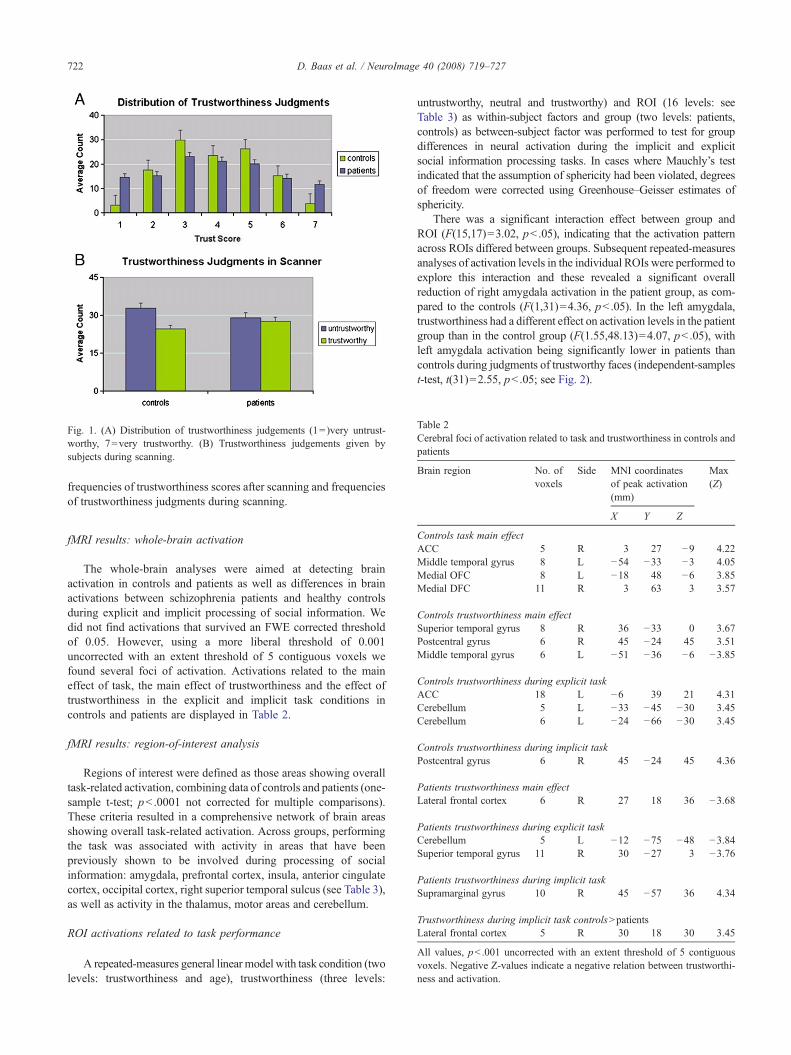
Fig. 1. (A) Distribution of trustworthiness judgements (1=)very untrust-worthy, 7=very trustworthy. (B) Trustworthiness judgements given bysubjects during scanning.
Table 2Cerebral foci of activation related to task and trustworthiness in controls andpatients
Brain region No. ofvoxels
Side MNI coordinatesof peak activation(mm)
Max(Z)
722 D. Baas et al. / NeuroImage 40 (2008) 719–727
frequencies of trustworthiness scores after scanning and frequenciesof trustworthiness judgments during scanning.
X Y Z
Controls task main effectACC 5 R 3 27 −9 4.22Middle temporal gyrus 8 L −54 −33 −3 4.05Medial OFC 8 L −18 48 −6 3.85Medial DFC 11 R 3 63 3 3.57
Controls trustworthiness main effectSuperior temporal gyrus 8 R 36 −33 0 3.67Postcentral gyrus 6 R 45 −24 45 3.51Middle temporal gyrus 6 L −51 −36 −6 −3.85
Controls trustworthiness during explicit taskACC 18 L −6 39 21 4.31Cerebellum 5 L −33 −45 −30 3.45Cerebellum 6 L −24 −66 −30 3.45
Controls trustworthiness during implicit taskPostcentral gyrus 6 R 45 −24 45 4.36
Patients trustworthiness main effectLateral frontal cortex 6 R 27 18 36 −3.68
Patients trustworthiness during explicit taskCerebellum 5 L −12 −75 −48 −3.84Superior temporal gyrus 11 R 30 −27 3 −3.76
Patients trustworthiness during implicit taskSupramarginal gyrus 10 R 45 −57 36 4.34
Trustworthiness during implicit task controlsNpatientsLateral frontal cortex 5 R 30 18 30 3.45
All values, pb .001 uncorrected with an extent threshold of 5 contiguousvoxels. Negative Z-values indicate a negative relation between trustworthi-ness and activation.
fMRI results: whole-brain activation
The whole-brain analyses were aimed at detecting brainactivation in controls and patients as well as differences in brainactivations between schizophrenia patients and healthy controlsduring explicit and implicit processing of social information. Wedid not find activations that survived an FWE corrected thresholdof 0.05. However, using a more liberal threshold of 0.001uncorrected with an extent threshold of 5 contiguous voxels wefound several foci of activation. Activations related to the maineffect of task, the main effect of trustworthiness and the effect oftrustworthiness in the explicit and implicit task conditions incontrols and patients are displayed in Table 2.
fMRI results: region-of-interest analysis
Regions of interest were defined as those areas showing overalltask-related activation, combining data of controls and patients (one-sample t-test; pb .0001 not corrected for multiple comparisons).These criteria resulted in a comprehensive network of brain areasshowing overall task-related activation. Across groups, performingthe task was associated with activity in areas that have beenpreviously shown to be involved during processing of socialinformation: amygdala, prefrontal cortex, insula, anterior cingulatecortex, occipital cortex, right superior temporal sulcus (see Table 3),as well as activity in the thalamus, motor areas and cerebellum.
ROI activations related to task performance
A repeated-measures general linear model with task condition (twolevels: trustworthiness and age), trustworthiness (three levels:
untrustworthy, neutral and trustworthy) and ROI (16 levels: seeTable 3) as within-subject factors and group (two levels: patients,controls) as between-subject factor was performed to test for groupdifferences in neural activation during the implicit and explicitsocial information processing tasks. In cases where Mauchly’s testindicated that the assumption of sphericity had been violated, degreesof freedom were corrected using Greenhouse–Geisser estimates ofsphericity.
There was a significant interaction effect between group andROI (F(15,17)=3.02, pb .05), indicating that the activation patternacross ROIs differed between groups. Subsequent repeated-measuresanalyses of activation levels in the individual ROIs were performed toexplore this interaction and these revealed a significant overallreduction of right amygdala activation in the patient group, as com-pared to the controls (F(1,31)=4.36, pb .05). In the left amygdala,trustworthiness had a different effect on activation levels in the patientgroup than in the control group (F(1.55,48.13)=4.07, pb .05), withleft amygdala activation being significantly lower in patients thancontrols during judgments of trustworthy faces (independent-samplest-test, t(31)=2.55, pb .05; see Fig. 2).
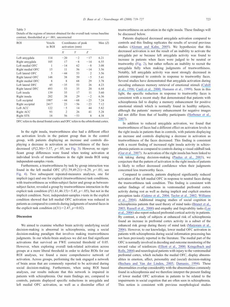
Table 3Details of the regions of interest obtained for the overall task versus baselinecontrast, thresholded at pb .001, uncorrected
ROI Voxelsin ROI
Coordinates of peakactivation (mm)
Max (Z)
X Y Z
Left amygdala 114 −20 −2 −17 6.21Right amygdala 105 17 −8 −14 6.55Left medial OFC 1 −14 62 −8 3.08Right medial OFC 15 11 56 −14 3.56Left lateral OFC 5 −44 53 2 3.56Right lateral OFC 148 38 59 −5 5.41Right medial DFC 8 8 68 29 3.78Left lateral DFC 45 −35 53 26 4.23Right lateral DFC 493 53 35 20 6.44Left insula 139 35 17 11 5.60Right insula 202 38 29 −2 6.36Left occipital 1907 −44 −68 −23 7.24Right occipital 2417 23 −56 −23 7.12Left ACC 122 −5 14 44 5.82Right ACC 93 2 26 41 5.28Right STS 18 56 −53 8 4.38
DFCrefers to the dorsal frontal cortex andOFC refers to the orbitofrontal cortex.
723D. Baas et al. / NeuroImage 40 (2008) 719–727
In the right insula, trustworthiness also had a different effecton activation levels in the patient group than in the controlgroup, with patients displaying an increase and controls dis-playing a decrease in activation as trustworthiness of the facesdecreased (F(2,30)=3.37, pb .05; see Fig. 3). However, no signi-ficant group differences were found when testing activation atindividual levels of trustworthiness in the right insula ROI usingindependent-samples t-tests.
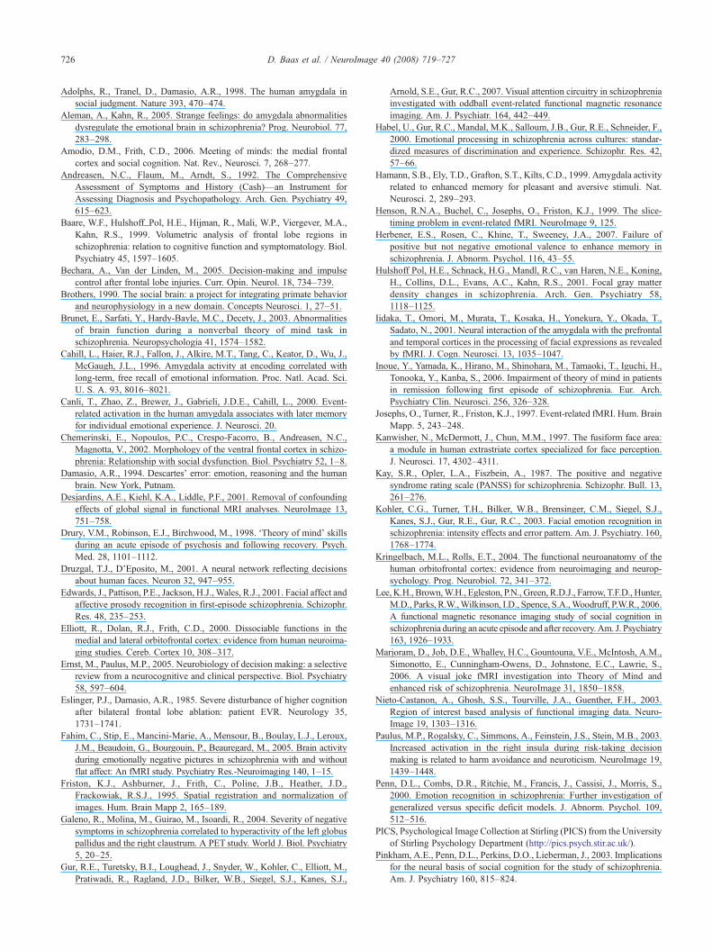
Furthermore, a trustworthiness by task by group interaction wasfound in the left medial OFC (F(1.59,49.21)=6.29, pb .01; seeFig. 4). Two subsequent repeated-measures analyses, one forimplicit (age) and one for explicit (trustworthiness) task conditions,with trustworthiness as within-subject factor and group as between-subject factor, revealed a group by trustworthiness interaction in theexplicit task condition (F(1.61,46.15)=5.45, pb .05), but not in theimplicit condition. Next, independent-samples t-tests in the explicitcondition showed that left medial OFC activation was reduced inpatients as compared to controls during judgments of neutral faces inthe explicit task condition (t(31)=2.47, p=0.02).
Discussion
We aimed to examine whether brain activity underlying socialdecision-making is abnormal in schizophrenia, using a socialdecision-making paradigm that involves making trustworthinessjudgments. In our whole-brain analyses we did not find significantactivations that survived an FWE corrected threshold of 0.05.However, when exploring overall task-related activation acrossgroups at a more liberal threshold in order to define ROIs for ourROI analyses, we found a more comprehensive network ofactivation. Across groups, performing the task engaged a networkof brain areas that are commonly reported to be activated duringsocial decision-making. Furthermore, when using ROI basedanalyses, our results indicate that this network is impaired inpatients with schizophrenia. Our main findings are, compared tocontrols, patients displayed specific reductions in amygdala andleft medial OFC activation, as well as a dissimilar effect of
trustworthiness on activation in the right insula. These findings willbe discussed below.
Patients displayed decreased amygdala activation compared tocontrols and this finding replicates the results of several previousstudies (Aleman and Kahn, 2005). We hypothesize that thisdecreased activation is not the result of an inability to activate theamygdala per se because left amygdala activity was found toincrease in patients when faces were judged to be neutral ortrustworthy (Fig. 2), but rather reflects an inability to recruit theamygdala fully when making judgments of trustworthiness.Notably, left amygdala activity was most strongly decreased inpatients compared to controls in response to trustworthy faces.Several studies have demonstrated that amygdala activation duringencoding enhances memory retrieval of emotional stimuli (Cahillet al., 1996; Canli et al., 2000; Hamann et al., 1999). Seen in thislight, the specific reduction in response to trustworthy faces isconsistent with a recent study that demonstrated that patients withschizophrenia fail to display a memory enhancement for positiveemotional stimuli which is normally found in healthy subjects,although the patients’ memory enhancement for negative imagesdid not differ from that of healthy participants (Herbener et al.,2007).
In addition to reduced amygdala activation, we found thattrustworthiness of faces had a different effect on activation levels inthe right insula in patients than in controls, with patients displayingan increase and controls displaying a decrease in activation astrustworthiness of the faces decreased. This finding is consistentwith a recent finding of increased right insula activity in schizo-phrenia patients as compared to controls during a visual oddball task(Gur et al., 2007). As activation of the right insula has been linked torisk taking during decision-making (Paulus et al., 2003), weconjecture that the pattern of activation in the right insula of patientsis likely to reflect decreased confidence when their judgmentsconcerned less trustworthy faces.
Compared to controls, patients displayed significantly reducedactivation of the left medial OFC in response to neutral faces duringthe trustworthiness task condition. This finding is consistent withearlier findings of reductions in ventromedial prefrontal cortexactivity during rest as well as during implicit and explicit emotionperception tasks (Galeno et al., 2004; Taylor et al., 2002; Williamset al., 2004). Additional imaging studies of social cognition inschizophrenia patients that used theory of mind tasks (Brunet et al.,2003; Russell et al., 2000) and empathy and forgivability tasks (Leeet al., 2006) also report reduced prefrontal cortical activity in patients.By contrast, a study of subjects at enhanced risk of schizophreniafound an increase in prefrontal cortex activity in a subset of theenhanced risk group during theory of mind task (Marjoram et al.,2006). However, to our knowledge, lower medial OFC activation inpatients with schizophrenia during social information processing hasnot been previously reported in the literature. The medial part of theOFC is normally involved in decoding and outcomemonitoring of thereward value of reinforcers (Elliott et al., 2000; Kringelbach andRolls, 2004) and neurological patients with injury to the ventromedialprefrontal cortex, which includes the medial OFC, display abnorm-alities in emotion, affect, personality and (social) decision-making(Bechara and Van der Linden, 2005; Damasio, 1994). Theseabnormalities overlap considerably with the abnormalities commonlyfound in schizophrenia and we therefore interpret the present findingof lower medial OFC activation in patients to be related to theimpairments in social cognition that are often seen in schizophrenia.This notion is consistent with previous morphological studies
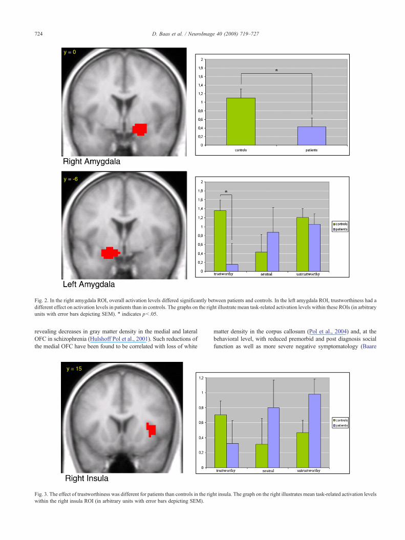
Fig. 2. In the right amygdala ROI, overall activation levels differed significantly between patients and controls. In the left amygdala ROI, trustworthiness had adifferent effect on activation levels in patients than in controls. The graphs on the right illustrate mean task-related activation levels within these ROIs (in arbitraryunits with error bars depicting SEM). ⁎ indicates pb .05.
724 D. Baas et al. / NeuroImage 40 (2008) 719–727
revealing decreases in gray matter density in the medial and lateralOFC in schizophrenia (Hulshoff Pol et al., 2001). Such reductions ofthe medial OFC have been found to be correlated with loss of white
Fig. 3. The effect of trustworthiness was different for patients than controls in the rigwithin the right insula ROI (in arbitrary units with error bars depicting SEM).
matter density in the corpus callosum (Pol et al., 2004) and, at thebehavioral level, with reduced premorbid and post diagnosis socialfunction as well as more severe negative symptomatology (Baare
ht insula. The graph on the right illustrates mean task-related activation levels
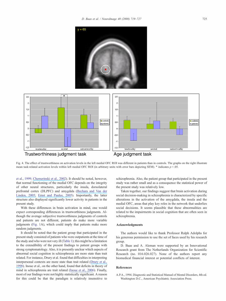
Fig. 4. The effect of trustworthiness on activation levels in the left medial OFC ROI was different in patients than in controls. The graphs on the right illustratemean task-related activation levels within left medial OFC ROI (in arbitrary units with error bars depicting SEM). ⁎ indicates p b .05.
725D. Baas et al. / NeuroImage 40 (2008) 719–727
et al., 1999; Chemerinski et al., 2002). It should be noted, however,that normal functioning of the medial OFC depends on the integrityof other neural structures, particularly the insula, dorsolateralprefrontal cortex (DLPFC) and amygdala (Bechara and Van derLinden, 2005; Ernst and Paulus, 2005). Importantly, the latterstructure also displayed significantly lower activity in patients in thepresent study.
With these differences in brain activation in mind, one wouldexpect corresponding differences in trustworthiness judgments. Al-though the average subjective trustworthiness judgments of controlsand patients are not different, patients do make more variablejudgments (Fig. 1A), which could imply that patients make morerandom judgments.
It should be noted that the patient group that participated in thepresent study consisted of patients who were outpatients at the time ofthe study andwhowere not very ill (Table 1); thismight be a limitationto the extendibility of the present findings to patient groups withstrong symptomatology. Also, it is presently unclear which aspects ofabnormal social cognition in schizophrenia are more state than traitrelated. For instance, Drury et al. found that difficulties in interpretinginterpersonal contexts are more state than trait related (Drury et al.,1998). Inoue et al., on the other hand, found that deficits in theory ofmind in schizophrenia are trait related (Inoue et al., 2006). Finally,most of our findings were not highly statistically significant. A reasonfor this could be that the paradigm is relatively insensitive to
schizophrenia. Also, the patient group that participated in the presentstudy was rather small and as a consequence the statistical power ofthe present study was relatively low.
Taken together, our findings suggest that brain activation duringsocial decision-making in schizophrenia is characterized by specificalterations in the activation of the amygdala, the insula and themedial OFC, areas that play key roles in the network that underliessocial decisions. It seems plausible that these abnormalities arerelated to the impairments in social cognition that are often seen inschizophrenia.
Acknowledgments
The authors would like to thank Professor Ralph Adolphs forhis generous permission to use the set of faces used by his researchgroup.
D. Baas and A. Aleman were supported by an InnovationalResearch grant from The Netherlands Organization for ScientificResearch (no. 016.026.027). None of the authors report anybiomedical financial interest or potential conflicts of interest.
References
A.P.A., 1994. Diagnostic and Statistical Manual of Mental Disorders, 4th ed.Washington D.C., American Psychiatric Association Press.
726 D. Baas et al. / NeuroImage 40 (2008) 719–727
Adolphs, R., Tranel, D., Damasio, A.R., 1998. The human amygdala insocial judgment. Nature 393, 470–474.
Aleman, A., Kahn, R., 2005. Strange feelings: do amygdala abnormalitiesdysregulate the emotional brain in schizophrenia? Prog. Neurobiol. 77,283–298.
Amodio, D.M., Frith, C.D., 2006. Meeting of minds: the medial frontalcortex and social cognition. Nat. Rev., Neurosci. 7, 268–277.
Andreasen, N.C., Flaum, M., Arndt, S., 1992. The ComprehensiveAssessment of Symptoms and History (Cash)—an Instrument forAssessing Diagnosis and Psychopathology. Arch. Gen. Psychiatry 49,615–623.
Baare, W.F., Hulshoff_Pol, H.E., Hijman, R., Mali, W.P., Viergever, M.A.,Kahn, R.S., 1999. Volumetric analysis of frontal lobe regions inschizophrenia: relation to cognitive function and symptomatology. Biol.Psychiatry 45, 1597–1605.
Bechara, A., Van der Linden, M., 2005. Decision-making and impulsecontrol after frontal lobe injuries. Curr. Opin. Neurol. 18, 734–739.
Brothers, 1990. The social brain: a project for integrating primate behaviorand neurophysiology in a new domain. Concepts Neurosci. 1, 27–51.
Brunet, E., Sarfati, Y., Hardy-Bayle, M.C., Decety, J., 2003. Abnormalitiesof brain function during a nonverbal theory of mind task inschizophrenia. Neuropsychologia 41, 1574–1582.
Cahill, L., Haier, R.J., Fallon, J., Alkire, M.T., Tang, C., Keator, D., Wu, J.,McGaugh, J.L., 1996. Amygdala activity at encoding correlated withlong-term, free recall of emotional information. Proc. Natl. Acad. Sci.U. S. A. 93, 8016–8021.
Canli, T., Zhao, Z., Brewer, J., Gabrieli, J.D.E., Cahill, L., 2000. Event-related activation in the human amygdala associates with later memoryfor individual emotional experience. J. Neurosci. 20.
Chemerinski, E., Nopoulos, P.C., Crespo-Facorro, B., Andreasen, N.C.,Magnotta, V., 2002. Morphology of the ventral frontal cortex in schizo-phrenia: Relationship with social dysfunction. Biol. Psychiatry 52, 1–8.
Damasio, A.R., 1994. Descartes’ error: emotion, reasoning and the humanbrain. New York, Putnam.
Desjardins, A.E., Kiehl, K.A., Liddle, P.F., 2001. Removal of confoundingeffects of global signal in functional MRI analyses. NeuroImage 13,751–758.
Drury, V.M., Robinson, E.J., Birchwood, M., 1998. ‘Theory of mind’ skillsduring an acute episode of psychosis and following recovery. Psych.Med. 28, 1101–1112.
Druzgal, T.J., D’Eposito, M., 2001. A neural network reflecting decisionsabout human faces. Neuron 32, 947–955.
Edwards, J., Pattison, P.E., Jackson, H.J., Wales, R.J., 2001. Facial affect andaffective prosody recognition in first-episode schizophrenia. Schizophr.Res. 48, 235–253.
Elliott, R., Dolan, R.J., Frith, C.D., 2000. Dissociable functions in themedial and lateral orbitofrontal cortex: evidence from human neuroima-ging studies. Cereb. Cortex 10, 308–317.
Ernst, M., Paulus, M.P., 2005. Neurobiology of decision making: a selectivereview from a neurocognitive and clinical perspective. Biol. Psychiatry58, 597–604.
Eslinger, P.J., Damasio, A.R., 1985. Severe disturbance of higher cognitionafter bilateral frontal lobe ablation: patient EVR. Neurology 35,1731–1741.
Fahim, C., Stip, E., Mancini-Marie, A., Mensour, B., Boulay, L.J., Leroux,J.M., Beaudoin, G., Bourgouin, P., Beauregard, M., 2005. Brain activityduring emotionally negative pictures in schizophrenia with and withoutflat affect: An fMRI study. Psychiatry Res.-Neuroimaging 140, 1–15.
Friston, K.J., Ashburner, J., Frith, C., Poline, J.B., Heather, J.D.,Frackowiak, R.S.J., 1995. Spatial registration and normalization ofimages. Hum. Brain Mapp 2, 165–189.
Galeno, R., Molina, M., Guirao, M., Isoardi, R., 2004. Severity of negativesymptoms in schizophrenia correlated to hyperactivity of the left globuspallidus and the right claustrum. A PET study. World J. Biol. Psychiatry5, 20–25.
Gur, R.E., Turetsky, B.I., Loughead, J., Snyder, W., Kohler, C., Elliott, M.,Pratiwadi, R., Ragland, J.D., Bilker, W.B., Siegel, S.J., Kanes, S.J.,
Arnold, S.E., Gur, R.C., 2007. Visual attention circuitry in schizophreniainvestigated with oddball event-related functional magnetic resonanceimaging. Am. J. Psychiatr. 164, 442–449.
Habel, U., Gur, R.C., Mandal, M.K., Salloum, J.B., Gur, R.E., Schneider, F.,2000. Emotional processing in schizophrenia across cultures: standar-dized measures of discrimination and experience. Schizophr. Res. 42,57–66.
Hamann, S.B., Ely, T.D., Grafton, S.T., Kilts, C.D., 1999. Amygdala activityrelated to enhanced memory for pleasant and aversive stimuli. Nat.Neurosci. 2, 289–293.
Henson, R.N.A., Buchel, C., Josephs, O., Friston, K.J., 1999. The slice-timing problem in event-related fMRI. NeuroImage 9, 125.
Herbener, E.S., Rosen, C., Khine, T., Sweeney, J.A., 2007. Failure ofpositive but not negative emotional valence to enhance memory inschizophrenia. J. Abnorm. Psychol. 116, 43–55.
Hulshoff Pol, H.E., Schnack, H.G., Mandl, R.C., van Haren, N.E., Koning,H., Collins, D.L., Evans, A.C., Kahn, R.S., 2001. Focal gray matterdensity changes in schizophrenia. Arch. Gen. Psychiatry 58,1118–1125.
Iidaka, T., Omori, M., Murata, T., Kosaka, H., Yonekura, Y., Okada, T.,Sadato, N., 2001. Neural interaction of the amygdala with the prefrontaland temporal cortices in the processing of facial expressions as revealedby fMRI. J. Cogn. Neurosci. 13, 1035–1047.
Inoue, Y., Yamada, K., Hirano, M., Shinohara, M., Tamaoki, T., Iguchi, H.,Tonooka, Y., Kanba, S., 2006. Impairment of theory of mind in patientsin remission following first episode of schizophrenia. Eur. Arch.Psychiatry Clin. Neurosci. 256, 326–328.
Josephs, O., Turner, R., Friston, K.J., 1997. Event-related fMRI. Hum. BrainMapp. 5, 243–248.
Kanwisher, N., McDermott, J., Chun, M.M., 1997. The fusiform face area:a module in human extrastriate cortex specialized for face perception.J. Neurosci. 17, 4302–4311.
Kay, S.R., Opler, L.A., Fiszbein, A., 1987. The positive and negativesyndrome rating scale (PANSS) for schizophrenia. Schizophr. Bull. 13,261–276.
Kohler, C.G., Turner, T.H., Bilker, W.B., Brensinger, C.M., Siegel, S.J.,Kanes, S.J., Gur, R.E., Gur, R.C., 2003. Facial emotion recognition inschizophrenia: intensity effects and error pattern. Am. J. Psychiatry. 160,1768–1774.
Kringelbach, M.L., Rolls, E.T., 2004. The functional neuroanatomy of thehuman orbitofrontal cortex: evidence from neuroimaging and neurop-sychology. Prog. Neurobiol. 72, 341–372.
Lee,K.H., Brown,W.H., Egleston, P.N., Green, R.D.J., Farrow, T.F.D., Hunter,M.D., Parks, R.W.,Wilkinson, I.D., Spence, S.A.,Woodruff, P.W.R., 2006.A functional magnetic resonance imaging study of social cognition inschizophrenia during an acute episode and after recovery.Am. J. Psychiatry163, 1926–1933.
Marjoram, D., Job, D.E., Whalley, H.C., Gountouna, V.E., McIntosh, A.M.,Simonotto, E., Cunningham-Owens, D., Johnstone, E.C., Lawrie, S.,2006. A visual joke fMRI investigation into Theory of Mind andenhanced risk of schizophrenia. NeuroImage 31, 1850–1858.
Nieto-Castanon, A., Ghosh, S.S., Tourville, J.A., Guenther, F.H., 2003.Region of interest based analysis of functional imaging data. Neuro-Image 19, 1303–1316.
Paulus, M.P., Rogalsky, C., Simmons, A., Feinstein, J.S., Stein, M.B., 2003.Increased activation in the right insula during risk-taking decisionmaking is related to harm avoidance and neuroticism. NeuroImage 19,1439–1448.
Penn, D.L., Combs, D.R., Ritchie, M., Francis, J., Cassisi, J., Morris, S.,2000. Emotion recognition in schizophrenia: Further investigation ofgeneralized versus specific deficit models. J. Abnorm. Psychol. 109,512–516.
PICS, Psychological Image Collection at Stirling (PICS) from the Universityof Stirling Psychology Department (http://pics.psych.stir.ac.uk/).
Pinkham, A.E., Penn, D.L., Perkins, D.O., Lieberman, J., 2003. Implicationsfor the neural basis of social cognition for the study of schizophrenia.Am. J. Psychiatry 160, 815–824.
727D. Baas et al. / NeuroImage 40 (2008) 719–727
Pol, H.E.H., Schnack, H.G., Mandl, R.C.W., Cahn, W., Collins, D.L., Evans,A.C., Kahn, R.S., 2004. Focal white matter density changes inschizophrenia: reduced inter-hemispheric connectivity. NeuroImage21, 27–35.
Russell, T.A., Rubia, K., Bullmore, E.T., Soni, W., Suckling, J., Brammer,M.J., Simmons, A., Williams, S.C., Sharma, T., 2000. Exploring thesocial brain in schizophrenia: left prefrontal underactivation duringmental state attribution. Am. J. Psychiatry 157, 2040–2042.
Schneider, F., Weiss, U., Kessler, C., Salloum, J.B., Posse, S., Grodd, W.,Muller_Gartner, H.W., 1998. Differential amygdala activation inschizophrenia during sadness. Schizophr. Res. 34, 133–142.
Sheehan, D.V., Lecrubier, Y., Sheehan, K.H., Amorim, P., Janavs, J., Weiller,E., Hergueta, T., Baker, R., Dunbar, G.C., 1998. The Mini-InternationalNeuropsychiatric Interview (MINI): the development and validation of astructured diagnostic psychiatric interview for DSM-IV and ICD-10.J. Clin. Psychiatry 59, 22–33.
Sprengelmeyer, R., Young, A.W., Calder, A.J., Karnat, A., Lange, H.W.,Hömberg, V., Perrett, D.I., Rowland, D., 1996. Loss of disgust:perception of faces and emotions in Huntington’s disease. Brain J.Neurol. 119, 1647–1665.
Taylor, S.F., Liberzon, I., Decker, L.R., Koeppe, R.A., 2002. A functionalanatomic study of emotion in schizophrenia. Schizophr. Res. 58, 159–172.
Williams, L.W., Das, P., Harris, A.W.F., Liddell, B.B., Brammer, M.J.,Olivieri, G., Skerrett, D., Phillips, M.L., David, A.S., Peduto, A.P.,Gordon, E., 2004. Dysregulation of arousal and amygdala–prefrontalsystems in paranoid schizophrenia. Am. J. Psychiatry 161, 480–489.
Winston, J.S., Strange, B.A., O’Doherty, J., Dolan, R.J., 2002. Automaticand intentional brain responses during evaluation of trustworthiness offaces. Nat. Neurosci. 5, 277–283.
Yamasaki, H., LaBar, K.S., McCarthy, G., 2002. Dissociable prefrontal brainsystems for attention and emotion. Proc. Natl. Acad. Sci. U. S. A. 99,11447–11451.
Copyright © 2022 FDOKUMEN