Preterm Labor and Later Maternal Cardiovascular Disease in ...
Infant Behavior & Development 31 (2008) 614–623
Contents lists available at ScienceDirect
Infant Behavior and Development
Developmental changes in the responses of preterm infants to apainful stressor
Rachel Lucas-Thompsona, Elise L. Townsendb, Megan R. Gunnarc, Michael K. Georgieffd,Sixto F. Guiangd, Raul F. Ciffuentese, Richard C. Lusskye, Elysia Poggi Davis f,g,∗
a Department of Psychology and Social Behavior, University of California Irvine, 300 Social Ecology I, Irvine, CA 92697-7050, United Statesb Graduate Programs in Physical Therapy, MGH Institute of Health Professions, Boston, MA, United Statesc Institute of Child Development and the Center for Neurobehavioral Development, University of Minnesota, United Statesd Department of Pediatrics, University of Minnesota, United Statese Department of Pediatrics, Hennepin County Medical Center, United Statesf Department of Psychiatry and Human Behavior, University of California Irvine, United Statesg Department of Pediatrics, University of California Irvine, United States
a r t i c l e i n f o
Article history:Received 23 January 2008Received in revised form 17 June 2008Accepted 18 July 2008
Keywords:StressPrematuritySelf-regulationPhysiological reactivity
a b s t r a c t
The purpose of this investigation was to examine longitudinally gestational age and devel-opmental differences in preterm infants’ self-regulatory abilities in response to a painfulstressor, as well as associations between behavioral and cardiovascular responses. Partici-pants included 49 healthy premature infants. Behavioral and cardiovascular responses to aheel stick blood draw were compared between infants of 28–31 and 32–34 weeks’ gestationage at birth. Both gestational age groups displayed behavioral and cardiovascular indica-tions of stress in response to the blood draw. However, both shortly after birth and severalweeks later, infants born at younger gestational ages (28–31 weeks) were more physio-logically reactive. Evidence that the behavioral stress responses of 28–31 weeks’ gestationage group preterm infants do not reflect their physiological responses suggests that evalu-ation of preterm infants’ experiences and risk require assessments of both physiology andbehavior. The greater stress vulnerability of the 28–31 weeks’ gestation group relative tothe 32–34 weeks’ gestation group and the implications of this for subsequent developmentare discussed.
Published by Elsevier Inc.
Medical and technological breakthroughs over the last few decades have increased the survival rates of infants born at theyoungest viable gestational ages. Yet many of the procedures that are a necessary part of their postnatal care can be, by nature,painful and stressful. In a Canadian survey, Johnston, Collinger, Henderson, and Anand (1997) found that infants residing ina NICU had an average of two invasive procedures a day, with some infants having as many as eight. One common procedureis the heel stick blood draw (Barker & Rutter, 1995). Although at one point the prevailing belief was that preterm infants
could not feel the pain of invasive medical procedures, more recent research has indicated that premature infants have theanatomic and brain structures necessary for nociception (Stevens, Johnston, & Grunau, 1995). The recognition that preterminfants experience pain has led to a proliferation of studies aimed at understanding the development of the pain systemand pain responses. It is now clear that preterm infants display appropriate hormonal stress responses (Barker & Rutter,∗ Corresponding author at: Department of Psychiatry and Human Behavior, University of California Irvine, City Tower, 333 City Boulevard West, Suite1200, Orange, CA 92628, United States. Tel.: +1 714 940 1924; fax: +1 714 940 1939.
E-mail address: [email protected] (E.P. Davis).
0163-6383/$ – see front matter. Published by Elsevier Inc.doi:10.1016/j.infbeh.2008.07.004
1L1sIF
ewoisrot
qGSecafl(bA(ai
biWdsr
edt4(nGw(ara
trcr
eitat
R. Lucas-Thompson et al. / Infant Behavior & Development 31 (2008) 614–623 615
996), increases in heart rate to painful procedures (Craig, Whitfield, Grunau, Linton, & Hadjistavropoulos, 1993; Grunau,inhares, Holsti, Oberlander, & Whitfield, 2004), and hypersensitivity to tissue damage (Fitzgerald, Millard, & McIntosh,989). In addition, research suggests that preterm infants are actually more sensitive to pain than their term counterparts, astructures in the central nervous system that prevent the spread of pain signals may be undeveloped (Stevens et al., 1995).ndeed, younger preterm infants have lower pain thresholds than their older preterm and term counterparts (Andrews &itzgerald, 1994).
An important question, therefore, is how preterm infants regulate responses to the painful stressors that they regularlyncounter. Preterm infants have a less organized self-regulation system than term infants (Als, Duffy, & McAnulty, 1988),hich may contribute to a less healthy stress response. A healthy stress response is one that is activated quickly in the face
f a stressor to enable the organism to manage the challenge and is then deactivated after the stressor has passed. Pretermnfants are overwhelmed more easily and, under extreme stress, may be unable to organize a response to the pain andtress (Als, Lester, Tronick, & Brazelton, 1982; Johnston, Franck, & Stremler, 1999). Rather than mounting an effective stressesponse, these infants may ‘shut down’ (Als, 1993; Als et al., 1994). Furthermore, preterm infants remain in a higher statef arousal after a stressor has passed as compared to full-term infants (Holsti, Grunau, Oberlander, & Whitfield, 2004) andake longer to recover from painful procedures (Craig et al., 1993).
A growing body of work suggests that pain and stress exposure early in infancy can have long-lasting negative conse-uences (Anand, Coskun, Thrivikraman, Nemeroff, & Plotsky, 1999; Anand, Grunau, & Oberlander, 1997; Bhutta et al., 2001;runau, Whitfield, & Petrie, 1998; Grunau, Whitfield, Petrie, & Fryer, 1994; Hack et al., 1995; Porter, Grunau, & Anand, 1999;hanks, Larocque, & Meaney, 1995). There has long been particular concern that the nature of life in the NICU is stressfulnough for the fragile systems of preterm infants that it may also do lasting damage manifested as health, sensory, andognitive deficits (Gorski, 1991; Gorski, Huntington, & Lewkowicz, 1990; Gottfried, Hodgman, & Brown, 1984; Gottfried etl., 1981; Long, Lucey, & Philip, 1980; Lucey, 1977). Increasing self-regulation abilities appears to be an important avenueor improving the medical and neurodevelopmental outcomes of preterm infants and perhaps ameliorating some of theseong-term consequences. Developmental care aimed at supporting infants’ self-regulation leads to better health outcomesAls et al., 2003; Als et al., 1994). At the root of these interventions is training health-care providers to identify and distinguishetween “stress” and “regulatory” behaviors, based on a scheme developed and validated by Als and colleagues (Als, 1984;ls et al., 1982). “Stress” behaviors, also called extension or avoidance behaviors, signal withdrawal, defense, and overload
Als, 1984) and have been linked with decreases in oxygen saturation levels (Peters, 2001). In contrast, “regulatory” behaviors,lso called flexion or approach behaviors, signal appropriate self-soothing (Als, 1984). The identification of these behaviorss then used to formulate specific suggestions for how to increase each infant’s self-regulatory competence (Als et al., 1994).
The success of these developmental programs emphasizes the importance of understanding stress and regulatoryehaviors. However, much about these behaviors remains relatively unexplored. Research suggests that younger preterm
nfants display more stress behaviors than older preterms during non-invasive care procedures (Holsti, Grunau, Oberlander,hitfield, & Weinberg, 2005). However, it is unclear whether there is a link between gestational age at birth (GAB) and the
isplay of stress and regulatory behaviors in response to a painful stressor such as a heel stick blood draw. In addition, althoughtress and regulatory behaviors are theoretically linked with physiological responses (Als, 1984), a link with physiologicalesponses other than oxygen saturation has not yet been empirically established.
A third aspect of adaptive and maladaptive behavioral responses to stress that warrants exploration is how the differentialxperiences of developing in utero versus in the NICU are associated with the ability to manage responses to a stressor. Evi-ence from cross-sectional studies suggests that preterm infants not only have a greater difficulty regulating their responseso stressors as compared to term infants, but also that they may not ‘catch up’ with increasing postconceptional age. Even at2 weeks postconception, infants born at the youngest GAB have the lowest stress thresholds and highest hypersensitivityAls et al., 1988). Furthermore, medically healthy preterm and term infants of the same postconceptional age (PCA) differ sig-ificantly on measures of self-regulation (Duffy, Als, & McAnulty, 1990; Ferrari, Grosoli, Fontana, & Cavazzuti, 1983; Holsti,runau, Oberlander, & Whitfield, 2005; Mouradian, Als, & Coster, 2000). However, evidence does suggest improvementith development. Over the 8-week period after birth, preterm infants become less likely to demonstrate facial flaccidity
Johnston, Stevens, Yang, & Horton, 1996), a behavioral pattern that is often argued to represent an inability to mount anppropriate pain response due to overload (Als, 1993; Als et al., 1994; Johnston et al., 1999). If facial flaccidity is in factepresentative of overload, this suggests that preterm infants may become more capable of appropriate self-soothing as theyge.
These findings suggest that early experiences may impact the development of the stress regulation system in wayshat have lasting consequences for the preterm infant. However, a longitudinal examination of the specific behaviors thatepresent overload and self-soothing is necessary to determine the pattern of associations between PCA and self-regulatoryompetence. This is of particular importance in terms of the consequences of self-regulatory competence for physiologicalegulation and the ability of preterm infants to manage a challenge.
Based on this review, a need exists for a greater understanding of the vulnerability of preterm infants by longitudinally
xamining developmental differences in preterm infants’ abilities to self-regulate in response to stressors. In addition, theres a need for integrating assessments of behavior and physiology to gain a clearer picture of whether behavioral responseso stress reflect physiological responses, or whether both must be considered to fully understand when preterm infantsre at risk. The goal of the present study was to advance understanding of preterm infants’ self-regulation by addressinghree questions: (1) How does GAB predict infants’ physiological, overload, and self-soothing responses to a painful stressor?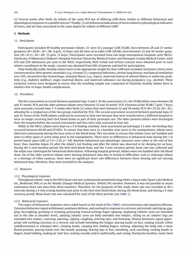
616 R. Lucas-Thompson et al. / Infant Behavior & Development 31 (2008) 614–623
(2) Several weeks after birth, do infants of the same PCA but of differing GAB show similar or different behavioral andphysiological responses to a painful stressor? Finally, (3) are behavioral indications of stress related to physiological indicatorsof stress, and are they associated to the same degree for infants of different GAB?
1. Methods
1.1. Participants
Participants included 49 healthy premature infants; 21 were of a younger GAB (YGAB), born between 28 and 31 weeks’gestation (M = 29.81; SD = .98; 9 girls, 12 boys) and 28 were of an older GAB (OGAB), born between 32 and 34 weeks’ gesta-tion (M = 33.11; SD = .69; 15 girls, 13 boys). Participants were recruited from two large metropolitan hospitals with NICUs:University of Minnesota Children’s Hospital-Fairview University Medical Center and Hennepin County Medical Center, with670 and 250 admissions per year to the NICU, respectively. Both verbal and written consent were obtained prior to eachinfant’s enrollment in the study; consent was obtained from 90% of parents solicited for participation.
Only medically stable, healthy infants born at the appropriate weight for their GAB were included. Exclusion criteria werechromosomal or other genetic anomalies (e.g., trisomy 21), congenital infections, chronic lung disease, mechanical ventilationover 24 h, intraventricular hemorrhage, neonatal illness (e.g., sepsis), maternal history of adrenal illness or endocrine prob-lems (e.g., diabetes mellitus), major maternal illness, and maternal substance use during pregnancy (e.g., alcohol). Theseexclusion criteria were designed to ensure that the resulting sample was comprised of relatively healthy infants born tomothers free of major health complications.
1.2. Procedures
The first assessment occurred between postnatal days 3 and 5. At this assessment (t1), the YGAB infants were between 28and 31 weeks’ PCA and the older preterm infants were between 32 and 34 weeks’ PCA. Fourteen of the YGAB (7 girls, 7 boys)were assessed a second time 3–5 weeks after birth (t2) when they were between 32 and 34 weeks PCA (M = 33.54; SD = .81),allowing for a comparison of the two groups matched for PCA. The second assessment occurred between postnatal days 21and 35. Seven of the YGAB infants could not be assessed at time two because they were transferred to a different hospital orwere no longer receiving heel stick blood draws as part of their postnatal care. The older preterm infants were dischargedfrom the hospital before the second assessment and thus were only assessed at time one.
While in the NICU, infants were kept on a 3-h feeding schedule. Each assessment period began 1 h after the feeding thatoccurred between 04:00 and 07:00 h. To ensure that they were in a baseline state prior to the manipulation, infants wereobserved continuously during the hour prior to the blood draw. This was done to ensure that infants were not handled andwere in either quiet or active sleep prior to the manipulation. There were no differences in behavioral state during this 1-hperiod based upon GAB [�2(3) = 2.18, p = .54]. Baseline behavioral and physiologic measures were taken at the end of thishour; thus, baseline began 2 h after the infant’s last feeding and after the infant was observed to be sleeping for an hour.During the 5-min baseline period, the heel stick blood draw, and the 5-min recovery period, heart rate was collected andthe infant was videotaped for behavioral observations. Following hospital protocol, infants were not handled after the blooddraw. Six of the older preterm infants were missing behavioral data due to technical difficulties such as videotape failureor a shortage of video cameras; there were no significant heart rate differences between those missing and not missingbehavioral data, therefore, they were included in the analyses.
1.3. Measures
1.3.1. Physiological responsesThroughout infants’ stay in the NICU heart rate was continuously monitored using either a Space Labs (Space Labs Medical
Inc., Redmond, WA) or an Air Shields (Draeger Medical Systems, Telford, PA) monitor. However, it was not possible to retaincontinuous heart rate data from these monitors. Therefore, for the purposes of this study, heart rate was recorded at 30-sintervals during a 5-min resting baseline just prior to the heel stick blood draw, during the blood draw, and during a 5-minrecovery period. Mean heart rate was calculated for each of the three periods (see Table 1).
1.3.2. Behavioral responsesTwo types of behavioral responses were coded based on the work of Als (1984): stress/extension and regulatory/flexion.
Extension behaviors signal withdrawal, avoidance defense, and overload in response to a stressor and include spitting up, gag-ging, hiccoughing, grunting or straining, grimacing, truncal arching, finger splaying, airplaning (infants’ arms are extendedout to the side at shoulder level), saluting (infants’ arms are fully extended into midair), sitting on air (infants’ legs are
extended into midair), sneezing, yawning, sighing, coughing, averting eyes, and frowning. Flexion behaviors signal appro-priate self-soothing reactions to a stressor and include extending the tongue, placing hands on face, making sounds (oftenundifferentiated or whimper-like), clasping hands, clasping feet, folding fingers, tucking, adjusting the body into a moreflexed position, placing hands over the mouth, grasping, bracing legs or feet, mouthing, suck searching, sucking hands orfingers, hand holding, making an ‘ooh’ face, locking visually and/or auditorially, and cooing. During the baseline, event (heel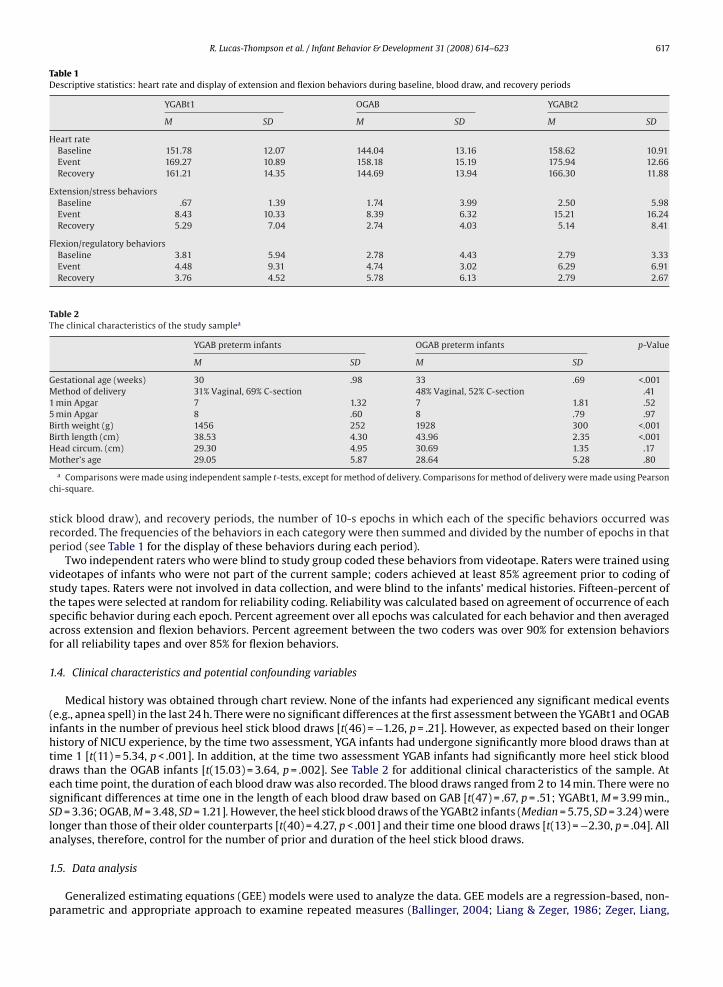
R. Lucas-Thompson et al. / Infant Behavior & Development 31 (2008) 614–623 617
Table 1Descriptive statistics: heart rate and display of extension and flexion behaviors during baseline, blood draw, and recovery periods
YGABt1 OGAB YGABt2
M SD M SD M SD
Heart rateBaseline 151.78 12.07 144.04 13.16 158.62 10.91Event 169.27 10.89 158.18 15.19 175.94 12.66Recovery 161.21 14.35 144.69 13.94 166.30 11.88
Extension/stress behaviorsBaseline .67 1.39 1.74 3.99 2.50 5.98Event 8.43 10.33 8.39 6.32 15.21 16.24Recovery 5.29 7.04 2.74 4.03 5.14 8.41
Flexion/regulatory behaviorsBaseline 3.81 5.94 2.78 4.43 2.79 3.33Event 4.48 9.31 4.74 3.02 6.29 6.91Recovery 3.76 4.52 5.78 6.13 2.79 2.67
Table 2The clinical characteristics of the study samplea
YGAB preterm infants OGAB preterm infants p-Value
M SD M SD
Gestational age (weeks) 30 .98 33 .69 <.001Method of delivery 31% Vaginal, 69% C-section 48% Vaginal, 52% C-section .411 min Apgar 7 1.32 7 1.81 .525 min Apgar 8 .60 8 .79 .97Birth weight (g) 1456 252 1928 300 <.001Birth length (cm) 38.53 4.30 43.96 2.35 <.001HM
c
srp
vstsaf
1
(ihtdesSla
1
p
ead circum. (cm) 29.30 4.95 30.69 1.35 .17other’s age 29.05 5.87 28.64 5.28 .80
a Comparisons were made using independent sample t-tests, except for method of delivery. Comparisons for method of delivery were made using Pearsonhi-square.
tick blood draw), and recovery periods, the number of 10-s epochs in which each of the specific behaviors occurred wasecorded. The frequencies of the behaviors in each category were then summed and divided by the number of epochs in thateriod (see Table 1 for the display of these behaviors during each period).
Two independent raters who were blind to study group coded these behaviors from videotape. Raters were trained usingideotapes of infants who were not part of the current sample; coders achieved at least 85% agreement prior to coding oftudy tapes. Raters were not involved in data collection, and were blind to the infants’ medical histories. Fifteen-percent ofhe tapes were selected at random for reliability coding. Reliability was calculated based on agreement of occurrence of eachpecific behavior during each epoch. Percent agreement over all epochs was calculated for each behavior and then averagedcross extension and flexion behaviors. Percent agreement between the two coders was over 90% for extension behaviorsor all reliability tapes and over 85% for flexion behaviors.
.4. Clinical characteristics and potential confounding variables
Medical history was obtained through chart review. None of the infants had experienced any significant medical eventse.g., apnea spell) in the last 24 h. There were no significant differences at the first assessment between the YGABt1 and OGABnfants in the number of previous heel stick blood draws [t(46) = −1.26, p = .21]. However, as expected based on their longeristory of NICU experience, by the time two assessment, YGA infants had undergone significantly more blood draws than atime 1 [t(11) = 5.34, p < .001]. In addition, at the time two assessment YGAB infants had significantly more heel stick bloodraws than the OGAB infants [t(15.03) = 3.64, p = .002]. See Table 2 for additional clinical characteristics of the sample. Atach time point, the duration of each blood draw was also recorded. The blood draws ranged from 2 to 14 min. There were noignificant differences at time one in the length of each blood draw based on GAB [t(47) = .67, p = .51; YGABt1, M = 3.99 min.,D = 3.36; OGAB, M = 3.48, SD = 1.21]. However, the heel stick blood draws of the YGABt2 infants (Median = 5.75, SD = 3.24) wereonger than those of their older counterparts [t(40) = 4.27, p < .001] and their time one blood draws [t(13) = −2.30, p = .04]. Allnalyses, therefore, control for the number of prior and duration of the heel stick blood draws.
.5. Data analysis
Generalized estimating equations (GEE) models were used to analyze the data. GEE models are a regression-based, non-arametric and appropriate approach to examine repeated measures (Ballinger, 2004; Liang & Zeger, 1986; Zeger, Liang,
618 R. Lucas-Thompson et al. / Infant Behavior & Development 31 (2008) 614–623
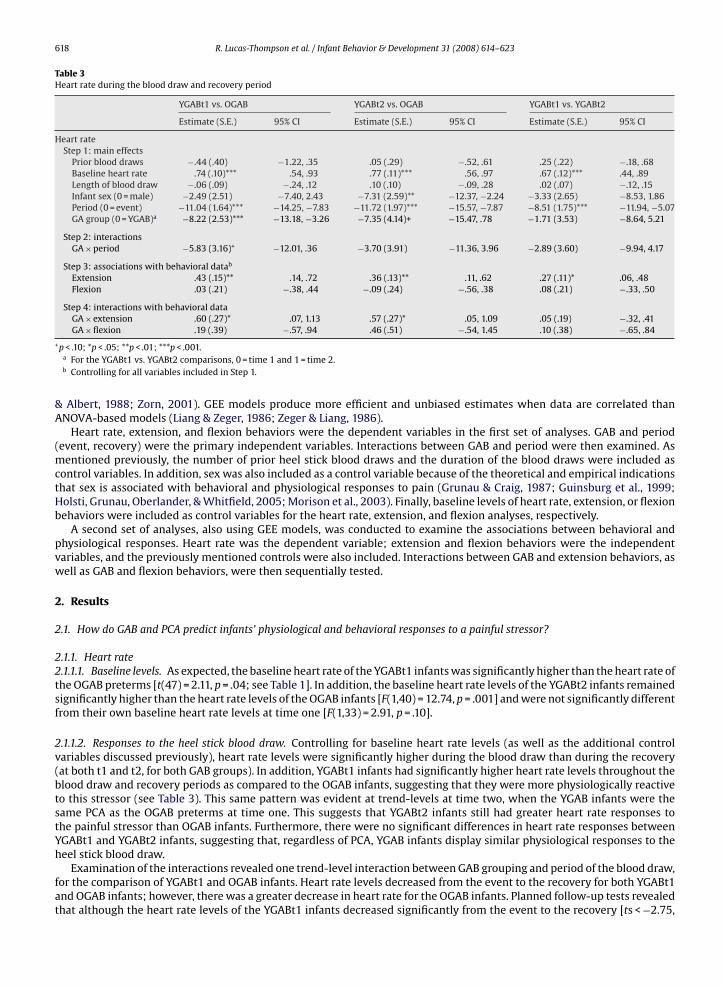
Table 3Heart rate during the blood draw and recovery period
YGABt1 vs. OGAB YGABt2 vs. OGAB YGABt1 vs. YGABt2
Estimate (S.E.) 95% CI Estimate (S.E.) 95% CI Estimate (S.E.) 95% CI
Heart rateStep 1: main effects
Prior blood draws −.44 (.40) −1.22, .35 .05 (.29) −.52, .61 .25 (.22) −.18, .68Baseline heart rate .74 (.10)*** .54, .93 .77 (.11)*** .56, .97 .67 (.12)*** .44, .89Length of blood draw −.06 (.09) −.24, .12 .10 (.10) −.09, .28 .02 (.07) −.12, .15Infant sex (0 = male) −2.49 (2.51) −7.40, 2.43 −7.31 (2.59)** −12.37, −2.24 −3.33 (2.65) −8.53, 1.86Period (0 = event) −11.04 (1.64)*** −14.25, −7.83 −11.72 (1.97)*** −15.57, −7.87 −8.51 (1.75)*** −11.94, −5.07GA group (0 = YGAB)a −8.22 (2.53)*** −13.18, −3.26 −7.35 (4.14)+ −15.47, .78 −1.71 (3.53) −8.64, 5.21
Step 2: interactionsGA × period −5.83 (3.16)+ −12.01, .36 −3.70 (3.91) −11.36, 3.96 −2.89 (3.60) −9.94, 4.17
Step 3: associations with behavioral datab
Extension .43 (.15)** .14, .72 .36 (.13)** .11, .62 .27 (.11)* .06, .48Flexion .03 (.21) −.38, .44 −.09 (.24) −.56, .38 .08 (.21) −.33, .50
Step 4: interactions with behavioral dataGA × extension .60 (.27)* .07, 1.13 .57 (.27)* .05, 1.09 .05 (.19) −.32, .41GA × flexion .19 (.39) −.57, .94 .46 (.51) −.54, 1.45 .10 (.38) −.65, .84
+p < .10; *p < .05; **p < .01; ***p < .001.a For the YGABt1 vs. YGABt2 comparisons, 0 = time 1 and 1 = time 2.b Controlling for all variables included in Step 1.
& Albert, 1988; Zorn, 2001). GEE models produce more efficient and unbiased estimates when data are correlated thanANOVA-based models (Liang & Zeger, 1986; Zeger & Liang, 1986).
Heart rate, extension, and flexion behaviors were the dependent variables in the first set of analyses. GAB and period(event, recovery) were the primary independent variables. Interactions between GAB and period were then examined. Asmentioned previously, the number of prior heel stick blood draws and the duration of the blood draws were included ascontrol variables. In addition, sex was also included as a control variable because of the theoretical and empirical indicationsthat sex is associated with behavioral and physiological responses to pain (Grunau & Craig, 1987; Guinsburg et al., 1999;Holsti, Grunau, Oberlander, & Whitfield, 2005; Morison et al., 2003). Finally, baseline levels of heart rate, extension, or flexionbehaviors were included as control variables for the heart rate, extension, and flexion analyses, respectively.
A second set of analyses, also using GEE models, was conducted to examine the associations between behavioral andphysiological responses. Heart rate was the dependent variable; extension and flexion behaviors were the independentvariables, and the previously mentioned controls were also included. Interactions between GAB and extension behaviors, aswell as GAB and flexion behaviors, were then sequentially tested.
2. Results
2.1. How do GAB and PCA predict infants’ physiological and behavioral responses to a painful stressor?
2.1.1. Heart rate2.1.1.1. Baseline levels. As expected, the baseline heart rate of the YGABt1 infants was significantly higher than the heart rate ofthe OGAB preterms [t(47) = 2.11, p = .04; see Table 1]. In addition, the baseline heart rate levels of the YGABt2 infants remainedsignificantly higher than the heart rate levels of the OGAB infants [F(1,40) = 12.74, p = .001] and were not significantly differentfrom their own baseline heart rate levels at time one [F(1,33) = 2.91, p = .10].
2.1.1.2. Responses to the heel stick blood draw. Controlling for baseline heart rate levels (as well as the additional controlvariables discussed previously), heart rate levels were significantly higher during the blood draw than during the recovery(at both t1 and t2, for both GAB groups). In addition, YGABt1 infants had significantly higher heart rate levels throughout theblood draw and recovery periods as compared to the OGAB infants, suggesting that they were more physiologically reactiveto this stressor (see Table 3). This same pattern was evident at trend-levels at time two, when the YGAB infants were thesame PCA as the OGAB preterms at time one. This suggests that YGABt2 infants still had greater heart rate responses tothe painful stressor than OGAB infants. Furthermore, there were no significant differences in heart rate responses betweenYGABt1 and YGABt2 infants, suggesting that, regardless of PCA, YGAB infants display similar physiological responses to the
heel stick blood draw.Examination of the interactions revealed one trend-level interaction between GAB grouping and period of the blood draw,for the comparison of YGABt1 and OGAB infants. Heart rate levels decreased from the event to the recovery for both YGABt1and OGAB infants; however, there was a greater decrease in heart rate for the OGAB infants. Planned follow-up tests revealedthat although the heart rate levels of the YGABt1 infants decreased significantly from the event to the recovery [ts < −2.75,
R. Lucas-Thompson et al. / Infant Behavior & Development 31 (2008) 614–623 619
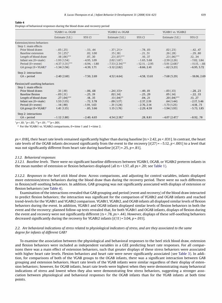
Table 4Displays of behavioral responses during the blood draw and recovery period
YGABt1 vs. OGAB YGABt2 vs. OGAB YGABt1 vs. YGABt2
Estimate (S.E.) 95% CI Estimate (S.E.) 95% CI Estimate (S.E.) 95% CI
Extension/stress behaviorsStep 1: main effects
Prior blood draws −.05 (.25) −.53, .44 −.37 (.21)+ −.78, .03 .02 (.23) −.42, .47Baseline extension .51 (.25)* .02, 1.00 .15 (.18) −.21, .51 .26 (.28) −.29, .80Length of blood draw .18 (.06)*** .07, .30 .23 (.07)*** .10, .37 .23 (.06)*** .12, .34Infant sex (0 = male) −1.93 (1.54) −4.95, 1.09 2.02 (1.87) −1.65, 5.68 −2.59 (2.26) −7.02, 1.84Period (0 = event) −4.37 (1.31)*** −6.94, −1.80 −7.53 (2.34)*** −12.11, −2.95 −5.91 (2.68)* −11.15, −.66GA group (0 = YGAB)a −1.34 (1.56) −4.39, 1.71 −3.12 (2.82) −8.66, 2.41 −.62 (3.23) −6.95, 5.72
Step 2: interactionsGA × period −2.40 (2.60) −7.50, 2.69 4.52 (4.64) −4.58, 13.61 −7.68 (5.29) −18.06, 2.69
Flexion/self-soothing behaviorsStep 1: main effects
Prior blood draws .31 (.19) −.06, .68 −.24 (.13)+ −.49, .01 −.03 (.13) −.28, .23Baseline flexion −.03 (.11) −.25, .19 .02 (.14) −.25, .29 .05 (.14) −.22, .33Length of blood draw .27 (.04)*** .18, .35 .13 (.04)** .04, .21 .20 (.04)*** .13, .27Infant sex (0 = male) 1.53 (1.15) −.72, 3.78 −.09 (1.17) −2.37, 2.19 .64 (1.44) −2.17, 3.46Period (0 = event) −.14 (.90) −1.91, 1.63 −.31 (1.24) −2.76, 2.14 −1.73 (1.25) −4.18, .73GA group (0 = YGAB)a 1.41 (1.15) −.85, 3.66 1.15 (1.76) −2.29, 4.59 −1.94 (1.93) −5.73, 1.85
Step 2: interactionsGA × period −1.12 (1.80) −2.40, 4.65 4.54 (2.18)* .28, 8.81 −4.07 (2.47)+ −8.92, .78
+
prw
22t
2mifl
ttbetd
2d
aiwtgsicp
p < .10; *p < .05; **p < .01; ***p < .001.a For the YGABt1 vs. YGABt2 comparisons, 0 = time 1 and 1 = time 2.
s < .018], their heart rate levels remained significantly higher than during baseline [ts > 2.42, ps < .031]. In contrast, the heartate levels of the OGAB infants decreased significantly from the event to the recovery [t(27) = −5.12, p < .001] to a level thatas not significantly different from heart rate during baseline [t(27) = .25, p = .81].
.1.2. Behavioral responses
.1.2.1. Baseline levels. There were no significant baseline differences between YGABt1, OGAB, or YGABt2 preterm infants inhe mean number of extension or flexion behaviors displayed (all ts < 1.57, all ps > .20; see Table 1).
.1.2.2. Responses to the heel stick blood draw. Across comparisons, and adjusting for control variables, infants displayedore extension/stress behaviors during the blood draw than during the recovery period. There were no such differences
n flexion/self-soothing behaviors. In addition, GAB grouping was not significantly associated with displays of extension orexion behaviors (see Table 4).
Examination of the interactions revealed that GAB grouping and period (event and recovery) of the blood draw interactedo predict flexion behaviors; the interaction was significant for the comparison of YGABt2 and OGAB infants and reachedrend-levels for the YGABt1 and YGABt2 comparison. YGABt1, YGABt2, and OGAB infants all displayed similar levels of flexionehaviors during the event. In addition, YGABt1 and OGAB infants displayed similar levels of flexion behaviors in both thevent and the recovery; planned follow-up tests revealed that, for both YGABt1 and OGAB infants, displays of flexion duringhe event and recovery were not significantly different (ts < .78, ps > .44). However, displays of these self-soothing behaviorsecreased significantly during the recovery for YGABt2 infants [t(11) = 3.04, p = .011].
.2. Are behavioral indications of stress related to physiological indicators of stress, and are they associated to the sameegree for infants of different GAB?
To examine the association between the physiological and behavioral responses to the heel stick blood draw, extensionnd flexion behaviors were included as independent variables in a GEE predicting heart rate responses. For all compar-sons there was a main effect of extension behaviors, such that greater displays of these stress behaviors were associated
ith higher heart rate levels. Flexion behaviors and heart rate were never significantly associated (see Table 3). In addi-
ion, for comparisons of both of the YGAB groups to the OGAB infants, there was a significant interaction between GABrouping and extension behaviors. Heart rate levels of the YGAB infants were similar regardless of their display of exten-ion behaviors; however, for OGAB infants, heart rate levels were highest when they were demonstrating many behavioralndications of stress and lowest when they also were demonstrating few stress behaviors, suggesting a stronger asso-iation between physiological and behavioral responses for the OGAB infants than for the YGAB infants at both timeoints.
620 R. Lucas-Thompson et al. / Infant Behavior & Development 31 (2008) 614–623
3. Discussion
The first goal of the present investigation was to longitudinally examine developmental differences in preterm infants’ability to self-regulate in response to stressors to gain a better understanding of the vulnerability of preterm infants to stressexposure. The second goal was to integrate assessments of both physiology and behavior to examine whether behavioralresponses reflect physiological responses, and whether the association between behavioral and physiological responsesdiffers by GAB. The results of this study indicate that there are both GAB and developmental differences in the self-regulatoryand physiological responses of preterm infants to a painful stressor. All of the preterm infants displayed both physiologicaland behavioral indications of stress in response to the heel stick blood draw. However, preterm infants of a younger GAB(28–31 weeks, YGAB) were more physiologically reactive to the blood draw than infants of an older GAB (32–34 weeks;OGAB), suggesting that younger GAB preterm infants may be more vulnerable to stress than older GAB preterm infants.Furthermore, associations between behavioral and physiological stress responses differed depending on GAB. AlthoughOGAB infants displayed a positive association between extension behaviors and heart rate levels, there was no associationbetween the physiological and behavioral responses of the YGAB infants. This suggests that integrating measurements ofboth behavior and physiology will allow for a better understanding of when younger preterm infants are at risk.
Compared to OGAB preterm infants, YGABt1 and YGABt2 preterm infants had greater heart rate increases in response tothe heel stick blood draw; this elevation continued into the recovery period. This pattern was evident at both assessmentpoints, during postnatal days 3–5 and again during postnatal weeks 3–5. This suggests that YGAB infants remain morephysiologically reactive to a painful stressor several weeks after birth, even when assessed at the same PCA as OGAB infants.These results are consistent with previous indications that physiological responses to pain remain similar over the 8 weeksafter birth (Johnston et al., 1996), and suggest that the physiological stress responses of young GAB infants do not matureover this period of time. Furthermore, although the heart rate levels of the OGAB infants returned to baseline during the 5-min recovery period, the heart rate levels of the YGAB infants remained significantly elevated. Taken together, these resultssuggest that, shortly after birth, younger preterm infants are less capable than older preterm infants of regulating theirphysiological responses to a stressor. Thus, this research provides evidence that the less developed self-regulatory abilitiesof infants born at younger GAs may render them more vulnerable to early postnatal stressors. These infants may lack thecontrol to organize a competent response to pain and stress (Als et al., 1982; Johnston et al., 1999), leading to physiologicalpatterns that indicate high-levels of arousal and overload well after the end of the stressor. Prolonged physiological arousalbeyond the end of a stressor may contribute to the behavioral and physiologic sensitization to repeated pain and stress that isoften demonstrated by younger preterms (Andrews & Fitzgerald, 1994; Fitzgerald et al., 1989; Grunau, Oberlander, Whitfield,& Fitzgerald, 2001; Holsti, Grunau, Oberlander, & Whitfield, 2005; Morison et al., 2003; Storm, 2000).
Previous research suggests that infants of older and younger GAB differ in their behavioral responses to pain, as measuredby facial pain responses (e.g., Goubet, Clifton, & Shah, 2001; Johnston, Stevens, Craig, & Grunau, 1993; Johnston et al., 1999),and that, with increasing PCA, preterm infants respond with increasing robustness to a painful stressor (Johnston et al., 1996).However, the results of the current study suggest that GAB is not associated with either behavioral stress or self-regulatoryresponses to the heel stick blood draw. In response to the blood draw, preterm infants across GAB and PCA groups increaseddisplays of stress behaviors; in contrast, no such increase in self-soothing behaviors was demonstrated. However, there wereindications that there were changes in the behavioral responses of the YGAB infants when observed 3–5 weeks after birth,suggesting a possible effect of their NICU experience on their behavioral stress responses. Although all infants displayedsimilar levels of self-soothing behaviors during the blood draw, only YGABt2 infants decreased their self-soothing behaviorsduring the recovery period. This suggests that preterm infants who remain for longer periods in the NICU are less capable ofbehavioral self-soothing.
Taken together, the results indicate that the experience of developing in a NICU (instead of in utero) may have last-ing consequences for stress regulation. Infants born at an earlier GA display less well-regulated stress responses, evenafter controlling for PCA. Specifically, these infants are not able to deactivate their physiological response after a stres-sor has passed; again, this continued activation and overload may lead to negative outcomes for preterm infants’ in thelong-term. As a whole, these results indicate that YGAB and OGAB infants display similar behavioral responses to pain, yetYGAB infants display more immature regulation of their physiological responses. This suggests the possibility that youngerinfants are less effective self-regulators than older preterms in that these behaviors do not lead to a reduction in physiologicalarousal.
In support of this idea, the results of the current study suggest that behavioral and physiological responses to pain arenot associated in the same way for OGAB and YGAB infants. This study is the first to directly examine the link betweencardiovascular responses and the behaviors identified by Als and colleagues as representing overload and self-soothing. Theexpected positive association between cardiovascular and behavioral responses was demonstrated by the OGAB infants,such that the infants who had the highest physiological arousal were also displaying the most behavioral indications ofstress; this indicates that the behavioral responses of older preterm infants reflect their physiologic responses. However, the
physiological stress responses of the YGAB infants were not reflected in their behavioral responses, as these infants showedsimilar levels of stress behaviors regardless of their heart rate levels. These results underscore that a reliance on measuringbehavioral responses is insufficient, and needs to be balanced by a consideration of physiological responses to more fullyunderstand when preterm infants are at risk because they have difficulty regulating stress (e.g., Morison, Grunau, Oberlander,& Whitfield, 2001). Furthermore, these findings have particularly meaningful implications for developmental care programs
aloEa
amampo1
wleolbs
3
oesihartdgur
3
ltrtptbtpep
A
aiCw
R. Lucas-Thompson et al. / Infant Behavior & Development 31 (2008) 614–623 621
imed at improving self-regulatory competence by tailoring support based on infants’ displays of stress/extension and regu-atory/flexion behaviors (e.g., Als, 1984; Als et al., 1982). Although younger infants may be less likely to be identified as beingverloaded by stressors, they may still be vulnerable to the long-term consequences of chronic arousal and reactivity (e.g.,pel et al., 2006; Silberman, Wald, & Genaro, 2003). Furthermore, these results suggest that the utility of using extensionnd flexion behaviors as indicators of physiological reactivity and vulnerability differs based on GAB.
An additional implication of these findings is that they do not support an association between flexion behaviors andmore rapid physiological recovery. Peters (2001) found that flexion behaviors were associated physiological recovery, aseasured by oxygen saturation levels. This raises the possibility that flexion behaviors are distinctly related to different
spects of physiological arousal and recovery. It is important to determine the behavioral responses to pain that reflectore adaptive cardiovascular responses. Clearly, based on the results of this study, it remains equivocal whether improving
reterm infants’ abilities to self-regulate and soothe in response to a stressor may ameliorate the long-term consequencesf the pain and stress that they are exposed to as part of NICU care (Bhutta et al., 2001; Grunau et al., 1994; Grunau et al.,998; Hack et al., 1995; Porter et al., 1999; Shanks et al., 1995).
Although evidence of this less adaptive behavioral pattern has important implications for researchers and care providersorking with preterm infants, the cause of this pattern remains unclear. Experiences in the NICU, including painful stressors
ike heel stick blood draws and overstimulating visual and auditory stimuli (e.g., Gorski, 1991; Gottfried et al., 1981; Longt al., 1980), may lead to this less adaptive behavioral pattern. It is also possible, however, that infants who are less capablef self-soothing are more likely to remain in the NICU for extended periods of time. In addition, it is possible that theess mature behavioral responses displayed by the YGAB infants resulted from prenatal experiences that affect stress andehavioral regulation such as maternal stress (Davis et al., 2007; De Weert, Van Hees, & Buitelaar, 2003) or treatment withynthetic glucocorticoids (Davis et al., 2006).
.1. Limitations and future directions
Although illuminative, the present study is not without limitations. As with much work in this area, our findings are basedn a naturalistic design. A more controlled administration of the painful stressor may have been theoretically preferable, butthical concerns preclude the use of a more methodologically rigorous design. Similarly, it would have been preferable totudy the infants over a longer time-span. However, we were only able to study the infants while they were being treatedn the NICU. Although following preterm infants for a longer period of time would allow us a better understanding ofow their stress responses continue to develop, it is ethically undesirable to expose these already vulnerable infants todditional discomfort that is not medically necessary after their release from the NICU. Of particular importance for futureesearch, therefore, is further investigation of how self-regulatory behaviors are associated with physiological responseso stress, and whether they play a role in buffering the risks of early, repetitive stress exposure. In addition, it would beesirable to have a continuous measure of heart rate, along with measures of oxygen saturation and respiratory rate toet a broader picture of physiological responses to stressors. It is meaningful that differences based on GAB were foundsing a less nuanced measure; however, future studies should utilize more continuous and varied measures of physiologicalesponses.
.2. Conclusions
The aim of this research was to investigate longitudinally the previously unexplored links between GAB and stress, regu-atory, and physiological responses to a painful stressor in order to more fully understand the vulnerability of preterm infantso stress, as well as the associations between behavioral and physiological stress responses and GAB. The findings of thisesearch indicate that younger preterm infants are less able to physiologically regulate their responses to a painful stressorhan are older preterm infants, a pattern that remains consistent over the first several weeks after birth. This study alsorovides evidence that associations between behavioral and physiological responses to pain differ based on GAB, suggestinghat younger preterms may be less effective at regulating their physiological responses to stressors even though they displayehavioral cues of self-soothing. Therefore, they may continue to be vulnerable to the consequences of pain and chronic reac-ivity even though they are not displaying behaviors that signal overload. Taken together, these results suggest that youngerreterm infants are more vulnerable to stress than older preterm infants and that future developmental care programs andvaluations of preterm infants’ experiences and risk would benefit from a simultaneous consideration of behavioral andhysiological responses.
cknowledgements
This research was supported by a grant from the Minnesota Medical Foundation (643-7051) to the last author and a K05ward (MH66208) to the third author. The authors wish to give a very special thank you to the families who participatedn this research and to the nurses, lab technicians, and physicians at the Fairview University Medical Center and Hennepinounty Medical Center, Minneapolis, MN. Thanks also to the undergraduates at the University of Minnesota who assistedith data collection for this study.
622 R. Lucas-Thompson et al. / Infant Behavior & Development 31 (2008) 614–623
References
Als, H. (1984). Manual for the naturalistic observation of newborn behavior (pre-term & full term). Boston, MA: The Children’s Hospital.Als, H. (1993). Individualized, family-focused developmental care for the very low birthweight preterm infant in the NICU. In S. L. Friedman & M. D. Sigman
(Eds.), Advances in applied developmental psychology: The psychological development of low birthweight children. Norwood, NJ: Ablex Publishing.Als, H., Duffy, F. H., & McAnulty, G. B. (1988). Behavioral differences between preterm and full-term newborns as measured with the APIB System Scores: I.
Infant Behavior and Development, 11, 305–318.Als, H., Gilkerson, L., McAnulty, G. B., Buehler, D. M., Vandenberg, K., Sweet, N., et al. (2003). A three-center, randomized, controlled trial of individualized
developmental care for very low birth weight infants: Medical, neurodevelopmental, parenting, and caregiving effects. Developmental and BehavioralPediatrics, 24(6), 399–408.
Als, H., Lawhon, G., Duffy, F. H., McAnulty, G. B., Gibes-Grossman, R., & Blickman, J. G. (1994). Individualized developmental care for the very low-birth-weightpreterm infant: Medical and neurofunctional effects. Journal of the American Medical Association, 272, 853–858.
Als, H., Lester, B. M., Tronick, E. Z., & Brazelton, T. B. (1982). Toward a research instrument for the assessment of preterm infants’ behavior (APIB). In H.Fitzgerald, B. M. Lester, & M. W. Yogman (Eds.), Theory and research in behavioral pediatrics (pp. 35–63). New York: Plenum Press.
Anand, K. J., Coskun, V., Thrivikraman, K. V., Nemeroff, C. B., & Plotsky, P. M. (1999). Long-term behavioral effects of repetitive pain in neonatal rat pups.Physiology & Behavior, 66, 627–637.
Anand, K. J., Grunau, R. E., & Oberlander, T. F. (1997). Developmental character and long-term consequences of pain in infants and children. Child andAdolescent Psychiatric Clinics of North America, 6, 703–724.
Andrews, K., & Fitzgerald, M. (1994). The cutaneous withdrawal reflex in human neonates: Sensitization, receptive fields, and the effects of contralateralstimulation. Pain, 56, 95–101.
Ballinger, G. A. (2004). Using generalized estimating equations for longitudinal data analysis. Organizational Research Methods, 7(2), 127–150.Barker, D. P., & Rutter, N. (1995). Exposure to invasive procedures in neonatal intensive care unit admissions. Archives of Disease in Childhood: Fetal and
Neonatal Edition, 72, 47–48.Barker, D. P., & Rutter, N. (1996). Stress, severity of illness, and outcome in ventilated preterm infants. Archives of Disease in Childhood: Fetal and Neonatal
Edition, 75, 187–190.Bhutta, A. T., Rovnaghi, C., Simpson, P. M., Gossett, J. M., Scalzo, F. M., & Anand, K. J. (2001). Interactions of inflammatory pain and morphine in infant rats:
Long-term behavioral effects. Physiology & Behavior, 73, 51–58.Craig, K. D., Whitfield, M. F., Grunau, R. V., Linton, J., & Hadjistavropoulos, H. D. (1993). Pain the preterm neonate: Behavioral and physiological indices. Pain,
52, 287–299.Davis, E. P., Glynn, L. M., Dunkel Schetter, C., Hobel, C., Chicz-Demet, A., & Sandman, C. A. (2007). Prenatal exposure to maternal depression and cortisol
influences infant temperament. Journal of the American Academy of Child and Adolescent Psychiatry, 46(6), 737–746.Davis, E. P., Townsend, E. L., Gunnar, M. R., Guiang, S. F., Lussky, R. C., Cifuentes, R. F., et al. (2006). Antenatal betamethasone treatment has a persisting
influence on infant HPA axis regulation. Journal of Perinatology, 26, 139–151.De Weerth, C., Van Hees, Y., & Buitelaar, J. (2003). Prenatal maternal cortisol levels and infant behavior during the first 5 months. Early Human Development,
74, 139–151.Duffy, F. H., Als, H., & McAnulty, G. B. (1990). Behavioral and electrophysiological evidence for gestational age effects in healthy preterm and fullterm infants
studied two weeks after expected due date. Child Development, 61, 1271–1286.Epel, E. S., Lin, J., Wilhelm, F. H., Wolkowitz, O. M., Cawthon, R., Adler, N. E., et al. (2006). Cell aging in relation to stress arousal and cardiovascular disease
risk factors. Psychoneuroendocrinology, 31, 277–287.Ferrari, F., Grosoli, M. V., Fontana, G., & Cavazzuti, G. B. (1983). Neurobehavioral comparison of low-risk preterm and fullterm infants at term conceptional
age. Developmental Medicine and Child Neurology, 25, 450–458.Fitzgerald, M., Millard, C., & McIntosh, N. (1989). Cutaneous hypersensitivity following peripheral tissue damage in newborn infants and its reversal with
topical anaesthesia. Pain, 39, 31–36.Gorski, P. A. (1991). Promoting infant development during neonatal hospitalization: Critiquing the state of the science. Children’s Health Care, 20, 250–257.Gorski, P. A., Huntington, L., & Lewkowicz, D. J. (1990). Handling preterm infants in hospitals: Stimulating controversy about timing of stimulation. Clinics
in Perinatology, 17, 103–112.Gottfried, A. W., Hodgman, J. E., & Brown, K. W. (1984). How intensive is newborn intensive care? An environmental analysis. Pediatrics, 74, 292–294.Gottfried, A. W., Wallace-Lande, P., Sherman-Brown, S., King, J., Coen, C., & Hodgman, J. E. (1981). Physical and social environment of newborn infants in
special care units. Science, 214(4521), 673–675.Goubet, N., Clifton, R. K., & Shah, B. (2001). Learning about pain in preterm newborns. Developmental and Behavioral Pediatrics, 22, 418–424.Grunau, R. V., & Craig, K. D. (1987). Pain expression in neonates: Facial action and cry. Pain, 28, 395–410.Grunau, R. E., Linhares, M. B., Holsti, L., Oberlander, T. F., & Whitfield, M. F. (2004). Does prone or supine position influence pain responses in preterm infants
a 32 weeks gestational age? The Clinical Journal of Pain, 20(2), 76–82.Grunau, R. E., Oberlander, T. F., Whitfield, M. F., Fitzgerald, C., & Less, S. K. (2001). Demographic and therapeutic determinants of pain reactivity in very low
birth weight neonates at 32 weeks’ postconceptional age. Pediatrics, 107, 105–112.Grunau, R. E., Whitfield, M. F., & Petrie, J. (1998). Children’s judgments about pain at age 8–10 years: Do extremely low birthweight (≤1000 g) children differ
from full birthweight peers? Journal of Child Psychology and Psychiatry, 39, 587–594.Grunau, R. V., Whitfield, M. F., Petrie, J. H., & Fryer, E. L. (1994). Early pain experience, child and family factors, as precursors of somatization: A prospective
study of extremely premature and fullterm children. Pain, 56, 353–359.Guinsburg, R., de Araujo Peres, C., de Almeida, M. F., de Cassia Xavier Balda, R., Berenguel, R. C., Tonelotto, J., et al. (1999). Differences in pain expression
between male and female newborn infants. Pain, 85, 127–133.Hack, M., Horbar, J. D., Malloy, M. H., Tyson, J. E., Wright, E., & Wright, L. (1995). Very low birth weight outcomes of the National Institute of Child Health
and Human Development Neonatal Network. American Journal of Obstetrics and Gynecology, 172, 457–464.Holsti, L., Grunau, R. E., Oberlander, T. F., & Whitfield, M. F. (2004). Specific newborn individualized developmental care and assessment program movements
are associated with acute pain in preterm infants in the neonatal intensive care unit. Pediatrics, 114, 65–72.Holsti, L., Grunau, R. E., Oberlander, T. F., & Whitfield, M. F. (2005). Prior pain induces heightened motor responses during clustered care in preterm infants
in the NICU. Early Human Development, 81, 293–302.Holsti, L., Grunau, R. E., Oberlander, T. F., Whitfield, M. F., & Weinberg, J. (2005). Body movements: An important additional factor in discriminating pain
from stress in preterm infants. The Clinical Journal of Pain, 21(6), 491–498.Johnston, C. C., Collinger, J., Henderson, S. J., & Anand, K. J. (1997). A cross-sectional survey of pain and pharmacological analgesia in Canadian Neonatal
Intensive Care Units. The Clinical Journal of Pain, 13(4), 308–312.Johnston, C. C., Franck, L. S., & Stremler, R. (1999). Factors explaining lack of response to heel stick in preterm newborns. Journal of Obstetric, Gynecologic, and
Neonatal Nursing, 28, 587–594.
Johnston, C. C., Stevens, B., Craig, K. D., & Grunau, R. V. (1993). Developmental changes in pain expression in premature, full-term, two- and four-month-oldinfants. Pain, 52, 201–208.Johnston, C. C., Stevens, B., Yang, F., & Horton, L. (1996). Developmental changes in response to heelstick in preterm infants: A prospective cohort study.
Developmental Medicine and Child Neurology, 38, 438–445.Liang, K. Y., & Zeger, S. L. (1986). Longitudinal data analysis using generalized linear model. Biometrika, 73, 13–22.Long, J. G., Lucey, J. F., & Philip, A. G. (1980). Noise and hypoxemia in the intensive care nursery. Pediatrics, 65, 143–145.

LM
M
M
PPS
S
S
S
ZZZ
R. Lucas-Thompson et al. / Infant Behavior & Development 31 (2008) 614–623 623
ucey, J. F. (1977). Is intensive care becoming too intensive? Pediatrics, 59, 1064–1065.orison, S. J., Grunau, R. E., Oberlander, T. F., & Whitfield, M. F. (2001). Relations between behavioral and cardiac autonomic reactivity to acute pain in
preterm neonates. The Clinical Journal of Pain, 17, 350–358.orison, S. J., Holsti, L., Grunau, R. E., Whitfield, M. F., Oberlander, T. F., Chan, H. W., et al. (2003). Are there developmentally distinct motor indicators of
pain in preterm infants? Early Human Development, 72, 131–146.ouradian, L. E., Als, H., & Coster, W. J. (2000). Neurobehavioral functioning of healthy preterm infants of varying gestational ages. Journal of Developmental
& Behavioral Pediatrics, 21(6), 408–416.eters, K. L. (2001). Association between autonomic and motoric systems in the preterm infants. Clinical Nursing Research, 10, 82–90.orter, F. L., Grunau, R. E., & Anand, K. J. (1999). Long-term effects of pain in infants. Developmental and Behavioral Pediatrics, 20(4), 253–261.hanks, N., Larocque, S., & Meaney, M. J. (1995). Neonatal endotoxin exposure alters the development of the hypothalamic-pituitary-adrenal axis: Early
illness and later responsivity to stress. The Journal of Neuroscience, 15, 376–384.ilberman, D. M., Wald, M. R., & Genaro, A. M. (2003). Acute and chronic stress exert opposing effects on antibody responses associated with changes in
stress hormone regulation of T-lymphocyte reactivity. Journal of Neuroimmunology, (144), 53–60.tevens, B. J., Johnston, C. C., & Grunau, R. V. (1995). Issues of assessment of pain and discomfort in neonates. Journal of Obstetric, Gynecologic, and Neonatal
Nursing, 24, 849–855.torm, H. (2000). Skin conductance and the stress response from heel stick in preterm infants. Archives of Disease in Childhood: Fetal and Neonatal Edition,
83, 143–147.eger, S. L., & Liang, K. Y. (1986). Longitudinal data analysis for discrete and continuous outcomes. Biometrics, 42, 121–130.eger, S. L., Liang, K. Y., & Albert, P. S. (1988). Models for longitudinal data: A generalized estimating equations approach. Biometrics, 44, 1049–1060.orn, C. J. (2001). Generalized estimating equation models for correlated data: A review with applications. American Journal of Political Science, 45, 470–490.
Copyright © 2022 FDOKUMEN