The Nucleocapsid of Paramyxoviruses - MDPI
28
viruses Review The Nucleocapsid of Paramyxoviruses: Structure and Function of an Encapsidated Template Louis-Marie Bloyet Citation: Bloyet, L.-M. The Nucleocapsid of Paramyxoviruses: Structure and Function of an Encapsidated Template. Viruses 2021, 13, 2465. https://doi.org/10.3390/ v13122465 Academic Editors: Rob W Ruigrok and Martin Blackledge Received: 17 November 2021 Accepted: 7 December 2021 Published: 9 December 2021 Publisher’s Note: MDPI stays neutral with regard to jurisdictional claims in published maps and institutional affil- iations. Copyright: © 2021 by the author. Licensee MDPI, Basel, Switzerland. This article is an open access article distributed under the terms and conditions of the Creative Commons Attribution (CC BY) license (https:// creativecommons.org/licenses/by/ 4.0/). Department of Molecular Microbiology, School of Medicine, Washington University in Saint Louis, St. Louis, MO 63110, USA; [email protected] Abstract: Viruses of the Paramyxoviridae family share a common and complex molecular machinery for transcribing and replicating their genomes. Their non-segmented, negative-strand RNA genome is encased in a tight homopolymer of viral nucleoproteins (N). This ribonucleoprotein complex, termed a nucleocapsid, is the template of the viral polymerase complex made of the large protein (L) and its co-factor, the phosphoprotein (P). This review summarizes the current knowledge on several aspects of paramyxovirus transcription and replication, including structural and functional data on (1) the architecture of the nucleocapsid (structure of the nucleoprotein, interprotomer contacts, interaction with RNA, and organization of the disordered C-terminal tail of N), (2) the encapsidation of the genomic RNAs (structure of the nucleoprotein in complex with its chaperon P and kinetics of RNA encapsidation in vitro), and (3) the use of the nucleocapsid as a template for the polymerase complex (release of the encased RNA and interaction network allowing the progress of the polymerase complex). Finally, this review presents models of paramyxovirus transcription and replication. Keywords: paramyxoviruses; nucleocapsid; nucleoprotein; phosphoprotein; polymerase complex; intrinsic disorder 1. Introduction Members of the Paramyxoviridae family are enveloped viruses with non-segmented negative-strand RNA genomes. These viruses share similar gene expression and genome replication mechanisms to the members of the other ten families of the Mononegavirales order. Paramyxoviruses are currently divided into 4 subfamilies, 17 genera, and 78 species (Figure 1)[1] (information as of November 2021, for an update see https://talk.ictvonline. org/taxonomy/). Paramyxoviruses are found in a broad range of animals, including mammals, fishes, reptiles, or birds and include several human and animal pathogens such as measles virus (MeV), mumps virus (MuV), canine distemper virus (CDV), and Newcastle disease virus (NDV). Some, such as Nipah virus (NiV) and Hendra virus (HeV), regularly emerge in human or animal populations where they cause deadly epidemics. Although vaccines exist against some paramyxoviruses, effective antiviral treatments for post-exposure prophylaxis are urgently needed [2,3]. Viral transcription and replication are both mediated by an RNA synthesis machinery composed of a viral polymerase complex and its encapsidated template. The polymerase complex consists of the large protein (L) and its co-factor the phosphoprotein (P) [4–7]. This complex uses a genome (or antigenome) embedded into a homopolymer of viral nucleo- proteins (N) as a template [8–10]. The encapsidation of the RNA in this ribonucleoprotein structure, known as the nucleocapsid, prevents the annealing of positive and negative RNA strands into double-stranded RNA structures, as well as the folding of genomic RNA into secondary RNA structures, which helps to prevent recognition by innate immune receptors [11,12]. It also protects the genome from degradation by nucleases [13,14] and from being targeted by siRNA [15,16]. Viruses 2021, 13, 2465. https://doi.org/10.3390/v13122465 https://www.mdpi.com/journal/viruses
-
Upload
khangminh22 -
Category
Documents
-
view
1 -
download
0
Transcript of The Nucleocapsid of Paramyxoviruses - MDPI

viruses
Review
The Nucleocapsid of Paramyxoviruses: Structure and Functionof an Encapsidated Template
Louis-Marie Bloyet
�����������������
Citation: Bloyet, L.-M. The
Nucleocapsid of Paramyxoviruses:
Structure and Function of an
Encapsidated Template. Viruses 2021,
13, 2465. https://doi.org/10.3390/
v13122465
Academic Editors: Rob W Ruigrok
and Martin Blackledge
Received: 17 November 2021
Accepted: 7 December 2021
Published: 9 December 2021
Publisher’s Note: MDPI stays neutral
with regard to jurisdictional claims in
published maps and institutional affil-
iations.
Copyright: © 2021 by the author.
Licensee MDPI, Basel, Switzerland.
This article is an open access article
distributed under the terms and
conditions of the Creative Commons
Attribution (CC BY) license (https://
creativecommons.org/licenses/by/
4.0/).
Department of Molecular Microbiology, School of Medicine, Washington University in Saint Louis,St. Louis, MO 63110, USA; [email protected]
Abstract: Viruses of the Paramyxoviridae family share a common and complex molecular machineryfor transcribing and replicating their genomes. Their non-segmented, negative-strand RNA genomeis encased in a tight homopolymer of viral nucleoproteins (N). This ribonucleoprotein complex,termed a nucleocapsid, is the template of the viral polymerase complex made of the large protein (L)and its co-factor, the phosphoprotein (P). This review summarizes the current knowledge on severalaspects of paramyxovirus transcription and replication, including structural and functional dataon (1) the architecture of the nucleocapsid (structure of the nucleoprotein, interprotomer contacts,interaction with RNA, and organization of the disordered C-terminal tail of N), (2) the encapsidationof the genomic RNAs (structure of the nucleoprotein in complex with its chaperon P and kinetics ofRNA encapsidation in vitro), and (3) the use of the nucleocapsid as a template for the polymerasecomplex (release of the encased RNA and interaction network allowing the progress of the polymerasecomplex). Finally, this review presents models of paramyxovirus transcription and replication.
Keywords: paramyxoviruses; nucleocapsid; nucleoprotein; phosphoprotein; polymerase complex;intrinsic disorder
1. Introduction
Members of the Paramyxoviridae family are enveloped viruses with non-segmentednegative-strand RNA genomes. These viruses share similar gene expression and genomereplication mechanisms to the members of the other ten families of the Mononegaviralesorder. Paramyxoviruses are currently divided into 4 subfamilies, 17 genera, and 78 species(Figure 1) [1] (information as of November 2021, for an update see https://talk.ictvonline.org/taxonomy/). Paramyxoviruses are found in a broad range of animals, includingmammals, fishes, reptiles, or birds and include several human and animal pathogenssuch as measles virus (MeV), mumps virus (MuV), canine distemper virus (CDV), andNewcastle disease virus (NDV). Some, such as Nipah virus (NiV) and Hendra virus (HeV),regularly emerge in human or animal populations where they cause deadly epidemics.Although vaccines exist against some paramyxoviruses, effective antiviral treatments forpost-exposure prophylaxis are urgently needed [2,3].
Viral transcription and replication are both mediated by an RNA synthesis machinerycomposed of a viral polymerase complex and its encapsidated template. The polymerasecomplex consists of the large protein (L) and its co-factor the phosphoprotein (P) [4–7]. Thiscomplex uses a genome (or antigenome) embedded into a homopolymer of viral nucleo-proteins (N) as a template [8–10]. The encapsidation of the RNA in this ribonucleoproteinstructure, known as the nucleocapsid, prevents the annealing of positive and negativeRNA strands into double-stranded RNA structures, as well as the folding of genomic RNAinto secondary RNA structures, which helps to prevent recognition by innate immunereceptors [11,12]. It also protects the genome from degradation by nucleases [13,14] andfrom being targeted by siRNA [15,16].
Viruses 2021, 13, 2465. https://doi.org/10.3390/v13122465 https://www.mdpi.com/journal/viruses

Viruses 2021, 13, 2465 2 of 28Viruses 2021, 13, x FOR PEER REVIEW 2 of 29
Figure 1. Phylogenetic tree of the Paramyxoviridae family. The phylogenetic tree was generated from an alignment of full-length L proteins. One sequence per species was used. The 78 sequences were selected based on the GenBank accession numbers given by the 2020 taxonomy of the International Committee on Taxonomy of Viruses. The names of the genera are indicated in italics. In bold, viruses for which structural data on the N protein is available. Structures of rings or single helical turns of the nucleocapsid-like complexes are shown from a top view (Newcastle disease virus (NDV), reconstructed from PDB: 6JC3; Sendai virus (SeV), reconstructed from PDB: 6M7D; Nipah virus (NiV), PDB: 7NT5; cetacean morbillivirus (CeMV), PDB: 7OI3; measles virus (MeV), PDB: 6h5Q; parainfluenza virus 5 (PIV5), PDB: 4XJN; mumps virus (MuV), PDB: 7EWQ). For human parainfluenza 3 (hPIV3), PIV5, MeV, and NiV, the structures of the N0-P complexes are shown (hPIV3, PDB: 7EV8; PIV5, PDB: 5WKN; MeV, PDB: 5E4V; NiV, PDB: 4CO6).
Viral transcription and replication are both mediated by an RNA synthesis machin-ery composed of a viral polymerase complex and its encapsidated template. The polymer-ase complex consists of the large protein (L) and its co-factor the phosphoprotein (P) [4–7]. This complex uses a genome (or antigenome) embedded into a homopolymer of viral nucleoproteins (N) as a template [8–10]. The encapsidation of the RNA in this ribonucle-oprotein structure, known as the nucleocapsid, prevents the annealing of positive and negative RNA strands into double-stranded RNA structures, as well as the folding of ge-nomic RNA into secondary RNA structures, which helps to prevent recognition by innate immune receptors [11,12]. It also protects the genome from degradation by nucleases [13,14] and from being targeted by siRNA [15,16].
The mechanisms used by the polymerase complex to access the encased RNA, pro-gress along the nucleocapsid, and generate new encapsidated products with soluble nu-cleoprotein monomers are yet to be fully deciphered. However, in the past few years, the atomic structures of the nucleoprotein of a number of paramyxoviruses from different genera have been solved in either assembled [17–23] or non-assembled form [24–27] (Fig-ure 1). Moreover, since the atomic structure of the polymerase complex of parainfluenza virus 5 (PIV5) has been solved, structural information is now available for all the compo-nents of the RNA synthesis machinery of paramyxoviruses [28]. Significant progress has
Figure 1. Phylogenetic tree of the Paramyxoviridae family. The phylogenetic tree was generated from an alignment offull-length L proteins. One sequence per species was used. The 78 sequences were selected based on the GenBank accessionnumbers given by the 2020 taxonomy of the International Committee on Taxonomy of Viruses. The names of the genera areindicated in italics. In bold, viruses for which structural data on the N protein is available. Structures of rings or singlehelical turns of the nucleocapsid-like complexes are shown from a top view (Newcastle disease virus (NDV), reconstructedfrom PDB: 6JC3; Sendai virus (SeV), reconstructed from PDB: 6M7D; Nipah virus (NiV), PDB: 7NT5; cetacean morbillivirus(CeMV), PDB: 7OI3; measles virus (MeV), PDB: 6h5Q; parainfluenza virus 5 (PIV5), PDB: 4XJN; mumps virus (MuV), PDB:7EWQ). For human parainfluenza 3 (hPIV3), PIV5, MeV, and NiV, the structures of the N0-P complexes are shown (hPIV3,PDB: 7EV8; PIV5, PDB: 5WKN; MeV, PDB: 5E4V; NiV, PDB: 4CO6).
The mechanisms used by the polymerase complex to access the encased RNA, progressalong the nucleocapsid, and generate new encapsidated products with soluble nucleopro-tein monomers are yet to be fully deciphered. However, in the past few years, the atomicstructures of the nucleoprotein of a number of paramyxoviruses from different genera havebeen solved in either assembled [17–23] or non-assembled form [24–27] (Figure 1). More-over, since the atomic structure of the polymerase complex of parainfluenza virus 5 (PIV5)has been solved, structural information is now available for all the components of the RNAsynthesis machinery of paramyxoviruses [28]. Significant progress has also been made onthe use of the nucleocapsid as a template, especially on the complex interaction network atplay between the nucleoprotein and the phosphoprotein. This review summarizes bothstructural and functional insights on the nucleocapsid of paramyxoviruses, its assembly,and its role as a template. Models of transcription and replication are proposed.
Viruses 2021, 13, 2465 3 of 28
2. Gene Expression and Genome Replication2.1. The Viral Genome
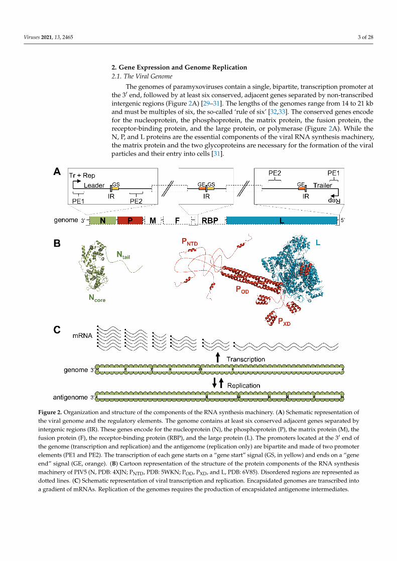
The genomes of paramyxoviruses contain a single, bipartite, transcription promoter atthe 3′ end, followed by at least six conserved, adjacent genes separated by non-transcribedintergenic regions (Figure 2A) [29–31]. The lengths of the genomes range from 14 to 21 kband must be multiples of six, the so-called ‘rule of six’ [32,33]. The conserved genes encodefor the nucleoprotein, the phosphoprotein, the matrix protein, the fusion protein, thereceptor-binding protein, and the large protein, or polymerase (Figure 2A). While theN, P, and L proteins are the essential components of the viral RNA synthesis machinery,the matrix protein and the two glycoproteins are necessary for the formation of the viralparticles and their entry into cells [31].
Viruses 2021, 13, x FOR PEER REVIEW 3 of 29
also been made on the use of the nucleocapsid as a template, especially on the complex interaction network at play between the nucleoprotein and the phosphoprotein. This re-view summarizes both structural and functional insights on the nucleocapsid of paramyx-oviruses, its assembly, and its role as a template. Models of transcription and replication are proposed.
2. Gene Expression and Genome Replication 2.1. The Viral Genome
The genomes of paramyxoviruses contain a single, bipartite, transcription promoter at the 3′ end, followed by at least six conserved, adjacent genes separated by non-tran-scribed intergenic regions (Figure 2A) [29–31]. The lengths of the genomes range from 14 to 21 kb and must be multiples of six, the so-called ‘rule of six’ [32,33]. The conserved genes encode for the nucleoprotein, the phosphoprotein, the matrix protein, the fusion protein, the receptor-binding protein, and the large protein, or polymerase (Figure 2A). While the N, P, and L proteins are the essential components of the viral RNA synthesis machinery, the matrix protein and the two glycoproteins are necessary for the formation of the viral particles and their entry into cells [31].
Figure 2. Organization and structure of the components of the RNA synthesis machinery. (A) Schematic representation of the viral genome and the regulatory elements. The genome contains at least six conserved adjacent genes separated by intergenic regions (IR). These genes encode for the nucleoprotein (N), the phosphoprotein (P), the matrix protein (M), the fusion protein (F), the receptor-binding protein (RBP), and the large protein (L). The promoters located at the 3′ end of the genome (transcription and replication) and the antigenome (replication only) are bipartite and made of two promoter elements (PE1 and PE2). The transcription of each gene starts on a “gene start” signal (GS, in yellow) and ends on a “gene end” signal (GE, orange). (B) Cartoon representation of the structure of the protein components of the RNA synthesis machinery of PIV5 (N, PDB: 4XJN; PNTD, PDB: 5WKN; POD, PXD, and L, PDB: 6V85). Disordered regions are represented as
Figure 2. Organization and structure of the components of the RNA synthesis machinery. (A) Schematic representation ofthe viral genome and the regulatory elements. The genome contains at least six conserved adjacent genes separated byintergenic regions (IR). These genes encode for the nucleoprotein (N), the phosphoprotein (P), the matrix protein (M), thefusion protein (F), the receptor-binding protein (RBP), and the large protein (L). The promoters located at the 3′ end ofthe genome (transcription and replication) and the antigenome (replication only) are bipartite and made of two promoterelements (PE1 and PE2). The transcription of each gene starts on a “gene start” signal (GS, in yellow) and ends on a “geneend” signal (GE, orange). (B) Cartoon representation of the structure of the protein components of the RNA synthesismachinery of PIV5 (N, PDB: 4XJN; PNTD, PDB: 5WKN; POD, PXD, and L, PDB: 6V85). Disordered regions are represented asdotted lines. (C) Schematic representation of viral transcription and replication. Encapsidated genomes are transcribed intoa gradient of mRNAs. Replication of the genomes requires the production of encapsidated antigenome intermediates.

Viruses 2021, 13, 2465 4 of 28
2.2. Components of the RNA Synthesis Machinery
The nucleoproteins of paramyxoviruses are made of a folded domain (Ncore) followedby a long intrinsically disordered tail (Ntail) [34] (Figure 2B). While Ncore is responsible forthe oligomerization on the RNA and constitutes the main body of the nucleocapsid [35–38],Ntail mediates the recruitment of the polymerase complex [34,39,40]. The nucleoproteinsself-assemble on the genome due to their affinity for RNA and the interactions betweenadjacent protomers, as discussed later.
The L protein is a large, multi-domain protein, made of five globular domains linkedtogether by flexible linkers [28,41] (for a review see [42,43]) (Figure 2B). It carries all theenzymatic activities required for transcribing and replicating the genome: RNA synthe-sis [5,6], mRNA cap addition [44,45], guanine-N-7-methylation of the cap, and ribose2′O-methylation of the first nucleotide [46,47]. Polyadenylation of the mRNA occurswhen the polymerase stutters on a short poly(U) sequence located in a polyadenylationsignal [48,49].
The phosphoprotein plays several essential roles: it stabilizes the L protein [7,50–52],recruits the L protein on its template [53], stabilizes the N protein in a monomeric andRNA-free state [54–56], and provides N proteins for the encapsidation of nascent RNAduring replication [7,55]. The phosphoprotein’s sequence is poorly conserved but itsorganization in three modules linked by long disordered regions is shared within theviral order [57–62] (Figure 2B). The N-terminal domain (PNTD) binds the nucleoproteinto prevent its self-assembly on RNAs other than viral genomes and antigenomes [55,56].This interaction forms the N0-P complex, used as a substrate for the encapsidation ofthe nascent RNA during genome replication [7]. The central domain, or oligomerizationdomain (POD), forms a long, parallel, coiled-coil tetramer for Sendai virus (SeV) [63],MeV [64,65], NiV [62,66], PIV5 [28], and human parainfluenza virus 3 (hPIV3) [67], butan antiparallel dimer of two parallel coiled-coil dimers for MuV [68,69]. The C-terminaldomain, or X domain (PXD), interacts with the nucleoprotein and recruits the polymeraseon the nucleocapsid [50,70–75].
2.3. Transcription and Replication
Following fusion of viral and host cell membranes during entry, the polymerasecomplexes packaged in virions transcribe the viral genes into capped and polyadenylatedmRNAs (Figure 2C) [31]. The viral genes are separated by short non-transcribed intergenicregions and contain a gene start (GS) and a gene end (GE) signal at their 3′ and 5′ ends,respectively (Figure 2A). The current model suggests that RNA synthesis starts at the3′ end of the genome where the polymerase recognizes the transcription promoter. TheL protein first synthesizes the leader RNA, a short, uncapped RNA, which is releasedupon recognition of the first intergenic region. The polymerase then scans the genometo find the first GS signal and re-initiates RNA synthesis. The nascent RNA is cappedand methylated by the L protein. Upon recognition of the GE signal, the polymeraseadds a poly(A) tail, releases the mRNA, and scans the genome to find the next gene startsignal. As the transcription re-initiation is not perfectly efficient, the polymerase generatesa decreasing gradient of mRNAs (Figure 2C).
Once the concentration of nucleoproteins reaches a certain threshold, the polymerasecan switch from a transcription mode to a replication mode [76,77]. Although the accumu-lation of N proteins may not be the only trigger, artificially increasing the amount of Nproteins advances the onset of replication [77,78]. Initiation of replication occurs when thenascent leader RNA is encapsidated by monomers of N and the polymerase ignores thetranscription signals, instead synthesizing a full-length encapsidated copy of the genome(antigenome) [76,79]. This antigenome is then used as a template to generate full-lengthencapsidated copies of the viral genome (Figure 2C).
Viral transcription and replication take place in the cytoplasm in membraneless inclu-sion bodies. Viral genomes, together with N, P, and L proteins, are concentrated in theseinclusions formed by liquid-liquid phase separations. The disordered regions of the N and

Viruses 2021, 13, 2465 5 of 28
P proteins play a critical role in the formation of these liquid-like structures (for a review,see [80–82]).
Finally, viral transcription and replication are also affected by the phosphorylation sta-tus of N and P. Indeed, both Ntail [83–85] and the disordered region upstream of POD [86–94]contain major phosphorylation sites targeted by cellular kinases [95–101]. Although theroles of these phosphorylations are still unclear, data suggest the phosphorylation ofsome residues can downregulate [85,92,93,100,102] or upregulate RNA synthesis [83,84,91],modify the balance between transcription and replication [103], alter the stability of en-capsidated genomes [104], participate in RNA encapsidation [105], tune the interactionbetween N and P [94], or facilitate the growth of the inclusion bodies [106].
3. Structure of the Nucleocapsid3.1. Overall Architecture
Nucleocapsids extracted from disrupted virions or infected cells form left-handed, rod-like helical structures with a herringbone appearance under an electron-microscope [107–110](Figure 3A). The coiling and rigidity of the helix depend on the conditions of the milieu,such as the ionic strength [111]. Indeed, while, at low salt concentration, the nucleocapsidsare loose and flexible, and at high ionic strength, the helixes are tight and rigid with alength of about 1 µm and a diameter of 15–20 nm. The flexibility also varies from one virusto another, with the nucleocapsids of MuV being more flexible than the nucleocapsids ofMeV, themselves more flexible than the nucleocapsids of SeV or PIV5 [109,112]. Purifiedintact nucleocapsids from the same virus, or even sections of a single nucleocapsid, canalso adopt condensed conformations with different pitches ranging from 5.3 to 6.8 nmfor SeV [110], 5.2 to 6.6 nm for MeV [113], or 5.8 to 6.7 nm for MuV [114] (Figure 3C). Asmaller pitch correlates with a more rigid and straight helix [112]. In intact MeV particles,the nucleocapsids have a higher pitch (7–8 nm), thus raising the question of whether apreferred pitch exists in physiological conditions and which value it adopts [115]. Theaverage number of N subunits per helical turn is relatively conserved and varies from12.3 to 13.4 [18,110,113,114]. Removal of the encapsidated RNA from MuV nucleocapsidincreases the flexibility of the helix but does not affect its helical conformation, indicatingthat the nucleocapsid assembly is stable even without RNA [114].
Viruses 2021, 13, x FOR PEER REVIEW 6 of 29
Figure 3. Conformations of nucleocapsids. (A) Disrupted measles particle by negative-stain microscopy. Republished with permission of Journal of Virology, from [116]; permission conveyed through Copyright Clearance Center, Inc. (B) Intact (left) and trypsin-digested (right) nucleocapsid-like complexes (NCLC) of MeV. Republished with permission of Journal of Virology, from [117]; permission conveyed through Copyright Clearance Center, Inc. (C) Top row: density maps of MuV nucleocapsid (left, EMDB: EMD-2630) and MuV NCLC (center, EMBD: EMD-31368; right, EMBD: EMD-31369). Bot-tom row: cross sections of the density maps shown above with schematic representation of Ncore in green. The pitch and number of N protomers per helical turn are indicated at the bottom. (D) Top: density map of a clam-shaped structure of NDV (EMBD: EMD-9793). Bottom: cross section of the density map shown above with schematic representation of Ncore in green.
Expression of the nucleoprotein in avian cells [118], mammalian cells [36], insect cells [119], or bacteria [120], induces the formation of nucleocapsid-like complexes (NCLC) con-taining cellular RNAs. These complexes can form helices [118,121], rings [17,122], or clam-shaped structures [19,21,22] (Figure 3B–D). The last has been observed for NDV, SeV, and NiV, and is made of two helical turns interacting in a back-to-back manner (Figure 3D). The detection of this structure for paramyxoviruses from different genera and its presence in nucleocapsids extracted from NDV and SeV virions, suggests it may be biologically relevant [19,21]. The clam-shaped structure has been proposed to play a role in seeding the assembly of double-headed helices, protecting the 5′ end of the genome from nucle-ases, and promoting the encapsidation of multiple nucleocapsids per virion [123–125].
3.2. Structure of the Nucleoprotein The sequence and the organization of Ncore are well conserved across the Paramyxo-
viridae family, while the sequence and length of Ntail vary greatly (Figure 4A). The struc-ture of the N protein in an oligomeric, RNA-bound conformation (i.e., engaged in an NCLC) have been solved for PIV5 [17], MeV [18,20], NDV [19], cetacean morbillivirus (CeMV) [23], NiV [22], SeV [21], and MuV [126], while the structure of N in complex with
Figure 3. Cont.

Viruses 2021, 13, 2465 6 of 28
Viruses 2021, 13, x FOR PEER REVIEW 6 of 29
Figure 3. Conformations of nucleocapsids. (A) Disrupted measles particle by negative-stain microscopy. Republished with permission of Journal of Virology, from [116]; permission conveyed through Copyright Clearance Center, Inc. (B) Intact (left) and trypsin-digested (right) nucleocapsid-like complexes (NCLC) of MeV. Republished with permission of Journal of Virology, from [117]; permission conveyed through Copyright Clearance Center, Inc. (C) Top row: density maps of MuV nucleocapsid (left, EMDB: EMD-2630) and MuV NCLC (center, EMBD: EMD-31368; right, EMBD: EMD-31369). Bot-tom row: cross sections of the density maps shown above with schematic representation of Ncore in green. The pitch and number of N protomers per helical turn are indicated at the bottom. (D) Top: density map of a clam-shaped structure of NDV (EMBD: EMD-9793). Bottom: cross section of the density map shown above with schematic representation of Ncore in green.
Expression of the nucleoprotein in avian cells [118], mammalian cells [36], insect cells [119], or bacteria [120], induces the formation of nucleocapsid-like complexes (NCLC) con-taining cellular RNAs. These complexes can form helices [118,121], rings [17,122], or clam-shaped structures [19,21,22] (Figure 3B–D). The last has been observed for NDV, SeV, and NiV, and is made of two helical turns interacting in a back-to-back manner (Figure 3D). The detection of this structure for paramyxoviruses from different genera and its presence in nucleocapsids extracted from NDV and SeV virions, suggests it may be biologically relevant [19,21]. The clam-shaped structure has been proposed to play a role in seeding the assembly of double-headed helices, protecting the 5′ end of the genome from nucle-ases, and promoting the encapsidation of multiple nucleocapsids per virion [123–125].
3.2. Structure of the Nucleoprotein The sequence and the organization of Ncore are well conserved across the Paramyxo-
viridae family, while the sequence and length of Ntail vary greatly (Figure 4A). The struc-ture of the N protein in an oligomeric, RNA-bound conformation (i.e., engaged in an NCLC) have been solved for PIV5 [17], MeV [18,20], NDV [19], cetacean morbillivirus (CeMV) [23], NiV [22], SeV [21], and MuV [126], while the structure of N in complex with
Figure 3. Conformations of nucleocapsids. (A) Disrupted measles particle by negative-stain microscopy. Republished withpermission of Journal of Virology, from [116]; permission conveyed through Copyright Clearance Center, Inc. (B) Intact(left) and trypsin-digested (right) nucleocapsid-like complexes (NCLC) of MeV. Republished with permission of Journal ofVirology, from [117]; permission conveyed through Copyright Clearance Center, Inc. (C) Top row: density maps of MuVnucleocapsid (left, EMDB: EMD-2630) and MuV NCLC (center, EMBD: EMD-31368; right, EMBD: EMD-31369). Bottom row:cross sections of the density maps shown above with schematic representation of Ncore in green. The pitch and number of Nprotomers per helical turn are indicated at the bottom. (D) Top: density map of a clam-shaped structure of NDV (EMBD:EMD-9793). Bottom: cross section of the density map shown above with schematic representation of Ncore in green.
Expression of the nucleoprotein in avian cells [118], mammalian cells [36], insectcells [119], or bacteria [120], induces the formation of nucleocapsid-like complexes (NCLC)containing cellular RNAs. These complexes can form helices [118,121], rings [17,122], orclam-shaped structures [19,21,22] (Figure 3B–D). The last has been observed for NDV, SeV,and NiV, and is made of two helical turns interacting in a back-to-back manner (Figure 3D).The detection of this structure for paramyxoviruses from different genera and its presencein nucleocapsids extracted from NDV and SeV virions, suggests it may be biologicallyrelevant [19,21]. The clam-shaped structure has been proposed to play a role in seeding theassembly of double-headed helices, protecting the 5′ end of the genome from nucleases,and promoting the encapsidation of multiple nucleocapsids per virion [123–125].
3.2. Structure of the Nucleoprotein
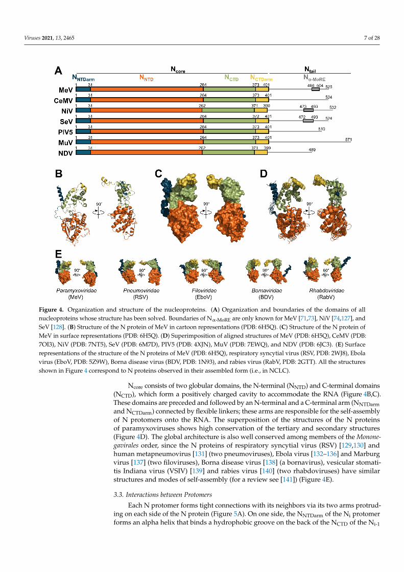
The sequence and the organization of Ncore are well conserved across the Paramyxoviri-dae family, while the sequence and length of Ntail vary greatly (Figure 4A). The structureof the N protein in an oligomeric, RNA-bound conformation (i.e., engaged in an NCLC)have been solved for PIV5 [17], MeV [18,20], NDV [19], cetacean morbillivirus (CeMV) [23],NiV [22], SeV [21], and MuV [126], while the structure of N in complex with PNTD (i.e., N0-Pcomplex) was solved for NiV [24], MeV [25], PIV5 [26], and hPIV3 [27] (Figure 1).
Viruses 2021, 13, 2465 7 of 28
Viruses 2021, 13, x FOR PEER REVIEW 7 of 29
PNTD (i.e., N0-P complex) was solved for NiV [24], MeV [25], PIV5 [26], and hPIV3 [27] (Figure 1).
Figure 4. Organization and structure of the nucleoproteins. (A) Organization and boundaries of the domains of all nucle-oproteins whose structure has been solved. Boundaries of Nα-MoRE are only known for MeV [71,73], NiV [74,127], and SeV [128]. (B) Structure of the N protein of MeV in cartoon representations (PDB: 6H5Q). (C) Structure of the N protein of MeV in surface representations (PDB: 6H5Q). (D) Superimposition of aligned structures of MeV (PDB: 6H5Q), CeMV (PDB: 7OI3), NiV (PDB: 7NT5), SeV (PDB: 6M7D), PIV5 (PDB: 4XJN), MuV (PDB: 7EWQ), and NDV (PDB: 6JC3). (E) Surface representations of the structure of the N proteins of MeV (PDB: 6H5Q), respiratory syncytial virus (RSV, PDB: 2WJ8), Ebola virus (EboV, PDB: 5Z9W), Borna disease virus (BDV, PDB: 1N93), and rabies virus (RabV, PDB: 2GTT). All the structures shown in Figure 4 correspond to N proteins observed in their assembled form (i.e., in NCLC).
Ncore consists of two globular domains, the N-terminal (NNTD) and C-terminal do-mains (NCTD), which form a positively charged cavity to accommodate the RNA (Figure 4B,C). These domains are preceded and followed by an N-terminal and a C-terminal arm (NNTDarm and NCTDarm) connected by flexible linkers; these arms are responsible for the self-assembly of N protomers onto the RNA. The superposition of the structures of the N pro-teins of paramyxoviruses shows high conservation of the tertiary and secondary struc-tures (Figure 4D). The global architecture is also well conserved among members of the Mononegavirales order, since the N proteins of respiratory syncytial virus (RSV) [129,130] and human metapneumovirus [131] (two pneumoviruses), Ebola virus [132–136] and Marburg virus [137] (two filoviruses), Borna disease virus [138] (a bornavirus), vesicular stomatitis Indiana virus (VSIV) [139] and rabies virus [140] (two rhabdoviruses) have sim-ilar structures and modes of self-assembly (for a review see [141]) (Figure 4E).
Figure 4. Organization and structure of the nucleoproteins. (A) Organization and boundaries of the domains of allnucleoproteins whose structure has been solved. Boundaries of Nα-MoRE are only known for MeV [71,73], NiV [74,127], andSeV [128]. (B) Structure of the N protein of MeV in cartoon representations (PDB: 6H5Q). (C) Structure of the N protein ofMeV in surface representations (PDB: 6H5Q). (D) Superimposition of aligned structures of MeV (PDB: 6H5Q), CeMV (PDB:7OI3), NiV (PDB: 7NT5), SeV (PDB: 6M7D), PIV5 (PDB: 4XJN), MuV (PDB: 7EWQ), and NDV (PDB: 6JC3). (E) Surfacerepresentations of the structure of the N proteins of MeV (PDB: 6H5Q), respiratory syncytial virus (RSV, PDB: 2WJ8), Ebolavirus (EboV, PDB: 5Z9W), Borna disease virus (BDV, PDB: 1N93), and rabies virus (RabV, PDB: 2GTT). All the structuresshown in Figure 4 correspond to N proteins observed in their assembled form (i.e., in NCLC).
Ncore consists of two globular domains, the N-terminal (NNTD) and C-terminal domains(NCTD), which form a positively charged cavity to accommodate the RNA (Figure 4B,C).These domains are preceded and followed by an N-terminal and a C-terminal arm (NNTDarmand NCTDarm) connected by flexible linkers; these arms are responsible for the self-assemblyof N protomers onto the RNA. The superposition of the structures of the N proteinsof paramyxoviruses shows high conservation of the tertiary and secondary structures(Figure 4D). The global architecture is also well conserved among members of the Monone-gavirales order, since the N proteins of respiratory syncytial virus (RSV) [129,130] andhuman metapneumovirus [131] (two pneumoviruses), Ebola virus [132–136] and Marburgvirus [137] (two filoviruses), Borna disease virus [138] (a bornavirus), vesicular stomati-tis Indiana virus (VSIV) [139] and rabies virus [140] (two rhabdoviruses) have similarstructures and modes of self-assembly (for a review see [141]) (Figure 4E).
3.3. Interactions between Protomers
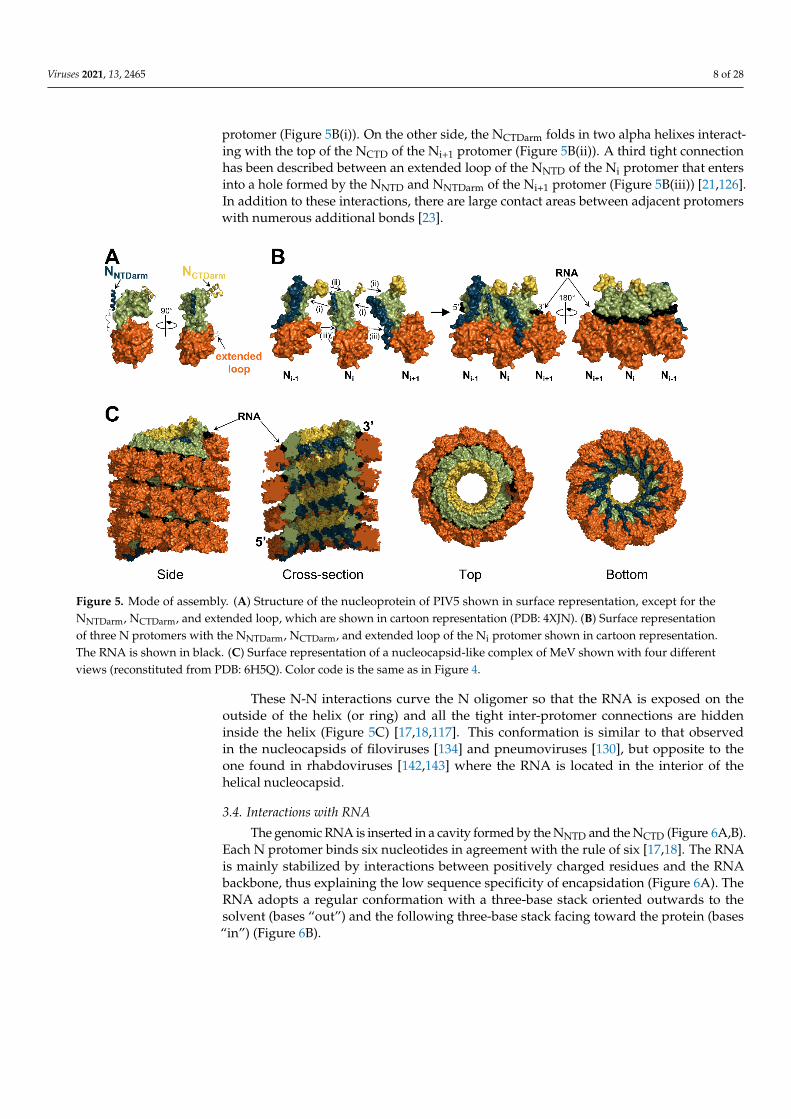
Each N protomer forms tight connections with its neighbors via its two arms protrud-ing on each side of the N protein (Figure 5A). On one side, the NNTDarm of the Ni protomerforms an alpha helix that binds a hydrophobic groove on the back of the NCTD of the Ni-1
Viruses 2021, 13, 2465 8 of 28
protomer (Figure 5B(i)). On the other side, the NCTDarm folds in two alpha helixes interact-ing with the top of the NCTD of the Ni+1 protomer (Figure 5B(ii)). A third tight connectionhas been described between an extended loop of the NNTD of the Ni protomer that entersinto a hole formed by the NNTD and NNTDarm of the Ni+1 protomer (Figure 5B(iii)) [21,126].In addition to these interactions, there are large contact areas between adjacent protomerswith numerous additional bonds [23].
Viruses 2021, 13, x FOR PEER REVIEW 8 of 29
3.3. Interactions Between Protomers Each N protomer forms tight connections with its neighbors via its two arms pro-
truding on each side of the N protein (Figure 5A). On one side, the NNTDarm of the Ni pro-tomer forms an alpha helix that binds a hydrophobic groove on the back of the NCTD of the Ni-1 protomer (Figure 5B(i)). On the other side, the NCTDarm folds in two alpha helixes in-teracting with the top of the NCTD of the Ni+1 protomer (Figure 5B(ii)). A third tight con-nection has been described between an extended loop of the NNTD of the Ni protomer that enters into a hole formed by the NNTD and NNTDarm of the Ni+1 protomer (Figure 5B(iii)) [21,126]. In addition to these interactions, there are large contact areas between adjacent protomers with numerous additional bonds [23].
Figure 5. Mode of assembly. (A) Structure of the nucleoprotein of PIV5 shown in surface representation, except for the NNTDarm, NCTDarm, and extended loop, which are shown in cartoon representation (PDB: 4XJN). (B) Surface representation of three N protomers with the NNTDarm, NCTDarm, and extended loop of the Ni protomer shown in cartoon representation. The RNA is shown in black. (C) Surface representation of a nucleocapsid-like complex of MeV shown with four different views (reconstituted from PDB: 6H5Q). Color code is the same as in Figure 4.
These N-N interactions curve the N oligomer so that the RNA is exposed on the out-side of the helix (or ring) and all the tight inter-protomer connections are hidden inside the helix (Figure 5C) [17,18,117]. This conformation is similar to that observed in the nu-cleocapsids of filoviruses [134] and pneumoviruses [130], but opposite to the one found in rhabdoviruses [142,143] where the RNA is located in the interior of the helical nucleocap-sid.
3.4. Interactions with RNA The genomic RNA is inserted in a cavity formed by the NNTD and the NCTD (Figure
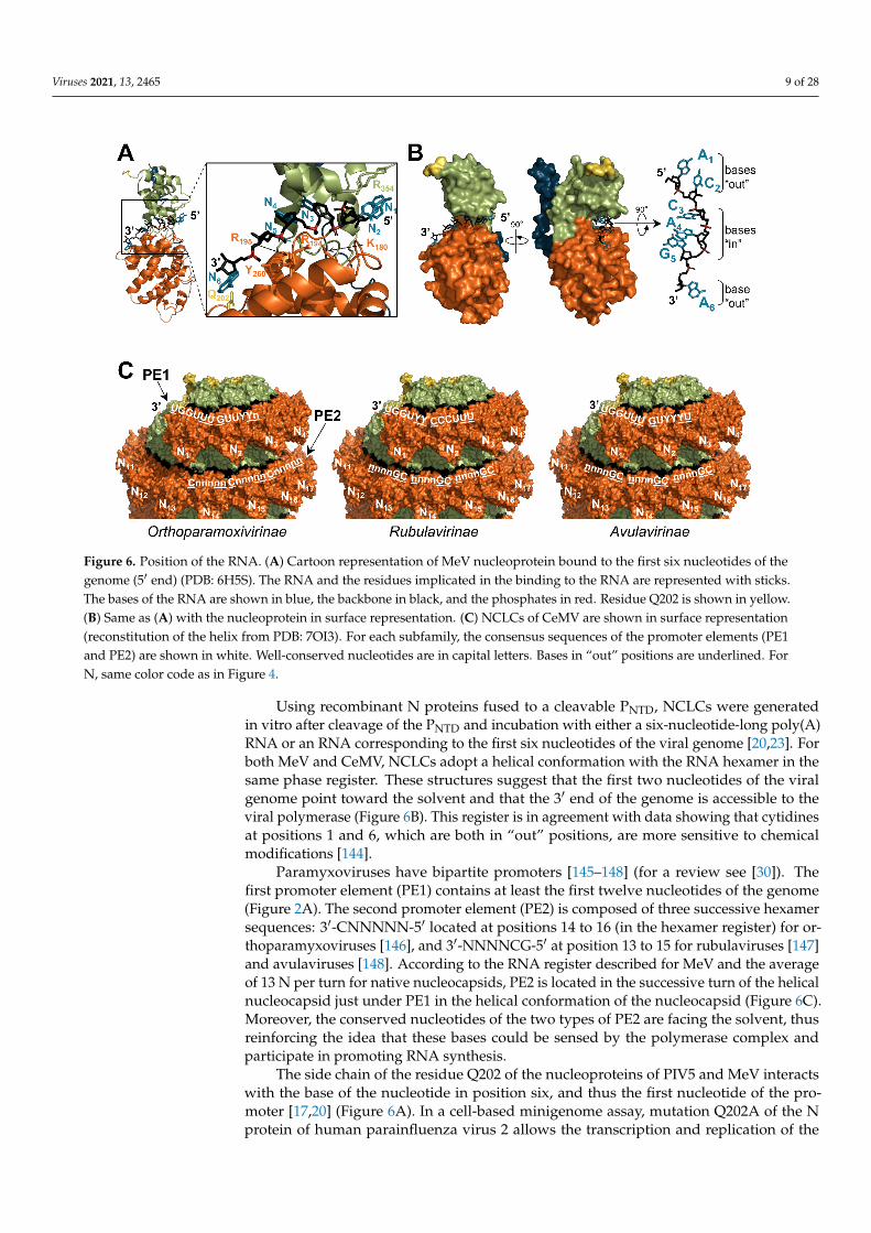
6A,B). Each N protomer binds six nucleotides in agreement with the rule of six [17,18]. The RNA is mainly stabilized by interactions between positively charged residues and the RNA backbone, thus explaining the low sequence specificity of encapsidation (Figure 6A). The RNA adopts a regular conformation with a three-base stack oriented outwards to the solvent (bases “out”) and the following three-base stack facing toward the protein (bases “in”) (Figure 6B).
Figure 5. Mode of assembly. (A) Structure of the nucleoprotein of PIV5 shown in surface representation, except for theNNTDarm, NCTDarm, and extended loop, which are shown in cartoon representation (PDB: 4XJN). (B) Surface representationof three N protomers with the NNTDarm, NCTDarm, and extended loop of the Ni protomer shown in cartoon representation.The RNA is shown in black. (C) Surface representation of a nucleocapsid-like complex of MeV shown with four differentviews (reconstituted from PDB: 6H5Q). Color code is the same as in Figure 4.
These N-N interactions curve the N oligomer so that the RNA is exposed on theoutside of the helix (or ring) and all the tight inter-protomer connections are hiddeninside the helix (Figure 5C) [17,18,117]. This conformation is similar to that observedin the nucleocapsids of filoviruses [134] and pneumoviruses [130], but opposite to theone found in rhabdoviruses [142,143] where the RNA is located in the interior of thehelical nucleocapsid.
3.4. Interactions with RNA
The genomic RNA is inserted in a cavity formed by the NNTD and the NCTD (Figure 6A,B).Each N protomer binds six nucleotides in agreement with the rule of six [17,18]. The RNAis mainly stabilized by interactions between positively charged residues and the RNAbackbone, thus explaining the low sequence specificity of encapsidation (Figure 6A). TheRNA adopts a regular conformation with a three-base stack oriented outwards to thesolvent (bases “out”) and the following three-base stack facing toward the protein (bases“in”) (Figure 6B).
Viruses 2021, 13, 2465 9 of 28Viruses 2021, 13, x FOR PEER REVIEW 9 of 29
Figure 6. Position of the RNA. (A) Cartoon representation of MeV nucleoprotein bound to the first six nucleotides of the genome (5′ end) (PDB: 6H5S). The RNA and the residues implicated in the binding to the RNA are represented with sticks. The bases of the RNA are shown in blue, the backbone in black, and the phosphates in red. Residue Q202 is shown in yellow. (B) Same as (A) with the nucleoprotein in surface representation. (C) NCLCs of CeMV are shown in surface rep-resentation (reconstitution of the helix from PDB: 7OI3). For each subfamily, the consensus sequences of the promoter elements (PE1 and PE2) are shown in white. Well-conserved nucleotides are in capital letters. Bases in “out” positions are underlined. For N, same color code as in Figure 4.
Using recombinant N proteins fused to a cleavable PNTD, NCLCs were generated in vitro after cleavage of the PNTD and incubation with either a six-nucleotide-long poly(A) RNA or an RNA corresponding to the first six nucleotides of the viral genome [20,23]. For both MeV and CeMV, NCLCs adopt a helical conformation with the RNA hexamer in the same phase register. These structures suggest that the first two nucleotides of the viral genome point toward the solvent and that the 3′ end of the genome is accessible to the viral polymerase (Figure 6B). This register is in agreement with data showing that cyti-dines at positions 1 and 6, which are both in “out” positions, are more sensitive to chemical modifications [144].
Paramyxoviruses have bipartite promoters [145–148] (for a review see [30]). The first promoter element (PE1) contains at least the first twelve nucleotides of the genome (Figure 2A). The second promoter element (PE2) is composed of three successive hexamer se-quences: 3′-CNNNNN-5′ located at positions 14 to 16 (in the hexamer register) for ortho-paramyxoviruses [146], and 3′-NNNNCG-5′ at position 13 to 15 for rubulaviruses [147] and avulaviruses [148]. According to the RNA register described for MeV and the average of 13 N per turn for native nucleocapsids, PE2 is located in the successive turn of the hel-ical nucleocapsid just under PE1 in the helical conformation of the nucleocapsid (Figure 6C). Moreover, the conserved nucleotides of the two types of PE2 are facing the solvent, thus reinforcing the idea that these bases could be sensed by the polymerase complex and participate in promoting RNA synthesis.
The side chain of the residue Q202 of the nucleoproteins of PIV5 and MeV interacts with the base of the nucleotide in position six, and thus the first nucleotide of the promoter [17,20] (Figure 6A). In a cell-based minigenome assay, mutation Q202A of the N protein of human parainfluenza virus 2 allows the transcription and replication of the minige-nome, irrespectively of whether its length is a multiple of six and the presence of the PE2
Figure 6. Position of the RNA. (A) Cartoon representation of MeV nucleoprotein bound to the first six nucleotides of thegenome (5′ end) (PDB: 6H5S). The RNA and the residues implicated in the binding to the RNA are represented with sticks.The bases of the RNA are shown in blue, the backbone in black, and the phosphates in red. Residue Q202 is shown in yellow.(B) Same as (A) with the nucleoprotein in surface representation. (C) NCLCs of CeMV are shown in surface representation(reconstitution of the helix from PDB: 7OI3). For each subfamily, the consensus sequences of the promoter elements (PE1and PE2) are shown in white. Well-conserved nucleotides are in capital letters. Bases in “out” positions are underlined. ForN, same color code as in Figure 4.
Using recombinant N proteins fused to a cleavable PNTD, NCLCs were generatedin vitro after cleavage of the PNTD and incubation with either a six-nucleotide-long poly(A)RNA or an RNA corresponding to the first six nucleotides of the viral genome [20,23]. Forboth MeV and CeMV, NCLCs adopt a helical conformation with the RNA hexamer in thesame phase register. These structures suggest that the first two nucleotides of the viralgenome point toward the solvent and that the 3′ end of the genome is accessible to theviral polymerase (Figure 6B). This register is in agreement with data showing that cytidinesat positions 1 and 6, which are both in “out” positions, are more sensitive to chemicalmodifications [144].
Paramyxoviruses have bipartite promoters [145–148] (for a review see [30]). Thefirst promoter element (PE1) contains at least the first twelve nucleotides of the genome(Figure 2A). The second promoter element (PE2) is composed of three successive hexamersequences: 3′-CNNNNN-5′ located at positions 14 to 16 (in the hexamer register) for or-thoparamyxoviruses [146], and 3′-NNNNCG-5′ at position 13 to 15 for rubulaviruses [147]and avulaviruses [148]. According to the RNA register described for MeV and the averageof 13 N per turn for native nucleocapsids, PE2 is located in the successive turn of the helicalnucleocapsid just under PE1 in the helical conformation of the nucleocapsid (Figure 6C).Moreover, the conserved nucleotides of the two types of PE2 are facing the solvent, thusreinforcing the idea that these bases could be sensed by the polymerase complex andparticipate in promoting RNA synthesis.
The side chain of the residue Q202 of the nucleoproteins of PIV5 and MeV interactswith the base of the nucleotide in position six, and thus the first nucleotide of the pro-moter [17,20] (Figure 6A). In a cell-based minigenome assay, mutation Q202A of the Nprotein of human parainfluenza virus 2 allows the transcription and replication of the

Viruses 2021, 13, 2465 10 of 28
minigenome, irrespectively of whether its length is a multiple of six and the presence ofthe PE2 element [149,150]. The authors suggest that residue Q202 may stabilize the firstnucleotide of the genome, thus inhibiting the initiation of RNA synthesis by the viral poly-merase. PE2 would then promote RNA synthesis initiation by stabilizing the polymerasecomplex on the promoter. Since the function of PE2 depends on its phase register, therequirement of PE2 would ensure the conservation of the rule of six [149,150].
In addition to its impact on the promoters, the phase of the RNA bases also affectsthe transcription signals. Indeed, during transcription, the P messenger RNA of paramyx-oviruses can be edited by the viral polymerase which adds one or several extra G residuesby stuttering along a short stretch of C residues [151]. Incremental modifications of thephase register of the editing site modify the efficiency of the editing and the number ofresidues added [144]. Moreover, within each genus, the gene start signals are found in afew preferred phases, suggesting the phasing of the gene start signal may also affect theefficiency of the re-initiation of transcription at gene junctions [33]. Overall, these resultsshow that the imprint of the N protomers on the RNA influences how the viral polymeraseuses its template.
3.5. Position and Influence of Ntail within the Nucleocapsid
Ntail belongs to the group of pre-molten globules within the class of intrinsicallydisordered proteins [34,57,59]. Although Ncore is sufficient to generate the helical core ofthe nucleocapsid [35,36,38], Ntail influences the conformation of the helix. Indeed, treatmentof the nucleocapsid or NCLC by trypsin cleaves off Ntail, reduces the pitch, and tightensthe helix [21,34,113,117,121] (Figure 3B). Moreover, while the expression of the full-lengthN protein of MuV generates NCLCs in a ring conformation, the cleavage of Ntail generateshelical structures [126].
In agreement with its disordered nature, Ntail is not visible by electron microscopy [34,113,126].Nuclear magnetic resonance spectroscopy and small-angle scattering showed that Ntailescapes from the nucleocapsid core toward the exterior between two helical turns [152](Figure 7A,B), in agreement with the location of epitopes recognized by antibodies targetingNtail [153]. While the first 50 residues of Ntail are spatially constrained by the helical turnsof the nucleocapsid core, the C-terminal residues have high angular freedom [75,127,152].This model is further supported by the recently solved structure of the NCLCs formedby SeV N proteins [21]. After cleavage of most of Ntail, the nucleocapsids were found tobecome more rigid and allowed a resolution of 2.9 Å. The atomic model was reconstitutedup to residue 414, thus including the first thirteen residues of the tail (aa 402-414). Theresidues bind the surface of NCTD and point toward the exterior of the nucleocapsid core(Figure 7A).
Viruses 2021, 13, x FOR PEER REVIEW 10 of 29
element [149,150]. The authors suggest that residue Q202 may stabilize the first nucleotide of the genome, thus inhibiting the initiation of RNA synthesis by the viral polymerase. PE2 would then promote RNA synthesis initiation by stabilizing the polymerase complex on the promoter. Since the function of PE2 depends on its phase register, the requirement of PE2 would ensure the conservation of the rule of six [149,150].
In addition to its impact on the promoters, the phase of the RNA bases also affects the transcription signals. Indeed, during transcription, the P messenger RNA of paramyx-oviruses can be edited by the viral polymerase which adds one or several extra G residues by stuttering along a short stretch of C residues [151]. Incremental modifications of the phase register of the editing site modify the efficiency of the editing and the number of residues added [144]. Moreover, within each genus, the gene start signals are found in a few preferred phases, suggesting the phasing of the gene start signal may also affect the efficiency of the re-initiation of transcription at gene junctions [33]. Overall, these results show that the imprint of the N protomers on the RNA influences how the viral polymerase uses its template.
3.5. Position and Influence of Ntail within the Nucleocapsid Ntail belongs to the group of pre-molten globules within the class of intrinsically dis-
ordered proteins [34,57,59]. Although Ncore is sufficient to generate the helical core of the nucleocapsid [35,36,38], Ntail influences the conformation of the helix. Indeed, treatment of the nucleocapsid or NCLC by trypsin cleaves off Ntail, reduces the pitch, and tightens the helix [21,34,113,117,121] (Figure 3B). Moreover, while the expression of the full-length N protein of MuV generates NCLCs in a ring conformation, the cleavage of Ntail generates helical structures [126].
In agreement with its disordered nature, Ntail is not visible by electron microscopy [34,113,126]. Nuclear magnetic resonance spectroscopy and small-angle scattering showed that Ntail escapes from the nucleocapsid core toward the exterior between two helical turns [152] (Figure 7A,B), in agreement with the location of epitopes recognized by antibodies targeting Ntail [153]. While the first 50 residues of Ntail are spatially constrained by the helical turns of the nucleocapsid core, the C-terminal residues have high angular freedom [75,127,152]. This model is further supported by the recently solved structure of the NCLCs formed by SeV N proteins [21]. After cleavage of most of Ntail, the nucleocap-sids were found to become more rigid and allowed a resolution of 2.9 Å. The atomic model was reconstituted up to residue 414, thus including the first thirteen residues of the tail (aa 402-414). The residues bind the surface of NCTD and point toward the exterior of the nucleocapsid core (Figure 7A).
Figure 7. Structure and position of Ntail. (A) Structure of the nucleoprotein of SeV in surface representation with the resi-dues of Ntail shown with spheres (PDB: 6M7D). (B) Surface representation of the nucleocapsid-like complex of CeMV with the Ntail represented as grey lines (reconstitution from PDB: 7OI3). (C) Structure of MeV PXD in complex with Nα-MoRE (PDB: 1T6O). For N, the color code is the same as in Figure 4.
Figure 7. Structure and position of Ntail. (A) Structure of the nucleoprotein of SeV in surface representation with theresidues of Ntail shown with spheres (PDB: 6M7D). (B) Surface representation of the nucleocapsid-like complex of CeMVwith the Ntail represented as grey lines (reconstitution from PDB: 7OI3). (C) Structure of MeV PXD in complex with Nα-MoRE
(PDB: 1T6O). For N, the color code is the same as in Figure 4.
Viruses 2021, 13, 2465 11 of 28
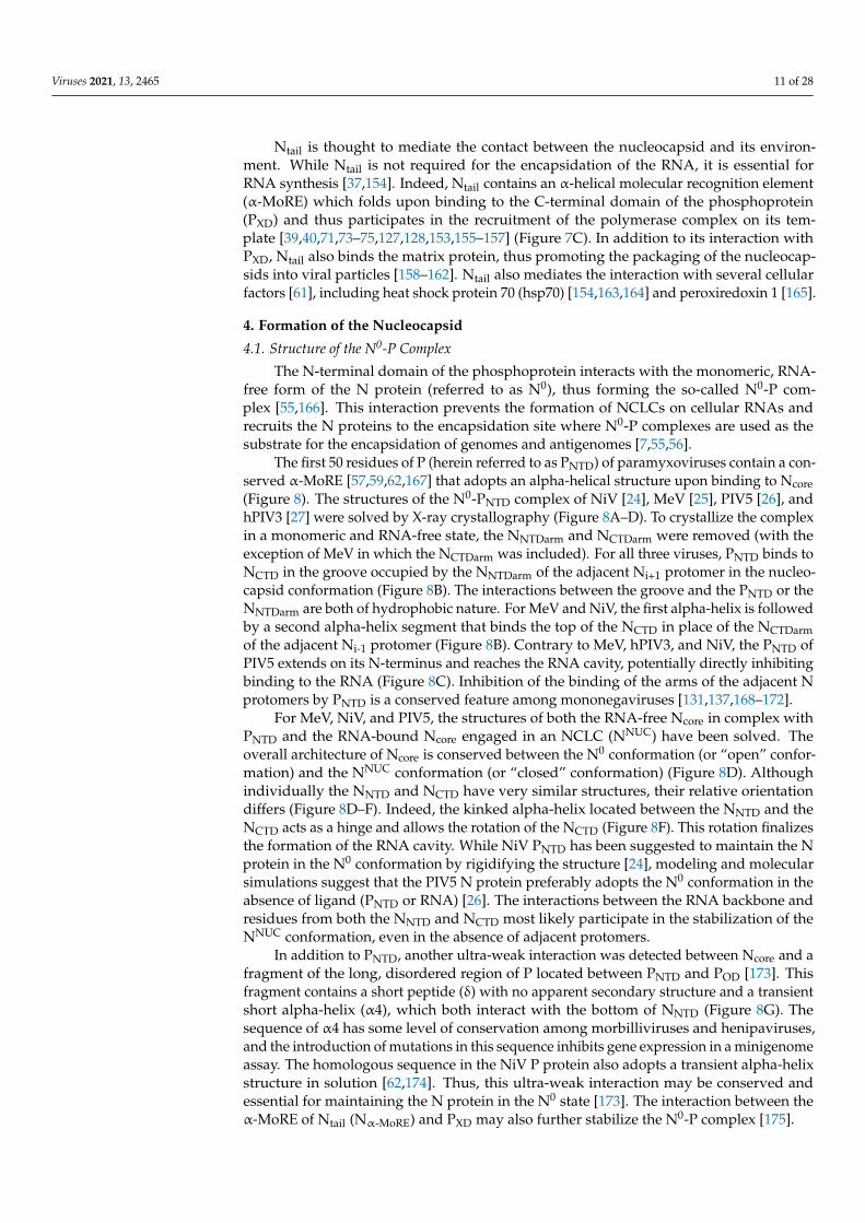
Ntail is thought to mediate the contact between the nucleocapsid and its environ-ment. While Ntail is not required for the encapsidation of the RNA, it is essential forRNA synthesis [37,154]. Indeed, Ntail contains an α-helical molecular recognition element(α-MoRE) which folds upon binding to the C-terminal domain of the phosphoprotein(PXD) and thus participates in the recruitment of the polymerase complex on its tem-plate [39,40,71,73–75,127,128,153,155–157] (Figure 7C). In addition to its interaction withPXD, Ntail also binds the matrix protein, thus promoting the packaging of the nucleocap-sids into viral particles [158–162]. Ntail also mediates the interaction with several cellularfactors [61], including heat shock protein 70 (hsp70) [154,163,164] and peroxiredoxin 1 [165].
4. Formation of the Nucleocapsid
4.1. Structure of the N0-P Complex
The N-terminal domain of the phosphoprotein interacts with the monomeric, RNA-free form of the N protein (referred to as N0), thus forming the so-called N0-P com-plex [55,166]. This interaction prevents the formation of NCLCs on cellular RNAs andrecruits the N proteins to the encapsidation site where N0-P complexes are used as thesubstrate for the encapsidation of genomes and antigenomes [7,55,56].
The first 50 residues of P (herein referred to as PNTD) of paramyxoviruses contain a con-served α-MoRE [57,59,62,167] that adopts an alpha-helical structure upon binding to Ncore(Figure 8). The structures of the N0-PNTD complex of NiV [24], MeV [25], PIV5 [26], andhPIV3 [27] were solved by X-ray crystallography (Figure 8A–D). To crystallize the complexin a monomeric and RNA-free state, the NNTDarm and NCTDarm were removed (with theexception of MeV in which the NCTDarm was included). For all three viruses, PNTD binds toNCTD in the groove occupied by the NNTDarm of the adjacent Ni+1 protomer in the nucleo-capsid conformation (Figure 8B). The interactions between the groove and the PNTD or theNNTDarm are both of hydrophobic nature. For MeV and NiV, the first alpha-helix is followedby a second alpha-helix segment that binds the top of the NCTD in place of the NCTDarmof the adjacent Ni-1 protomer (Figure 8B). Contrary to MeV, hPIV3, and NiV, the PNTD ofPIV5 extends on its N-terminus and reaches the RNA cavity, potentially directly inhibitingbinding to the RNA (Figure 8C). Inhibition of the binding of the arms of the adjacent Nprotomers by PNTD is a conserved feature among mononegaviruses [131,137,168–172].
For MeV, NiV, and PIV5, the structures of both the RNA-free Ncore in complex withPNTD and the RNA-bound Ncore engaged in an NCLC (NNUC) have been solved. Theoverall architecture of Ncore is conserved between the N0 conformation (or “open” confor-mation) and the NNUC conformation (or “closed” conformation) (Figure 8D). Althoughindividually the NNTD and NCTD have very similar structures, their relative orientationdiffers (Figure 8D–F). Indeed, the kinked alpha-helix located between the NNTD and theNCTD acts as a hinge and allows the rotation of the NCTD (Figure 8F). This rotation finalizesthe formation of the RNA cavity. While NiV PNTD has been suggested to maintain the Nprotein in the N0 conformation by rigidifying the structure [24], modeling and molecularsimulations suggest that the PIV5 N protein preferably adopts the N0 conformation in theabsence of ligand (PNTD or RNA) [26]. The interactions between the RNA backbone andresidues from both the NNTD and NCTD most likely participate in the stabilization of theNNUC conformation, even in the absence of adjacent protomers.
In addition to PNTD, another ultra-weak interaction was detected between Ncore and afragment of the long, disordered region of P located between PNTD and POD [173]. Thisfragment contains a short peptide (δ) with no apparent secondary structure and a transientshort alpha-helix (α4), which both interact with the bottom of NNTD (Figure 8G). Thesequence of α4 has some level of conservation among morbilliviruses and henipaviruses,and the introduction of mutations in this sequence inhibits gene expression in a minigenomeassay. The homologous sequence in the NiV P protein also adopts a transient alpha-helixstructure in solution [62,174]. Thus, this ultra-weak interaction may be conserved andessential for maintaining the N protein in the N0 state [173]. The interaction between theα-MoRE of Ntail (Nα-MoRE) and PXD may also further stabilize the N0-P complex [175].

Viruses 2021, 13, 2465 12 of 28Viruses 2021, 13, x FOR PEER REVIEW 12 of 29
Figure 8. Structure of the N0-P complex. (A) Structure of the N0-P complex of NiV (PDB: 4CO6). The NNTD and the NCTD are in pale orange and pale green, respectively. (B) Superimposition of the structure of three N protomers of NiV NLPC (PDB: 7NT5) with NiV N0-P (PDB: 4CO6). The NCTD of the N0-P complex was aligned onto the NCTD of the Ni promoter. The N protomers are presented in surface representation, while the NNTDarm, NCTDarm, and the extended loop of the Ni protomer are shown in cartoon representation. PNTD is shown in cartoon representation. (C) Structure of the N0-P complex of PIV5 with PNTD in cartoon representation (PDB: 5WKN). The RNA is shown as a transparent black surface. (D) Side-by-side comparison of the structures of the N0-P complex (PDB: 5E4V) and of an N protomer of the NCLC of MeV (PDB: 6H5Q) (NNTDarm and NCTDarm are not shown). The NNTD and the NCTD of N0 are in pale orange and pale green, respectively. (E) Superimposition of the NCTD (top) and of the NNTD (bottom) of the N0-P complex and of an N protomer of the NCLC of MeV. (F) Superimposition of the structures of MeV N0 and NNUC (NNTDarm and NCTDarm are not shown). The structures are aligned based on their NCTD. (G) Structure of the MeV N0-P complex with the disordered region downstream PNTD shown as a dotted line. The transient alpha-helices (α3 and α4) are shown in cartoon representation.
For MeV, NiV, and PIV5, the structures of both the RNA-free Ncore in complex with PNTD and the RNA-bound Ncore engaged in an NCLC (NNUC) have been solved. The overall architecture of Ncore is conserved between the N0 conformation (or “open” conformation) and the NNUC conformation (or “closed” conformation) (Figure 8D). Although individu-ally the NNTD and NCTD have very similar structures, their relative orientation differs (Fig-ure 8D–F). Indeed, the kinked alpha-helix located between the NNTD and the NCTD acts as a hinge and allows the rotation of the NCTD (Figure 8F). This rotation finalizes the for-mation of the RNA cavity. While NiV PNTD has been suggested to maintain the N protein in the N0 conformation by rigidifying the structure [24], modeling and molecular simula-
Figure 8. Structure of the N0-P complex. (A) Structure of the N0-P complex of NiV (PDB: 4CO6). The NNTD and theNCTD are in pale orange and pale green, respectively. (B) Superimposition of the structure of three N protomers of NiVNLPC (PDB: 7NT5) with NiV N0-P (PDB: 4CO6). The NCTD of the N0-P complex was aligned onto the NCTD of the Ni
promoter. The N protomers are presented in surface representation, while the NNTDarm, NCTDarm, and the extended loop ofthe Ni protomer are shown in cartoon representation. PNTD is shown in cartoon representation. (C) Structure of the N0-Pcomplex of PIV5 with PNTD in cartoon representation (PDB: 5WKN). The RNA is shown as a transparent black surface.(D) Side-by-side comparison of the structures of the N0-P complex (PDB: 5E4V) and of an N protomer of the NCLC of MeV(PDB: 6H5Q) (NNTDarm and NCTDarm are not shown). The NNTD and the NCTD of N0 are in pale orange and pale green,respectively. (E) Superimposition of the NCTD (top) and of the NNTD (bottom) of the N0-P complex and of an N protomer ofthe NCLC of MeV. (F) Superimposition of the structures of MeV N0 and NNUC (NNTDarm and NCTDarm are not shown). Thestructures are aligned based on their NCTD. (G) Structure of the MeV N0-P complex with the disordered region downstreamPNTD shown as a dotted line. The transient alpha-helices (α3 and α4) are shown in cartoon representation.
4.2. Encapsidation of the RNA
The fusion of full-length N protein (or Ncore) to a cleavable PNTD allows the productionof soluble N0-P complexes [20,23,176]. Incubation of these complexes with RNA triggersthe formation of NCLCs in vitro. This experimental setup revealed that the encapsidationof the RNA is sequence-specific and that the N protein of MeV preferably encapsidatesa poly(A) hexamer (but not poly(U)), as well as an RNA corresponding to the first sixnucleotides of the genome. Similar specificity was observed for other mononegaviruses,suggesting that the N protein has a higher affinity for poly(A) RNA and the first nucleotides

Viruses 2021, 13, 2465 13 of 28
of the corresponding genome [177–180]. The formation of long helical NCLCs from shortRNA hexamers indicates that a continuous RNA molecule is not required for the assemblyof N proteins. The kinetics of the encapsidation was analyzed by real-time nuclear magneticresonance and fluorescence spectroscopy and revealed a two-step mechanism. The firststep is rapid and could correspond to the binding of N monomers to the RNA, while thesecond step consists of the assembly of RNA-bound N proteins [176].
Finally, the local concentration of N0-P complexes seems critical to efficient encapsida-tion of the RNA and generation of NCLCs. Indeed, the formation of droplets formed byliquid-liquid phase separation increases both the local concentration of N0-P complexesand the rate of formation of NCLCs in vitro [181]. For VSIV (Rhabdoviridae family), al-though dispensable [182], POD enhances genome replication possibly by increasing thelocal concentration of N0 at the site of encapsidation [183].
5. The Nucleocapsid as a Template
Unlike the distantly related members of the Articulavirales and Bunyavirales ordersfor which the polymerase can directly bind the extremity of the viral genomes, the viralpolymerase of mononegaviruses cannot interact with its nucleocapsid template without itscofactor P [43]. Moreover, the genomes of mononegaviruses are deeply encased in helicalnucleocapsids, thus raising the question of how the polymerase accesses the RNA.
5.1. Recruitment and Progression of the Polymerase Complex
Since PXD mediates the interaction of L with the nucleocapsid and the polymerasecomplex is made of the L protein and a tetramer of P proteins, the polymerase complexpossesses four potential nucleocapsid-binding sites. This architecture is the basis of the so-called “cartwheeling” model in which the polymerase complex progresses on its templatedue to cycles of association/dissociation between PXD and the nucleocapsid [184,185].
The PXD of paramyxoviruses adopts a three-helix bundle structure [71–73,75,186,187].PXD binds to the α-MoRE of Ntail [73–75,127,128], the L protein [28,188,189], andNcore [68,114,190,191] (Figure 9). PXD has a prism-like shape with three faces: α1 + α2form face 1 (F1), α2 + α3 form face 2 (F2), and α3 + α1 form face 3 (F3) (Figure 9A). Eachinteraction is mediated by a different face, with Nα-MoRE interacting with F2 [73], the Lprotein with F3 [28,189], and Ncore with F1 [191] (Figure 9).
Viruses 2021, 13, x FOR PEER REVIEW 14 of 29
is mediated by a different face, with Nα-MoRE interacting with F2 [73], the L protein with F3 [28,189], and Ncore with F1 [191] (Figure 9).
Figure 9. Interaction network of PXD. (A) Structure of MeV PXD (PDB: 1T6O). The three faces of the “prism” are indicated (F1, F2, F3). F1, F2, and F3 are shown with a color code corresponding to the color used to draw the protein partner. (B) Structure of MeV PXD in complex with Nα-MoRE (PDB: 1T6O). (C) Structure of a protomer of MeV N, as observed in the NCLC (PDB: 6H5Q) with the RNA shown in black. Amino acids shown as red spheres correspond to CDV N residues that once mutated restore the growth of a recombinant CDV bearing deletions in the NCDR [191]. The amino acids correspond-ing to the residues of NiV identified as important for the interaction with PXD are shown as pink spheres [192]. Bottom: enlargements of the NNTD with the acidic loop shown in pink. (D) Surface representation of a nucleocapsid-like complex of MeV (reconstituted from PDB: 6H5Q) with the acidic loop shown in pink. (E) Schematic representation of two adjacent N protomers bound to PXD in complex with Nα-MoRE. (F) Structure of the polymerase complex of PIV5 (PDB: 6V85). For panels C–E, N protomers are colored according to the color code in Figure 4.
The interaction of PXD with Nα-MoRE is weak, with a constant of dissociation (KD) in the µM range [74,128,157,193]. While the disordered Nα-MoRE folds into an alpha-helix upon binding to PXD, the flanking regions of Ntail remain disordered [74,75,127,187,194–197]. This phenomenon, called fuzziness [198], affects the interaction between Ntail and its part-ners. Indeed, the long-disordered region upstream of Nα-MoRE slows the disorder-to-order transition of Nα-MoRE upon binding to PXD [199,200]. Accordingly, the deletion of the disor-dered region upstream MeV, NiV, and HeV Nα-MoRE increases the affinity for PXD (and also HSP70 for MeV) [199]. The dampening effect of the disordered regions does not directly depend on the sequence, but rather on a combination of length and disorder. Although not directly involved, the poorly conserved disordered region of Ntail upstream the Nα-MoRE affects the interaction with PXD [199–201].
Figure 9. Cont.

Viruses 2021, 13, 2465 14 of 28
Viruses 2021, 13, x FOR PEER REVIEW 14 of 29
is mediated by a different face, with Nα-MoRE interacting with F2 [73], the L protein with F3 [28,189], and Ncore with F1 [191] (Figure 9).
Figure 9. Interaction network of PXD. (A) Structure of MeV PXD (PDB: 1T6O). The three faces of the “prism” are indicated (F1, F2, F3). F1, F2, and F3 are shown with a color code corresponding to the color used to draw the protein partner. (B) Structure of MeV PXD in complex with Nα-MoRE (PDB: 1T6O). (C) Structure of a protomer of MeV N, as observed in the NCLC (PDB: 6H5Q) with the RNA shown in black. Amino acids shown as red spheres correspond to CDV N residues that once mutated restore the growth of a recombinant CDV bearing deletions in the NCDR [191]. The amino acids correspond-ing to the residues of NiV identified as important for the interaction with PXD are shown as pink spheres [192]. Bottom: enlargements of the NNTD with the acidic loop shown in pink. (D) Surface representation of a nucleocapsid-like complex of MeV (reconstituted from PDB: 6H5Q) with the acidic loop shown in pink. (E) Schematic representation of two adjacent N protomers bound to PXD in complex with Nα-MoRE. (F) Structure of the polymerase complex of PIV5 (PDB: 6V85). For panels C–E, N protomers are colored according to the color code in Figure 4.
The interaction of PXD with Nα-MoRE is weak, with a constant of dissociation (KD) in the µM range [74,128,157,193]. While the disordered Nα-MoRE folds into an alpha-helix upon binding to PXD, the flanking regions of Ntail remain disordered [74,75,127,187,194–197]. This phenomenon, called fuzziness [198], affects the interaction between Ntail and its part-ners. Indeed, the long-disordered region upstream of Nα-MoRE slows the disorder-to-order transition of Nα-MoRE upon binding to PXD [199,200]. Accordingly, the deletion of the disor-dered region upstream MeV, NiV, and HeV Nα-MoRE increases the affinity for PXD (and also HSP70 for MeV) [199]. The dampening effect of the disordered regions does not directly depend on the sequence, but rather on a combination of length and disorder. Although not directly involved, the poorly conserved disordered region of Ntail upstream the Nα-MoRE affects the interaction with PXD [199–201].
Figure 9. Interaction network of PXD. (A) Structure of MeV PXD (PDB: 1T6O). The three faces of the “prism” are indicated(F1, F2, F3). F1, F2, and F3 are shown with a color code corresponding to the color used to draw the protein partner.(B) Structure of MeV PXD in complex with Nα-MoRE (PDB: 1T6O). (C) Structure of a protomer of MeV N, as observedin the NCLC (PDB: 6H5Q) with the RNA shown in black. Amino acids shown as red spheres correspond to CDV Nresidues that once mutated restore the growth of a recombinant CDV bearing deletions in the NCDR [191]. The amino acidscorresponding to the residues of NiV identified as important for the interaction with PXD are shown as pink spheres [192].Bottom: enlargements of the NNTD with the acidic loop shown in pink. (D) Surface representation of a nucleocapsid-likecomplex of MeV (reconstituted from PDB: 6H5Q) with the acidic loop shown in pink. (E) Schematic representation of twoadjacent N protomers bound to PXD in complex with Nα-MoRE. (F) Structure of the polymerase complex of PIV5 (PDB:6V85). For panels (C–E), N protomers are colored according to the color code in Figure 4.
The interaction of PXD with Nα-MoRE is weak, with a constant of dissociation (KD) in theµM range [74,128,157,193]. While the disordered Nα-MoRE folds into an alpha-helix uponbinding to PXD, the flanking regions of Ntail remain disordered [74,75,127,187,194–197].This phenomenon, called fuzziness [198], affects the interaction between Ntail and itspartners. Indeed, the long-disordered region upstream of Nα-MoRE slows the disorder-to-order transition of Nα-MoRE upon binding to PXD [199,200]. Accordingly, the deletion ofthe disordered region upstream MeV, NiV, and HeV Nα-MoRE increases the affinity for PXD(and also HSP70 for MeV) [199]. The dampening effect of the disordered regions doesnot directly depend on the sequence, but rather on a combination of length and disorder.Although not directly involved, the poorly conserved disordered region of Ntail upstreamthe Nα-MoRE affects the interaction with PXD [199–201].
The low affinity between PXD and Nα-MoRE permits efficient progression of the poly-merase complex on the nucleocapsid. Indeed, modifications of the binding strength be-tween MeV PXD and Nα-MoRE alter the activity of the polymerase [202–204]. Indeed, thereis a positive correlation between the PXD/Nα-MoRE interaction strength and the efficiencyof the re-initiation of the transcription at gene junctions [204] (Figure 2A). This effect isfurther enhanced when the polymerase crosses long non-transcribed intergenic regions,suggesting the PXD/Nα-MoRE interaction keeps the polymerase in an active state whencrossing the gap between transcription units.
Although the PXD/Nα-MoRE interaction regulates the activity of the polymerase, itis not strictly required for RNA synthesis. Indeed, while the truncation of MeV N from

Viruses 2021, 13, 2465 15 of 28
Nα-MoRE to its C-terminus abrogates gene expression in a minigenome assay, furtherdeletion of the 43 upstream residues restores activity [205] (Figure 10). This suggests thatthe central disordered region (NCDR) upstream of the Nα-MoRE has a negative effect onthe activity of the polymerase and the binding of PXD to Nα-MoRE counteracts this effect.Accordingly, deletions in the NCDR of CDV and NiV increase activity [206], and whenMeV Nα-MoRE is relocated to a variable region of NNTD, removing NCDR enhances geneexpression [207] (Figure 10). However, a recombinant MeV harboring Nα-MoRE on Ncoreand devoid of NCDR produces more polycistronic mRNAs. Similarly, two recombinantCDV with deletions in NCDR produce more polycistronic mRNA and show an alteredRNA synthesis balance with enhanced transcription over replication [206]. Therefore, thecentral disordered region of Ntail inhibits RNA synthesis, and PXD counterbalances thisnegative effect. This interplay would finely tune the polymerase’s activity and ensurethe quality of the generated mRNAs by improving the recognition of the signals at theintergenic junctions.
Viruses 2021, 13, x FOR PEER REVIEW 15 of 29
The low affinity between PXD and Nα-MoRE permits efficient progression of the poly-merase complex on the nucleocapsid. Indeed, modifications of the binding strength be-tween MeV PXD and Nα-MoRE alter the activity of the polymerase [202–204]. Indeed, there is a positive correlation between the PXD/Nα-MoRE interaction strength and the efficiency of the re-initiation of the transcription at gene junctions [204] (Figure 2A). This effect is further enhanced when the polymerase crosses long non-transcribed intergenic regions, suggest-ing the PXD/Nα-MoRE interaction keeps the polymerase in an active state when crossing the gap between transcription units.
Although the PXD/Nα-MoRE interaction regulates the activity of the polymerase, it is not strictly required for RNA synthesis. Indeed, while the truncation of MeV N from Nα-MoRE to its C-terminus abrogates gene expression in a minigenome assay, further deletion of the 43 upstream residues restores activity [205] (Figure 10). This suggests that the central disordered region (NCDR) upstream of the Nα-MoRE has a negative effect on the activity of the polymerase and the binding of PXD to Nα-MoRE counteracts this effect. Accordingly, de-letions in the NCDR of CDV and NiV increase activity [206], and when MeV Nα-MoRE is relo-cated to a variable region of NNTD, removing NCDR enhances gene expression [207] (Figure 10). However, a recombinant MeV harboring Nα-MoRE on Ncore and devoid of NCDR produces more polycistronic mRNAs. Similarly, two recombinant CDV with deletions in NCDR pro-duce more polycistronic mRNA and show an altered RNA synthesis balance with en-hanced transcription over replication [206]. Therefore, the central disordered region of Ntail inhibits RNA synthesis, and PXD counterbalances this negative effect. This interplay would finely tune the polymerase’s activity and ensure the quality of the generated mRNAs by improving the recognition of the signals at the intergenic junctions.
Figure 10. Summary of experimental data on the effect of Ntail on the polymerase’s activity. Schematic representation of recombinant N proteins analyzed in minigenome assays. The levels of gene expression are normalized to the level of the wild-type protein (first protein of each set). Activity levels: 0–10%: “-”; 50–75%: “+++”; 75–100%: “++++”; >100%: “+++++”. For mutants (6) and (7) Nα-MoRE is inserted in a loop of NNTD at residue 138. Mutants (4), (5), (6), (9), and (11) contain internal deletion shown with a “^”. Activity levels are inferred from the references indicated on the right.
In addition to interacting with the α-MoRE, PXD of CDV was also shown to directly interact with Ncore [191]. This interaction is mediated by an acidic loop of NNTD (residues 146 to 161) and requires the presence of Nα-MoRE in complex with PXD (Figure 9C–E). Ac-cordingly, mutations in the homologous region of NiV Ncore were previously shown to inhibit the P-N interaction [192]. After several passages of a recombinant CDV expressing a Ntail with a large deletion in NCDR, compensatory mutations appeared in PXD and NNTD that decreased the affinity between the two domains. Thus, NCDR may inhibit the interac-tion of PXD with Ncore. Modeling and molecular docking predict that the PXD/Nα-MoRE com-plex binds between two N protomers. On one side the F1 of PXD binds to the acidic loop
Figure 10. Summary of experimental data on the effect of Ntail on the polymerase’s activity. Schematic representation ofrecombinant N proteins analyzed in minigenome assays. The levels of gene expression are normalized to the level of thewild-type protein (first protein of each set). Activity levels: 0–10%: “-”; 50–75%: “+++”; 75–100%: “++++”; >100%: “+++++”.For mutants (6) and (7) Nα-MoRE is inserted in a loop of NNTD at residue 138. Mutants (4), (5), (6), (9), and (11) containinternal deletion shown with a “ˆ”. Activity levels are inferred from the references indicated on the right.
In addition to interacting with the α-MoRE, PXD of CDV was also shown to directlyinteract with Ncore [191]. This interaction is mediated by an acidic loop of NNTD (residues146 to 161) and requires the presence of Nα-MoRE in complex with PXD (Figure 9C–E).Accordingly, mutations in the homologous region of NiV Ncore were previously shown toinhibit the P-N interaction [192]. After several passages of a recombinant CDV expressinga Ntail with a large deletion in NCDR, compensatory mutations appeared in PXD andNNTD that decreased the affinity between the two domains. Thus, NCDR may inhibitthe interaction of PXD with Ncore. Modeling and molecular docking predict that thePXD/Nα-MoRE complex binds between two N protomers. On one side the F1 of PXD bindsto the acidic loop of one protomer and on the other side, Nα-MoRE interacts with a differentloop (residues 133 to 142) of the NNTD of the other protomer (Figure 9D,E). The requirementof Nα-MoRE for this interaction suggests PXD first binds Nα-MoRE and then Ncore. A similarinteraction with the acidic loop may explain the singular behavior of MuV PXD, which canbind Ncore in the absence of Ntail [68,190]. This atypical phenotype may be linked to thelower stability of the MuV PXD tertiary structure in solution and to a folding-upon-bindingmechanism [186,190]. The binding of CDV PXD to NNTD also agrees well with the densitymap of MuV nucleocapsids bound to PXD [114]. The P protein of the respiratory syncytialvirus (Pneumoviridae family) also binds the nucleocapsid on the same location on NNTD,

Viruses 2021, 13, 2465 16 of 28
suggesting an ancestral mode of interaction between the polymerase complex and itstemplate [191,208,209].
P binds the L protein via the C-terminal part of the POD and the third face of the PXDof one P protomer [28,188,189] (Figure 9F). Using different combinations of mutants in aminigenome assay, it was shown that at least one PXD of the tetramer must have the abilityto bind the L protein and one different PXD must be able to bind Nα-MoRE [189]. The PXDthat binds L does not have to be able to bind Nα-MoRE and it is unclear whether PXD cansimultaneously bind L and Nα-MoRE. It is also unknown whether the interaction between Land PXD is stable or undergoes rounds of association/dissociation during RNA synthesis.
Of note, some cellular factors influence the progression of the polymerase complex.For instance, hsp70 [163] and peroxiredoxin 1 [165] both compete with PXD for the bindingto Ntail and enhance RNA synthesis.
Taken together, these results show that PXD is at the core of an interaction network,which is more complex than initially anticipated. PXD participates in a finely regulateddance allowing the progression of the polymerase complex onto the nucleocapsid and theoptimal recognition of the transcription signals.
5.2. Access to the RNA
The viral genome is encased between the NNTD and NCTD of each N protomers,with only half of its bases facing the solvent. Thus, copying of the genome requiressignificant conformational rearrangements to allow the RNA to reach the catalytic site ofthe polymerase.
First, in the straight helical conformation of purified nucleocapsids, the RNA is coveredby the N protomers of the adjacent helical turn, leaving an insufficient empty spaceabove the RNA to accommodate the large polymerase complex (Figure 5C). Therefore,a local modification of the conformation of the helical nucleocapsid must take place.Nucleocapsids are flexible and can adopt different conformations [21,109–111]. Suchuncoiling and bending could enable the polymerase complex to reach the RNA cavity.Although Ntail affects the rigidity and the pitch of the helical nucleocapsids [21,113,117,121],the addition of monomeric PXD to intact nucleocapsids of HeV or MuV does not affect thecoiling of the helix [75,114]. For HeV, the addition of PXD does not make the RNA moreaccessible to nucleases either. Similarly, the binding of the C-terminal domain of VSIV P toNCLC does not significantly modify the structure of the N proteins [210]. This suggeststhat either the binding of PXD does not participate in the uncoiling of the nucleocapsid, orthe oligomerization of PXD is required. Some cellular factors may also contribute to theuncoiling of the nucleocapsid, such as hsp70, which binds MeV Ntail [154,163], stimulatesMeV and CDV polymerase activity [154,211], and seems to favor the production of “light”nucleocapsids (versus “dense” nucleocapsids) of CDV [212,213]. Conversely, the PNTD ofMuV can uncoil the helical nucleocapsid and increase gene expression in a minigenomeassay [68,114]. Similar to the binding of MuV PXD to Ncore, the atypical behavior of MuVPNTD could reflect a possibly common uncoiling mechanism.
Second, the RNA cavity of the N proteins must open to release the RNA upon thepassage of the polymerase. A first model proposes that the N protomers undergo a largeconformational rearrangement reverting from the NNUC to the N0 conformation [24,214].Molecular dynamics simulations show that in absence of ligands (RNA or PNTD), the Nprotein of PIV5, initially in the NNUC conformation, adopts a conformation similar to theN0 conformation. This suggests that the N0 conformation is the most stable in the absenceof RNA and adjacent N proteins [26]. However, the removal of the RNA from nucleocapsid-like structures of MuV or VSIV does not significantly change the relative orientation ofNNTD and NCTD [179,215]. Moreover, when the simulations are made on a short RNA-free nucleocapsid of three N proteins of PIV5, only the conformations of the Ni+1 and Niprotomers slightly change toward the N0 conformation, with the conformation of the Niprotomer being modified to a lower extent. Thus, the conformational rearrangement from

Viruses 2021, 13, 2465 17 of 28
the NNUC to the N0 conformation may partially take place only at the 3′ end of the genometo facilitate the release of the RNA terminus and the engagement of the polymerase.
Another model proposes that a local conformational change of the α-helix 7 and theupstream flexible loop is enough to release the RNA [215] (Figure 8E). Indeed, this helix,which is located at the surface of the protein, constitutes most of the bottom part of the RNAcavity and contains several amino acids implicated in RNA stabilization. Moreover, whenthe NNTD of MeV NNUC and N0 are superimposed, the loop-α7 region is the only fragmentthat does not properly align (Figure 8E). Thus, in addition to the rotation of the NNTD, thetransition from the N0 to the NNUC conformation also implies a local rearrangement ofthis region. Mutations in this helix affect gene expression, which is in agreement with apotential role in releasing the RNA during RNA synthesis [215].
PNTD has also been proposed to participate in the release of the RNA. Due to the tightN-N interactions and the high stability of the nucleocapsid, it seems unlikely that duringthe progression of the polymerase PNTD would bind the N protomers in an N0-P-likemanner and sequentially “open” the N proteins. However, the NNTDarm-binding groove ofthe last N protomer of the nucleocapsid (or the first protomer on the 3′ terminus) is notoccupied and may therefore be engaged by PNTD to help to release the RNA. Due to thebinding register of the first six nucleotides of the genome and the rule of six, the terminal 3′
nucleotides are positioned such that they are exposed to the solvent and are therefore moreaccessible [20]. Therefore, the binding of PNTD only to the terminal N protomer may besufficient for the polymerase to access the RNA. However, for SeV and hPIV3, the deletionof the PNTD abolishes genome replication but not transcription in vitro, thus suggestingthat the potential role of PNTD in releasing the RNA is not essential (at least for members ofthe Respirovirus genus) [216,217].
6. Model of Transcription and Replication6.1. Transcription
Upon fusion of viral and cellular membranes and the subsequent release of the nucle-ocapsid in the cytoplasm, the polymerase complexes previously packaged in the virioninitiate primary transcription. Given that the nucleocapsid adopts a helical conformation,the two elements of the bipartite promoter are exposed on the same side. PXD binds Ntailand recruits the polymerase complex on the nucleocapsid (Figure 11A). When the poly-merase reaches the 3′ end of the nucleocapsid, it is somehow stabilized by the secondelement of the bi-partite promoter (PE2). The phase register of the encapsidation exposesthe 3′ end of the genome to the solvent. The release of the RNA from the first N proteinmay be enhanced by the lodging of one PNTD into the empty NNTDarm-binding grooveof the first protomer (Figure 11B). A conformational change of the first two N protomersfrom the NNUC to the N0 conformation may also assist the release. When the polymerasecomplex passes on an N protomer, a transient local conformational change of the helix α7of NNTD and the preceding loop allows the release of the RNA (Figure 11C).
The progression of the polymerase along the genome induces a local conformationalchange of the helical nucleocapsid (Figure 11D). A dynamic dance is orchestrated by PXD,which periodically binds the α-MoRE of Ntail and Ncore. The fuzziness of Ntail inhibitsthe passage of the polymerase complex, which uses PXD to “clear the way”. First, PXDbinds Nα-MoRE which folds upon binding into an alpha-helix (Figure 11E). Second, thePXD/Nα-MoRE complex binds the Ncore of the same protomer or an adjacent protomer(model 1), thus stabilizing the Ntail, decreasing the fuzziness, and facilitating the pro-gression of the polymerase (Figure 11F,G). As the binding of the PXD/Nα-MoRE complexto the NNTD of N protomer from the same helical turn would potentially further blockthe way (Figure 11F), the complex could alternatively bind the NNTD of a protomer fromthe previous helical turn (model 2). In this alternative model, the anchoring of the Ntailto the previous helical turn would further remove it from the path of the polymerase(Figure 11H–K).

Viruses 2021, 13, 2465 18 of 28
This dynamic interaction network regulates the speed of the RNA synthesis andensures an optimal recognition of the transcription signals at the intergenic regions (IR)(Figure 2A). Indeed, upon the recognition of the first IR, the polymerase stops the synthesisof the leader RNA and scans the genome to find the next gene start signal (GS). Recognitionof the GS triggers the re-initiation of RNA synthesis and capping of the mRNA. If recog-nized, the gene end signal triggers the synthesis of the poly(A) tail (for more details on themechanisms of RNA synthesis, see [218]). The efficiency with which the polymerase crossesthe IR and scans the genome depends on PXD and its interactions with the nucleocapsid. Itis yet to be deciphered if the PXD that binds the L protein can simultaneously bind Ntailand if it periodically associates with and dissociates from L.
Viruses 2021, 13, x FOR PEER REVIEW 18 of 29
Figure 11. Models of different steps of RNA synthesis. Schematic representations of the recruitment of the polymerase complex (A), the initiation of RNA synthesis (B), the release of the RNA (C), the dynamics of the interaction between PXD and Ntail and of PXD-Nα-MoRE with Ncore (D–K), and the encapsidation of nascent RNA during replication (L–N). (A) The polymerase complex is recruited on its template via the interaction between PXD and Ntail. (B) The polymerase complex finds the 3′ end of the genome to initiate RNA synthesis. Bottom panel: enlargement of the top panel. The binding of PNTD to the first protomer may enhance the release of the RNA and its engagement in the template entrance channel. (C) The progression of the polymerase complex induces a local rearrangement of the helix α7 and the preceding loop. This con-formational change releases the RNA. (D–K) Two models are proposed for the dynamics of the interaction between PXD
Figure 11. Cont.
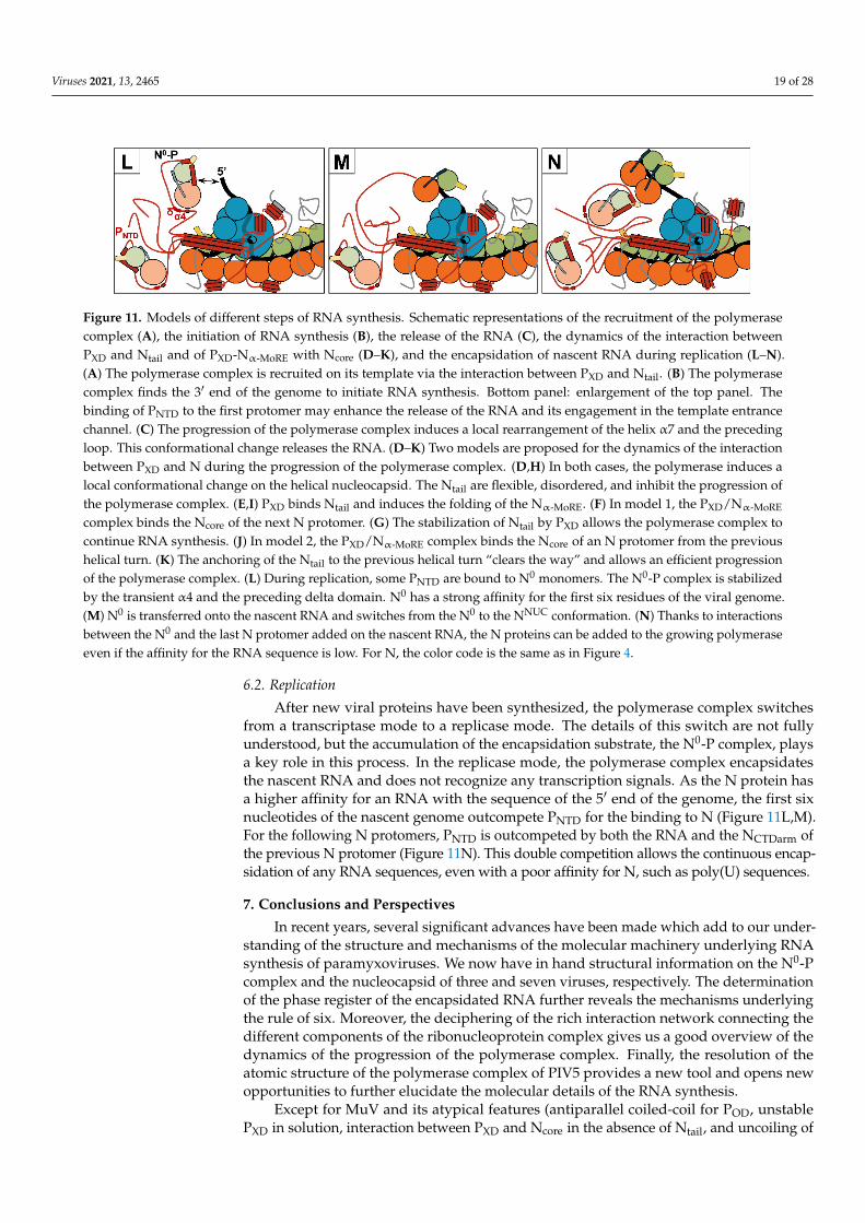
Viruses 2021, 13, 2465 19 of 28
Viruses 2021, 13, x FOR PEER REVIEW 18 of 29
Figure 11. Models of different steps of RNA synthesis. Schematic representations of the recruitment of the polymerase complex (A), the initiation of RNA synthesis (B), the release of the RNA (C), the dynamics of the interaction between PXD and Ntail and of PXD-Nα-MoRE with Ncore (D–K), and the encapsidation of nascent RNA during replication (L–N). (A) The polymerase complex is recruited on its template via the interaction between PXD and Ntail. (B) The polymerase complex finds the 3′ end of the genome to initiate RNA synthesis. Bottom panel: enlargement of the top panel. The binding of PNTD to the first protomer may enhance the release of the RNA and its engagement in the template entrance channel. (C) The progression of the polymerase complex induces a local rearrangement of the helix α7 and the preceding loop. This con-formational change releases the RNA. (D–K) Two models are proposed for the dynamics of the interaction between PXD
Figure 11. Models of different steps of RNA synthesis. Schematic representations of the recruitment of the polymerasecomplex (A), the initiation of RNA synthesis (B), the release of the RNA (C), the dynamics of the interaction betweenPXD and Ntail and of PXD-Nα-MoRE with Ncore (D–K), and the encapsidation of nascent RNA during replication (L–N).(A) The polymerase complex is recruited on its template via the interaction between PXD and Ntail. (B) The polymerasecomplex finds the 3′ end of the genome to initiate RNA synthesis. Bottom panel: enlargement of the top panel. Thebinding of PNTD to the first protomer may enhance the release of the RNA and its engagement in the template entrancechannel. (C) The progression of the polymerase complex induces a local rearrangement of the helix α7 and the precedingloop. This conformational change releases the RNA. (D–K) Two models are proposed for the dynamics of the interactionbetween PXD and N during the progression of the polymerase complex. (D,H) In both cases, the polymerase induces alocal conformational change on the helical nucleocapsid. The Ntail are flexible, disordered, and inhibit the progression ofthe polymerase complex. (E,I) PXD binds Ntail and induces the folding of the Nα-MoRE. (F) In model 1, the PXD/Nα-MoRE
complex binds the Ncore of the next N protomer. (G) The stabilization of Ntail by PXD allows the polymerase complex tocontinue RNA synthesis. (J) In model 2, the PXD/Nα-MoRE complex binds the Ncore of an N protomer from the previoushelical turn. (K) The anchoring of the Ntail to the previous helical turn “clears the way” and allows an efficient progressionof the polymerase complex. (L) During replication, some PNTD are bound to N0 monomers. The N0-P complex is stabilizedby the transient α4 and the preceding delta domain. N0 has a strong affinity for the first six residues of the viral genome.(M) N0 is transferred onto the nascent RNA and switches from the N0 to the NNUC conformation. (N) Thanks to interactionsbetween the N0 and the last N protomer added on the nascent RNA, the N proteins can be added to the growing polymeraseeven if the affinity for the RNA sequence is low. For N, the color code is the same as in Figure 4.
6.2. Replication
After new viral proteins have been synthesized, the polymerase complex switchesfrom a transcriptase mode to a replicase mode. The details of this switch are not fullyunderstood, but the accumulation of the encapsidation substrate, the N0-P complex, playsa key role in this process. In the replicase mode, the polymerase complex encapsidatesthe nascent RNA and does not recognize any transcription signals. As the N protein hasa higher affinity for an RNA with the sequence of the 5′ end of the genome, the first sixnucleotides of the nascent genome outcompete PNTD for the binding to N (Figure 11L,M).For the following N protomers, PNTD is outcompeted by both the RNA and the NCTDarm ofthe previous N protomer (Figure 11N). This double competition allows the continuous encap-sidation of any RNA sequences, even with a poor affinity for N, such as poly(U) sequences.
7. Conclusions and Perspectives
In recent years, several significant advances have been made which add to our under-standing of the structure and mechanisms of the molecular machinery underlying RNAsynthesis of paramyxoviruses. We now have in hand structural information on the N0-Pcomplex and the nucleocapsid of three and seven viruses, respectively. The determinationof the phase register of the encapsidated RNA further reveals the mechanisms underlyingthe rule of six. Moreover, the deciphering of the rich interaction network connecting thedifferent components of the ribonucleoprotein complex gives us a good overview of thedynamics of the progression of the polymerase complex. Finally, the resolution of theatomic structure of the polymerase complex of PIV5 provides a new tool and opens newopportunities to further elucidate the molecular details of the RNA synthesis.
Except for MuV and its atypical features (antiparallel coiled-coil for POD, unstablePXD in solution, interaction between PXD and Ncore in the absence of Ntail, and uncoiling of

Viruses 2021, 13, 2465 20 of 28
the nucleocapsid by PNTD), the molecular machinery and the mechanisms at play duringRNA synthesis appear to be well conserved among paramyxoviruses. The exposure of theRNA on the outside of the helical nucleocapsid, the binding site of the C-terminal domainof P to the NNTD, the coiled-coil structure of POD, and the interaction sites between P and Lsuggest these mechanisms are shared with pneumoviruses and filoviruses but are quitedifferent for rhabdoviruses.
Despite these new insights, several questions remain unanswered. The helical nucleo-capsid must uncoil to allow the progression of the polymerase. What factors induce thisconformational change? It is also still unclear how the nucleoproteins release the RNAand what signals trigger this release. Although structural information is available on theindividual components, there is little insight into the organization or the structure of thepolymerase complex on the nucleocapsid while synthesizing RNA. In addition, how doesthe second element of the bipartite promoter enhance initiation? How does the polymerasecomplex add nucleoproteins to the nascent RNA? How does the phosphoprotein orches-trate its interactions with Ntail, Ncore, and L? How do other viral proteins such as the V, C,and M proteins interact with the ribonucleoprotein complex and regulate RNA synthesis?
Answering these questions should bring significant insight into the mechanisms atplay and assist the development of safe and potent vaccine vectors, oncolytic viruses,and antiviral therapeutics. With the momentum provided by the recent advances andthe constant improvement of powerful tools such as cryo-electron microscopy, there isno doubt our understanding of the viral molecular machinery of paramyxoviruses willcontinue to rapidly improve.
Funding: This work was supported by NIH grant AI059371 to Sean P.J. Whelan.
Acknowledgments: I thank Denis Gerlier and Sonia Longhi for their helpful comments and sug-gestions, Paul W. Rothlauf for the English proofreading of the manuscript, Sean P.J. Whelan for hissupport, and Soazig Friguel for her help in assembling the figures.
Conflicts of Interest: The author declares no conflict of interest.
References1. Rima, B.; Balkema-Buschmann, A.; Dundon, W.G.; Duprex, P.; Easton, A.; Fouchier, R.; Kurath, G.; Lamb, R.; Lee, B.; Rota, P.; et al.
ICTV Virus Taxonomy Profile: Paramyxoviridae. J. Gen. Virol. 2019, 100, 1593–1594. [CrossRef] [PubMed]2. Cox, R.; Plemper, R.K. The paramyxovirus polymerase complex as a target for next-generation anti-paramyxovirus therapeutics.
Front. Microbiol. 2015, 6, 459. [CrossRef] [PubMed]3. Aggarwal, M.; Plemper, R.K. Structural Insight into Paramyxovirus and Pneumovirus Entry Inhibition. Viruses 2020, 12, 342.
[CrossRef] [PubMed]4. Emerson, S.U.; Wagner, R.R. Dissociation and reconstitution of the transcriptase and template activities of vesicular stomatitis B
and T virions. J. Virol. 1972, 10, 297–309. [CrossRef] [PubMed]5. Emerson, S.U.; Yu, Y. Both NS and L proteins are required for in vitro RNA synthesis by vesicular stomatitis virus. J. Virol. 1975,
15, 1348–1356. [CrossRef]6. Hamaguchi, M.; Yoshida, T.; Nishikawa, K.; Naruse, H.; Nagai, Y. Transcriptive complex of Newcastle disease virus. I. Both L and
P proteins are required to constitute an active complex. Virology 1983, 128, 105–117. [CrossRef]7. Horikami, S.M.; Curran, J.; Kolakofsky, D.; Moyer, S.A. Complexes of Sendai virus NP-P and P-L proteins are required for
defective interfering particle genome replication in vitro. J. Virol. 1992, 66, 4901–4908. [CrossRef]8. Stone, H.O.; Kingsbury, D.W.; Darlington, R.W. Sendai virus-induced transcriptase from infected cells: Polypeptides in the
transcriptive complex. J. Virol. 1972, 10, 1037–1043. [CrossRef]9. Colonno, R.J.; Stone, H.O. Isolation of a transcriptive complex from Newcastle disease virions. J. Virol. 1976, 19, 1080–1089.
[CrossRef]10. Portner, A.; Murti, K.G.; Morgan, E.M.; Kingsbury, D.W. Antibodies against Sendai virus L protein: Distribution of the protein in
nucleocapsids revealed by immunoelectron microscopy. Virology 1988, 163, 236–239. [CrossRef]11. Mura, M.; Combredet, C.; Najburg, V.; David, R.Y.S.; Tangy, F.; Komarova, A.V. Nonencapsidated 5′ Copy-Back Defective
Interfering Genomes Produced by Recombinant Measles Viruses Are Recognized by RIG-I and LGP2 but Not MDA5. J. Virol.2017, 91. [CrossRef] [PubMed]
12. Nilsson-Payant, B.E.; Blanco-Melo, D.; Uhl, S.; Escudero-Pérez, B.; Olschewski, S.; Thibault, P.; Panis, M.; Rosenthal, M.; Muñoz-Fontela, C.; Lee, B.; et al. Reduced Nucleoprotein Availability Impairs Negative-Sense RNA Virus Replication and Promotes HostRecognition. J. Virol. 2021, 95, e02274-20. [CrossRef] [PubMed]

Viruses 2021, 13, 2465 21 of 28
13. Compans, R.W.; Choppin, P.W. The nucleic acid of the parainfluenza virus SV5. Virology 1968, 35, 289–296. [CrossRef]14. Lynch, S.; Kolakofsky, D. Ends of the RNA within Sendai virus defective interfering nucleocapsids are not free. J. Virol. 1978, 28,
584–589. [CrossRef]15. Bitko, V.; Barik, S. Phenotypic silencing of cytoplasmic genes using sequence-specific double-stranded short interfering RNA and
its application in the reverse genetics of wild type negative-strand RNA viruses. BMC Microbiol. 2001, 1, 34. [CrossRef] [PubMed]16. Mottet-Osman, G.; Iseni, F.; Pelet, T.; Wiznerowicz, M.; Garcin, D.; Roux, L. Suppression of the Sendai Virus M Protein through a
Novel Short Interfering RNA Approach Inhibits Viral Particle Production but Does Not Affect Viral RNA Synthesis. J. Virol. 2007,81, 2861–2868. [CrossRef]
17. Alayyoubi, M.; Leser, G.P.; Kors, C.A.; Lamb, R.A. Structure of the paramyxovirus parainfluenza virus 5 nucleoprotein-RNAcomplex. Proc. Natl. Acad. Sci. USA 2015, 112, E1792–E1799. [CrossRef]
18. Gutsche, I.; Desfosses, A.; Effantin, G.; Ling, W.L.; Haupt, M.; Ruigrok, R.W.H.; Sachse, C.; Schoehn, G. Near-atomic cryo-EMstructure of the helical measles virus nucleocapsid. Science 2015, 348, 704–707. [CrossRef]
19. Song, X.; Shan, H.; Zhu, Y.; Hu, S.; Xue, L.; Chen, Y.; Ding, W.; Niu, T.; Gu, J.; Ouyang, S.; et al. Self-capping of nucleoproteinfilaments protects the Newcastle disease virus genome. eLife 2019, 8. [CrossRef]
20. Desfosses, A.; Milles, S.; Jensen, M.R.; Guseva, S.; Colletier, J.-P.; Maurin, D.; Schoehn, G.; Gutsche, I.; Ruigrok, R.W.H.;Blackledge, M. Assembly and cryo-EM structures of RNA-specific measles virus nucleocapsids provide mechanistic insight intoparamyxoviral replication. Proc. Natl. Acad. Sci. USA 2019, 116, 4256–4264. [CrossRef] [PubMed]
21. Zhang, N.; Shan, H.; Liu, M.; Li, T.; Luo, R.; Yang, L.; Qi, L.; Chu, X.; Su, X.; Wang, R.; et al. Structure and assembly ofdouble-headed Sendai virus nucleocapsids. Commun. Biol. 2021, 4, 494. [CrossRef]
22. Ker, D.-S.; Jenkins, H.T.; Greive, S.J.; Antson, A.A. CryoEM structure of the Nipah virus nucleocapsid assembly. PLoS Pathog.2021, 17, e1009740. [CrossRef]
23. Zinzula, L.; Beck, F.; Klumpe, S.; Bohn, S.; Pfeifer, G.; Bollschweiler, D.; Nagy, I.; Plitzko, J.M.; Baumeister, W. Cryo-EM structureof the cetacean morbillivirus nucleoprotein-RNA complex. J. Struct. Biol. 2021, 213, 107750. [CrossRef] [PubMed]
24. Yabukarski, F.; Lawrence, P.; Tarbouriech, N.; Bourhis, J.-M.; Delaforge, E.; Jensen, M.R.; Ruigrok, R.W.H.; Blackledge, M.;Volchkov, V.; Jamin, M. Structure of Nipah virus unassembled nucleoprotein in complex with its viral chaperone. Nat. Struct. Mol.Biol. 2014, 21, 754–759. [CrossRef]
25. Guryanov, S.G.; Liljeroos, L.; Kasaragod, P.; Kajander, T.; Butcher, S.J. Crystal Structure of the Measles Virus Nucleoprotein Corein Complex with an N-Terminal Region of Phosphoprotein. J. Virol. 2016, 90, 2849–2857. [CrossRef]
26. Aggarwal, M.; Leser, G.P.; Kors, C.A.; Lamb, R.A. Structure of the Paramyxovirus Parainfluenza Virus 5 Nucleoprotein inComplex with an Amino-Terminal Peptide of the Phosphoprotein. J. Virol. 2018, 92, e01304-17. [CrossRef] [PubMed]
27. Dong, X.; Wang, X.; Xie, M.; Wu, W.; Chen, Z. Structural Basis of Human Parainfluenza Virus 3 Unassembled Nucleoprotein inComplex with Its Viral ChaperoneHPIV3 Nucleoprotein-Phosphoprotein Structure. J. Virol. 2021, JVI0164821. [CrossRef]
28. Abdella, R.; Aggarwal, M.; Okura, T.; Lamb, R.A.; He, Y. Structure of a paramyxovirus polymerase complex reveals a uniquemethyltransferase-CTD conformation. Proc. Natl. Acad. Sci. USA 2020, 117, 4931–4941. [CrossRef]
29. Glazier, K.; Raghow, R.; Kingsbury, D.W. Regulation of Sendai virus transcription: Evidence for a single promoter in vivo. J. Virol.1977, 21, 863–871. [CrossRef]
30. Le Mercier, P.; Kolakofsky, D. Bipartite promoters and RNA editing of paramyxoviruses and filoviruses. RNA 2019, 25, 279–285.[CrossRef] [PubMed]
31. Plemper, R.K.; Lamb, R.A. Paramyxoviridae: The viruses and their replication. In Fields Virology: Emerging Viruses, 7th ed.;Howley, P.M., Knipe, D.M., Eds.; Lippincott Williams and Wilkins, Wolters Kluwer: Philadelphia, PA, USA, 2020; pp. 504–558.
32. Calain, P.; Roux, L. The rule of six, a basic feature for efficient replication of Sendai virus defective interfering RNA. J. Virol. 1993,67, 4822–4830. [CrossRef] [PubMed]
33. Kolakofsky, D.; Pelet, T.; Garcin, D.; Hausmann, S.; Curran, J.; Roux, L. Paramyxovirus RNA synthesis and the requirement forhexamer genome length: The rule of six revisited. J. Virol. 1998, 72, 891–899. [CrossRef]
34. Longhi, S.; Receveur-Brechot, V.; Karlin, D.; Johansson, K.; Darbon, H.; Bhella, D.; Yeo, R.; Finet, S.; Canard, B. The C-terminalDomain of the Measles Virus Nucleoprotein Is Intrinsically Disordered and Folds upon Binding to the C-terminal Moiety of thePhosphoprotein. J. Biol. Chem. 2003, 278, 18638–18648. [CrossRef] [PubMed]
35. Mountcastle, W.E.; Compans, R.W.; Lackland, H.; Choppin, P.W. Proteolytic cleavage of subunits of the nucleocapsid of theparamyxovirus simian virus 5. J. Virol. 1974, 14, 1253–1261. [CrossRef] [PubMed]
36. Buchholz, C.J.; Spehner, D.; Drillien, R.; Neubert, W.J.; Homann, H.E. The conserved N-terminal region of Sendai virusnucleocapsid protein NP is required for nucleocapsid assembly. J. Virol. 1993, 67, 5803–5812. [CrossRef]
37. Curran, J.; Homann, H.; Buchholz, C.; Rochat, S.; Neubert, W.; Kolakofsky, D. The hypervariable C-terminal tail of the Sendaiparamyxovirus nucleocapsid protein is required for template function but not for RNA encapsidation. J. Virol. 1993, 67, 4358–4364.[CrossRef] [PubMed]
38. Bankamp, B.; Horikami, S.M.; Thompson, P.; Huber, M.; Billeter, M.; Moyer, S.A. Domains of the Measles Virus N ProteinRequired for Binding to P Protein and Self-Assembly. Virology 1996, 216, 272–277. [CrossRef] [PubMed]
39. Buchholz, C.J.; Retzler, C.; Homann, H.E.; Neubert, W.J. The Carboxy-terminal Domain of Sendai Virus Nucleocapsid Protein IsInvolved in Complex Formation between Phosphoprotein and Nucleocapsid-like Particles. Virology 1994, 204, 770–776. [CrossRef][PubMed]

Viruses 2021, 13, 2465 22 of 28
40. Liston, P.; Batal, R.; Diflumeri, C.; Briedis, D.J. Protein interaction domains of the measles virus nucleocapsid protein (NP). Arch.Virol. 1997, 142, 305–321. [CrossRef]
41. Rahmeh, A.A.; Schenk, A.; Danek, E.I.; Kranzusch, P.J.; Liang, B.; Walz, T.; Whelan, S.P.J. Molecular architecture of the vesicularstomatitis virus RNA polymerase. Proc. Natl. Acad. Sci. USA 2010, 107, 20075–20080. [CrossRef]
42. Liang, B. Structures of the Mononegavirales Polymerases. J. Virol. 2020, 94, e00175-20. [CrossRef]43. Pyle, J.D.; Whelan, S.P.; Bloyet, L.-M. Structure and function of negative-strand RNA virus polymerase complexes. Enzymes 2021,
50, 21–78. [CrossRef]44. Abraham, G.; Rhodes, D.P.; Banerjee, A.K. Novel initiation of RNA synthesis in vitro by vesicular stomatitis virus. Nature 1975,
255, 37–40. [CrossRef] [PubMed]45. Ogino, T.; Banerjee, A.K. Unconventional Mechanism of mRNA Capping by the RNA-Dependent RNA Polymerase of Vesicular
Stomatitis Virus. Mol. Cell 2007, 25, 85–97. [CrossRef]46. Hercyk, N.; Horikami, S.M.; Moyer, S.A. The vesicular stomatitis virus L protein possesses the mRNA methyltransferase activities.
Virology 1988, 163, 222–225. [CrossRef]47. Ogino, T.; Kobayashi, M.; Iwama, M.; Mizumoto, K. Sendai Virus RNA-dependent RNA Polymerase L Protein Catalyzes Cap
Methylation of Virus-specific mRNA. J. Biol. Chem. 2005, 280, 4429–4435. [CrossRef]48. Hunt, D.M.; Mehta, R.; Hutchinson, K.L. The L Protein of Vesicular Stomatitis Virus Modulates the Response of the Polyadenylic
Acid Polymerase to S-Adenosylhomocysteine. J. Gen. Virol. 1988, 69, 2555–2561. [CrossRef]49. Jordan, P.C.; Liu, C.; Raynaud, P.; Lo, M.; Spiropoulou, C.F.; Symons, J.A.; Beigelman, L.; Deval, J. Initiation, extension, and
termination of RNA synthesis by a paramyxovirus polymerase. PLoS Pathog. 2018, 14, e1006889. [CrossRef] [PubMed]50. Smallwood, S.; Ryan, K.W.; Moyer, S.A. Deletion Analysis Defines a Carboxyl-Proximal Region of Sendai Virus P Protein That
Binds to the Polymerase L Protein. Virology 1994, 202, 154–163. [CrossRef] [PubMed]51. Canter, D.; Perrault, J. Stabilization of Vesicular Stomatitis Virus L Polymerase Protein by P Protein Binding: A Small Deletion in
the C-Terminal Domain of L Abrogates Binding. Virology 1996, 219, 376–386. [CrossRef] [PubMed]52. Bloyet, L.-M.; Welsch, J.; Enchery, F.; Mathieu, C.; de Breyne, S.; Horvat, B.; Grigorov, B.; Gerlier, D. HSP90 Chaperoning in
Addition to Phosphoprotein Required for Folding but Not for Supporting Enzymatic Activities of Measles and Nipah Virus LPolymerases. J. Virol. 2016, 90, 6642–6656. [CrossRef]
53. Mellon, M.G.; Emerson, S.U. Rebinding of transcriptase components (L and NS proteins) to the nucleocapsid template of vesicularstomatitis virus. J. Virol. 1978, 27, 560–567. [CrossRef] [PubMed]
54. Masters, P.S.; Banerjee, A.K. Complex formation with vesicular stomatitis virus phosphoprotein NS prevents binding of nucleo-capsid protein N to nonspecific RNA. J. Virol. 1988, 62, 2658–2664. [CrossRef] [PubMed]
55. Curran, J.; Marq, J.B.; Kolakofsky, D. An N-terminal domain of the Sendai paramyxovirus P protein acts as a chaperone for theNP protein during the nascent chain assembly step of genome replication. J. Virol. 1995, 69, 849–855. [CrossRef]
56. Spehner, D.; Drillien, R.; Howley, P.M. The Assembly of the Measles Virus Nucleoprotein into Nucleocapsid-like Particles IsModulated by the Phosphoprotein. Virology 1997, 232, 260–268. [CrossRef] [PubMed]
57. Karlin, D.; Ferron, F.; Canard, B.; Longhi, S. Structural disorder and modular organization in Paramyxovirinae N and P. J. Gen.Virol. 2003, 84, 3239–3252. [CrossRef]
58. Gerard, F.C.; Ribeiro, E.D.A.; Leyrat, C.; Ivanov, I.; Blondel, D.; Longhi, S.; Ruigrok, R.W.; Jamin, M. Modular Organization ofRabies Virus Phosphoprotein. J. Mol. Biol. 2009, 388, 978–996. [CrossRef] [PubMed]
59. Habchi, J.; Mamelli, L.; Darbon, H.; Longhi, S. Structural Disorder within Henipavirus Nucleoprotein and Phosphoprotein: FromPredictions to Experimental Assessment. PLoS ONE 2010, 5, e11684. [CrossRef]
60. Pereira, N.; Cardone, C.; Lassoued, S.; Galloux, M.; Fix, J.; Assrir, N.; Lescop, E.; Bontems, F.; Eléouët, J.-F.; Sizun, C. New Insightsinto Structural Disorder in Human Respiratory Syncytial Virus Phosphoprotein and Implications for Binding of Protein Partners.J. Biol. Chem. 2017, 292, 2120–2131. [CrossRef] [PubMed]
61. Longhi, S.; Bloyet, L.-M.; Gianni, S.; Gerlier, D. How order and disorder within paramyxoviral nucleoproteins and phosphopro-teins orchestrate the molecular interplay of transcription and replication. Cell. Mol. Life Sci. 2017, 74, 3091–3118. [CrossRef]
62. Jensen, M.R.; Yabukarski, F.; Communie, G.; Condamine, E.; Mas, C.; Volchkova, V.; Tarbouriech, N.; Bourhis, J.-M.; Volchkov, V.;Blackledge, M.; et al. Structural Description of the Nipah Virus Phosphoprotein and Its Interaction with STAT1. Biophys. J. 2020,118, 2470–2488. [CrossRef]
63. Tarbouriech, N.; Curran, J.; Ruigrok, R.W.; Burmeister, W.P. Tetrameric coiled coil domain of Sendai virus phosphoprotein. Nat.Genet. 2000, 7, 777–781. [CrossRef]
64. Communie, G.; Crepin, T.; Maurin, D.; Jensen, M.R.; Blackledge, M.; Ruigrok, R.W.H. Structure of the Tetramerization Domain ofMeasles Virus Phosphoprotein. J. Virol. 2013, 87, 7166–7169. [CrossRef] [PubMed]
65. Blocquel, D.; Habchi, J.; Durand, E.; Sevajol, M.; Ferron, F.; Erales, J.; Papageorgiou, N.; Longhi, S. Coiled-coil deformations incrystal structures: The measles virus phosphoprotein multimerization domain as an illustrative example. Acta Crystallogr. D Biol.Crystallogr. 2014, 70, 1589–1603. [CrossRef] [PubMed]
66. Bruhn, J.F.; Barnett, K.; Bibby, J.; Thomas, J.M.H.; Keegan, R.; Rigden, D.J.; Bornholdt, Z.A.; Saphire, E.O. Crystal Structure of theNipah Virus Phosphoprotein Tetramerization Domain. J. Virol. 2013, 88, 758–762. [CrossRef]
Viruses 2021, 13, 2465 23 of 28
67. Galvan, J.R.; Donner, B.; Veseley, C.H.; Reardon, P.; Forsythe, H.M.; Howe, J.; Fujimura, G.; Barbar, E. Human Parainfluenza Virus3 Phosphoprotein Is a Tetramer and Shares Structural and Interaction Features with Ebola Phosphoprotein VP35. Biomolecules2021, 11, 1603. [CrossRef] [PubMed]
68. Cox, R.; Green, T.J.; Purushotham, S.; Deivanayagam, C.; Bedwell, G.J.; Prevelige, P.E.; Luo, M. Structural and FunctionalCharacterization of the Mumps Virus Phosphoprotein. J. Virol. 2013, 87, 7558–7568. [CrossRef] [PubMed]
69. Pickar, A.; Elson, A.; Yang, Y.; Xu, P.; Luo, M.; He, B. Oligomerization of Mumps Virus Phosphoprotein. J. Virol. 2015, 89,11002–11010. [CrossRef]
70. Ryan, K.W.; Kingsbury, D.W. Carboxyl-terminal region of sendai virus P protein is required for binding to viral nucleocapsids.Virology 1988, 167, 106–112. [CrossRef]
71. Johansson, K.; Bourhis, J.-M.; Campanacci, V.; Cambillau, C.; Canard, B.; Longhi, S. Crystal Structure of the Measles VirusPhosphoprotein Domain Responsible for the Induced Folding of the C-terminal Domain of the Nucleoprotein. J. Biol. Chem. 2003,278, 44567–44573. [CrossRef] [PubMed]
72. Blanchard, L.; Tarbouriech, N.; Blackledge, M.; Timmins, P.; Burmeister, W.P.; Ruigrok, R.W.; Marion, D. Structure and dynamicsof the nucleocapsid-binding domain of the Sendai virus phosphoprotein in solution. Virology 2004, 319, 201–211. [CrossRef]
73. Kingston, R.L.; Hamel, D.J.; Gay, L.S.; Dahlquist, F.W.; Matthews, B.W. Structural basis for the attachment of a paramyxoviralpolymerase to its template. Proc. Natl. Acad. Sci. USA 2004, 101, 8301–8306. [CrossRef]
74. Habchi, J.; Blangy, S.; Mamelli, L.; Jensen, M.R.; Blackledge, M.; Darbon, H.; Oglesbee, M.; Shu, Y.; Longhi, S. Characterizationof the Interactions between the Nucleoprotein and the Phosphoprotein of Henipavirus. J. Biol. Chem. 2011, 286, 13583–13602.[CrossRef] [PubMed]
75. Communie, G.; Habchi, J.; Yabukarski, F.; Blocquel, D.; Schneider, R.; Tarbouriech, N.; Papageorgiou, N.; Ruigrok, R.W.H.; Jamin,M.; Jensen, M.R.; et al. Atomic Resolution Description of the Interaction between the Nucleoprotein and Phosphoprotein ofHendra Virus. PLoS Pathog. 2013, 9, e1003631. [CrossRef] [PubMed]
76. Blumberg, B.M.; Leppert, M.; Kolakofsky, D. Interaction of VSV leader RNA and nucleocapsid protein may control VSV genomereplication. Cell 1981, 23, 837–845. [CrossRef]
77. Plumet, S.; Duprex, W.P.; Gerlier, D. Dynamics of Viral RNA Synthesis during Measles Virus Infection. J. Virol. 2005, 79, 6900–6908.[CrossRef]
78. Ranadheera, C.; Proulx, R.; Chaiyakul, M.; Jones, S.; Grolla, A.; Leung, A.; Rutherford, J.; Kobasa, D.; Carpenter, M.; Czub, M. Theinteraction between the Nipah virus nucleocapsid protein and phosphoprotein regulates virus replication. Sci. Rep. 2018, 8, 15994.[CrossRef] [PubMed]
79. Gubbay, O.; Curran, J.; Kolakofsky, D. Sendai virus genome synthesis and assembly are coupled: A possible mechanism topromote viral RNA polymerase processivity. J. Gen. Virol. 2001, 82, 2895–2903. [CrossRef] [PubMed]
80. Guseva, S.; Milles, S.; Jensen, M.R.; Schoehn, G.; Ruigrok, R.W.; Blackledge, M. Structure, dynamics and phase separation ofmeasles virus RNA replication machinery. Curr. Opin. Virol. 2020, 41, 59–67. [CrossRef]
81. Nikolic, J.; Lagaudrière-Gesbert, C.; Scrima, N.; Blondel, D.; Gaudin, Y. Structure and Function of Negri Bodies. Adv. Exp. Med.Biol. 2019, 1215, 111–127. [CrossRef] [PubMed]
82. Brocca, S.; Grandori, R.; Longhi, S.; Uversky, V. Liquid-Liquid Phase Separation by Intrinsically Disordered Protein Regions ofViruses: Roles in Viral Life Cycle and Control of Virus-Host Interactions. Int. J. Mol. Sci. 2020, 21, 9045. [CrossRef] [PubMed]
83. Hagiwara, K.; Sato, H.; Inoue, Y.; Watanabe, A.; Yoneda, M.; Ikeda, F.; Fujita, K.; Fukuda, H.; Takamura, C.; Kozuka-Hata, H.; et al.Phosphorylation of measles virus nucleoprotein upregulates the transcriptional activity of minigenomic RNA. Proteomics 2008, 8,1871–1879. [CrossRef] [PubMed]
84. Huang, M.; Sato, H.; Hagiwara, K.; Watanabe, A.; Sugai, A.; Ikeda, F.; Kozuka-Hata, H.; Oyama, M.; Yoneda, M.; Kai, C.Determination of a phosphorylation site in Nipah virus nucleoprotein and its involvement in virus transcription. J. Gen. Virol.2011, 92, 2133–2141. [CrossRef] [PubMed]
85. Zengel, J.; Pickar, A.; Xu, P.; Lin, A.; He, B. Roles of Phosphorylation of the Nucleocapsid Protein of Mumps Virus in RegulatingViral RNA Transcription and Replication. J. Virol. 2015, 89, 7338–7347. [CrossRef]
86. Vidal, S.; Curran, J.; Orvell, C.; Kolakofsky, D. Mapping of monoclonal antibodies to the Sendai virus P protein and the location ofits phosphates. J. Virol. 1988, 62, 2200–2203. [CrossRef]
87. Das, T.; Schuster, A.; Schneider-Schaulies, S.; Banerjee, A.K. Involvement of Cellular Casein Kinase II in the Phosphorylation ofMeasles Virus P Protein: Identification of Phosphorylation Sites. Virology 1995, 211, 218–226. [CrossRef] [PubMed]
88. Huntley, C.C.; De, B.P.; Murray, N.R.; Fields, A.P.; Banerjee, A.K. Human parainfluenza virus type 3 phosphoprotein: Identificationof serine 333 as the major site for PKC zeta phosphorylation. Virology 1995, 211, 561–567. [CrossRef]
89. Byrappa, S.; Pan, Y.-B.; Gupta, K.C. Sendai Virus P Protein Is Constitutively Phosphorylated at Serine249: High PhosphorylationPotential of the P Protein. Virology 1996, 216, 228–234. [CrossRef]
90. Timani, K.A.; Sun, D.; Sun, M.; Keim, C.; Lin, Y.; Schmitt, P.T.; Schmitt, A.; He, B. A Single Amino Acid Residue Change in the PProtein of Parainfluenza Virus 5 Elevates Viral Gene Expression. J. Virol. 2008, 82, 9123–9133. [CrossRef]
91. Sun, D.; Luthra, P.; Xu, P.; Yoon, H.; He, B. Identification of a Phosphorylation Site within the P Protein Important for mRNATranscription and Growth of Parainfluenza Virus 5. J. Virol. 2011, 85, 8376–8385. [CrossRef]
92. Sugai, A.; Sato, H.; Yoneda, M.; Kai, C. Phosphorylation of measles virus phosphoprotein at S86 and/or S151 downregulates viraltranscriptional activity. FEBS Lett. 2012, 586, 3900–3907. [CrossRef] [PubMed]

Viruses 2021, 13, 2465 24 of 28
93. Pickar, A.; Xu, P.; Elson, A.; Li, Z.; Zengel, J.; He, B. Roles of Serine and Threonine Residues of Mumps Virus P Protein in ViralTranscription and Replication. J. Virol. 2014, 88, 4414–4422. [CrossRef]
94. Qiu, X.; Zhan, Y.; Meng, C.; Wang, J.; Dong, L.; Sun, Y.; Tan, L.; Song, C.; Yu, S.; Ding, C. Identification and functional analysis ofphosphorylation in Newcastle disease virus phosphoprotein. Arch. Virol. 2016, 161, 2103–2116. [CrossRef]
95. De, B.P.; Gupta, S.; Banerjee, A.K. Cellular protein kinase C isoform zeta regulates human parainfluenza virus type 3 replication.Proc. Natl. Acad. Sci. USA 1995, 92, 5204–5208. [CrossRef]
96. Liu, Z.; Huntley, C.C.; De, B.P.; Das, T.; Banerjee, A.K.; Oglesbee, M.J. Phosphorylation of canine distemper virus P protein byprotein kinase C-zeta and casein kinase II. Virology 1997, 232, 198–206. [CrossRef]
97. Huntley, C.C.; De, B.P.; Banerjee, A.K. Phosphorylation of Sendai virus phosphoprotein by cellular protein kinase C zeta. J. Biol.Chem. 1997, 272, 16578–16584. [CrossRef]
98. Kaushik, R.; Shaila, M.S. Cellular casein kinase II-mediated phosphorylation of rinderpest virus P protein is a prerequisite for itsrole in replication/transcription of the genome. J. Gen. Virol. 2004, 85, 687–691. [CrossRef] [PubMed]
99. Sun, M.; Fuentes, S.M.; Timani, K.; Sun, D.; Murphy, C.; Lin, Y.; August, A.; Teng, M.N.; He, B. Akt Plays a Critical Role inReplication of Nonsegmented Negative-Stranded RNA Viruses. J. Virol. 2008, 82, 105–114. [CrossRef]
100. Sun, D.; Luthra, P.; Li, Z.; He, B. PLK1 Down-Regulates Parainfluenza Virus 5 Gene Expression. PLoS Pathog. 2009, 5, e1000525.[CrossRef]
101. Sugai, A.; Sato, H.; Yoneda, M.; Kai, C. PIM 3 kinase, a proto-oncogene product, regulates phosphorylation of the measles virusnucleoprotein tail domain at Ser 479 and Ser 510. Biochem. Biophys. Res. Commun. 2020, 531. [CrossRef]
102. Young, D.F.; Wignall-Fleming, E.B.; Busse, D.C.; Pickin, M.J.; Hankinson, J.; Randall, E.M.; Tavendale, A.; Davison, A.J.; Lamont,D.; Tregoning, J.S.; et al. The switch between acute and persistent paramyxovirus infection caused by single amino acidsubstitutions in the RNA polymerase P subunit. PLoS Pathog. 2019, 15, e1007561. [CrossRef] [PubMed]
103. Saikia, P.; Gopinath, M.; Shaila, M.S. Phosphorylation status of the phosphoprotein P of rinderpest virus modulates transcriptionand replication of the genome. Arch. Virol. 2008, 153, 615–626. [CrossRef] [PubMed]
104. Sugai, A.; Sato, H.; Yoneda, M.; Kai, C. Phosphorylation of Measles Virus Nucleoprotein Affects Viral Growth by Changing GeneExpression and Genomic RNA Stability. J. Virol. 2013, 87, 11684–11692. [CrossRef] [PubMed]
105. Sugai, A.; Sato, H.; Hagiwara, K.; Kozuka-Hata, H.; Oyama, M.; Yoneda, M.; Kai, C. Newly Identified Minor PhosphorylationSite Threonine-279 of Measles Virus Nucleoprotein Is a Prerequisite for Nucleocapsid Formation. J. Virol. 2013, 88, 1140–1149.[CrossRef]
106. Zhou, Y.; Su, J.M.; Samuel, C.E.; Ma, D. Measles Virus Forms Inclusion Bodies with Properties of Liquid Organelles. J. Virol. 2019,93, e00948-19. [CrossRef]
107. Horne, R.W.; Waterson, A.P.; Wildy, P.; Farnham, A.E. The structure and composition of the myxoviruses. I. Electron microscopestudies of the structure of myxovirus particles by negative staining techniques. Virology 1960, 11, 79–98. [CrossRef]
108. Compans, R.; Choppin, P.W. Isolation and properties of the helical nucleocapsid of the parainfluenza virus SV5. Proc. Natl. Acad.Sci. USA 1967, 57, 949–956. [CrossRef]
109. Finch, J.T.; Gibbs, A.J. Observations on the Structure of the Nucleocapsids of some Paramyxoviruses. J. Gen. Virol. 1970, 6,141–150. [CrossRef]
110. Egelman, E.H.; Wu, S.S.; Amrein, M.; Portner, A.; Murti, G. The Sendai virus nucleocapsid exists in at least four different helicalstates. J. Virol. 1989, 63, 2233–2243. [CrossRef]
111. Heggeness, M.H.; Scheid, A.; Choppin, P.W. Conformation of the helical nucleocapsids of paramyxoviruses and vesicularstomatitis virus: Reversible coiling and uncoiling induced by changes in salt concentration. Proc. Natl. Acad. Sci. USA 1980, 77,2631–2635. [CrossRef]
112. Bhella, D.; Ralph, A.; Murphy, L.B.; Yeo, R.P. Significant differences in nucleocapsid morphology within the Paramyxoviridae. J.Gen. Virol. 2002, 83, 1831–1839. [CrossRef]
113. Bhella, D.; Ralph, A.; Yeo, R.P. Conformational Flexibility in Recombinant Measles Virus Nucleocapsids Visualised by Cryo-negative Stain Electron Microscopy and Real-space Helical Reconstruction. J. Mol. Biol. 2004, 340, 319–331. [CrossRef]
114. Cox, R.; Pickar, A.; Qiu, S.; Tsao, J.; Rodenburg, C.; Dokland, T.; Elson, A.; He, B.; Luo, M. Structural studies on the authenticmumps virus nucleocapsid showing uncoiling by the phosphoprotein. Proc. Natl. Acad. Sci. USA 2014, 111, 15208–15213.[CrossRef] [PubMed]
115. Ke, Z.; Strauss, J.D.; Hampton, C.M.; Brindley, M.A.; Dillard, R.S.; Leon, F.; Lamb, K.M.; Plemper, R.K.; Wright, E.R. Promotion ofvirus assembly and organization by the measles virus matrix protein. Nat. Commun. 2018, 9, 1736. [CrossRef]
116. Nakai, M.; Imagawa, D.T. Electron microscopy of measles virus replication. J. Virol. 1969, 3, 187–197. [CrossRef]117. Desfosses, A.; Goret, G.; Estrozi, L.F.; Ruigrok, R.W.H.; Gutsche, I. Nucleoprotein-RNA Orientation in the Measles Virus
Nucleocapsid by Three-Dimensional Electron Microscopy. J. Virol. 2011, 85, 1391–1395. [CrossRef] [PubMed]118. Spehner, D.; Kirn, A.; Drillien, R. Assembly of nucleocapsidlike structures in animal cells infected with a vaccinia virus
recombinant encoding the measles virus nucleoprotein. J. Virol. 1991, 65, 6296–6300. [CrossRef] [PubMed]119. Fooks, A.R.; Stephenson, J.R.; Warnes, A.; Dowsett, A.B.; Rima, B.K.; Wilkinson, G.W.G. Measles virus nucleocapsid protein
expressed in insect cells assembles into nucleocapsid-like structures. J. Gen. Virol. 1993, 74, 1439–1444. [CrossRef] [PubMed]120. Warnes, A.; Fooks, A.R.; Dowsett, A.; Wilkinson, G.W.; Stephenson, J.R. Expression of the measles virus nucleoprotein gene in
Escherichia coli and assembly of nucleocapsid-like structures. Gene 1995, 160, 173–178. [CrossRef]

Viruses 2021, 13, 2465 25 of 28
121. Schoehn, G.; Mavrakis, M.; Albertini, A.; Wade, R.; Hoenger, A.; Ruigrok, R.W. The 12 Å Structure of Trypsin-treated MeaslesVirus N-RNA. J. Mol. Biol. 2004, 339, 301–312. [CrossRef] [PubMed]
122. Cox, R.; Green, T.J.; Qiu, S.; Kang, J.; Tsao, J.; Prevelige, P.E.; He, B.; Luo, M. Characterization of a Mumps Virus NucleocapsidlikeParticle. J. Virol. 2009, 83, 11402–11406. [CrossRef] [PubMed]
123. Rager, M.; Vongpunsawad, S.; Duprex, W.P.; Cattaneo, R. Polyploid measles virus with hexameric genome length. EMBO J. 2002,21, 2364–2372. [CrossRef]
124. Loney, C.; Mottet-Osman, G.; Roux, L.; Bhella, D. Paramyxovirus Ultrastructure and Genome Packaging: Cryo-ElectronTomography of Sendai Virus. J. Virol. 2009, 83, 8191–8197. [CrossRef] [PubMed]
125. Goff, P.H.; Gao, Q.; Palese, P. A Majority of Infectious Newcastle Disease Virus Particles Contain a Single Genome, while aMinority Contain Multiple Genomes. J. Virol. 2012, 86, 10852–10856. [CrossRef]
126. Shan, H.; Su, X.; Li, T.; Qin, Y.; Zhang, N.; Yang, L.; Ma, L.; Bai, Y.; Qi, L.; Liu, Y.; et al. Structural plasticity of mumps virusnucleocapsids with cryo-EM structures. Commun. Biol. 2021, 4, 833. [CrossRef]
127. Baronti, L.; Erales, J.; Habchi, J.; Felli, I.C.; Pierattelli, R.; Longhi, S. Dynamics of the Intrinsically Disordered C-Terminal Domainof the Nipah Virus Nucleoprotein and Interaction with the X Domain of the Phosphoprotein as Unveiled by NMR Spectroscopy.ChemBioChem 2014, 16, 268–276. [CrossRef]
128. Houben, K.; Marion, D.; Tarbouriech, N.; Ruigrok, R.W.H.; Blanchard, L. Interaction of the C-Terminal Domains of Sendai VirusN and P Proteins: Comparison of Polymerase-Nucleocapsid Interactions within the Paramyxovirus Family. J. Virol. 2007, 81,6807–6816. [CrossRef] [PubMed]
129. Tawar, R.G.; Duquerroy, S.; Vonrhein, C.; Varela, P.F.; Damier-Piolle, L.; Castagné, N.; MacLellan, K.; Bedouelle, H.; Bricogne, G.;Bhella, D.; et al. Crystal Structure of a Nucleocapsid-Like Nucleoprotein-RNA Complex of Respiratory Syncytial Virus. Science2009, 326, 1279–1283. [CrossRef]
130. Bakker, S.E.; Duquerroy, S.; Galloux, M.; Loney, C.; Conner, E.; Eléouët, J.-F.; Rey, F.; Bhella, D. The respiratory syncytial virusnucleoprotein-RNA complex forms a left-handed helical nucleocapsid. J. Gen. Virol. 2013, 94, 1734–1738. [CrossRef]
131. Renner, M.; Bertinelli, M.; Leyrat, C.; Paesen, G.C.; De Oliveira, L.F.S.; Huiskonen, J.T.; Grimes, J.M. Nucleocapsid assembly inpneumoviruses is regulated by conformational switching of the N protein. eLife 2016, 5, e12627. [CrossRef]
132. Dong, S.; Yang, P.; Li, G.; Liu, B.; Wang, W.; Liu, X.; Xia, B.; Yang, C.; Lou, Z.; Guo, Y.; et al. Insight into the Ebola virusnucleocapsid assembly mechanism: Crystal structure of Ebola virus nucleoprotein core domain at 1.8 Å resolution. Protein Cell2015, 6, 351–362. [CrossRef]
133. Wan, W.; Kolesnikova, L.; Clarke, M.; Koehler, A.; Noda, T.; Becker, S.; Briggs, J.A.G. Structure and assembly of the Ebola virusnucleocapsid. Nature 2017, 551, 394–397. [CrossRef] [PubMed]
134. Sugita, Y.; Matsunami, H.; Kawaoka, Y.; Noda, T.; Wolf, M. Cryo-EM structure of the Ebola virus nucleoprotein-RNA complex at3.6 A resolution. Nature 2018, 563, 137–140. [CrossRef]
135. Su, Z.; Wu, C.; Shi, L.; Luthra, P.; Pintilie, G.D.; Johnson, B.; Porter, J.R.; Ge, P.; Chen, M.; Liu, G.; et al. Electron Cryo-microscopyStructure of Ebola Virus Nucleoprotein Reveals a Mechanism for Nucleocapsid-like Assembly. Cell 2018, 172, 966–978.e12.[CrossRef]
136. Kirchdoerfer, R.N.; Saphire, E.O.; Ward, A.B. Cryo-EM structure of the Ebola virus nucleoprotein-RNA complex. Acta Crystallogr.Sect. F Struct. Biol. Commun. 2019, 75, 340–347. [CrossRef]
137. Liu, B.; Dong, S.; Li, G.; Wang, W.; Liu, X.; Wang, Y.; Yang, C.; Rao, Z.; Guo, Y. Structural Insight into Nucleoprotein ConformationChange Chaperoned by VP35 Peptide in Marburg Virus. J. Virol. 2017, 91, e00825-17. [CrossRef]
138. Rudolph, M.G.; Kraus, I.; Dickmanns, A.; Eickmann, M.; Garten, W.; Ficner, R. Crystal Structure of the Borna Disease VirusNucleoprotein. Structure 2003, 11, 1219–1226. [CrossRef] [PubMed]
139. Green, T.J.; Zhang, X.; Wertz, G.W.; Luo, M. Structure of the Vesicular Stomatitis Virus Nucleoprotein-RNA Complex. Science2006, 313, 357–360. [CrossRef]
140. Albertini, A.A.V.; Wernimont, A.K.; Muziol, T.M.; Ravelli, R.B.G.; Clapier, C.R.; Schoehn, G.; Weissenhorn, W.; Ruigrok, R.W.H.Crystal Structure of the Rabies Virus Nucleoprotein-RNA Complex. Science 2006, 313, 360–363. [CrossRef]
141. Luo, M.; Terrell, J.R.; McManus, S.A. Nucleocapsid Structure of Negative Strand RNA Virus. Viruses 2020, 12, 835. [CrossRef]142. Ge, P.; Tsao, J.; Schein, S.; Green, T.J.; Luo, M.; Zhou, Z.H. Cryo-EM Model of the Bullet-Shaped Vesicular Stomatitis Virus. Science
2010, 327, 689–693. [CrossRef]143. Riedel, C.; Vasishtan, D.; Pražák, V.; Ghanem, A.; Conzelmann, K.-K.; Rümenapf, T. Cryo EM structure of the rabies virus
ribonucleoprotein complex. Sci. Rep. 2019, 9, 9639. [CrossRef]144. Iseni, F.; Baudin, F.; Garcin, D.; Marq, J.-B.; Ruigrok, R.W.; Kolakofsky, D. Chemical modification of nucleotide bases and mRNA
editing depend on hexamer or nucleoprotein phase in Sendai virus nucleocapsids. RNA 2002, 8, 1056–1067. [CrossRef]145. Pelet, T.; Delenda, C.; Gubbay, O.; Garcin, D.; Kolakofsky, D. Partial Characterization of a Sendai Virus Replication Promoter and
the Rule of Six. Virology 1996, 224, 405–414. [CrossRef]146. Tapparel, C.; Maurice, D.; Roux, L. The activity of Sendai virus genomic and antigenomic promoters requires a second element
past the leader template regions: A motif (GNNNNN)3 is essential for replication. J. Virol. 1998, 72, 3117–3128. [CrossRef][PubMed]
147. Murphy, S.K.; Parks, G.D. RNA replication for the paramyxovirus simian virus 5 requires an internal repeated (CGNNNN)sequence motif. J. Virol. 1999, 73, 805–809. [CrossRef] [PubMed]

Viruses 2021, 13, 2465 26 of 28
148. Marcos, F.; Ferreira, L.; Cros, J.; Park, M.-S.; Nakaya, T.; García-Sastre, A.; Villar, E. Mapping of the RNA promoter of Newcastledisease virus. Virology 2005, 331, 396–406. [CrossRef]
149. Matsumoto, Y.; Ohta, K.; Kolakofsky, D.; Nishio, M. A Point Mutation in the RNA-Binding Domain of Human ParainfluenzaVirus Type 2 Nucleoprotein Elicits Abnormally Enhanced Polymerase Activity. J. Virol. 2017, 91, e02203-16. [CrossRef] [PubMed]
150. Matsumoto, Y.; Ohta, K.; Kolakofsky, D.; Nishio, M. The control of paramyxovirus genome hexamer length and mRNA editing.RNA 2018, 24, 461–467. [CrossRef]
151. Vidal, S.; Curran, J.; Kolakofsky, D. A stuttering model for paramyxovirus P mRNA editing. EMBO J. 1990, 9, 2017–2022.[CrossRef] [PubMed]
152. Jensen, M.R.; Communie, G.; Ribeiro, E.A.; Martinez, N.; Desfosses, A.; Salmon, L.; Mollica, L.; Gabel, F.; Jamin, M.; Longhi,S.; et al. Intrinsic disorder in measles virus nucleocapsids. Proc. Natl. Acad. Sci. USA 2011, 108, 9839–9844. [CrossRef]
153. Ryan, K.W.; Portner, A.; Murti, K. Antibodies to Paramyxovirus Nucleoproteins Define Regions Important for Immunogenicityand Nucleocapsid Assembly. Virology 1993, 193, 376–384. [CrossRef]
154. Zhang, X.; Glendening, C.; Linke, H.; Parks, C.L.; Brooks, C.; Udem, S.A.; Oglesbee, M. Identification and Characterization of aRegulatory Domain on the Carboxyl Terminus of the Measles Virus Nucleocapsid Protein. J. Virol. 2002, 76, 8737–8746. [CrossRef]
155. Chan, Y.P.; Koh, C.L.; Lam, S.K.; Wang, L.-F. Mapping of domains responsible for nucleocapsid protein-phosphoprotein interactionof henipaviruses. J. Gen. Virol. 2004, 85, 1675–1684. [CrossRef] [PubMed]
156. Bourhis, J.-M.; Johansson, K.; Receveur-Bréchot, V.; Oldfield, C.J.; Dunker, K.A.; Canard, B.; Longhi, S. The C-terminal domain ofmeasles virus nucleoprotein belongs to the class of intrinsically disordered proteins that fold upon binding to their physiologicalpartner. Virus Res. 2004, 99, 157–167. [CrossRef] [PubMed]
157. Dosnon, M.; Bonetti, D.; Morrone, A.; Erales, J.; Di Silvio, E.; Longhi, S.; Gianni, S. Demonstration of a Folding after BindingMechanism in the Recognition between the Measles Virus NTAIL and X Domains. ACS Chem. Biol. 2014, 10, 795–802. [CrossRef][PubMed]
158. Coronel, E.C.; Takimoto, T.; Murti, K.G.; Varich, N.; Portner, A. Nucleocapsid Incorporation into Parainfluenza Virus Is Regulatedby Specific Interaction with Matrix Protein. J. Virol. 2001, 75, 1117–1123. [CrossRef]
159. Iwasaki, M.; Takeda, M.; Shirogane, Y.; Nakatsu, Y.; Nakamura, T.; Yanagi, Y. The matrix protein of measles virus regulates viralRNA synthesis and assembly by interacting with the nucleocapsid protein. J. Virol. 2009, 83, 10374–10383. [CrossRef]
160. Schmitt, P.T.; Ray, G.; Schmitt, A. The C-Terminal End of Parainfluenza Virus 5 NP Protein Is Important for Virus-Like ParticleProduction and M-NP Protein Interaction. J. Virol. 2010, 84, 12810–12823. [CrossRef]
161. Liljeroos, L.; Huiskonen, J.; Ora, A.; Susi, P.; Butcher, S.J. Electron cryotomography of measles virus reveals how matrix proteincoats the ribonucleocapsid within intact virions. Proc. Natl. Acad. Sci. USA 2011, 108, 18085–18090. [CrossRef]
162. Ray, G.; Schmitt, P.T.; Schmitt, A.P. C-Terminal DxD-Containing Sequences within Paramyxovirus Nucleocapsid ProteinsDetermine Matrix Protein Compatibility and Can Direct Foreign Proteins into Budding Particles. J. Virol. 2016, 90, 3650–3660.[CrossRef]
163. Zhang, X.; Bourhis, J.-M.; Longhi, S.; Carsillo, T.; Buccellato, M.; Morin, B.; Canard, B.; Oglesbee, M. Hsp72 recognizes a P bindingmotif in the measles virus N protein C-terminus. Virology 2005, 337, 162–174. [CrossRef]
164. Couturier, M.; Buccellato, M.; Costanzo, S.; Bourhis, J.-M.; Shu, Y.; Nicaise, M.; Desmadril, M.; Flaudrops, C.; Longhi, S.; Oglesbee,M. High affinity binding between Hsp70 and the C-terminal domain of the measles virus nucleoprotein requires an Hsp40co-chaperone. J. Mol. Recognit. 2009, 23, 301–315. [CrossRef]
165. Watanabe, A.; Yoneda, M.; Ikeda, F.; Sugai, A.; Sato, H.; Kai, C. Peroxiredoxin 1 Is Required for Efficient Transcription andReplication of Measles Virus. J. Virol. 2010, 85, 2247–2253. [CrossRef]
166. Mavrakis, M.; Méhouas, S.; Réal, E.; Iseni, F.; Blondel, D.; Tordo, N.; Ruigrok, R.W. Rabies virus chaperone: Identification of thephosphoprotein peptide that keeps nucleoprotein soluble and free from non-specific RNA. Virology 2006, 349, 422–429. [CrossRef]
167. Karlin, D.; Belshaw, R. Detecting Remote Sequence Homology in Disordered Proteins: Discovery of Conserved Motifs in theN-Termini of Mononegavirales phosphoproteins. PLoS ONE 2012, 7, e31719. [CrossRef]
168. Leyrat, C.; Yabukarski, F.; Tarbouriech, N.; Ribeiro, E.A., Jr.; Jensen, M.R.; Blackledge, M.; Ruigrok, R.W.H.; Jamin, M. Structure ofthe Vesicular Stomatitis Virus N0-P Complex. PLoS Pathog. 2011, 7, e1002248. [CrossRef]
169. Kirchdoerfer, R.N.; Abelson, D.M.; Li, S.; Wood, M.R.; Saphire, E.O. Assembly of the Ebola Virus Nucleoprotein from a ChaperonedVP35 Complex. Cell Rep. 2015, 12, 140–149. [CrossRef]
170. Leung, D.W.; Borek, D.; Luthra, P.; Binning, J.M.; Anantpadma, M.; Liu, G.; Harvey, I.B.; Su, Z.; Endlich-Frazier, A.; Pan, J.; et al.An Intrinsically Disordered Peptide from Ebola Virus VP35 Controls Viral RNA Synthesis by Modulating Nucleoprotein-RNAInteractions. Cell Rep. 2015, 11, 376–389. [CrossRef]
171. Zhu, T.; Song, H.; Peng, R.; Shi, Y.; Qi, J.; Gao, G.F. Crystal Structure of the Marburg Virus Nucleoprotein Core Domain Chaperonedby a VP35 Peptide Reveals a Conserved Drug Target for Filovirus. J. Virol. 2017, 91, e00996-17. [CrossRef]
172. Landeras-Bueno, S.; Oda, S.-I.; Norris, M.J.; Salie, Z.L.; Guenaga, J.; Wyatt, R.T.; Saphire, E.O. Sudan Ebolavirus VP35-NP CrystalStructure Reveals a Potential Target for Pan-Filovirus Treatment. mBio 2019, 10, e00734-19. [CrossRef]
173. Milles, S.; Jensen, M.R.; Lazert, C.; Guseva, S.; Ivashchenko, S.; Communie, G.; Maurin, D.; Gerlier, D.; Ruigrok, R.W.H.;Blackledge, M. An ultraweak interaction in the intrinsically disordered replication machinery is essential for measles virusfunction. Sci. Adv. 2018, 4, eaat7778. [CrossRef]

Viruses 2021, 13, 2465 27 of 28
174. Schiavina, M.; Salladini, E.; Murrali, M.G.; Tria, G.; Felli, I.C.; Pierattelli, R.; Longhi, S. Ensemble description of the intrinsicallydisordered N-terminal domain of the Nipah virus P/V protein from combined NMR and SAXS. Sci. Rep. 2020, 10, 19574.[CrossRef]
175. Cevik, B.; Kaesberg, J.; Smallwood, S.; Feller, J.A.; Moyer, S.A. Mapping the phosphoprotein binding site on Sendai virus NPprotein assembled into nucleocapsids. Virology 2004, 325, 216–224. [CrossRef]
176. Milles, S.; Jensen, M.R.; Communie, G.; Maurin, D.; Schoehn, G.; Ruigrok, R.W.; Blackledge, M. Self-Assembly of Measles VirusNucleocapsid-like Particles: Kinetics and RNA Sequence Dependence. Angew. Chem. Int. Ed. 2016, 55, 9356–9360. [CrossRef]
177. Blumberg, B.M.; Giorgi, C.; Kolakofsky, D. N protein of vesicular stomatitis virus selectively encapsidates leader RNA in vitro.Cell 1983, 32, 559–567. [CrossRef]
178. Moyer, S.A.; Smallwood-Kentro, S.; Haddad, A.; Prevec, L. Assembly and transcription of synthetic vesicular stomatitis virusnucleocapsids. J. Virol. 1991, 65, 2170–2178. [CrossRef]
179. Green, T.J.; Rowse, M.; Tsao, J.; Kang, J.; Ge, P.; Zhou, Z.H.; Luo, M. Access to RNA Encapsidated in the Nucleocapsid of VesicularStomatitis Virus. J. Virol. 2010, 85, 2714–2722. [CrossRef]
180. Gao, Y.; Cao, D.; Ahn, H.M.; Swain, A.; Hill, S.; Ogilvie, C.; Kurien, M.; Rahmatullah, T.; Liang, B. In vitro trackable assembly ofRNA-specific nucleocapsids of the respiratory syncytial virus. J. Biol. Chem. 2020, 295, 883–895. [CrossRef]
181. Guseva, S.; Milles, S.; Jensen, M.R.; Salvi, N.; Kleman, J.-P.; Maurin, D.; Ruigrok, R.W.H.; Blackledge, M. Measles virus nucleo-and phosphoproteins form liquid-like phase-separated compartments that promote nucleocapsid assembly. Sci. Adv. 2020,6, eaaz7095. [CrossRef]
182. Gérard, F.C.A.; Jamin, M.; Blackledge, M.; Blondel, D.; Bourhis, J.-M. Vesicular Stomatitis Virus Phosphoprotein DimerizationDomain Is Dispensable for Virus Growth. J. Virol. 2020, 94, e01789-19. [CrossRef] [PubMed]
183. Bloyet, L.-M.; Morin, B.; Brusic, V.; Gardner, E.; Ross, R.A.; Vadakkan, T.; Kirchhausen, T.; Whelan, S.P.J. Oligomerization of theVesicular Stomatitis Virus Phosphoprotein Is Dispensable for mRNA Synthesis but Facilitates RNA Replication. J. Virol. 2020,94, e00115-20. [CrossRef] [PubMed]
184. Curran, J. A role for the Sendai virus P protein trimer in RNA synthesis. J. Virol. 1998, 72, 4274–4280. [CrossRef]185. Kolakofsky, D.; Le Mercier, P.; Iseni, F.; Garcin, D. Viral DNA polymerase scanning and the gymnastics of Sendai virus RNA
synthesis. Virology 2004, 318, 463–473. [CrossRef]186. Kingston, R.L.; Gay, L.S.; Baase, W.S.; Matthews, B.W. Structure of the Nucleocapsid-Binding Domain from the Mumps Virus
Polymerase; an Example of Protein Folding Induced by Crystallization. J. Mol. Biol. 2008, 379, 719–731. [CrossRef]187. Gely, S.; Lowry, D.F.; Bernard, C.; Jensen, M.R.; Blackledge, M.; Costanzo, S.; Bourhis, J.-M.; Darbon, H.; Daughdrill, G.; Longhi,
S. Solution structure of the C-terminal X domain of the measles virus phosphoprotein and interaction with the intrinsicallydisordered C-terminal domain of the nucleoprotein. J. Mol. Recognit. 2010, 23, 435–447. [CrossRef]
188. Bloyet, L.-M.; Schramm, A.; Lazert, C.; Raynal, B.; Hologne, M.; Walker, O.; Longhi, S.; Gerlier, D. Regulation of measles virusgene expression by P protein coiled-coil properties. Sci. Adv. 2019, 5, eaaw3702. [CrossRef] [PubMed]
189. Du Pont, V.; Jiang, Y.; Plemper, R.K. Bipartite interface of the measles virus phosphoprotein X domain with the large polymeraseprotein regulates viral polymerase dynamics. PLoS Pathog. 2019, 15, e1007995. [CrossRef] [PubMed]
190. Kingston, R.L.; Baase, W.A.; Gay, L.S. Characterization of nucleocapsid binding by the measles virus and mumps virus phospho-proteins. J. Virol. 2004, 78, 8630–8640. [CrossRef]
191. Sourimant, J.; Thakkar, V.D.; Cox, R.M.; Plemper, R.K. Viral evolution identifies a regulatory interface between paramyxoviruspolymerase complex and nucleocapsid that controls replication dynamics. Sci. Adv. 2020, 6, eaaz1590. [CrossRef] [PubMed]
192. Omi-Furutani, M.; Yoneda, M.; Fujita, K.; Ikeda, F.; Kai, C. Novel Phosphoprotein-Interacting Region in Nipah Virus NucleocapsidProtein and Its Involvement in Viral Replication. J. Virol. 2010, 84, 9793–9799. [CrossRef] [PubMed]
193. Yegambaram, K.; Kingston, R.L. The feet of the measles virus polymerase bind the viral nucleocapsid protein at a single site.Protein Sci. 2010, 19, 893–899. [CrossRef]
194. Bourhis, J.-M.; Receveur-Brechot, V.; Oglesbee, M.; Zhang, X.; Buccellato, M.; Darbon, H.; Canard, B.; Finet, S.; Longhi, S. Theintrinsically disordered C-terminal domain of the measles virus nucleoprotein interacts with the C-terminal domain of thephosphoprotein via two distinct sites and remains predominantly unfolded. Protein Sci. 2005, 14, 1975–1992. [CrossRef]
195. Belle, V.; Rouger, S.; Costanzo, S.; Liquière, E.; Strancar, J.; Guigliarelli, B.; Fournel, A.; Longhi, S. Mapping α-helical inducedfolding within the intrinsically disordered C-terminal domain of the measles virus nucleoprotein by site-directed spin-labelingEPR spectroscopy. Proteins Struct. Funct. Bioinform. 2008, 73, 973–988. [CrossRef]
196. Kavalenka, A.; Urbancic, I.; Belle, V.; Rouger, S.; Costanzo, S.; Kure, S.; Fournel, A.; Longhi, S.; Guigliarelli, B.; Strancar, J.Conformational analysis of the partially disordered measles virus N(TAIL)-XD complex by SDSL EPR spectroscopy. Biophys. J.2010, 98, 1055–1064. [CrossRef]
197. Martinho, M.; Habchi, J.; El Habre, Z.; Nesme, L.; Guigliarelli, B.; Belle, V.; Longhi, S. Assessing induced folding within theintrinsically disordered C-terminal domain of the Henipavirus nucleoproteins by site-directed spin labeling EPR spectroscopy.J. Biomol. Struct. Dyn. 2013, 31, 453–471. [CrossRef]
198. Tompa, P.; Fuxreiter, M. Fuzzy complexes: Polymorphism and structural disorder in protein-protein interactions. Trends Biochem.Sci. 2008, 33, 2–8. [CrossRef]
199. Gruet, A.; Dosnon, M.; Blocquel, D.; Brunel, J.; Gerlier, D.; Das, R.; Bonetti, D.; Gianni, S.; Fuxreiter, M.; Longhi, S.; et al. Fuzzyregions in an intrinsically disordered protein impair protein-protein interactions. FEBS J. 2016, 283, 576–594. [CrossRef]

Viruses 2021, 13, 2465 28 of 28
200. Troilo, F.; Bonetti, D.; Bignon, C.; Longhi, S.; Gianni, S. Understanding Intramolecular Crosstalk in an Intrinsically DisorderedProtein. ACS Chem. Biol. 2019, 14, 337–341. [CrossRef] [PubMed]
201. Gruet, A.; Dosnon, M.; Vassena, A.; Lombard, V.; Gerlier, D.; Bignon, C.; Longhi, S. Dissecting Partner Recognition by anIntrinsically Disordered Protein Using Descriptive Random Mutagenesis. J. Mol. Biol. 2013, 425, 3495–3509. [CrossRef]
202. Shu, Y.; Habchi, J.; Costanzo, S.; Padilla, A.; Brunel, J.; Gerlier, D.; Oglesbee, M.; Longhi, S. Plasticity in Structural and FunctionalInteractions between the Phosphoprotein and Nucleoprotein of Measles Virus. J. Biol. Chem. 2012, 287, 11951–11967. [CrossRef]
203. Brunel, J.; Chopy, D.; Dosnon, M.; Bloyet, L.-M.; Devaux, P.; Urzua, E.; Cattaneo, R.; Longhi, S.; Gerlier, D. Sequence of Eventsin Measles Virus Replication: Role of Phosphoprotein-Nucleocapsid Interactions. J. Virol. 2014, 88, 10851–10863. [CrossRef][PubMed]
204. Bloyet, L.-M.; Brunel, J.; Dosnon, M.; Hamon, V.; Erales, J.; Gruet, A.; Lazert, C.; Bignon, C.; Roche, P.; Longhi, S.; et al. Modulationof Re-initiation of Measles Virus Transcription at Intergenic Regions by PXD to NTAIL Binding Strength. PLoS Pathog. 2016,12, e1006058. [CrossRef] [PubMed]
205. Krumm, S.A.; Takeda, M.; Plemper, R.K. The Measles Virus Nucleocapsid Protein Tail Domain Is Dispensable for Viral PolymeraseRecruitment and Activity. J. Biol. Chem. 2013, 288, 29943–29953. [CrossRef] [PubMed]
206. Thakkar, V.; Cox, R.M.; Sawatsky, B.; Budaszewski, R.D.F.; Sourimant, J.; Wabbel, K.; Makhsous, N.; Greninger, A.; von Messling,V.; Plemper, R.K. The Unstructured Paramyxovirus Nucleocapsid Protein Tail Domain Modulates Viral Pathogenesis throughRegulation of Transcriptase Activity. J. Virol. 2018, 92, e02064-17. [CrossRef] [PubMed]
207. Cox, R.M.; Krumm, S.A.; Thakkar, V.D.; Sohn, M.; Plemper, R.K. The structurally disordered paramyxovirus nucleocapsid proteintail domain is a regulator of the mRNA transcription gradient. Sci. Adv. 2017, 3, e1602350. [CrossRef] [PubMed]
208. Galloux, M.; Tarus, B.; Blazevic, I.; Fix, J.; Duquerroy, S.; Eléouët, J.-F. Characterization of a Viral Phosphoprotein Binding Site onthe Surface of the Respiratory Syncytial Nucleoprotein. J. Virol. 2012, 86, 8375–8387. [CrossRef]
209. Ouizougun-Oubari, M.; Pereira, N.; Tarus, B.; Galloux, M.; Lassoued, S.; Fix, J.; Tortorici, M.A.; Hoos, S.; Baron, B.; England, P.;et al. A Druggable Pocket at the Nucleocapsid/Phosphoprotein Interaction Site of Human Respiratory Syncytial Virus. J. Virol.2015, 89, 11129–11143. [CrossRef] [PubMed]
210. Green, T.J.; Luo, M. Structure of the vesicular stomatitis virus nucleocapsid in complex with the nucleocapsid-binding domain ofthe small polymerase cofactor, P. Proc. Natl. Acad. Sci. USA 2009, 106, 11713–11718. [CrossRef]
211. Oglesbee, M.J.; Liu, Z.; Kenney, H.; Brooks, C.L. The Highly Inducible Member of the 70 kDa Family of Heat Shock ProteinsIncreases Canine Distemper Virus Polymerase Activity. J. Gen. Virol. 1996, 77, 2125–2135. [CrossRef]
212. Oglesbee, M.; Tatalick, L.; Rice, J.; Krakowka, S. Isolation and Characterization of Canine Distemper Virus Nucleocapsid Variants.J. Gen. Virol. 1989, 70, 2409–2419. [CrossRef]
213. Oglesbee, M.; Ringler, S.; Krakowka, S. Interaction of canine distemper virus nucleocapsid variants with 70K heat-shock proteins.J. Gen. Virol. 1990, 71, 1585–1590. [CrossRef] [PubMed]
214. Jamin, M.; Yabukarski, F. Nonsegmented Negative-Sense RNA Viruses—Structural Data Bring New Insights into NucleocapsidAssembly. Adv. Virus Res. 2017, 97, 143–185. [CrossRef]
215. Severin, C.; Terrell, J.R.; Zengel, J.R.; Cox, R.; Plemper, R.K.; He, B.; Luo, M. Releasing the Genomic RNA Sequestered in theMumps Virus Nucleocapsid. J. Virol. 2016, 90, 10113–10119. [CrossRef] [PubMed]
216. Curran, J. Reexamination of the Sendai Virus P Protein Domains Required for RNA Synthesis: A Possible Supplemental Role forthe P Protein. Virology 1996, 221, 130–140. [CrossRef]
217. De, B.P.; Hoffman, M.A.; Choudhary, S.; Huntley, C.C.; Banerjee, A.K. Role of NH2- and COOH-Terminal Domains of the PProtein of Human Parainfluenza Virus Type 3 in Transcription and Replication. J. Virol. 2000, 74, 5886–5895. [CrossRef]
218. Ogino, T.; Green, T.J. RNA Synthesis and Capping by Non-segmented Negative Strand RNA Viral Polymerases: Lessons from aPrototypic Virus. Front. Microbiol. 2019, 10, 1490. [CrossRef] [PubMed]