The Neuroanatomy of Verbal Working Memory in Schizophrenia: A Voxel-Based Morphometry Study
9
Translatisna I Mediclne The Neuroanatomy of Verbal Working Memory in Sch izophrenia: A Voxel-Based Morphometry Study Gianfranco Spallettu 7'2, Franc€scaTomaiuoloî, Margtterits Di Paola t, AlbertoTrequattrini 3, Pietrs Briaa, Emilisna Macalusa t, Ríchard 5. J. Frackowiúk t,s, {arla Calt*girane î'2 Abstract Background: Abnormalities of language expression and verbal working memory impairment have been described in schizophrenic subjects. Purpose: To investigate the relationship between verbal working memory performance and cerebral structure in schizophrenic patients and control subjects. Method: Twenty-one D iagnostic and Statistical Manual of Mental Disorders-Fourth Edition (DSM-IV) schizophrenic subjects and twenty-one control subjects underwent T1- weighted magnetic resonance imaging (MRI). Grey (GM) and white matter (WM) densities were evaluated using voxel- based morphometry (VBM). We administered the verbal n-back task to assess verbal working memory performance. Findings: A linear regression model, with illness duration included as a covariate, showed that WM density values in the pars opercularis of the left inferior frontal gyrus were positively and specifically correlated (t>7.I6,DF=18, r=0.878, p<0.05 corrected for multiple comparisons, at voxel level) with verbal working memory performance in schizophrenic patients. The cluster showing this relationship between performance and WM density extended from the inferior frontal gyrus to the parietal operculum (p-corrected<0.05, at cluster level). Because ofits shape and position, the cluster is most probably located in the third component of the superior longitudinal fasciculus. This finding was specific for WM and was not found in control subjects. Conclusions: The hlpothesis that there is a direct and specific relationship between verbal working memory performance and the integrity of the WM in frontal language areas in schizophrenia is confrrmed by the results of this study. Poor working memory is reflected in abnormal frontal WM in the dominant hemisphere and, hence, probably reflects a failure of intercortical connectivity. Key Words: Schizophrenia, Working Memory, White Matter, Voxel-Based Morphometry (VBM) Introduction Schizophrenia is a mental disorder with variable phe- nomenology that includes positive and negative symptoms and cognitive impairment (1, 2). Cognitive impairment is often present before overt clinical onset in schizophrenic patients; it is also found in attenuated form in nonschizo- phrenic relatives of patients and is an important risk factor for social dysfunction and unemployment. One hlpothesis concerning the cause of schizophrenia is that there is an impairment in the function of cerebral language areas (3). This results in abnormalities of language expression and dificulty discriminating inner from heard speech. Furthermore, some schizophrenic subjects show working memory impairment (4-6). It has been suggested that the basis for such abnormalities may be an anatomical disconnection between language areas (3, 7). t IRCCS Santa Lucia Foundation, Rome 2 Department of Neuroscience, University of Rome "Tor Vergata" 3 Department of Mental Health,ASLCittò di Castello (PG) a lnstitute of Psychiatry, Catholic University of Sacred Heart, Rome 5 Wellcome Department of lmoging Neuroscience, lnstitute of Neurology, University College of London, London Note:G.Spalletto and F.Tomaiuolo contributed equally to this work. Address for correspondence: Dr. Gianfranco Spalletta, IRCCS Fondazione Sonto Lucia,Via Ardeatina, 306-001 79 Roma, ltaly Phone: 0039-065 1 501 575; Fax: 0039-0651 501 575; E- m a i I : g.s pa I I etto@ h s o nta I uci a.i t Submitted: September 25, 2007; Revised: December 20, 2007; Accepted: December 22. 2007 Clinical Schizophrenia & Related Psychoses April 2008 r 79
-
Upload
univ-lyon1 -
Category
Documents
-
view
0 -
download
0
Transcript of The Neuroanatomy of Verbal Working Memory in Schizophrenia: A Voxel-Based Morphometry Study
Translatisna I Mediclne
The Neuroanatomy of Verbal WorkingMemory in Sch izophrenia:
A Voxel-Based Morphometry StudyGianfranco Spallettu 7'2, Franc€scaTomaiuoloî, Margtterits Di Paola t,
AlbertoTrequattrini 3, Pietrs Briaa, Emilisna Macalusa t,
Ríchard 5. J. Frackowiúk t,s, {arla Calt*girane î'2
Abstract
Background: Abnormalities of language expression and verbal working memory impairment have been described inschizophrenic subjects. Purpose: To investigate the relationship between verbal working memory performance andcerebral structure in schizophrenic patients and control subjects. Method: Twenty-one D iagnostic and Statistical Manualof Mental Disorders-Fourth Edition (DSM-IV) schizophrenic subjects and twenty-one control subjects underwent T1-weighted magnetic resonance imaging (MRI). Grey (GM) and white matter (WM) densities were evaluated using voxel-based morphometry (VBM). We administered the verbal n-back task to assess verbal working memory performance.Findings: A linear regression model, with illness duration included as a covariate, showed that WM density values inthe pars opercularis of the left inferior frontal gyrus were positively and specifically correlated (t>7.I6,DF=18, r=0.878,p<0.05 corrected for multiple comparisons, at voxel level) with verbal working memory performance in schizophrenicpatients. The cluster showing this relationship between performance and WM density extended from the inferiorfrontal gyrus to the parietal operculum (p-corrected<0.05, at cluster level). Because ofits shape and position, the clusteris most probably located in the third component of the superior longitudinal fasciculus. This finding was specific forWM and was not found in control subjects. Conclusions: The hlpothesis that there is a direct and specific relationshipbetween verbal working memory performance and the integrity of the WM in frontal language areas in schizophreniais confrrmed by the results of this study. Poor working memory is reflected in abnormal frontal WM in the dominanthemisphere and, hence, probably reflects a failure of intercortical connectivity.
Key Words: Schizophrenia, Working Memory, White Matter, Voxel-Based Morphometry (VBM)
IntroductionSchizophrenia is a mental disorder with variable phe-
nomenology that includes positive and negative symptomsand cognitive impairment (1, 2). Cognitive impairment is
often present before overt clinical onset in schizophrenicpatients; it is also found in attenuated form in nonschizo-phrenic relatives of patients and is an important risk factorfor social dysfunction and unemployment.
One hlpothesis concerning the cause of schizophreniais that there is an impairment in the function of cerebrallanguage areas (3). This results in abnormalities of languageexpression and dificulty discriminating inner from heardspeech. Furthermore, some schizophrenic subjects showworking memory impairment (4-6). It has been suggested
that the basis for such abnormalities may be an anatomicaldisconnection between language areas (3, 7).
t IRCCS Santa Lucia Foundation, Rome2 Department of Neuroscience, University of Rome "Tor Vergata"3 Department of Mental Health,ASLCittò di Castello (PG)a lnstitute of Psychiatry, Catholic University of Sacred Heart, Rome5 Wellcome Department of lmoging Neuroscience,
lnstitute of Neurology, University College of London, London
Note:G.Spalletto and F.Tomaiuolo contributed equally to this work.
Address for correspondence: Dr. Gianfranco Spalletta,IRCCS Fondazione Sonto Lucia,Via Ardeatina, 306-001 79 Roma, ltalyPhone: 0039-065 1 501 575; Fax: 0039-0651 501 575;E- m a i I : g.s pa I I etto@ h s o nta I uci a.i t
Submitted: September 25, 2007; Revised: December 20, 2007;Accepted: December 22. 2007
Clinical Schizophrenia & Related Psychoses April 2008 r 79
Neuroanatomy, Warking Memory and Schizophrenia
Working memory is composed of several subsystems - a
central executive, the phonological or articulatory loop and
a visuospatial sketch pad (8). Each subsystem is associated
with activations in different cerebral areas (9). Optimal
working memory performance may then be the result of
efficient connectivity between component cerebral structures
implicated in this system (9). Verbal working memory may
have a particularly crucial role in schizophrenia given its
function in the temporary storage and manipulation ofverbal
information and in subvocal rehearsal. It has been shown to
be abnormal in schizophrenic subjects in its auditory (10'
11) and visualverbal (12, 13) forms.
Functional imaging studies of verbal working memory
in normal subjects have shown activation of the left inferior
frontal gyrus and the Ieft posterior parietal cortex by verbal
storage and subvocal rehearsal (9, l4). Schizophrenic sub-
jects show reduced activation in the Ieft inferior frontal
gyrus (11) and bilaterally in the dorsolateral prefrontal
cortex (9, 11), the frontal operculum and both inferior
and superior parietal cortices (10). These results support
the hypothesis that functional abnormalities of the verbal
working memory system in schizophrenic subjects, found
mainly in the frontal lobes, are part of the core pathology of
schizophrenia. Nevertheless, these results do not distinguish
between intrinsic abnormalities of cortical function and/or
impaired connections between them (7, 15).
The aim of our study was to explore whether there is a
relationship between verbal working memory performance
and cerebral WM or GM morphometry in schizophrenic
patients. We used the classical verbal "n-back' task to probe
working memory (9) and VBM to assess local structural
integrity (16, I7). The same analysis was carried out ina group of control subjects matched for age, gender and
educational level.
MethodsS chi zo p h re n i c S u bj ects
The clinical sample consisted of twenty-one consec-
utively recruited outpatients with a diagnosis of schizo-
phrenia in a phase of illness stability. Age at onset was
defined as age at first hospitalization. Additional inclusion
criteria were: age between eighteen and sixty-five years,
Mini-Mental State Examination (MMSE) >2a 0Ù @
characteristic indicating no cognitive deterioration in the
Italian population) ( 19,20), no major medical or neurological
illness, no history of alcohol or drug dependence or head
trauma, no tardive dyskinesia as assessed by the Abnormal
Involuntary Movement Scale (AIMS) (21) and no additional
psychiatric disorder. All patients were receiving stable
oral doses of atlpical antipsychotic drugs. Antipsychotic
dosages were converted to estimated equivalent dosages of
olanzapine.
ffi
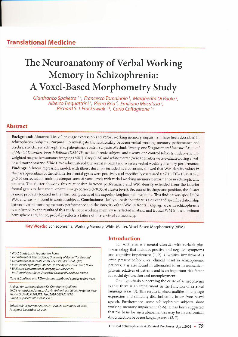
A:Thegreenarrowindicatesonecompleten-backsequenceatthen-1 level fromthe"studyphase"tothe"response'"Theyellowarrowindicatesone complete n-back sequence at the n-2 level.The blue arrow indicates one complete n-back sequence at the n-3 level. The word "Oven" is
the correct item at the n-1, n-2 and n-3 levels.
B: Indicates one complete n-back sequence in terms of time.
80 r Chnical Schizophrenia & Related Psychoses April 2008

Gianfranco Spalletta, Francesco Tomaiuolo et al.
The research protocol was approved by the Ethics
Committee of the IRCCS Fondazione Santa Lucia. Allpatients gave written consent after a full verbal explanationofthe procedures in the study.
Diag n osti c and N e uropsych ologÍ calEvaluations
A trained psychiatrist diagnosed DSM-IV schizophrenia
using the Structured Clinical Interview for DSM-IV Patient
Edition (SCID-P) (22). A second trained psychiatristassessed verbal working memory performance (i.e., accuracy
as memory load) of patients using the "n-back' verbalworking memory task. The experiment was conducted in a
soundproof room with soft lighting. Subjects sat comfortablyin an armchair approximately 50 cm from a computermonitor (ViewSonic, 19 inches); the center of the monitorwas aligned with the subjects' eyes. Subjects were requiredto monitor continuously a sequence of verbal stimuli (a
total of twenty-two items presented as short words) and toselect items that appeared n items back in any sequence.(This sequence was generated using locally written softwareinstalled on a Pentium@ 4IBM computer.) Furthermore, theitem selection was done by the participants using a specialkeyboard with three keys, one for each stimulus. Also,the computer software automatically generated a file withresults of the task with corrected-uncorrected responses.
We administered three n-back subtasks at different levels ofdifficulty. At the n-l level subjects were required to select
an item that appeared one back in a sequence, at the n-2level subjects selected an item that appeared two back ina sequence, and at the n-3 level they selected an item thatappeared three items back in a sequence. The number ofcorrected responses for each sequence (i.e., n-1, n-2, and n-3) was considered as a possible index of working memoryperformance. The experimental procedure is described inFigure 1 and has been used with nonverbal stimuli in several
previous studies (23-25).
All patients were trained to obtain their maximalperformance score. In particular, all subjects were trainedusing the n-1 back paradigm by means of verbal and writtenexplanations. Furthermore, for training, patients practicedthe n-1 back sequence three times before starting the actualexperimental procedure. Those subjects who scored less thanten on the n-1 back sequence (n=1) were excluded from thestudy; "n-back'tasks were performed within fifteen days ofMRI.
ControlSubjectsTwenty-one control subjects were selected and matched
with the schizophrenic patients for age, gender and
educational level. Theywere interviewed with the Structured
Clinical Interview for DSM-IV Non-Patient Edition (SCID-NP) (26). None of them suffered from a mental disorder.A different clinical psychiatrist administered the n-backverbal working memory task to the control subjects. Timesand modalities of administration for the tasks to the controlsubjects were identical to those used with the schizophrenicsubiects.
Image AcquísitionMagnetic resonance T I -weighted images, Magnetization
Prepared Rapid Gradient Echo Sequence (MPRAGE;
Erlangen, Germany; I mm isotropic voxel, repetition time[TR]=11.4 ms, echo time [TE]=4.4 ms, flip angle=15 deg),were obtained with a Siemens 1.5 T Vision Magnetom MRsystem. Image distortions and head motion of each imagewere carefully evaluated by a trained neuroradiologist.Only images without artifact or with very small acceptable
problems were used for the analysis.
Image ProcessingThe procedure we used for VBM followed the
methodology described by Bernasconi et al. (27). Thefollowing steps were applied to the scans of each subject:
1) The MPRAGE brain volume images underwent a
nonuniformity correction to remove variations in MRIintensity related to radio frequency inhomogeneity (28);
2) Subsequently, MRI images were transformedlinearly to standardized stereotactic space (29) to adjustfor differences in total brain volume and brain orientation.Correct registration is critical for VBM analysis (30);
the quality of the transformation was verified using thesoftware Register that allows for the simultaneousvisualization in 3D of two brain volumes (www.bic.mni.
mcgill.calsoftware/distribution). Each individual brain of thesubjects who participated in our study was compared withthe template of the Montreal Neurological Institute (average
of 305 brains) to verify correct resampling in stereotacticspace. Variability caused by head structure other than thebrain (31) was reduced using the MacDonald algorithm(32);
3) Then the tissue-classification algorithm (INSECT)(33) was applied to classify each voxel into one of threeclasses: namely, GM, WM or cerebralspinal fluid. A binaryvolume consisting of GM voxels and another consisting ofWM voxels were extracted from the classified image; and,
4) GM and WM binary masks were smoothed usinga Gaussian smoothing kernel of l0 mm full width at halfmaximum to generate 3D maps of GM and WM 'density''
Smoothing allows one to convert the binary data into a rangeof continuous data for statistical analyses used in this study(34). The brainstem and cerebellum were not included in theanalyses.
ClinicalSchizophrenia&RelatedPsychoses April2008 r 8l

Neuroanatomy, Working Mernory and Sehizophrenia
Characteristics
Age (year)
Educational Level (year)
Age at the Onset of
the lllness (year)
Duration of lllness (year)
Olanzapine Equivalents
(mg/day)
Verbal Working Memory
(l -back)*
Verbal Working Memory
(2-back)*
VerbalWorking Memory
(3-back)*
Mini-Mental State
Examination (MMSE)
Gender (male)
Marital status (married)
DSM-IVSubtypes:
Paranoid
Schizophrenic Patients(n=21)
Mean+SD (Range)-*-t;'rtóitìzosài '"- -
1 1.013.1 (8-l7)
23.3+7.0 (15-40)
11.8+8.3 (1-34)
21 .5+16.3 (3-60)
19.5+3.2 \10-22)
16.4+4.6 (S-21)
9.0+3.7 (4-151
27.9+1 .6 (24-30)
N (o/o)
12 (s7)
2 (9)
1l!67)
111_1ì
_, I q4l1 (s)
Control Subjects
!"=-2llMean+5D (Range)
-":i.6ià.ì i;#;j--1 1.8+3.7 (g_17)
20.9!2.0 (15-22)
1 9.013.4 (9-21)
15.1+4.7 (8-20)
28.6+1.1 (27-30)
N (o/o)
12 (s7)
7 (33)
4.677 : <.0001
ll
>.u5i
:pi-.------- -
i >.05i \n<
>.05
>.05
-"t".-
l
;
l
p
-0.187 ;
0.763 l
i
I
:
i
1.782
2.144
t.ool
chi,l!,1"1"
0.000
3.535
: >.05:
\
i
t
--:-
:
-..!-
Residual
SD=standard deviation, * Number of corrected responses
Statistícal AnalysisComparisons with respect to categorical and
continuous sociodemographic and clinical variables were
made using chi-square and Student's t-tests, respectively.
Pearson's correlation was used to analyze the relationship
between verbal working memory performance during the
n-back task at the 2-back level and antipsychotic dosages
in schizophrenic patients. This was done in order to verify
whether a statistically significant effect of antipsychotic
treatment on verbal working memory performance existed
(35). This efect, if present, could influence results of the
study.
Using six multivariate linear regression models, we
correlated the individual GM and WM density maps
with patient and control subject verbal working memory
performances during the n-back task. In the f,rst analysis,
we correlated the WM density map of the schizophrenic
patients with the number of corrected responses at the n-2
level ofthe n-backtask. In the second analysis, we correlated
the WM density map of the control subjects with the number
ofcorrected responses at the n-2 level ofthe n-back task' In
the third analysis, we correlated the WM density map oÎ the
control subiects with the number of corrected responses at
the n-3 level of the n-back task. The same three analyses were
repeated for GM. In all these anaiyses, the duration of illness
was entered as covariate, to correct for possible impact of
this variable on the cerebral structures. In schizophrenic
subjects we chose to limit statistical analysis to the n-2 level as
schizophrenics scored at chance for the n-3 task and at ceiling
for the n-1 task. In control subjects, we also analyzed the n-
3 level because a range of performances was obtained (36)'
The voxel-level statistical threshold for GM maps was
set to t>6.85 (r>0.843), based on a voxel size of 1 mm:',
smoothness of 10 mm, a volume of interest of 500 cm3, 18
degrees of freedom (DF) and a significance ievel of p<0'05
corrected for multiple comparisons (3a). lhe voxel-level
threshold for WM maps was set to t>7.16 (r>0.860), based
on a voxei size of 1 mm3, smoothness of l0 mm, a volume of
interest of 800 cm3, 18 degrees of freedom and a significance
level of p<0.05 corrected for multiple comparisons (34). A
positive regression indicated greater GM or WM density
associated with more correct items on the n-back task' In
82 r Clinical Schizophrenia & Related Psychoses April 2008
Gianfranco Spalletta, Francesco Tomaiuolo et al.
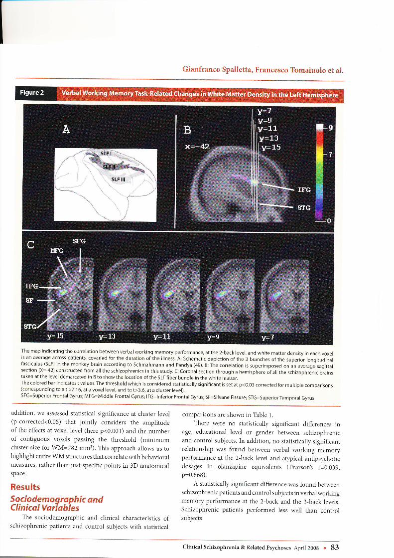
The map indicating the correlation between verbal working memory performance, atthe 2-back level, and white matter density in each voxelis an average across patients, covaried for the duration ofthe illness. A: Schematic depiction ofthe 3 branches ofthe superior longitudinalfasciculus (SLF) in the monkey brain according to Schmahmann and Pandya (49). B: The correlation is superimposed on an average sagittalsection (x=-42) constructed from all the schlzophrenics in this study. C: Coronal section through a hemisphere ofall the schizophrenic biainstaken at the level demarcated in B to show the location of the SLF fiber bundle in the white matter.The colored bar indicates t values.The threshold which is considered statistically significant is set at p<0.05 corrected for multiple comparisons(corresponding Ío at>7.16, at a voxel level, and to t>3.6, at a cluster level).SFG=Superior Frontal Gyrus; MFG=Middle Frontal Gyrus; IFG=lnferior Frontal Gyrus; SF=Silvane Fissure; STG=SuperiorTemporal Gyrus
addition, we assessed statistical significance at cluster level(p-corrected<0.05) that jorntly considers the amplitudeof the efects at voxel level (here p<0.001) and the numberof contiguous voxels passing the threshold (minimumcluster size for WM=782 mm3). This approach allows us tohighlight entire WM structures that correlate with behavioralmeasures, rather than just specific points in 3D anatomicalspace.
ResultsSociodemoqraphic ondClinical Vorlabies
The sociodemographic and clinical characteristics ofschizophrenic patients and control subjects with statistical
comparisons are shown in Table 1.
There were no statistically significant differences inage, educational level or gender between schizophrenicand control subjects. In addition, no statistically significantrelationship was found between verbal working memoryperformance at the 2-back level and atypical antipsychoticdosages in olanzapine equivalents (pearson's r=0.039,p=0.868).
A statistically significant difference was found betweenschizophrenic patients and control subjects inverbal workingmemory performance at the 2-back and the 3-back levels.Schizophrenic patients performed less well than controlsubjects.
Clinical Schizophrenia & Related Psychoses April 2008 r 83

Neuroanatomy, Working Memory and Schizophrenia
Re I atí o n sh i p b etwe en Ve rb al Wor ki n gMemory, GM and WM Density
We found a significant positive correlation (see Figure
2) between verbal working memory performance ofschizophrenic subjects at the 2-back level and WM density
in the pars opercularis of the left inferior frontal gyrus with a
maximum peakatx=-42,y=12,2=14 (f>7.16, DF= 18' r=0.878,
p<0.05 corrected for multiple comparisons, at voxel level).
No significant relationship was found with GM density.
To investigate the distribution of the WM densityvalues
in the two groups ofpatients and control subjects at the level
of the superior longitudinal fasciculus (SLF), we extracted,
for each subject, the value of the white matter density at
the voxel with the maximum significant difference (x=
-42,y=12, z=14) ín the previous analysis. Then, we plotted
these values comparing patients versus control subjects (see
Figure 3). In this cerebral area, the WM density statistically
differed between schizophrenic patients and control subjects
(t=2.r44, DF=40, p=0.038).
Figure 4 shows the scatterplot of the correlation between
working memory scores and white matter density values
of schizophrenic patients at the coordinates (x=-42,Y=12,
z=14) in the schizophrenic patients.
We found no significant relationship in control
subjects between either GM or WM density and n-back
verbal working memory performance at the 2-back and
3-back levels. Statistical tests for cluster-level significance
(p-corrected<0.05) demonstrated that WM density of the
fibertracks connecting the pars opercularis ofthe left inferior
frontal gyrus and the parietal operculum in schizophrenic
patients showed positive correlation with verbal working
memory performance (cluster size=962 mm3). Because ofthe characteristic shape and position (see Figure 2) we
believe that the result is located and confined to the
sLF (37).
DiscussionOur study finds that the WM density of the pars
opercularis ofthe left inferior frontal gyrus ofschizophrenic
subjects correlates positively with the ability to correctly
perform the verbal working memory n-back task at the
2-back level. The peak of this correlation is located in a
cluster centered on the frontoparietal operculum. The shape
and position of the cluster suggests it is part of the thirdcomponent of the SLF. We found no significant correlation
between verbal working memory and GM density. In
control subjects we found no correlation between GM or
WM density and verbal working memory performance.
The absence of any correlation cannot be explained by a
statistical bias induced by a ceiling effect. In control subjects
there was also no correlation between performance of the
more diffìcult 3-back task and WM or GM density. The
change in WM density found in schizophrenic subjects is,
therefore, specific. It could be the result of a modification ofaxon diameter or myelination, either of which could modify
speeds of neural transmission (38, 39).
A very recent study, using diffusion tensor imaging to
examine fractional anisotropy, described that recent-onset
schizophrenic patients, with a mean illness duration offifteen months, showed anatomical changes in the left SLF,
and this abnormality was associated with the performance
in a verbal working memory task (40). Our results, in an
older group of patients with longer illness duration, using
a different method for assessing WM morphometry, that is
VBM, found similar results. This suggests that verbal working
memory impairment and its structural correlate may be a
constant marker of schizophrenia both at the onset of the
AA*,
&th
**
&
^.**
*
*
&*
;: t'.1
;1,rrl
.L2000
a)
o6
Q)
Ctl
!o
=.cGózN
2500 3000 3500 4000 4500 5000 5500
White Matter Density
4800 -
î 47oo-vl! aooo-
3 +soo -
.4400-'; 4JUU -
E qzoo-f]b 4100-
È 4000-E .onn-o
= 3800 -
7 ztoo-
84 r Clinical Schizophrenia & Related Psychoses April 2008
Gianfranco Spalletta, Francesco Tomaiuolo et al.
illness and during the later stages. The results are interestingbecause the affected area is connected to Broca's area (41)
and frontal fibers of the SLF. The SLF consists of association
fibers that link the frontal to the parietal lobes (37, 42).
Anatomical studies in monkeys indicate that it consists ofthree reciprocal components (37). The third component(i.e., that which is implicated by our results) connects the
supramarginal gyrus and the parietal operculum with the
premotor area, the pars opercularis of the inferior frontalgyrus, the frontal opercular region and the ventral part ofthe middorsolateral frontal lobe. Complex somatosensory
stimuli related to the face and arm are carried by the thirdcomponent of the SLF (43,44) . Neural areas connected by the
third component of the SLF respond both when a monkeyperforms a specific movement and when it observes anothermonkey performing the same action (45). Therefore, the
interconnected areas and connecting pathways are involvedin action imitation that may be at the basis of gestural
communication; a function that probably preceded the
evolution of linguistic communication.From a theoretical point ofview, it is possible to interpret
our data in the light of Baddeley's model of working mem-ory in which he introduced the concept of "fractionation
of working memory" (8). In this model, working memoryis a cognitive function composed of various subsystems.
The phonological loop constitutes a temporary storage
system for language information that may be maintainedin memory for two to three seconds. However, whennecessary, such information may be retained in memory bycontinuous subvocal rehearsal, an operation that is med-iated by Broca's area (8, 9). Thus, the modification of WMdensity found in our patients that correlates with verbalworking memory performance could represent the structuralbasis for a specific impairment of phonological loop activityin schizophrenia.
There are some issues to be discussed. Firstly, despite
the fact that we found no correlation between atlpicalantipsychotic dosage and verbal working memoryperformance, we believe it is important to confirmour findings in drug-naive patients with first-episode
schizophrenia. Secondly, how can we explain that: 1) those
schizophrenic patients who have a reduced verbal workingmemory performance have a reduced WM density in the
SLF, a WM tract which is involved in the rehearsal process;
and that, 2) this was found in schizophrenics only and notin control subjects? On the one hand, it is possible that some
insult (e.g., developmental), specifically in this area, can
primarily impair the structure of the SLF and, consequently,
the working memory performance. Thus, schizophrenics
who have decreased verbal working memory performancedo not use rehearsal as a strategic choice and may hold verbalmaterials in other preserved cerebral areas rather than in the
damaged phonological areas. However, this strategy couldnot fully compensate the good performance associated
with a preserved SLF structure. An alternative explanationcan be found in a precocious primary linguistic problemof environmental or reactive nature. such as a defective
phonological problem, that secondarily may compromise the
development of language cerebral areas. In reality, there isevidence that those children who will develop schizophreniahave impaired capacity for social interaction, solitary playpreference and language problems and, therefore, may use
differently, either quantitatively or qualitatively, language
for communication (46-48). If this is caused by problems
of primary biological nature or secondary environmentalor psychological nature, it is difficult to explain. However,
the fact that schizophrenic patients have reduced WMdensity in the SLF area with the strongest correlation withverbal working memory performance, in comparisonwith control subjects, strongly suggests that this area isprimarily damaged in schizophrenia independently fromthe cognitive performance. Apart from these speculations,
the finding of reduced verbal working memory performancein schizophrenia associated with reduced WM density of the
SLF is in line with the concept that the structure of fibers
connecting Broca's area of the left premotor frontal regionand supramarginal gyrus of the inferior parietal lobule mayhave a pathogenetic role in schizophrenia.
Given the limitations of this studywe suggest that futurestudies should investigate the specificity ofthese results and
analyze the relationship between WM structure (i.e., density,
volume or diffusion tensor imaging) and a larger set ofcognitive domains.
ConclusionsResults of this study confirm the hlpothesis that WM
structural abnormality of cerebral language areas, namelythe pars opercularis ofthe left inferior frontal gyrus and the
SLF, is implicated in verbal working memory impairment inschizophrenia.
AcknowledgmentsThe Italian Ministry of Health RC-06/A.
References1. Andreasen NC. Schizophrenia: the fundamental questions.
Brain Res Brain Res Rev 2000;3 1 (2- 3):706-112.
2. Mitchell RL, EIIiott R, Woodruff PW fMRI and cognitivedysfunction in schizophrenia. Trends Cogn Sci200I ;512):7 1 -
81.
3. Crow Tl. Schizophrenia as the price that homo sapiens
pays for language: a resolution ofthe central paradox in theorigin ofthe species. Brain Res Brain Res Rev 2000;31(2-3):778-129.
4. Carter CS, Perlstein W, Ganguli R, Brar J, Mintun M,
Clinical Schizophrenia & Related Psychoses April 200S o 85

Neuroanatomy, Working Memory and Schizophrenia
Cohen iD. Functional hlpofrontality and workingmemory dysfunction in schizophrenia. Am I Psychiatry| 998r I ss(9):1 2B5- | 287.
5. Goldman-Rakic PS. Working memory dysfunctionin schizophrenia. J Neuropsychiatry Clin Neurosci
I994:6(4):348-357 .
6. Pierri JN, Volk CL, Auh S, Sampson A, Lewis DA.
Decreased somal size of deep layer 3 pyramidal neurons inthe prefrontal cortex of subjects with schizophrenia. ArchGen Psychiatry 2001 ;58(5):466 - 47 3.
7. Spalletta G, Tomaiuolo R Marino V Bonaviri G,
Trequattrini A, Caltagirone C. Chronic schizophrenia as a
brain misconnection syndrome: a white matter voxel-based
morphometry study. Schizophr Res 2003;64(1):15-23.
8. Baddeley A. The fractionation of working memory. Proc
Natl Acad Sci USA 1996;93(24):13468-13472.
9. Smith EE, Jonides ]. Storage and executive processes in the
frontal lobes. Science 1999;283(5408):1657 -7667.
10. Menon V Anagnoson RT, Mathalon DH, Glover GH,
Pfefferbaum A. Functional neuroanatomy of auditoryworking memory in schizophrenia: relation to positive and
negative symptoms. Neuroimage 2001;13(3):433-446.
11. Stevens AA, Goldman-Rakic PS, Gore JC, Fulbright RK,
Wexler BE. Cortical dysfunction in schizophrenia duringauditory word and tone working memory demonstrated
by functional magnetic resonance imaging. Arch Gen
Psychiatry 1 998;55( r2): 1097 -II03.
12. Conklin HM, Curtis CE, Katsanis J, Iacono WG. Verbal
working memory impairment in schizophrenia patientsand their first-degree relatives: evidence from the digit span
task. Am J Psychiatry 2000;157(2):275-277.
13. Perlstein WM, Carter CS, NolI DC, Cohen JD. Relation
of prefrontal cortex dysfunction to working memoryand symptoms in schizophrenia. Am ] Psychiatry200 1;1s8(7):1 105- I r 13.
14. Honey GD, Bullmore ET, Sharma T. Prolonged reaction
time to a verbal working memory task predicts increased
power of posterior parietal cortical activation. Neuroimage
2000;12( 5):49s-503.
15. Crow TJ. Schizophrenia as failure ofhemispheric dominancefor language. Trends Neuro s ci 19 97 ;20 (8) :339 - 3 43.
16. Paus I Zijdenbos A, Worsley K, Collins DL, Blumenthal I,Giedd JN, et al. Structural maturation of neural pathways
in chiidren and adolescents: in vivo study. Science
1999 ;283 (5409) : 1 908 - 1 9 1 1.
17. Tisserand DJ, van Boxtel MR Pruessner JC, Hofman P,
Evans AC, Jolles J. A voxel-based morphometric study todetermine individual differences in gray matter densityassociated with age and cognitive change over time. Cereb
Cortex 2004; I 4 (9 ) :9 66 -97 3.
18. Folstein MR Folstein SE, McHugh PR. "Mini-mental state'l
A practical method for grading the cognitive state of patientsfor the clinician. J Psychiatr Res 1975;12(3):189-198.
19. Measso G, Cavarzeran F,Zappalìt G, et al. The Mini-MentalState Examination: normative study of an Italian randomsample. Dev Neuropsychol 1993 ;9 :7 7 -85.
Grigoletto F, Zappala G, Anderson DW, Lebowitz BD.
Norms for the Mini-Mental State Examination in a healthypopulation. Neurology 1999 ;53 (2) :375 -320.
Guy W ECDEU assessment manual for psychopharma-cology. DHEW publication, no. (ADM) 76-338. Rockville,MD, U.S. Dept. of Health, Education, and Welfare, PublicHealth Service, Alcohol, Drug Abuse, and Mental HealthAdministration, National Institute of Mental Health,Psychopharmacology Research Branch, Division ofExtramural Research Programs. 1976.
First MB, Spitzer RL, Gibbon M, Williams JBW. Structuredclinical interview for Axis I DSM-IV disorders. Research
version-patient edition (SCID-I/R Ver. 2.0). New York(NY): New York State Psychiatric Institute, BiometricsResearch Department; 1996.
23. Oliveri M, Turriziani B Carlesimo GA, Koch G, TomaiuoÌoF. Panella M. et al. Parieto-frontal interactions in visual-object and visual-spatial working memory: evidence
from transcranial maenetic stimulation. Cereb Cortex2001;1 1(7):606-618.
24. Owen AM, Stern CE, Look RB, Tracey I, Rosen BR, Petrides
M. Functional organization of spatial and nonspatialworking memory processing within the human lateralfrontal cortex. Proc Natl Acad Sci USA 1998;95(f3):7721-7726.
25. Smith EE, ionides J. Working memory: a view fromneuroimaging. Cognit Psychol 7997 ;33 (7) :5 - 42.
26. First MB, Spitzer RL, Gibbon M, Williams JBW Structuredclinical interview for DSM-IV Axis I Disorders (SCID). NewYork (NY): New York State Psychiatric Institute, BiometricResearch Department; 1995.
27. Bernasconi N, Duchesne S, lanke A, Lerch J, Collins DL,Bernasconi A. Whole-brain voxel-based statistical analysis
of gray matter and white matter in temporal lobe epilepsy.
Neuroimage 200 4;23 (2) :7 17 -7 23.
28. Sled lG, Zijdenbos AP, Evans AC. A nonparametric methodfor automatic correction of intensity nonuniformity in MRIdata. IEEE Trans Med Imaging 1998;17 (1):87 -97 .
29. Collins DL, Neelin P, Peters TM, Evans AC. Automatic3D intersubject registration of MR volumetric data instandardized Talairach space. J Comput Assist Tomogr1994;18(2):192-205.
30. Ashburner ], Friston Kl. Why voxel-based morphometryshould be used. Neuroimage 200 1 ; 14(6) :1238 - 1243.
31. Burton EJ, Karas G, Paling SM, Barber R, Williams ED,
Ballard CG, et a1. Patterns of cerebral atrophy in dementiawith Lewy bodies using voxel-based morphometry.Neuroimage 200217 (2) :618 - 630.
20.
21
22.
86 r Ctnical Schizophrenia & Related Psychoses April 2008

Gianfranco Spalletta, Francesco Tomaiuolo et al"
4t.
32.
33.
MacDonald D, Kabani N, Avis D, Evans AC. Automated 3-
D extraction of inner and outer surfaces of cerebral cortex
from MRI. Neuroimage 2000 ;12(3):3 40 -3 56
Zijdenbos AP, Forghani R, Evans AC' Automatic
quantification of MS lesion in 3D MRI brain data sets:
validation of INSECT. Proceedings of International
Conference on Medical Image Computing and Computer-
Assisted Intervention (MICCAI). Berlin (Germany):
Springer Verlag; 1998.
Worsley KJ, Marret S, Neelin P, Vandal AC, Friston Ki,
Evans AC. A unified statistical approach for determining
significant signals in image of cerebral activation. Hum
Brain Mapp 1996;4:58-7 3.
Keefe RS, Young CA, Rock SL, Purdon SE' Gold fM, Breier
A; HGGN Study Group. One-year double-blind study of
the neurocognìtive eficacy of olanzapine, risperidone, and
haloperidol in schizophrenia. Schizophr Res 2006;81(1):1-
15.
Honey GD, Fu CH, Kim J, Brammer MI, Croudace TJ,
Suckling J, et al. Effects of verbal working memory load on
corticocortical connectivity modeled by path analysis of
functional magnetic resonance imaging data. Neuroìmage
2002;17 (2):573-582.
37. Petrides M, Pandya DN. Association pathways of the
prefrontal cortex and functional observations. In: Stuss DT,
Knight RI, editors. Principles of frontal lobe function' New
York: Oxford University Press; 2002.
38. Aboitiz R Scheibel AB, Fisher RS, Zaidel E. Fiber
composition of the human corpus callosum. Brain Res
1992;598(r -2) :1 43 - | 53.
Paus T, Coliins DL, Evans AC, Leonard G, Pike B, Zijden-
bos A. Maturation of white matter in the human bratn:
a review of magnetic resonance studies. Brain Res Bull
200I:54(3):2s5-266.
Karlsgodt KH, van Erp TG, Poldrack RA, Bearden CE,
Nuechterlein KH, Cannon TD. Diffusion tensor imaging of
the superior longitudinal fasciculus and working memory
in recent-onset schizophrenia. Biol Psychiatry 2008;
63(5):s12-5 18.
Tomaiuolo F, MacDonald |D, Caramanos Z' Posner G,
Chiavaras M, Evans AC, et al. Morphology, morphometry
and probability mapping of the pars opercularìs of the
inferior frontal gyrus: an in vivo MRI analysis' Eur ]Neurosci 1999;1 1 (9):303 3-3046.
Tailarach f, Tournoux P. Referentially oriented cerebral
MRI anatomy: atlas of sterotaic anatomical correlations for
gray and white matter. New York, Stuttgart: Georg Thieme
Verlag; i993.
43. Hyvarinen |, Shelepin Y. Distribution of visual and somatic
functions in the parietal associative area 7 of the monkey'
Brain Res 197 9 :769 (3) :561 - 564.
44. Leinonen L, Nyman G. II. Functional properties of cells in
anterolateral part of area 7 associative face area of awake
monkeys. Exp Brain F.es 1979:34(2):321-333.
45. Rizzolatti G, Fadiga L, Gallese V Fogassi L. Premotor cor-
tex and the recognition of motor actions. Brain Res Cogn
Brain Res 1996;3(2):13I - l4l.
46. Done DL Crow TJ, Johnstone EC, Sacker A. Childhood
antecedents of schizophrenia and afective illness: social
adjustment at ages 7 and 11. BMJ 1994;309(6956):699-703.
47. Jones P, Rodgers B, Murray R, Marmot M' Child develop-
ment risk factors for adult schizophrenia in the British 1946
birth cohort. Lancer 199 4;344(893 4):1398 - 1402'
48. Nicolson R, Lenane M, Singaracharlu S, Malaspina D,
Giedd JN, Hamburger SD, et al' Premorbid speech and
language impairments in childhood-onset schizophrenia:
association with risk factors. Am I Psychiatry
2000;157 (5):794-800.
49. Schmahmann JD, Pandya DN. Fiber pathways of the brain'
New York Oxford University Press; 2006. Adopted with
permission from fìgures 13-1, f3-3, 13-4.
42.
15.
36.
39.
Clinical Schizophrenia & Related Psychoses April 2008 .97