
3D moment invariant based morphometry
8
3D Moment Invariant Based Morphometry J.-F. Mangin , F. Poupon , D. Rivi` ere , A. Cachia , D. L. Collins , A. C. Evans , and J. R´ egis Service Hospitalier Fr´ ed´ eric Joliot, CEA, 91401 Orsay, France [email protected], http://anatomist.info Institut F´ ed´ eratif de Recherche 49 (Imagerie Neurofonctionnelle), Paris Montreal Neurological Institute, McGill University, Montreal Service de Neurochirurgie Fonctionnelle et Stereotaxique, CHU La Timone, Marseille Abstract. This paper advocates the use of shape descriptors based on moments of 3D coordinates for morphometry of the cortical sulci. These descriptors, which have been introduced more than a decade ago, are invariant relatively to rotations, symmetry and scale and can be computed for any topology. A rapid insight of the derivation of these invariants is proposed first. Then, their potential to character- ize shapes is shown from a principal component analysis of the 12 first invariants computed for 12 different deep brain structures manually drawn from 7 different brains. Finally, these invariants are used to find some correlates of handedness among the shapes of 116 different cortical sulci automatically identified in 144 brains of the ICBM database. 1 Introduction This paper advocates the use of shape descriptors based on moments of 3D coordinates for morphometry purpose. These 3D descriptors, which are invariant relatively to rota- tion, symmetry and scale, have been introduced more than a decade ago [8], as a 3D extension to the 2D moment invariants widely used in pattern recognition [6]. These 3D moment invariants have not gained a lot of attention in the medical imaging community. In our opinion, however, they provide a powerful way to perform global morphome- try of anatomical entities because they impose no constraints on the object’s topology. Therefore, they appear as an interesting alternative to the spherical harmonics based ap- proach, which implies simply connected objects for 2D parameterization of the surface [1, 5]. While the theoretical derivation of the invariants from the coordinate moments is complex, they can be computed in a simple and robust way from a binary volume based description of the objects of interest. This simplicity of use makes these invariants good candidates for mining large databases of objects before using more sophisticated shape analysis tools providing locality to the study [11, 5]. In this paper, the interest of the 3D moment invariants is illustrated through the study of the shapes of the cortical sulci of 144 subjects of the ICBM database. A pre- vious study has shown some correlates of handedness on the global size of some of the sulci of the motor and premotor areas [10]. These correlates are supposed to stem from handedness-related discrepancies in the pressure to increase the local folding induced by discrepancies in the development of the surrounding cortical areas. This first study has shown that sulcus based morphometry is a compelling complement to the usual
-
Upload
independent -
Category
Documents
-
view
2 -
download
0
Transcript of 3D moment invariant based morphometry

3D Moment Invariant Based Morphometry
J.-F. Mangin��� �
, F. Poupon��� �
, D. Riviere��� �
, A. Cachia��� �
,D. L. Collins
�, A. C. Evans
�, and J. Regis
��Service Hospitalier Frederic Joliot, CEA, 91401 Orsay, [email protected], http://anatomist.info�
Institut Federatif de Recherche 49 (Imagerie Neurofonctionnelle), ParisMontreal Neurological Institute, McGill University, Montreal
Service de Neurochirurgie Fonctionnelle et Stereotaxique, CHU La Timone, Marseille
Abstract. This paper advocates the use of shape descriptors based on momentsof 3D coordinates for morphometry of the cortical sulci. These descriptors, whichhave been introduced more than a decade ago, are invariant relatively to rotations,symmetry and scale and can be computed for any topology. A rapid insight of thederivation of these invariants is proposed first. Then, their potential to character-ize shapes is shown from a principal component analysis of the 12 first invariantscomputed for 12 different deep brain structures manually drawn from 7 differentbrains. Finally, these invariants are used to find some correlates of handednessamong the shapes of 116 different cortical sulci automatically identified in 144brains of the ICBM database.
1 Introduction
This paper advocates the use of shape descriptors based on moments of 3D coordinatesfor morphometry purpose. These 3D descriptors, which are invariant relatively to rota-tion, symmetry and scale, have been introduced more than a decade ago [8], as a 3Dextension to the 2D moment invariants widely used in pattern recognition [6]. These 3Dmoment invariants have not gained a lot of attention in the medical imaging community.In our opinion, however, they provide a powerful way to perform global morphome-try of anatomical entities because they impose no constraints on the object’s topology.Therefore, they appear as an interesting alternative to the spherical harmonics based ap-proach, which implies simply connected objects for 2D parameterization of the surface[1, 5]. While the theoretical derivation of the invariants from the coordinate moments iscomplex, they can be computed in a simple and robust way from a binary volume baseddescription of the objects of interest. This simplicity of use makes these invariants goodcandidates for mining large databases of objects before using more sophisticated shapeanalysis tools providing locality to the study [11, 5].
In this paper, the interest of the 3D moment invariants is illustrated through thestudy of the shapes of the cortical sulci of 144 subjects of the ICBM database. A pre-vious study has shown some correlates of handedness on the global size of some of thesulci of the motor and premotor areas [10]. These correlates are supposed to stem fromhandedness-related discrepancies in the pressure to increase the local folding inducedby discrepancies in the development of the surrounding cortical areas. This first studyhas shown that sulcus based morphometry is a compelling complement to the usual

voxel based morphometry. VBM, indeed, could not reveal any significant handedness-related result [16], although the existence of some correlates could be forecast fromprevious manual studies. Such disagreement between both morphometry strategies maystem from the loss of statistical power induced by the non perfect gyral matching per-formed by the spatial normalization underlying VBM. Therefore, sulcus based mor-phometry may become a new probe to test the assumption that certain neuroanatomicalstructures may be preferentially modified by particular cognitive skills or diseases.
The first study mentioned above was relying on the size of the pieces of skele-ton used to represent the sulci of interest [9]. This measure of size is analogous tothe volume calculation used in standard morphometric studies. It is evident that sulcuscharacterization by size does only capture one of the multiple aspects of the foldingpatterns [5]. We propose the use of 3D moment invariants as a richer description ofthe sulcus shapes. They appear especially adapted to sulcus morphometry, because thenumerous and variable sulcus interruptions prevent a simple parameterization strategyfor most of the sulci [1, 7]. The next section provides an insight into the origin of theinvariants, and a few experiments about their invariance properties and their slow varia-tions in the shape space. The last section reports some invariant-based results relative tothe handedness-correlate study of about 116 sulci automatically labeled in each of 144brains by a system described elsewhere [14].
2 3D moment invariants
The 3D moments of order � ����������������� IN of a 3D density function ��� �!�#"���$&% aredefined by ')(+*-, � .0/1
2 /.0/1
2 /.!/1
2 / � ( " * $ , ��� �0��"���$&%435�635"738$:9 (1)
In the following, ��� �0��"���$&%;�=< , because we deal with objects defined by binaryimages. The moments of order higher than 3 will not be considered in this paper forthe sake of simplicity, but the derivation of moment invariants is theoretically possiblefor any order. By discarding moments of order higher than 3, we get a small set ofglobal descriptors which should embed simple shape information like bending, taper-ing, pinching, etc... The derivation of the invariants aims at filtering out the influence oflocalization, orientation and scale on the 3D moments in order to obtain “pure shape”descriptors. Translation invariance is simply obtained using central moments. In the fol-lowing, for the sake of clarity, the origin of the coordinate system is assumed to be atthe centroid of the object and the corresponding central moments will be written > (+*-, .As shown in [6] for the 2D case, the similitude invariance is obtained by normalizingmoments with the suitable power of the volume >@?A?�? . Therefore, in the following weconsider B (+*-, � > (+*#,> ?A?�?DCAEGFHE8IJ . � 9 (2)
Rotation invariants can be derived from group theory techniques usual in quantummechanics [4]. When a rotation is applied to the underlying object, central moments of

order � are transformed into linear combinations of moments of the same order. Thisresult stems from the fact that homogeneous polynomials of order � form a subspaceK7L
of the functions of IR�
stable under the rotation group. The coefficients of the linearcombinations mentioned above are the matrix elements of a representation of the 3Drotation group (corresponding to a group homomorphism) [4]. This representation isreducible, which means that
K Lcan be decomposed into a direct sum of smaller sub-
spaces stable under the rotation group. Rotation invariants stem from the finest possibledecomposition leading to irreducible representations. In this new basis, the effect of therotation operator on a vector of
KMLcorresponds to a block diagonal matrix
N �OPPPQ N ? � N � %
. . . � N;R %SUTTTV � (3)
whereN ? , 9U9+9 , N;R
are irreducible representations [4].
The basis corresponding to this decomposition is the basis of harmonic polynomials"&WX �Y� X[Z WX , \]�_^`�+9U9U9U��a , ' �cbM\-�+9U9U9��A\ whereZ WX are the spherical harmonics and���ed � � �@" � �@$ �
. In the following, f X denotes the subspace defined by harmonicpolynomials of order \ . The space
KMLof homogeneous polynomials of order � decom-
poses itself into subspaces f L, f L 2 � , f L 2 � , etc 9U9+9 For instance
K � splits into f � and f&? ,K � splits into f � and f � , etc... In the new basis ofKML
, moments
B (+*-, are transformedinto complex moments g`WX and rotation invariants are derived from gDWX using tensorproducts, which may be understood as a generalization of scalar or vector products [8,4].
Rotation invariants are inferred from all the possible applications of the tensor prod-uct to g`W? , g4W� , g4W� and g4W� yielding rank-0 tensors, namely scalars which are the 3Dmoment invariants. These invariants turn out to be homogeneous polynomials of cen-tral moments. Because of various symmetries, the tensor products results only in twelveinvariants h&ij , where k denotes the order of the underlying central moments and l de-notes the subspace indices of the different tensors used in the application of the products[8, 12]. Thus, we get four norms h �?A? �Ah ���� ��h ���� , and h ��A� , five scalar products and normsof new tensors h ��A�A� �Ah ����A�A� �Ah ����#��� �Ah ����A��� , and h ��A���A� , and three last invariants derived bycombining the second- and third-order moments h �m� ��A�-� ��h �U� ����#� , and h �m� ��A�A� . Since moment in-variants are expressed by homogeneous polynomials, they can be finally reduced by thesuitable power of the invariant [2]. Then each invariant h`ij is transformed innh ij �po+qsrG�t�Hh ij %�9vu h ij u ��w�x � (4)
wherenh&ij is the reduced moment invariant and 3 the polynomial degree.
In order to check that the theoretical properties of invariance stand for discrete repre-sentations of objects relying on binary images, two simple shapes have been resampledwith 28 different orientations. Each invariant has been computed for each orientation.The obtained standard deviations are almost negligible relatively to the means, whichshows that the rotation invariance is respected (see Fig 1). The standard deviations are

Invariants Superquadric Ventricley z y z{| �}~} 0.569 0.0002 1.572 0.002{| ��~� 0.271 0.0004 1.147 0.001{| ��~�~� -0.299 0.0005 -1.263 0.002{| ~ -0.233 0.0004 -0.732 0.001{| �~� -0.235 0.0008 -0.825 0.003{| ~~~ 0.223 0.0004 0.711 0.001{| A�[A� 0.177 0.0004 0.581 0.002{| ~~A� -0.198 0.0004 -0.621 0.002{| A�~�~� -0.176 0.0005 -0.555 0.005{| ��� ~-� 0.231 0.0004 0.771 0.001{| ��� A�H� -0.194 0.0004 -0.677 0.001{| ��� �~�H� 0.189 0.0007 0.540 0.0190 1119 8836 29822 70867 138063 238572 378844 567151
0.566
0.567
0.568
0.569
0.57
0.571Effet de la resolution sur l invariant 1
Nombre de voxels pour un volume donne (initial=8836)
Val
eur
de l
inva
riant
0.568
58715188381119 29822
0.571
0.57
0.569
0.567
0.566
Number of voxels
First invariant
Fig. 1. left: Means and standard deviations of the twelve invariants for 28 different orientations ofthe two objects visualized in the figure. right: Typical variations observed for different samplingof the pinched superquadric. NB: in all figures, for the sake of visualization, voxel-based objectsare triangulated before 3D rendering.
higher for the ventricle than for the pinched superquadric, which can be understoodfrom the fact that the resampling induces more modifications of the thinest shape.
In order to check that the 3D moment invariants vary sufficiently slowly in the shapespace to be interesting as shape descriptors, we have performed a simple principal com-ponent analysis of the invariants obtained for 6 different kinds of shapes correspondingto deep brain nuclei and lateral ventricles. These objects have been manually drawn bya neuroanatomist in the two hemispheres of 7 different brains and can be visualized inFig. 2. Plotting the 84 objects in a chart corresponding to the three first axes yieldedby the PCA shows that the instances of each anatomical entity gather in one localizedregion of the shape space, as described by the invariants. Furthermore, the regions corre-sponding to two nuclei with similar shapes, are closed in this space. This is for instanceclear for the pairs (caudate nucleus, lateral ventricle), or (putamen, globus pallidus).These properties have been used previously to design shape probability distributionsembedded in a Bayesian framework to bias a multi-object deformable model dedicatedto brain basal ganglia [13].
3 Result
The large scale morphometric studies of the handedness correlates has been performedon a sulcus-by-sulcus basis, each sulcus being identified automatically by a computervision system freely available on http://anatomist.info. The subjects scanned were 144unselected normal volunteers previously used for one of the VBM studies mentionedabove [16]. On a short handedness questionnaire, 14 subjects were dominant for left-hand use on a number of tasks; the remaining 130 subjects preferred to use their righthand. The 144 T1-weighted brain volumes were stereotaxically transformed using nineparameters [3] to match the Montreal Neurological Institute 305 average template. The

Cd
LVThGP,Pu
Acb
Acb
Axis 2 versus axis 1
Cd
LV
Th
Acb
Pu
GP
Axis 3 versus axis 1
Fig. 2. The 84 deep anatomical objects used to analyze the shape representation provided by the3D invariants. The 12 invariants have been computed for each nucleus. Then a standard PCAhas been performed and each nucleus is plotted in two charts corresponding to the 3 principalaxes of the PCA. Abbreviations denote Acb: accumbens, Th: thalamus, Cd: caudate, GP: globuspallidus, Pu: putamen, LV: lateral ventricle. For each point, lower and upper case letters denoteleft and right hemispheres.
cortical folds were then automatically extracted using a 3D skeletonization [9]. Finally,58 cortical sulci were recognized in each hemisphere [14].
The size of each sulcus was computed from its skeletonized representation. Then,left (L) and right (R) sizes were used to obtain a normalized asymmetry index ((L-R)/(L+R)/2) for each sulcus and each brain. For each sulcus, the Mann-Whitney U Testwas used to compare the asymmetry indices of left-handed and right-handed groups.This test relies on rank order statistics, which are robust to potential outliers stemmingfrom sulcus recognition errors. Several significant differences were revealed by ouranalysis (����^D9 ^G� , not corrected for multiple comparisons), including most of the sulciof the motor areas: inferior precentral sulcus (p=0.013), intermediate precentral sulcus(p=0.019), central sulcus (p=0.031) [10].
To test if the 3D invariants could capture additional information about the hand-edness correlates on the folding patterns, the 12 invariants have been computed foreach sulcus and each brain. For this computation, each sulcus is represented by a setof voxels of the global skeleton. For each sulcus, the Mann-Whitney U Test was usedto compare the invariant distributions of left-handed and right-handed groups. It shouldbe noted that this analysis involved 12x116=1392 tests, which calls for some correctionfor multiple testing. This correction however requires further work to take into accountthe complex dependences between these tests. With �Y��^`9 ^5^D< , only two sulci yield
Left handed Right handed
14 right handedversus
14 left handed
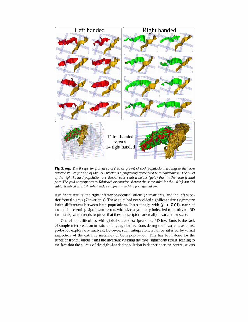
Fig. 3. top: The 8 superior frontal sulci (red or green) of both populations leading to the moreextreme values for one of the 3D invariants significantly correlated with handedness. The sulciof the right handed population are deeper near central sulcus (gold) than in the more frontalpart. The grid corresponds to Talairach orientation. down: the same sulci for the 14 left handedsubjects mixed with 14 right handed subjects matching for age and sex.
significant results: the right inferior postcentral sulcus (2 invariants) and the left supe-rior frontal sulcus (7 invariants). These sulci had not yielded significant size asymmetryindex differences between both populations. Interestingly, with (����^D9 ^D< ), none ofthe sulci presenting significant results with size asymmetry index led to results for 3Dinvariants, which tends to prove that these descriptors are really invariant for scale.
One of the difficulties with global shape descriptors like 3D invariants is the lackof simple interpretation in natural language terms. Considering the invariants as a firstprobe for exploratory analysis, however, such interpretation can be inferred by visualinspection of the extreme instances of both population. This has been done for thesuperior frontal sulcus using the invariant yielding the most significant result, leading tothe fact that the sulcus of the right-handed population is deeper near the central sulcus

Left handed
14 right handed
14 left handedversusRight handed
Fig. 4. left: The 8 subjects of both populations leading to the more extreme values for one of the3D invariants, computed for the left motor complex, significantly correlated with handedness. Themotor complex is made up of three sulci, which have yielded handedness correlated asymmetryindices [10]: central (cyan/gold), intermediate precentral (violet/yellow) and inferior precentral(blue/green) sulci. The intermediate precentral sulcus seems more parallel to the central sulcusin right handed subjects. right: the same sulci for the 14 left handed subjects mixed with 14right handed subjects matching for age and sex. The central sulcus of left handed subjects seemsshifted toward the back of the brain.
than in the more frontal part (see Fig. 3). This observation may be related to models ofthe folding process, like the tension-based mechanism introduced by Van Essen [15].
Another experiment has consisted in gathering the sulci of the motor areas, whichhave led to handedness correlated asymmetry indices, to test if their global patternsembed some handedness correlate. For this purpose, central, intermediate precentraland inferior precentral sulci have been merged into a “motor complex”, for which the

twelve invariants have been computed. These invariants have led to significant resultsfor the left hemisphere (p=0.02). Visual inspection led to observe a specific pattern inthe right handed population, namely an intermediate precentral sulcus more parallel tothe central sulcus than in the left handed population (see Fig. 4).
4 Conclusion
This paper has shown that invariants of 3D coordinate moments could allow the de-velopment of a sulcus based morphometry of the cerebral cortex. They provide shapedescriptors that can be used to compare sulci with different topologies. Some morework, however, has to be done to deal with the correction for multiple testing. Anyway,this new morphometry strategy can already be used for exploratory purpose, in order tofocus morphometric studies on specific regions of the folding patterns.
References
1. C. Brechbuler, G. Gerig, and O. Kubler. Parametrization of closed surfaces for 3D shapedescription. Computer Vision and Image Understanding, 61(2):154–170, 1995.
2. G. Burel and H. Henocq. Three-dimensional invariants and their application to object recog-nition. Signal Processing, 45(1):1–22, July 1995.
3. D. L. Collins, P. Neelin, T. M. Peters et al. Automatic 3D intersubject registration of MRvolumetric data in standardized talairach space. JCAT, 18(2):192–205, 1994.
4. A. R. Edmonds. Angular momentum in quantum mechanics. Princeton Univ. Press, 1960.5. G.Gerig, M Styner, ME Shenton et al. Shape versus size: Improved understanding of the
morphology of brain structures. In MICCAI 2001, LNCS 2208, Springer, pp 24–32, 2001.6. M.-K. Hu. Visual pattern recognition by moment invariants. IRE Trans. Inf. Theory, 8:179–
187, February 1962.7. G. Le Goualher et al. Statistical sulcal shape comparisons: application to the detection of
genetic encoding of the central sulcus shape. Neuroimage, 11(5):564–574, 2000.8. C.-H. Lo and H.-S. Don. 3D moment forms: their construction and application to object
identification and positioning. IEEE PAMI, 11:1053–1064, October 1989.9. J.-F. Mangin, V. Frouin, I. Bloch, et al. From 3D magnetic resonance images to structural
representations of the cortex topography using topology preserving deformations. Journalof Mathematical Imaging and Vision, 5(4):297–318, 1995.
10. J.-F. Mangin, D. Riviere, A. Cachia et al. Object-based strategy for morphometry of thecerebral cortex. In IPMI, LNCS 2732, Springer Verlag, pp 160–171, 2003.
11. S. M. Pizer, D. S. Fritsch, P. A. Yushkevich et al. Segmentation, registration, and measure-ment of shape variation via image object shape. IEEE T. Med. Imaging, 18:851–865, 1999.
12. F. Poupon, J.-F. Mangin, V. Frouin, and I. Magnin. 3D multi-object deformable templatesbased on moment invariants. In 10th SCIA, volume I, pages 149–155, 1997.
13. F. Poupon et al. Multi-object Deformable Templates Dedicated to the Segmentation of BrainDeep Structures. In MICCAI’98, MIT, LNCS-1496, pp 1134–1143. Springer, 1998.
14. D. Riviere, J.-F. Mangin, et al. Automatic recognition of cortical sulci of the human brainusing a congregation of neural networks. Med Image Anal, 6(2):77–92, 2002.
15. D. C. Van Essen. A tension-based theory of morphogenesis and compact wiring in the centralnervous system. Nature, 385:313–318, 1997.
16. K. E. Watkins, T. Paus, J. P. Lerch, et al. Structural asymmetries in the human brain: avoxel-based statistical analysis of 142 mri scans. Cereb Cortex, 11(9):868–877, 2001.