The Neural Substrates of In-Group Bias A Functional Magnetic Resonance Imaging Investigation
10
Research Article The Neural Substrates of In-Group Bias A Functional Magnetic Resonance Imaging Investigation Jay J. Van Bavel, Dominic J. Packer, and William A. Cunningham The Ohio State University ABSTRACT—Classic minimal-group studies found that people arbitrarily assigned to a novel group quickly dis- play a range of perceptual, affective, and behavioral in-group biases. We randomly assigned participants to a mixed-race team and used functional magnetic resonance imaging to identify brain regions involved in processing novel in-group and out-group members independently of preexisting attitudes, stereotypes, or familiarity. Whereas previous research on intergroup perception found amygdala activity—typically interpreted as negativity—in response to stigmatized social groups, we found greater activity in the amygdala, fusiform gyri, orbitofrontal cortex, and dorsal striatum when participants viewed novel in-group faces than when they viewed novel out-group faces. Moreover, activity in orbitofrontal cortex mediated the in-group bias in self-reported liking for the faces. These in-group biases in neural activity were not moderated by race or by whether participants explicitly attended to team membership or race, a finding suggesting that they occur relatively automatically. This study helps clarify the role of neural substrates involved in perceptual and affective in-group biases. People who accurately identify, value, and cooperate with in- group members enjoy numerous functional benefits, including the fulfillment of their basic psychological needs (Allport, 1954; Correll & Park, 2005). The value humans place on group membership is illustrated by the ease with which humans form groups and favor in-group members. Mere assignment to an arbitrary group (i.e., the minimal-group paradigm) elicits strong preferences for in-group relative to out-group members in the absence of factors typically thought to account for intergroup discrimination, such as prior contact with in-group or out-group members and competition over resources (Tajfel, 1970). Such minimal-group research laid the foundation for two seminal concepts in intergroup relations: People rapidly and flexibly categorize themselves in terms of currently salient social groupings (Turner, Hogg, Oakes, Reicher, & Wetherell, 1987), and social identities are a central motivational force in human life (Tajfel, 1982). In-group identities shape not only preferences, thoughts, and behavior, but also basic social perception. For example, people are better at recognizing members of their own race or ethnicity than at recognizing members of other races—the own-race bias (ORB; Malpass & Kravitz, 1969; Sporer, 2001). Although the ORB was originally explained in terms of experience with racial in-group members (i.e., people have a lifetime of experience recognizing members of their own race), there is increasing evidence that self-categorization with a group leads to in-depth or individuated processing of in-group members (Sporer, 2001). Indeed, the so-called ORB has been replicated across a variety of nonracial social categories, including minimal groups. These studies demonstrate that mere categorization with a group is sufficient to produce greater recognition of in-group than of out- group faces when prior exposure to members of the two groups is equivalent (Bernstein, Young, & Hugenberg, 2007). This sug- gests that self-categorization may motivate in-group biases in social perception (Balcetis & Dunning, 2006), including the more in-depth or individuated processing of in-group members. Extending this research, Golby, Gabrieli, Chiao, and Eber- hardt (2001) found that Black and White participants showed heightened activity in the fusiform face area while viewing own-race faces during functional magnetic resonance imaging (fMRI). Participants exhibiting the strongest effect also dis- played the greatest ORB on a subsequent recognition task. Because the fusiform face area appears to play an important role in processing and individuating faces (Kanwisher, McDermott, & Chun, 1997; Rhodes, Byatt, Michie, & Puce, 2004) and PSCI 2214 B Dispatch: 24.10.08 Journal: PSCI CE: Preetha Journal Name Manuscript No. Author Received: No. of pages: 9 PE: Chris/Aswani Address correspondence to William A. Cunningham, The Ohio State University Department of Psychology, Lazenby Hall, 1827 Neil Ave., Columbus, OH 43210, e-mail: [email protected]. PSYCHOLOGICAL SCIENCE 1130 Volume 19—Number 11 Copyright r 2008 Association for Psychological Science PSCI 2214 (BWUS PSCI 2214.PDF 24-Oct-08 16:33 422671 Bytes 9 PAGES n operator=jnm.Christina)
Transcript of The Neural Substrates of In-Group Bias A Functional Magnetic Resonance Imaging Investigation
Research Article
The Neural Substrates ofIn-Group BiasA Functional Magnetic Resonance Imaging InvestigationJay J. Van Bavel, Dominic J. Packer, and William A. Cunningham
The Ohio State University
ABSTRACT—Classic minimal-group studies found that
people arbitrarily assigned to a novel group quickly dis-
play a range of perceptual, affective, and behavioral
in-group biases. We randomly assigned participants to a
mixed-race team and used functional magnetic resonance
imaging to identify brain regions involved in processing
novel in-group and out-group members independently of
preexisting attitudes, stereotypes, or familiarity. Whereas
previous research on intergroup perception found amygdala
activity—typically interpreted as negativity—in response to
stigmatized social groups, we found greater activity in the
amygdala, fusiform gyri, orbitofrontal cortex, and dorsal
striatum when participants viewed novel in-group faces
than when they viewed novel out-group faces. Moreover,
activity in orbitofrontal cortex mediated the in-group bias
in self-reported liking for the faces. These in-group biases in
neural activity were not moderated by race or by whether
participants explicitly attended to team membership or race,
a finding suggesting that they occur relatively automatically.
This study helps clarify the role of neural substrates involved
in perceptual and affective in-group biases.
People who accurately identify, value, and cooperate with in-
group members enjoy numerous functional benefits, including
the fulfillment of their basic psychological needs (Allport, 1954;
Correll & Park, 2005). The value humans place on group
membership is illustrated by the ease with which humans form
groups and favor in-group members. Mere assignment to an
arbitrary group (i.e., the minimal-group paradigm) elicits strong
preferences for in-group relative to out-group members in the
absence of factors typically thought to account for intergroup
discrimination, such as prior contact with in-group or out-group
members and competition over resources (Tajfel, 1970). Such
minimal-group research laid the foundation for two seminal
concepts in intergroup relations: People rapidly and flexibly
categorize themselves in terms of currently salient social
groupings (Turner, Hogg, Oakes, Reicher, & Wetherell, 1987),
and social identities are a central motivational force in human
life (Tajfel, 1982).
In-group identities shape not only preferences, thoughts, and
behavior, but also basic social perception. For example, people
are better at recognizing members of their own race or ethnicity
than at recognizing members of other races—the own-race bias
(ORB; Malpass & Kravitz, 1969; Sporer, 2001). Although the
ORB was originally explained in terms of experience with racial
in-group members (i.e., people have a lifetime of experience
recognizing members of their own race), there is increasing
evidence that self-categorization with a group leads to in-depth
or individuated processing of in-group members (Sporer, 2001).
Indeed, the so-called ORB has been replicated across a variety
of nonracial social categories, including minimal groups. These
studies demonstrate that mere categorization with a group is
sufficient to produce greater recognition of in-group than of out-
group faces when prior exposure to members of the two groups is
equivalent (Bernstein, Young, & Hugenberg, 2007). This sug-
gests that self-categorization may motivate in-group biases in
social perception (Balcetis & Dunning, 2006), including the
more in-depth or individuated processing of in-group members.
Extending this research, Golby, Gabrieli, Chiao, and Eber-
hardt (2001) found that Black and White participants showed
heightened activity in the fusiform face area while viewing
own-race faces during functional magnetic resonance imaging
(fMRI). Participants exhibiting the strongest effect also dis-
played the greatest ORB on a subsequent recognition task.
Because the fusiform face area appears to play an important role
in processing and individuating faces (Kanwisher, McDermott,
& Chun, 1997; Rhodes, Byatt, Michie, & Puce, 2004) and
P S C I 2 2 1 4 B Dispatch: 24.10.08 Journal: PSCI CE: Preetha
Journal Name Manuscript No. Author Received: No. of pages: 9 PE: Chris/Aswani
Address correspondence to William A. Cunningham, The Ohio StateUniversity Department of Psychology, Lazenby Hall, 1827 Neil Ave.,Columbus, OH 43210, e-mail: [email protected].
PSYCHOLOGICAL SCIENCE
1130 Volume 19—Number 11Copyright r 2008 Association for Psychological Science
PSCI 2214(BW
US
PSC
I 22
14.P
DF
24-O
ct-0
8 16
:33
4226
71 B
ytes
9 P
AG
ES
n op
erat
or=
jnm
.Chr
istin
a)
in perceptual expertise (Gauthier, Tarr, Anderson, Skudlarski, &
Gore, 1999), Golby et al. concluded that own-race biases in
fusiform activity were due to superior perceptual expertise with
own-race faces. However, it remains unclear whether own-race
biases in fusiform activity stem from perceptual expertise with
own-race faces, motivated aspects of self-categorization,
or some combination of these factors.
The amygdala is also believed to play an important role in
processing faces from different social groups. Viewing images of
racial out-group members activates the amygdala more than
does viewing images of racial in-group members (Hart et al.,
2000), and this difference in amygdala activity correlates with
implicit measures of racial bias (Cunningham et al., 2004;
Phelps et al., 2000). These correlations with racial bias, coupled
with evidence linking the amygdala and fear conditioning
(LeDoux, 1996), have led researchers to interpret differences in
amygdala activation in intergroup contexts as evidence of nega-
tivity toward certain groups (e.g., Harris & Fiske, 2006; Lieber-
man, Hariri, Jarcho, Eisenberger, & Bookheimer, 2005). However,
recent research has shown that the amygdala is sensitive to rep-
resentations of any valence when they are motivationally relevant
(Cunningham, Van Bavel, & Johnsen, 2008). Accordingly, when
race is the most salient social category, the amygdala may respond
to members of groups that are stereotypically associated with
threat or novelty (Dubois et al., 1999). However, when race is not
salient, the amygdala may be responsive to members of groups that
are currently relevant, including the in-group (Allport, 1954).
The present study was designed to clarify the role of neural
substrates involved in intergroup processing. We used a variant
of the minimal-group paradigm in which White participants
were randomly assigned to a mixed-race team without a history
of contact or conflict (see Kurzban, Tooby, & Cosmides, 2001, for
a similar paradigm). To enhance intergroup encoding, we
showed faces to the participants and told them that the teams
were competing, unlike in the classic minimal-group paradigm.
In previous studies, participants had different experiences and
associations with the social groups, so it was possible that
familiarity or novelty elicited the observed fusiform (Gauthier
et al., 1999) or amygdala (Dubois et al., 1999) activity, respec-
tively. However, the mixed-race teams in the present study were
equated in familiarity-novelty, so these variables were eliminated
as potential explanations. Assigning participants to mixed-race
teams allowed us to examine the role of self-categorization in
neural processing (Turner et al., 1987): Would a current team
membership—however minimal—override racial biases in social
perception? On the basis of previous research, we hypothesized
that in-group, compared with out-group, members would be as-
sociated with greater activity in the fusiform gyri; however, it was
unclear whether fusiform activity would increase in response
to familiar racial in-group members (Golby et al., 2001) or in
response to current in-group members (Sporer, 2001) regardless of
race. Previous research suggested that amygdala activity might
be greater in response to out-group members, or at least racial
out-group members, than in response to in-group members (Hart
et al., 2000). However, recent research linking the amygdala to
motivational relevance (Cunningham et al., 2008) led us to pro-
pose that the most relevant faces—those of novel in-group mem-
bers—would be associated with heightened amygdala activity.
METHOD
Participants
Twenty-two White participants (8 males, 14 females; mean age 5
25 years, SD 5 4) were paid $50 for completing the study.1
Participants reported no abnormal neurological history and had
normal or corrected-to-normal vision.
Materials and Procedure
Group Assignment
Participants arrived at the imaging center and posed for a digital
photograph. They were informed that they were in a study ex-
ploring learning about groups and that they had been (randomly)
assigned to the Leopards (n 5 11) or Tigers (n 5 11).2 They were
also told that it was important for them to learn the members of
their team and a competing team before moving to other phases
of the study. Participants then completed two learning tasks
taking about 15 min. During the first learning task, participants
spent 3 min memorizing the team membership of 24 faces pre-
sented simultaneously: 12 members of the Leopards and 12
members of the Tigers. Race was orthogonal to team member-
ship; there were 6 Black and 6 White males on each team. Faces
were randomly assigned to the teams, and assignment was fully
counterbalanced so that nothing in the appearance of the faces
allowed participants to visually sort them into teams (the experi-
mental design logically guaranteed that participants were equally
likely to see each face as an in-group or out-group member).
During the second learning task, the faces were shown one at a
time, and participants categorized each according to whether it
was affiliated with the Leopards or the Tigers. We had partici-
pants categorize their own face as part of this task, to enhance
their identification with their team. Thus, during this task only,
each participant viewed his or her own face, which was randomly
interspersed at three points within each block of learned faces.
During the first block of trials, a label reminded participants
whether each face was a Leopard or Tiger. Participants cate-
gorized each face once and their own face three times during the
first block, for a total of 27 trials. During the second block of
trials, the label was removed so that participants needed to rely
on their memory. Following each trial in the second block,
feedback indicated if the response was correct. Participants
categorized each face three times and their own face three times
during the second block, for a total of 75 trials.
1None of the observed effects were moderated by participants’ gender.2There were no main effects of team assignment.
Volume 19—Number 11 1131
Jay J. Van Bavel, Dominic J. Packer, and William A. Cunningham
PSCI 2214(BW
US
PSC
I 22
14.P
DF
24-O
ct-0
8 16
:33
4226
71 B
ytes
9 P
AG
ES
n op
erat
or=
jnm
.Chr
istin
a)

Categorization Task
Next, participants completed a categorization task during fMRI.
This phase of the experiment included six runs of four blocks;
each block contained 12 trials, for a total of 288 trials. In each
block, 12 of the 24 faces were randomly selected and presented
to participants. Participants categorized each face according to
team membership or race (see Fig. 1). During explicit blocks,
pictorial labels representing the teams (Leopards and Tigers)
appeared in the top corners of the screen, and participants
categorized 12 faces according to team membership. We refer to
these blocks as ‘‘explicit’’ because participants’ attention was
explicitly focused on group membership. During implicit
blocks, pictorial labels representing race (Black and White)
appeared in the top corners of the screen, and participants
categorized 12 faces according to skin color. At the beginning of
each block, participants saw a 4-s directions screen calling at-
tention to the new labels at the top corners and then a 2-s fixation
cross. The directions screen appeared only at the beginning of
each block and was designed to cue the categorization required
on the following 12 trials. On each trial, participants saw a face
for 2 s and categorized that face according to team membership
or race (depending on the block), using two buttons on a button
box held in their right hand. Each face was followed by a fixation
cross to allow for estimation of the hemodynamic signal. The
duration of this fixation cross varied (2, 4, 6, or 8 s, in pseudo-
random order). This pattern was repeated for the 12 trials in each
block. The left/right positions of team and race labels were
counterbalanced within runs. Blocks were presented in random
order within each run. Each of the 24 faces was categorized
twice in each run (once by team membership and once by race).
The fMRI acquisition parameters were identical to those in our
previous research (see Cunningham et al., 2008).
Rating and Memory Tasks
After scanning, participants completed two computerized
questionnaires. First, participants completed a face-rating task
in which they were told that ‘‘people can often quickly determine
who they like or dislike based on subtle facial features and
expressions’’ and then rated each of the 24 faces on a 6-point
liking scale (1 5 dislike, 6 5 like). Second, participants com-
pleted a memory task in which they reported whether each face
was a Leopard or a Tiger. Faces were presented in random order
in both tasks.
RESULTS
Behavioral Results
fMRI Categorization Speed and Accuracy
We examined reaction time (in milliseconds) and accuracy
(proportion of trials with a correct categorization) as a function of
team membership and race during each fMRI categorization
Directions: 4 s
Note new labels+ +
+
+
+
+
Fixation: 2 s
Stimulus: 2 s
Fixation:2, 4, 6, or 8 s
Fig. 1. The team-categorization task (left) and race-categorization task (right) performed duringfunctional magnetic resonance imaging. Each block started with a directions screen. After thedirections screen, participants completed 12 trials. On each trial, participants categorized a ran-domly presented face according to the pictorial labels (representing either team membership or race,depending on the task) shown at the top corners of the screen and then saw a fixation cross. After thecompletion of each block, directions for the next block appeared. Each of six runs contained twoblocks in which the race-categorization task was performed and two blocks in which the team-categorization task was performed.
1132 Volume 19—Number 11
Neural Substrates of In-Group Bias
PSCI 2214(BW
US
PSC
I 22
14.P
DF
24-O
ct-0
8 16
:33
4226
71 B
ytes
9 P
AG
ES
n op
erat
or=
jnm
.Chr
istin
a)

task (see Table 1). We first removed all trials on which the re-
sponse time was less than or equal to 300 ms. Responses were
faster, F(1, 21) 5 113.29, prep> .99, and more accurate, F(1, 21) 5
19.40, prep > .99, during the implicit than during the explicit
categorization task. As in previous research (Levin, 1996), a
Task � Race interaction indicated that participants were faster
to categorize Black than White faces during the race-categori-
zation task, F(1, 21) 5 5.05, prep 5 .90, but not during the team-
categorization task. A Task � Team interaction indicated that
participants were faster to recognize and categorize in-group than
out-group faces during the team-categorization task, F(1, 21) 5
7.73, prep 5 .95, but not during the race-categorization task. There
were no other main effects or interactions for either reaction time
or accuracy (preps < .77).
Ratings and Memory
We analyzed participants’ ratings of accurately recalled faces
(as determined from the memory task).3 Rating and memory
results for 1 participant were lost because of a data-collection
error. The mean rating of the sample was used as a rating score
for this participant in subsequent brain-behavior correlations.
Excluding this participant did not change the correlations or
main effects.
To determine if ratings were characterized by in-group bias,
out-group derogation, or some combination of the two (see Fig.
2), we subtracted the midpoint of the rating scale, 3.5, from every
rating and analyzed these adjusted scores. As in previous re-
search (see Brewer, 1979), participants preferred novel in-group
to out-group members, F(1, 20) 5 4.59, prep 5 .89. Moreover,
this preference for in-group members was not moderated by
race (prep < .68). Specifically, it was driven by in-group liking
(M 5 0.40), t(20) 5 3.23, prep 5 .97. There was no evidence of
out-group disliking (M 5 �0.03), t(20) 5 �0.15, prep 5 .20.
These data are consistent with the view that in-group bias and
out-group derogation are not necessarily reciprocal—liking an
in-group does not necessitate disliking an out-group (Allport,
1954; Brewer, 1999).
fMRI Results
fMRI Preprocessing and Analysis
Data were preprocessed and analyzed using SPM5 (Wellcome
Department of Cognitive Neurology, London, United Kingdom).
They were corrected for slice-acquisition time and motion,
co-registered to structural images, transformed to conform to the
default T1 Montreal Neurological Institute (MNI) brain inter-
polated to 3� 3� 3 mm, and smoothed using a 9-mm full-width/
half-maximum kernel. The blood-oxygenation-level-dependent
(BOLD) signal was modeled as a function of a canonical
hemodynamic response function and its temporal derivative,
using a 128-s high-pass filter. First-level images were analyzed
at the second level using a 2 (group membership: in-group, out-
group) � 2 (race: Black, White) � 2 (categorization task: ex-
plicit, implicit) repeated measures analysis of variance. BOLD
activity during directions screens was modeled using separate
regressors so they would not affect analyses.
For whole-brain analyses, the level of significance was a prep
value of at least .99, p � .001 (uncorrected), and a significant
effect was reported if activity in at least 10 contiguous voxels
met this criterion. For region-of-interest (ROI) analyses, the
level of significance was a prep value of at least .95, p � .01
(uncorrected), and a significant effect was reported if activity in
at least 10 contiguous voxels met this criterion. ROIs were based
on voxels from 5-mm spheres centered on the maxima of the
amygdala (defined using an anatomical mask) and significant
regions identified in the whole-brain analysis. To examine
moderation and correlations, we exported ROI data from SPM5
to SAS.
Data from 17 participants (7 males, 10 females; 9 Tigers, 8
Leopards) who successfully learned the group membership of the
faces (average 5 87% correct) and completed the categorization
task were included in the fMRI analyses. The other 5 participants
were excluded because of head motion (> 3 mm; n 5 1), random
responding during implicit categorization (> 3.6 standard devia-
tions below the mean in accuracy; n 5 1), and failure to learn the
teams (< 71% correct, with 50% 5 chance; n 5 3).
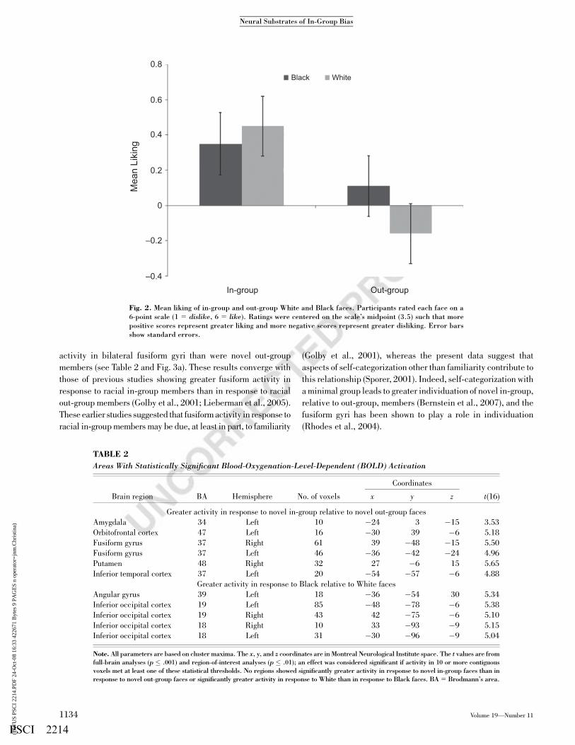
Novel Group Membership
Neuroimaging results were consistent with the idea that self-
categorization motivates social perception (see Table 2 for all
statistically significant effects of self-categorization on brain
activity). Novel in-group members were associated with greater
TABLE 1
Accuracy and Reaction Time During the Categorization Tasks
Task and face category
Accuracy Reaction time
Mean SD Mean SD
Implicit (race) task
White, in-group .96 .19 896 298
White, out-group .95 .22 902 310
Black, in-group .94 .24 862 282
Black, out-group .96 .19 849 264
Explicit (group) task
White, in-group .86 .35 1,210 329
White, out-group .79 .41 1,250 322
Black, in-group .79 .41 1,233 319
Black, out-group .79 .41 1,270 316
Note. Accuracy is the proportion of trails responded to correctly during the 2-sface presentation. Reaction time is the time in milliseconds between the pre-sentation of a face and the response to it. Trials on which the reaction time wasless than or equal to 300 ms are excluded.
3Although participants were more accurate at recognizing the team mem-bership of White than Black faces during the final memory task, F(1, 20) 5
5.43, prep 5 .91, there were no differences in accuracy between novel in-groupand out-group faces, regardless of race (preps < .77).
Volume 19—Number 11 1133
Jay J. Van Bavel, Dominic J. Packer, and William A. Cunningham
PSCI 2214(BW
US
PSC
I 22
14.P
DF
24-O
ct-0
8 16
:33
4226
71 B
ytes
9 P
AG
ES
n op
erat
or=
jnm
.Chr
istin
a)
activity in bilateral fusiform gyri than were novel out-group
members (see Table 2 and Fig. 3a). These results converge with
those of previous studies showing greater fusiform activity in
response to racial in-group members than in response to racial
out-group members (Golby et al., 2001; Lieberman et al., 2005).
These earlier studies suggested that fusiform activity in response to
racial in-group members may be due, at least in part, to familiarity
(Golby et al., 2001), whereas the present data suggest that
aspects of self-categorization other than familiarity contribute to
this relationship (Sporer, 2001). Indeed, self-categorization with
a minimal group leads to greater individuation of novel in-group,
relative to out-group, members (Bernstein et al., 2007), and the
fusiform gyri has been shown to play a role in individuation
(Rhodes et al., 2004).
0.8
0.6
0.4M
ean
Liki
ng
0.2
–0.2
In-group
Black White
Out-group–0.4
0
Fig. 2. Mean liking of in-group and out-group White and Black faces. Participants rated each face on a6-point scale (1 5 dislike, 6 5 like). Ratings were centered on the scale’s midpoint (3.5) such that morepositive scores represent greater liking and more negative scores represent greater disliking. Error barsshow standard errors.
TABLE 2
Areas With Statistically Significant Blood-Oxygenation-Level-Dependent (BOLD) Activation
Brain region BA Hemisphere No. of voxels
Coordinates
t(16)x y z
Greater activity in response to novel in-group relative to novel out-group faces
Amygdala 34 Left 10 �24 3 �15 3.53
Orbitofrontal cortex 47 Left 16 �30 39 �6 5.18
Fusiform gyrus 37 Right 61 39 �48 �15 5.50
Fusiform gyrus 37 Left 46 �36 �42 �24 4.96
Putamen 48 Right 32 27 �6 15 5.65
Inferior temporal cortex 37 Left 20 �54 �57 �6 4.88
Greater activity in response to Black relative to White faces
Angular gyrus 39 Left 18 �36 �54 30 5.34
Inferior occipital cortex 19 Left 85 �48 �78 �6 5.38
Inferior occipital cortex 19 Right 43 42 �75 �6 5.10
Inferior occipital cortex 18 Right 10 33 �93 �9 5.15
Inferior occipital cortex 18 Left 31 �30 �96 �9 5.04
Note. All parameters are based on cluster maxima. The x, y, and z coordinates are in Montreal Neurological Institute space. The t values are fromfull-brain analyses (p � .001) and region-of-interest analyses (p � .01); an effect was considered significant if activity in 10 or more contiguousvoxels met at least one of these statistical thresholds. No regions showed significantly greater activity in response to novel in-group faces than inresponse to novel out-group faces or significantly greater activity in response to White than in response to Black faces. BA 5 Brodmann’s area.
1134 Volume 19—Number 11
Neural Substrates of In-Group Bias
PSCI 2214(BW
US
PSC
I 22
14.P
DF
24-O
ct-0
8 16
:33
4226
71 B
ytes
9 P
AG
ES
n op
erat
or=
jnm
.Chr
istin
a)

Although previous research has suggested that amygdala
activity in response to Black faces reflects the processing of
negative information, recent models posit a more general role for
the amygdala in processing motivationally relevant stimuli (e.g.,
Cunningham et al., 2008). Results were consistent with this
latter model in that novel in-group members were associated
with greater amygdala activity than were novel out-group
members (see Table 2 and Fig. 3b). Although this result may
appear to differ from experimental results showing greater
amygdala activity in response to racial out-group faces relative
to racial in-group faces (e.g., Cunningham et al., 2004; Hart
et al., 2000), racial attitudes and stereotypes are likely to be
motivationally relevant in some intergroup contexts and irrele-
vant in others. For example, amygdala activity is greater for
racial out-group faces than for racial in-group faces when people
think of the faces in terms of their social category membership,
but amygdala activity is greater for racial in-group faces than for
racial out-group faces when people think of the faces as indi-
viduals (Wheeler & Fiske, 2005).
The contention that amygdala activity in response to novel
in-group members may reflect motivational consequences of
belonging to a group rather than negativity is corroborated by
our finding of greater activity associated with novel in-group
members, relative to out-group members, in the orbitofrontal
cortex (OFC; see Table 2 and Fig. 3c) and dorsal striatum
(putamen). The OFC plays a key role in linking social and
appetitive stimuli to hedonic experience (Kringelbach, 2005),
and the dorsal striatum is active during acts of mutual cooper-
ation (Rilling et al., 2002) and viewing of pictures of loved ones
(Bartels & Zeki, 2000).
No regions showed significantly greater activity for out-group
than for in-group faces.
Correlations Between Neural Activity and In-Group Bias
We tested whether the greater neural activity in response to
viewing novel in-group, relative to out-group, members was
associated with individual differences in in-group bias (i.e.,
liking in-group members more than out-group members). For
each ROI (fusiform gyri, amygdala, OFC, and striatum), we
calculated individual differences in brain activity (in-group –
out-group) and then correlated these scores with individual
differences in self-reported liking of the faces (in-group – out-
group). Participants with greater in-group bias in OFC activity
reported a stronger preference for in-group over out-group
members (r 5 .54, prep 5 .94).4 Moreover, OFC activity medi-
ated the in-group bias in self-reported liking for the faces.
Specifically, in-group bias was significantly reduced (from
b 5 0.389, prep 5 .92, to b 5 �0.131, prep 5 .63) when we
controlled for increases in OFC activity (in-group – out-group;
Sobel z 5 2.22, prep 5 .94).
Racial Group Membership
To further examine the neural components involved in pro-
cessing social groups, we compared brain activity in response to
Black and White faces. Black faces were associated with greater
activity in the visual cortex than were White faces (see Table 2);
no regions were more active in response to White faces than in
response to Black faces.5 These data are consistent with studies
showing that race may be largely ignored when it is orthogonal to
current group membership (Kurzban et al., 2001).
Interactions of Team With Race and Task
To examine the automaticity of these in-group biases in neural
processing, we tested whether the observed in-group biases in
brain activity were moderated by race (Black vs. White),
attention (explicit vs. implicit) to team membership, or both. For
each ROI (i.e., fusiform gyri, amygdala, OFC, and dorsal stria-
tum), we compared mean activity for Black versus White faces
and the implicit versus the explicit categorization tasks. In-
group biases in neural activity were not moderated by race,
categorization task, or a Race � Task interaction. Whereas
Fig. 3. Maps showing brain areas in which activity was greater for in-group than for out-group faces.Areas showing this effect were (a) the fusiform gyri (coronal view; y 5 �48), (b) the amygdala(coronal view; y 5 0), and (c) orbitofrontal cortex (sagittal view; x 5 �24).
4As in previous research, in-group bias (in-group – out-group) in self-reported liking was not correlated with amygdala activity (Phelps et al., 2000),or with activity in other regions showing in-group bias. We also conductedcorrelations between activity in regions showing in-group bias. Activity in theleft fusiform gyrus was correlated with activity in the right fusiform gyrus (r 5.62, prep 5 .97) and in the putamen (r 5 .59, prep 5 .97). No other correlationswere statistically significant.
5We extracted an ROI from each brain region showing in-group bias, and race(Black vs. White) did not have a significant effect on activity in any of theseregions.
Volume 19—Number 11 1135
Jay J. Van Bavel, Dominic J. Packer, and William A. Cunningham
PSCI 2214(BW
US
PSC
I 22
14.P
DF
24-O
ct-0
8 16
:33
4226
71 B
ytes
9 P
AG
ES
n op
erat
or=
jnm
.Chr
istin
a)

previous studies have shown that activity in these regions can be
modulated by explicit processing goals (Cunningham, Johnson,
Gatenby, Gore, & Banaji, 2003), the in-group biases in neural
activity reported here do not appear to require explicit attention
to team membership, nor do they pertain strictly to Black or
White faces.
GENERAL DISCUSSION
This study reveals a constellation of neural activity consistent
with models of flexible self-categorization (Turner et al., 1987).
Viewing novel in-group members was associated with greater
activation in the fusiform gyri, amygdala, OFC, and dorsal
striatum, relative to viewing novel out-group members. More-
over, OFC activity mediated the in-group bias in self-reported
liking for the faces. In-group biases in neural processing
occurred within minutes of team assignment, in the absence of
explicit team-based rewards or punishments, and independently
of preexisting attitudes, stereotypes, or familiarity. In-group
biases in neural processing were not moderated by the target’s
race or by the categorization task, which suggests that they did
not require explicit attention to team membership and may have
occurred relatively automatically.
This study provides neural evidence that in-group members
are processed in greater depth than out-group members—
placing in-group biases in perception firmly within the realm of
motivated social perception (Balcetis & Dunning, 2006). By
virtue of their motivational significance in a variety of contexts
(e.g., economic, psychological, and evolutionary), in-group
members often warrant greater, or deeper, processing than out-
group members (Brewer, 1979, 1999). By assigning participants
to novel groups and providing equal exposure to in-group and
out-group faces, we were able to minimize the roles of familiarity
and novelty as causal variables in the observed neural in-group
biases. The absence of expertise with the faces also raises the
possibility that the fusiform gyri may be associated with at-
tentional biases (Wojciulik, Kanwisher, & Driver, 1998) toward
in-group members, greater individuation (Rhodes et al., 2004) of
in-group relative to out-group members, or both.
Whereas earlier studies reported greater amygdala activity in
response to racial out-group, relative to in-group, faces—often
interpreted as reflecting negativity or fear toward out-group or
stigmatized-group members (e.g., Lieberman et al., 2005)—
participants in the current study had greater amygdala activity
in response to novel in-group, relative to out-group, members
(see also Chiao et al., in press). Wheeler and Fiske (2005) found
both patterns: Amygdala activity was greater in response to ra-
cial out-group faces during a social categorization task (deciding
whether a person was young or old), but was greater in response
to racial in-group faces during an individuation task. Wheeler
and Fiske’s study captures the flexibility of the amygdala, which
can respond to positive and negative stimuli, stimulus intensity,
and, more generally, the motivational relevance of stimuli
(Cunningham et al., 2008). The amygdala may be involved in
segregating relevant from irrelevant stimuli in order to enhance
perception of important stimuli (Anderson & Phelps, 2001;
Vuilleumier, 2005; Whalen, 1998). This view of amygdala
function offers an alternative explanation for previous results
showing that amygdala activity of both White and Black par-
ticipants is greater for Black than for White faces (Lieberman et
al., 2005). Amygdala activity among White participants may
reflect negative stereotypes toward Blacks, whereas amygdala
activity among Black participants (who generally have a stronger
racial identity and may therefore view Black faces as more rele-
vant than White faces; Crocker, Luhtanen, Blaine, & Broadnax,
1994) may reflect increased processing of in-group members. In
other words, different psychological mechanisms may guide the
processing of racial in-group and out-group members. We propose
that the amygdala activity in response to in-group members in the
current study stemmed from their motivational relevance and
salience in the current group context.
The relevance of most social category memberships also
varies according to social context (Turner et al., 1987). As-
signing people to mixed-race teams may change the way they
construe race, and sensitize perceptual and affective processes
to the currently-salient social category (i.e., team membership).
Indeed, people categorize others according to race when it is the
salient social category, but categorize according to team mem-
bership (and ignore race) when team membership is salient
(Kurzban et al., 2001). Whereas race was the most salient
difference between faces in previous fMRI studies, team mem-
bership was highly salient in the study reported here. In the
current study, in-group biases in neural activity were not mod-
erated by race; however, in contexts in which race provides the
most salient group distinction, attitudes, cultural stereotypes
(especially threat), and personal values (egalitarianism) may
provide the most relevant motivational guides to perception,
evaluation, and behavior. Moreover, people may process others
according to race when these others are unaffiliated with the
in-group or out-group (Van Bavel & Cunningham, in press).
The pattern of in-group bias in the current study extended to
self-reported preferences for in-group members. Participants
with a stronger preference for in-group members exhibited
stronger OFC activity in response to in-group relative to out-
group members. This brain-behavior relationship is consistent
with a recent study showing a strong correlation between activity
in a similar region of the OFC while participants tasted liquids
and self-reported pleasantness of the liquids (Kringelbach,
O’Doherty, Rolls, & Andrews, 2003) and, more generally, with
the idea that this region plays a central role in representing
and processing subjective value (Kringelbach, 2005). To our
knowledge, this is the first fMRI study to identify the neural
mediators of self-reported intergroup biases, and it demon-
strates an important link between the pervasive preference for
novel in-group members (Brewer, 1979) and brain regions that
process reward and subjective value (Kringelbach, 2005).
1136 Volume 19—Number 11
Neural Substrates of In-Group Bias
PSCI 2214(BW
US
PSC
I 22
14.P
DF
24-O
ct-0
8 16
:33
4226
71 B
ytes
9 P
AG
ES
n op
erat
or=
jnm
.Chr
istin
a)

In-group biases in neural activity did not require explicit
attention to team membership. Although the tasks differed in
difficulty (judging by the faster reaction times and higher
accuracy in the implicit task), neural in-group biases did not
differ across tasks. This finding suggests that these biases are
relatively automatic. Similarly, Bernstein et al. (2007) proposed
that ‘‘ingroup faces are processed in a default, automatic man-
ner, characterized by holistic processing’’ (p. 711). Indeed,
several regions activated by in-group members respond auto-
matically to social stimuli; for example, the amygdala responds
to subliminal presentations of arousing faces (Whalen et al.,
1998), and the fusiform gyri respond to faces within 100 to 200
ms (Liu, Harris, & Kanwisher, 2002). Future research will need
to use electroencephalography and other techniques to examine
the time course of the in-group biases demonstrated in this study
and whether or not they emerge prior to conscious awareness.
CONCLUSION
This study elucidates the neural substrates that underlie in-
group biases in perceptual and affective processing and clarifies
the role of the fusiform gyri and amygdala in intergroup contexts.
Although we have referred to the observed patterns of activity
using the term in-group bias, future research will need to include
neutral control faces to more precisely dissociate the neural
components of perceiving and evaluating both in-group and out-
group members. Humans are a fundamentally social species,
and understanding the neural processes that underlie intergroup
perception and evaluation promises to yield important insights
into how people navigate their complex social worlds.
REFERENCES
Allport, G.W. (1954). The nature of prejudice. Reading, MA: Addison
Wesley.
Anderson, A.K., & Phelps, E.A. (2001). Lesions of the human amyg-
dala impair enhanced perception of emotionally salient events.
Nature, 411, 305–309.
Balcetis, E., & Dunning, D. (2006). See what you want to see: Motiva-
tional influences on visual perception. Journal of Personality andSocial Psychology, 91, 612–625.
Bartels, A., & Zeki, S. (2000). The neural basis of romantic love.
NeuroReport, 11, 3829–3834.
Bernstein, M., Young, S., & Hugenberg, K. (2007). The cross-category
effect: Mere social categorization is sufficient to elicit an
own-group bias in face recognition. Psychological Science, 18,
709–712.
Brewer, M.B. (1979). In-group bias in the minimal intergroup situa-
tion: A cognitive-motivational analysis. Psychological Bulletin,
86, 307–324.
Brewer, M.B. (1999). The psychology of prejudice: Ingroup love or
outgroup hate? Journal of Social Issues, 55, 429–444.
Chiao, J.Y., Iidaka, T., Gordon, H.L., Nogawa, J., Bar, M., Aminoff, E.,
et al. (in press) Cultural specificity in amygdala response to fear
faces. Journal of Cognitive Neuroscience.
Correll, J., & Park, B. (2005). A model of the ingroup as a social
resource. Personality and Social Psychology Review, 9, 341–359.
Crocker, J., Luhtanen, R., Blaine, B., & Broadnax, S. (1994). Collec-
tive self-esteem and psychological well-being among White,
Black, and Asian college students. Personality and SocialPsychology Bulletin, 20, 503–513.
Cunningham, W.A., Johnson, M.K., Gatenby, J.C., Gore, J.C., &
Banaji, M.R. (2003). Neural components of social evaluation.
Journal of Personality and Social Psychology, 85, 639–649.
Cunningham, W.A., Johnson, M.K., Raye, C.L., Gatenby, J.C., Gore,
J.C., & Banaji, M.R. (2004). Separable neural components in the
processing of Black and White faces. Psychological Science, 15,
806–813.
Cunningham, W.A., Van Bavel, J.J., & Johnsen, I.R. (2008). Affective
flexibility: Evaluative processing goals shape amygdala activity.
Psychological Science, 19, 152–160.
Dubois, S., Rossion, B., Schiltz, C., Bodart, J.M., Michel, C., Bruyer,
R., & Crommelinck, M. (1999). Effect of familiarity on the
processing of human faces. NeuroImage, 9, 278–289.
Gauthier, I., Tarr, M.J., Anderson, A.W., Skudlarski, P., & Gore, J.C.
(1999). Activation of the middle fusiform ‘face area’ increases
with expertise in recognizing novel objects. Nature Neuroscience,
2, 568–573.
Golby, A.J., Gabrieli, J.D.E., Chiao, J.Y., & Eberhardt, J.L. (2001).
Differential fusiform responses to same- and other-race faces.
Nature Neuroscience, 4, 845–850.
Harris, L.T., & Fiske, S.T. (2006). Dehumanizing the lowest of the low:
Neuroimaging responses to extreme out-groups. PsychologicalScience, 17, 847–853.
Hart, A.J., Whalen, P.J., Shin, L.M., McInerney, S.C., Fischer, H., &
Rauch, S.L. (2000). Differential response in the human amygdala
to racial outgroup versus ingroup face stimuli. NeuroReport, 11,
2351–2355.
Kanwisher, N., McDermott, J., & Chun, M. (1997). The fusiform
face area: A module in human extrastriate cortex specialized
for the perception of faces. Journal of Neuroscience, 17, 4302–
4311.
Kringelbach, M.L. (2005). The human orbitofrontal cortex: Linking
reward to hedonic experience. Nature Reviews Neuroscience, 6,
691–702.
Kringelbach, M.L., O’Doherty, J., Rolls, E.T., & Andrews, C. (2003).
Activation of the human orbitofrontal cortex to a liquid food
stimulus is correlated with its subjective pleasantness. Cerebral
Cortex, 13, 1064–1071.
Kurzban, R., Tooby, J., & Cosmides, L. (2001). Can race be erased?
Coalitional computation and social categorization. Proceedings of
the National Academy of Sciences, USA, 98, 15387–15392.
LeDoux, J.E. (1996). The emotional brain: The mysterious underpin-
nings of emotional life. New York: Simon & Schuster.
Levin, D.T. (1996). Classifying faces by race: The structure of face
categories. Journal of Experimental Psychology: Learning,Memory, and Cognition, 22, 1364–1382.
Lieberman, M.D., Hariri, A., Jarcho, J.M., Eisenberger, N.I., &
Bookheimer, S.Y. (2005). An fMRI investigation of race-related
amygdala activity in African-American and Caucasian-American
individuals. Nature Neuroscience, 8, 720–722.
Liu, J., Harris, A., & Kanwisher, N. (2002). Stages of processing in face
perception: An MEG study. Nature Neuroscience, 5, 910–916.
Malpass, R.S., & Kravitz, J. (1969). Recognition for faces of own and
other ‘‘race.’’. Journal of Personality and Social Psychology, 13,
330–334.
Phelps, E.A., O’Connor, K.J., Cunningham, W.A., Funayama, E.S.,
Gatenby, J.C., Gore, J.C., & Banaji, M.R. (2000). Performance on
Volume 19—Number 11 1137
Jay J. Van Bavel, Dominic J. Packer, and William A. Cunningham
PSCI 2214(BW
US
PSC
I 22
14.P
DF
24-O
ct-0
8 16
:33
4226
71 B
ytes
9 P
AG
ES
n op
erat
or=
jnm
.Chr
istin
a)
indirect measures of race evaluation predicts amygdala activa-
tion. Journal of Cognitive Neuroscience, 12, 729–738.
Rhodes, G., Byatt, G., Michie, P.T., & Puce, A. (2004). Is the fusiform
face area specialized for faces, individuation, or expert individ-
uation? Journal of Cognitive Neuroscience, 16, 189–203.
Rilling, J.K., Gutman, D.A., Zeh, T.R., Pagnoni, G., Berns, G.S., &
Kilts, C.D. (2002). A neural basis for social cooperation. Neuron,
35, 395–405.
Sporer, S.L. (2001). Recognizing faces of other ethnic groups: An inte-
gration of theories. Psychology, Public Policy, and Law, 7, 36–97.
Tajfel, H. (1970). Experiments in intergroup discrimination. ScientificAmerican, 223, 96–102.
Tajfel, H. (1982). Social identity and intergroup behavior. Cambridge,
England: Cambridge University Press.
Turner, J.C., Hogg, M.A., Oakes, P.J., Reicher, S.D., & Wetherell, M.S.
(1987). Rediscovering the social group: A self-categorizationtheory. Oxford, England: Basil Blackwell.
Van Bavel, J.J., & Cunningham, W.A. (in press). Self-categorization
with a novel mixed-race group moderates automatic social and
racial biases. Personality and Social Psychology Bulletin.
Vuilleumier, P. (2005). How brains beware: Neural mechanisms of
emotional attention. Trends in Cognitive Sciences, 9, 585–594.
Whalen, P.J. (1998). Fear, vigilance and ambiguity: Initial neuro-
imaging studies of the human amygdala. Current Directions inPsychological Science, 7, 177–188.
Whalen, P.J., Rauch, S.L., Etcoff, N.L., McInerney, S.C., Lee, M., &
Jenike, M.A. (1998). Masked presentations of emotional facial
expressions modulate amygdala activity without explicit knowl-
edge. Journal of Neuroscience, 18, 411–418.
Wheeler, M.E., & Fiske, S.T. (2005). Controlling racial prejudice:
Social-cognitive goals affect amygdala and stereotype activation.
Psychological Science, 16, 56–63.
Wojciulik, E., Kanwisher, N., & Driver, J. (1998). Modulation of
activity in the fusiform face area by covert attention: An fMRI
study. Journal of Neurophysiology, 79, 1574–1579.
(RECEIVED 8/24/07; REVISION ACCEPTED 5/7/08)
1138 Volume 19—Number 11
Neural Substrates of In-Group Bias
PSCI 2214(BW
US
PSC
I 22
14.P
DF
24-O
ct-0
8 16
:33
4226
71 B
ytes
9 P
AG
ES
n op
erat
or=
jnm
.Chr
istin
a)

Author Query Form
_______________________________________________________
_______________________________________________________
Dear Author,
During the copy-editing of your paper, the following queries arose. Please respond to these by marking up your proofs with the necessary changes/additions. Please write your answers clearly on the query sheet if there is insufficient space on the page proofs. If returning the proof by fax do not write too close to the paper's edge. Please remember that illegible mark-ups may delay publication.
Journal PSCIArticle 2214
Query No. Description Author Response
.No Queries