Rehabilitation and Repression: Reassessing their Ideological Embeddedness
Upload
independentCategory
view
2download
0
The mechanism of repression of themyeloid-specific c-fms gene by Pax5 duringB lineage restriction
Hiromi Tagoh1, Richard Ingram1,5,Nicola Wilson1,5, Giorgia Salvagiotto2,Alan J Warren3,4, Deborah Clarke1,Meinrad Busslinger2 andConstanze Bonifer1,*1Division of Experimental Haematology, LIMM, University of Leeds,St James’s University Hospital, Leeds, UK, 2Research Institute ofMolecular Pathology, Vienna Biocenter, Vienna, Austria, 3MRCLaboratory of Molecular Biology, Cambridge, UK and 4Department ofHaematology, University of Cambridge, Cambridge, UK
The transcription factor Pax5 (BSAP) is required for the
expression of a B-cell-specific genetic program and for
B-cell differentiation, and also to suppress genes of alter-
native lineages. The molecular mechanism by which
repression of myeloid genes occurs during early B-lineage
restriction is unknown and in this study we addressed this
question. One of the genes repressed by Pax5 in B cells
is the colony-stimulating factor receptor 1 gene (csf1r or
c-fms). We examined the changes in chromatin caused by
Pax5 activity, and we show that Pax5 is directly recruited
to c-fms resulting in the rapid loss of RNA polymerase II
binding, followed by loss of transcription factor binding
and DNaseI hypersensitivity at all cis-regulatory elements.
We also show that Pax5 targets the basal transcription
machinery of c-fms by interacting with a binding site
within the major transcription start sites. Our results
support a model by which Pax5 does not lead to major
alterations in chromatin modification, but inhibits tran-
scription by interfering with the action of myeloid tran-
scription factors.
The EMBO Journal (2006) 25, 1070–1080. doi:10.1038/
sj.emboj.7600997; Published online 16 February 2006
Subject Categories: chromatin & transcription; immunology
Keywords: B-lineage restriction; c-fms; chromatin; gene
silencing; Pax5
Introduction
All blood cells originate from hematopoietic stem cells
(HSCs), which in the adult mammalian organism reside in
the bone marrow and have the capacity to self-renew as well
as differentiate (Weissman et al, 2001). Differentiating HSCs
undergo a gradual loss of developmental potential in favor of
functional specialization. This generates defined precursor
cell types, which give rise to functionally specialized blood
cells, and this differentiation process is concurrent with the
execution of different genetic programs. A conceptual break-
through in how cell fate decisions are made came from
experiments demonstrating that HSCs express a lineage pro-
miscuous gene expression program and that the chromatin of
genes not yet expressed in precursor cells is already partially
reorganized (Jimenez et al, 1992; Enver and Greaves, 1998;
Kontaraki et al, 2000; Tagoh et al, 2004a). Once cells start
to differentiate into specific blood cell types, genes for
inappropriate lineages are silenced, whereas lineage-appro-
priate genes are further activated. Lineage-specific transcrip-
tion factors play an essential role in this process. It is well
known that shifting the balance of specific transcription
factors in hematopoietic precursor cells dictates the outcome
of cell differentiation (reviewed in Orkin, 2000; Graf, 2002)
and this principle holds true for all differentiation decisions in
all multicellular organisms that have been studied so far.
Overexpression of the transcription factor PU.1 in multi-
potent progenitor cells shifts differentiation toward myeloid
cells (Nerlov and Graf, 1998; DeKoter and Singh, 2000;
Yamada et al, 2001; McIvor et al, 2003). PU.1 functions in
opposition to GATA-1, which regulates erythropoieses and
inhibits GATA-1 activity (Kulessa et al, 1995; Nerlov et al,
2000; Zhang et al, 2000; Heyworth et al, 2002). It has even
been shown that the overexpression of myeloid-specific
transcription factors in mature B cells can reprogram these
cells into macrophages (Xie et al, 2004).
Recent experiments have identified the B-cell-specific tran-
scription factor Pax5 as an important transcription factor
regulating commitment to the B lymphoid lineage and have
given crucial insights into how transcription factors regulate
cell fate decisions. Pax5 null cells in the bone marrow of
knockout mice are blocked in B-cell differentiation (Urbanek
et al, 1994; Nutt et al, 1997a; Hayashi et al, 2003), demon-
strating that Pax5 is required for the activation of B-cell-
specific genes (Nutt et al, 1998). Subsequent experiments
have shown that the conditional elimination of Pax5 in
immature and mature B cells leads to the re-expression of
myeloid-specific genes. This includes the colony-stimulating
factor 1 receptor gene (csf1r or c-fms) or the myeloperoxidase
gene (mpo), confirming that not only activators but also
repressors are required for the establishment of a specific
genetic program (Nutt et al, 1999; Mikkola et al, 2002; Tagoh
et al, 2004b). These experiments also showed that Pax5 is
able to act as activator and repressor in one cell type. How
Pax5 represses a subset of lymphoid-specific genes has been
studied in some detail. It interacts with the Groucho co-
repressor family member Grg4 to repress the activity of the
immunoglobulin heavy chain enhancer (Eberhard et al, 2000;
Linderson et al, 2004). However, ectopic expression of Pax5
in the myeloid lineage has no effect on myelopoiesis (SouabniReceived: 20 September 2005; accepted: 19 January 2006; publishedonline: 16 February 2006
*Corresponding author. Leeds Institute of Molecular Medicine, The JIFBuilding, St James University Hospital, University of Leeds, Leeds LS97TF, UK. Tel.: þ 44 113 343 8525; Fax: þ 44 113 343 8702;E-mail: [email protected] authors contributed equally to this work
The EMBO Journal (2006) 25, 1070–1080 | & 2006 European Molecular Biology Organization | All Rights Reserved 0261-4189/06
www.embojournal.org
The EMBO Journal VOL 25 | NO 5 | 2006 &2006 European Molecular Biology Organization
EMBO
THE
EMBOJOURNAL
THE
EMBOJOURNAL
1070
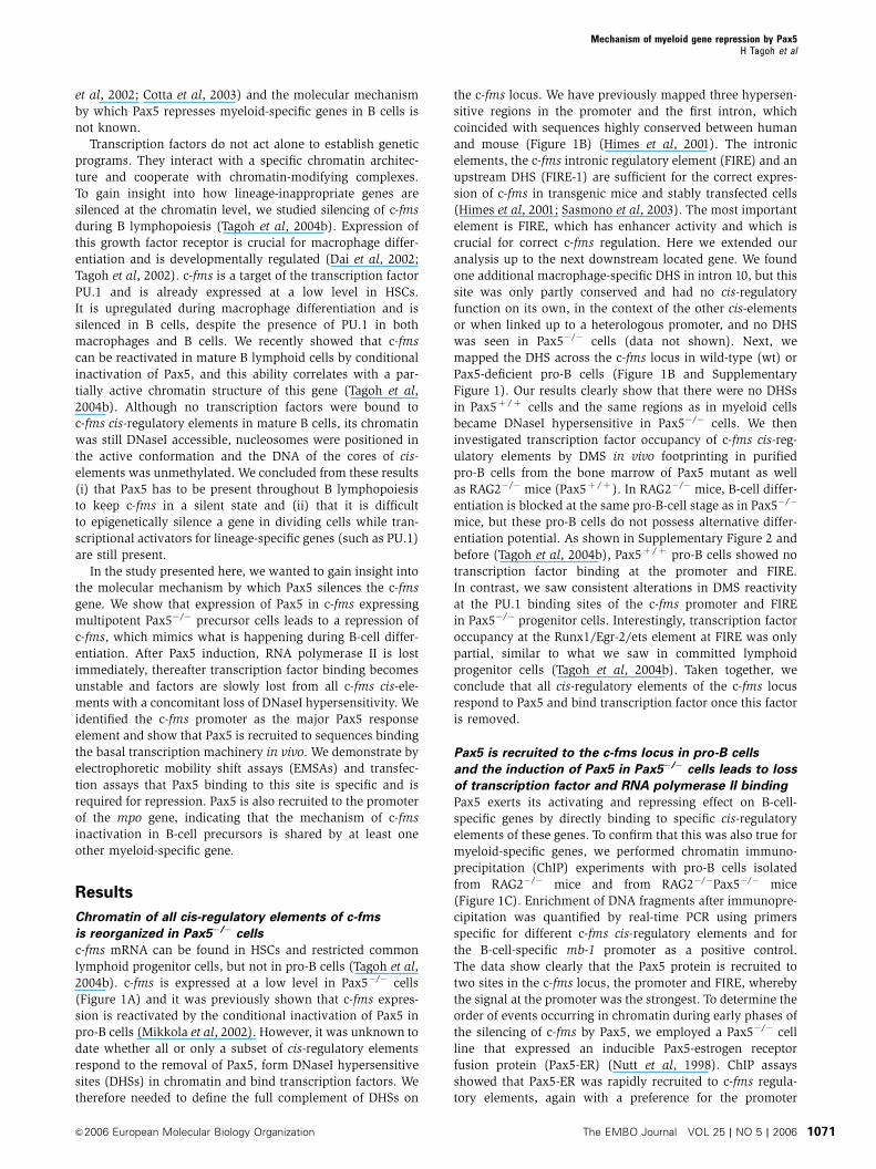
et al, 2002; Cotta et al, 2003) and the molecular mechanism
by which Pax5 represses myeloid-specific genes in B cells is
not known.
Transcription factors do not act alone to establish genetic
programs. They interact with a specific chromatin architec-
ture and cooperate with chromatin-modifying complexes.
To gain insight into how lineage-inappropriate genes are
silenced at the chromatin level, we studied silencing of c-fms
during B lymphopoiesis (Tagoh et al, 2004b). Expression of
this growth factor receptor is crucial for macrophage differ-
entiation and is developmentally regulated (Dai et al, 2002;
Tagoh et al, 2002). c-fms is a target of the transcription factor
PU.1 and is already expressed at a low level in HSCs.
It is upregulated during macrophage differentiation and is
silenced in B cells, despite the presence of PU.1 in both
macrophages and B cells. We recently showed that c-fms
can be reactivated in mature B lymphoid cells by conditional
inactivation of Pax5, and this ability correlates with a par-
tially active chromatin structure of this gene (Tagoh et al,
2004b). Although no transcription factors were bound to
c-fms cis-regulatory elements in mature B cells, its chromatin
was still DNaseI accessible, nucleosomes were positioned in
the active conformation and the DNA of the cores of cis-
elements was unmethylated. We concluded from these results
(i) that Pax5 has to be present throughout B lymphopoiesis
to keep c-fms in a silent state and (ii) that it is difficult
to epigenetically silence a gene in dividing cells while tran-
scriptional activators for lineage-specific genes (such as PU.1)
are still present.
In the study presented here, we wanted to gain insight into
the molecular mechanism by which Pax5 silences the c-fms
gene. We show that expression of Pax5 in c-fms expressing
multipotent Pax5�/� precursor cells leads to a repression of
c-fms, which mimics what is happening during B-cell differ-
entiation. After Pax5 induction, RNA polymerase II is lost
immediately, thereafter transcription factor binding becomes
unstable and factors are slowly lost from all c-fms cis-ele-
ments with a concomitant loss of DNaseI hypersensitivity. We
identified the c-fms promoter as the major Pax5 response
element and show that Pax5 is recruited to sequences binding
the basal transcription machinery in vivo. We demonstrate by
electrophoretic mobility shift assays (EMSAs) and transfec-
tion assays that Pax5 binding to this site is specific and is
required for repression. Pax5 is also recruited to the promoter
of the mpo gene, indicating that the mechanism of c-fms
inactivation in B-cell precursors is shared by at least one
other myeloid-specific gene.
Results
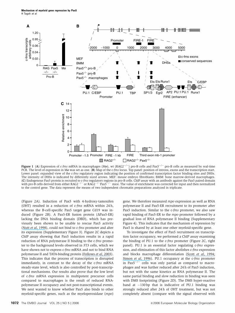
Chromatin of all cis-regulatory elements of c-fms
is reorganized in Pax5�/� cells
c-fms mRNA can be found in HSCs and restricted common
lymphoid progenitor cells, but not in pro-B cells (Tagoh et al,
2004b). c-fms is expressed at a low level in Pax5�/� cells
(Figure 1A) and it was previously shown that c-fms expres-
sion is reactivated by the conditional inactivation of Pax5 in
pro-B cells (Mikkola et al, 2002). However, it was unknown to
date whether all or only a subset of cis-regulatory elements
respond to the removal of Pax5, form DNaseI hypersensitive
sites (DHSs) in chromatin and bind transcription factors. We
therefore needed to define the full complement of DHSs on
the c-fms locus. We have previously mapped three hypersen-
sitive regions in the promoter and the first intron, which
coincided with sequences highly conserved between human
and mouse (Figure 1B) (Himes et al, 2001). The intronic
elements, the c-fms intronic regulatory element (FIRE) and an
upstream DHS (FIRE-1) are sufficient for the correct expres-
sion of c-fms in transgenic mice and stably transfected cells
(Himes et al, 2001; Sasmono et al, 2003). The most important
element is FIRE, which has enhancer activity and which is
crucial for correct c-fms regulation. Here we extended our
analysis up to the next downstream located gene. We found
one additional macrophage-specific DHS in intron 10, but this
site was only partly conserved and had no cis-regulatory
function on its own, in the context of the other cis-elements
or when linked up to a heterologous promoter, and no DHS
was seen in Pax5�/� cells (data not shown). Next, we
mapped the DHS across the c-fms locus in wild-type (wt) or
Pax5-deficient pro-B cells (Figure 1B and Supplementary
Figure 1). Our results clearly show that there were no DHSs
in Pax5þ /þ cells and the same regions as in myeloid cells
became DNaseI hypersensitive in Pax5�/� cells. We then
investigated transcription factor occupancy of c-fms cis-reg-
ulatory elements by DMS in vivo footprinting in purified
pro-B cells from the bone marrow of Pax5 mutant as well
as RAG2�/� mice (Pax5þ /þ ). In RAG2�/� mice, B-cell differ-
entiation is blocked at the same pro-B-cell stage as in Pax5�/�
mice, but these pro-B cells do not possess alternative differ-
entiation potential. As shown in Supplementary Figure 2 and
before (Tagoh et al, 2004b), Pax5þ /þ pro-B cells showed no
transcription factor binding at the promoter and FIRE.
In contrast, we saw consistent alterations in DMS reactivity
at the PU.1 binding sites of the c-fms promoter and FIRE
in Pax5�/� progenitor cells. Interestingly, transcription factor
occupancy at the Runx1/Egr-2/ets element at FIRE was only
partial, similar to what we saw in committed lymphoid
progenitor cells (Tagoh et al, 2004b). Taken together, we
conclude that all cis-regulatory elements of the c-fms locus
respond to Pax5 and bind transcription factor once this factor
is removed.
Pax5 is recruited to the c-fms locus in pro-B cells
and the induction of Pax5 in Pax5�/� cells leads to loss
of transcription factor and RNA polymerase II binding
Pax5 exerts its activating and repressing effect on B-cell-
specific genes by directly binding to specific cis-regulatory
elements of these genes. To confirm that this was also true for
myeloid-specific genes, we performed chromatin immuno-
precipitation (ChIP) experiments with pro-B cells isolated
from RAG2�/� mice and from RAG2�/�Pax5�/� mice
(Figure 1C). Enrichment of DNA fragments after immunopre-
cipitation was quantified by real-time PCR using primers
specific for different c-fms cis-regulatory elements and for
the B-cell-specific mb-1 promoter as a positive control.
The data show clearly that the Pax5 protein is recruited to
two sites in the c-fms locus, the promoter and FIRE, whereby
the signal at the promoter was the strongest. To determine the
order of events occurring in chromatin during early phases of
the silencing of c-fms by Pax5, we employed a Pax5�/� cell
line that expressed an inducible Pax5-estrogen receptor
fusion protein (Pax5-ER) (Nutt et al, 1998). ChIP assays
showed that Pax5-ER was rapidly recruited to c-fms regula-
tory elements, again with a preference for the promoter
Mechanism of myeloid gene repression by Pax5H Tagoh et al
&2006 European Molecular Biology Organization The EMBO Journal VOL 25 | NO 5 | 2006 1071
(Figure 2A). Induction of Pax5 with 4-hydroxy-tamoxifen
(OHT) resulted in a reduction of c-fms mRNA within 24 h,
whereas the B-cell-specific Pax5 target gene Cd19 was in-
duced (Figure 2B). A Pax5-ER fusion protein (DPax5-ER)
lacking the DNA binding domain (DBD), which has pre-
viously been shown to be unable to rescue Pax5 activity
(Nutt et al, 1998), could not bind to c-fms promoter and alter
its expression (Supplementary Figure 3). Figure 2C depicts a
ChIP assay showing that Pax5 induction results in a rapid
reduction of RNA polymerase II binding to the c-fms promo-
ter to the background levels observed in 3T3 cells, which we
have shown not to express c-fms mRNA and not to bind RNA
polymerase II and TATA-binding protein (Follows et al, 2003).
This indicates that the process of transcription is disrupted
immediately, in contrast to the decay of the c-fms mRNA
steady-state level, which is also controlled by post-transcrip-
tional mechanisms. Our results also prove that the low level
of c-fms mRNA expression in multipotent precursor cells
compared to macrophages is the result of reduced RNA-
polymerase II occupancy and not post-transcriptional events.
We next wanted to know whether Pax5 also binds to other
myeloid-specific genes, such as the myeloperoxidase (mpo)
gene. We therefore measured mpo expression as well as RNA
polymerase II and Pax5-ER recruitment to its promoter after
Pax5 induction. Similar to the c-fms promoter, we also saw
rapid binding of Pax5-ER to the mpo promoter followed by a
gradual loss of RNA polymerase II binding (Supplementary
Figure 4). This indicates that the mechanism of repression by
Pax5 is shared by at least one other myeloid-specific gene.
To investigate the effect of Pax5 recruitment on transcrip-
tion factor occupancy, we performed a ChIP assay examining
the binding of PU.1 to the c-fms promoter (Figure 2C, right
panel). PU.1 is an essential factor regulating c-fms expres-
sion, and elimination of this factor abolishes c-fms expression
and blocks macrophage differentiation (Scott et al, 1994;
Simon et al, 1996). PU.1 occupancy at the c-fms promoter
in Pax5�/� cells was only partial as compared to macro-
phages and was further reduced after 24 h of Pax5 induction,
but not with the same kinetics as RNA polymerase II. The
same partial binding and slow reduction in binding was seen
with DMS footprinting (Figure 2D). The DMS hyper-reactive
band at �130 bp that is indicative of PU.1 binding was
strongly reduced after 24 h of OHT treatment, but was not
completely absent (compare with the signal observed with
B
C
A
−2000 −1000 0 1000 2000 3000 4000 5000
c-fms exonsconserved sequences
Pax5+/+ pro-B
Pax5−/− macrophages
MEF
Pax5−/− pro-B
BMM DHSs
FIREFIRE-1Promoter
AAAAAAAA
PromoterC/EBP PU.1PU.1 TBP PU.1 PU.1
EtsEts Ets
Egr2SP1/3
Runx1
Runx1AP2
C/EBP
c-fm
s tr
ansc
ripts
arbi
trar
y un
its
0
0.02
0.04
0.06
0.95
1.20
RAG–/– –/–
Pax5 Mø
Pro-B
0
2
4
6
8
10
12
14
Promoter −1.5 Promoter FIRE −1 kb FIRE Third exon mb-1 promoter
RAG2–/– RAG2–/– Pax5–/–
Rel
ativ
e en
richm
ent
FIRE (enhancer)
Figure 1 (A) Expression of c-fms mRNA in macrophages (M^), wt (RAG2�/�) pro-B cells and Pax5�/� pro-B cells as measured by real-timePCR. The level of expression in M^ was set as one. (B) Map of the c-fms locus. Top panel: position of introns, exons and the transcription start.Lower panel: expanded view of the c-fms regulatory region indicating the position of confirmed transcription factor binding sites and DHSs.The intensity of DHSs is indicated by differently sized arrows. MEF: mouse embryo fibroblasts; BMM: bone marrow-derived macrophages.(C) Endogenous Pax5 protein is recruited to c-fms regulatory regions in pro-B cells. ChIP assay with an antibody against the Pax5 paired domainwith pro-B cells derived from either RAG2�/� or RAG2�/�Pax5�/� mice. The value of enrichment was corrected for input and then normalizedto the control gene. The data represent the means of two independent chromatin preparations analyzed in triplicate.
Mechanism of myeloid gene repression by Pax5H Tagoh et al
The EMBO Journal VOL 25 | NO 5 | 2006 &2006 European Molecular Biology Organization1072

T cells). We also noted increased DMS reactivity of a G at
�113 bp after OHT induction (marked by an asterisk). By
contrast, transcription factors binding to the FIRE enhancer
were completely lost (Figure 2D, right panel). This held true
for PU.1 binding at þ 2781 bp, and for other transcription
factors such as those binding to the Runx1/Egr-2/ets cluster
at þ 2717 bp.
In order to gain insights into the mechanism of repression
activity by Pax5 at the level of chromatin, we studied the
effect of Pax5 induction on different features of c-fms chro-
matin fine structure (Figures 3 and 4 and Supplementary
Figures 5 and 6). We first examined the effect of Pax5
induction on DNaseI hypersensitivity using a novel high-
resolution ligation-mediated PCR (LM-PCR) assay. To this
end, we treated uninduced, induced and control cells with
DNaseI, followed by direct linker ligation and amplification.
This method only amplifies double-strand cuts that are
indicative of highly distorted regions in chromatin, which
allows the enzyme to simultaneously access opposite DNA
strands. Figure 3A shows that the region over the main
transcription start sites at the promoter was strongly
DNaseI hypersensitive, and this hypersensitivity was progres-
sively but not immediately lost after Pax5 induction. The
same held true for the FIRE enhancer. This loss of DNaseI
hypersensitivity was not observed with a ribosomal RNA
gene analyzed as a control (Supplementary Figure 5A).
General DNaseI accessibility at these elements, which is an
indication of the extent of chromatin condensation as mea-
sured by single-strand specific LM-PCR, was not altered
(Supplementary Figure 6). The c-fms promoter in c-fms
non-expressing cells such as 3T3 cells is organized in a
positioned nucleosome, which is relocated once the gene is
expressed, thus exposing the different transcription start sites
to RNA polymerase II binding (Tagoh et al, 2004b). Pax5�/�
cells show a partially shifted nucleosome, and nucleosome
positioning was not altered by induction of Pax5 as measured
by LM-PCR on micrococcal nuclease (MNase)-digested chro-
matin (Figure 4). This is in keeping with our earlier data
showing that c-fms chromatin is still in a partially active and
accessible conformation in B cells (Tagoh et al, 2004b).
0
1.5
1.0
0.5
90
60
30
00 h 24 h15 h1 h
Time after induction ofPax5 by OHT
c-fms
CD19
Tra
nscr
ipts
arb
itrar
y un
its(t
he le
vel i
n un
indu
ced
cells
: 1)
G T MØ – +OHT
induction G T MØ – +OHT
induction
−130
+2717
+2781
PU.1
Runx1
Egr-2
Ets
PU.1
PU.1Promoter upper strand
FIRE lower strand
*
0 h 12 h2 h 24 hTime after induction of Pax5 by OHT
0
2
10
8
4
6
Promoter–1.5 kb
Promoter FIRE –1 kb Third exonFIRE 5′
Rel
ativ
e en
richm
ent
Pax5-ERR
elat
ive
enric
hmen
t
0
1.5
1.0
0.5
25
30
0
4.0
1.5
1.0
5.0
0.5
RNA polymerase II PU.1
Pro-Bcell line
0 h 24 h12 h2 h3T3 Pax5–/– Pax5–/–WTRAW264
Pax5-ER cells+OHT
Pro-Bcell line
0 h 24 h12 h2 h3T3 WTRAW264
Pax5-ER cells+OHT
A
C D
B
Figure 2 (A) Pax5-ER fusion protein is rapidly recruited to c-fms cis-regulatory elements. ChIP assay with a human ERa antibody. The value ofenrichment was corrected for input and normalized to the control gene. The data represent the means of three independent chromatinpreparations analyzed in triplicate. (B) c-fms mRNA expression is repressed after Pax5 induction with OHT (upper panel), whereas CD19mRNA is induced (lower panel). The scale was set with the levels in non-induced cells as one. (C) RNA polymerase II binding to the c-fmspromoter is reduced to background levels as seen in NIH3T3 cells (3T3) or wt pro-B cells (wt) after Pax5 induction (left panel). PU.1 is onlystarting to be lost after 12 h of OHT induction (right panel). The recruitment of these proteins was measured by a ChIP assay in the indicatedcell types. The experiment also demonstrates that PU.1 and RNA polymerase II binding is only partial (compare with the signals obtained withRAW264 cells). The value of enrichment was corrected for input and then normalized to the control gene. Relative enrichment in non-inducedPax5-ER cells was presented as one. The data represent the means of three independent chromatin preparations analyzed in triplicate. (D) DMSin vivo footprinting experiment examining transcription factor binding in the indicated cell types. G: DMS-piperidine cleavage reactionperformed on naked DNA; T: CD3þ thymocytes; M^: bone marrow-derived macrophages; �/þ : Pax5-ER cells without OHT (�) or after 24 hof OHT treatment (þ ). Black circles indicate DMS hyper-reactivity, gray circles indicate different degrees of weak hyper-reactivity and whitecircles indicate DMS hypo-reactivity. The numbers indicate the nucleotide position relative to the ATG. Positions of transcription factor bindingsites are indicated.
Mechanism of myeloid gene repression by Pax5H Tagoh et al
&2006 European Molecular Biology Organization The EMBO Journal VOL 25 | NO 5 | 2006 1073
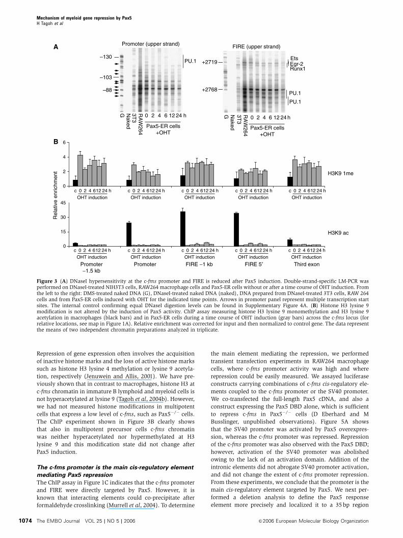
Repression of gene expression often involves the acquisition
of inactive histone marks and the loss of active histone marks
such as histone H3 lysine 4 methylation or lysine 9 acetyla-
tion, respectively (Jenuwein and Allis, 2001). We have pre-
viously shown that in contrast to macrophages, histone H3 at
c-fms chromatin in immature B lymphoid and myeloid cells is
not hyperacetylated at lysine 9 (Tagoh et al, 2004b). However,
we had not measured histone modifications in multipotent
cells that express a low level of c-fms, such as Pax5�/� cells.
The ChIP experiment shown in Figure 3B clearly shows
that also in multipotent precursor cells c-fms chromatin
was neither hyperacetylated nor hypermethylated at H3
lysine 9 and this modification state did not change after
Pax5 induction.
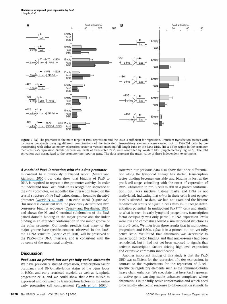
The c-fms promoter is the main cis-regulatory element
mediating Pax5 repression
The ChIP assay in Figure 1C indicates that the c-fms promoter
and FIRE were directly targeted by Pax5. However, it is
known that interacting elements could co-precipitate after
formaldehyde crosslinking (Murrell et al, 2004). To determine
the main element mediating the repression, we performed
transient transfection experiments in RAW264 macrophage
cells, where c-fms promoter activity was high and where
repression could be easily measured. We assayed luciferase
constructs carrying combinations of c-fms cis-regulatory ele-
ments coupled to the c-fms promoter or the SV40 promoter.
We co-transfected the full-length Pax5 cDNA, and also a
construct expressing the Pax5 DBD alone, which is sufficient
to repress c-fms in Pax5�/� cells (D Eberhard and M
Busslinger, unpublished observations). Figure 5A shows
that the SV40 promoter was activated by Pax5 overexpres-
sion, whereas the c-fms promoter was repressed. Repression
of the c-fms promoter was also observed with the Pax5 DBD;
however, activation of the SV40 promoter was abolished
owing to the lack of an activation domain. Addition of the
intronic elements did not abrogate SV40 promoter activation,
and did not change the extent of c-fms promoter repression.
From these experiments, we conclude that the promoter is the
main cis-regulatory element targeted by Pax5. We next per-
formed a deletion analysis to define the Pax5 response
element more precisely and localized it to a 35 bp region
–130
–103
–88
G Naked
3T3 0 2412642
RA
W264
h G Naked
3T3 0 2412642
RA
W264
h
+2719
+2768
PU.1
PU.1
PU.1
Runx1Egr-2Ets
Promoter (upper strand) FIRE (upper strand)
0
2
6
4
0 2412642 hcOHT induction
0 2412642 hcOHT induction
0 2412642 hcOHT induction
0 2412642 hcOHT induction
0 2412642 hcOHT induction
0 2412642 hcOHT induction
0 2412642 hcOHT induction
0 2412642 hcOHT induction
0 2412642 hcOHT induction
0 2412642 hcOHT induction
15
H3K9 1me
0
45
30
H3K9 ac
Promoter−1.5 kb
Third exonFIRE 5′FIRE −1 kbPromoter
Rel
ativ
e en
richm
ent
Pax5-ER cells+OHT
Pax5-ER cells+OHT
A
B
Figure 3 (A) DNaseI hypersensitivity at the c-fms promoter and FIRE is reduced after Pax5 induction. Double-strand-specific LM-PCR wasperformed on DNaseI-treated NIH3T3 cells, RAW264 macrophage cells and Pax5-ER cells without or after a time course of OHT induction. Fromthe left to the right: DMS-treated naked DNA (G), DNaseI-treated naked DNA (naked), DNA prepared from DNaseI-treated 3T3 cells, RAW 264cells and from Pax5-ER cells induced with OHT for the indicated time points. Arrows in promoter panel represent multiple transcription startsites. The internal control confirming equal DNaseI digestion levels can be found in Supplementary Figure 4A. (B) Histone H3 lysine 9modification is not altered by the induction of Pax5 activity. ChIP assay measuring histone H3 lysine 9 monomethylation and H3 lysine 9acetylation in macrophages (black bars) and in Pax5-ER cells during a time course of OHT induction (gray bars) across the c-fms locus (forrelative locations, see map in Figure 1A). Relative enrichment was corrected for input and then normalized to control gene. The data representthe means of two independent chromatin preparations analyzed in triplicate.
Mechanism of myeloid gene repression by Pax5H Tagoh et al
The EMBO Journal VOL 25 | NO 5 | 2006 &2006 European Molecular Biology Organization1074

between �135 and �100 bp relative to the ATG (Figure 5B).
Pax5 repressed transcription in all constructs carrying trans-
activator (mostly PU.1) binding sites, but repression was lost
once we deleted the most proximal PU.1 site.
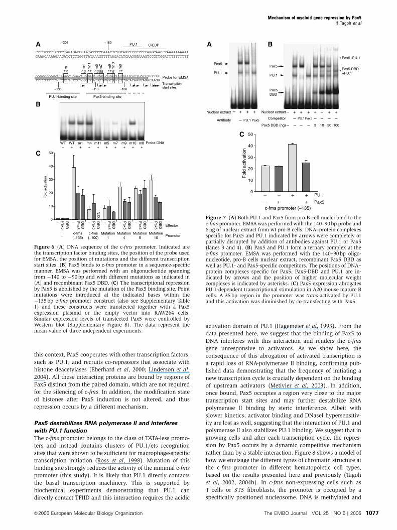
Pax5 binds to region harboring the main c-fms
transcription start sites and represses transcription
without abrogating PU.1 binding
To define the Pax5 binding site and to identify the mechanism
by which Pax5 represses c-fms, we performed EMSAs with a
51 bp oligonucleotide harboring the Pax5 responsive region
and recombinant Pax5 protein (DBD) (Figure 6A). Figure 6B
demonstrates that the Pax5 DBD bound to this oligonucleo-
tide. Pax5 DBD bound with approximately the same affinity
to the binding site for the B-cell-specific gene Pax5 target gene
mb-1, where Pax5 acts as an activator. However, compared to
the Pax5 binding site in the B-cell-specific CD19 gene, the
binding affinity was significantly lower (Supplementary
Figure 7). In order to test whether binding was specific and
to obtain information about binding site specificity, we
performed EMSAs with oligonucleotides in which specific
bases were mutated (Figure 6B), and tested the effect of these
mutations on the transcriptional activity of the c-fms promo-
ter in the presence and absence of Pax5 (Figure 6C). Mutation
1 abrogated binding of PU.1 (data not shown) but not of
Pax5. A construct harboring this mutation had the same low
basal activity as the �100 bp promoter in a transfection assay
and was not further repressed by Pax5. This indicates that the
elimination of PU.1 binding and the repression by Pax5
reduce transcription to a similarly low level. Constructs that
harbored mutations 4 and 10 that did not eliminate Pax5
binding, or mutation 7 that showed weaker binding activity
were still repressed by Pax5. Mutations 11, 5, 9 and 8 that
eliminated Pax5 binding compromised repression by the full-
length Pax5 protein or the DBD alone (Figure 6C and data not
shown). The guanine base at �113 bp that was altered by
mutation 5 is most likely in direct contact with Pax5, because
it became reproducibly hyper-reactive to DMS after Pax5
induction (see Figure 2D, marked with an asterisk).
With its paired domain Pax5 makes extended and variable
contacts to the DNA bases and phosphate backbone (Czerny
et al, 1993; Garvie et al, 2001). From our in vivo data, it was
already clear that Pax5 recruitment parallels the loss of RNA
polymerase II from the c-fms promoter. However, the same
experiments also showed a loss of PU.1, albeit with slower
kinetics (Figure 2C). We therefore wanted to investigate
whether destabilization of PU.1 binding contributes to the
repression of the c-fms promoter by Pax5. To this end, we
performed EMSAs with a probe harboring the Pax5 respon-
sive region, nuclear extracts from a wt pro-B-cell line and
increasing amounts of recombinant Pax5 (Figure 7). B cells
contain PU.1 as well as Pax5, and both bind to the c-fms
promoter as demonstrated by EMSA (Figure 7A and B).
Specificity of binding was confirmed by abrogation of binding
with Pax5- and PU.1-specific antibodies (Figure 7A) and by
competition with an excess of Pax5 or PU.1 consensus
oligonucleotide (Figure 7B). No Pax5-specific band was ob-
served with extracts from Pax5�/� cells, whereas the PU.1-
specific band was still present (data not shown). The addition
of increasing amounts of recombinant Pax5 led to the dis-
appearance of both bands, indicating that recombinant and
wt Pax5 compete for the same binding site. We also saw the
appearance of a new higher molecular weight complex
(asterisks in Figure 7B). This new complex contained Pax5
and PU.1, as shown by incubation of the reactions with
antibodies specific for PU.1 and Pax5 (data not shown).
The data outlined so far indicate that Pax5 binding did not
immediately interfere with PU.1 binding in vivo and in vitro,
but led to a rapid loss of RNA polymerase II and repression of
transcription in vivo. However, removal of PU.1 also abro-
gated Pax5 repression and reduced c-fms promoter activity to
the same background level. This indicated that Pax5 could
block activated expression rather than repressing it alto-
gether. To this end, we performed co-transfection experi-
ments in a B-cell line using the �135 bp minimal c-fms
promoter carrying the Pax5 response element. Owing to the
presence of endogenous Pax5, the activity of the c-fms
promoter was repressed to the same lowered activity as in
RAW264 cells overexpressing Pax5 (Figure 5B); however, this
repression could be overcome by overexpression of PU.1,
resulting in an approximately two-fold stimulation of reporter
gene activity (Figure 7C). This stimulation was dependent on
the PU.1 binding site (data not shown). When Pax5 was
coexpressed, this stimulation was abrogated and c-fms pro-
moter activity was reduced to background levels and was not
further repressed. These experiments clearly demonstrate
that in B cells PU.1 is in direct competition with Pax5 and
that Pax5 interferes with PU.1 activity.
Pax5-ER+ OHT
0 2 4 6 h24G
PU.1
PU.1
C/EBP
–103
–130
–88
c-fms promoter (lower strand)
Naked
3T3
RA
W
Figure 4 Nucleosome positioning at the c-fms promoter is in apartially active conformation in Pax5�/� cells and is unchangedafter Pax5 induction. Naked DNA and chromatin prepared from theindicated cell populations was treated with MNase and double-strand breaks were detected by LM-PCR using primers specific forthe c-fms promoter. Horizontal arrows indicate the positions oftranscription start sites and transcription factor binding sitesare shown on the left. Alternate nucleosome positions in thedifferent cell types are depicted on the right. The internal controlconfirming equal MNase digestion levels can be found in Supple-mentary Figure 4B.
Mechanism of myeloid gene repression by Pax5H Tagoh et al
&2006 European Molecular Biology Organization The EMBO Journal VOL 25 | NO 5 | 2006 1075
A model of Pax5 interaction with the c-fms promoter
In contrast to a previously published report (Maitra and
Atchison, 2000), our data show that binding of Pax5 to
DNA is required to repress c-fms promoter activity. In order
to understand how Pax5 binds to its recognition sequence at
the c-fms promoter, we modelled the interaction based on the
crystal structure of the Pax5 paired domain bound to the mb-1
promoter (Garvie et al, 2001, PDB code 1K78) (Figure 8A).
Our model is consistent with the previously determined Pax5
consensus binding sequence (Czerny and Busslinger, 1995)
and shows the N- and C-terminal subdomains of the Pax5
paired domain binding in the major groove and the linker
binding in an extended conformation in the minor groove of
the c-fms promoter. Our model predicts that many of the
major groove base-specific contacts observed in the Pax5-
mb-1 DNA structure (Garvie et al, 2001) will be preserved at
the Pax5-c-fms DNA interface, and is consistent with the
outcome of the mutational analysis.
Discussion
Pax5 acts on primed, but not yet fully active chromatin
We have previously studied expression, transcription factor
occupancy and DNA-methylation status of the c-fms locus
in HSCs, and early restricted myeloid as well as lymphoid
progenitor cells, and we could show that c-fms mRNA is
expressed and occupied by transcription factors in the entire
early progenitor cell compartment (Tagoh et al, 2004b).
However, our previous data also show that once differentia-
tion along the lymphoid lineage has started, transcription
factor binding becomes unstable and binding is lost at the
pro-B-cell stage, coinciding with the onset of expression of
Pax5. Chromatin in pro-B cells is still in a poised conforma-
tion, but lacks inactive histone marks and DNA is not
methylated, indicating that c-fms in these cells is not epigen-
etically silenced. To date, we had not examined the histone
modification status of c-fms in cells with multilineage differ-
entiation potential. In multipotent Pax5�/� cells and similar
to what is seen in early lymphoid progenitors, transcription
factor occupancy was only partial, mRNA expression levels
were low and chromatin showed a similar poised structure as
in pro-B cells. We infer from these results that in multipotent
progenitors and HSCs, c-fms is in a primed but not yet fully
active state. We found that chromatin was accessible to
transcription factor binding and that nucleosomes had been
remodelled, but it had not yet been exposed to signals that
activate transcription factors driving high-level expression
and extensive chromatin modification.
Another important finding of this study is that the Pax5
DBD was sufficient for the repression of c-fms expression, in
contrast to the requirements for the repression of B-cell-
specific cis-regulatory elements such as the immunoglobulin
heavy chain enhancer. We speculate that here Pax5 represses
an active gene carrying stable enhancer complexes where
chromatin is in the fully active conformation and which need
to be rapidly silenced in response to differentiation stimuli. In
LucSV40
Luc
LucSV40 FIRE
FIRE-1LucSV40
FIRE-1 FIRELucc-fms
Lucc-fms
Lucc-fms FIRE
FIRE-1Lucc-fms
EmptyPax5DBD
EmptyPax5DBD
EmptyPax5DBD
EmptyPax5DBD
EmptyPax5DBD
EmptyPax5DBD
EmptyPax5DBD
EmptyPax5DBD
0 12060 180
Lucc-fms
Lucc-fms
Lucc-fms
Lucc-fms
LucEmpty
Pax5
Empty
Pax5
Empty
Pax5
Empty
Pax5
Empty
Pax5
0 10050 150
–201 –1
–180 –1
–135 –1
–100 –1
Fold activation Fold activation
–425 –1
–425 –1
–425 –1
–425 –1
+2470 +2958
+1578 +1928
+1578 +2958
A B
Figure 5 (A) The promoter is the main target of Pax5 repression and the DBD is sufficient for repression. Transient transfection studies withluciferase constructs carrying different combinations of the indicated cis-regulatory elements were carried out in RAW264 cells by co-transfecting with either an empty expression vector or vectors encoding full-length Pax5 or the Pax5 DBD. (B) A 35 bp region in the promotermediates Pax5 repression. Similar expression levels of transfected Pax5 were controlled by Western blot (Supplementary Figure 8). The foldactivation was normalized to the promoter-less reporter gene. The data represent the mean value of three independent experiments.
Mechanism of myeloid gene repression by Pax5H Tagoh et al
The EMBO Journal VOL 25 | NO 5 | 2006 &2006 European Molecular Biology Organization1076
this context, Pax5 cooperates with other transcription factors,
such as PU.1, and recruits co-repressors that associate with
histone deacetylases (Eberhard et al, 2000; Linderson et al,
2004). All these interacting proteins are bound by regions of
Pax5 distinct from the paired domain, which are not required
for the silencing of c-fms. In addition, the modification state
of histones after Pax5 induction is not altered, and thus
repression occurs by a different mechanism.
Pax5 destabilizes RNA polymerase II and interferes
with PU.1 function
The c-fms promoter belongs to the class of TATA-less promo-
ters and instead contains clusters of PU.1/ets recognition
sites that were shown to be sufficient for macrophage-specific
transcription initiation (Ross et al, 1998). Mutation of this
binding site strongly reduces the activity of the minimal c-fms
promoter (this study). It is likely that PU.1 directly contacts
the basal transcription machinery. This is supported by
biochemical experiments demonstrating that PU.1 can
directly contact TFIID and this interaction requires the acidic
activation domain of PU.1 (Hagemeier et al, 1993). From the
data presented here, we suggest that the binding of Pax5 to
DNA interferes with this interaction and renders the c-fms
gene unresponsive to activators. As we show here, the
consequence of this abrogation of activated transcription is
a rapid loss of RNA-polymerase II binding, confirming pub-
lished data demonstrating that the frequency of initiating a
new transcription cycle is crucially dependent on the binding
of upstream activators (Metivier et al, 2003). In addition,
once bound, Pax5 occupies a region very close to the major
transcription start sites and may further destabilize RNA
polymerase II binding by steric interference. Albeit with
slower kinetics, activator binding and DNaseI hypersensitiv-
ity are lost as well, suggesting that the interaction of PU.1 and
polymerase II also stabilizes PU.1 binding. We suggest that in
growing cells and after each transcription cycle, the repres-
sion by Pax5 occurs by a dynamic competitive mechanism
rather than by a stable interaction. Figure 8 shows a model of
how we envisage the different types of chromatin structure at
the c-fms promoter in different hematopoietic cell types,
based on the results presented here and previously (Tagoh
et al, 2002, 2004b). In c-fms non-expressing cells such as
T cells or 3T3 fibroblasts, the promoter is occupied by a
specifically positioned nucleosome. DNA is methylated and
A
B
C
WT WT m10m9m7m5m11m4 m8m1− +++++++++
Probe DNA
AAAAAAAAAAGGGGAAGAGGAGCCAGTGCAACAGACAGGAACGTGTTCATCTGTTCCTTTTTTTTTTCCCCTTCTCCTCGGTCACGTTGTGTGTCCTTGCACAAGTAGACAAGG
tc cg cgtt gg ttg ct
m1
m11
m5
m7
m10
m9
m8
m4
PU.1-binding site Pax5-binding site
Transcription start sites–130 –113 –100
CTTTGTTTTCTTCTAGAGACCCAATATTTCCAAATTCTGTAGTTCCCTTTCAGGCAACCTAAAAAAAAAAGAAACAAAAGAAGATCTCTGGGTTATAAAGGTTTAAGACATCAAGGGAAAGTCCGTTGGATTTTTTTTTT
–201 –180 PU.1 C/EBP
Pax5
– – – – – – –
Promoter−c-fms(−135)
c-fms(−100)
Mutation1
Mutation10
Mutation5
Mutation4
Fol
d ac
tivat
ion
0
10
20
50
40
30
DB
D
Pax5
DB
D
Pax5
DB
D
Pax5
Pax5
DB
D
Pax5
DB
D
Pax5
DB
D Effector
DB
DN
.D.
Probe for EMSA
Figure 6 (A) DNA sequence of the c-fms promoter. Indicated arethe transcription factor binding sites, the position of the probe usedfor EMSA, the position of mutations and the different transcriptionstart sites. (B) Pax5 binds to c-fms promoter in a sequence-specificmanner. EMSA was performed with an oligonucleotide spanningfrom �140 to �90 bp and with different mutations as indicated in(A) and recombinant Pax5 DBD. (C) The transcriptional repressionby Pax5 is abolished by the mutation of the Pax5 binding site. Pointmutations were introduced at the indicated bases within the�135 bp c-fms promoter construct (also see Supplementary Table1) and these constructs were transfected together with a Pax5expression plasmid or the empty vector into RAW264 cells.Similar expression levels of transfected Pax5 were controlled byWestern blot (Supplementary Figure 8). The data represent themean value of three independent experiments.
BA
C
Pax5+– +–+– +–
c-fms promoter (–135)
Fol
d ac
tivat
ion
– +Nuclear extract
Antibody
+ +
– PU.1 Pax5
Pax5
PU.1
– + + + + + ++Competitor
Pax5 DBD (ng)
– PU.1 Pax5
– – – – 3 10 30 100
– – – –
*Pax5 DBD+PU.1
Nuclear extract
Pax5
PU.1
Pax5DBD
* Pax5+PU.1
10
0
20
50
40
30
PU.1
Figure 7 (A) Both PU.1 and Pax5 from pro-B-cell nuclei bind to thec-fms promoter. EMSA was performed with the 140–90 bp probe and6mg of nuclear extract from wt pro-B cells. DNA–protein complexesspecific for Pax5 and PU.1 indicated by arrows were completely orpartially disrupted by addition of antibodies against PU.1 or Pax5(lanes 3 and 4). (B) Pax5 and PU.1 form a ternary complex at thec-fms promoter. EMSA was performed with the 140–90 bp oligo-nucleotide, pro-B cells nuclear extract, recombinant Pax5 DBD aswell as PU.1- and Pax5-specific competitors. The positions of DNA–protein complexes specific for Pax5, Pax5-DBD and PU.1 are in-dicated by arrows and the position of higher molecular weightcomplexes is indicated by asterisks. (C) Pax5 expression abrogatesPU.1-dependent transcriptional stimulation in A20 mouse mature Bcells. A 35 bp region in the promoter was trans-activated by PU.1and this activation was diminished by co-transfecting with Pax5.
Mechanism of myeloid gene repression by Pax5H Tagoh et al
&2006 European Molecular Biology Organization The EMBO Journal VOL 25 | NO 5 | 2006 1077
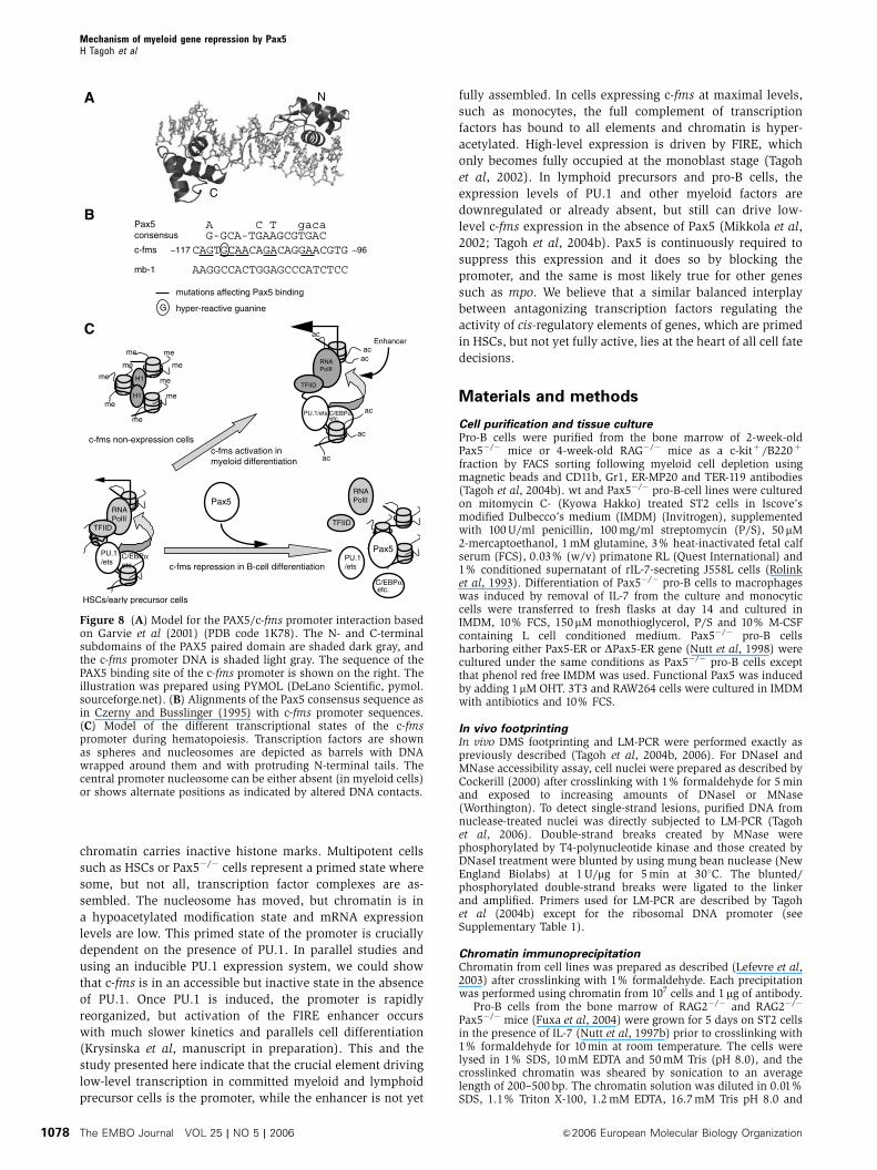
chromatin carries inactive histone marks. Multipotent cells
such as HSCs or Pax5�/� cells represent a primed state where
some, but not all, transcription factor complexes are as-
sembled. The nucleosome has moved, but chromatin is in
a hypoacetylated modification state and mRNA expression
levels are low. This primed state of the promoter is crucially
dependent on the presence of PU.1. In parallel studies and
using an inducible PU.1 expression system, we could show
that c-fms is in an accessible but inactive state in the absence
of PU.1. Once PU.1 is induced, the promoter is rapidly
reorganized, but activation of the FIRE enhancer occurs
with much slower kinetics and parallels cell differentiation
(Krysinska et al, manuscript in preparation). This and the
study presented here indicate that the crucial element driving
low-level transcription in committed myeloid and lymphoid
precursor cells is the promoter, while the enhancer is not yet
fully assembled. In cells expressing c-fms at maximal levels,
such as monocytes, the full complement of transcription
factors has bound to all elements and chromatin is hyper-
acetylated. High-level expression is driven by FIRE, which
only becomes fully occupied at the monoblast stage (Tagoh
et al, 2002). In lymphoid precursors and pro-B cells, the
expression levels of PU.1 and other myeloid factors are
downregulated or already absent, but still can drive low-
level c-fms expression in the absence of Pax5 (Mikkola et al,
2002; Tagoh et al, 2004b). Pax5 is continuously required to
suppress this expression and it does so by blocking the
promoter, and the same is most likely true for other genes
such as mpo. We believe that a similar balanced interplay
between antagonizing transcription factors regulating the
activity of cis-regulatory elements of genes, which are primed
in HSCs, but not yet fully active, lies at the heart of all cell fate
decisions.
Materials and methods
Cell purification and tissue culturePro-B cells were purified from the bone marrow of 2-week-oldPax5�/� mice or 4-week-old RAG�/� mice as a c-kitþ/B220þ
fraction by FACS sorting following myeloid cell depletion usingmagnetic beads and CD11b, Gr1, ER-MP20 and TER-119 antibodies(Tagoh et al, 2004b). wt and Pax5�/� pro-B-cell lines were culturedon mitomycin C- (Kyowa Hakko) treated ST2 cells in Iscove’smodified Dulbecco’s medium (IMDM) (Invitrogen), supplementedwith 100 U/ml penicillin, 100 mg/ml streptomycin (P/S), 50 mM2-mercaptoethanol, 1 mM glutamine, 3% heat-inactivated fetal calfserum (FCS), 0.03% (w/v) primatone RL (Quest International) and1% conditioned supernatant of rIL-7-secreting J558L cells (Rolinket al, 1993). Differentiation of Pax5�/� pro-B cells to macrophageswas induced by removal of IL-7 from the culture and monocyticcells were transferred to fresh flasks at day 14 and cultured inIMDM, 10% FCS, 150mM monothioglycerol, P/S and 10% M-CSFcontaining L cell conditioned medium. Pax5�/� pro-B cellsharboring either Pax5-ER or DPax5-ER gene (Nutt et al, 1998) werecultured under the same conditions as Pax5�/� pro-B cells exceptthat phenol red free IMDM was used. Functional Pax5 was inducedby adding 1mM OHT. 3T3 and RAW264 cells were cultured in IMDMwith antibiotics and 10% FCS.
In vivo footprintingIn vivo DMS footprinting and LM-PCR were performed exactly aspreviously described (Tagoh et al, 2004b, 2006). For DNaseI andMNase accessibility assay, cell nuclei were prepared as described byCockerill (2000) after crosslinking with 1% formaldehyde for 5 minand exposed to increasing amounts of DNaseI or MNase(Worthington). To detect single-strand lesions, purified DNA fromnuclease-treated nuclei was directly subjected to LM-PCR (Tagohet al, 2006). Double-strand breaks created by MNase werephosphorylated by T4-polynucleotide kinase and those created byDNaseI treatment were blunted by using mung bean nuclease (NewEngland Biolabs) at 1 U/mg for 5 min at 301C. The blunted/phosphorylated double-strand breaks were ligated to the linkerand amplified. Primers used for LM-PCR are described by Tagohet al (2004b) except for the ribosomal DNA promoter (seeSupplementary Table 1).
Chromatin immunoprecipitationChromatin from cell lines was prepared as described (Lefevre et al,2003) after crosslinking with 1% formaldehyde. Each precipitationwas performed using chromatin from 107 cells and 1mg of antibody.
Pro-B cells from the bone marrow of RAG2�/� and RAG2�/�
Pax5�/� mice (Fuxa et al, 2004) were grown for 5 days on ST2 cellsin the presence of IL-7 (Nutt et al, 1997b) prior to crosslinking with1% formaldehyde for 10 min at room temperature. The cells werelysed in 1% SDS, 10 mM EDTA and 50 mM Tris (pH 8.0), and thecrosslinked chromatin was sheared by sonication to an averagelength of 200–500 bp. The chromatin solution was diluted in 0.01%SDS, 1.1% Triton X-100, 1.2 mM EDTA, 16.7 mM Tris pH 8.0 and
RNAPolII
TFIID
PU.1/ets
C/EBPαetc.
Pax5RNAPolII
TFIID
PU.1/ets
c-fms activation inmyeloid differentiation
c-fms repression in B-cell differentiation
c-fms non-expression cells
HSCs/early precursor cells
B
C
C/EBPαetc.
C
N
CAGTGCAACAGACAGGAACGTG
AAGGCCACTGGAGCCCATCTCC
−117 −96c-fms
mb-1
Pax5 consensus
mutations affecting Pax5 binding
G hyper-reactive guanine
A C T gacaG-GCA-TGAAGCGTGAC
A
Pax5
me
me
me me
me
me
me
me
me
H1
H1
ac
RNAPolII
TFIID
PU.1/ets C/EBPαetc.
ac
acac
ac
ac
Enhancer
Figure 8 (A) Model for the PAX5/c-fms promoter interaction basedon Garvie et al (2001) (PDB code 1K78). The N- and C-terminalsubdomains of the PAX5 paired domain are shaded dark gray, andthe c-fms promoter DNA is shaded light gray. The sequence of thePAX5 binding site of the c-fms promoter is shown on the right. Theillustration was prepared using PYMOL (DeLano Scientific, pymol.sourceforge.net). (B) Alignments of the Pax5 consensus sequence asin Czerny and Busslinger (1995) with c-fms promoter sequences.(C) Model of the different transcriptional states of the c-fmspromoter during hematopoiesis. Transcription factors are shownas spheres and nucleosomes are depicted as barrels with DNAwrapped around them and with protruding N-terminal tails. Thecentral promoter nucleosome can be either absent (in myeloid cells)or shows alternate positions as indicated by altered DNA contacts.
Mechanism of myeloid gene repression by Pax5H Tagoh et al
The EMBO Journal VOL 25 | NO 5 | 2006 &2006 European Molecular Biology Organization1078
167 mM NaCl, and incubated overnight at 41C with an affinity-purified antibody (5mg) directed against the Pax5 paired domain(Adams et al, 1992). After addition of protein A-Sepharose beads,the immune complexes were collected by centrifugation, washed inRIPA buffer and eluted in 1% SDS and 100 mM NaHCO3 followed byreversal of the crosslinks by heating at 651C for 6 h. Genomic DNAwas isolated by phenol extraction and ethanol precipitation beforereal-time PCR detection of c-fms gene sequences. Precipitated DNAwas quantified by using real-time quantitative PCR with SYBRGreen. Primers used in this assay are listed in Tagoh et al (2004b).Primers specific for mpo are listed in Supplementary Table 1.
Additional information on materials and methods can be foundin Supplementary data.
Supplementary dataSupplementary data are available at The EMBO Journal Online.
Acknowledgements
This work was supported by the Leukaemia Research Fund, theBiotechnology and Biological Sciences Research Council andYorkshire Cancer Research. H Tagoh is a Kay Kendall LeukaemiaFund fellow. M Busslinger’s research is supported by BoehringerIngelheim. We thank Elisabeth Straszinski (Leeds) and GabrieleStengl (Vienna) for cell sorting. Work in AJ Warren’s laboratory issupported by the Leukaemia Research Fund and the MRC.
References
Adams B, Dorfler P, Aguzzi A, Kozmik Z, Urbanek P, Maurer-Fogy I,Busslinger M (1992) Pax-5 encodes the transcription factor BSAPand is expressed in B lymphocytes, the developing CNS, and adulttestis. Genes Dev 6: 1589–1607
Cockerill PN (2000) Identification of DNaseI hypersensitive siteswithin nuclei. Methods Mol Biol 130: 29–46
Cotta CV, Zhang Z, Kim HG, Klug CA (2003) Pax5 determinesB- versus T-cell fate and does not block early myeloid-lineagedevelopment. Blood 101: 4342–4346
Czerny T, Busslinger M (1995) DNA-binding and transactivationproperties of Pax-6: three amino acids in the paired domain areresponsible for the different sequence recognition of Pax-6 andBSAP (Pax-5). Mol Cell Biol 15: 2858–2871
Czerny T, Schaffner G, Busslinger M (1993) DNA sequence recogni-tion by Pax proteins: bipartite structure of the paired domain andits binding site. Genes Dev 7: 2048–2061
Dai XM, Ryan GR, Hapel AJ, Dominguez MG, Russell RG, Kapp S,Sylvestre V, Stanley ER (2002) Targeted disruption of the mousecolony-stimulating factor 1 receptor gene results in osteopetrosis,mononuclear phagocyte deficiency, increased primitive progeni-tor cell frequencies, and reproductive defects. Blood 99: 111–120
DeKoter RP, Singh H (2000) Regulation of B lymphocyte andmacrophage development by graded expression of PU.1. Science288: 1439–1441
Eberhard D, Jimenez G, Heavey B, Busslinger M (2000)Transcriptional repression by Pax5 (BSAP) through interactionwith corepressors of the Groucho family. EMBO J 19: 2292–2303
Enver T, Greaves M (1998) Loops, lineage, and leukemia. Cell 94:9–12
Follows GA, Tagoh H, Lefevre P, Morgan GJ, Bonifer C (2003)Differential transcription factor occupancy but evolutionarilyconserved chromatin features at the human and mouse M-CSF(CSF-1) receptor loci. Nucleic Acids Res 31: 5805–5816
Fuxa M, Skok J, Souabni A, Salvagiotto G, Roldan E, Busslinger M(2004) Pax5 induces V-to-DJ rearrangements and locus contrac-tion of the immunoglobulin heavy-chain gene. Genes Dev 18:411–422
Garvie CW, Hagman J, Wolberger C (2001) Structural studies ofEts-1/Pax5 complex formation on DNA. Mol Cell 8: 1267–1276
Graf T (2002) Differentiation plasticity of hematopoietic cells. Blood99: 3089–3101
Hagemeier C, Bannister AJ, Cook A, Kouzarides T (1993) Theactivation domain of transcription factor PU.1 binds the retino-blastoma (RB) protein and the transcription factor TFIID in vitro:RB shows sequence similarity to TFIID and TFIIB. Proc Natl AcadSci USA 90: 1580–1584
Hayashi K, Yamamoto M, Nojima T, Goitsuka R, Kitamura D (2003)Distinct signaling requirements for Dmu selection, IgH allelicexclusion, pre-B cell transition, and tumor suppression in B cellprogenitors. Immunity 18: 825–836
Heyworth C, Pearson S, May G, Enver T (2002) Transcription factor-mediated lineage switching reveals plasticity in primary com-mitted progenitor cells. EMBO J 21: 3770–3781
Himes SR, Tagoh H, Goonetilleke N, Sasmono T, Oceandy D, ClarkR, Bonifer C, Hume DA (2001) A highly conserved c-fms geneintronic element controls macrophage-specific and regulatedexpression. J Leukoc Biol 70: 812–820
Jenuwein T, Allis CD (2001) Translating the histone code. Science293: 1074–1080
Jimenez G, Griffiths SD, Ford AM, Greaves MF, Enver T (1992)Activation of the beta-globin locus control region precedes com-mitment to the erythroid lineage. Proc Natl Acad Sci USA 89:10618–10622
Kontaraki J, Chen HH, Riggs A, Bonifer C (2000) Chromatin finestructure profiles for a developmentally regulated gene: reorga-nization of the lysozyme locus before trans-activator binding andgene expression. Genes Dev 14: 2106–2122
Kulessa H, Frampton J, Graf T (1995) GATA-1 reprograms avianmyelomonocytic cell lines into eosinophils, thromboblasts, anderythroblasts. Genes Dev 9: 1250–1262
Lefevre P, Melnik S, Wilson N, Riggs AD, Bonifer C (2003)Developmentally regulated recruitment of transcription factorsand chromatin modification activities to chicken lysozyme cis-regulatory elements in vivo. Mol Cell Biol 23: 4386–4400
Linderson Y, Eberhard D, Malin S, Johansson A, Busslinger M,Pettersson S (2004) Corecruitment of the Grg4 repressor by PU.1is critical for Pax5-mediated repression of B-cell-specific genes.EMBO Rep 5: 291–296
Maitra S, Atchison M (2000) BSAP can repress enhancer activity bytargeting PU.1 function. Mol Cell Biol 20: 1911–1922
McIvor Z, Hein S, Fiegler H, Schroeder T, Stocking C, Just U, CrossM (2003) Transient expression of PU.1 commits multipotentprogenitors to a myeloid fate whereas continued expressionfavors macrophage over granulocyte differentiation. ExpHematol 31: 39–47
Metivier R, Penot G, Hubner MR, Reid G, Brand H, Kos M, Gannon F(2003) Estrogen receptor-alpha directs ordered, cyclical, andcombinatorial recruitment of cofactors on a natural target pro-moter. Cell 115: 751–763
Mikkola I, Heavey B, Horcher M, Busslinger M (2002) Reversion ofB cell commitment upon loss of Pax5 expression. Science 297:110–113
Murrell A, Heeson S, Reik W (2004) Interaction between differen-tially methylated regions partitions the imprinted genes Igf2 andH19 into parent-specific chromatin loops. Nat Genet 36: 889–893
Nerlov C, Graf T (1998) PU.1 induces myeloid lineage commitmentin multipotent hematopoietic progenitors. Genes Dev 12:2403–2412
Nerlov C, Querfurth E, Kulessa H, Graf T (2000) GATA-1 interactswith the myeloid PU.1 transcription factor and represses PU.1-dependent transcription. Blood 95: 2543–2551
Nutt SL, Heavey B, Rolink AG, Busslinger M (1999) Commitment tothe B-lymphoid lineage depends on the transcription factor Pax5.Nature 401: 556–562
Nutt SL, Morrison AM, Dorfler P, Rolink A, Busslinger M (1998)Identification of BSAP (Pax-5) target genes in early B-cell devel-opment by loss- and gain-of-function experiments. EMBO J 17:2319–2333
Nutt SL, Urbanek P, Rolink A, Busslinger M (1997a) Essentialfunctions of Pax5 (BSAP) in pro-B cell development: differencebetween fetal and adult B lymphopoiesis and reduced V-to-DJrecombination at the IgH locus. Genes Dev 11: 476–491
Nutt SL, Urbanek P, Rolink A, Busslinger M (1997b) Essentialfunctions of Pax5 (BSAP) in pro-B cell development: differencebetween fetal and adult B lymphopoiesis and reduced V-to-DJrecombination at the IgH locus. Genes Dev 11: 476–491
Orkin SH (2000) Diversification of haematopoietic stem cells tospecific lineages. Nat Rev Genet 1: 57–64
Mechanism of myeloid gene repression by Pax5H Tagoh et al
&2006 European Molecular Biology Organization The EMBO Journal VOL 25 | NO 5 | 2006 1079

Rolink A, Haasner D, Nishikawa S, Melchers F (1993) Changes infrequencies of clonable pre B cells during life in different lym-phoid organs of mice. Blood 81: 2290–2300
Ross IL, Yue X, Ostrowski MC, Hume DA (1998) Interaction betweenPU.1 and another Ets family transcription factor promotes macro-phage-specific basal transcription initiation. J Biol Chem 273:6662–6669
Sasmono RT, Oceandy D, Pollard JW, Tong W, Pavli P, WainwrightBJ, Ostrowski MC, Himes SR, Hume DA (2003) A macrophagecolony-stimulating factor receptor-green fluorescent proteintransgene is expressed throughout the mononuclear phagocytesystem of the mouse. Blood 101: 1155–1163
Scott EW, Simon MC, Anastasi J, Singh H (1994) Requirement oftranscription factor PU.1 in the development of multiple hema-topoietic lineages. Science 265: 1573–1577
Simon MC, Olson M, Scott E, Hack A, Su G, Singh H (1996)Terminal myeloid gene expression and differentiation requiresthe transcription factor PU.1. Curr Top Microbiol Immunol 211:113–119
Souabni A, Cobaleda C, Schebesta M, Busslinger M (2002) Pax5promotes B lymphopoiesis and blocks T cell development byrepressing Notch1. Immunity 17: 781–793
Tagoh H, Cockerill PN, Bonifer C (2006) In vivo genomic footprint-ing using LM-PCR methods. In Nuclear Reprogramming, Pells S(ed) pp 285–314. Totowa, NJ: Humana Press
Tagoh H, Himes R, Clarke D, Leenen PJ, Riggs AD, Hume D, BoniferC (2002) Transcription factor complex formation and chromatinfine structure alterations at the murine c-fms (CSF-1 receptor)
locus during maturation of myeloid precursor cells. Genes Dev 16:1721–1737
Tagoh H, Melnik S, Lefevre P, Chong S, Riggs AD, Bonifer C (2004a)Dynamic reorganization of chromatin structure and selectiveDNA demethylation prior to stable enhancer complex formationduring differentiation of primary hematopoietic cells in vitro.Blood 103: 2950–2955
Tagoh H, Schebesta A, Lefevre P, Wilson N, Hume D, Busslinger M,Bonifer C (2004b) Epigenetic silencing of the c-fms locus duringB-lymphopoiesis occurs in discrete steps and is reversible. EMBOJ 23: 4275–4285
Urbanek P, Wang ZQ, Fetka I, Wagner EF, Busslinger M (1994)Complete block of early B cell differentiation and altered pattern-ing of the posterior midbrain in mice lacking Pax5/BSAP. Cell 79:901–912
Weissman IL, Anderson DJ, Gage F (2001) Stem and progenitorcells: origins, phenotypes, lineage commitments, and transdiffer-entiations. Annu Rev Cell Dev Biol 17: 387–403
Xie H, Ye M, Feng R, Graf T (2004) Stepwise reprogramming of Bcells into macrophages. Cell 117: 663–676
Yamada T, Abe M, Higashi T, Yamamoto H, Kihara-Negishi F,Sakurai T, Shirai T, Oikawa T (2001) Lineage switch induced byoverexpression of Ets family transcription factor PU.1 in murineerythroleukemia cells. Blood 97: 2300–2307
Zhang P, Zhang X, Iwama A, Yu C, Smith KA, Mueller BU,Narravula S, Torbett BE, Orkin SH, Tenen DG (2000) PU.1 inhibitsGATA-1 function and erythroid differentiation by blocking GATA-1DNA binding. Blood 96: 2641–2648
Mechanism of myeloid gene repression by Pax5H Tagoh et al
The EMBO Journal VOL 25 | NO 5 | 2006 &2006 European Molecular Biology Organization1080
Copyright © 2022 FDOKUMEN