The magnocellular visual pathway and facial emotion misattribution errors in schizophrenia
6
The magnocellular visual pathway and facial emotion misattribution errors in schizophrenia Jeffrey S. Bedwell a, ⁎, Chi C. Chan a , Ovad Cohen b , Yinnon Karbi b , Eyal Shamir c , Yuri Rassovsky d, e, f a Department of Psychology, 4000 Central Florida Blvd., University of Central Florida, Orlando, FL 32816-1390, USA b Academic College of Tel Aviv-Yafo, 2 Rabenu Yeruham St., Tel-Aviv Yaffo, 61083, Israel c Abarbanel Mental Health Center, 15 Keren-Kayement St., Bat Yam, 59100, Israel d Department of Psychology, Bar-Ilan University, Ramat-Gan, 52900, Israel e Gonda Multidisciplinary Brain Research Center, Bar-Ilan University, Ramat-Gan, 52900, Israel f UCLA Semel Institute for Neuroscience and Human Behavior, 760 Westwood Plaza, Los Angeles, CA 90095, USA abstract article info Article history: Received 12 December 2012 Received in revised form 15 January 2013 Accepted 20 January 2013 Available online 28 January 2013 Keywords: Anger Emotion Fear Schizophrenia Social cognition Visual processing Many individuals with schizophrenia show impairment in labeling the emotion depicted by faces, and tend to ascribe anger or fear to neutral expressions. Preliminary research has linked some of these difficulties to dysfunc- tion in the magnocellular (M) visual pathway, which has direct projections to subcortical emotion processing re- gions. The current study attempted to clarify these relationships using a novel paradigm that included a red background. Diffuse red light is known to suppress the M-pathway in nonpsychiatric adults, and there is prelim- inary evidence that it may have the opposite (stimulating) effect in schizophrenia-spectrum disorders (SSDs). Twenty-five individuals with SSDs were compared with 31 nonpsychiatric controls using a facial emotion iden- tification task depicting happy, angry, fearful, and sad emotions on red, green, and gray backgrounds. There was a robust interaction of group by change in errors to the red (vs. green) background for misattributing fear expres- sions as depicting anger (p = .001, ή 2 =.18). Specifically, controls showed a significant decrease in this type of error with the red background (p = .003, d =0.77), while the SSD group tended to increase this type of error (p =.07, d =0.54). These findings suggest that the well-established M-pathway abnormalities in SSDs may con- tribute to the heightened misperception of other emotions such as anger, which in turn may cause social misper- ceptions in the environment and elicit symptoms such as paranoia and social withdrawal. As the ventral striatum plays a primary role in identifying anger and receives efferent input from the M-pathway, it may serve as the neuroanatomical substrate in the perception of anger. © 2013 Elsevier Inc. All rights reserved. 1. Introduction Among the diverse cognitive deficits described in schizophrenia (Irani et al., 2011; Schmidt et al., 2011), impairment in social cogni- tion may be particularly important, as it has been shown to be related to poorer quality of life, social support, and work outcome in these indi- viduals (Maat et al., 2012; Schmidt et al., 2011). Many individuals with schizophrenia have difficulty accurately identifying emotions depicted by photographs of faces, particularly for fear expressions (Chan et al., 2010; Kohler et al., 2010). This difficulty appears to be relatively stable over time and independent of general cognitive ability (Chan et al., 2010; Kohler et al., 2010). Research has also shown that individuals with schizophrenia are more likely to mislabel a neutral facial expres- sion as depicting fear or anger (Habel et al., 2010; Pinkham et al., 2011; Premkumar et al., 2008). Two studies have suggested that re- duced performance on facial emotion identification tasks in individuals with schizophrenia is related to poorer magnocellular visual pathway (M-pathway) functioning (Butler et al., 2009; Norton et al., 2009). The M-pathway has relatively direct projections from the thalamus to sub- cortical regions such as the amygdala and ventral striatum (see Fig. 1), which are thought to quickly process the low spatial frequencies of fa- cial emotional expressions (Vuilleumier et al., 2003). It also appears that these connections may prime the perception of threat-based emo- tions such as fear and anger to allow for a fast response (West et al., 2010; Williams et al., 2011). The precise abnormality within the M-pathway in individuals with schizophrenia remains unclear, as one study has suggested that the dysfunction begins at the level of the rod photoreceptors in the retina (Hebert et al., 2010), while other research has suggested that the dysfunction begins at later stages in M-pathway processing (Chen et al., 2004; Guthrie et al., 2006). One way of manipulating M-pathway activity involves the use of diffuse red light, which suppresses the M-pathway (Bedwell et al., Progress in Neuro-Psychopharmacology & Biological Psychiatry 44 (2013) 88–93 Abbreviations: M-pathway, magnocellular pathway; SSD, schizophrenia-spectrum disorder; SCID-I, Structured Clinical Interview for DSM-IV Axis I Disorders; SCID-II, Structured Clinical Interview for DSM-IV Axis II Disorders; fMRI, functional magnetic resonance imaging. ⁎ Corresponding author. Tel.: +1 407 823 5858; fax: +1 407 823 5862. E-mail addresses: [email protected] (J.S. Bedwell), [email protected] (C.C. Chan), [email protected] (O. Cohen), [email protected] (Y. Karbi), [email protected] (E. Shamir), [email protected] (Y. Rassovsky). 0278-5846/$ – see front matter © 2013 Elsevier Inc. All rights reserved. http://dx.doi.org/10.1016/j.pnpbp.2013.01.015 Contents lists available at SciVerse ScienceDirect Progress in Neuro-Psychopharmacology & Biological Psychiatry journal homepage: www.elsevier.com/locate/pnp
Transcript of The magnocellular visual pathway and facial emotion misattribution errors in schizophrenia

Progress in Neuro-Psychopharmacology & Biological Psychiatry 44 (2013) 88–93
Contents lists available at SciVerse ScienceDirect
Progress in Neuro-Psychopharmacology & BiologicalPsychiatry
j ourna l homepage: www.e lsev ie r .com/ locate /pnp
The magnocellular visual pathway and facial emotion misattributionerrors in schizophrenia
Jeffrey S. Bedwell a,⁎, Chi C. Chan a, Ovad Cohen b, Yinnon Karbi b, Eyal Shamir c, Yuri Rassovsky d,e,f
a Department of Psychology, 4000 Central Florida Blvd., University of Central Florida, Orlando, FL 32816-1390, USAb Academic College of Tel Aviv-Yafo, 2 Rabenu Yeruham St., Tel-Aviv Yaffo, 61083, Israelc Abarbanel Mental Health Center, 15 Keren-Kayement St., Bat Yam, 59100, Israeld Department of Psychology, Bar-Ilan University, Ramat-Gan, 52900, Israele Gonda Multidisciplinary Brain Research Center, Bar-Ilan University, Ramat-Gan, 52900, Israelf UCLA Semel Institute for Neuroscience and Human Behavior, 760 Westwood Plaza, Los Angeles, CA 90095, USA
Abbreviations: M-pathway, magnocellular pathway;disorder; SCID-I, Structured Clinical Interview for DSMStructured Clinical Interview for DSM-IV Axis II Disordresonance imaging.⁎ Corresponding author. Tel.: +1 407 823 5858; fax:
E-mail addresses: [email protected] (J.S. Bedw(C.C. Chan), [email protected] (O. Cohen), [email protected] (E. Shamir), [email protected] (Y
0278-5846/$ – see front matter © 2013 Elsevier Inc. Allhttp://dx.doi.org/10.1016/j.pnpbp.2013.01.015
a b s t r a c t
a r t i c l e i n f oArticle history:Received 12 December 2012Received in revised form 15 January 2013Accepted 20 January 2013Available online 28 January 2013
Keywords:AngerEmotionFearSchizophreniaSocial cognitionVisual processing
Many individuals with schizophrenia show impairment in labeling the emotion depicted by faces, and tend toascribe anger or fear to neutral expressions. Preliminary research has linked some of these difficulties to dysfunc-tion in themagnocellular (M) visual pathway, which has direct projections to subcortical emotion processing re-gions. The current study attempted to clarify these relationships using a novel paradigm that included a redbackground. Diffuse red light is known to suppress theM-pathway in nonpsychiatric adults, and there is prelim-inary evidence that it may have the opposite (stimulating) effect in schizophrenia-spectrum disorders (SSDs).Twenty-five individuals with SSDs were compared with 31 nonpsychiatric controls using a facial emotion iden-tification task depicting happy, angry, fearful, and sad emotions on red, green, and gray backgrounds. Therewas arobust interaction of group by change in errors to the red (vs. green) background for misattributing fear expres-sions as depicting anger (p=.001, ή2=.18). Specifically, controls showed a significant decrease in this type oferror with the red background (p=.003, d=0.77), while the SSD group tended to increase this type of error(p=.07, d=0.54). These findings suggest that the well-establishedM-pathway abnormalities in SSDsmay con-tribute to the heightenedmisperception of other emotions such as anger, which in turnmay cause socialmisper-ceptions in the environment and elicit symptoms such as paranoia and socialwithdrawal. As the ventral striatumplays a primary role in identifying anger and receives efferent input from the M-pathway, it may serve as theneuroanatomical substrate in the perception of anger.
© 2013 Elsevier Inc. All rights reserved.
1. Introduction
Among the diverse cognitive deficits described in schizophrenia(Irani et al., 2011; Schmidt et al., 2011), impairment in social cogni-tion may be particularly important, as it has been shown to be relatedto poorer quality of life, social support, andwork outcome in these indi-viduals (Maat et al., 2012; Schmidt et al., 2011). Many individuals withschizophrenia have difficulty accurately identifying emotions depictedby photographs of faces, particularly for fear expressions (Chan et al.,2010; Kohler et al., 2010). This difficulty appears to be relatively stableover time and independent of general cognitive ability (Chan et al.,2010; Kohler et al., 2010). Research has also shown that individuals
SSD, schizophrenia-spectrum-IV Axis I Disorders; SCID-II,
ers; fMRI, functional magnetic
+1 407 823 5862.ell), [email protected]@yahoo.com (Y. Karbi),. Rassovsky).
rights reserved.
with schizophrenia are more likely to mislabel a neutral facial expres-sion as depicting fear or anger (Habel et al., 2010; Pinkham et al.,2011; Premkumar et al., 2008). Two studies have suggested that re-duced performance on facial emotion identification tasks in individualswith schizophrenia is related to poorer magnocellular visual pathway(M-pathway) functioning (Butler et al., 2009; Norton et al., 2009). TheM-pathway has relatively direct projections from the thalamus to sub-cortical regions such as the amygdala and ventral striatum (see Fig. 1),which are thought to quickly process the low spatial frequencies of fa-cial emotional expressions (Vuilleumier et al., 2003). It also appearsthat these connections may prime the perception of threat-based emo-tions such as fear and anger to allow for a fast response (West et al.,2010; Williams et al., 2011). The precise abnormality within theM-pathway in individuals with schizophrenia remains unclear, as onestudy has suggested that the dysfunction begins at the level of the rodphotoreceptors in the retina (Hebert et al., 2010), while other researchhas suggested that the dysfunction begins at later stages in M-pathwayprocessing (Chen et al., 2004; Guthrie et al., 2006).
One way of manipulating M-pathway activity involves the use ofdiffuse red light, which suppresses the M-pathway (Bedwell et al.,
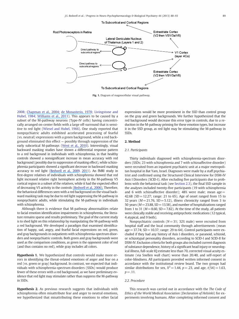
Fig. 1. Diagram of magnocellular visual pathway.
89J.S. Bedwell et al. / Progress in Neuro-Psychopharmacology & Biological Psychiatry 44 (2013) 88–93
2008; Chapman et al., 2004; de Monasterio, 1978; Livingstone andHubel, 1984; Williams et al., 2011). This appears to be caused by asubset of the M-pathway neurons (Type-IV cells) having concentri-cally arranged on-center fields with a large off-surround that is sensi-tive to red light (Wiesel and Hubel, 1966). One study reported thatnonpsychiatric adults exhibited accelerated processing of fearful(vs. neutral) expressions with a green background, while a red back-ground eliminated this effect — possibly through suppression of theearly subcortical M-pathways (West et al., 2010). Interestingly, visualbackward masking studies have shown a differential response patternto a red background in individuals with schizophrenia, in that healthycontrols showed a nonsignificant increase in mean accuracy with redbackground (possibly due to suppressionofmasking effect),while schizo-phrenia participants showed a significant decrease in backward maskingaccuracy to red light (Bedwell et al., 2009, 2011). An fMRI study infirst-degree relatives of individuals with schizophrenia showed that redlight increased relative right hemisphere activity in the M-pathway V5cortical region in a subset of the relatives, while it had the expected effectof decreasing V5 activity in the controls (Bedwell et al., 2006). Therefore,the behavioral differences seenwith a red background on the visual back-wardmasking taskmay be due to red light suppressing theM-pathway innonpsychiatric adults, while stimulating the M-pathway in individualswith schizophrenia.
Although there is evidence that M-pathway abnormalities relateto facial emotion identification impairments in schizophrenia, the litera-ture remains sparse and results preliminary. The goal of the current studyis to shed light on this relationship bymanipulating theM-pathwaywitha red background. We developed a paradigm that examined identifica-tion of happy, sad, angry, and fearful facial expressions on red, green,and gray backgrounds in outpatientswith schizophrenia-spectrumdisor-ders and nonpsychiatric controls. Both green and gray backgroundswereused as the comparison conditions, as green is the opponent color of red(and thus contains no red), while gray includes all colors.
Hypothesis 1. We hypothesized that controls would make more er-rors in identifying the threat-related emotions of anger and fear on ared (vs. green or gray) background. In contrast, we expected that indi-viduals with schizophrenia-spectrum disorders (SSDs) would producefewer of these errors with a red background, as we have preliminary ev-idence that red light may stimulate rather than suppress theM-pathwayin SSDs.
Hypothesis 2. As previous research suggests that individuals withschizophrenia often misattribute fear and anger to neutral emotions,we hypothesized that misattributing these emotions to other facial
expressions would be more prominent in the SSD than control groupon the gray and green backgrounds. We further hypothesized that thered background would decrease this error type in controls, due to a re-duction in theM-pathway priming for these emotion types, but increaseit in the SSD group, as red light may be stimulating the M-pathway inSSDs.
2. Method
2.1. Participants
Thirty individuals diagnosed with schizophrenia-spectrum disor-ders (SSDs; 23 with schizophrenia and 7 with schizoaffective disorder)were recruited from an inpatient psychiatric unit at a major metropoli-tan hospital in Bat Yam, Israel. Diagnoses were made by a staff psychia-trist and confirmed using the Structured Clinical Interview for DSM-IVAxis I Disorders (SCID-I). After excluding five participants due to prob-lems with the behavioral task (see Section 2.3), the final SSD sample inthe analyses included twenty-five participants (19 with schizophreniaand 6 with schizoaffective disorder); 48% were male; mean age=42.68 (SD=12.27; range: 21 to 65). Age of onset ranged from 15 to32 years (M=21.76, SD=5.12), illness chronicity ranged from 3 to50 years (M=23.88, SD=13.58), and number of hospitalizations rangedfrom 1 to 31 (M=8.60, SD=7.82). At the time of the study, all patientswere clinically stable and receiving antipsychoticmedications (12 typical,4 atypical, and 9 both).
Nonpsychiatric controls (N=31; 32% male) were recruited fromhospital staff and the local community through advertisements (meanage=37.74; SD=10.37; range: 29 to 64). Control participants were ex-cluded if they had any history of Axis I disorders, or paranoid, schizoid,or schizotypal personality disorders, according to SCID-I and SCID-II forDSM-IV. Exclusion criteria for both groups also included current diagnosisof substance dependence, history of a significant head injury or neurolog-ical illness, full-scale IQ estimate less than 70, corrected visual acuity es-timate (via Snellen wall chart) worse than 20/40, and self-report ofcolor-blindness. All participants provided written informed consent inaccordance with the institutional review board. The two groups hadsimilar distributions for sex, Χ2=1.44, p=.23, and age, t(54)=1.63,p=.11.
2.2. Procedure
This research was carried out in accordance with the The Code ofEthics of the World Medical Association (Declaration of Helsinki) for ex-periments involving humans. After completing informed consent and

90 J.S. Bedwell et al. / Progress in Neuro-Psychopharmacology & Biological Psychiatry 44 (2013) 88–93
diagnostic interviews, participants completed the facial emotion identi-fication task. Stimuli consisted of facial pictures expressing one of fouremotions: happy, sad, angry, or afraid. The pictures were selectedfrom the Karolinska Directed Emotional Faces set (Lundqvist et al.,1998), and were trimmed to exclude hair and non-facial contours inthe manner described by Holmes et al. (2005). Colored filters wereapplied across the entire background and face stimulus to create threecolor background conditions: red, green, and gray (matched for physicalluminance; see Fig. 2). The stimuli were displayed centrally usingE-prime software and a 17″ Sony Multiscan 200PS monitor. Partici-pants were seated 57 cm from the monitor, received instructions,and practiced the task. For the experimental trials, the color back-grounds were presented in a random blocked order and the emotiontypes were presented in a random order within each color back-ground. Each face was presented for 500 ms, followed by four emo-tion labels (“happy,” “sad,” “angry,” and “afraid”) presented for 10 s.Participants were instructed to select the facial affect displayed bypressing a number on a keyboard, which triggered the subsequenttrial. If the participant did not press any button within 10 s, the nexttrial was presented. There were five different pictures for each of thefour emotion types (i.e., 20 total pictures) presented within each ofthe three color conditions.
2.3. Statistical procedures
Five participants from the SSD group failed to respond to morethan 3 (>15%) of the 20 trials within at least one color block andwere excluded from the analysis to help reduce the potential confoundof attentional lapses. Therefore, all analyses were conducted on theremaining 25 SSD participants and 31 control participants.
Mixed three-way ANOVAs, using the Greenhouse–Geisser correc-tion, examined the effects of color, emotion error type, and groupon errors. All post-hoc analyseswere conductedwith Bonferroni correc-tion for multiple comparisons. An independent-sample t-test was usedto examine group differences on errors to each emotion depicted withthe gray background.
3. Results
3.1. Errors classified by type of emotion depicted (Hypothesis 1)
To test the first hypothesis (Hypothesis 1), emotion error type wasdefined as the type of emotion depicted by the face on error trials.There was no interaction of color by group, F(2,100)=0.67, p=.50,ή2=.01, or color by group by type of emotion depicted, F(5,267)=1.20, p=.31, ή2=.02, on errors. See Table 1 for descriptive statistics.
For the sake of comparisonwith other studies, we examined the pat-tern of errors classified by emotion depicted in both groups with thegray background, as previous studies have typically used a white orgray background. On the gray background, the SSD groupmade signifi-cantly more errors than controls only when asked to identify a sad ex-pression, t(54)=2.91, p=.01, d=0.85 (see Table 1).
Fig. 2. Example facial
3.2. Errors classified by the type of emotion chosen on error trials(misattribution error types; Hypothesis 2)
For this analysis, misattribution error type was defined as the par-ticular wrong emotion chosen when making an error (e.g., incorrectlylabeling other expressions as angry). TheANOVA revealed a significant in-teraction of color by emotion by group, F(4,223)=3.47, p=.01, ή2=.06.The interaction of color by emotion by group was then examined withineach pairwise color comparison, which showed that this was only statis-tically significant with the red and green backgrounds, F(3,155)=4.26,p=.01, ή2=.07. The color (green vs. red) by group interactions werethen examined for each of the four emotion error types, which revealedthat only errors of misidentifying other expressions as anger was statisti-cally significant for the four comparisons, F(1,54)=6.75, p=.01, ή2=.11.We then examined the three possible error types in which “angry” wasthe incorrect answer and found that only the error type of mislabeling afearful face as angry showed a statistically significant group by color inter-action, F(1,54)=12.20, p=.001, ή2=.18. For this error type, control par-ticipants were less likely to mislabel a fearful face as angry with the redbackground when compared to the green background, t(30)=3.18, p=.003, d=0.77. In contrast, the SSD group showed a statistical trend forchange in the opposite direction to the red background, as they mademore of these errors with the red (vs. green) background, t(24)=1.90,p=.07, d=0.54. Within the SSD group, there was no interaction of anti-psychotic medication type (atypical, typical, both) and background coloron mislabeling a fearful face as angry, F(2,22)=0.58, p=.57, ή2=.05.
We then examined group differences for this error type (i.e.,misattributing anger to fearful expressions) within each color con-dition. The groups did not differ in this type of error on the greenbackground, t(54)=0.86, p=.39, d=0.23, but the SDD group madesignificant more of these errors with the gray background, t(54)=2.32, p=.02, d=0.62, and the difference was most pronounced withthe red background t(54)=3.99, pb .001, d=1.12 (see Table 1). Oneprediction in the second hypothesis (Hypothesis 2) was that the SSDgroup would make more general misattribution errors for both fearand anger misattributions on both the green and gray backgrounds.This was true for misattributing fear on other expressions for both thegreen, t(36)=2.47, p=.02, d=0.73, and gray, t(35)=2.75, p=.01,d=0.82, backgrounds, but only was true for misattributing anger onother expressions for the gray background, t(32)=2.59, p=.01, d=0.80.
4. Discussion
Weused a novel task to examine the effects ofM-pathway processingon facial emotion recognition in individualswith schizophrenia. Contraryto our first hypothesis (Hypothesis 1), when categorizing the errors bythe type of emotion depicted, we did not find the expected modulationin performance to red light within either group. However, when we ex-amined types of misattribution for our second hypothesis (Hypothesis2), the control group showed a significant decrease in misattributinganger to fearful expressions with a red background, while individualswith schizophrenia showed the opposite tendency (see Fig. 3). In other
emotion stimuli.

Table 1Descriptive statistics and comparisons of errors made by emotion depicted, emotion identified, color background, and group.
Type of error Control group Schizophrenia-spectrum group
Gray background Green background Red background Gray background Green background Red background
All errors to feara 1.58 (0.92) 1.42 (1.06) 1.32 (0.83) 1.88 (1.17) 1.96 (1.49) 2.40 (1.26)###
Fear as anger 0.23 (0.50) 0.90 (0.87) 0.35 (0.55)⁎⁎ 0.60 (0.71)# 0.72 (0.68) 1.16 (0.94)###
Fear as sad 1.29 (0.86) 0.48 (0.68) 0.90 (0.83)⁎⁎ 1.16 (1.14) 0.84 (0.80) 1.08 (1.12)Fear as happy 0.06 (0.25) 0.03 (0.18) 0.06 (0.25) 0.12 (0.33) 0.40 (1.04) 0.16 (0.55)
All errors to angera 0.55 (0.81) 0.90 (0.79) 0.65 (0.88) 0.96 (0.98) 1.68 (1.14)## 1.28 (1.14)#
Anger as fear 0.29 (0.46) 0.55 (0.57) 0.32 (0.54) 0.80 (0.91)# 0.92 (0.76)# 0.92 (0.95)##
Anger as sad 0.23 (0.62) 0.29 (0.46) 0.29 (0.69) 0.12 (0.44) 0.56 (0.65) 0.20 (0.50)⁎
Anger as happy 0.03 (0.18) 0.06 (0.25) 0.03 (0.18) 0.04 (0.20) 0.20 (0.50) 0.16 (0.47)All errors to sada 1.26 (1.03) 1.10 (1.04) 1.00 (1.24) 2.40 (1.73)## 2.08 (0.77)# 1.92 (1.66)#
Sad as fear 0.61 (0.76) 0.74 (0.82) 0.84 (1.07) 1.08 (1.26) 1.16 (1.18) 1.28 (1.34)Sad as anger 0.58 (0.56) 0.35 (0.71) 0.10 (0.40) 1.12 (1.45) 0.72 (0.98) 0.64 (1.08)#
Sad as happy 0.06 (0.25) 0.00 (0.00) 0.06 (0.25) 0.20 (0.41) 0.20 (0.50) 0.00 (0.00)All errors to happya 0.00 (0.00) 0.26 (0.63) 0.23 (0.50) 0.12 (0.44) 0.56 (0.77) 0.28 (0.54)
Happy as fear 0.00 (0.00) 0.06 (0.25) 0.10 (0.30) 0.04 (0.20) 0.20 (0.41) 0.08 (0.28)Happy as anger 0.00 (0.00) 0.00 (0.00) 0.13 (0.34)⁎ 0.08 (0.40) 0.16 (0.47) 0.12 (0.33)Happy as sad 0.00 (0.00) 0.19 (0.48) 0.00 (0.00)⁎ 0.00 (0.00) 0.20 (0.50) 0.08 (0.28)
Numbers in cells represent: mean (standard deviation).⁎Significant within-group difference for red-to-green comparison at pb .05; ⁎⁎pb .01.#=Significant difference compared to controls within same color at pb .05; ##pb .01; ###pb .001.
a These categories are the number of total errors made to faces depicting the listed emotion.
91J.S. Bedwell et al. / Progress in Neuro-Psychopharmacology & Biological Psychiatry 44 (2013) 88–93
words, while a red background attenuated the perception of anger incontrols, it increased perception of anger in patients, specifically forfaces depicting fear (i.e., mislabeling fear expressions as angry).Whereasthe groups did not differ on this type of error with the green back-ground, they differed with the gray background (which contains somered; d=0.62), with the most robust difference between groups wasfoundwith the red background (d=1.12). Our results were also largelyconsistent with our hypothesis that the SSD group would make moremisattribution errors involving labeling another expression as fearfulor angry across both green and gray backgrounds; however, this wasonly found for anger misattributions with the gray background.
Angry expressions appear to be primarily processed by the ventralstriatum (Baggio et al., 2012; Calder et al., 2004; Phillips et al.,1999). The ventral striatum has relatively direct M-pathway input(Buxbaum-Conradi and Ewert, 1999), and comparative research hasshown that the aggression-related activity in the ventral striatumappears
Fig. 3. Differential change to red light in number of erro
to be primarily influenced by an increase of dopamine activity (Ferrari etal., 2003; Louilot et al., 1986).Notably, psychotic symptomsof schizophre-nia are also thought to be caused by increased dopamine in the same re-gion (Howes and Kapur, 2009). Additionally, administration of sulpiride(a dopamine D2-class receptor antagonist) caused a specific reduction inhealthyhumanparticipants' accuracy in identifying anger in facial expres-sions (Lawrence et al., 2002). These findings thus offer a neurophysio-logical framework for the repeated behavioral findings indicating thatindividuals with schizophrenia are more likely to ascribe anger to neu-tral expressions (Habel et al., 2010; Pinkham et al., 2011; Premkumar etal., 2008). Consistent with this past research, we found that the SSDgroup made more errors than controls in misidentifying fear expres-sions as angry with the gray background, which is closest to the gray/white backgrounds used in those previous studies. This mechanismmay also partially explain the increased rate of violent behavior foundin a subset of individuals with schizophrenia, particularly those who
rs of misattributing anger to a fearful face by group.
92 J.S. Bedwell et al. / Progress in Neuro-Psychopharmacology & Biological Psychiatry 44 (2013) 88–93
have comorbid substance abuse, which further increases dopamine inthe ventral striatum (Erkiran et al., 2006; Swanson et al., 1990).
Considering theM-pathway input to the ventral striatum,which playsa role in identifying anger expressions, the decrease inmistakenly ascrib-ing anger to other emotions in response to red light seen in our controlgroup may be due to the red light suppression of the M-pathway inputsto the ventral striatum. Thus, the priming effect of the M-pathway indetecting threat (West et al., 2010; Williams et al., 2011), which mayresult in some false positive identifications of threat, appears to besuppressed by red light in the control group. Conversely, the tendencyto increase anger attribution to fear faces seen in the SSD group may bedue to red light stimulating (instead of suppressing) M-pathway inputto the ventral striatum in this group, a finding consistent with ourtheory of a differential M-pathway response to red light in the SSD.However, this theory would suggest that we would also see this pat-tern with misattributing other expressions such as fear, which is alsoa threat-relevant emotion. It is possible that we did not find this pat-tern because our task involved viewing faces with a gaze directed atthe participant. Seeing anger in these faces, even when anger is notdepicted, may be more related to protective anger-detection regionssuch as the ventral striatum, than seeing fear in the faces. In contrast,a fear expression with a gaze directed away from (rather than at) theparticipant may be more threatening as it may indicate an indirectthreatening object nearby. Further research is needed to clarify if andwhy this red light modulation is limited to misattributing anger tofear expressions in schizophrenia-spectrum disorders.
It is possible that our first hypothesis (Hypothesis 1) was notsupported due to the M-pathway playing a central role in priming thedetection of threat expressions. Thus, modulation of the M-pathway byred lightmay cause a change in preattentive rapid emotion identificationdecisions that are best indexed by the pattern of misattribution errorsrather than errors to the type of emotion depicted. It is possible thatthe M-pathway priming function causes one to be particularly vigilantfor threat related emotions, thus sometimes causing misattribution er-rors in this direction. Further research is needed to clarify this possibility.In addition, while performance on this task offers clues into the etiologyof SSDs, it is unlikely to be a helpful marker of effectiveness of currenttreatments. Abnormalities on measures of emotion identification andM-pathway dysfunction appear to be trait markers of liability for the ill-ness, rather than state-related dysfunctions, as these abnormalities arealso found in first-degree relatives of SSD probands and healthy analogsamples (e.g., college students with schizotypy).
Our study is limited by not having a neutral expression to serve as a“control” expression onto which participants could attribute saliencewithout potential bias from the stimuli. For example, we may havefound the samepattern of red light effects formislabeling neutral expres-sions as angry. In addition, we only examined SSD participants that werecurrently receiving antipsychotic medications, therefore, findings maydiffer with unmedicated participants. Despite these limitations, ourresults support an increased tendency for individuals with an SSDto misperceive anger in others, which may be modulated by themagnocellular visual pathway. This misperception may cause socialmisperceptions in the environment and elicit symptoms such as para-noia and social withdrawal. Further research using neuroimaging andneurophysiological techniques is needed to clarify this theory and relat-edmechanism. This line of research can inform treatment for social cog-nition difficulties, as well as for increased anger and violence found in asubset of individuals with schizophrenia.
5. Conclusions
The current study expands the existing literature on the influenceof M-pathway processing on emotion identification in schizophreniaby showing, for the first time, a differential effect of red light on a partic-ular type of emotion identification misattribution error. This providespreliminary evidence for a novel theory that an increased propensity to
misattribute non-angry faces as angry in schizophrenia, found in the cur-rent study as well as previous studies, may be due to well-establishedM-pathway abnormalities in SSDs (Brenner et al., 2009; Butler et al.,2007; Javitt, 2009; Martinez et al., 2011).
Acknowledgments
The authors would like to thank Scott Sutterby, Andrew Deptula,and Alixandra Burks for assistance with data processing. This studywas not funded by an external agency. The authors do not have anyfinancial/ethical conflicts related to this research.
References
Baggio HC, Segura B, Ibarretxe-Bilbao N, Valldeoriola F, Marti MJ, Compta Y, et al. Struc-tural correlates of facial emotion recognition deficits in Parkinson's disease patients.Neuropsychologia 2012;50:2121–8.
Bedwell JS, Brown JM, Yanasak NE, Miller LS. Schizophrenia and red light: fMRI evi-dence for a novel biobehavioral marker. Int J Neurosci 2006;116:881–94.
Bedwell JS, Brown JM, Orem DM. The effect of a red background on location backwardmasking by structure. Percept Psychophys 2008;70:503–7.
Bedwell JS, Orem DM, Rassovsky Y, Allen LG, Sutterby SR. A potential qualitativeendophenotype for schizophrenia: backward masking response to red light. Psy-chiatry Res 2009;166(2–3):166–73.
Bedwell JS, Rassovsky Y, Orem DM, Kamath V. The backward masking red light effect inschizophrenia: relationship to clinical features and neurocognitive performance.J Abnorm Psychol 2011;120(2):490–6.
Brenner CA, Krishnan GP, Vohs JL, AhnWY, Hetrick WP, Morzorati SL, et al. Steady stateresponses: electrophysiological assessment of sensory function in schizophrenia.Schizophr Bull 2009;35(6):1065–77.
Butler PD, Martinez A, Foxe JJ, Kim D, Zemon V, Silipo G, et al. Subcortical visual dysfunc-tion in schizophrenia drives secondary cortical impairments. Brain 2007;130(Pt. 2):417–30.
Butler PD, Abeles IY,Weiskopf NG, Tambini A, JalbrzikowskiM, LegattME, et al. Sensory con-tributions to impaired emotion processing in schizophrenia. Schizophr Bull 2009;35(6):1095–107.
Buxbaum-Conradi H, Ewert JP. Responses of single neurons in the toad's caudal ventralstriatum to moving visual stimuli and test of their efferent projection by extracel-lular antidromic stimulation/recording techniques. Brain Behav Evol 1999;54(6):338–54.
Calder AJ, Keane J, Lawrence AD, Manes F. Impaired recognition of anger followingdamage to the ventral striatum. Brain 2004;127(Pt. 9):1958–69.
Chan RC, Li H, Cheung EF, Gong QY. Impaired facial emotion perception in schizophrenia:a meta-analysis. Psychiatry Res 2010;178(2):381–90.
Chapman C, Hoag R, Giaschi D. The effect of disrupting the human magnocellular pathwayon global motion perception. Vision Res 2004;44(22):2551–7.
Chen Y, Levy DL, Sheremata S, Holtzman PS. Comprised late-stage motion processing inschizophrenia. Biol Psychiatry 2004;55:834–41.
de Monasterio FM. Properties of concentrically organized X and Y ganglion cells of ma-caque retina. J Neurophysiol 1978;41(6):1394–417.
Erkiran M, Ozunalan H, Evren C, Aytaclar S, Kirisci L, Tarter R. Substance abuse amplifiesthe risk for violence in schizophrenia spectrum disorder. Addict Behav 2006;31(10):1797–805.
Ferrari PF, van Erp AM, Tornatzky W, Miczek KA. Accumbal dopamine and serotonin inanticipation of the next aggressive episode in rats. Eur J Neurosci 2003;17(2):371–8.
Guthrie AH, McDowell JE, Hammond BR. Scotopic sensitivity in schizophrenia. SchizophrRes 2006;84:378–85.
Habel U, Chechko N, Pauly K, Koch K, Backes V, Seiferth N, et al. Neural correlates ofemotion recognition in schizophrenia. Schizophr Res 2010;122(1–3):113–23.
Hebert M, Gagne AM, Paradis ME, Jomphe V, Roy MA, Merett C, et al. Retinal responseto light in young nonaffected offspring at high genetic risk of neuropsychiatricbrain disorders. Biol Psychiatry 2010;67:270–4.
Holmes A,Winston JS, EimerM. The role of spatial frequency information for ERP compo-nents sensitive to faces and emotional facial expression. Brain Res Cogn Brain Res2005;25(2):508–20.
Howes OD, Kapur S. The dopamine hypothesis of schizophrenia: version III—the finalcommon pathway. Schizophr Bull 2009;35(3):549–62.
Irani F, Kalkstein S, Moberg EA, Moberg PJ. Neuropsychological performance in olderpatients with schizophrenia: a meta-analysis of cross-sectional and longitudinalstudies. Schizophr Bull 2011;37(6):1318–26.
Javitt DC. When doors of perception close: bottom-up models of disrupted cognition inschizophrenia. Annu Rev Clin Psychol 2009;5:249–75.
Kohler CG, Walker JB, Martin EA, Healey KM, Moberg PJ. Facial emotion perception inschizophrenia: a meta-analytic review. Schizophr Bull 2010;36(5):1009–19.
Lawrence AD, Calder AJ, McGowan SW, Grasby PM. Selective disruption of the recogni-tion of facial expressions of anger. Neuroreport 2002;13(6):881–4.
Livingstone MS, Hubel DH. Anatomy and physiology of a color system in the primatevisual cortex. J Neurosci 1984;4(1):309–56.
Louilot A, Le Moal M, Simon H. Differential reactivity of dopaminergic neurons in thenucleus accumbens in response to different behavioral situations. An in vivovoltammetric study in free moving rats. Brain Res 1986;397(2):395–400.

93J.S. Bedwell et al. / Progress in Neuro-Psychopharmacology & Biological Psychiatry 44 (2013) 88–93
Lundqvist D, Flykt A, Ohman A. Karolinksa directed emotional faces. Department ofNeurosciences, Karolinska Hospital, Stockholm, Sweden, 1998.
Maat A, Fett AK, Derks E. Social cognition and quality of life in schizophrenia. SchizophrRes 2012(1–3):212–8.
Martinez A, Hillyard SA, Bickel S, Dias EC, Butler PD, Javitt DC. Consequences ofmagnocellular dysfunction on processing attended information in schizophrenia.Cereb Cortex 2011;22(6):1282–93.
Norton D, McBain R, Holt DJ, Ongur D, Chen Y. Association of impaired facial affect recog-nition with basic facial and visual processing deficits in schizophrenia. Biol Psychiatry2009;65(12):1094–8.
Phillips ML,Williams L, Senior C, Bullmore ET, Brammer MJ, Andrew C, et al. A differentialneural response to threatening and non-threatening negative facial expressions inparanoid and non-paranoid schizophrenics. Psychiatry Res 1999;92(1):11–31.
Pinkham AE, Brensinger C, Kohler C, Gur RE, Gur RC. Actively paranoid patients withschizophrenia over attribute anger to neutral faces. Schizophr Res 2011;125(2–3):174–8.
Premkumar P, Cooke MA, Fannon D, Peters E, Michel TM, Aasen I, et al. Misattributionbias of threat-related facial expressions is related to a longer duration of illness and
poor executive function in schizophrenia and schizoaffective disorder. Eur Psychiatry2008;23(1):14–9.
Schmidt SJ, Mueller DR, Roder V. Social cognition as a mediator variable betweenneurocognition and functional outcome in schizophrenia: empirical review andnew results by structural equation modeling. Schizophr Bull 2011;37(Suppl. 2):S41–54.
Swanson JW, Holzer III CE, Ganju VK, Jono RT. Violence and psychiatric disorder in thecommunity: evidence from the epidemiologic catchment area surveys. Hosp Com-munity Psychiatry 1990;41(7):761–70.
Vuilleumier P, Armony JL, Driver J, Dolan RJ. Distinct spatial frequency sensitivities forprocessing faces and emotional expressions. Nat Neurosci 2003;6(6):624–31.
West GL, Anderson AAK, Bedwell JS, Pratt J. Red diffuse light suppresses the acceleratedperception of fear. Psychol Sci 2010;21(7):992–9.
Wiesel TN, Hubel DH. Spatial and chromatic interactions in the lateral geniculate nucle-us body of the rhesus monkey. J Neurophysiol 1966;29:1115–56.
Williams CK, Grierson LE, Carnahan H. Colour-induced relationship between affect andreaching kinematics during a goal-directed aiming task. Exp Brain Res 2011;212(4):555–61.