The Journal of Pharmacy and Pharmacology 1963 Volume 15 ...
74
J O U R N A L O F P H A R M A C Y P ublished by D irection, o f the C ouncil o f THE PHARMACEUTICAL SOCIETY OF CT. BRITAIN
-
Upload
khangminh22 -
Category
Documents
-
view
0 -
download
0
Transcript of The Journal of Pharmacy and Pharmacology 1963 Volume 15 ...

J O U R N A L O F P H A R M A C Y
P u b l i s h e d b y D i r e c t i o n , o f t h e C o u n c i l o f
THE PHARMACEUTICAL SOCIETY OF CT. BRITAIN

N E W F R O M T h e
W A T S O NS E R V I C E 3
M i c r o s c o p e
If you would like io know more about tin's outstanding new instrument, please write for full details to
A Watson quality microscope designed to meet changing needs from student use to
advanced graduate studies
c h o i c e of inc lined m c n o c u l a r h e a d , inc lined m o n o c u l a r h e a d with d ra w tu b e , or inc lined b in o c u la r h e a d . All u n i t s c a n b e ro ta ted t h r o u g h 360° a n d a r e quickly in : e r c h a n g e - ab le . A ver t ica l m o n o c u l a r h e a d is a l so available .
ro ta t in g q u a d r u p l e n c s e p i e c e a c c e p t i n g * t h e full r a n g e of W a t s o n h ig h - p e r f o r m a n c e
s l e e v e - m o u n te d ob je c t iv e s .
v s p r in g - lo a d e d 4 m m a n d 2 m m p a r a c h r o - m a t ic o b je c t i v e s .
all o b je c t i v e s p a r c e i t r e d , p a r fo ca l le d a n d c o l o u r - c o d e d fo r e a s y identi f ica tion .
la rg e a c id - r e s i s t i n g pla in s t a c e a c c e p t s W a t s o n a t t a c h a b le m e c h a n ic a l s t a g e s .
f o c u s i n g c o n d e n s e r a s s e m b l y fo r o re - c e n t r e d o r ce n t r in g m o u n t s e a c h fitted with iris d ia p h r a g m a n d filter h o lde r .
v t # lo w -p la ced c o a r s e a n d f ine a d j u s t m e n t * £ * c o n t ro l s .
c h o i c e of t h r e e b a s e s : m i r ro r a s s e m b l y ;• V c o n c e a l e d 25 w. m a in s v o l tag e la m p a n d
c o n d e n s i n g l e n s ; c o n c e a l e d buil t- in low v o l t a g e la m p (6 v. 13w.) , l ight c o n t ro l unit a n d c o n d e n s i n g l e n s
In b a s ic fo rm , th e new W a ts o n S e rv ic e 3 M ic ro sc o p e is a re lia b le , ro b u s t and e a sy -to -u se in s tru m e n t fo r s tu d e n t b io lo g ica l m ic ro sc o p y . W ith a c c e s s o r ie s it is fu lly ca p a b le of sa tis fy in g m any o f th e d e m an d s of ad van ced w o rk e rs . U p-to -date in a p p e a ra n c e an c d e s ig n , it p o s s e s s e s m any o u tstan d in g fe a tu re s .
Service 3 Microscope with low-voltage lamp base
;;ew d u r a b l e ‘e v e r c l e a n ’ fin ish .
A n extensive range of W atscn optical and m echanical a cce sso rie s
can be used with the new S E R V I C E 3
W. WATSON & SONS 1-D • BARrttT ■ HERTS B A R N E T 4 4 0 4 ■ C A B L E S : ‘O P T I C S ’ B A R N E T

J O U R N A L O F P H A R M A C Y A N D
P H A R M A C O L O G YE d i t o r : G e o r g e B r o w n le e , D . S c . , P h .D . . F .P .S .
A s r i s t a n t E d i to r : J . R . F o w le r , B .P h a r m . , F .P .S .
A nnual Subscription £5 Os. Od. Single Copies 10s.
17 BLOOMSBURY SQUARE, LONDON, W.C.ICables- Pharm akon , L ondon , W .C .I Telephone: H O L born 8967
V ol. XV N o . 10 October, 1963
C O N T E N T SResearch Papers p a g e s
S u b s t it u t e d D ih y d r o x y b f .n z o ic A c id s a s P o ssib le A n t i - I n f l a m m a t d r y A g e n t s . By J. E. L ig h to w le r a n d H . J. R y lan ce 633-638
I n t e r a c t i o n o f C o c a in e a n d T y r a m in e o n t h e I s o l a t e d M a m m a l ia n H e a r t . By J. B. F a rm e r a n d B. P e tch . . . . . 639-643
T h e R e l a t iv e Po t e n c ie s o f T h y r o x in e a n d L io t h y r o n in e b y O r a l a n d Su b c u t a n e o u s A d m in is t r a t io n in t h e R a t .By G . S. W berg , W . F. D evlin , N . R . S tep h en so n a n d J. R . C a rte r 644-651
T h e E f f e c t s o f H is t a m in e R e l e a s e o n t h e L ip id C o n t e n t o f t h e Is o l a t e d Pe r f u s e d L u n g s o f Se n s it is e d G u in e a -P ig s . ByV. O . M arq u is a n d W . G . S m ith . . . . . . . . . . 652-659
T h e E f f e c t s o f T w o P h e n y l a c e t i c A c id D e r iv a t iv e s (C F T 1201 a n d C F T 1208) o n T h e A n a l g e s ic A c t i o n o f M o r p h in e in M ic e . By M . M ed ak o v ic a n d B. Banic. . . . . . . 660-665
A n I n v e s t ig a t io n i n t o t h e E f f e c t s o f G a m m a - R a y s o n A q u e o u s So l u t io n s o f /i - A m in o b e n z o ic A c i d . By P. H . M a rrio tt . . 666 -6 7 0
S e l f - a d m in is t r a t io n o f Pr a l id o x im e in N e r v e G a s Po is o n in g w i t h a N o t e o n t h e St a b i l i t y o f t h e D r u g . By R o lf B ark- m an . Bo E dgren a n d A n d e rs S u ndw all . . . . . . . . 671 -6 7 7
D a tu ra le icn h a rd rii M u e l l . E x B e n t h . St r u c t u r e o f
t h e F l o w e r . By A . C o rre ia A lves a n d W . C . E vans . . . . 678-687A N o t e o n t h e T o x i c i t y a n d So l v e n t Pr o p e r t ie s o f D im e t h y l
S u l p h o x i d e . By V. K . B row n, J. R o b in so n a n d D . E. S tevenson 688-692
L etters to the EditorC h l o r o q u in e in B r o n c h ia l A s t h m a . By S o h a n L. A g a rw a l,
B. S. D e sh m a n k a r a n d V in o d B h arg av a . . . . . . . . 693H is t a m in e in N o n - v e r t e b r a t e A n im a l s . By E . F. M e ttr ick a n d
J . M . T e lfo rd ............................................................................................ 694-697Pa p a v e r in e - l ik e P h a m a c o l o g i c a l P r o p e r t ie s o f R o t e n o n e . By
R . S an ti, M . F e rra r i a n d C . E. T o th . . 697-698A M o d i f ic a t i o n o f t h e B u t a n o l E x t r a c t i o n M e t h o d fo r
t h e F l u o r im e t r ic A s sa y o f C a t e c h o l a m in e s in B io l o g ic a l M a t e r ia l s By B. A. C a llin g h am a n d R o se m ary C ass . . 699 700
E D IT O R IA L BOARDH . S. Bean, B.Piarm., Ph.D., F.P.S., J. C. H aNBURY, M.A., B Pharm., F.P.S., F.R.I.C., F. H artley, b.s.c., Ph.D., f.p.s., f.r .i.c ., E. F. H ersant, a.pharm., Ph.D., f.p .s., f.r.i.c ., J. J. Lewis, m.sc., f.p.s., A. D . M acdonald, m.d ., m.a., m.sc., A.McCoUBREY, B.Sc., Ph.D., M.P.S., F.R.I.C., D . W. MATHIESON, B.SC., Ph.D., F.R.I.C.,H . G. R olfe, b.sc., f.p .s., f.r.i .c., G. F. Somers, b.sc., ph.D., f.p .s., J. B. Stenlake,D.Sc., Ph.D., F.P.S., F.R.I.C., G. B. W est B.Pharm., D.Sc., Ph.D., F.P.S., R .T . WILLIAMS, D.Sc., Ph.D.Secretary: F. W. A dams, b.sc., f.p .s., f.r .i .c .

Journal of Pharmacy and Pharmacology October, 1963
t f a , u /i< Ù S > s ig tu fc c z u ic e o f
l o c a l d e c a m e t h y l e n e - b i s -
( 4 - a m i n o q u i n a l d i n i u m c h l o r i d e )
B y p r o v i d i n g e f f e c t i v e l o c a l a n t i b a c t e r i a l a n d a n t i f u n g a l t h e r a p y f o r i n f e c t i o n s o f t h e s k i n a n d m u c o u s m e m b r a n e . D e q u a d in f r e q u e n t l y s p a r e s t h e u s e o f a n t i b i o t i c s .
D e q u a d in h a s a w id e r a n t i m i c r o b i a l s p e c t r u m t h a n p e n i c i l l i n a n d i t i s a c t i v e a g a i n s t o r g a n i s m s i e s i s t a n t t o a n t i b i o t i c s . F u r t h e r m o r e , n o r e s i s t a n t s t r a i n s h a v e b e e n r e p o r t e d f o l l o w i n g t h e u s e o f D e q u a d in .
R e c e n t l a b o r a t o r y w o r k h a s s h o w n t h a t D e q u a d in i s r e t a i n e d o n t i s s u e . T o d e m o n s t r a t e t h i s u n u s u a l p r o p e r t y , a s p e c i a l l a b o r a t o r y t e s t w a s d e v i s e d i n v o l v i n g t h e u s e o f 14C D e q u a d in . V i s u a l r e c o r d in g o f t h e r e t e n t i o n o f D e q u a d in o n t i s s u e w a s s u p p l i e d b y a t e c h n i q u e i n v o l v i n g t h e u s e o f a u t o - r a d i o g r a p h s .
m D E Q U A D I Na product c f Allen & HanburysresearchinL O Z E N G E S C R E A M P A I N T T U L L E D R E S S I N G S
• E . 2A L L E N & H A N B U R Y S L T D L O N D O NS61 720 H

R E S E A R C H P A P E R S
SUBSTITUTED DIHYDROXYBENZOIC ACIDS AS POSSIBLE ANTI-INFLAMMATORY AGENTS
By J. E. Lightowler* and H. J. Rylance From Edinburgh Pharmaceutical Industries Limited, Wheatfield Road, Edinburgh, 11
Received A pril 30, 1963
A series of substituted dihydroxybenzoic acids was prepared and examined for anti-inflammatory activity. The compounds which showed activity were derived from y-resorcylic acid. The introduction of a halogen atom at positions 3 and 5 and a benzyl or methyl group at position 4 gave marked activity but also increased toxicity.
As a result of the reported anti-inflammatory activity of y-resorcylic acid (I) (Reid and others, 1951) a series of related compounds was prepared and examined for anti-inflammatory activity. These compounds were based on the six isomeric dihydroxybenzoic acids (I-VI). The enhancement of the activity by the introduction of a halogen atom was noted early in this investigation and this paper therefore deals mainly with the halogenatec, methyl and benzyl derivatives of these acids, of which a number have previously been reported (see Experimental).
CO„H c o 2h c o 2hHO | " OH 1 OH | OH
V 7\ /T Y; 1
\ / A )HO
\ /1
2,6-dihyd roxybenzoic 2,5-dihydroxybenzoic OHac d acid 2,4-dihydroxybenzoic
acid(y-resorcylic acid) (Gentisic acid) (/3-resorcylic acid)
(I) (II) (HI)
c o 2h c o 2h1
c o 2hi OH
i Y A A \¡1 1
\ A \ A A AOH | OH HC OH
2,3-dihydroxybenzoic OH 3,5-dihyd roxybenzoicacid 3,4-dihyd roxybenzoic
acidacid
(IV) (V) (VI)
ExperimentalC h e m ic a l
All m.ps. are uncorrected. Micro analyses were by Messrs. Weiler and Strauss.
2,6-Dihydroxy-4-methylbenzoic acid (Robertson and Robinson, 1927),3,5-dibromo-2,6-dihydroxybenzoic acid (Beilstein, 10, I, 186), 3,5-di- bromo-, 3,5-dichloro- and 3-chloro-2,6-dihydroxy-4-methylbenzoic acid
* Present a d d re ss : R iker L aboratories, D epartm en t o f Pharm acology, W elwyn G arden City.
633

J. E. LIGHTOW LER A N D H. J. RYLANCE
(British Patent 877,355), 2,5-dihydroxy-3-methylbenzoic acid (Beilstein, 10, 419), 2,5-dihydroxy-4-methylbenzoic acid (Beilstein, 10, 421), 2,4-di- hydroxy-5-methylbenzoic acid (Beilstein, 10, II, 275), 5-chloro-2,4- dihydroxybenzoic acid (Sandin and McKee, 1935), 3,5-dichloro-2,4-di- hydroxybenzoic acid (Pectyrin and Kirchine, 1947), 4,5-dibromo-2,3- dihydroxybenzoic acid (Beilstein, 10, I, 175), 5-bromD-3,4-dihydroxy- benzoic acid (Beilstein, 10. 400) and 3,5-dihydroxybenzoic acid (Birkin- shaw and Bracken, 1942) were known compounds and were prepared by standard methods.
The unhalogenated acids in general were prepared by carboxylation of the corresponding m-dihydroxybenzene using KHC03/C02. The general preparation was as follows.
1 part by weight of the dihydroxybenzene and 2 parts by weight of KHC03 were mixed with 2 parts by volume of glycerol. The mixture was then heated, preferably with stirring to 140° (bath or jacket temperature) with CO, passing through during the reaction. After 6 hr. the mixture was taken up in water, strongly acidified with sulphuric acid and filtered. The solid obtained was then recrystallised from water or ethanol/water.
The carboxylation of 4-benzylresorcinol gave a mixture of the isomers3-benzyl-2,6-dihydroxybenzoic acid and 5-benzyl-2,4-dihydroxybenzoic acid. These were separated chromatographically on an alumina column (Table I).
TABLE IA cids obtained by the carboxylation of «i-dihydroxybenzenes
Namem.p.°C
Found TheoryC K
c !H
2,6-Dihydroxy-4-(hydroxymethyl)benzoic acid 190-193 52-2 4-3 52-2 4-354-Benzvl-2,6-dihydroxybenzoic acid . . 172-173 68-8 51 68-85 1 4.93-Benzyl-2,6-dihydroxybenzoic acid 174-176 67-7 4-5 68-85 4-92,6-Dihvdroxy-4-(/MolvImethyl)benzoic acid .. 168-170 69-7 5-5 69-8 1 5-42,4-Dihvdroxy-3-methylbenzoic acid .. 204-206 570 5 3 57-1 : 4-85-Benzyl-2,4-dihydroxybenzoic acid 211-214 68-2 4-8 68-85 i 4-9
Halogenated acids generally were prepared by the addition of sulphuryl chloride or bromine to a solution of the dihydroxybenzoic acid in ether or acetic acid.
The phenolic acid (0-05m) was dissolved in ether (50 ml.) and sulphuryl chloride (0-05 or OTm depending on whether mono- or di-chlorination was required) added slowly in the cold. After standing overnight the reaction mixture was poured into water, extracted with ether and the ethereal layer extracted with aqueous sodium bicarbonate; after acidification of the alkaline solution, the acid was precipitated and recrystallised from water or ethanol/water (using charcoal for purification if necessary).
A similar procedure substituting bromine for sulphuryl chloride gave the bromo-compounds (see Table II).
634
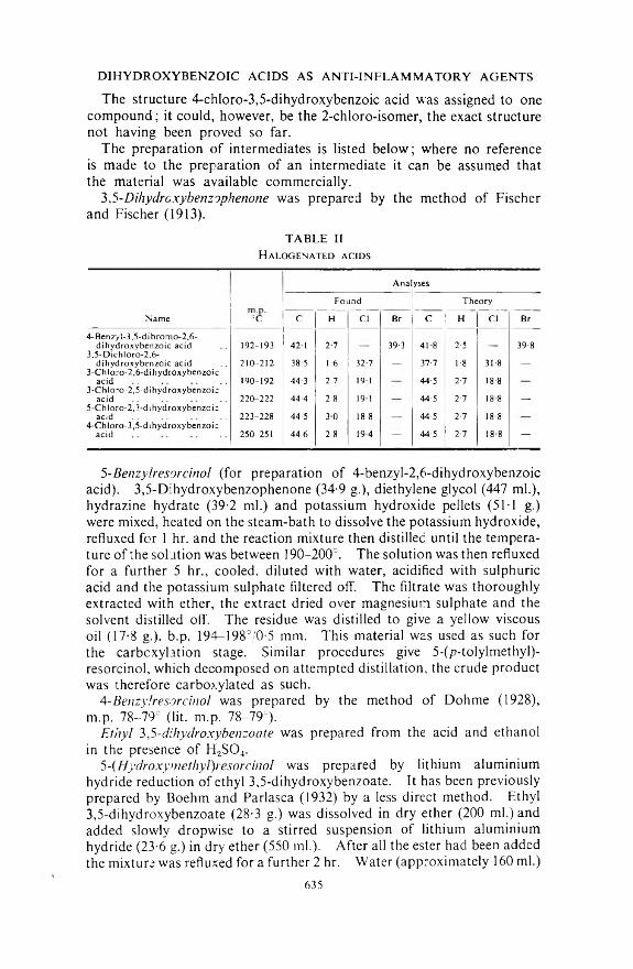
DIHYDROXYBENZOIC ACIDS AS ANTI-INFLAMMATORY AGENTS
The structure 4-chloro-3,5-dihydroxybenzoic acid was assigned to one compound; it could, however, be the 2-chloro-isomer, the exact structure not having been proved so far.
The preparation of intermediates is listed below; where no reference is made to the preparation of an intermediate it can be assumed that the material was available commercially.
3,5-Dihydroxybenzophenone was prepared by the method of Fischer and Fischer (1913).
TABLE IIH alogenated acids
AnalysesFound Theory
Name °C C H Cl Br C H Cl Br4- Benzyl-3,5-dibromo2,6-
dihydroxybenzoic acid 192-193 42 1 2-7 39-3 418 2-5 39-83,5-Dichloro-2,6-
di hydroxy benzoic acid 210-212 38-5 1-6 32-7 _ 37-7 1-8 31-8 _3-Chloro-2,6-dihydroxybenzoic
acid 190-192 44-3 2-7 191 44-5 2-7 18-83-ChIoro-2,5-d ihydroxybenzoic
acid 220-222 44-4 2-8 191 44-5 2-7 18 8 _5-Chloro-2,3-dihydroxybenzoic
acid 223-228 44-5 30 18-8 44'5 2-7 18-84-Chloro-3,5-dihydroxybenzoic
acid 250-251 44-6 2-8 19-4 - 44-5 2-7 18-8 -
5-Benzylresorcinol (for preparation of 4-benzyl-2,6-dihydroxybenzoic acid). 3,5-Dihydroxybenzophenone (34-9 g.), diethylene glycol (447 ml.), hydrazine hydrate (39-2 ml.) and potassium hydroxide pellets (5IT g.) were mixed, heated on the steam-bath to dissolve the potassium hydroxide, refluxed for 1 hr. and the reaction mixture then distilled until the temperature of the solution was between 190-200°. The solution was then refluxed for a further 5 hr., cooled, diluted with water, acidified with sulphuric acid and the potassium sulphate filtered off. The filtrate was thoroughly extracted with ether, the extract dried over magnesium sulphate and the solvent distilled off. The residue was distilled to give a yellow viscous oil (17-8 g.), b.p. 194—198°/'0-5 mm. This material was used as such for the carbcxylation stage. Similar procedures give 5-(/?-tolylmethyl)- resorcinol, which decomposed on attempted distillation, the crude product was therefore carboxylated as such.
4- Benzylresorcinol was prepared by the method of Dohme (1928), m.p. 78-79° (lit. m.p. 78-79°).
Ethyl 3,5-dihydroxybenzoate was prepared from the acid and ethanol in the presence of H2S04.
5- (Hydroxymethyl)resorcinol was prepared by lithium aluminium hydride reduction of ethyl 3,5-dihydroxybenzoate. It has been previously prepared by Boehm and Parlasca (1932) by a less direct method. Ethyl3,5-dihydroxybenzoate (28-3 g.) was dissolved in dry ether (200 ml.) and added slowly dropwise to a stirred suspension of lithium aluminium hydride (23-6 g.) in dry ether (550 ml.). After all the ester had been added the mixture was refluxed for a further 2 hr. Water (approximately 160 ml.)
635
was then added slowly with vigorous stirring to the cooled mixture to decompose excess lithium aluminium hydride and the reaction mixture poured into ice-cold diluted sulphuric acid. The ether layer was separated, the aqueous solution saturated with sodium sulphate and continuously extracted with ether for 2 days. The ether extracts v/ere dried over MgS04, the ether removed by distillation to give 5-(hydroxymethyl)- resorcinol (151 g.), m.p. 170-178°. This material is satisfactory for carboxylation. A sample was recrystallised from ether/60-80° light petroleum to give material m.p. 175-179°. Found: C, 59T ; H, 5-8. C7Hs0 3 requires C, 60-0; H, 5-7.
Methyl 3,5-dichloro-2,6-dihydro.xy-4-methyIbenzoate. 3,5-Dichloro-2.6- dihydroxy-4-methylbenzoic acid (52 g.) was dissolved in acetone (1200 ml.) and sodium bicarbonate (17-8 g.) and dimethyl sulphate (22-2 ml.) were added. The mixture was refluxed for 2 hr., cooled and water (500 ml.) added, whereupon the excess of sodium bicarbonate dissolved and the methyl ester was precipitated. After standing for several hours the mixture was filtered to give the ester m.p. 166-169°.
3,5-Dichloro-2,6-dihydroxy-4-methy!benzamide. The methyl 3.5-di- chloro-2,6-dihydroxy-4-methylbenzoate (3 g.) was suspended in concentrated ammonia solution (20 ml.) and shaken mechanically overnight. The amide was filtered off" and recrystallised from water to give material (2g.)m.p.210-212°. Found: C,40-5; H, 3-0; Cl, 30-4;N,5-9. C8fTCl2N03 requires C, 40-7; H, 3-0; Cl, 30-1; N, 5-9.
3-ChloroA-methylbenzoic acid. This compound has been prepared before (Beilstein, 9, 498, and 9, II, 331) by various methods.
Sulphuryl chloride (36 ml.) was added to p-toluic acid (20 g.) in glacial acetic acid (100 ml.) and the solution refluxed for 5 hr. After standing overnight the product was filtered off and recrystallised from ethanol to give material (7 g.) m.p. 201-203° (lit. m.p. 200-202°). Mol. wt. found (by titration) 169-2; mol. wt. theory 170-5.
M ethods
Anti-inflammatory assessment was made using either or both the granuloma pellet test and reduction of yeast-induced oedema.Granuloma Pellet Test
This was modified from the method of Meyer, Stucki and Auslebrook(1953) and Meier, Schuler and Desaulles (1950). Groups of 10 albino rats of either sex, within the weight range 50-60 g. were anaesthetized with ether. Dental cotton wool pellets of 6-10 mg. were implanted via a trochar and cannula under the skin of the groin and axilla. Subsequently the animals received five daily subcutaneous injections of the solution (or vehicle) into the scapular region and were killed on the sixth day for examination. Tissue infiltration was measured by determining the increase in weight of pellets after drying 24 hr. at 110° and by micro-Kjeldahl estimation of the nitrogen content of the dried pellet. The two values obtained gave good agreement. Values obtained with treated animals were compared with those from controls by Students test.
J. E. LIGHTOW LER A N D H. J. RYLANCE
636

This was modified from the method of Selitto and Randall (1954), and Eckhart, Thomas and Garner (1958). Groups of 15 albino rats within the weight range 55-60 g. were used for each dose level. The paw circumference was measured immediately before the injection of 0T ml. of a 20 per cent suspension of brewer’s yeast into the plantar surface of the right hind foot. One min. after the yeast injection the compound was given intraperitoneally and the paw circumference then measured 2 hr. and 4 hr. later. Pooled values obtained with a total of 135 control animals, over the period of the tests, showed that the percentage increase (with standard deviation) in the circumference of yeast injected paws was 35 ± 3 at 2 hr. and 40 4 4 at 4 hr. Compounds were considered to be effective if the swelling did not exceed 20 per cent.
In the rat-paw test two dose levels only (changed by a factor of two) were used. Table III shows either the lowest active dose or the highest tolerated dose which failed to confer protection.
TABLE IIIA nti-inflammatory activity in a series of substituted dihydroxybenzoic acids
D IH Y D R O X Y BEN ZO IC ACIDS AS A N TI-IN FLA M M A TO R Y AG ENTS
R eduction o f Y east-induced O edem a
Name (mg./kg.)
Act-vityApproximate
LD50 i.v. mouse (mg./kg.)
Granulomapellettest
Rat paw oedema
test2,6-Dihydroxv-4-msthylbenzcic acid 150 0 j- 3803,5-Dibromo-2,6-dihydroxybenzoic acid 300 — 3503,5-Dibromo-2,6-dihvdroxy-4-methylbenzoic acid 150 0 -f 1383,5-Dichloro-2,6-dihydroxy-4-methylbenzoic acid 150 -p -r 125
(P = 0 001)2,6-Dihydroxv-4-(hydroxymethyl)benzoic acid. . 600 0 12004-Benzyl-2,6-dihvdroxybenzoic acid 150 1003-Benzyl-2,6-dihydroxvbenzoic acid 752,6-Dihydrox\-4-(/>-toiylmethyl)benzoic acid 37-5 454-Benzyl-3,5-dibromo-2,6-dihydroxybenzoic acid 75 1053,5-Dichloro-2,6-dihydroxy-4-methvlbenzamide 75 1102,4-Dihvdroxy-5-methylbenzcic acid 150 0 0 5455-Chloro-2,4-dihvdroxybenzoic acid 150 0 0 10753,5-Dichloro-2.4-dihydroxybenzoic acid 150 0 0 10002,4-Dihydroxy-3-methylbenzcic acid 300 0 7805-Benzyl-2,4-dihyd-oxybenzoic acid 150 04,5-Dibromo-2,3-dihydroxybenzoic acid 75 0 75*5-Chioro-2,3-dihydroxybenzoic acid . . 150 05-Bromo-3,4-dihydroxybenzoic acid 150 0 1803,5-Dihydroxybenzoic acid 150 0 20004,Chlcro-3,5-üihydroxybenzoic acid 150 0 20003-Chloro-4-methylbenzoic acid . . 160 -f -p 208
II o ©
0 Ineffective at the highest tolerated dose.4- Effective, swelling less than 20 per cent.
* Miss M. E. Farquharson (personal communication).
Toxicity. Albino mice within the weight range 18-22 g. were used. LD50 values were determined graphically by plotting log dose against probability using not less than 5 groups of 5 animals.
R esults
The results are given in Table III.The undermentioned compounds were tested only by the granuloma
pellet test and found to be inactive. P = >0-05 (“i” test): 3-Chloro-2,6- dihydroxy-4-methylbenzoic acid, 3,5-dichloro-2,6-dihydroxybenzoic acid,
637

J. E. LIGHTOW LER A N D H. J. RY LANC E
3- chloro-2,6-dihydroxybenzoic acid, 2,5-dihydroxy-3-methylbenzoic acid,2,5-dihydroxy-4-methylbenzoic acid and 3-chloro-2,5-dihydroxybenzoic acid.
D iscussion
Anti-inflammatory activity, in the compounds tested in this series, was apparently restricted to the 2,6-dihydroxybenzoic acids (Table III). Activity was enhanced by the presence of halogen atoms at positions 3 and 5. This was further increased when a methyl or benzyl group was also present at position 4 in the ring. 4-Benzyl-2,6-dihydroxybenzoic acid, with a benzyl group at position 4 but without halogens in the ring, was also active.
Clarke and Mosher (1953) have claimed that both the hydroxyl and carboxyl groups are essential for anti-inflammatory activity; 3-chloro-4- methylbenzoic acid, which possesses no hydroxyl groups has been shown to be active and is thus an exception to the general statement. The most active compound was 3,5-dichloro-2,6-dihydroxy-4-methyl- benzoic acid. Unfortunately whenever anti-inflammatory activity was found, the doses given were very near to the toxic levels (Table III) and attempts to divorce the two (i.e. give a permissible therapeutic index) met with failure.
Acknowledgements. The authors wish to thank Miss J. G. Peggie and Mr. A. B. Ritchie for technical assistance.
R eferencesBirkinshaw, J. H. and Bracken, A. (1942). J. chem. Soc., 368-370.Boehm, T. and Parlasca, H. (1932). Arch. Pharm., 270, 168-182.Clarke, N. E., and Mosher, R. E. (1953). Circulation, 7, 337-344.Dohme, A. R. L. (1928). U.S. Patent 1,658,229.Eckhart, E. T., Thomas, G. P. and Garner, W. M. (1958). Prcc. Soc. exp. Biol.,
N.Y., 98, 211-212.Fischer, E. and Fischer, H. O. L. (1913). Ber., 46, 1138-1148.Meier, R., Schuler, W. and Desaulles, P. (1950). Experientia, 6, 469-471.Meyer, R. K., Stucki, J. C. and Auslebrook, K. A. (1953). Proc. Soc. exp. Biol.,
N.Y., 84, 624-628.Pectyrin, P. A. and Kirchine, A. S. (1947). J. gen. Chem., 17, 278-282.Reid, J., Watson, R. D., Cochran, J. B. and Sproull, D. H. (1951). Brit. med. J.,
2, 321-325.Robertson, A. and Robinson, R. (1927). J. chem. Soc., 2196-2206.Sandin, R. B. and McKee, R. A. (1935). J. Amer. chem. Soc., 57, 1077-1078. Selitto, J. J. and Randall, L. O. (1954). Fed. Proc., 13, 403-404.
638
IN T E R A C T IO N O F C O C A IN E A N D T Y R A M IN E O N T H E IS O L A T E D M A M M A L IA N H E A R T
By J. B. F armer and B. Petch
From the Department o f Pharmacology, Pfizer Limited, Sandwich, Kent
Received April 24, 1963
Cocaine added to the fluid perfusing the isolated guinea-pig heart antagonised the action of tyramine in reducing the noradrenaline content of the heart. The extent of the antagonism depended on the concentration of cocaine in the perfusion fluid and on the amount of tyramine administered to the heart. Cocaine exerted its antagonistic action in concentrations which had no effect on the release of noradrenaline from the isolated heart, as judged by the absence of change in the noradrenaline content of the heart, and by the absence of a decrease in heart rate. These results may account for the antagonism by cocaine of the sympathomimetic actions of tyramine.
T ainter and Chang (1927) observed that cocaine antagonised the pressor action of tyramine. Eakins and Lockett (1960) showed that the rise in catecholamine content of arterial blood produced by intravenous injections of tyramine was prevented after administration of cocaine. However the nature of the antagonism could not be ascertained from these experiments. The effect of cocaine on dose-effect curves for tyramine on the nictitating membrane, heart rate and blood pressure of the cat indicated that the antagonism was competitive (Trendelenburg, 1961.)
The sympathomimetic effect of tyramine on the guinea-pig and rat isolated heart is mediated by noradrenaline released from tissue stores (Davey, Farmer and Reinert, 1962, 1963; Davey and Farmer, 1963; Axelrod, Gordon, Hertting, Kopin and Potter, 1962). Since the mode of action of tyramine on the isolated heart has been established, this preparation was used for experiments with cocaine and its interaction with tyramine.
M ethods
Guinea-pig hearts were perfused by the method of Langendorff, with Krebs’ solution of the following composition (g./litre NaCl 6-9; KC1 0-35; CaCl, 0-28; MgS04.7H20 0-28; NaHCOs 2 09; KH2P04 0-16; glucose 1 -0) ; the solution was at 36= and was gassed with oxygen 95 per cent and carbon dioxide 5 per cent. Tyramine hydrochloride (100 /u,g.) was injected into a cannula close to the heart every 5-10 min. In some experiments cocaine hydrochloride was added to the reservoir of Krebs’ solution in the required concentration. The noradrenaline content of the whole heart was estimated by the method of Merrills (1962). The heart was homogenised in 0-3m perchloric acid, the noradrenaline was adsorbed on alumina from a neutralised aliquot of the perchloric acid extract and eluted by adjusting the pH. Noradrenaline was fluori- metrically estimated in the eluate. Thioglycollic acid was used as a
639

J. B. FAR M ER A N D B. PETCH
stabilising agent to make the method specific for the estimation of noradrenaline; tyramine, adrenaline, isoprénaline, 3/4-dihydroxyphenyl- alanine and cocaine produced no interference. The recovery of noradrenaline added was 90-95 per cent. Amounts of tyramine and cocaine are expressed as hydrochloride.
R esults
The Effect of Tyramine on the Isolated Guinea-pig Heart The repeated administration of tyramine to the isolated perfused
guinea-pig heart led to a gradual decrease in the positive inotropic response. The loss of a response to tyramine was accompanied by a decrease in the noradrenaline content of the heart and an increase in the noradrenaline content of the perfusate. These results on the loss of noradrenaline from the heart have been published previously (Davey and Farmer, 1963) but they are included in Fig. 1 to serve as a control for the later experiments.
The Effect of Cocaine on the Isolated Guinea-pig Heart Perfusion of cocaine (1 to 5 jug./ml.) through the isolated hearts for
3 hr. was without effect on the noradrenaline content of the myocardium (see Table I), or on the force of myocardial contraction. Larger concentrations of cocaine (10 jug./ml.) slowed the rate and decreased the force of contractions. In some experiments, when the heart was perfused with 10 /xg./ml. of cocaine, the heart stopped and the perfusion was discontinued. The time of perfusion was noted and the noradrenaline content of the heart was determined. After perfusion of the isolated heart with 10 jug./ml. of cocaine there was an increase in the noradrenaline concentration. The increase was proportional to the time of perfusion with cocaine (see Table I).
TABLE IN oradrenaline content of isolated guinea-pig hearts perfused with cocaine
Cocaine [¿g./ml.Perfusion time
hr.Noradrenaline content
n-g./g.0 3 1*21 4- 0 08 (5)1 3 106 - 0-2 (4)2 3 1-20 - 0 04 (4)5 3 1 28 - 0-27 (3)
10 1 1 92 - 017 (3H0 2-49 I 2 322i 2-53 - 012 (3) r —0-193 2-8 j
The Effect of Tyramine and Cocaine on the Noradrenaline Content o f the Isolated Guinea-pig Heart
Cocaine antagonised the effect of tyramine in depleting noradrenaline. In experiments with a concentration of cocaine of 5 /xg./ml. larger amounts of tyramine were required to diminish the noradrenaline content of the heart; the dose-response line for this effect of tyramine was shifted to the right but remained parallel to that obtained in the control experiment
640

COCAINE A N D TYR A M IN E O N THE H EART
(Fig. 1). When isolated hearts were given 3 mg. of tyramine (30 X 100 fig. doses) the depletion of noradrenaline produced by tyramine was progressively blocked by increasing concentrations of cocaine (1, 2 and 5 fig.lml) (Fig. 2).
Fig.1. The effect of tyramine on the noradrenaline content of the isolated perfused guinea-pig heart. Open circles hearts perfused with Kreb’s solution, closed circles hearts perfused with Kreb’s solution containing 5 /¿g./ml. cocaine.
The Effect of Cocaine on the Response of the Isolated Guinea-pig Heart to Tyramine and Noradrenaline
Guinea-pig hearts were perfused at the start of the experiments with Krebs’ solution and control responses to tyramine and noradrenaline were obtained. The perfusion fluid was then altered to one which contained 5 fig./ml. of cocaine. After 10 min. had elapsed the response
hear
t 1-2
W)W)
1-0
Vc
08 -"c3c<1>■O
0-6
rti_OZ
0-t
0-3Log cone, cocaine HCI (/Mg./ml.)
Fig. 2. The effect of increasing concentrations of cocaine on the depletion of heart noradrenaline produced by a standard dose of 3 mg. of tyramine (30 x 100 /Mg. doses).
to tyramine was almost abolished but the effects of noradrenaline were potentiated. Restoration of the perfusion fluid to Krebs’ solution without added cocaine caused the partial return of the response to tyramine (Fig. 3).
641

D iscussion
Macmillan (1959) suggested that cocaine prevented the sympathomimetic actions of tyramine on rabbits’ isolated atria by blocking the release of noradrenaline from tissues stores. If these tissue stores of noradrenaline are those acted upon by impulses in postganglionic sympathetic nerves then it might be expected that cocaine would abolish the effects of nerve stimulation. But Trendelenburg (1959), found that cocaine has no effect on the output of noradrenaline from isolated spleens during stimulation of the postganglionic splenic sympathetic nerves. We have obtained similar results in experiments on the isolated cross perfused spleen of the cat. Noradrenaline output may even be increased during stimulation by intra-arterial injections of cocaine. Also, Hukovic and Muscholl (1962) found that cocaine increased the output of noradrenaline during stimulation of the sympathetic nerves to the isolated perfused rabbit heart.
A _________ B_________
S9O • X O • X
F i g . 3. The effect of cocaine on the positive inotropic response of the isolated guinea-pig heart to tyramine 10 ^g. (O); 100 /¿g. (# ) and noradrenaline 10 ng. (X). Between A and B, cocaine was added to the perfusion fluid at tie concentration of 5 /ig./ml. Between B and C, the perfusion fluid was altered to one without added cocaine.
The experiments of Trendelenburg (1961) indicated that there was a competitive antagonism between cocaine and tyramine. If the action of cocaine were to prevent the uptake of sympathomimetic amines into tissue stores then there would be no reason to suppose that the release of noradrenaline by nerve stimulation would be affected.
Our results show that the noradrenaline content of the heart muscle remained unchanged for up to 3 hr. in the presence of cocaine in concentrations of up to 5 jug./ml. With concentrations of 10 /¿g./ml. of cocaine the heart rate was slowed and there was an increase in the heart’s content of noradrenaline. Our interpretation of these results is that the lower concentration of cocaine (less than 5 /ug./ml.) did not interfere with the spontaneous release of noradrenaline but that higher concentrations (10 /¿g./ml.) caused a diminished release of noradrenaline. Macmillan (1959) observed a slowing of the rate of isolated rabbit atria with 7-5 to 10 ^g./ml. of cocaine. Cocaine blocked the action of tyramine
J. B. FA R M ER A N D B. PETCH
642

CO CAINE A N D T Y R A M IN E O N THE H EART
and potentiated the action of noradrenaline on the guinea-pig heart in concentrations that did not affect the noradrenaline content of the heart. Therefore, it seems that cocaine can impair the uptake of tyramine and noradrenaline into tissue stores without affecting the spontaneous release of noradrenaline from the stores.
Davey and Farmer (1963) showed that tyramine depleted noradrenaline from the isolated guinea-pig heart; the degree of depletion was proportional to the amount of tyramine administered. Now it has been shown that cocaine antagonised the depleting effect of tyramine on the isolated heart. The line relating the dose of tyramine given to the amount of noradrenaline in the heart was shifted to the right by cocaine, but, it remained parallel to the original line. It was also shown that the depletion produced by a dose of tyramine was increasingly antagonised by increasing amounts of cocaine. These results are consistent with a competitive antagonism of tyramine by cocaine. Muscholl(1961) has shown that cocaine competitively antagonised the uptake of noradrenaline by the rat heart.
These results may explain the apparent discrepancy between the effect of cocaine on the release of noradrenaline from tissues by nerve stimulation on the one hand and by tyramine on the other.
Cocaine antagonises competitively the displacement of noradrenaline from tissue stores by tyramine and thus abolishes the sympathomimetic effect of tyramine. The enhanced release of noradrenaline from the spleen and heart during stimulation of the sympathetic nerves occurs because the re-entry of transmitter (noradrenaline) into tissue storage sites is prevented by cocaine.
R eferencesAxelrod, J., Gordon, E., Hertting, G., Kopin, I. J. and Potter, L. T. (1962). Brit.
J. Pharmacol., 19, 56-63.Davey, M. J., Farmer, J. B. and Reinert, H. (1962). Communication to the Winter
Meeting of the British Pharmacological Society, Jan., 1962.Davey, M. J. and Farmer, J. B. (1963). J. Pharm. Pharmacol., 15, 178-182.Davey, M. J., Farmer, J. B. and Reinert, H. (1963). Brit. J. Pharmacol., 20,
121-134.Eakins, K. E. and Lockett, M. F. (1960). Ibid., 12, 513-517.Hukovic, S. and Muscholl, E. (1962). Arch. exp. Path. Pharmak., 244, 81-96. Macmillan, W. H. (1959). Brit. J. Pharmacol., 14, 385-391.Merrills, R. J. (1962). Nature Load., 193, 988.Muscholl, E. (1961). Brit. J. Pharmacol., 16, 352-359.Tainter, M. L. and Chang, D. K. (1927). J. Pharmacol., 39, 193-207. Trendelenburg, U. (1959). Ibid., 125, 55-65.Trendelenburg, U. (1961). Ibid., 134, 8-17.
643
THE RELATIVE POTENCIES OF THYROXINE AND LIOTHYRONINE BY ORAL AND SUBCUTANEOUS
ADMINISTRATION IN THE RATBy G. S. W iberg, W. F. D evlin, N. R. Stephenson and
J. R. Carter
From the Physiology and Hormones Section, Food and Drug Laboratories, Department o f National Health and Welfare, Ottawa, Canada
Received May 17, 1963
In goitre prevention assays in adult female rats, thyroxine was only one-fifth to one-sixth as active by mouth as by subcutaneous injection whereas liothyronine had about the same biological activity for the two routes of administration. These findings provide an explanation for the fact that thyroxine was one-sixth to one-seventh as active as liothyronine when injected subcutaneously but only one-twentieth to one-thirtieth as potent when the hormones were administered orally. Chemical examination of three commercially available thyroxine samples which were labelled as “chromatographically pure” revealed that one of them contained approximately 10 per cent liothyronine.The presence of this contaminant had a marked influence on the biological responses of the test animals to this preparation.
Recently Wiberg, Devlin, Stephenson, Carter and Bayne (1962) demonstrated that the liothyronine (tri-iodothyronine) content of orally administered desiccated thyroid accounted for most of the biological activity as measured by the goitre-prevention response in adult female rats treated with thiouracil. This conclusion was based on the results of a series of bioassays of pork, beef, and sheep thyroid preparations for which the content of liothyronine and thyroxine had been determined by the method of Devlin and Stephenson (1962). A possible reason for this observation could be that the availability of an oral dose of liothyronine is substantially greater than that of thyroxine (Gross and Pitt-Rivers. 1953). Inasmuch as the parenteral potency of liothyronine is known to be 5-7 times that of thyroxine (Danowski, 1962), the oral administration of these substances would effect a still greater disparity in the comparative biological activity.
The present investigation was designed to secure quantitative proof of this contention. The availability of liothyronine and thyroxine from the oral route has now been measured and in addition, the relative potency of the two hormones by subcutaneous injection and gastric intubation has been compared.
M ethods
Three different samples of “pure” sodium L-thyroxine pentahydrate purchased directly from the manufacturers were examined. The liothyronine was kindly supplied by Smith, Kline and French Co. Ltd. as well as a thyroxine preparation which served as a chemical standard.*
* Data accompanying the Smith, Kline and French samples indicated that for their sodium liothyronine salt (“Cytomel” sample RM 3678) 1 -137 mg. was equivalent to 1-0 mg. of the free base, and that for their sodium L-thyroxine pentahydrate preparation (Elthrin sample BS 7861) 1-123 mg. was equivalent to TO mg. of the free base.
644

Chromatographic analyses of the various preparations were made by the method of Devlin and Stephenson (1962) and the total iodine content of the thyroxine samples were determined by the oxygen flask method of Johnson and Smith (1961).
The thyroid hormones were dissolved in a solution containing 95 per cent ethanol (9 vols.) and 20 per cent acetic acid in water (1 voh), such that each ml. contained either 100 p,g. thyroxine or 10 p.g. liothyronine. Final dilutions were made from these stock solutions with 1 per cent sodium bicarbonate immediately before dosing.
Adult female rats, weighing 150—160 g., derived from an inbred Wistar Strain were used. They were fed ad libitum a diet of ground chow containing 3 per cent maize oil and 0-3 per cent thiouracil. Each dose group contained eight animals. The dose-response relation for six doses, separated by a dose interval of 1-35 was investigated. The doses (1-0 ml./rat) were adminstered for 14 consecutive days, orally by a blunted No. 17 gauge 2\ in. hypodermic needle, or subcutaneously by an interscapular injection. At the end of this period, the animals were killed and the relative thyroid weights determined.
Log dose-response curves for each substance were plotted and only those doses which produced a response lying on the linear portion of the curve were used in subsequent calculations. The relative potency and confidence limits were calculated by conventional statistical procedures (Bliss, 1952; Finney, 1952). The simultaneous determination of the oral and subcutaneous log dose-response lines for each substance permitted an evaluation of the availability from the gut of the various thyroactive preparations. The oral and subcutaneous potencies of liothyronine relative to each thyroxine sample were determined in a separate series of assays.
TH YRO XINE A N D LIO TH YRO NINE
R esults
A comparison of the effectiveness of thyroxine and liothyronine by the oral and subcutaneous routes is presented in Table F Thyroxine
TABLE IA COMPARISON OF THE BIOLOGICAL AVAILABILITY OF VARIOUS THYROXINE SAMPLES AND A LIOTHYRONINE PREPARATION FROM THE ORAL ROUTE IN ADULT FEMALE RATS
USING THE GOITRE PREVENTION RESPONSE
Availability from the oral route withType of 95 per cent confidence limits Index of
Sample assay (Subcutaneous potency = 100) Precision
Thyroxine A 3 x 2 21-8 per cent 20-6-23-6 per cent 0-034Thyroxine B 3 x 3 16 8 15-5-18-2 0-054Thyroxine C 2 x 3 30-3 27-8-32-9 0-054Liothyronine 3 x 3 103 6 95-9-112 3 0-060
Experiment 1 Liothyronine 2 x 3 740 63-9-85-5 0-050
Experiment 2
samples A and B were only one-fifth to one-sixth as active by the oral route as when injected whereas thyroxine C retained one-third of its parenteral potency. The availability of liothyronine by the oral route
1ffjE y m îîif îc f iM fm u

was even greater. In the first experiment, liothyronine had the same level of biological activity for each of the two routes of administration but in a second experiment the oral dose possessed 26 per cent less activity than the equivalent dose administered subcutaneously. The weight of the thyroid gland in the goitre prevention assay has an upper and lower limit and the slope of the log dose response lire is steep. Thus it is necessary to use a small dose interval between successive doses in order to have two or three responses fall on the linear portion of the curve. Therefore slight variations in sensitivity between groups of test animals to a thyroactive substance can produce significant differences in the estimated potency in replicate assays of the same materials. However, the experiments show that from 75 to 100 per cent of an oral dose of liothyronine is available. These results are of the same order of magnitude as those of Gross and Pitt-Rivers (1953) who estimated an oral dose of liothyronine to be 86 per cent as active as the comparable subcutaneous dose. The availability of an oral dose of thyroxine on the other hand is considerably less.
The potency of liothyronine relative to the three thyroxine samples by the subcutaneous route is recorded in Table II, and the estimates are in
TABLE IISubcutaneous potency of liothyronine relative to various thyroxine samples
BY THE GOITRE PREVENTION ASSAY
G. S. W IBERG, W. F. DEV LIN, N . R. STEPH ENSON A N D J. R. CARTER
Sample
Type ofassay
(S x U)Relative potency with 95 per
cent confidence limits*Index of Precision
Thyroxine A . . Liothyronine .. 3 x 3
I 00 — 6-25 5-86-6-68 0-043
Thyroxine B . . Liothyronine .. 3 x 3
1 -00 — 6-97 6-45-7-59 0-046
Thyroxine C .. Liothyronine .. 3 x 3
100 — 5-24 4-82-5-71 0-053
* Potencies were computed on an equimolar basis.
good agreement with the results of other workers, i.e., that liothyronine is from 5 to 7 times as potent as thyroxine on a molar basis (Danowski,1962). However, when the oral activity of liothyronine is compared to that of the three thyroxine samples (Table III) a vastly different relationship was observed. Here liothyronine was much more active than any of the thyroxine preparations investigated : thus, it was 23 times as potent as thyroxine “A”, 30 times more potent than thyroxine “B”, and 12 times as effective as thyroxine “C”. These estimates of oral activity were in general agreement with those found in a second senes of bioassays also shown in Table III.
As the same liothyronine preparation was used in each of the assays, it would seem that some of the thyroxine samples must contain other active components, or conversely, inert materials to account for these marked differences in oral potency between the three products. Accordingly the purity of the thyroxine samples was checked by total iodine analysis and paper chromatographic examination. The results of
646

these investigations are presented in Table IV. Total iodine determinations did not reveal any great variation between the three samples, certainly not enough to be detected by biological assay. However examination of the paper chromatograms showed that thyroxine “C” contained lio- thyronine. Quantitative elution of “liothyronine” and “thyroxine” spots from the three thyroxine samples and their subsequent chemical
TABLE IIIO r a l p o t e n c y o f l io t h y r o n in e r e l a t iv e t o v a r io u s t h y r o x in e s a m p l e s b y t h e
GOITRE PREVENTION ASSAY
T H Y R O X IN E A N D LIO TH YRO NINE
Sample
Type of assay
(S x U)Relative potency with 95 per
cent confidence limits*Index of Precision
Thyroxine A .. Liothyronine . . 3 x 2
1023-2 21-7-250 0-057
Thyroxine A . . Liothyronine .. 3 x 2
10 21 2 18-6-23-2 0-064
Thyroxine B . . Liothyronine . . 3 x 3
1030-1 28-4-34-1 0-063
Thyroxine B .. Liothyronine .. 3 x 2
10240 220-26-2 0-058
Thyroxine C . . Liothyronine . . 3 x 3
1012-2 112-13-3 0044
Thyroxine C . . Liothyronine . . 3 x 3
1011-9 11-0-12-8 0-052
* Potencies were computed on an equimolar basis.
assay against the standards also revealed that thyroxine “A” contained slightly more thyroxine than did samples B and C. It also showed that that thyroxine “C” was not pure but that it contained approximately 10 per cent liothyronine. Since it has been found that liothyronine is 20 to 30 times more active orally than thyroxine, then paradoxically this contaminant would account for a major proportion of the biological activity of thyroxine “C” at least when given by mouth to rats.
TABLE IVC h e m ic a l a n a l y s is o f c o m m e r c ia l t h y r o x in e s a m p l e s
Total iodine Chromatographic analysis
Sample oer cent foundper cent of
theory*Thyroxine (T4)
per cent recovered tLiothyronine (T3)
per cent recovered+
Thyroxine “ A” .| 57 3 100-4 109 —
Thyroxine “ B” .! 56 4 98 8 101 —
Thyroxine “ C” 56 0 98-1 102 10
* Pure sodium thyroxine oentahydrate contains 57-10 per cent iodine.t These values rep-esent the amount o f T4 and T3 eluted from chromatograms o f the various samples
in comparison to ths chemical standards which underwent identical treatment.
DiscussionThyroxine “C” will not be considered in the first part of this discussion
since it contained liothyronine.Quantitative proof has been obtained that thyroxine is much less
active by the oral route than by subcutaneous injection in rats. This647
loss of biological activity by thyroxine could result from various mechanisms. The simplest and a frequently advanced explanation is that of incomplete absorption of thyroxine from the gastrointestinal tract (Clayton, Free, Page, Sommers and Woollett, 1950; Albert, Tenney and Lorenz, 1952; Levy and Knox, 1961). However proof of this hypothesis would be difficult since a number of complications are involved. Thyroxine in rats undergoes an entero-hepatic circulation (Albert and Keating, 1952; Pitt-Rivers and Tata, 1959), hence studies of faecal thyroxine levels will not provide conclusive evidence of incomplete absorption. Furthermore, certain dietary components, including ground chow, may increase the faecal loss of thyroxine (van Middlesworth, 1957: Beck, 1958). Also, Stasilli, Kroc and Edlin (1960) have reported that thiouracil increases the faecal thyroxine level above that of control animals. Acceptable evidence for the incomplete absorption of thyroxine from the gut would have to make allowance for these factors.
Alternative explanations for the loss of activity after oral ingestion of thyroxine include such possibilities as metabolic transformation of the hormone by the intestinal flora, e.g., deiodination or decarboxylation; chemical degradation at the alkaline pH of the intestinal tract or perhaps racemisation. Whatever the mechanism or mechanisms involved, an cral dose of liothyronine does not seem to be subject to the same influences as those acting on thyroxine. The availability of these hormones from the gastrointestinal tract can be discussed without specifying a particular mechanism and this we have done.
Probably one-sixth or one-quarter of the oral dose of thyroxine is available whereas at least three-quarters and perhaps the entire oral dose of liothyronine is available. Bioassays confirmed that liothyronine is 5 to 7 times as active as thyroxine by the subcutaneous route. Consequently if the factors of gastrointestinal availability and parenteral potency operate in conjunction, then the oral activity of liothyronine in rats could range from 20 to 40 times that of thyroxine. The results obtained with thyroxine samples A and B fully support this conclusion.
This marked difference in the oral potencies of liothyronine and thyroxine has immediate relevance to the biological activity of desiccated thyroid. Analyses made in this laboratory have shown that the molar ratio of thyroxine to liothyronine usually varies from 2:1 to 3:1 for samples of pig, ox and sheep thyroid (Devlin and Stephenson, 1961; Wiberg and others, 1962)*. Provided there is no interaction between liothyronine and thyroxine, it is obvious that the greater part of the activity of thyroid powder, by mouth, is due to the liothyronine content and not to thyroxine. The data leading to these conclusions were obtained with the goitre-prevention response in adult female rats and may not be applicable to man. Nevertheless, in man, thyroxine has been reported to be less active by mouth than by the parenteral route (Thompson, Thompson and Dickie, 1933; Blackburn and Keating, 1954),
* Investigations made by one of us (W. F. D.) now include more than 25 different samples of thyroid powder from these three species and the molar thyroxire: liothyronine ratio has never been greater than 3:1.
G. S. W IBERG, W. F. DEV LIN, N . R. STEPH ENSON A N D J. R. CARTER
648
whereas liothyronine has about the same activity for both routes (Lerman, 1953; Blackburn and Keating, 1954). The literature comparing the physiological responses of man to thyroxine and liothyronine is extensive and not always in agreement (Starr and Liebhold-Schuek, 1953, Selenkow and Asper, 1955; Strisower, Gofman, Strisower and deLalla, 1958; Kyle, Canary, Meyer and Pac, 1958; Wellby, Good, Charnock and Hetzel, 1960). However the validity of any comparison is dependent upon the purity of the hormones used.
With commercially available sodium thyroxine, we have observed some discrepancies in the total iodine content which could be due to varying water content, other inert material, or iodinated substances. Sodium thyroxine pentahydrate and anhydrous sodium liothyronine have virtually the same theoretical iodine content: 57-iO per cent for the thyroxine salt and 57-07 per cent for the liothyronine salt. Therefore total iodine analysis of a supposedly pure sample of sodium thyroxine pentahydrate will not be altered by the presence of liothyronine. Similarly contamination of sodium liothyronine by the thyroxine salt will not change the iodine content. Accordingly, an alternative method for assessing the purity of the iodothyronines must be used.
Paper chromatographic procedures for the resolving of liothyronine, thyroxine and other iodinated compounds are available and would appear to be the method of choice for establishing purity but for this purpose they are subject to the limits of sensitivity in detecting contaminants. Not all methods are equally sensitive in detecting small amounts of iodinated substances. Provided a solvent system has been used which separates liothyronine and thyroxine, the extremely sensitive Bowden, Maclagan, Wilkinson (1955) staining procedure (in which iodinated compounds act catalytically in the reduction of ceric sulphate by arsenious acid reagent) would be superior to a stoichiometric chemical reaction such as the diazotisation stain of Gross and LeBlond (1951). Considering the ceric sulphate-arsenious acid stain alone, (a) the relative concentrations of the ceric ion and arsenious acid, (b) the acidity of reagents, and (c) the reaction time, can be varied to reach a sensitivity of detection of 0-05 fxg. iodinated thyronine. Further modification, such as that suggested by Stoic (1958), which involved spraying the paper wtih fluorescein and subsequent examination under ultraviolet light, can be used to obtain greater sensitivity. Similarly, Gawienowski (1957) advocated spraying with brucine sulphate and Gmelin and Virtanen(1959) have employed a "ferrichloride-ferricyanide-arsenic acid” spray to increase the sensitivity. Consequently the term, “chromatographically pure” applied to thyroxine and liothyronine preparations depends upon the methods used.
The presence of liothyronine in thyroxine sample C appreciably affected its biological activity, especially by mouth. Thus the apparent oral potency of liothyronine relative to thyroxine was reduced about two-fold. As would be expected biological assays of the three thyroxine samples indicated that thyroxine “C” was much more potent orally than thyroxine samples A and B.
T H Y R O X IN E A N D LIO TH YRO NINE
649
Wiberg and Stephenson (1961) noted earlier that the slope of the log dose-response curve for L-thyroxine in the goitre prevention assay was significantly less steep than that for desiccated thyroid. The thyroxine preparation used in those studies was thyroxine sample A. The slope of log dose-response lines for thyroxine sample C in similar tests was steeper than that for thyroxine samples A and B and approached that obtained for desiccated thyroid in the goitre prevention assays.
The choice of a satisfactory solvent for stock solutions of liothyronine and thyroxine is of importance. Traditionally aqueous solutions of sodium bicarbonate or carbonate have been used, since the pH of the solution is suitable for parenteral adminstration and the chance of racemisation is reduced. However, in our experience, the liothyronine and thyroxine preparations lose some of their activity in these mecia. For example, a sample of thyroxine lost 33 per cent of its biological activity over the 14-day dosing schedule when administered in sodium bicarbonate compared to the same substance dissolved in the acetic acid-ethanol solvent. In addition, Maclagan, Bowden and Wilkinson (1957) report that thyroxine undergoes chemical decomposition in an aqueous solution of sodium carbonate. Chromatographic studies in this laboratory not only confirmed this observation but also indicated that samples of liothyronine and thyroxine dissolved in acetic acid- ethanol were stable up to two weeks.
G. S. W IBERG, W. F. DEV LIN, N . R. STEPH ENSON A N D J. R. CARTER
R eferences
Albert, A. and Keating, F. R. (1952). Endocrinol., 51, 427-443Albert, A., Tenney, A. and Lorenz, N. (1952). Ibid., 50, 374-376.Beck, R. N. (1958). Ibid., 62, 587-592.Blackburn, C. M. and Keating, F. R. (1954). J. clin. Invest., 33, 918.Bliss, C. I. (1952). The Statistics o f Bioassay. New York: Academic Press Inc.Bowden, N. F., Maclagan, C. H. and Wilkinson, J. H. (1955). Biochem. J., 59,
93-97.Clayton, J. C., Free, A. A., Page, J. E., Sommers, G. F. and Woollett, E. A.
(1950). Ibid., 46, 598-604.Danowski, T. S. (1962). Clinical Endocrinology. Vol. II. Thyroid. Baltimore:
The Williams and Wilkins Co.Devlin, W. F. and Stephenson, N. R. (1962). J. Pharm. Pharmacol., 14, 597-604.Finney, D. J. (1952). Statistical Method in Biological Assay. New York: Hafner
Publishing Co.Gawienowski, A. M. (1957). Analyst, 82, 452-453.Gmelin, R. and Virtanen, A. I. (1959). Acta chem. scand., 13, 1469-1470.Gross, J. and LeBlond, C. P. (1951). Endocrinol., 48, 714-725.Gross, J. and Pitt-Rivers, R. (1953). Biochem. J., 53, 652-657.Johnson, C. A. and Smith, K. L. (1961). J. Pharm. Pharmacol., 13, Sttppl. 133 T-
135 T.Kyle, L. H., Canary, J. J., Meyer, R. J. and Pac, F. P. (1958). J. din. Endocrinol.,
18, 950-965.Lerman, J. (1953). Ibid., 13, 1341-1346.Levy, G. and Knox, F. G. (1961). Amer. J. Pharm., 133, 255-266.Maclagan, N. F., Bowden, C. H. and Wilkinson, J. H. (1957). Biochem. J., 67,
5-11.Pitt-Rivers, R. and Tata, J. R. (1959). The Thyroid Hormones. London: Pergamon
Press.Selenkow, H. A. and Asper, S. P. (1955). J. clin. Endocrinol., 15, 285-296.Starr, P. and Liebhold-Schuek, R. (1953). Proc. Soc. exp. Biol. N.Y., 83, 52-56.Stasilli, N. R. and Kroc, R. L. (1956). J. din. Endocrinol., 16, 1595-1606.Stasilli, N. R., Kroc, R. L. and Edlin, R. (1960). Endocrinol., 66, 872-885.
650

Strisower, E. H., Gofman, J. W., Strisower, A. B. and deLalla, O. (1958). J. din.Endocrinol., 18, 721-735.
Stoic, V. (1958). Nature Lond., 182, 52.Thomason, W. O. Thompson, P. K. and Dickie, L. F. N. (1933). Arch. int. Med.,
52, 576-592.van Middlesworth, L. (1957). Endocrinol., 61, 570-573.Wellby, M. L„ Good, B. F., Charnock, J. S. and Hetzel, B. S. (1960). J. din.
Endocrinol., 20, 1384-1391.Wiberg, G. S., Devlin, W. F., Stephenson, N. R., Carter, J. R. and Bayne, A. J.
(1962). J. Phaim. Pharmacol., 14, 777-783.Wiberg, G. S. anc Stephenson, N. R. (1961). Ibid., 13, 416-421.
TH Y R O X IN E A N D LIO TH YRO NINE
651
THE EFFECTS OF HISTAMINE RELEASE ON THE LIPID CONTENT OF THE ISOLATED PERFUSED LUNGS OF
SENSITISED GUINEA-PIGS
By V. O. M arquis and W. G. Smith
From the Research Laboratory in Biochemical Pharmacology, School o f Pharmacy, Sunderland Technical College, County Durham
Received May 3, 1963
The histamine release induced in isolated perfused sensitised guinea-pig lungs by antigen, trypsin, Russell viper venom, and compound 48/80 has been compared. At equi-active dosage for histamine release, these four substances released varying amounts of slow reacting substances. Neither histamine release nor the release of slow reacting substances appeared to be responsible for the changes in cholesterol, glyceride or lipid phosphorus of the lung tissue observed in these experiments.
The release of lipid from guinea-pig lungs as a result of an anaphylactic reaction has recently been described by Smith (1962a). These observations have been extended by Goadby and Smith (1962) who have reported that anaphylaxis in vivo caused marked changes in the lipid metabolism of guinea-pig lung tissue. Since the loss of lipid from lung tissue that occurs under these conditions is accompanied by the release of histamine and the slow reacting substance of anaphylaxis (srs-a), a comparative study has now been made of antigen and three other releasers of histamine and slow reacting substances (Russell viper venom, compound 48/80 and crystalline trypsin).
Experimental
Release of HistamineGuinea-pigs of either sex weighing 200 g. were sensitised by the sub
cutaneous injection of 100 mg. of commercial egg albumin in 1 ml. of normal saline. They were fed on Diet 18 pellets and received 50 mg. of ascorbic acid each morning in drinking water contained in amber glass bottles. Overnight they were given tap water. Three weeks after the sensitising dose of antigen, the animals were killed and their excised lungs perfused with Tyrode solution through the pulmonary artery as described by Brocklehurst (1960). After injection of the histamine releaser into the blood-free lungs, perfusion was stopped for a 2 min. period and then restarted at the rate of 2 ml./min. The perfusate was collected for 30 min., centrifuged to remove blood cells, and then examined for histamine and slow reacting substances. Increasing doses of each of the four histamine releasers were administered to groups of between five and ten guinea-pigs. The total yield of histamine from each lung was calculated from the result of each histamine assay ; and for each dose level of histamine releaser a mean quantity of histamine released and a standard deviation was calculated.
652

EFFECTS OF HISTAMINE ON LUNG LIPIDS
Isolation of Histamine from PerfusateWith all histamine releasers except antigen it was found to be necessary
to extract histamine from the perfusate before the biological assay to determine its histamine content. Russell viper venom, compound 4 8 /8 0 , and crystalline trypsin exhibit pharmacological actions on isolated ileum which interfere with the assay of histamine. After examining a number of methods for the extraction of histamine from perfusate, the column chromatographic technique of Roberts and Adams (1 9 5 0 ) using Decalso F was selected for use. A short column of the material was prepared in a 5 0 ml. capacity burette as follows. A small pad of cotton wool was inserted into the burette tube just above the tap. Decalso F ( 6 0 - 8 0 mesh,L. Light & Co.) without pretreatment was introduced in small quantities and packed down with a glass rod until a column 10 cm. high X 1 cm. diameter containing about 5 g. of Decalso F was obtained. A small pad of cotton wool was placed on the top of the column followed by 0-5 g. of washed white sand. The pH of the perfusate was adjusted to between pH 8 -2 and 8 6 by the addition of 0 -2 n sodium hydroxide. The perfusate was then passed through the column at a flow rate of 1-0 ml./min. The eluate from the column at this stage was freeze-dried if required for assay of slow reacting substance. The adsorbed histamine was eluted from the column using 4 -0 ml. of 0 -8 8 0 ammonia followed by 5 0 ml. of chloroform saturated with ammonia gas, prepared by bubbling dry ammonia gas through chloroform until gas ceased to dissolve. The collected eluate was taken to dryness under reduced pressure and the dry residue dissolved in 5 0 ml. of absolute ethanol containing 4 per cent hydrochloric acid. After evaporation under reduced pressure, the final residue was dissolved in 3 0 ml. Tyrode solution ready for biological assay.
Assay of HistamineThis was made on isolated guinea-pig ileum suspended in 4 ml. of
aerated Tyrode solution using the usual four point Latin square design.
Isolation of Slew Reacting Substances from PerfusateAfter adsorption of the histamine onto Decalso F, the column eluate
was freeze-dried and then extracted with ethanol as described by Chak- ravarty (1960).
Assay of Slow Reacting SubstancesThe isolated slow reacting substances were dissolved in Tyrode solution
and assayed on isolated guinea-pig ileum in 4 ml. aerated Tyrode solution containing atropine 1 0 ~ 7m and mepyramine I O ^ m . The assay design used was the four point Latin square design described by Chakravarty(1960) for the assay of srs-a. The guinea-pig ileum gives a contraction to 1 unit/ml. of srs-a similar in size to that seen with 0-01 /u.g./ml. of histamine (Smith, 1962b). The standard used in these assays was a laboratory standard containing 20 units of srs-a per ml.
653

V. O. MARQUIS AND W. G. SMITH
Estimation of the Lipid Content of Perfused LungsImmediately after perfusion, the lung lobes were dissected from the
bronchi, coarsely chopped and freeze-dried. Each freeze-dried lung was extracted with 200 times its own weight of chloroform: methanol (2:1) for 24 hr. Lipid analyses were confined to cholesterol (Hanel and Dam, 1955), glyceride (Van Handel and Zilversmit, 1957), and lipid phosphorus (Bartlett, 1959) after preliminary treatment with silicic acid as described by Goadby and Smith (1962). The final results were calculated as mg./g. of freeze-dried lung tissue.Materials
Crystalline trypsin was obtained from British Drug Houses Ltd. and compound 48/80 and Russell viper venom were generously given by Burroughs Welcome & Co. Ltd.
R esults
The Quantitative Release of Histamine The amounts of histamine released by increasing doses of the four
histamine releasers investigated are shown in Ligs. 1 to 4. No two
Dose of antigen (mg.)
Fig. 1. Histamine released by antigen from sensitised guinea-pig lur.g.
histamine releasers showed identical dose-effect relationships. The curve relating histamine release to dosage of compound 48/80 had an unusual shape. Doses in excess of 5 mg. appeared to cause less histamine release than some lower doses. These yields of histamine were confirmed in duplicate experiments throughout the whole dose range. Further investigation showed that the anomaly was due to interference by compound 48/80 in the assays of histamine from which the histamine yields were calculated.
654
EFFECTS OF H ISTAM INE O N L U N G LIPIDS
D ose of releaser (mg.)
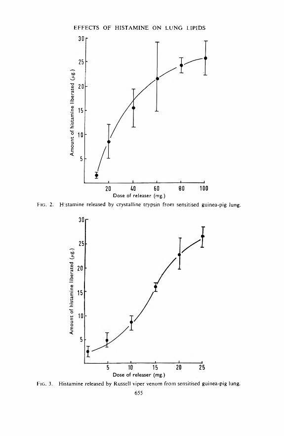
Fig. 2. Histamine released by crystalline trypsin from sensitised guinea-pig lung.
Fig. 3. Histamine released by Russell viper venom from sensitised guinea-pig lung.655

At concentrations in excess of 80 /xg./ml. in Tyrode solution (the concentration of compound 48/80 in perfusate after a histamine releasing dose of 5 mg.), compound 48/80 was adsorbed onto Decalso F. It could subsequently be eluted, in the manner used for the elution of adsorbed histamine, with ammonia and then chloroform saturated with ammonia.
V. O. M ARQ UIS A N D W. G. SM ITH
Dose of releaser (mg.)
Fig. 4. Histamine released by compound 48/80 from sensitised guinea-pig lung.
When the ammonia-chloroform eluate was taken to dryness and made up in Tyrode solution, the resultant solution was found to reduce the sensitivity of guinea-pig ileum to histamine. It was thus concluded that the histamine released by compound 48/80 in doses in excess of 5 mg. was contaminated with compound 48/80 in a concentration sufficient to reduce the sensitivity of guinea-pig ileum to histamine. During subsequent biological assay, the contamination was confined to the test solutions and thus produced low estimates of the histamine actually released.
Effect of Histamine Release on Lipid Content o f Lung TissueFrom the data given in Figs. 1 to 4 it was concluded that the release of
15 pg. of histamine from perfused guinea-pig lungs could be induced by16 mg. of antigen (egg albumin), 34 mg. of trypsin, 15 mg. of Russell viper venom, or 4 mg. of compound 48/80. These doses were each administered to a group of ten sensitised guinea-pigs and the observed histamine release agreed very closely with that expected (15-0 ± 1-05 pg.). The mean amounts of slow reacting substance released are shown in Fig. 5 together with the standard deviation of each mean. A similar yield of slow reacting substance was noted with antigen, trypsin and Russell viper venom. The amount released by compound 48/80 was appreciably less than that released by the other three compounds.
656

The lipid present in the lungs of these animals was compared with that found in an “experimental control” group which had been perfused with Tyrode solution without administration of a histamine releaser. The results are shown in Fig. 6.
EFFECTS OF H IST AM INE O N L U N G LIPIDS
320 -
240 -
160
80
n r
oFig. 5. The slow reacting substances released from sensitised guinea-pig lung by equi-active doses of different histamine releasers. A, Antigen 16 mg. B, Trypsin 34 mg. C, Russell viper venom 15 mg. D, Compound 48/80 4 mg.
The amounts of lipid present in the “experimental control” group were the same as these observed in chopped blood free lungs. Comparative figures are given in Table I.
TABLE ILipid content of lungs
Controls Treatment controlsCholesterol mg./g... 18-90 ± 1-41 18-20 ± 4 03Glyceride mg./g. .. 30-40 ± 15-69 30-40 ± 15-27Lipid phosphorus mg./g. .. 4-86 ± 0-25 4-90 ± 0-30
Fig. 6a shows that whereas antigen, Russell viper venom, and compound 48/80 inducec falls in the lipid phosphorus content of the lung, trypsin was without an effect on this lipid fraction. From Fig. 6b, only the Russell viper venom causes a significant rise in the lung cholesterol. Fig. 6c shows that although all four histamine releasers induced an increase in the glyceride content of the lung tissue there was considerable variation in the magnitude of the change.
DiscussionIn the event that histamine release in anaphylaxis is due to the activation
of a proteolytic enzyme, activation of phospholipase A, or degranulation of mast cells, it is reasonable to expect that the release of histamine and the changes in the lipid content of the lung tissue in anaphylaxis would be closely paralleled by trypsin, Russell viper venom, or compound 48/80 respectively. The results obtained do not support any of these possibilities.
657

The release of histamine from sensitised guinea-pig lung tissue by antigen is most closely duplicated by trypsin. An accurate comparison on a molar basis is not possible since the molecular weights of these substances are not known with accuracy. Even so, there is a quantitative similarity between the histamine-releasing effects of trypsin and antigen, which is not shared by Russell viper venom and compound 48/80. A comparison confined to the shape of the dose-effect curves might be interpreted as a suggestion that antigen released histamine by a proteolytic action, and that compound 48/80 released histamine by a phospholipase effect.
V. O. M ARQ UIS A N D W. G. SM ITH
Fig. 6. The lipid content of sensitised guinea-pig lungs after treatment with equi- active doses of different histamine releasers. See F ig. 5 for Key.
The similarity between the histamine-releasing effects of antigen and trypsin is not supported by the changes induced in the lipid content of the lungs. Trypsin did not produce a fall in lipid phosphorus content of the tissue. The effects of antigen on the lung lipids reported here ditfer slightly from those reported earlier by Smith (1962a). On that occasion loss of glyceride was noted after antigen administration, whereas in the
658

present study there was no significant change. These two studies agree, however, in that they both show a loss of lipid phosphorus and to a lesser degree of cholesterol.
Since the amount of histamine release was constant in all four groups of lungs whose lipid content was compared, the marked differences in the lipid fractions of the respective groups indicates that histamine release itself is not the primary cause of the changes in the lung lipids. The amounts of slov/ reacting substance liberated varied from one histamine liberator to another, but the amounts liberated also show no obvious relationship to the lipid changes. The liberated slow reacting substances are not identical in all four cases; the slow reacting substance released by Russell viper venom differs from that liberated by antigen (Smith, 1962b; Schütz and Vogt, 1961).
It is thus concluded that the release of histamine or :he release of slow reacting substance during anaphylaxis in guinea-pig lung are not themselves responsible for the changes in lipid metabolism induced by that condition. Such changes are presumably due to alterations in intermediary metabolism induced in sensitised tissue when antigen combines with antibody bound within it, and are manifestations of effects on the tissue metabolism which occur in parallel with effects due to the release of chemical mediators of anaphylaxis.
EFFECTS OF H IST AM INE O N L U N G LIPIDS
ReferencesBartlett, G. R. (1959). /. biol. Chem., 234, 466-468.Brocklehurst, W. E. (1960). J. Physiol., 151, 416-435.Chakravarty, N. (1960). Acta physiol, scand., 48, 167.Goadby, P. and Smith, W. G. (1962). J. Phann. Pharmacol., 14, 739-745. Hanel. H. K. and Dam, H. (1955). Acta. chem. scand., 9, 677-682.Roberts, M. and Adam, H. M. (1950). Brit. J. Pharmacol., 5, 526-541.Schütz, R. M. and Vogt, W. (1961). Arch. exp. Path. Pharmak., 240, 504-513. Smith, W. G. (1962a). Biochem. ¡Pharmacol., 11, 183-186.Smith, W. G. (1962b). Life Sciences, 1, 133-140.
659

T H E E F F E C T S O F T W O P H E N Y L A C E T IC A C ID D E R IV A T IV E SO N T H E A N A L G E S IC A C T IO N O F M O R P H I N E I N M IC E
By M . M edakovic and B. Banic
From the Department of Pharmacology, Medical Faculty, Belgrade, Yugoslavia
Received April 1, 1963
After intracerebral injection in mice, the analgesic effect of morphine depended on the dose. Two phenylacetic acid esters, CFT 1201 (di- ethylaminoethylphenyldiallyl acetate) and CFT 1208 (diethylamino- ethylphenylallyl acetate), injected intraperitoneally, potentiated the analgesic action of 0-5 ng. of morphine, injected into the posterior portion of the brain 90 min. later. When the phenyl acetates were injected (50 /*g.) into the brain, they potentiated the analgesic action of morphine, injected subcutaneously (10 mg./kg.) 80 min. later. The results show that the inhibitors of the hepatic microsomal enzymes may affect the action of analgesics by an action on the central nervous system.
Substances which inhibit hepatic microsomal enzymes can potentiate and prolong the action of drugs being inactivated in the liver. Thus, carbon tetrachloride and some phenyl acetates potentiate and prolong the analgesic effect of morphine and similar analgesics. This phenomenon has been explained by an inhibitory action on the hepatic microsomal enzymes. However, some authors (Cook, Navis, Tonner and Fellows, 1953; Swinyard, Madsen and Goodman, 1954; Herken, Neubert and Timmler, 1959) suggested that the potentiating action of some of these agents (SKF 525-A and CFT 1201) was not only due to their action on the metabolic demethylation of drugs in the liver. Our recent experiments (Medakovic and Banic, 1963) suggested also that carbon tetrachloride may increase the analgesic action of morphine not only by its action on the liver, but also by some mechanism which our data suggested might be an action on the central nervous system.
To further elucidate this possibility, experiments were arranged so as to minimise the influence of the inhibitors of the hepatic microsomal enzymes on the liver. This was achieved by using various routes of injection, and by combining these routes. Thus in some experiments morphine was injected in mice intracerebrally and the microsomal inhibitor intraperitoneally, while in other experiments the reverse order v/as followed. Further, the actions of two phenyl acetates on morphine analgesia have been studied. One of these, diethylaminoethylphenyl- diallyl acetate (CFT 1201) is a potent inhibitor of hepatic microsomal enzymes, while the other, diethylaminoethylphenylallyl acetate (CFT 1208) has no such effects (Maibauer, Neubert and Rottka, 1958).
M ethods
Male white mice of approximately 20 g. were used. Analgesia was tested according to the method of Woolfe and Macdonald (1944). Each mouse in turn was placed on a hot plate (53°) and the reaction time
660

for the appearance of the paw licking reflex recorded. Groups of 10 mice were used and comprised only those animals which responded in a10-15 sec. period. To prevent paw tissue damage in animals with analgesia, they were removed from the hot plate before this could occur. An arbitrary interval equivalent to double the control mean reaction time of each given group was selected for the purpose. The values were expressed on the graphs as percentages of the cut-off time. Thus the 50 per cent value in the graphs corresponds to the control mean reaction time of the corresponding group.
Intracerebral injections were according to Matthies and Schmidt (1961), into the hind brain, 2 mm. deep, with fine intradermal needles, approximately 3 mm. long. The drugs were dissolved in saline, to give a total volume of 0-01 ml. for the intracerebral injections.
The drugs used were: morphine hydrochloride, diethylaminoethyl- phenyldiallyl acetate hydrochloride (CFT 1201) and diethylaminoethyl- phenyallyl acetate hydrochloride (CFT 1208).
The results are presented graphically, but some of the animals were allowed to remain in contact with the hot plate until the cut-off time had expired, thus the analgesic effect as plotted is not exact and represents low values. Mean values which include two or more cut-off times have been represented in the graphs by open signs (circles, squares or triangles).
PHENYLACETIC A C ID DERIVATIVES A N D A N A LG ESIA
Fig. 1. Analgesic effect of three doses of morphine, injected intracerebrally. Triangles, 0-5 f i g . , squares, 1 fig. and circles, 2 fig. of morphine hydrochloride.
R esults
The action of intracerebrally injected morphine. Saline, injected into the brain, did not change the control mean reaction time. However, when morphine, dissolved in saline, was injected by the same route, the reaction times were prolonged (Fig. 1). Three groups of animals
661

were injected with 0-5, 1 and 2 /xg. respectively. Fig. 1 shows that the effect of morphine was dependent on the dose injected.
The pH of the saline remained unchanged when morphine hydrochloride was dissolved either in concentrations used in these experiments or in much higher ones. Because of this, and also because of the dose : response relation, the prolongation of the reaction time was considered to be caused by the analgesic action of morphine.
Effects o f compound CFT 1201 and CFT 1208 on the action of intra- cerebrally injected morphine. Compounds CFT 1201 and CFT 1208 respectively, were injected intraperitoneally (50 mg./kg.), 90 min. before morphine (0-5 /xg.) was given intracerebrally. The animals in the control group received saline intraperitoneally. The results are in Fig. 2. Both
M. M EDAK O VlC A N D B. B A N lC
Time after injection
F ig . 2. The effects of compound CFT 1201 and 1208, injected intraperitoneally (50 mg./kg. of each) on the analgesic action of morphine, injected intracerebrally (0-5 f tg.) 90 min. after the respective CFT compound. Triangles, morphine after saline; squares, morphine after CFT 1208; circles, morphine after CFT 1201.
morphine (min.)
Fig. 3. The effects of CFT 1201 and CFT 1208, injected intracerebrally (50 u.g. each) on the analgesic action of morphine, injected subcutaneously (10 mg./ kg.) 80 min. after the respective CFT compound. Triangles, morphine after saline; squares, morphine after CFT 1208; circles, morphine after CFT 1 2 0 1 .
CFT compounds were able to potentiate the analgesic action of morphine. Fig. 2 shows also that CFT 1201 had a higher potentiating activity than CFT 1208.
The control time before the injection of morphine revealed that compound CFT 1201 itself caused a moderate and short-lasting prolongation of the control mean reaction time. Neither compound CFT 1208 nor saline produced this effect. This effect of the compound CFT 1201 reached a maximum 15 min. after injection, but was insignificant when the morphine was injected.
662

PHENYLACETIC A C ID DERIVATIVES A N D AN ALG ESIA
The effect of the morphine (0-5 /xg.) in animals which had previously received compound CFT 1201, was comparable to the effect of 2 /xg. of morphine alone (Fig. 1). Thus the mean reaction time was prolonged to 99 per cent o: the cut-off time 20 min. after the injection of morphine. At this time cut-off time was recorded in 8 out of 10 mice. In the control experiment (Fig. 1), 0-5 /xg. of morphine produced a prolongation of the reaction time to 78 per cent of the cut-off time and only 4 out of 10 mice did not respond until the cut-off time.
As almost all mice receiving morphine after CFT 1201 did not respond until the cut-off time, the curve of the analgesic action shown in Fig. 2 is not the true effect, and it can be assumed that the potentiation of the effect of morphine is higher than is shown in Fig. 2.
In the mice receiving CFT 1208, the analgesic action of morphine was also potentiated, but less than by compound CFT 1201. Thus, only 5 out of 10 mice reached the cut-off time, and the mean reaction time was 89 per cent of the cut-off time. The potentiating effect of the compound CFT 1208 was also higher than presented in the graph. Consequently, the true difference between the potentiating activity of these two compounds cannot be clearly established.
Effect of intracerebrally injected CFT compounds on the analgesic action of subcutaneously injected morphine. In this experiment, CFT 1201 and CFT 1208 were injected intracerebrally (50 /xg.) and then morphine was injected subcutaneously (10 mg./kg.) 80 min. later.
The first group of mice received CFT 1201. Immediately after, signs of central stimulation were observed. The animals appeared quiet, but, on touch, started running and some had convulsions. None died but those which convulsed were rejected.
Compound CFT 1201 caused moderate and reversible prolongation of the reaction time upon intracerebral injection. This effect was short- lasting and the reaction time returned to its starting control level long before morphine was injected.
The effect after morphine is shown in Fig. 3. Its action was potentiated by the previous ejection of the compound CFT 1201. Potentiation was also obtained with the compound CFT 1208, both compounds apparently potentiating the effect of morphine to a similar degree. However, it can be assumed that the potentiating activity of CFT compounds was partly masked in this experiment also. Consequently, the relative potentiating activity of CFT compounds in this experimental design cannot be clearly established.
D iscussion
The present experiments were aimed at throwing more light on the hypothesis that the so-called inhibitors of hepatic microsomal enzymes may affect the analgesic action of morphine by a mechanism independent from their action on the liver. Therefore, the routes of administration of drugs were chosen to minimise the influence of the metabolising capacity of the liver on the analgesic action of morphine.
663

It can be assumed that the analgesic action of morphine upon intracerebral injection was caused mainly by the direct effect of the drug; the concentration in the blood at this dose level having an insignificant effect, as 10 mg. /kg. of morphine (approximately 200 fig. per mouse) has to be injected subcutaneously or intraperitoneally to obtain an effect comparable to that of 0-5 /Mg. of morphine injected into the brain.
The CFT compounds both intraperitoneally and intracerebrally potentiated the analgesic action of morphine. The potentiating intracerebral dose of the compound CFT 1201, was only 50 fig. while to inhibit hepatic microsomes, as much as 100 mg./kg. (approximately 2000 fig. per mouse) is usually injected intraperitoneally. It is therefore, improbable that after the intracerebral injection of 50 fig. of CFT 1201 the hepatic microsomes could be so strongly inhibited as to cause a delayed inactivation of morphine, and consequently a potentiation of its effect. That some other mechanism is probably involved is suggested also by the fact that both CFT 1201 and CFT 1208 potentiated the action of morphine. As already mentioned, compound CFT 1208 has been found not to inhibit hepatic microsomal enzymes in in vitro experiments (Maibauer and others, 1958).
When the CFT compounds were injected intraperitoneally, the effect of morphine was potentiated, and not prolonged. But, if CFT compounds acted by involving only the rate of the hepatic inactivation of morphine, the result would be prolongation rather than a potentiation.
Hence, the findings suggest that the so-called inhibitors of the hepatic microsomal enzymes may affect the action of analgesics not only by inhibiting their inactivation in the liver, but also by some other mechanism, a view already expressed by others (Cook and others, 1953; Swinyard and others, 1954; Herken and others, 1959; Herz, 1961). Thus, for example, that SKF 525-A, potentiated the analgesic action but did not affect the toxicity of morphine, could not be explained by the inhibition of the hepatic microsomes only (Cook and others, 1953). Similarly, SKF 525-A and CFT 1201 failed to potentiate the sedative stupor- inducing action of morphine (Herz, 1961). Further, carbon tetrachloride increased the analgesic action of morphine and codeine, but failed to affect the antidiuretic action of these drugs in our earlier experiments (Medakovic and Banic, 1963). Hence, it can be assumed that these inhibitors of the hepatic microsomes can affect the action of analgesics by some more specific mechanism.
Carbon tetrachloride was shown to potentiate the inhibitory action of morphine on the twitch of the electrically stimulated isolated guinea-pig ileum (Medakovic and Banic, 1963). Since this organ has been proposed as a suitable model for the analysis of actions of drugs which affect the central nervous system (Schaumann, Giovannini and Jochum, 1952; Patón, 1957), our finding suggested that carbon tetrachloride may potentiate the action of analgesics acting on the central nervous system. The present in vivo experiments indicated that the potentiating action of CFT compounds on the analgesic action of morphine was elicited by a mechanism involving the brain.
M. M EDAK O VIC A N D B. BANIC
664

The question arises as to whether the potentiating effect shown by the CFT compounds had some connection with their ability to affect the enzymes. While their effect might possibly be a pharmacological synergism only, a connection between the effect of the CFT compounds and their ability to act upon enzymatic processes, is possible and is further suggested by the fact that CFT 1201 appeared to have a higher activity than CFT 1208.
Axelrod (1956) expressed the view that the metabolic changes, which the molecule of morphine undergoes in the liver, may be a model for the processes which occur at the receptors of the morphine action in the brain. In fact, the in vivo transformation of morphine in the brain has been demonstrated (Beckett, Casy and Harper, 1956; Milters, 1962), as well as a remarkable demethylation of pethidine by the rat brain tissue (Herken and others, 1959). It would be therefore interesting to examine the possible action of the inhibitors of hepatic micrcsomal enzymes on these and similar processes in the brain.
PHENYLACETIC A C ID DERIVATIVES A N D A N A L G E SIA
Acknowledgements.—CFT compounds were kindly supplied by Prof. Dr. H. Herken (Berlin) and Dr. Pflugk (Chemische Fabrik Tempelhof, Berlin).
R eferences
Axelrod, J. (1956). J. Pharmacol., 117, 322-330.Beckett, A. H., Casy, A. F. and Harper, N. J. (1956). J. Pharm. Pharmacol., 8, 874-
883Cock, L., Navis, G. G., Tonner, L. and Fellows, E. G. (1953). Fed. Proc., 12, 299. Herken, H., Neubert, D. and Timmler, R. (1959). Arch. exp. Path. Pharmak., 237,
319-333Herz, A. (1961). Ibid., 241, 236-253.Maibauer, D., Neubert, D. and Rottka, H. (1958). Ibid., 234, 474-489.Matthies, H. and Schmidt, J. (1961). Ibid., 241, 508-509.Medakovic, M and Banic, B. (1963). Acta med. Jug. (in the press).Milters, K. (1962). Acta pharm. tox. Kbh., 19, 235.Paton, W. D. M. (1957). Brit. J. Pharmacol., 12, 119-127.Schaumann, O., Giovannini, M. and Jochum, K. (1952). Arch. exp. Path. Pharmak.,
215, 460-468.Swinyard, E. A., Madsen, J. A. and Goodman, L. S. (1954) J. Pharmacol., I l l ,
54-63.Woolfe, G. and Macdonald, A. D. (1944). Ibid., 80, 300-307.
665

B y P. H . M a r r i o t t *
AN INVESTIGATION INTO THE EFFECTS OF GAMMA-RAYSON AQUEOUS SOLUTIONS OF p-AMINOBENZOIC ACID
From the Department o f Pharmacy, University o f Manchester
Received April 29, 1963
Aqueous solutions of /?-aminobenzoic acid (0-3 per cent) were irradiated with gamma-rays under both oxygen-saturated and oxygen-free conditions. By means of paper chromatography, /?-nitrobe:izoic acid has been isolated from the irradiated, oxygenated aqueous solution and4-amino-3-hydroxybenzoic acid isolated from the irradiated, oxygen- free aqueous solution.
These products were probably produced by indirect action of the Co-60 gamma-rays.
T h e a b i l i t y o f h ig h -e n e r g y e le c t r o m a g n e t ic a n d p a r t ic u la te r a d ia t io n t o d e s tr o y (w ith in c e r ta in l im it s o f p r o b a b i l i t y ) m ic r o -o r g a n is m s w i t h o u t a p p r e c ia b le t e m p e r a tu r e r ise in th e s u b s tr a te m ig h t o f fe r a m e a n s o f s t e r i l is in g h e a t - la b i le d r u g s .
In June, 1960, a report was published (A.B.P.I. Report) on the use of y-radiation sources for the sterilisation of pharmaceutical products. This report served to emphasise the need for more work in this field.
An investigation into the effects of y-irradiation on aqueous solutions of p-aminobenzoic acid (paba) should yield information about the action of the radiation on esters of paba, e.g. procaine, in aquecus solution.
The major primary path by which any high-energy particles or photons interact with matter is by interaction with electron shells of molecules. Energy is absorbed, electrons are ejected to produce ions, and bonds are broken to produce free-radicals. The particles produced are often unstable and decompose with the result that much of the energy is quickly dispersed with the formation of excited molecules and radicals. It is these species that undergo the postulated reactions and “account” for many of the products that are ultimately observed (Hart, 1954a,b).
Initial radical production appears to be a highly indiscriminate process, the number of radicals produced from any component being roughly proportional to the fraction of the component, by weight, present in the system (Henley and Barr, 1956). Accordingly, the reactions of a dilute solution of a reactive substrate in an unreactive solvent are chiefly those of the solvent radicals with the substrate and it may be relatively easy to interpret these. Irradiation of a single complex organic molecule, or mixtures such as those found in biochemical systems, may give results which are of almost meaningless complexity.
Products to be expected on y-irradiation of aqueous solutions of paba are those produced from the original solute by oxidation, reduction, deamination and decarboxylation. The relative amounts of the products cannot be predicted.
* Present address: School of Pharmacy, University of London, 29/39 Brunswick Square, London, W.C.l.
666

EFFECTS OF y-RAYS ON p-AMINOBENZOIC ACID
E x p e r im e n t a l M e t h o d s
Radiation Source and DosimetryThe radiation source was a 250c Cobalt-60 source, which was
calibrated as described by Davies (1960).Solutions Prepared and Dose Given
p-Aminobenzoic acid (0-3 g.) was dissolved in glass-distilled water (100 ml.). The pH of the solution was not specially adjusted. The solution was divided into two equal portions (by volume), each was contained in a large glass tube, of approximately 70 ml. capacity, fitted with a ground-glass stopper.
Oxygen was bubbled through one of the portions, designated I0, and oxygen-free nitrogen gas through the other portion, designated In, for 2 hr., after which the tubes were well stoppered. The air above solution I0 was assumed to be saturated with oxygen whereas the enclosed air above the second solution, In, contained little oxygen. Each portion of the solution was colourless and free from solid particles. The tubes containing the solutions were then irradiated. Solution I0 received a dose of approximately 2 Megarads and solution In a dose of approximately 4 Megarads.Detection o f Breakdown Products Using Paper Chromatography
The solvent was a mixture of n-butanol and aqueous ammonium hydroxide solution (1-5n) (35:65 v/v). Whatman No. 1 paper 57 X 46 cm., or smaller sizes cut as required, was used and after equilibration (at 24° ± 1° for 4 to 6 hr.) developed by the descending technique for 17 to 18 hr. The paper was then dried by warm air.
Substances Applied to the ChromatogramsThe irradiated solutions I0 and In were concentrated by reduced pressure
distillation and then applied as such to the chromatograms.The following possible products of degradation were applied to the
chromatograms as spots in ether. o-Nitrophenol, w-nitrophenol, p- nitrophenol, and 4-amino-3-hydroxybenzoic acid were applied as separate spots. A mixture ofp-aminobenzoic acid, salicylic acid, w-hydroxybenzoic acid, p-hydroxybenzoic acid and p-nitrobenzoic acid and a mixture of p-aminobenzoic acid, p-aminosalicylic acid, o-aminophenol, w-amino- phenol, and p-aminophenol were applied to the chromatograms as single spots and run alongside the spots of I0 and In-
p-Aminosalicylic acid was prepared from the mono-sodium salt of4-aminosalicylic acid, and 4-amino-3-hydroxybenzoic acid was prepared from 3-hydroxy-4-nitrobenzoic acid by reduction using tin and hydrochloric acid (Froelicher and Cohen, 1921). All other substances were obtained from commercial sources and purified before use.
Reagents for Spot DetectionEhrlich reagent. p-Dimethylaminobenzaldehyde (2 g.) is dissolved by
shaking in 20 per cent aqueous hydrochloric acid (100 ml.).667
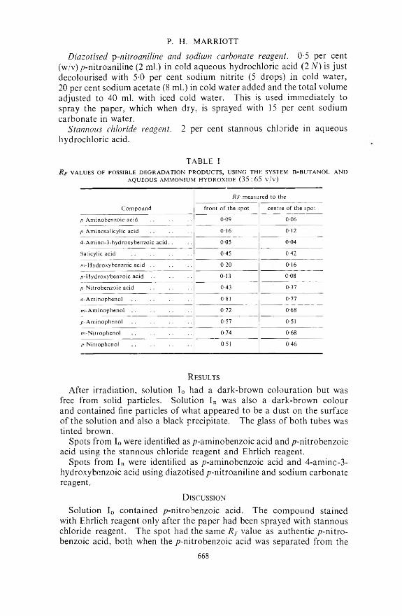
P. H. MARRIOTT
Diazotised y-nitroaniline and sodium carbonate reagent. 0-5 per cent (w/v) p-nitroaniline (2 ml.) in cold aqueous hydrochloric acid (2 N) is just decolourised with 5-0 per cent sodium nitrite (5 drops) in cold water, 20 per cent sodium acetate (8 ml.) in cold water added and the total volume adjusted to 40 ml. with iced cold water. This is used immediately to spray the paper, which when dry, is sprayed with 15 per cent sodium carbonate in water.
Stannous chloride reagent. 2 per cent stannous chloride in aqueous hydrochloric acid.
TABLE IR jr VALUES OF POSSIBLE DEGRADATION PRODUCTS, USING THE SYSTEM n-B U TA N O L AND
AQUEOUS AMMONIUM HYDROXIDE (35:65 v/v)
Rf measured to theCompound front of the spot centre of the spot
p-Aminobenzoic acid 009 0-06p-Aminosalicylic acid 016 0124-Amino-3-hydroxybenzoic acid.. 0 05 004Salicylic acid 0-45 0-42m-Hydroxybenzoic acid .. 0-20 016p-Hydroxybenzoic acid 0-13 008p-Nitrobenzoic acid 0-43 0-37o-Aminophenol 0-81 0-77m-Aminophenol 0-72 0-68p-Aminophenol 0-57 0-51w-Nitrophenol 0-74 0-68p-Nitrophenol 0-51 0-46
R esults
After irradiation, solution I0 had a dark-brown colouration but was free from solid particles. Solution In was also a dark-brown colour and contained fine particles of what appeared to be a dust on the surface of the solution and also a black precipitate. The glass of both tubes was tinted brown.
Spots from I0 were identified as p-aminobenzoic acid and p-nitrobenzoic acid using the stannous chloride reagent and Ehrlich reagent.
Spots from In were identified as p-aminobenzoic acid and 4-aminc-3- hydroxybenzoic acid using diazotised p-nitroaniline and sodium carbonate reagent.
D iscussion
Solution I0 contained p-nitrobenzoic acid. The compound stained with Ehrlich reagent only after the paper had been sprayed with stannous chloride reagent. The spot had the same RF value as authentic p-nitro- benzoic acid, both when the p-nitrobenzoic acid was separated from the
668
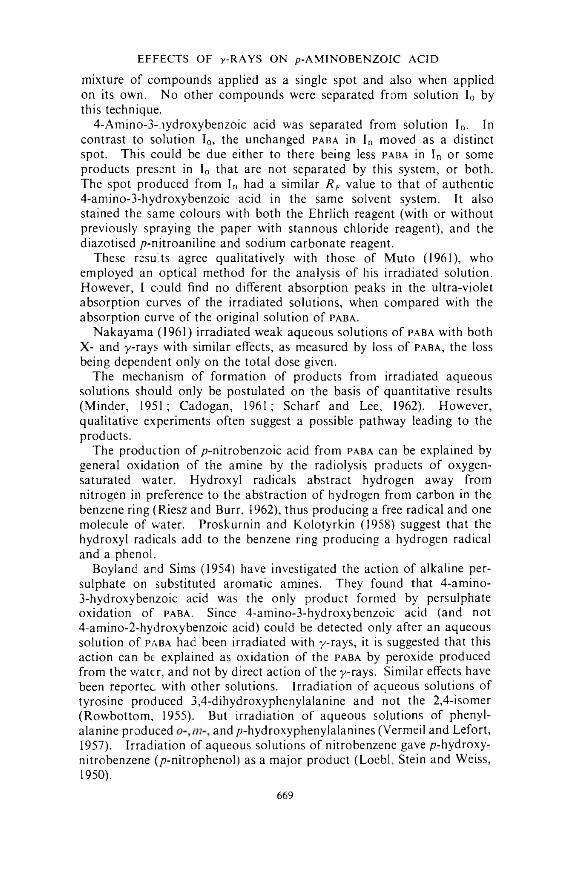
mixture of compounds applied as a single spot and also when applied on its own. No other compounds were separated from solution I0 by this technique.
4-Amino-3-/iydroxybenzoic acid was separated from solution In. In contrast to solution I0, the unchanged paba in In moved as a distinct spot. This could be due either to there being less paba in In or some products present in I0 that are not separated by this system, or both. The spot produced from In had a similar RF value to that of authentic4-amino-3-hydroxybenzoic acid in the same solvent system. It also stained the same colours with both the Ehrlich reagent (with or without previously spraying the paper with stannous chloride reagent), and the diazotised /7-nitroaniline and sodium carbonate reagent.
These results agree qualitatively with those of Muto (1961), who employed an optical method for the analysis of his irradiated solution. However, I could find no different absorption peaks in the ultra-violet absorption curves of the irradiated solutions, when compared with the absorption curve of the original solution of paba.
Nakayama (1961) irradiated weak aqueous solutions of paba with both X- and y-rays with similar effects, as measured by loss of paba, the loss being dependent only on the total dose given.
The mechanism of formation of products from irradiated aqueous solutions should only be postulated on the basis of quantitative results (Minder, 1951; Cadogan, 1961; Scharf and Lee, 1962). However, qualitative experiments often suggest a possible pathway leading to the products.
The production of p-nitrobenzoic acid from paba can be explained by general oxidation of the amine by the radiolysis products of oxygen- saturated water. Hydroxyl radicals abstract hydrogen away from nitrogen in preference to the abstraction of hydrogen from carbon in the benzene ring (Riesz and Burr, 1962), thus producing a free radical and one molecule of water. Proskurnin and Kolotyrkin (1958) suggest that the hydroxyl radicals add to the benzene ring producing a hydrogen radical and a phenol.
Boyland and Sims (1954) have investigated the action of alkaline persulphate on substituted aromatic amines. They found that 4-amino-3- hydroxybenzoic acid was the only product formed by persulphate oxidation of paba. Since 4-amino-3-hydroxybenzoic acid (and not4- amino-2-hydroxybenzoic acid) could be detected only after an aqueous solution of paba had been irradiated with y-rays, it is suggested that this action can be explained as oxidation of the paba by peroxide produced from the water, and not by direct action of the y-rays. Similar effects have been reported with other solutions. Irradiation of aqueous solutions of tyrosine produced 3,4-dihydroxyphenylalanine and not the 2,4-isomer (Rowbottom, 1955). But irradiation of aqueous solutions of phenylalanine produced and p-hydroxyphenylalanines (Vermeil and Lefort,1957). Irradiation of aqueous solutions of nitrobenzene gave p-hydroxy- nitrobenzene (/?-nitrophenol) as a major product (Loebl, Stein and Weiss, 1950).
EFFECTS OF y-RAYS ON p-AMINOBENZOIC ACID
669
P. H. MARRIOTT
No p-nitrobenzoic acid was detected in solution In and no 4-amino-3- hydroxybenzoic acid detected in solution I0. The lack of p-nitrobenzoic acid in In indicates that dissolved oxygen gas plays a part in oxidation procedures that occur on y-radiolysis of oxygenated aqueous solutions. The degree of oxidation is controlled by the tensions of oxygen, nitrogen and other gases present. In oxygen-free solutions monophenolic compounds would be stable whereas in oxygen-saturated solutions further oxidation would almost certainly occur giving rise to poly-phenolic compounds or nitro-compounds from paba. In oxygen-free solutions any free-radicals produced would tend to combine, forming stable compounds such as diphenyl derivatives. The black precipitate in solution In could well be a polyphenyl derivative but identification of the precipitate has not been completed.
The future prospects for y-radiation sterilisation of drugs in aqueous solution would seem to lie with compounds that decompose, into nontoxic products, in preference to the breakdown of the drug.
Acknowledgements. The author wishes to express his gratitude to Professor K. Bullock for the encouragement and help given him, and to the Department of Scientific and Industrial Research for the award of a Research Studentship, throughout the course of this work.
R eferences
A.B.P.I. Report (1960). “The use of Gamma-radiation Sources for the Sterilisation of Pharmaceutical Products,” Report of a Working Party Established by The Association of British Pharmaceutical Industry in collaboration with The School of Pharmacy, University of London and The Isotope Division, Atomic Energy Research Establishment. London: A.B.P.I., Tavistock Square.
Boyland, E. and Sims, P. (1954). J. chem. Soc., 980-985.Cadogan, J. I. G. (1961). The Roval Institute o f Chemistry, Lecture Series, No. 6,
1-28.Davies, D. J. G. (1960). M .Sc. Thesis, University of Manchester.Froelicher, V. and Cohen, J. B. (1921). J. chem. Soc., 1425-1431.Hart, E. J. (1954a). Rad. Res., 1, 53-61.Hart, E. J. (1954b). Ann. Rev. Phys. Chem., 5, 139-162.Henley, E. J. and Barr, N. F. (1956). Advances in Chemical Engineering, 1, pp. 1-76,
New York: Academic Press Inc.Loebl, H., Stein, C. and Weiss, J. (1950). J. chem. Soc, 2704-2739.Minder, W. (1951). Brit. J. Radiol., 24, 435-440.Muto, T. (1961). Nuclear Science Abstracts, 20739.Nakayama, Y. (1961). Ibid., 20765.Proskurnin, M. A., and Kolotyrkin, Ya. M. (1958). Proc. U.N. Intern. Cortf.
Peaceful Uses At. Energy, 2nd., Geneva, P/2022, 29, 52-61.Riesz, P. and Burr, B. E. (1962). Rad. Res., 16, 661-667; 668-673.Rowbottom, J. (1955). J. biol. Chem., 212, 877-885.Scharf, K. and Lee, R. M. (1962). Rad. Res., 16, 115-124.Vermeil, C. and Lefort, M. (1957). C.R. Acad. Sci. Paris, 244, 889-891.
670
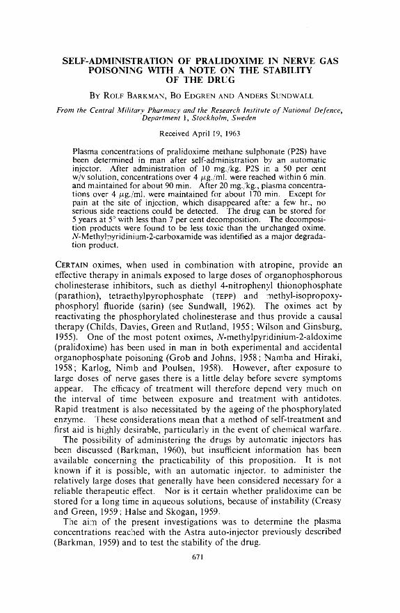
S E L F -A D M IN IS T R A T IO N O F P R A L ID O X IM E IN N E R V E G A S P O IS O N IN G W IT H A N O T E O N T H E ST A B IL IT Y
O F T H E D R U G
B y R o l f B a r k m a n , B o E d g r e n a n d A n d e r s S u n d w a l l
From the Central Military Pharmacy and the Research Institute o f National Defence, Department 1, Stockholm, Sweden
Received April 19, 1963
Plasma concentrations of pralidoxime methane sulphonate (P2S) have been determined in man after self-administration by an automatic injector. After administration of 10 mg./kg. P2S in a 50 per cent w/v solution, concentrations over 4 /rg./ml. were reached within 6 min. and maintained for about 90 min. After 20 mg./kg., plasma concentrations over 4 /ag./ml. were maintained for about 170 min. Except for pain at the site of injection, which disappeared after a few hr., no serious side reactions could be detected. The drug can be stored for 5 years at 5° with less than 7 per cent decomposition. The decomposition products were found to be less toxic than the unchanged oxime. jV-Methylpyridinium-2-carboxamide was identified as a major degradation product.
C e r t a i n oximes, when used in combination with atropine, provide an effective therapy in animals exposed to large doses of organophosphorous cholinesterase inhibitors, such as diethyl 4-nitrophenyl thionophosphate (parathion), tetraethylpyrophosphate ( t e p p ) and methyl-isopropoxy- phosphoryl fluoride (sarin) (see Sundwall, 1962). The oximes act by reactivating the phosphorylated cholinesterase and thus provide a causal therapy (Childs, Davies, Green and Rutland, 1955; Wilson and Ginsburg, 1955). One of the most potent oximes, TV-methylpyndinium-2-aldoxime (pralidoxime) has been used in man in both experimental and accidental organophosphate poisoning (Grob and Johns, 1958; Namba and Hiraki, 1958; Karlog, Nimb and Poulsen, 1958). However, after exposure to large doses of nerve gases there is a little delay before severe symptoms appear. The efficacy of treatment will therefore depend very much on the interval of time between exposure and treatment with antidotes. Rapid treatment is also necessitated by the ageing of the phosphorylated enzyme. These considerations mean that a method of self-treatment and first aid is highly desirable, particularly in the event of chemical warfare.
The possibility of administering the drugs by automatic injectors has been discussed (Barkman, 1960), but insufficient information has been available concerning the practicability of this proposition. It is not known if it is possible, with an automatic injector, to administer the relatively large doses that generally have been considered necessary for a reliable therapeutic effect. Nor is it certain whether pralidoxime can be stored for a long time in aqueous solutions, because of instability (Creasy and Green, 1959; Halse and Skogan, 1959.
The aim of the present investigations was to determine the plasma concentrations reached with the Astra auto-injector previously described (Barkman, 1959) and to test the stability of the drug.
671

R. BARKMAN, B. EDGREN AND A. SUNDWALL
M e t h o d s
Intramuscular Administration of PralidoximeThe injections were made with an automatic injector as described by
Barkman (1960) (see Fig. 1). The injector was loaded with 1-5 ml. of a
F i g . 1. Above: Closed view o f the injector. Middle: Exploded view of the injector. The ring and loop function as a safety catch. Below: Case with two spare needles and ampoules attached.
50 per cent w/v aqueous solution of pralidoxime methane sulphonate (P2S). The solutions were tilled in the plastic ampoules of the injector and sterilized by autoclaving at 120° for 20 min. This procedure produces about 5 per cent loss of oxime (Barkman. 1959), without influencing the acute toxicity of the solution. The experiments were done on 24 healthy human volunteers of both sexes (age 21 to 35 years). The injections were administered by the test persons themselves on the outside of the thigh. Blood samples were taken at intervals from an antecubital vein and the plasma analysed for P2S by an ultra-violet method (Sundwall, 1960).Long-term Storage of P2S in Aqueous Solutions
The pH for maximum stability at 120: was about 3 when measured at 25° (Barkman, 1959). For this reason solutions of P2S (16 per cent w/v) were buffered at pH 2-5, 3-0 and 3-5 although the pFI of maximum stability is 4 at room temperature. The solutions were stored in sealed glass ampoules at 5, 15, 25 and 45° for 2 years. The buffer solutions were prepared from 0 - 2 m disodium phosphate and 0 1 m citric acid, according to Mcllvaine and Sorensen.
The solutions were analysed for unchanged pralidoxime after 1, 2. 3. 6, 12 and 24 months by an ultra-violet procedure (Ellin and Kondritzer.1959). The precision of this method under the experimental conditions used is -2 per cent (Barkman. 1959). The specificity of the method has been examined by Ellin (1958) and by Ellin and Kondritzer (1959). who found that neither acid nor alkaline hydrolysis of pralidoxime produces substances that interfered. Hydroxylamine and hydrogen cyanide are initially formed during breakdown of P2S (Creasy and Green, 1959; Ellin and Kondritzer, 1959). Hydroxylamine was determined colori- metrically with a diazo reaction after conversion to nitrite (Csaky. 1948)
672

and hydrogen cyanide with a modified Zincke-Konig reaction (Asntus and Garschagen, 1953). To be able to determine eventual hydrogen cyanide, the ampoules used were broken in a plastic test-tube together with n sulphuric acid. The liberated hydrocyanic acid was absorbed in 0- I n sodium hydroxide and determined.
The decomposition products were separated by descending paper chromatography with n-butanol:acetic acid: water (5:1:3) (Ellin and Easterday, 1961). The spots were examined under an ultra-violet lamp and by spraying with a modified Dragendorff spray reagent (Ellin and Easterday, 196 ). A-Methylpyridinium-2-nitrile methane sulphonate and TV-methylpyridinium-Z-carboxyamide iodide were synthesized by the method described in the literature (Enander, Sundwall and Sorbo, 1961,1962).
Acute toxicky of the solutions using the intraperitoneal route of injection (10 ml. kg.) was determined in male and femaie mice (20 ± 1 g.). LD50 calculations were made by the method of Miller and Tainter (1944).
R esults a n d D iscussion
Intramuscular Administration of PIS with the Automatic InjectorIn a previous investigation, 8 to 10 ml. of 25 per cent (30 mg./kg.
body weight) aqueous solutions of P2S were injected intramuscularly in human volunteers without severe pain or other signs of severe local reactions (Sundwall, 1960).
In the present investigation, 1-5-3-0 ml. of a 50 per cent aqueous solution of the drug w'as used.
PRALIDOXIME IN NERVE GAS POISONING
TABLE IRate of absorption and maintenance of therapeutic plasma concentrations
OF P2S AFTER DIFFERENT DOSES AND ROUTES OF ADMINISTRATION IN MAN
Length of time whichTime after which cone, of above 4 ug. ml.
Number of cone, of P2S reached was maintained — s.e.experiments Dose of oxime i 4 v.g. ml. :r s.e. (min.) (min.)
12 0-75 g. i.m. 5-9 : : M 90 124 2 . C-75 g. i.m. 4-3 ; : 1-3 169 136 0-75 g. i.m. H- 2 g. orally 3-8 0-7 208 ± 283 0-75 g. i.m. followed by 2
after 30 min.. orally 6-7 - © sb
___i 200 - 17
Twelve experiments were made using one injection corresponding to about 10 mg./kg. body weight, and four experiments with two injections, about 20 mg./kg., given within 1 min. After most injections, the subjects complained of pain at the site of the injection. This was most pronounced in those who w'ere physically trained. The pains disappeared within a few hr. The results of the absorption studies are summarised in Fig. 2 and Table I. As can be seen, this form of administration gives a rapid absorption. The maximum plasma concentrations were reached
673

within 20 min. The peak plasma concentrations after two injections are roughly double those after a single injection. Earlier experiments indicated that plasma concentrations over 4 jug./ml. are needed for any therapeutic effect (Sundwall, 1961). This concentration was reached
R. BARKMAN, B. EDGREN AND A. SUNDWALL
Fig. 2. Plasma concentrations of P2S in man following self administration with an automatic injector. Above: One injection (about 10 mg. kg.). Below: Two injections within 1 min. (about 20 mg./kg.).
within 4-6 min. (see Table I) and was maintained for about ly hr. after a single injection and nearly twice that time after two injections.
A combination of intramuscular and oral administration was then tested, to see if rapid absorption and prolonged maintenance of therapeutic plasma concentrations could be obtained (Fig. 3). Table I shows
674

that this combination gave no better results than two intramuscular injections.
No serious side reactions were noticed. About one third of the subjects complained of drowsiness but this effect is difficult to evaluate as no blind test was made. One person experienced diplopia for a few min. after two injections.
PRALIDOXIME IN NERVE GAS POISONING
£
hr.Fig. 3. Plasma concentrations of P2S in man after a combination of self-injection and administration by mouth. Above: One injection and 2g. P2S orally within 1 min. (lOmg./.sg. and 30mg./kg. respectively). Below: One injection followed by oral administration of 2g. P2S 30 min. later (10 and 30 mg./kg. respectively.
In animal experiments, myositis has been described after repeated intramuscular injections (Albanus, Jarplid and Sundwall, 1961). Special attention was therefore paid to the possibility of necroses at the site of injection but no signs could be detected.Long-term Storage of P2S in Aqueous Solution
The unchanged amounts of P2S remaining in buffered solutions of pH 2-5, 3-0 and 3-5 after 2 years storage at different temperatures are
675

summarised in Table II. At temperatures of 5, 15 and 25= there is less than 6 per cent deterioration of pralidoxime. But there was much decomposition at 45°.
All solutions were analysed for hydrogen cyanide and hydroxylamine which were present in amounts corresponding to about 0-05 and 0-3 per cent respectively.
Paper chromatography of the solutions with 80 per cent decomposition revealed that the two main decomposition products had the same .Revalues in acetic acid: n-butanol: water (5:1:3) as authentic samples of A-methylpyridinium-l-carboxyamide and Ar-methylpyridinium-2-nitrile (Rr 0-36 and 0-41 respectively). The amide, not previously identified as a degradation product of pralidoxime, was identified as follows. The presumed amide spot was eluted and re-chromatographed in another solvent system, 95 per cent ethanol: strong ammonia (s.g. 0-880) (19:1), together with marker amide. The spots which were localised with the modified Dragendorff spray reagent were found to have the same R, value (0-29). This solvent system resolves A-methylpyridinium-2-carboxylic acid from yV-methyIpyridinium-2-carboxyamide (Enander and others.
R. BARKMAN, B. EDGREN AND A. SUNDWALL
TABLE IIU nchanged pralidoxime, per cent, after two years storage in buffered water
SOLUTIONS (16 PER CENT w/v) AT pH 2'5-3'5 FROM 5-45J
Per cent unchangedTemDerature
C. pH 2-5 pH 3 0 pH 3-55 98-4 98-4 97-8
15 98-1 970 96-325 95-7 960 94045 550 25-0 200
1962). In another experiment, the spots of the unknown and authentic samples were eluted and their ultra-violet absorption spectra compared in 0 - I n sodium hydroxide. The same spectrum was found.
No difference in the acute toxicity could be demonstrated between freshly prepared solutions and the solutions stored at between 5 and 25 (LD50. 125 mg./kg.). The solutions stored at 45: w'ith an 80 per cent decomposition were considerably less toxic (LD50 about ¿00-500 mg- kg.) The TD50 of the amide was over 400 mg. kg.
From practical and economic considerations it is essential to store P2S for at least 5 year periods. It is of fairly minor importance, however, if 10 per cent active material is lost after storage, provided that toxic decomposition products are not produced. Our experiments suggest that after 5 years storage at 5: the decomposition should be less than 7 per cent. The acute toxicity of the more decomposed solutions reveals that the decomposition products are less toxic than the parent compounds. Results in agreement with our stability tests have recently been published by Kondritzer and others (1961) for pralidoxime chloride.
676

R e f e r e n c e s
Albanus, L., Järplid, B. and Sundwall, A. (1961). A c ta p h a rm , to x . K b h ., 18, 321 — 328.
Asmus, E. and Garschagen, H. (1953). Z ts c h r . A n a l. C h em ., 138, 414-422. Barkman, R. (1960). T id s k r if t i m ili tä r h ä lso vd rd , 85, 1-32.Childs, A. F., Davies, D. R., Green, A. L. and Rutland, J. P. (1955). B rit. J.
P h a rm a co l., 10, 462-465.Creasy, N. H. and Green, A. L. (1959). J . P h a rm a co l., 11, 485-489.Csäky, Z. (1948). A c ta ch em . sc a n d ., 2, 450-454.Davies, D. R., Green, A. L. and Willey, G. L. (1959). B rit. J . P h a rm a c o l., 14, 5-8. Ellin, R. I. (1958) J . A m e r . ch em . S o c ., 80, 6588-6990.Ellin, R. I. and Kondritzer, A. A. (1959). A n a ly t . C h em ., 31, 200-201.Ellin, R. I. and Easterday, D. E. (1961). J . P h a rm . P h a rm a co l., 13, 370-373. Enander, I.. Sundwall, A. and Sörbo, B. (1961). B io ch em . P h a rm a co l., 7, 232-236. Enander, I.. Sundwall, A. and Sörbo, B. (1962). Ib id ., 11, 377-382.Grob, D. and Johns, R. J. (1958). A m e r . J . M e d ., 24, 497-515.Halse, M. and Skogan, O. (1959). M e d d . N o r s k F arm . S e ls k ., 11, 141-157.Karlog, O., Nimt., M. and Poulsen, E. (1958). U g esk r . L a e g ., 120, 177-183. Kondritzer, A. A., Ellin, R. I. and Edberg, L. J. (1961). J . p h a rm . S e i., 50, 109-112. Miller, L. C. and Tainter, M. L. (1944). P ro c . S o c . e x p . B io l., N .Y . , 57, 261-265. Namba, T. and Hiraki, K. (1958). J . m ed . A s s ., 166, 1834-1839.Sundwall, A. (1960). B io ch em . P h a r m a c o l., 5, 225-230.Sundwall, A. (1961). I b id ., 8, 413-417.Sundwall, A. (1962). T id s k r if t i m ili tä r h ä lso vä rd , 87, 3-31.Wilsen, I. B. and Ginsburg, S. (1955). B io ch em . B io p h ys . A c ta , 18, 168-170.
PRALIDOXIME IN NERVE GAS POISONING
677

D A T U R A L E I C H H A R D T I 1 MUELL. EX BENTH. STRUCTURE OF THE FLOWER
By A . C orreia A lves a n d W . C. E vans
From the U niversity, N ottingham
Received March 18, 1963
The structure of the flower of D a tu ra le ich h a rd tii is described and compared with the other species of D a tu ra so far investigated.
T he characters of the leaf and stem and the nature of the alkaloidal constituents of Datura leichhardtii Muell. ex Benth. have already been described (Evans and Stevenson, 1962a; Evans and Stevenson, 1962b). As a prerequisite to a breeding programme involving this and other species of Datura it is desirable to have available descriptions of the flowers to facilitate comparison of the hybrid and parent plants. The only two species of Datura, the flowers of which appear to have been studied in detail, are D. stramonium (Wallis and Rohatgi, 1952a) and D. innoxia (Santos, 1927; Wallis and Rohatgi, 1952b). A study of D. leichhardtii flowers was therefore undertaken. The source of the plant material was cited previously (Evans and Stevenson, 1962a) and differences between samples of different origin are noted in the text below; otherwise the descriptions given apply generally to all the samples examined.
D. leichhardtii and D. pruinosa differ from all other species of Datura in the small size of their flowers. With D. leichhardtii the corolla is up to about 3-5 cm. in length and the calyx one-half to three-quarters this length. After fertilisation the flower becomes pendent, the calyx shrivels above and becomes accrescent below the line of abscission, and a globose spiny fruit develops.
A n a to m ic a l St r u c t u r e
CalyxAt the bud stage the calyx is conical and slightly ribbed, becoming
inflated and cylindrical before the corolla emerges. It splits into five small, often unequal, teeth and its form, above the line of abscission, when slit longitudinally and flattened is indicated in Fig. 1, A. Each sepal has a well-marked midrib passing from the base of the calyx to the tip of the lobe. The midrib is flanked on either side by another vein which joins it near the base of the lobe. Between these veins, smaller ones anastomose throughout the length of the tube. A transverse section of the calyx, some aspects of the outer and inner epidermis and trichomes, are shown in Fig. 1.
The outer epidermal cells are covered externally with a smooth cuticle and in general, in surface view, they appear polygonal with either straight or slightly sinuous anticlinal walls; their form varies according to the position on the calyx. Cells of the lobe, L and T = 9 to 21 to 27 to 33u and R = 12 to 18 to 24 to 30p, (Fig. 1 ,C), are somewhat smaller than those lower down the tube, L and T = 15 to 27 to 35 to 61/uand R = 12 to 30/x.
678

STRUCTURE OF FLOWER OF D. LEICHHARDTil
F i g . 1. D atura leichhardtii Muell. ex Benth. Calyx. A, calyx above the line of abscission, slit longitudinally and spread out, showing venation, x 2. B, transverse section through the midrib at the middle of the lobe. C, outer epidermis of lobe. D, ditto at line of abscission. E, inner epidermis above the line of abscission, cr, crystals of calcium oxalate; g .tr , glandular trichome; ph, phloem; tr, covering trichomes; x y , xylem. All x 240.
679

A. CORREIA ALVES AND W. C. EVANS
F i g . 2. D atura leichhardtii Muell. ex Benth. Corolla. A, slit longitudinally and spread out to show the form, venation and stamens, x 5. B, transverse section through midrib of corolla at base of tube. C, D, E, outer epidermis of lobes, middle and lower part of tube respectively, cr, crystals of calcium oxalate; g .tr , clavate glandular trichomes; ph, phloem; tr, covering trichomes; x y , xylem. All x 240.
680

STRUCTURE OF FLOWER OF D. LEICHHARDTII
Those epidermal cells overlying the main veins are longitudinally elongated, L = 18 to 63ju, and T = 15 to 42p,. Cells along the line of abscission possess a granular lining to the walls, L = 30 to 70/x and T= 15 to 60f i (Fig. 1, D), and those below the line of abscission measure L = 18 to 69/a and T = 12 to 39ju,. Stomata of the anisocytic or anomocytic type are of frequent occurrence over the whole of the calyx, except over the veins where they are sparse. Isolated crystals of calcium oxalate occur as prisms, rosettes and rounded masses throughout the epidermal layer. Both glandular and covering trichomes occur over the outer surface of the calyx and are most numerous along the margins of the lobes, especially on samples raised in the greenhouse. The glandular clavate trichomes possess a multicellular head of usually 4 to 8 cells, diameter 33 to bO/x and a single celled stalk of length 18 to 27/x (Fig. 1, g.tr). Uniseriate covering trichomes, 75 to 210¡x in length and consisting of 1 to 2 to 3 to 8 cells possess somewhat warty walls and often an enlarged basal cell of diameter 12 to 48p.. Frequently, one cell may be flattened at right angles to the planes of adjacent cells. A few branched trichomes were observed on greenhouse plants (Fig. 1, g.tr).
The inner ep.dermis of the lobes of the calyx resembles the outer epidermis. At a point above the line of abscission the epidermal cells of greenhouse samples have a somewhat sinuous outline in surface view, L and T = 15 to 82/u, (Fig. 1, E,); they may possess a few glandular and covering trichomes, the latter generally of three cells, narrower than those trichomes of the outer epidermis and with no swollen basal cell. The inner epidermis of the calyx of Australian-grown flowers, taken at the same level, differed in having less sinuous anticlinal walls, more stomata and more trichomes of both types. At the line of abscission the inner epidermal cells resemble those of the outer epidermis and contain the characteristic granular deposit along the walls. Just below the line of abscission the cells are polygonal, or slightly sinuous, in outline and may contain irregular masses of calcium oxalate. At this level there were clothing and glandular trichomes but these were not observed on the inner epidermis towards the base of the calyx.
The structure of the calyx in transverse section and the form of the calcium oxalate crystals in the mesophyll are similar to those of D. stramonium (Wallis and Rohatgi, 1952a) (Fig. 1, B).
CorollaIn general the yellowish-white corollas of flowers formed in the green
house in Nottingham remained unopened each as a ribbed tube with five incurved pointed lobes. Avery and Satina (1959) illustrate D. leich- hardtii with similar corollas, but several of the Kew herbarium specimens of Australian plants and dried material obtained from Australia have open corollas. The form of the corolla and its attached stamens when slit longitudinally and spread out is shown in Fig. 2, A.
Surface features of the outer and inner epidermal cells at different levels of the corolla are shown in Fig. 2, C to E. They have straight, or slightly sinuous anticlinal walls, their form and size varying according to
681
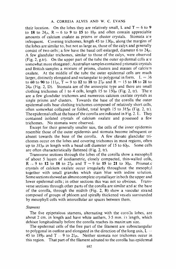
their location. On the lobes they are relatively small, L and T = 6 to 9 to 18 to 24)a, R = 6 to 9 to 15 to 18/x and often contain appreciable amounts of calcium oxalate as prisms or cluster crystals. Stomata are infrequent. Covering trichomes, length 45 to 130/j,, along the margins of the lobes are similar to, but not as large as, those of the calyx and generally consist of two cells; a few have the basal cell enlarged, diameter 6 to 24/x. A few glandular trichomes, similar to those of the calyx, were observed (Fig. 2, g.tr). On the upper part of the tube the outer epidermal cells are somewhat more elongated; Australian samples contained prismatic crystals and British samples a mixture of prisms, clusters and masses of calcium oxalate. At the middle of the tube the outer epidermal cells are much larger, distinctly elongated and rectangular to polygonal in form. L = 36 to 60 to 90 to 11 l(i, T = 9 to 12 to 18 to 27q, and R = 15 to 18 to 21 to 24/x (Fig. 2, D). Stomata are of the anisocytic type and there are small clothing trichomes of 1 to 4 cells, length 15 to 150/x (Fig. 2, tr). There are a few glandular trichomes and numerous calcium oxalate crystals as single prisms and clusters. Towards the base of the corolla the outer epidermal cells bear clothing trichomes composed of relatively short cells, often somewhat collapsed or folded, total length 15 to 315/x (Fig. 2, tr). The epidermal cells at the base of the corolla are indicated in Fig. 2. E. They contained isolated crystals of calcium oxalate and possessed a few trichomes. No stomata were observed.
Except for their generally smaller size, the cells of the inner epidermis resemble those of the outer epidermis and stomata become infrequent or absent towards the base of the corolla. A few clavate glandular trichomes occur on the lobes and covering trichomes in most regions, often up to 315¡i in length with a basal cell diameter of 15 to 60/x. Some cells are often characteristically flattened (Fig. 2, tr).
Transverse sections through the lobes of the corolla show a mesophyll of about 5 layers of isodiametric, closely compacted, thin-walled cells, R = 9 to 12 to 18 to 27/x and T = 9 to 15 to 21 to 30fj.. Prismatic crystals of calcium oxalate occur irregularly throughout the mesophyl together with small granules which stain blue with iodine solution. Some sections showed an almost complete crystal layer in both the upper and lower epidermal cells; in other sections this was not so obvious. Transverse sections through other parts of the corolla are similar and at the base of the corolla, through the midrib (Fig. 2, B) show a vascular strand composed of groups of phloem and spirally thickened vessels surrounded by mesophyll cells with intercellular air spaces between them.
StamensThe five epipetalous stamens, alternating with the corolla lobes, are
about 2 cm. in length and have white anthers, 3-5 mm. in length, which dehisce longitudinally before the corolla reaches its maximum size.
The epidermal cells of the free part of the filament are subrectangular to polygonal in outline and elongated in the direction of the long axis, L = 45 to 189)u. and T = 9 to 21/x. Neither stomata nor trichomes occur in this region. That part of the filament adnated to the corolla has epidermal
A. CORREIA ALVES AND W. C. EVANS
682
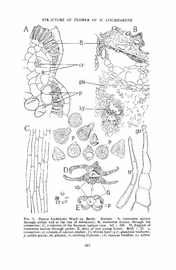
STRUCTURE OF FLOWER OF D. LEICHHARDTil
F i g . 3. D a tw a leichhardtii Muell. ex. Benth. Stamen. A, transverse section through anther wall at the line of dehiscence; B, transverse section through the connective; C, eoidermis of the filament, surface view. All x 240. D, diagram of transverse section through anther; E, ditto of very young flower. Both x 25. c, connective; cr, crystals of calcium oxalate; /./, fibrous layer; g .tr , glandular trichome; p , pollen grains; ph , phloem; tr, clothing trichome; v.b, vascular bundles; x y , xylem.
683

A. CORREIA ALVES AND W. C. EVANS
F ig. 4. D atura leichhardtii Muell. ex Benth. A, transverse section through the style, x 240; B, gynaecium and forms of the stigma x 1; C, D, transverse sections through the style, x 60; E, transverse section through spine on ovary wall, x 240. F, transverse section through ovary just below insertion of style. G, ditto middle portion, both x 7. g .tr , glandular trichomes x 240; id , idioblast; 1, lacuna; ov, ovules; sp, spine; st, starch granules; tr, covering trichomes of the outer epidermis x 240; v.b, vascular bundles.
684
STRUCTURE OF FLOWER OF D. LEICHHARDTII
cells shorter and broader than those of the free part and resembling those cells of the corresponding level of the corolla. Covering trichomes are frequent, generally conical or filamentous in form, often up to 480/x in length and composed of 2 to 3 to 4 to 6 cells (Fig. 3, tr). Some terminate in a relatively small single-celled gland (Fig. 3, g.tr). Other unicellular trichomes are similar to those on the corolla. A transverse section through the free portion of the filament is similar to that of D. stramonium (Wallis and Rohatgi, 1952a).
The outer portion of the connective is composed of spongy parenchyma, having large numbers of lacunae, and this loosely links the lobes to the central area. Several layers of parenchyma, the outer ones coloured yellow or brown, and containing many small rounded bodies of unknown composition, surround the vascular cylinder. The latter consists of small groups of phloem around a group of small, spirally thickened vessels which are somewhat elliptical in transverse section. A lacuna is generally evident (Fig. 3, 3, D). In the young anther, spongy parenchyma and air spaces are absent from the connective (Fig. 3, E). The outer epidermis of the anther lobes is composed of cells elongated in the longitudinal direction, has a well-marked cuticle and possesses many filamentous trichomes of 2 to 5 cells and up to 390/x in length. For the epidermal cells, R — 12 to 18 to 30 to 39/x and T =24 to 42 to 60 to 90¡i. In the proximity of the line of dehiscence, the fibrous layer of the anther wall is one cell thick, but becomes 2 to 4 cells thick in the region where it joins the connective; in surface view it resembles that of D. stramonium and it gives a slight reaction for lignin when tested with phloroglucinol solution followed by concentrated hydrochloric acid (Fig. 3, A, B, D, E). The pollen grains (Fig. 3, p), mounted in cold lactophenol, are generally spherical in outline having three, usually well-marked, germinal pores 6 to 12 to 18 to 21^ in diameter and faint germinal furrows. The surface of the exine is irregularly granular. Pollen grain diameter varied somewhat with the many samples examined; 1,500 grains were measured and the diameters, excluding projecting pores, were 24 to45 to54to 69/x. 8 per cent of the grains measured exceeded 54/x and 0-7 per cent exceeded 60/x.
GynaeciumThe conical ovary is about 3 mm. wide at the base, 4 mm. long and is
covered by four areas of spines. The style when fully developed is about 3 cm. in length and thickens towards the stigma; the latter is usually capitate but may form two or three divergent lobes (Fig. 4, B).
Transverse sections indicate a single lacuna directly under the insertion of the style but the ovary is two-celled below this level (Fig. 4, F, G). The ovary wall bears spines and trichomes. In surface view the epidermal cells of the ovary wall are irregular in shape, L = 9 to 45/x, T = 9 to 33/x and R = 21 to 30/x, The structure and contents of the mesophyll and septum resemble those of D. stramonium.
The epidermal cells of the spines are elongated in the direction of the axis, L = 22 to 120/;., T = 9 to 54/x and R = 15 to 24/x, and contain variously shaped crystals of calcium oxalate. In transverse section the
685

spines show a ring of small vascular bundles surrounded by almost isodiametric parenchymatous cells many of which contain crystal sand (Fig. 4, E). The spines of mature fruits also show in their basal portions a ring of about 12 to 13 groups of lignified elements, each group consisting of a few spirally thickened vessels surrounded by lignified parenchyma. Towards the apex of the spine the ring of lignified tissue is complete. Clothing and glandular trichomes are associated with the spines of the ovary, the two- or three-celled clothing forms being most numerous. The glandular trichomes are of two types, the common clavate type with a multicellular head and those with a multicellular uniseriate stalk and a single-celled glandular head (Fig. 4, g.tr).
The epidermal cells of the style are elongated in the direction of the main axis; they may contain rosettes, prisms or irregular masses of calcium oxalate and in transverse section show convex outer surfaces, L = 39 to 105/u, T = 9 to 21/x and R = 18 to 27p.. The cuticle is longitudinally striated, stomata are rare and no trichomes were observed. In transverse sections, two different forms of the style were noted. One (Fig. 4, C) is irregular and flattened in outline with an oval central area of thin-walled cells and irregular lacunae. On either side of the central area is a vascular strand. The other form (Fig. 4, D) is rounded or slightly oval in transverse section with a triangular to three-lobed central area of thin-walled, rounded cells (L and T = 6 to 15/x) and in the centre of this area is a large lacuna of the same shape. The outer parenchyma supporting the central zone consists of 2 to 12 rows of almost isodiametric cells (L and T = 9 to 36/x) and containing small round starch grains and isolated clusters a id prisms of calcium oxalate. Three vascular strands, alternating with the lobes of the central area, are situated in the outer parenchyma (Fig. 4, A). These two styles are correlated with the form of the stigma which may be capitate with a vertical groove on either side, or two- or three-lobed, with the lobes spreading and attached to one another and the top of the style, at their bases (Fig. 4, B). The three-lobed stigma corresponds to the style with three vascular strands. The oval-shaped stigma is covered, except for the vertical grooves, by a papillose receptive surface which arises from a central area of small-celled tissue; pollen grains may be attached. There is a vascular bundle on either side of the central area. In those stigmas with divergent lobes, each lobe corresponds essentially to one half of the oval shaped stigma described above and in transverse section shows a reniform outline.
C o nclusio n
The flowers of D. leichhardtii are smaller than those of all but one other species of Datura; their histological features closely resemble those of the flowers of D. stramonium and D. innoxia which are the only other flowers of the genus Datura which have been described in detail (Wallis and Rohatgi, 1952a, b). The pollen grains afford some distinguishing characters; they are very similar to those of D. stramonium but are slightly smaller in size being about 24 to 45 to 54 to 69/x in diameter, while those ofD. stramonium are about 48 to 60 to 78/x in diameter. The pollen grains
A. CORREIA ALVES AND W. C. EVANS
686
are, however, markedly different from those of D. innoxia, being slightly smaller in size and bearing quite different marking of the exine. The clothing trichomes of the calyx, with flattened cells similar to those observed on the leaves, and the uniseriate trichomes with a single glandular head are characters not common to the flowers of D. innoxia and D. stramonium respectively.
Acknowledgement. One of us (A.C.A.) is indebted to the “Comissao Coordenadora da Investiga^ao para a OTAN”, Lisbon, Portugal, for the award of a scholarship, during the term of which this study was undertaken.
STRUCTURE OF FLOWER OF D. LEICHHARD Til
R e f e r e n c e s
Avery, A. G. and Satina, S. (1959). B ta k e s le e : The G en u s D a tu ra , p. 25, New York : Ronald Press.
Evans, W. C. and Stevenson, N. A. (1962a). J . P h a rm . P h a rm a co l., 14, 664-678. Evans, W. C. and Stevenson, N. A. (1962b). Ib id ., 14, 107 7M10 T.Santos, J. K. (1927). P h ilip p . J . S e i ., 32, 275-296.Wallis, T. E. and Rohatgi, S. (1952a). J . P h a rm . P h a rm a co l., 4, 243-258.Wallis. T. E. and Rohatgi, S. (1952b). Ib id ., 4, 471-478.
687
B y V . K . B r o w n , J. R o b in s o n a n d D . E. S t e v e n s o n
F rom “S h e ll” R esea rch L im ite d , T u n sta ll L a b o ra to ry , S ittin g b o u rn e , K e n t
Received May 10, 1963
Dimethyl sulphoxide has good solvent properties for a wide range of chemicals, and a low toxicity to mammals. The acute oral LD50 for rats is about 20 ml./'kg. and for chickens about 12-5 ml./kg. Rats tolerate up to 10 daily doses of 10 ml./kg. weight. Dimethyl sulphoxide is therefore a useful solvent for chemical compounds under toxicological evaluation.
T h e selection of a solvent for use in toxicological investigations requires considerable care. On the one hand, solvents with desirable physicochemical characteristics may be sufficiently toxic to affect significantly the results obtained with only slightly toxic solutes either as a result of the toxicity of the solvent per se, or as a result of an increase in the rate of absorption of the solute through the alimentary tract or via the skin. On the other hand, physiologically inert substances may have poor solvent properties. Sanderson (1959) has discussed the desirable characteristics of a solvent to be used in toxicity testing and recommended glycerol formal as a satisfactory solvent with a low toxicity. However, many compounds under development for use in the plastics industry and in agriculture have very low acute toxicities and consequently large doses of the compounds must be administered. The solubility of some of these compounds in glycerol formal is low and it is difficult to prepare solutions of the necessary concentration for LD50 determinations. We are, therefore, continually searching for compounds with good solvent properties combined with low toxicity (LD50>20 ml./kg.). One such compound of great promise is dimethyl sulphoxide.
P h y s ic a l C h a r a c t e r is t ic s
Dimethyl sulphoxide, Me2S:0, is the simplest member of the homologous series of organic sulphoxides. It is prepared by oxidation of dimethyl sulphide commercially available from both the petroleum and sulphite-pulp industries.
Pure dimethyl sulphoxide (d m s o ) is a colourless liquid, with a slight odour and a slightly bitter taste; the commercial material, 99-5 per cent purity, has a strong characteristic odour of sulphur compounds. It is very hygroscopic, absorbing over 70 per cent of its own weight of water from air at 20° and 65 per cent relative humidity. The physical properties are summarised in Table I.
d m s o is a dipolar aprotic solvent in which many classes of compound are soluble. Thus, the gases acetylene, ammonia and sulphur dioxide and most organic compounds are soluble. At 20° d m s o mixes in all proportions with methanol, octanol, glycerol, acetalcehyde, acetone, propionic acid, formic acid, diethylether, ethyl acetate, triacetin, dioxane,
A NOTE ON THE TOXICITY AND SOLVENT PROPERTIES OFDIMETHYL SULPHOXIDE
688

pyridine, benzene, toluene and xylene. The chlorinated hydrocarbon insecticides, many proteins, and polynuclear hydrocarbons, e.g., chrysene and benzpyrene, are also soluble in dmso.
SOLVENT PROPERTIES OF DIMETHYL SULPHOXIDE
TABLE IP h y s i c a l p r o p e r t i e s o f d i m e t h y l s l l p h o x i d e
M e ltin g -p o in t 18-4°B o ilin g -p o in t (760 m m . H g) 189°V a p o u r p re s su re a t 10° 0-417 m m . H gV a p o u r p re s su re a t 50° 3-07 m m H gD e n s ity (20°) 1-100R e fra c tiv e in d ex n ^ 1-4783V isco sity (20°) 2-14 c e n d p o iseD ie le c tr ic c o n s ta n t (20°) 48-9D ip o le m o m e n t 4-3 D eb y e
Organic compounds that are almost insoluble in dmso include paraffin hydrocarbons, glycerides of higher fatty acids and other compounds with a highly paraffinic moiety, and plastic materials such as nylon, polystyrene and polyethylene.
dmso is a good solvent for many inorganic compounds. It is miscible in all proportions with water. Lithium chloride, ferric chloride and ammonium thiocyanate are very soluble, whereas mercurous chloride, lead chromate and potassium ferrocyanide are almost insoluble. More detailed information concerning the solubility of inorganic and oganic compounds in dmso has been published by Ranky and Nelson (1961) and Schlafer and Schaffernicht (1960).
Experimental
Rats derived from Carworth Farm strains, Carworth Farm No. 1 and AH (hairless) mice and Pirbright strain guinea-pigs were obtained from the Tunstall Laboratory Animal Breeding Unit. White Leghorn chickens aged 8-9 weeks were obtained from an accredited dealer.
The rats and mice were maintained at an environmental temperature of 73° F i 33F and a relative humidity of 45 per cent. Guinea-pigs were kept in a building where the temperature ranged from 60-70° F during the experimental period.
All animals were fasted overnight before weighing, and dosing using a ball point needle. Water was allowed ad libitum throughout the experiment and food was also always available after dosing, dmso (British Drug Houses) Laboratory Reagent Grade was used throughout these experiments.
ResultsOral Toxicity
Acute ora! toxicity in rats. 20 ml./kg. killed one of four animals in a group of male and also in a group of female rats. Below this dose there were no deaths. This indicates that the LD50 is over 20 ml./kg. Above this dose it is considered that mechanical factors such as gastric distension play an important part in determining toxicity. A parallel
689

experiment was also made with rats maintained in an unheated room at about 50° F and the results were similar to those reported above. (2/8 deaths at 17-5 ml./kg., 1/8 at 20-0 ml./kg.). It is therefore considered that the ambient temperature is unlikely to materially affect the results.
Acute oral toxicity in mice. A small experiment indicated that the acute oral toxicity of dimethyl sulphoxide to mice was about the same as that of rats, 1/4 animals dying at a dose rate of 17-5 ml./kg. and 2/4 at 20-0 ml./kg. weight.
Acute oral toxicity in guinea-pigs. No deaths occurred with coses up to 10 ml./kg. body weight. Higher doses were not investigated.
Acute oral toxicity in chickens. Chickens gave results for 10-0 ml.,12-5 ml. and 15 ml. of 0/3, 2/3 and 3/3 respectively for cocks and 1/3, 1/3 and 3/3 respectively for hens. Thus the LD50 for this species is therefore approximately 12-5 ml./kg.
Repeated oral dosing to rats. Experiments were made in groups of 5 of each sex in which the rats were dosed 5 days a week for two successive weeks at dose levels of 1, 3-5, 5 and 10 ml./kg. Control groups, 10 animals of each sex, and groups dosed with water, 1 and 3-5 ml., were also included to determine the effect on weight which might be caused by recaging and other manipulatory procedures. The only animals that died were 2 males on the 5 ml./kg. and 1 male and 1 female on the 10 ml./kg. dose. The deaths were due to injuries caused by dosing.
Animals were weighed before dosing and during the first week of dosing the regrouping and caging of both control and treated animals was found to produce an adverse effect on weight. Losses of frcm 0T to 1-2 per cent were recorded for water dosage and with the solvent, as little as 1-0 and as much as 17 per cent loss was recorded in the first week. This was largely recovered during the 6th and 7th days, when no dosing took place, and the subsequent changes during the second week were not so marked. The animals were killed about 10 days after exposure ceased and at that time no changes in the formed elements of the blood of the treated animals were noted.Dermal Irritation and Sensitisation
Sensitisation. 5 male and 2 female adult guinea-pigs had a course of intradermal injections of 0T ml. of a 10 per cent v/v aqueous solution of dmso, given on alternate days for 3 weeks. Two weeks after the last injection a further intradermal injection of 0-1 ml. of the same solution was given but no signs of sensitisation were noticed. The experiment was then repeated on the same animals, but again no sensitisation was seen.
Irritation. 28 daily applications of undiluted dmso to the clipped backs of guinea-pigs produced no macro- or microscopic signs of injury. dmso was also painted on to the dorsal scapula region of 5 male AH (hairless) mice twice a week for 30 weeks. At the end of this period there was no discernable effect on the skin of the treated animals.
V. K. BROWN, J. ROBINSON AND D. E. STEVENSON
690

SOLVENT PROPERTIES OF DIMETHYL SULPHOXIDE
Although dmso has been used in a large number of dermal toxicity experiments in rats as a solvent for other materials under investigation there has been no evidence of dermal injury.
Ocular damage. Undiluted dmso was dropped into the conjunctival sac of the right eye of 6 adult New Zealand White rabbits. Two rabbits had the eye irrigated immediately with warm water and a further 2 rabbits were irrigated after a delay of 2-3 min. The eyes of the remaining 2 rabbits were not irrigated. No ill effects were noted in any of the treated eyes.Intraperitoneal Toxicity
Both adult rats and guinea-pigs tolerated doses of 5 ml. dmso per kg. weight. Eight male rats were given four successive daily intraperitoneal injections of dmso at a dose of 7-5 ml./kg. weight. One rat died 2 days after the last injection but the remaining animals remained healthy during an observation period cf 10 days after the last dose.Pathology
No specific micro- or macroscopic evidence of toxicity has been noted at the autopsies of exposed animals. In a few animals some fatty degeneration and congestion of the sinusoids was seen in the liver. Where rats were given repeated intraperitoneal injections of large doses of dmso there was evidence of rounding of the liver margin and of fibrinous strands within the peritoneum.
D iscussion
It is plausible to argue that the ability of a compound to dissolve large amounts of a wide range of chemical compounds excludes the possibility of its biological inertness. The formulation of chemical compounds for toxicity testing can therefore be a very real problem. A volume of 20 ml. solution/kg. weight may be regarded as the maximum tolerated oral dose. Thus a saturated solution containing 10 per cent w/v of toxicant cannot be used to give a dose greater than 2 g./kg. Sometimes it is possible to overcome this difficulty by administering the material as a suspension in water; however, the preparation of stable suspensions containing more than 10 per cent w/v of the material may be difficult, particularly with solids which have high elasticity.
Dimethyl sulphoxide fulfils most of the criteria proposed by Sanderson for the ideal solvent in toxicity tests. It is completely miscible with water, it not known to interfere with absorption or metabolism, has good solvent properties and a low viscosity and volatility. However, while large doses may be tolerated orally, dermally or intraperitoneally, even small doses may cause transient fall in body weight. After the administration of a large dose of dmso animals may become depressed, but do not lose consciousness, dmso causes dénaturation of blood proteins and is therefore not suitable for intravenous injection.
Furthermore, in view of its dipolar aprotic nature, the possibility exists that a compound dissolved in dimethyl sulphoxide ma> undergo a chemical
691

change. Thus certain trivalent phosphorus compounds react with dimethyl sulphoxide, and two organophosphorus compounds showed colour changes of their solutions in dimethyl sulphoxide after 24 hr.
Dimethyl sulphoxide has two main disadvantages. It is more expensive than many other more readily available solvents and it has a characteristic odour which may be considered obnoxious.
References
Ranky, W. O. and Nelson, D. C. (1961). Organic Sulphur Compounds, Vol. 1, Ch. 17. Editor, Kharasch, N. Oxford: Pergamon.
Sanderson, D. M. (1959). J. Pharm, and Pharmacol., 55, 150-156.Schläfer, H. L. and Schaffernicht, W. (1960). Angew. Chem., 72, 618-626.
V. K. BROWN, J. ROBINSON AND D. E. STEVENSON
692

LETTERS TO THE EDITOR
Chloroquine in Bronchial Asthma
Sir,—Chlorocuine possesses specific antihistamine and anti-anaphylactic activity (Agarwal and Deshmankar, 1963) and this could account for the anti-asthmatic action of the drug (Juul-Mollcr, 1961). Since allergic reactions are mediated through histamine releases from tissues, we have studied the tissue levels of histamine after chronic administration of chloroquine in rats.
Groups of 4 male albino rats (100 to 125 g.) were used in all experiments. Chloroquine sulphate (5 mg./kg.) was injected intraperitoneally daily into 2 groups for 21 days. An equivalent quantity of normal saline was injected into another 2 groups of control rats. The animals were killed on the 21st day and samples of lungs and stomach were pooled, extracted and assayed for histamine as described by ?arratt and West (1957).
The results, as summarised in Table I, clearly indicate chloroquine to cause a significant reduction in the histamine content of the lung, but the histamine content of the stomach was found to increase. Similar results have been obtained with glucocorticoids for lung (Chowdhuri, 1962) and stomach (Telford and West,1960) in rats.
TABLE IT h e e f f e c t o f c h l o r o q u i n e o n h i s t a m i n e c o n t e n t ( a s b a s e ) o f t i s s u e s o f r a t s
T issu e H is ta m in e (u g ./g .) ± (S .E .)
C o n tro l j A fte r C h lo ro q u in e
L u n g s 4-3 2-4SS to m a c h 8-66 11*75
The histamine depleting action of chloroquine in lungs further substantiates its value in bronchial asthma. Juul-Moller, in a personal communication, told us he had founc asthmatic patients under chloroquine therapy to be free from attacks of status asthmaticus and this persistant action of the drug may be due to the histamine depletion in the lungs.
Department of Pharmacology, Sohan L. AgarwalG. R. Medical College, B. S. D eshmankarGwalior (M.P.), India. V inod Bhargava
July 17, 1963
References
Agarwal, Sohan. L. and Deshmankar, B. S. (1963). Arch. int. Pharmacodvn., 143, 401-407.
Chowdhur:, B. N. (1962). J. Ind. med. Ass., 39, 343-345.Juul-Moller, O. (1961). Ugeskr. Laege., 123, 105-108.Parratt, J. R. and West, G. B. (1957). J. Phvsiol. (Lond.), 137, 169-178.Telford, J. M. and West, G. B. (1960). Brit. J. Pharmacol., 15, 532-539.
693

LETTERS TO THE EDITOR Histamine in Non-vertebrate Animals
Sir,—Histamine is widely distributed in nature. In the plant kingdom, histamine is formed in large amounts by several bacterial organisms, is present in various fungi, and is of common occurrence in the higher plants (Emmelin and Feldberg, 1947 ; Werle and Raub, 1948 ; Gale, 1953 ; Fowler, 1962). In the animal kingdom, investigation of the distribution of histamine has been confined mainly to the vertebrates : the amine is known to occur commonly in fish, and to be present in greater or lesser amount in almost every mammalian organ, tissue and fluid (Guggenheim, 1951; Feldberg, 1956). Among invertebrates, high concentrations of histamine have been detected in various stings and venoms ( Marcou, Derevici and Derevici, 1937 ; Jaques and Schachter, 954b ; Rothsch ld and Parsons, 1962), in the gnat (Eckert, Paasonen and Vartiainen, 1951), in 2 coelenterates (Mathias, Ross and Schachter, 1960), and in a sponge (Ackermarm and List, 1957). It is not known, however, whether histamine is of general as opposed to isolated occurrence among the lower animal phyla, particularly in the non-coelomates. Studies on the distribution and significance of histamine in comparatively simple organisms may provide valuable clues to the function of the amine in higher species, and a survey has therefore been made of its occurrence in a wide range of diplo- and triploblastic animals. A study of the ability to form histamine from histidine has also been made in some species.
Freshly collected organisms were extracted for 24 hr. in 10 per cent trichloroacetic acid, and after removal of excess acid with ether, the extracts were assayed for histamine or histamine-like substance on the isolated ileum of the guinea- pig and on the blood pressure of the anaesthetised dog. The specificity of the responses was checked on each preparation using mepyramine maleate. Histidine decarboxylase activity was measured as the amount of histamine formed from added histidine during an incubation period of 3 hr. in buffered physiological saline solution. The methods have been described in detail elsewhere (Telford and West, 1961a).
The results, presented in Table I, indicate that histamine is widely distribut in non-vertebrate animals. The species investigated are drawn from all the major phyla in the animal kingdom with the exception of tne protozoa, and though the number of species studied is small, at least one species from each of the 10 phyla represented has been found to contain histamine. Furthermore, the amine has been detected in 19 of the 26 species examined. Particularly high concentrations have been found in the sea-anemone Aiptasici tagetes (Coelenterata : Anthozoa), in the parasite Mesocoelium monodi i Platyhelminthes : Trematoda) obtained from the intestine of Bufo mcirinus, and in the sea-urchin Echinaster echinophorus (Echinodermata : Echinoidea). The amount of histamine per unit weight in each of these species is comparable with the large amount of histamine present in certain mammalian tissues such as rat skin ar.d guinea-pig lung. Some other species from the same phyla, however, contain little or no histamine. Hence, as may be seen from Table I where the species are classified according to Rothschild (1961), there is no relationship in the non-vertebrate animals between the distribution of histamine and biological complexity.
Calculation of histamine content as /¿g. histamine per unit weight gives little information about the total amount of amine that a single specimen contains. However, calculation as /¿g. histamine per specimen is impractical as the weights of individual specimens from any one species sometimes vary greatly. Such approximate estimates as can be made nevertheless indicate that among those species containing histamine there is no relation between total histamine content and size.
694

Histamine was found in all the parasitic species studied. Too few animals have been investigated to postulate that histamine is more widely distributed among parasitic species than among invertebrate free-living species, but among the species examined, the parasitic forms contain a significantly greater amount of histamine per unit weight than the free-living forms (P > 0 05).
Histidine decarboxylase activity was detected in two histamine-containing species, Aiptasia tagetes and Mesocoelium monodi. In both, enzyme activity was found in the range pH 4 0-9 0, in contrast to bacterial histidine decarboxylases which are most active at an acid pH (Gale, 1953), and in contrast to mammalian histidine decarboxylases which are usually most active at an alkaline pH (Schayer, 1957; Telford and West, 1961a, 1961b; Weissbach, Lovenberg and Udenfriend, 1961). The addition of organic solvent did not increase enzyme activity in either species, neither did the addition of pyridoxal-5-phos- phoric acid. T ie addition of organic solvent usually increases the histidine decarboxylase activity of mammalian tissues (Waton, 1956; Telford and West, 1961a, 1961b), whilst the addition of pyridoxal-5-phosphoric acid has been found to increase enzyme activity in some mammalian tissues (Rothschild and Schayer, 1958; Telford, unpublished observations) and in bacteria (Gale, 1953). It is possible, therefore, that the histamine-forming enzymes in Aiptasia tagetes and Mesocoelium monodi represent a different type of histidine decarboxylase from those which have been previously described.
TABLE I
LETTERS TO THE EDITOR
Histamine content (/¿g./g.) and histidine decarboxylase activity (/xg. histamine FDRMED/g./3 hr.) OF VARIOUS INVERTEBRATE SPECIES
SpeciesM o d e o f
lifeH is ta m in e
c o n te n t
H is tid in ed e c a rb o x y la se
a c tiv ity
P h y lu m P o rife raHaV.clona sp . F L 3 0 —A1 y cale Cecilia F L ;o —
P h y lu m C o e le n te ra taRhodactis sanctiiho nae F L 0-4 —A iptasia tagetes F L 32-5 1 5 0Cassiopeia xaymaccna F L I -8 —Condactvlis gigantea F L 0 —
P h v lu m P la tv h e lm in t.ie sStviochus megalcps F L 0 —Styioplanocera fasrata F L 0 —Syndesmis franc ¡see na S P Ó-9 0Fasciola hepática .. P 5-2 0Mesocoelium monodi P 53-3 154-4Oochoristica ameivee P 1 3 0 0
P h v lu m A sc h e lm ir th e s0Stephanurus dentatus P 1-9
P h v lu m A c a n th o c e p h a la3-2 0Macracantkorynchus hirudinaceus P
P h v lu m M o llu scaA ustralorbis glabra us F L 3 —Acra zebra F L to —Pleurodonta sp . F L >1 0Domax deniiculatus F L >6 —
P h v lu m A n n e lid a> 2Pheretima sp . F L —
Hermodice caruncidata F L —P h v lu m A r th ro p o d a
F LMysidium columbiae 0 —Coenobita clypeatu ; F L 4-3 —
P h v lu m E c h in o d e rm a ta28-3Echinaster echinoplorus .. F L
0/ .vtechinus variegaius F L 0Diadema antillarum F L 2-5 —Holothuria sp . F L 1 8 0
F L . free liv in g : P , p a ra s i t ic : S P . s e m i-p a ra s itic : ?. in te rfe r in g su b s ta n c e — , n o t te s ted . A ll valu es o f h is ta m in e re fe r to :h e b ase , a n d a re th e m e a n o f a t le a s t 3 d e te rm in a tio n s .
695

LETTERS TO THE EDITOROne of the species studied, Aiptasia tagetes, is a sea-anemone whose tentacles
release an unidentified substance which on contact with human skin produces symptoms resembling those produced by histamine. The symptoms consist of a burning sensation accompanied by itching, redness, and sometimes local oedema. The present experiments strongly suggest that these symptoms are in fact due, at least in part, to histamine being introduced into the skin. However, other substances such as histamine releasers, acetylcholine or 5-hydroxy- tryptamine may be involved; it should be noted that potent histamine releasers and 5-hydroxytryptamine have been extracted from a number of coelenterate species (Jaques and Schachter, 1954a; Mathias, Ross and Schachter, 1957; Mathias, Ross and Schachter, 1960; Uvnas, 1960; Welsh, 1960).
Histamine may be widely distributed in sea-anemones. Large amounts have been found in the tentacles of Actinia equina and Anemonica sulcata (Mathias, Ross and Schachter, 1960), but Aiptasia tagetes differs from these species in that its histamine is localised mainly in the body of the animal. Only 20-30 per cent of the total histamine was found to be present in the tentacles, the remaining 70-80 per cent being present in the column and coelenteric structures. We have not so far established whether the histamine is confined to one type of cell structure, but it does not appear to be localised in the nematocysts. Thus, while the total number of nematocysts in the column and coelenteric structures is probably greater than in the tentacles, the number of nematocysts per unit area is higher in the tentacles than elsewhere.
The experiments reported here provide a further demonstration of the apparently haphazard fashion in which histamine is distributed in nature. At the present time it is not possible to say whether the amine serves a common function in all tissues which contain it, or whether during the course of evolution its presence has been adapted to serve new functions. However, the occurrence of particularly high concentrations of histamine in a variety of invertebrate poisonous secretions indicates that in the lower animal groups the amine may play a part in defence and attack. In mammals, it has been suggested by various authors (Feldberg, 1954; Kahlson, Nilsson, Rosengren and Zederfeldt, 1960; Riley, 1962) that the amine exerts an important role in tissue defence and repair, and it is possible that histamine serves a related function throughout the animal kingdom.
Department of Zoology, D. F. M ettrick
Department of Physiology, J. M. TelfordUniversity of the West Indies,Kingston 7, Jamaica.August 20, 1963
References
Ackermann, D. and List, P. H. (1957). Hoppe-Seyl. Z., 308, 27-1-276.Eckert, D., Paasonen, M. and Vartiainen, A. (1951). Acta pharm, tox., Kbh.,
7, 16-22.Emmelin, H. and Feldberg, W. (1947). J. Physiol., 106, 440-448.Feldberg, W. (1954). J. Pharm. Pharmacol., 6, 281-301.Feldberg, W. (1956). Ciba Symposium on Histamine, London: Churchill.Fowler, H. D. (1962). Nature, Lond., 193, 582-583.Gale, E. F. (1953). Brit. med. Bull., 9, 135-136.Guggenheim, M. (1951). Die biogenen Amine, Basle: Karger, and New York:
Interscience.Jaques, R. and Schachter, M. (1954a). Brit. J. Pharmacol., 9, 49-52.Jaques, R. and Schachter, M. (1954b). Brit. J. Pharmacol., 9, 53-58.
696

Kahlson, G., Nilsson, K., Rosengren, E. and Zederfeldt, R. (1960). Lancet, 2, 230-234.
Marcou, I., Derevici, A. and Derevici, M. (1937). C.R. Soc. Biol., Paris, 126, 726-728.
Mathias, A. P., Ross, D. M. and Schachter, M. (1957). Nature, Load., 180, 658-659. Mathias, A. P., Ross, D. M. and Schachter, M. (1960). J. Physiol. 151, 296-311. Riley, J. F. (1962). Lancet, 2, 40-41.Rothschild, Lord. (1961). A Classification o f Living Animals, London : Longmans. Rothschild, The Hon. M. and Parsons, J. (1962). Proc. R. Ent. Soc. (c), 27, 21-22. Rothschild, A. M. and Schayer, R. W. (1958). Fed. Proc., 17, 136.Schayer, R. W. (1957). Amer. J. Physiol., 189, 533-536.Telford, J. M. and West, G. B. (1961a). J. Pharm. Pharmacol., 13, 75-82.Telford, J. M. and West. G. B. (1961b). J. Physiol., 157, 306-314.Uvnas, B. (1960). Ann. N.Y. Acad. Sci., 90, 751-759.Waton, N. G. (1956). Brit. J. Pharmacol., 11, 119-127.Weissbach, H., Lovenberg, W. and Udenfriend, S. (1961). Biochim. biophys. acta,
50, 177-179.Welsh, J. H. (1960). Nature, Load., 186, 811-812.Werle, E. and Raub, A. (1948). Biochem. Z., 318, 538-544.
LETTERS TO THE EDITOR
Papaverine-like Pharmacological Properties of Rotenone
S ir ,—Recent investigations showed papaverine and some structurally related compounds to be powerful inhibitors of oxidative phosphorylation (Santi, Ferrari and Contessa, 1963). A relation also seemed possible between the spasmolytic activity of papaverine-like drugs and their inhibition of the respiratory chain, thus explaining the similarity of effects elicited by anoxia, by some enzyme inhibitors and by papaverine, on the drug-induced contraction of intestinal smooth muscle. Under these experimental conditions it was observed (West, Hadden and Farah, 1951; Santi Contessa and Ferrari, 1963) that the isolated gut failed to give normal tonic responses to acetylcholine, histamine and BaCl2 showing only an immediate, short-lasting contraction. This inability of smooth muscle to maintain tone was considered as “the first evidence of interference with energy production” (West, Hadden and Farah, 1951). For papaverine and allied drugs we pointed out that a similar effect may be elicited through a strong inhibition of electron-transfer reactions, between d pn and cytochrome b. The site of action is therefore the same as the one recognised for amytal (Emster, Jailing, Low and Lindberg, 1955), rotenone (Ernster, Dallner and Azzone, 1963) and allyloxibenzamide (Bruni and Contessa, 1961). With regard to the selectivity of the action and to the degree of activity, among the inhibitors of oxidative phosphorylation rotenone appears as the one most closely related to papaverine (Ernster, Dallner and Azzone, 1963; Santi, Contessa and Ferrari, 1963). This similarity of biochemical properties prompted a pharmacological comparison between rotenone and papaverine. The purpose was to examine the reliability of the previously proposed hypothesis on the mechanism of action of papaverine.
In the present investigations on rotenone and papaverine we have studied the spasmolytic activity on isolated gut (guinea-pig ileum and rabbit duodenum) ; the vasodilator effect on the hind-limb of the dog, by recording the arterial femoral flow with a Shipley and Wilson rotameter ; the effects on respiration and arterial blood pressure in rabbits and dogs.
Rotenone at final concentrations ranging from 10 8 to 10~9 (w/v) inhibits selectively the tonic response of intestinal smooth muscle to acetylcholine and histamine without preventing the immediate short-lasting contraction. Thus rotenone mimics the spasmolytic effect of papaverine, but its degree of activity is
697
LETTERS TO THE EDITOR
m uch greater (about 100 tim es) and the m yolytic effect is persistent and n ot readily abolished by w ashing.
W hen injected intra-arterially roten on e (1 5 -2 0 /xg.) elicits a rem arkable vaso dilator effect by increasing b lood flow 7 0 -8 0 per cent for a tim e o f 120-150 sec. ; papaverine show s a sim ilar but w eaker activity.
R oten on e exhibits a clear respiratory stim ulant effect both w hen injected in to the com m on carotid artery (1 5 -2 0 /¿g. through the thyroid inferior artery) and w hen adm inistered intravenously (1 5 -25 /xg./kg.) ; w hen given intravenously there is evidence o f a short-lasting hypotensive effect resem bling that elicited by papaverine.
T he data so far obtained dem onstrate a c lose resem blance betw een the pharm aco log ica l properties o f rotenone and papaverine. R oten on e appears the m ore active com pound in pharm acodynam ic and biochem ical properties. In respect to our previous investigations w hich led to the hypothesis that the spasm olytic effect o f papaverine is related to the inh ib ition o f oxidative phosphorylation , it is notew orthy that rotenone, the m ost selective inhibitor o f oxidative p h o sphorylation till n o w recognised, exhibits papaverine-like activities (spasm olytic, vasodilator, respiratory stim ulant effects). It seem s reasonable to adm it that other pharm acological properties o f papaverine, like the respiratory stim ulant one, are related to the inh ib ition o f oxidative phosphorylation .
Institute o f P harm acology, R . S a n t i
U niversity o f Padua, Italy. M . F e r r a r i
A ugust 19 ,1963 C. E. T oth
R e f e r e n c e s
Bruni, A. and C ontessa, A. R. (1961). Biochem. Pharmacol., 6, 275-277.E rnster, L ., D allner, G . and Azzone, G . F. (1963). J. biol. Chem., 238, 1124-1131. E rnster, L., Jailing, O ., Low, H . and Lindberg, O. (1955). Exp. Cell Res., Suppl. 3,
124—132.Santi, R ., C ontessa, A. R . and Ferrari, M. (1963). Biochem. Biophys. Res. Comm.,
11, 156-159.Santi, R ., Ferrari, M . and C ontessa, A. R . (1963). Biochem. Pharmacol., (in press). W est, T. C., H adden, G . and F arah , A. (1951). Amer. J. Physio!., 164, 565-572.
698

A Modification of the Butanol Extraction Method for the Fluorimetric Assay of Catecholamines in Biological Materials
S ir .— A satisfactory and w idely used m ethod for the extraction o f catecholam ines from tissues is that described by Shore and O lin (1958). H ow ever, errors m ay arise due to the destruction o f the am ines during hom ogenisation and during the extraction period. The use o f a m otor-driven glass hom ogeniser m ay produce severe local over-heating even when the rotor is filled w ith ice.
LETTERS TO THE EDITOR
punch
die
cose
tissue
gap
die base
base
bolt
F ig . 1. A longitudinal section o f the tab le tting die and punch. The punch and die base are m ade of tool steel and the rem ainder is m ade of stainless steel. The die and die base are rem ovable. A die o f internal d iam eter o f 1-2 cm. is suitable for the hearts o f ra ts o f 200 g. o r less. F o r ra t brains and hearts o f anim als o f m ore than 200 g. a 2-0 cm. die is used.
T his is particularly so w ith tough tissues such as heart and skin. Errors m ay also be produced by the presence o f froth in som e extracts, especially those from brain. T his causes variations in the am ounts o f tissue in the sam ples when m easured by pipette. A ll these factors lead to losses o f the catecholam ines and low assay results. A m odification o f the m ethod w hich overcom es these difficulties has been developed.
T he tissue to be assayed is placed in a stainless steel m esh basket and im m ersed in liquid n itroger contained in a w ide m outhed vacuum jar. W hen the nitrogen stops boiling the tissue is rem oved and dropped w hole into a su itably sized stainless steel tablet die (see F ig. 1). T he frozen tissue is crushed by striking the punch w ith a ham m er. The entire com pressed pellet o f still-frozen tissue is expelled into -.he salt saturated butanol w hich is used in the Shore and Olin m ethod. A volum e o f 0 0 In HC1 sufficient to produce a total aqueous phase o f approxim ately 1/10th o f the volum e o f butanol is added, together w ith 10 mg. o f sodium m etabisulphite to reduce oxidation during the extraction process. The rem ainder o f the assay procedure is the sam e as that described by Cass and
699
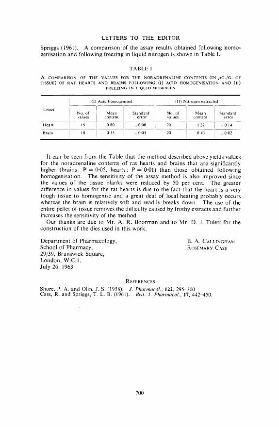
Spriggs (1961). A com parison o f the assay results obtained fo llow ing h o m o genisation and fo llow ing freezing in liquid n itrogen is show n in T able I.
LETTERS TO THE EDITOR
T A B LE I
A C O M PA R ISO N O F T H E V ALUES FO R T H E N O R A D R E N A L IN E C O N T EN TS ( IN /¿ G ./C . OF TISSU E ) O F R A T H EA R TS A N D B R A IN S F O L L O W IN G ( i ) A C ID H O M O G EN ISA TIO N A N D ( i l )
F R E E Z IN G IN L IQ U ID N IT R O G E N
(I) Acid homogenised (II) Nitrogen extracted
TissueNo. of values
Meancontent
Standarderror
No. of values
Meancontent
Standarderror
Heart 15 0-80 ± 0 0 8 20 1 37 ±0-14
Brain 18 0-35 ± 0 0 3 20 0 43 ±0 02
It can be seen from the T able that the m ethod described above yields values for the noradrenaline contents o f rat hearts and brains that are significantly higher (brains: P = 0 0 5 , hearts: P = 0-01) than those obtained fo llow ing hom ogenisation . T he sensitivity o f the assay m ethod is a lso im proved since the values o f the tissue blanks were reduced by 50 per cent. T he greater difference in values for the rat hearts is due to the fact that the heart is a very tough tissue to hom ogenise and a great deal o f local heating probably occurs whereas the brain is relatively soft and readily breaks dow n. T he use o f the entire pellet o f tissue rem oves the difficulty caused by frothy extracts and further increases the sensitivity o f the m ethod.
Our thanks are due to M r. A . R . B oorm an and to Mr. D . J. T ulett for the construction o f the dies used in this work.
D epartm ent o f Pharm acology, B. A . CallinghamS chool o f Pharm acy, Rosemary Cass29/39, Brunswick Square,L ondon , W .C .l.July 26, 1963
R e f e r e n c e s
Shore, P. A. and Olin, J. S. (1958). J. Pharmacol., 122, 295-300.Cass, R . and Spriggs, T . L. B. (1961). Brit. J. Pharmacol, 17, 442-450.
700

U N I V E R S I T Y O F S Y D N E Y
S E N IO R L E C T U R E S H IP
I N P H A R M A C O L O G Y
Applications are invited for the position o f Senior Lecturer in Pharm acology. The salary for a Senior Lecturer is within the range o f £A2,745 x 100 — £3,245 per annum . Cost o f living adjustm ents will be allowed and the salary will be subject to deductions under the State Superannuation Act. The com m encing salary will be fixed according to the qualifications and experience o f the successful applicant.
U nder the Staff M em bers’ Housing scheme in cases approved by the University and its Bankers, m arried m en m ay be assisted by loans to purchase a house.
F urther particulars and inform ation as to the m ethod o f application should be obtained from the Secretary, A ssociation o f C om m onw ealth U niversities (Branch Office), M arlborough House, Pall M all, London, S .W .l.
A pplications close in A ustralia and L ondon on 2 5 th O c t o b e r , 1 9 6 3 .

Save tim e and m oney w ith R egent D isposable Gloves by elimin a t in g m ulti-stage reconditioning processes, th u s sim plifying sterilisa tion and liberating valuable skill for nursing ■ Because hands are assum ed to be an im p o rtan t vehicle of cross-infection, th e use of a new glove of guaran teed in teg rity each tim e also m akes a con tribu tion tow ards th e elim ination of th is problem *R egent D isposable gloves reduce han d fatigue and provide’bare han d ’ tac tile sen sitiv ity ■ T hey are supplied packed ready for in s tan t sterilisation , com plete w ith E th icon Bio-sorb powder.
R e g e n t S u r g e o n s ' D i s p o s a b l e G l o v e s
British Standard 1803 : 5952
Available under Ministry o f Health Central Contract Manufactured by LONDON RUBBER INDUSTRIES LIMITED
H A L L L A N E • L O N D O N E4-(Surgical Division)