The investigation of acute optic neuritis: a review and proposed protocol
13
NATURE REVIEWS | NEUROLOGY VOLUME 10 | AUGUST 2014 | 447 Departments of Neurology and Ophthalmology, VU University Medical Center, De Boelelaan 1117, 1081 HV Amsterdam, Netherlands (A.P.). Department of Radiology, Nuclear Medicine and PET Research, VU University Medical Center, Amsterdam, Netherlands (M.P.W.). University of Calgary, Canada (F.C.). The National Institute of Neurology and Neurosurgery, Mexico City, Mexico (J.F.‑R.). University of Sydney, Australia (C.L.F.). Tohoku University Graduate School of Medicine, Japan (K.F.). Mayo Clinic, Rochester, USA (J.L., B.W.). Hôpital Neurologique Pierre Wertheimer, Bron Cedex, France (R.M.). NeuroCure Clinical Research Center, Charité- Universitätsmedizin Berlin, Germany (F.P.). University Hospital Zürich, Switzerland (S.S.). Cliniques Universitaires Saint- Luc, Brussels, Belgium (C.S.). August Pi i Sunyer Biomedical Research Institute (IDIBAPS)–Hospital Clinic, Barcelona, Spain (P.V.). Moorfields Eye Hospital and The National Hospital for Neurology and Neurosurgery, London, UK (G.T.P.). Correspondence to: A.P. [email protected] The investigation of acute optic neuritis: a review and proposed protocol Axel Petzold, Mike P. Wattjes, Fiona Costello, Jose Flores-Rivera, Clare L. Fraser, Kazuo Fujihara, Jacqueline Leavitt, Romain Marignier, Friedemann Paul, Sven Schippling, Christian Sindic, Pablo Villoslada, Brian Weinshenker and Gordon T. Plant Abstract | Optic neuritis is an inflammatory optic neuropathy that affects many patients with multiple sclerosis (MS) at some point during their disease course. Differentiation of acute episodes of MS-associated optic neuritis from other autoimmune and inflammatory optic neuropathies is vital for treatment choice and further patient management, but is not always straightforward. Over the past decade, a number of new imaging, laboratory and electrophysiological techniques have entered the clinical arena. To date, however, no consensus guidelines have been devised to specify how and when these techniques can be most rationally applied for the diagnostic work-up of patients with acute optic neuritis. In this article, we review the literature and attempt to formulate a consensus for the investigation of patients with acute optic neuritis, both in standard care and in research with relevance to clinical treatment trials. Petzold, A. et al. Nat. Rev. Neurol. 10, 447–458 (2014); published online 8 July 2014; corrected online 18 July 2014; doi:10.1038/nrneurol.2014.108 Introduction Optic neuritis is characterized by inflammation of the optic nerve, and is frequently but not always associ- ated with multiple sclerosis (MS). 1 Once a patient is diagnosed with MS, visual symptoms may be summar- ily attributed to an MS relapse, potentially missing other treatable aetiologies of visual loss. Transient problems such as conduction block might be mistaken for relapses of optic neuritis, 2,3 and problems related to impaired eye movements may be overlooked. 4–6 Patients with forms of optic neuritis other than MS-associated optic neuritis (MSON)—including neuromyelitis optica (NMO) spec- trum disorder and chronic relapsing inflammatory optic neuropathy (CRION)—must be recognized as early as possible, because these individuals are at risk of severe visual loss. 7,8 In this Review, we emphasize that optic neuritis represents a pragmatic model for testing structural– functional relationships. 9,10 The visual system is the best- understood and most accessible part of the human CNS. In each eye, 100 million rod photoreceptors capture light, which is then converted by 12 types of bipolar cell into a digitally coded electrical signal, 11 which passes through the 1,158,000 axons of the optic nerve. 12 The nerve fibres decussate partially at the optic chiasm, synapse in the lateral geniculate nucleus, and finally project through the optic radiations into the well-defined cytoarchitec- ture of the visual cortex, 13 wherein plastic capabilities continuously shape our subjective visual experience. 14 A real opportunity exists to make use of this detailed knowledge in an increasingly complex diagnostic and therapeutic landscape. Table 1 summarizes the published criteria for a single episode of isolated optic neuritis (ION), 15 relapsing episodes of isolated optic neuritis (RION), 15 CRION, 7 MSON, 16 and optic neuritis as observed in NMO spec- trum disorder. 8 No consensus has been reached on how to investigate patients who present with acute optic neur- itis, and this Review aims to contribute to the develop- ment of such a consensus. Protocols for standard care and research (Supplementary Figure 1 online) should take associated costs and resources into account. History taking A structured history-taking process should search for warning signs (‘red flags’) that suggest an alterna- tive diagnosis to MSON (Box 1), thereby prompting further investigations. The classic clinical presentation of optic neuritis consists of (peri)ocular pain, often retrobulbar, which frequently precedes loss of vision and is typically associ- ated with dyschromatopsia. Pain that is exacerbated by eye movement suggests inflammation of the optic nerve adjacent to the ocular muscles. 17 Ocular pain or headache not worsened on eye movement, or painless optic neur- itis, may indicate inflammation within the optic canal or intracranial space. In addition, positive phenomena such as phosphenes and scintillations can be present. Following recovery from optic neuritis, patients might experience glare disability, reduced vision in bright light, visual fading, and/or the Uhthoff or Pulfrich phenomenon. Competing interests The authors declare no competing interests. REVIEWS © 2014 Macmillan Publishers Limited. All rights reserved
-
Upload
independent -
Category
Documents
-
view
0 -
download
0
Transcript of The investigation of acute optic neuritis: a review and proposed protocol

NATURE REVIEWS | NEUROLOGY VOLUME 10 | AUGUST 2014 | 447
Departments of Neurology and Ophthalmology, VU University Medical Center, De Boelelaan 1117, 1081 HV Amsterdam, Netherlands (A.P.). Department of Radiology, Nuclear Medicine and PET Research, VU University Medical Center, Amsterdam, Netherlands (M.P.W.). University of Calgary, Canada (F.C.). The National Institute of Neurology and Neurosurgery, Mexico City, Mexico (J.F.‑R.). University of Sydney, Australia (C.L.F.). Tohoku University Graduate School of Medicine, Japan (K.F.). Mayo Clinic, Rochester, USA (J.L., B.W.). Hôpital Neurologique Pierre Wertheimer, Bron Cedex, France (R.M.). NeuroCure Clinical Research Center, Charité-Universitätsmedizin Berlin, Germany (F.P.). University Hospital Zürich, Switzerland (S.S.). Cliniques Universitaires Saint-Luc, Brussels, Belgium (C.S.). August Pi i Sunyer Biomedical Research Institute (IDIBAPS)–Hospital Clinic, Barcelona, Spain (P.V.). Moorfields Eye Hospital and The National Hospital for Neurology and Neurosurgery, London, UK (G.T.P.).
Correspondence to: A.P. [email protected]
The investigation of acute optic neuritis: a review and proposed protocolAxel Petzold, Mike P. Wattjes, Fiona Costello, Jose Flores-Rivera, Clare L. Fraser, Kazuo Fujihara, Jacqueline Leavitt, Romain Marignier, Friedemann Paul, Sven Schippling, Christian Sindic, Pablo Villoslada, Brian Weinshenker and Gordon T. Plant
Abstract | Optic neuritis is an inflammatory optic neuropathy that affects many patients with multiple sclerosis (MS) at some point during their disease course. Differentiation of acute episodes of MS-associated optic neuritis from other autoimmune and inflammatory optic neuropathies is vital for treatment choice and further patient management, but is not always straightforward. Over the past decade, a number of new imaging, laboratory and electrophysiological techniques have entered the clinical arena. To date, however, no consensus guidelines have been devised to specify how and when these techniques can be most rationally applied for the diagnostic work-up of patients with acute optic neuritis. In this article, we review the literature and attempt to formulate a consensus for the investigation of patients with acute optic neuritis, both in standard care and in research with relevance to clinical treatment trials.
Petzold, A. et al. Nat. Rev. Neurol. 10, 447–458 (2014); published online 8 July 2014; corrected online 18 July 2014; doi:10.1038/nrneurol.2014.108
IntroductionOptic neuritis is characterized by inflammation of the optic nerve, and is frequently but not always associated with multiple sclerosis (MS).1 Once a patient is diagnosed with MS, visual symptoms may be summarily attributed to an MS relapse, potentially missing other treatable aetiologies of visual loss. Transient problems such as conduction block might be mistaken for relapses of optic neuritis,2,3 and problems related to impaired eye movements may be overlooked.4–6 Patients with forms of optic neuritis other than MSassociated optic neuritis (MSON)—including neuro myelitis optica (NMO) spectrum disorder and chronic relapsing inflammatory optic neuropathy (CRION)—must be recognized as early as possible, because these individuals are at risk of severe visual loss.7,8
In this Review, we emphasize that optic neuritis rep resents a pragmatic model for testing structural–functional relationships.9,10 The visual system is the bestunderstood and most accessible part of the human CNS. In each eye, 100 million rod photoreceptors capture light, which is then converted by 12 types of bipolar cell into a digitally coded electrical signal,11 which passes through the 1,158,000 axons of the optic nerve.12 The nerve fibres decussate partially at the optic chiasm, synapse in the lateral geniculate nucleus, and finally project through the optic radi ations into the welldefined cytoarchitecture of the visual cortex,13 wherein plastic capabilities continuously shape our subjective visual experience.14 A real opportunity exists to make use of this detailed
knowledge in an increasingly complex diagnostic and therapeutic landscape.
Table 1 summarizes the published criteria for a single episode of isolated optic neuritis (ION),15 relapsing episodes of isolated optic neuritis (RION),15 CRION,7 MSON,16 and optic neuritis as observed in NMO spectrum disorder.8 No consensus has been reached on how to investigate patients who present with acute optic neuritis, and this Review aims to contribute to the development of such a consensus. Protocols for standard care and research (Supplementary Figure 1 online) should take associated costs and resources into account.
History takingA structured historytaking process should search for warning signs (‘red flags’) that suggest an alternative diagnosis to MSON (Box 1), thereby prompting further investigations.
The classic clinical presentation of optic neuritis consists of (peri)ocular pain, often retrobulbar, which frequently precedes loss of vision and is typically associated with dyschromatopsia. Pain that is exacerbated by eye movement suggests inflammation of the optic nerve adjacent to the ocular muscles.17 Ocular pain or headache not worsened on eye movement, or painless optic neuritis, may indicate inflammation within the optic canal or intracranial space. In addition, positive phenomena such as phosphenes and scintillations can be present.
Following recovery from optic neuritis, patients might experience glare disability, reduced vision in bright light, visual fading, and/or the Uhthoff or Pulfrich phenomenon.
Competing interestsThe authors declare no competing interests.
REVIEWS
© 2014 Macmillan Publishers Limited. All rights reserved
448 | AUGUST 2014 | VOLUME 10 www.nature.com/nrneurol
Uhthoff phenomenonThe symptoms and signs of optic neuritis can be aggravated by a rise in body temperature due to exercise, taking a bath, fever due to infection, warm meals, cognitive and/or emotional stress, or high ambient temperatures. This socalled ‘Uhthoff phenomenon’ is caused by transient conduction block, and is typically observed during the recovery phase of optic neuritis.2,3
Pulfrich phenomenonPerception of movement in depth may be difficult following optic neuritis—a condition termed the Pulfrich phenom enon.18,19 Patients might report problems with judging the course of vehicles or bicycles in moving traffic, pouring liquid into jars, or judging the trajectory of a tennis or squash ball. We specifically ask whether these problems also occur if the patient is not moving, with best corrected visual acuity under binocular viewing conditions. The Pulfrich phenomenon is usually found in patients who have recovered from optic neuritis and exhibit good visual acuity, rather than in the acute situation.
Key points
■ Optic neuritis is frequently but not always associated with multiple sclerosis (MS), and patients who present with optic neuritis will want to know about their risk of developing MS
■ Early recognition of optic neuritis not caused by MS is important to prevent severe visual loss, and to avoid inappropriate use of MS-targeted treatments
■ No international consensus exists on the nosology of optic neuritis: the aetiology remains idiopathic in many cases, and attempts at classification fall short, in part because we lack a uniform investigation protocol
■ This Review on established and emerging diagnostic tools proposes a consensus on the investigation of patients with suspected optic neuritis in both standard care and research
■ The aims are to aid recognition of patients at risk of severe visual loss, to contribute to future attempts at classification of optic neuritis, and to provide end points for clinical studies
Clinical and bedside assessmentStandard care protocolAn example of the diagnostic workup for a patient with optic neuritis is presented in Supplementary Figure 1 online and Box 2.
The standard of care for optic neuritis includes measurement of best corrected highcontrast visual acuity, preferably with a LogMAR retroilluminated chart. For conversion of the various acuity tests used around the world to a LogMAR value, see Supplementary Table 1 online.
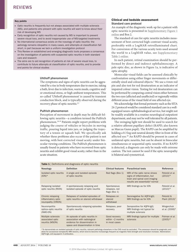
In each patient, retinal examination should be performed by direct and indirect ophthalmoscopy. A pale optic disc, as shown in Figure 1a, is indicative of optic atrophy.
Monocular visual fields can be assessed clinically by confrontation using either finger movements or differentially sized and coloured objects.1 We use a 4 mm red pin and also test for red desaturation as an indicator of impaired colour vision. Testing for red desaturation can be performed by comparing central vision either between the two eyes (affected and unaffected) or regionally within different areas of the visual field of one affected eye.
We acknowledge that formal perimetry such as the SITA 242 protocol would be considered standard care in a wellequipped neuroophthalmological service, but might not be readily available in a routine neurological outpatient department, and may not be well tolerated by all patients.
The swinging light test should be used to search for a relative afferent pupillary defect (RAPD, also known as Marcus Gunn pupil). The RAPD can be amplified by holding a 0.3 log unit neutral density filter in front of the affected eye.20 An RAPD should be present in cases of unilateral optic neuritis, but can be absent in binocular simultaneous or sequential optic neuritis. If no RAPD is detected, a diagnosis can only be made with extreme caution. The test cannot be used if the optic neuropathy is bilateral and symmetrical.
Table 1 | Definitions and diagnosis of optic neuritis
Nosology Definition Clinical features Paraclinical tests Reference
Isolated optic neuritis (ION)*
A single and isolated episode of optic neuritis
Red flags (Box 1) MRI of the optic nerve shows signs of inflammation, but brain and spinal cord imaging results are essentially‡ normal
Petzold et al. (2010)15
Relapsing isolated optic neuritis (RION)
A spontaneously relapsing and isolated episode of optic neuritis
Spontaneous relapses, red flags (Box 1)
MRI findings as for ION Petzold et al. (2010)15
Chronic relapsing inflammatory optic neuropathy (CRION)
Relapses of isolated episodes of optic neuritis on steroid withdrawal
Relapses on steroid withdrawal
Seronegative for AQP4-IgG; MRI findings as for ION
Petzold & Plant (2013)7
Neuromyelitis optica–optic neuritis (NMO-ON)
Spontaneously relapsing episodes of optic neuritis
Relapses, poor recovery
Seropositive for AQP4-IgG; MRI findings not typical for multiple sclerosis
Wingerchuk et al. (2006)8
Multiple sclerosis-associated optic neuritis (MSON)
An episode of optic neuritis in association with radiological evidence for dissemination in space and dissemination in time
Good recovery within ~2 months in most cases
MRI findings typical for multiple sclerosis
Polman et al. (2005)16
*To demonstrate an isolated episode of optic neuritis, relevant pathology elsewhere in the CNS must be excluded. ‡The assessor should be pragmatic and allow for occasional nonspecific MRI lesions, which tend to become increasingly frequent as magnetic field strength and resolution increase. Abbreviation: AQP4-IgG, anti-aquaporin 4 antibodies.
REVIEWS
© 2014 Macmillan Publishers Limited. All rights reserved
NATURE REVIEWS | NEUROLOGY VOLUME 10 | AUGUST 2014 | 449
Pain on eye movements is one of the strongest indicators of optic neuritis, and the few other conditions that manifest with this symptom—for example, myositis, orbital inflammation or subtarsal foreign body—can generally be excluded at the bedside by testing eyelid function and eye movements. Eye movements are best tested if the patient has sufficient visual acuity to focus on an appropriately moving target. We examine smooth pursuit, saccades and vergence movements binocularly. We are looking particularly for evidence of slow adduction of one or both eyes on examination of horizontal saccades as evidence of subclinical internuclear ophthalmoplegia, which is highly suggestive of demyelination elsewhere in the CNS.21 The
cover–uncover test helps us to identify transient visual problems that might be attributable to a phoria.
Research protocolAdditional tests that are not routinely used because of time and financial constraints can be incorporated into research protocols for optic neuritis. Lowcontrast visual acuity can be tested with best correction using Sloan charts.22 In addition, Pelli–Robson charts are useful for testing contrast sensitivity. Colour vision tests include, among others, the Ishihara Test, the Hardy–Rand–Ritter Pseudoisochromatic Plates, the Lanthony Desaturated D15 Test or, if time permits, the Farnsworth–Munsell 100 Hue Test. Both lowcontrast acuity and colour vision tests are sensitive for subtle impairment of visual function even years after the episode. Computerized testing of colour vision provides quantitative data.23 Importantly, this approach also considers relevant ageadjusted normal limits of colour vision, which are required for reliable recognition of acquired problems.23
Patients with the Pulfrich phenomenon can be assessed using a classic pendulum.18,19 A horizontal swinging pendulum is perceived as following an ellipsoid trajectory by patients who exhibit this symptom.24 The Pulfrich phenomenon is best assessed if the pendulum swings in front of a highcontrast patterned background, which enhances binocular disparity (stereoscopic vision).
Optical coherence tomographyOverviewRetinal optical coherence tomography (OCT) permits accurate documentation of changes in thickness of retinal layers.25,26 The full spectrum of retinal pathology that might be unravelled through the use of OCT in MS is not yet known, and new differential diagnoses are being added as the quality of OCT data improves. At present, recommendations for standard care and research protocols for optic neuritis are based largely on two types of pathology. First, thinning or atrophy of the inner retinal layers—that is, the retinal nerve fibre layer (RNFL) and ganglion cell layer (GCL, Figures 1, 2 and 3)—is commonly observed. Second, thickening or swelling of the peripapillary RNFL can be seen in cases with optic disc swelling. There might also be localized thickening of the inner nuclear layer (INL), which can be associated with microcystic macular oedema (MMO, also known as microcystic macular changes or retrograde maculopathy;27–29 Figure 4 and Supplementary Video 1 online).
ThinningLoss of the unmyelinated axons in the retina can readily be quantified by OCT, and is understood to represent several processes:30 first, pathology in the retinal layers resulting in anterograde (Wallerian) degeneration, leading to thinning of the RNFL; second, pathology of the optic nerve, such as an acute optic neuritis attack, causing axonal transection and loss (direct retrograde degeneration); and third, posterior visual pathway lesions that lead to thinning of the RNFL by trans synaptic (via the lateral geniculate nucleus) retrograde axonal degener ation.9,31,32
Box 1 | Red flags implying diagnosis other than MSON
Atypical clinical presentation ■ Pain or loss of vision presenting for more than 2 weeks ■ Absence of pain ■ Retinal abnormalities ■ Unexplained optic atrophy ■ Severe loss of vision in patients with a non-white
ethnic background ■ Severe loss of vision without early recovery
Atypical course ■ Progressive loss of vision ■ Absence of recovery for more than 3 months ■ Worsening of visual function after reducing or stopping
steroids or immunosuppression
Bilateral optic neuritis*
Past medical history of cancer*Simultaneous binocular visual loss must be distinguished from sequential bilateral visual loss. Both visual acuity and visual field need to be documented; sole assessment of visual acuity may miss a peripheral visual field defect. Abbreviation: MSON, multiple sclerosis-associated optic neuritis.
Box 2 | A case of relapsing isolated optic neuritis
A 35-year-old woman presented with blurred vision in her right eye for the second time in 3 months. Visual loss was preceded by several days with photopsia, but no pain on eye movements was reported. Uncorrected visual acuity in the right eye was 0.03 with a central scotoma. Oligoclonal bands were detected in the cerebrospinal fluid (CSF), and additional identical oligoclonal bands (OCBs) were found in CSF and serum (‘type 3’ or mirror plus pattern67). CSF white cell count was elevated (25 per mm3), with a normal total protein level (19 mg/l). The test for anti-aquaporin 4 antibodies was negative, as were the remaining autoimmune, infectious and metabolic laboratory investigations.
Fundus photography (Figure 1a) showed that the right optic disc was pale temporally, indicating optic atrophy, whereas the left optic disc was normal. Fluorescein angiography (Figure 1b) indicated normal retinal blood flow and absence of leakage. Short tau inversion recovery MRI (Figure 1c) demonstrated an increased signal in the right optic nerve, indicating inflammation.
The patient received intravenous steroids according to the optic neuritis treatment trial protocol, but her vision failed to improve over the subsequent 9 months. Serial optical coherence tomography measurements demonstrated the development of further peripapillary retinal nerve fibre layer (pRNFL) atrophy. Consistent with the photograph of the right optic disc, some pRNFL atrophy of the temporal nerve fibres was observed (thin black line in Figure 1d). Atrophy of the pRNFL progressed over the subsequent 9 months and mainly affected the temporal fibres. Automated layer segmentation (Figure 1e–h) demonstrated only a very small degree of atrophy in the inner plexiform layer and inner nuclear layer. By contrast, more-global thickening developed in the outer retinal layers (Figure 1h). This is a consistent finding in the literature, but its significance is unknown.
REVIEWS
© 2014 Macmillan Publishers Limited. All rights reserved

450 | AUGUST 2014 | VOLUME 10 www.nature.com/nrneurol
a cb d
e f
–180NAS
–135 –90INF
–45
Position (degrees)
Thic
knes
s (μ
m)
0TMP
45 90SUP
0
200
150
100
50
250
135 180NAS
–180NAS
–135 –90INF
–45
Position (degrees)
Thic
knes
s (μ
m)
0TMP
45 90SUP
0
175
150
125
100
200
135 180NAS
Thic
knes
s (μ
m)
0
100
75
50
25
125
Thic
knes
s (μ
m)
0
100
75
50
25
125
Thic
knes
s (μ
m)
0
240
180
120
60
300
g h
74 (–45)
20 (–1)
179 (+12)
53 (–4)
124 (–33)
Figure 1 | Ophthalmological and MRI investigations in a 35-year-old woman with blurred vision in the right eye. a | On fundus photographs, the right optic disc (top image) was pale temporally, whereas the left optic disc (bottom image) was normal. b | Fluorescein angiography was normal. c | Coronal section of a fat-suppressed MRI STIR sequence of the orbit demonstrates an increased signal in the right optic nerve. d | Optical coherence tomography shows pRNFL atrophy of the temporal nerve fibres (thin black line at baseline, thick black line at 9-month follow-up). Yellow and red areas indicate atrophy; green area describes the normal thickness profile around the optic disc; blue and purple areas indicate pRNFL thickening. Vertical green line shows the reference position in the corresponding confocal scanning laser ophthalmoscopy image (not shown). e | Automated layer segmentation demonstrated a small degree of atrophy (green areas) in the IPL and f | INL. g | Extent and location of atrophy is best appreciated by summation of the inner retinal layers (pRNFL, IPL and INL). h | More-global thickening developed in the outer retinal layers (outer plexiform layer to basal membrane; red area in summation graph). Abbreviations: INF, inferior; INL, inner nuclear layer; IPL, inner plexiform layer; NAS, nasal; pRNFL, peripapillary retinal nerve fibre layer; SUP, superior; TMP, temporal.
c
Thic
knes
s (μ
m)
0
240
180
120
60
30037 (–36)
–180NAS
–135 –90INF
–45
Position (degrees)
0TMP
45 90SUP
135 180NAS
ba
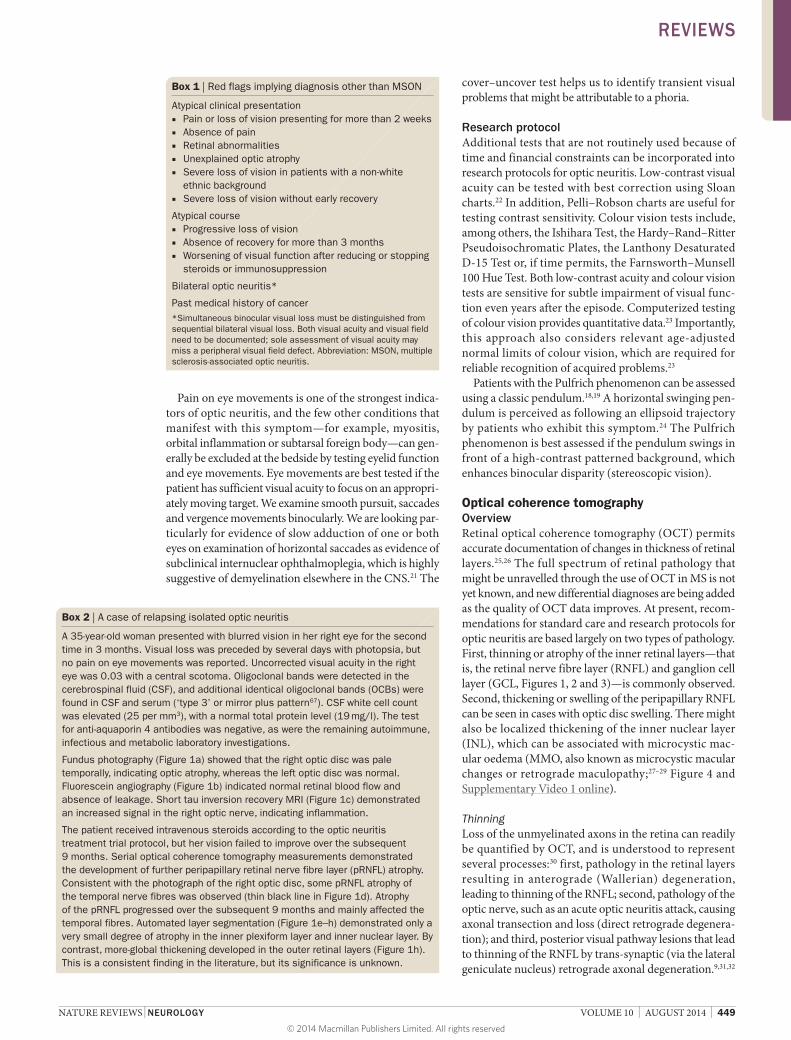
Figure 2 | Minimal retinal OCT protocol—part I: peripapillary ring scan. A 27-year-old woman experienced two episodes of NMO-ON in her left eye. Visual acuity in the left eye was ‘no light perception’ after the first episode of NMO-ON. The second episode started with pain on eye movements, but visual acuity was not helpful for clinical monitoring. a | Peripapillary ring scan (green circle in SLO image). b | Corresponding OCT B-scan. Distance between the red line (inner limiting membrane) and the horizontal green line indicates RNFL thickness. Vertical green line indicates the corresponding anatomical position in the SLO image. c | Thin black line indicates reference RNFL thickness after the first episode of NMO-ON and thick black line indicates additional RNFL loss manifesting 3 months after the second episode of NMO-ON. Abbreviations: INF, inferior; NAS, nasal; NMO-ON, neuromyelitis optica–optic neuritis; OCT, optical coherence tomography; RNFL, retinal nerve fibre layer; SLO, confocal scanning laser ophthalmoscopy; SUP, superior; TMP, temporal.
REVIEWS
© 2014 Macmillan Publishers Limited. All rights reserved
NATURE REVIEWS | NEUROLOGY VOLUME 10 | AUGUST 2014 | 451
In reality, axonotemesis in the hardwired visual pathways will always lead to bidirectional transsynaptic axonal degeneration.33 One should allow a 3month interval after an acute attack of optic neuritis before attempting to quantify peripapillary atrophy of the RNFL.34,35 Wedgelike thinning should prompt investi gation for vascular pathology, such as AION or Susac syndrome (Table 2).36 Occasionally, fluor escein angio graphy (FAG; Figure 1b) may be required to further assess retinal blood flow and leakage.37 RNFL and GCL atrophy is more severe in NMO and CRION than in MSON.38–43 Caution is required when interpreting data from patients with recurrent episodes, as moresevere thinning may be the result of cumulative damage.
ThickeningDisk swelling in the acute phase of optic neuritis is well recognized.1,35 Recent evidence indicates that MMO and thickening of the INL are more frequent in NMO than in MSON.44,45 Because of the association between MMO and NMO, we advocate testing for the presence of antiaquaporin 4 antibodies (AQP4IgG) in patients with MMO who experience relapsing optic neuritis, or in the presence of red flags (Box 1, Supplementary Figure 1 online).
MMO is not specific for either MS or NMO, and is also seen in RION, CRION and a range of other acute and chronic ophthalmological conditions (Figure 4).7,27,45–49 A large study involving 6,551 OCT scans from 1,370 patients indicated that MMO is transient in over 80% of cases, so we suggest longitudinal followup of patients who exhibit this sign.47 In neuroretinitis, acute subretinal fluid and chronic exudates can be effectively visualized by OCT.50
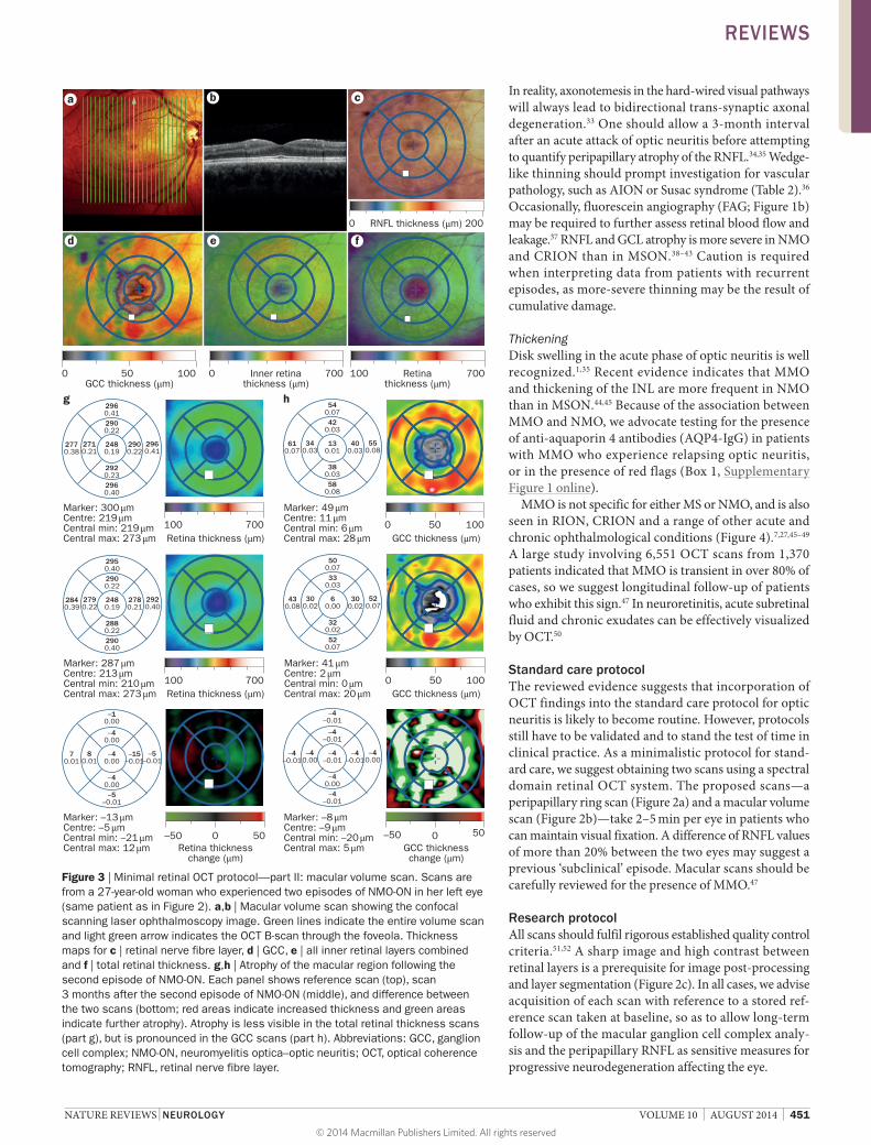
Standard care protocolThe reviewed evidence suggests that incorporation of OCT findings into the standard care protocol for optic neuritis is likely to become routine. However, protocols still have to be validated and to stand the test of time in clinical practice. As a minimalistic protocol for standard care, we suggest obtaining two scans using a spectral domain retinal OCT system. The proposed scans—a peripapillary ring scan (Figure 2a) and a macular volume scan (Figure 2b)—take 2–5 min per eye in patients who can maintain visual fixation. A difference of RNFL values of more than 20% between the two eyes may suggest a previous ‘subclinical’ episode. Macular scans should be carefully reviewed for the presence of MMO.47
Research protocolAll scans should fulfil rigorous established quality control criteria.51,52 A sharp image and high contrast between retinal layers is a prerequisite for image postprocessing and layer segmentation (Figure 2c). In all cases, we advise acquisition of each scan with reference to a stored reference scan taken at baseline, so as to allow longterm followup of the macular ganglion cell complex analysis and the peripapillary RNFL as sensitive measures for progressive neurodegeneration affecting the eye.
0 10050
c
d e f
b
2960.412900.22
2480.19
2900.22
2960.41
2770.38
2710.21
2920.232960.40
100 700Retina thickness (μm)
Marker: 300 μmCentre: 219 μmCentral min: 219 μmCentral max: 273 μm
540.0742
0.03
130.01
400.03
550.08
610.07
340.03
380.0358
0.08
GCC thickness (μm)
Marker: 49 μmCentre: 11 μmCentral min: 6 μmCentral max: 28 μm
2950.402900.22
2480.19
2780.21
2920.40
2840.39
2790.22
2880.222900.40
100 700Retina thickness (μm)
Marker: 287 μmCentre: 213 μmCentral min: 210 μmCentral max: 273 μm
500.0733
0.03
60.00
300.02
520.07
430.08
300.02
320.0252
0.07
0 100GCC thickness (μm)
Marker: 41 μmCentre: 2 μmCentral min: 0 μmCentral max: 20 μm
–10.00
–40.00
–40.00
–15–0.01
–5–0.01
70.01
80.01
–40.00–5
–0.01
–50 50Retina thickness
change (μm)
Marker: –13 μmCentre: –5 μmCentral min: –21 μmCentral max: 12 μm
–4–0.01
–4–0.01
–4–0.01
–4–0.01
–40.00
–4–0.01
–40.00
–40.00–4
–0.01
–50 50
GCC thicknesschange (μm)
Marker: –8 μmCentre: –9 μmCentral min: –20 μmCentral max: 5 μm
g h
0 0
50
100 700Retinathickness (μm)
0 700Inner retinathickness (μm)
0 10050GCC thickness (μm)
0 200RNFL thickness (μm)
a
Figure 3 | Minimal retinal OCT protocol—part II: macular volume scan. Scans are from a 27-year-old woman who experienced two episodes of NMO-ON in her left eye (same patient as in Figure 2). a,b | Macular volume scan showing the confocal scanning laser ophthalmoscopy image. Green lines indicate the entire volume scan and light green arrow indicates the OCT B-scan through the foveola. Thickness maps for c | retinal nerve fibre layer, d | GCC, e | all inner retinal layers combined and f | total retinal thickness. g,h | Atrophy of the macular region following the second episode of NMO-ON. Each panel shows reference scan (top), scan 3 months after the second episode of NMO-ON (middle), and difference between the two scans (bottom; red areas indicate increased thickness and green areas indicate further atrophy). Atrophy is less visible in the total retinal thickness scans (part g), but is pronounced in the GCC scans (part h). Abbreviations: GCC, ganglion cell complex; NMO-ON, neuromyelitis optica–optic neuritis; OCT, optical coherence tomography; RNFL, retinal nerve fibre layer.
REVIEWS
© 2014 Macmillan Publishers Limited. All rights reserved

452 | AUGUST 2014 | VOLUME 10 www.nature.com/nrneurol
A range of additional research techniques are available, such as en face OCT, polarizationsensitive OCT, adaptive optics, fluorescence labelling, and Doppler OCT.30
Laboratory investigationsOverviewIt is important to note that in most patients with typical MSON, there is no evidence to suggest a need for routine blood tests or cerebrospinal fluid (CSF) examination.1
Standard care protocolAdditional blood investigation is warranted in patients with an atypical presentation, such as lack of pain or severe visual loss (<6/60), bilateral visual loss, or severe disc swelling with haemorrhages, and/or with a relevant medical or family history. Blood tests can aid identification of conditions such as paraneoplastic optic neuropathy and cancerassociated retinopathy.
Metabolic pathologyIn suspected metabolic conditions, which can be characterized by bilateral and symmetrical visual loss, tests for vitamin B12, red blood cell folate and methylmalonic acid levels are recommended.53 Note that low vitamin B12 levels can coexist with NMO.54
Systemic pathologyFor suspected systemic disease (see systemic and ischaemic aetiologies in Table 2), tests for haematology, electrolytes, liver function, erythrocyte sedimentation rate, Creactive protein and serum angiotensinconverting enzyme levels are advised.
Inflammatory pathologySerological studies should be guided by the clinical history. Consideration should be given to syphilis, Lyme disease and Bartonella henselae (see infectious aetiologies in Table 2).
Immunological pathologyA resourcesaving screening approach to immunological blood tests is to start with antinuclear antibodies (ANA), following the international guidelines.55 In cases of ANA seropositivity, particularly in young patients, this test may be followed by a more targeted approach that includes assays for autoantibodies against extractable nuclear antigens, perinuclear antineutrophil cyto plasmic antibodies, cardiolipin and β2 glycoprotein 1. This approach facilitates the detection of autoimmune overlap syndromes, which occur in around 5% of patients with autoimmune disease.56,57 In one case, a patient with optic neuritis was found to be seropositive for antiGQ1b and antiGT1a IgG.58
If orbital disease causing a compressive optic neuropathy is suspected, thyroid function and antithyroid antibody tests should be requested. Tests for antiCRMP5 (also called antiCV2) and recoverin autoantibodies should be requested in patients with a history of cancer or suspected paraneoplastic disease, in order to identify conditions such as cancerassociated retinopathy.
In suspected NMO, stateoftheart assays for AQP4IgG are recommended because of their high diagnostic specifi city (>95%) and sensitivity (77%).59–61 Testing for AQP4IgG is advised in all cases with features atypical for MSON—in particular, severe relapsing or
db
a
MMO
Müllercell
Traction
Microglia
Blood–retinabarrier
AQP4KIR4.1
Autoantibodies
c
Anterograde Retrograde
Trans-synaptic degeneration
Figure 4 | MMO in a 44-year-old man with Harding disease (multiple sclerosis plus Leber hereditary optic neuropathy). 15 years earlier, he experienced an episode of optic neuritis in his left eye. Left optic disc was pale with severe atrophy of the peripapillary retinal nerve fibre layer (global average 42 μm). a | MMO in the perimacular rim of the left eye (see also Supplementary Video 1 online). Inset magnified to show retinal layer cytoarchitecture. b | In a diagram of the inset, a retinal ganglion cell is drawn in yellow; the INL, where MMO resides, contains bipolar cells (purple), horizontal cells (light blue) and amacrine cells (dark blue). Bipolar cells synapse with rods (pink) and cones (red) located in the outer retina. Müller cells (green) traverse all retinal layers. c | MMO may be attributable to persistent microcystic changes of the INL, probably following bidirectional trans-synaptic axonal degeneration (anterograde: from outer retina to brain; retrograde: from brain to eye). d | The transient nature of MMO has been related to traction at the vitreoretinal interface (arrow), and is proposed to reflect signs of inflammation from breakdown of the blood–retina barrier, microglial activation, presence of autoantibodies against AQP4 or KIR4.1, and/or Müller cell pathology. Abbreviations: AQP4, aquaporin 4; INL, inner nuclear layer; MMO, microcystic macular oedema.
REVIEWS
© 2014 Macmillan Publishers Limited. All rights reserved

NATURE REVIEWS | NEUROLOGY VOLUME 10 | AUGUST 2014 | 453
bilateral loss of vision—as AQP4IgG seropositivity has specific diagnostic and prognostic implications.62,63 Because of the hetero geneity of assays61 and the possible harmful and ethical consequences of a falsenegative or false positive diagnosis of NMO–optic neuritis, we
recommend that AQP4IgG detection should be performed in laboratories that use validated assays with high sensitivity and specificity. Evidence exists, however, for a subgroup of patients with NMO who are AQP4IgG seronegative but myelin oligodendrocyte glycoprotein
Table 2 | Differential diagnoses and mimics of optic neuritis
Aetiology Features Differential diagnoses Paraclinical tests
Infectious Subacute or progressive visual loss following exposure to infectious agent; frequently with broader cellular reaction in the eye
Spirochaetes (syphilis, Lyme) Serology, PCR, CSF, MRI
HIV MRI, serology
Bartonella henselae, neurocysticercosis, tuberculosis
Chest radiography, serology, CSF, MRI
Sinus pain Viral sinusitis* MRI
Reactive Bilateral and simultaneous; often in childhood and then mostly good prognosis. In contrast, in adults the outcome is more frequently poor
Post-infectious‡ Serology, CSF
Post-vaccination OCT, ERG
Acute disseminated encephalomyelitis (ADEM)
MRI
Neuroretinitis Macular star§
Vascular Sudden-onset visual loss, mostly painless (except GCA); acutely swollen optic disc (except PION); cardiovascular risk factors
AION ESR, CRP, glucose
PION Coagulation
GCA Biopsy
Diabetic papillopathy ECG, Doppler ultrasound
Frequent episodes, migraine Retinal vasospasm ECG, Doppler ultrasound
Hearing loss, encephalitis Susac syndrome Audiogram, OCT, visual fields
Proptosis, chemosis, orbital venous congestion, diplopia (intermittent)
CCF Orbital bruit, CTA/MRA
Seizures, neurological signs Vascular malformations CTA/MRA, DSA, CSF
Nutritional and toxic
Bilateral, painless, progressive; evidence is emerging that cobalt toxicity120 may also be relevant to (hip) joint implants containing cobalt. Pale discs, poor prognosis
Vitamin B12 deficiency Vitamin B12, MMA
Tobacco–alcohol, toxic Full blood, cobalt levels
Endemic|| OCT, visual fields
Methanol, ethambutol, ethylene glycol
Plasma osmolar gap, ethylene glycol, glycolic acid, formate
Compressive Painless, progressive, pale disc at presentation, cilioretinal shunt vessels, history of cancer; proptosis, lid lag, diplopia, history of thyroid disease
Primary tumours, metastases, tuberculoma, sinus mucoceles, Graves disease
CT or MRI, orbits and brain with contrast, MRA, OCT, biopsy, anti-thyroid antibodies (Graves disease only)
Systemic disease
Painful, progressive and often bilateral, more frequent in non-whites, subacute visual loss, history of migraine
All diagnoses Brain and orbits with contrast
Sarcoidosis ACE, CSF, biopsy (sarcoid)
Behçet disease OCT, chest radiography, MRI
SLE Coagulation, if ANA-positive search for specific antibodies
Cancer Paraneoplastic antibodies
Persistent migrainous visual aura Further tests not part of routine work-up
Ocular Pain Posterior scleritis OCT, ultrasound, ANCA121
Painless, metamorphosia Maculopathies OCT, ERG
Preserved colour vision Retinopathies Fluoroscein angiogram
Visual field loss, photopsias Big blind spot syndromes Visual fields, OCT
Preserved colour vision AZOOR ERG
Hereditary Family history. Bilateral, painless LHON; OPA1 and OPA3 mutations Genetic testing
*Sinusitis was high up in the differential diagnosis in the past but has almost disappeared over the past few decades.122 ‡It is absolutely mandatory to obtain a good history including recent travels to high-risk areas for certain infections (malaria, dengue virus, West Nile virus, cysticercosis) or other forms of exposure. §A macular star is suggestive of neuroretinitis and can be associated with peripheral retinal haemorrhages and speckled appearance of the pigment epithelium. ||Endemic optic neuropathies have been described in Cuba and Tanzania. Abbreviations: ACE, angiotensin-converting enzyme; ANA, antinuclear antibodies; ANCA, antineutrophil cytoplasmic antibodies; AZOOR, acute zonal occult outer retinopathy; CCF, carotid cavernous sinus fistula; CRP, c-reactive protein; CSF, cerebrospinal fluid; CTA, CT angiography; DSA, digital subtraction angiography; ECG, electrocardiogram; ERG, electroretinogram; ESR, erythrocyte sedimentation rate; GCA, giant cell arteritis; LHON, Leber hereditary optic neuropathy; MMA, methylmalonic acid; MRA, magnetic resonance angiography; OCT, optical coherence tomography; PION, posterior ischaemic optic neuropathy; SLE, systemic lupus erythematosus; VEP, visual evoked potentials.
REVIEWS
© 2014 Macmillan Publishers Limited. All rights reserved
454 | AUGUST 2014 | VOLUME 10 www.nature.com/nrneurol
IgG seropositive, so AQP4IgG seronegativity does not necessarily preclude a diagnosis of NMO.64,65
Cerebrospinal fluidCSF examination will occasionally be necessary in patients presenting with symptoms of optic neuritis, principally when there is doubt as to the aetiology. CSF examination is rarely contributory in cases deemed to be MSON on other criteria.16 The CSF examination should include cytology, total protein, glucose and oligo clonal bands.66 Of note, CSF OCBs are not specific for MS, and might also be found in over 30 other conditions.67 The type 2 OCB pattern, in which OCBs are found in the CSF but not in the serum, indicating isolated intrathecal oligoclonal IgG synthesis, is only found in about 10% of patients with NMO.68,69 However, CSF glial fibrillary acidic protein (GFAP) levels have a high sensitivity (85–100%) but low specificity (77–100%) for acute NMO.70–73
Research protocolIn view of the emerging body of literature on the role of vitamin D deficiency as a risk factor for demyelination, testing of vitamin D levels is becoming increasingly relevant for optic neuritis research.74,75 In addition, blood neurofilament levels might be of prognostic value in optic neuritis.76–78
Standardized sample collection, processing, storage and analyses are mandatory to ensure highquality biomarker research, enable multicentre analyses, make use of historical cohorts, and safeguard against bias.79 Collection of both plasma and serum is recommended. Samples should be well mixed prior to transport to the laboratory, and should be centrifuged at room temperature (2,000 g for 10 min) within 1 h of sampling. In situations where a longer interval between sampling and processing can be expected, transport and storage at 4 °C is advised. Some biomarkers are sensitive to repeated
freeze–thaw cycling, so we advise that each sample should be stored in at least four 500 μl aliquots at –80 °C.
We anticipate that the discovery of new autoantibodies, in part through retrospective analysis of stored CSF, serum and plasma samples, will be relevant for the differential diagnosis of optic neuritis.
MRIOverviewThis section focuses on MRI of the optic nerves, which is complementary to the wellestablished literature on brain MRI.80 MRI of the optic nerves helps us to rule out alternative diagnoses, demonstrate optic nerve inflammation, and conduct research into the assessment of optic nerve damage and atrophy.
Classic MRI findings in acute optic neuritis are highsignalintensity lesions in the optic nerve (occasionally extending to the chiasm and the optic tracts) on T2weighted MRI (preferably with fat suppression; Figure 1c). There is usually also swelling of the affected segment of the optic nerve. Moreextensive optic nerve lesions are associated with poor clinical outcomes such as slow and incomplete recovery.81,82
Depending on the severity of inflammation, contrast enhancement can be observed on (fatsuppressed) T1weighted images in about 94% of patients with acute optic neuritis. In established optic atrophy from previous damage, T2 high signal changes will also be seen, but these changes are associated with thinning of the nerve and do not change over time. Sensitivity of detection of contrastenhancing lesions in the optic nerve can be further increased by using a higher dosage of contrast medium (for example, a triple dose), but in our view this approach is not costeffective.83,84
Patients with NMO have a propensity towards extensive involvement of the anterior visual pathways, and of the intracranial as opposed to the intraorbital segments, making these individuals particularly prone to chiasmitis and simultaneous bilateral disease.85,86
Standard care protocolNo consensus guidelines have been formulated on how and when to perform optic nerve MRI in cases of optic neuritis. For assessment of the optic nerve, the MRI protocol must include fatsuppressed T2weighted images (for example, short tau inversion recovery or frequencyspecific selective partial inversion recovery)81 and contrast enhanced (fatsuppressed) T1weighted images (Box 3). The most costeffective approach is to use a standard dosage of gadoliniumbased contrast medium.
T1weighted and T2weighted sequences for the assessment of the whole orbit and brain are useful to simultaneously detect possible demyelinating brain lesions. For the best spatial resolution, images should be acquired without any interslice gap. The spatial resolution should be 3 mm slice thickness, in plane 1 × 1 mm (measured voxel size 3 × 1 × 1 mm). Additional sagittal proton density weighted/T2weighted (preferably spin echo) and T1weighted postcontrast images of the spinal cord are useful for detecting demyelinating spinal cord lesions
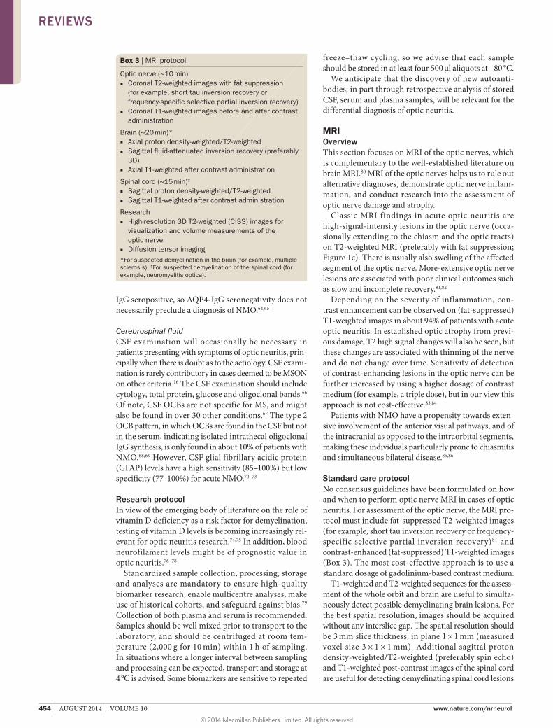
Box 3 | MRI protocol
Optic nerve (~10 min) ■ Coronal T2-weighted images with fat suppression
(for example, short tau inversion recovery or frequency-specific selective partial inversion recovery)
■ Coronal T1-weighted images before and after contrast administration
Brain (~20 min)* ■ Axial proton density-weighted/T2-weighted ■ Sagittal fluid-attenuated inversion recovery (preferably
3D) ■ Axial T1-weighted after contrast administration
Spinal cord (~15 min)‡
■ Sagittal proton density-weighted/T2-weighted ■ Sagittal T1-weighted after contrast administration
Research ■ High-resolution 3D T2-weighted (CISS) images for
visualization and volume measurements of the optic nerve
■ Diffusion tensor imaging*For suspected demyelination in the brain (for example, multiple sclerosis). ‡For suspected demyelination of the spinal cord (for example, neuromyelitis optica).
REVIEWS
© 2014 Macmillan Publishers Limited. All rights reserved

NATURE REVIEWS | NEUROLOGY VOLUME 10 | AUGUST 2014 | 455
and for differential diagnostic purposes (Box 3). Further radiological tests to exclude vascular malformations or vasculitis may be indicated, as summarized in Table 2.
Research protocolAlthough MRI at higher magnetic field strengths (for example, 3 T) does not necessarily lead to an earlier diagnosis of MS, it does result in a higher image quality.87,88 In general, a standard head coil is sufficient for the assessment of the orbit and the optic nerves. However, surface coils focusing on the orbit may further improve the image quality.89
Important MRI methods for research purposes include measures of optic nerve atrophy obtained using highresolution 3D T2weighted sequences,90 quantitative MRI techniques91 and diffusion tensor imaging.92 These advanced techniques can assist in the early detection of clinical impairment, and enable quantitative estimation of the presence and extent of damage to the optic nerve.93,94 These measures have all been proposed as markers of irreversible tissue damage and disease progression, and as predictors of clinical recovery.
Electrodiagnostic testsOverviewElectrodiagnostic tests have a role in the investigation of atypical presentations of optic neuritis.95 The tests should not be limited to visual evoked potentials (VEPs), as any retinal abnormality will affect the VEP signal.95,96 Indeed, the International Society for Clinical Electrophysiology of Vision (ISCEV) standards97 recommend that in cases of unexplained visual loss, the VEP results should be interpreted in conjunction with both a standard electroretinogram (ERG) and a pattern electroretinogram (PERG).98
Standard care protocolThe ISCEV standards97 state that electrophysiological tests are of little diagnostic value in acute retrobulbar optic neuritis, but may be useful in studies evaluating therapy. If the diagnosis is uncertain, however, a full series of electrophysiological tests should be performed, as outlined below.
Visual evoked potentialsThe VEP represents a specific change in an ongoing EEG recording due to the response of the visual pathway to either a pattern or a flash stimulus. VEPs have been shown to be a highly sensitive but not especially specific test for optic neuritis. A unilateral substantially delayed VEP with minimal amplitude reduction is highly suggestive of a demyelinating optic neuropathy, for example, recovered MSON or subclinical involvement of the optic nerve in MS. VEP abnormalities will also be seen in patients with refractive errors, pure retinal dysfunction, compressive lesions of the optic nerve, Parkinson disease99 or migraine.100 Normality of both the ERG and VEP supports the differential diagnosis of nonorganic aetiology if performed after other investigations failed to yield a diagnosis.
ElectroretinographyAn ERG provides diagnostic information about the rod and cone photoreceptors, and inner retinal function across the entire retina. The differential diagnosis of a pale optic disc with reduced central vision includes cone dystrophies and maculopathies, which will be incorrectly diagnosed if VEPs are used alone.
Research protocolMultifocal VEPs allow fine mapping of the electrophysiological abnormalities in the visual pathway. This mapping enables detection of abnormalities restricted to specific topographies, and may be more sensitive than standard VEPs.
As the macula comprises less than 5% of the retina, a maculopathy will generally not affect the fullfield ERG. The PERG stimulus parameters mean that the macula is the retinal location that is primarily stimulated.96 The PERG is a surrogate test of macular function, and can, therefore, be used to differentiate macular from optic nerve causes of central vision loss. The two main components of the PERG are N95 and P50,96,101,102 and the N95 component and N95:P50 ratio were found to be useful in optic neuritis.101–104
The optic nerve head component responses of multifocal ERG may also emerge as a useful tool in the assessment of optic neuritis.105
PerimetryOverviewThe classic visual field defect described in MSON is a central scotoma with a sloping border of the isopters.106 Of note, however, this feature has only been reported in 2% of cases,107 and becomes even rarer (0.5%) later in the disease course. Most patients experience a more generalized reduction in their visual field sensitivity (66%).107 By contrast, altitudinal visual field defects suggest a vascular differential diagnosis,108 and sparing of central vision suggests an optic perineuritis,109 but neither pattern will rule out optic neuritis.
Automated perimetry should be performed using threshold estimation strategies.110,111 An important limitation, particularly in neurological conditions, children or elderly patients, is that the test is timeconsuming and the failure rate may be up to 46%.112–114 One needs to consider operator instruction effects, ‘natural variation’ in test results, and learning effects.115,116 It may not be possible to detect a visual field defect until about 25–35% of retinal ganglion cells have been lost.117–119 Therefore, as a consensus, we hesitate to recommend perimetry as a mandatory part of a standard care protocol but, rather, advocate selective use in cases where the test results may guide the differential diagnosis (Table 2), or in the presence of red flags (Box 1).
Research protocolGiven that production of the Goldmann perimeter has stopped and experience is vanishing, we recommend use of standard threshold automated perimetry. The stimulus size should be Goldmann No. III (0.481°). A visual field
REVIEWS
© 2014 Macmillan Publishers Limited. All rights reserved

456 | AUGUST 2014 | VOLUME 10 www.nature.com/nrneurol
of 20–30° should suffice (242 or 302 protocols on the Humphrey Zeiss perimeter; full threshold 32 or static whiteonwhite fields on the Octopus 900 series).
Conclusions and further perspectivesThe clinical spectrum of optic neuritis has broadened over the past two decades. This development was driven by several factors: cumulative experience from trials on treatment of optic neuritis; clinical recognition of a divergent semiology; discovery of new autoantibodies and biomarkers; and availability of new highresolution imaging techniques for the optic nerve and retina.
A need exists to make use of this knowledge and these techniques to better investigate and define the clinical spectrum of optic neuritis with the aim of guiding future patient management. Importantly, some forms of optic neuritis (for example, NMO and CRION) will require rigor ous immunosuppression to protect patients from blindness. It is also essential that patients with these conditions are not included in MSON treatment trials.
We anticipate that the investigation protocol presented here will help us to reach a consensus on the classification of optic neuritis and how to optimize treatment. This consensus should, in turn, be pursued inter nationally so that different approaches can be validated in those parts of the world where MS is less common.
Review criteria
A search for original articles published between 1970 and 2014, focusing on optic neuritis, was performed in Google Scholar, MEDLINE and PubMed. The search terms used were “optic neuritis”, “imaging”, “MRI”, “OCT”, “cerebrospinal fluid”, “CSF”, “biomarker”, “immune”, “VEP”, “ERG”, “visual field”, “perimetry”, “vision”, “visual system”, “retina” and “optic nerve”, alone and in combination. All articles identified were full-text papers in the English language. We also searched the reference lists of identified articles for further relevant papers. Furthermore, specific papers from the literature archives of the authors were included.
1. Hickman, S., Dalton, C., Miller, D. & Plant, G. Management of acute optic neuritis. Lancet 360, 1953–1962 (2002).
2. Fraser, C. L., Davagnanam, I., Radon, M. & Plant, G. T. The time course and phenotype of Uhthoff phenomenon following optic neuritis. Mult. Scler. 18, 1042–1044 (2012).
3. Frohman, T. C. et al. Uhthoff’s phenomena in MS—clinical features and pathophysiology. Nat. Rev. Neurol. 9, 535–540 (2013).
4. Hess, K., Gresty, M. & Leech, J. Clinical and theoretical aspects of head movement dependent oscillopsia (HMDO). A review. J. Neurol. 219, 151–157 (1978).
5. Serra, A., Derwenskus, J., Downey, D. L. & Leigh, R. J. Role of eye movement examination and subjective visual vertical in clinical evaluation of multiple sclerosis. J. Neurol. 250, 569–575 (2003).
6. Sharpe, J. A., Goldberg, H. J., Lo, A. W. & Herishanu, Y. O. Visual–vestibular interaction in multiple sclerosis. Neurology 31, 427–433 (1981).
7. Petzold, A. & Plant, G. T. Chronic relapsing inflammatory optic neuropathy: a systematic review of 122 cases reported. J. Neurol. 261, 17–26 (2014).
8. Wingerchuk, D. M., Lennon, V. A., Pittock, S. J., Lucchinetti, C. F. & Weinshenker, B. G. Revised diagnostic criteria for neuromyelitis optica. Neurology 66, 1485–1489 (2006).
9. Gabilondo, I. et al. Trans-synaptic axonal degeneration in the visual pathway in multiple sclerosis. Ann. Neurol. 75, 98–107 (2014).
10. Jenkins, T. et al. Dissecting structure–function interactions in acute optic neuritis to investigate neuroplasticity. Hum. Brain Mapp. 31, 276–286 (2010).
11. Masland, R. H. The neuronal organization of the retina. Neuron 76, 266–280 (2012).
12. Jonas, J. B., Schmidt, A. M., Müller-Bergh, J. A., Schlötzer-Schrehardt, U. M. & Naumann, G. O. Human optic nerve fiber count and optic disc size. Invest. Ophthalmol. Vis. Sci. 33, 2012–2018 (1992).
13. Hubel, D. & Wiesel, T. David Hubel and Torsten Wiesel. Neuron 75, 182–184 (2012).
14. Gilbert, C. D. & Li, W. Top-down influences on visual processing. Nat. Rev. Neurosci. 14, 350–363 (2013).
15. Petzold, A. et al. Neuromyelitis optica-IgG (aquaporin-4) autoantibodies in immune mediated optic neuritis. J. Neurol. Neurosurg. Psychiatry 81, 109–111 (2010).
16. Polman, C. et al. Diagnostic criteria for multiple sclerosis: 2005 revisions to the “McDonald Criteria”. Ann. Neurol. 58, 840–846 (2005).
17. Lepore, F. E. The origin of pain in optic neuritis. Determinants of pain in 101 eyes with optic neuritis. Arch. Neurol. 48, 748–749 (1991).
18. Petzold, A. & Pitz, E. The historical origin of the Pulfrich Effect: a serendipitous astronomic observation at the border of the Milky Way. Neuro-Ophthalmology 33, 39–46 (2009).
19. McGowan, G., Ahmed, T. Y., Heron, G. & Diaper, C. The Pulfrich phenomenon; clumsiness and collisions which can be ameliorated. Pract. Neurol. 11, 173–176 (2011).
20. Kawasaki, A., Moore, P. & Kardon, R. H. Long-term fluctuation of relative afferent pupillary defect in subjects with normal visual function. Am. J. Ophthalmol. 122, 875–882 (1996).
21. Leigh, R. J. & Serra, A. Taking the temperature of MS with INO. Neurology 70, 1063–1064 (2008).
22. Balcer, L. J. et al. Contrast letter acuity as a visual component for the Multiple Sclerosis Functional Composite. Neurology 61, 1367–1373 (2003).
23. Rodriguez-Carmona, M., O’Neill-Biba, M. & Barbur, J. L. Assessing the severity of color vision loss with implications for aviation and other occupational environments. Aviat. Space Environ. Med. 83, 19–29 (2012).
24. Anzai, A., Ohzawa, I. & Freeman, R. D. Joint-encoding of motion and depth by visual cortical neurons: neural basis of the Pulfrich effect. Nat. Neurosci. 4, 513–518 (2001).
25. Frohman, E. M. et al. Relationship of optic nerve and brain conventional and non-conventional MRI measures and retinal nerve fiber layer thickness, as assessed by OCT and GDx: a pilot study. J. Neurol. Sci. 282, 96–105 (2009).
26. Menke, M. N., Dabov, S., Knecht, P. & Sturm, V. Reproducibility of retinal thickness measurements in healthy subjects using spectralis optical coherence tomography. Am. J. Ophthalmol. 147, 467–472 (2009).
27. Kisimbi, J. et al. Macular spectral domain optical coherence tomography findings in Tanzanian
endemic optic neuropathy. Brain 136, 3418–3426 (2013).
28. Abegg, M. et al. Microcystic macular edema: retrograde maculopathy caused by optic neuropathy. Ophthalmology 121, 142–149 (2014).
29. Mahroo, O. A. et al. Re: Abegg. et al.: Microcystic macular edema: retrograde maculopathy caused by optic neuropathy (Ophthalmology 2014; 121:142–9). Ophthalmology http://dx.doi.org/ 10.1016/j.ophtha.2014.01.035.
30. Petzold, A. et al. Optical coherence tomography in multiple sclerosis: a systematic review and meta-analysis. Lancet Neurol. 9, 921–932 (2010).
31. Balk, L. J. et al. A dam for retrograde axonal degeneration in multiple sclerosis? J. Neurol. Neurosurg. Psychiatry 85, 782–789 (2014).
32. Pfueller, C. F. et al. Metabolic changes in the visual cortex are linked to retinal nerve fiber layer thinning in multiple sclerosis. PLoS ONE 6, e18019 (2011).
33. Petzold, A. Neurodegeneration and multiple sclerosis. In Neurodegenerative Diseases: Clinical Aspects, Molecular Genetics and Biomarkers (eds Galimberti, D. & Scarpini, E.) 227–245 (Springer, 2014).
34. Costello, F. et al. Quantifying axonal loss after optic neuritis with optical coherence tomography. Ann. Neurol. 59, 963–969 (2006).
35. Toosy, A. T., Mason, D. F. & Miller, D. H. Optic neuritis. Lancet Neurol. 13, 83–99 (2014).
36. Brandt, A. U. et al. Patterns of retinal damage facilitate differential diagnosis between Susac syndrome and MS. PLoS ONE 7, e38741 (2012).
37. Nagia, L. & Eggenberger, E. Differentiating retinal from optic nerve syndromes. Curr. Opin. Ophthalmol. 24, 528–533 (2013).
38. Bichuetti, D. B. et al. The retinal nerve fiber layer of patients with neuromyelitis optica and chronic relapsing optic neuritis is more severely damaged than patients with multiple sclerosis. J. Neuroophthalmol. 33, 220–224 (2013).
39. Bouyon, M. et al. Longitudinal follow-up of vision in a neuromyelitis optica cohort. Mult. Scler. 19, 1320–1322 (2013).
40. Fernandes, D. B. et al. Evaluation of inner retinal layers in patients with multiple sclerosis or neuromyelitis optica using optical coherence tomography. Ophthalmology 120, 387–394 (2013).
REVIEWS
© 2014 Macmillan Publishers Limited. All rights reserved

NATURE REVIEWS | NEUROLOGY VOLUME 10 | AUGUST 2014 | 457
41. Kaufhold, F. et al. Optic neuritis is associated with inner nuclear layer thickening and microcystic macular edema independently of multiple sclerosis. PLoS ONE 8, e71145 (2013).
42. Nakamura, M. et al. Early high-dose intravenous methylprednisolone is effective in preserving retinal nerve fiber layer thickness in patients with neuromyelitis optica. Graefes Arch. Clin. Exp. Ophthalmol. 248, 1777–1785 (2010).
43. von Glehn, F. et al. Structural brain abnormalities are related to retinal nerve fiber layer thinning and disease duration in neuromyelitis optica spectrum disorders. Mult. Scler. http:// dx.doi.org/10.1177/1352458513519838.
44. Gelfand, J. M., Nolan, R., Schwartz, D. M., Graves, J. & Green, A. J. Microcystic macular oedema in multiple sclerosis is associated with disease severity. Brain 135, 1786–1793 (2012).
45. Schneider, E. et al. Optical coherence tomography reveals distinct patterns of retinal damage in neuromyelitis optica and multiple sclerosis. PLoS ONE 8, e66151 (2013).
46. Balk, L. J., Killestein, J., Polman, C. H., Uitdehaag, B. M. & Petzold, A. Microcystic macular oedema confirmed, but not specific for multiple sclerosis. Brain 135, e226 (2012).
47. Burggraaff, M. C., Trieu, J., de Vries-Knoppert, W. A., Balk, L. & Petzold, A. The clinical spectrum of microcystic macular oedema. Invest. Ophthalmol. Vis. Sci. 55, 952–961 (2014).
48. Gelfand, J. M., Cree, B. A., Nolan, R., Arnow, S. & Green, A. J. Microcystic inner nuclear layer abnormalities and neuromyelitis optica. JAMA Neurol 70, 629–633 (2013).
49. Sotirchos, E. S. et al. In vivo identification of morphologic retinal abnormalities in neuromyelitis optica. Neurology 80, 1406–1414 (2013).
50. Vishwanath, S. et al. Post-fever retinitis: a single center experience from south India. Int. Ophthalmol. http://dx.doi.org/10.1007/s10792-013-9891–7.
51. Tewarie, P. et al. The OSCAR-IB consensus criteria for retinal OCT quality assessment. PLoS ONE 7, e34823 (2012).
52. Schippling, S. et al. Quality control for retinal OCT in multiple sclerosis: validation of the OSCAR-IB criteria. Mult. Scler. http:// dx.doi.org/10.1177/1352458514538110.
53. Stabler, S. P. Clinical practice. Vitamin B12 deficiency. N. Engl. J. Med. 368, 149–160 (2013).
54. Jarius, S., Paul, F., Ruprecht, K. & Wildemann, B. Low vitamin B12 levels and gastric parietal cell antibodies in patients with aquaporin-4 antibody-positive neuromyelitis optica spectrum disorders. J. Neurol. 259, 2743–2745 (2012).
55. Agmon-Levin, N. et al. International recommendations for the assessment of autoantibodies to cellular antigens referred to as anti-nuclear antibodies. Ann. Rheum. Dis. 73, 17–23 (2014).
56. Jarius, S. et al. Contrasting disease patterns in seropositive and seronegative neuromyelitis optica: a multicentre study of 175 patients. J. Neuroinflammation 9, 14 (2012).
57. Myers, T. D. et al. Use of corticosteroid sparing systemic immunosuppression for treatment of corticosteroid dependent optic neuritis not associated with demyelinating disease. Br. J. Ophthalmol. 88, 673–680 (2004).
58. Biotti, D., Boucher, S., Ong, E., Tilikete, C. & Vighetto, A. Optic neuritis as a possible phenotype of anti-GQ1b/GT1a antibody syndrome. J. Neurol. 260, 2890–2891 (2013).
59. Fujihara, K. & Leite, M. Seronegative NMO: a sensitive AQP4 antibody test clarifies clinical features and next challenges. Neurology 80, 2176–2177 (2013).
60. Jarius, S. & Wildemann, B. Aquaporin-4 antibodies (NMO-IgG) as a serological marker of neuromyelitis optica: a critical review of the literature. Brain Pathol. 23, 661–683 (2013).
61. Waters, P. J. et al. Serologic diagnosis of NMO: a multicenter comparison of aquaporin-4-IgG assays. Neurology 78, 665–671 (2012).
62. Jarius, S. et al. Frequency and prognostic impact of antibodies to aquaporin-4 in patients with optic neuritis. J. Neurol. Sci. 298, 158–162 (2010).
63. Matiello, M. et al. NMO-IgG predicts the outcome of recurrent optic neuritis. Neurology 70, 2197–2200 (2008).
64. Kitley, J. et al. Neuromyelitis optica spectrum disorders with aquaporin-4 and myelin-oligodendrocyte glycoprotein antibodies: a comparative study. JAMA Neurol. 71, 276–283 (2014).
65. Sato, D. K. et al. Distinction between MOG antibody-positive and AQP4 antibody-positive NMO spectrum disorders. Neurology 82, 474–481 (2014).
66. Stangel, M. et al. The utility of cerebrospinal fluid analysis in patients with multiple sclerosis. Nat. Rev. Neurol. 9, 267–276 (2013).
67. Petzold, A. Intrathecal oligoclonal IgG synthesis in multiple sclerosis. J. Neuroimmunol. 262, 1–10 (2013).
68. Jarius, S. et al. Cerebrospinal fluid findings in aquaporin-4 antibody positive neuromyelitis optica: results from 211 lumbar punctures. J. Neurol. Sci. 306, 82–90 (2011).
69. Nakamura, M. et al. Clinical and laboratory features of neuromyelitis optica with oligoclonal IgG bands. Mult. Scler. 13, 332–335 (2007).
70. Misu, T. et al. Marked increase in cerebrospinal fluid glial fibrillar acidic protein in neuromyelitis optica: an astrocytic damage marker. J. Neurol. Neurosurg. Psychiatry 80, 575–577 (2009).
71. Petzold, A., Marignier, R., Verbeek, M. M. & Confavreux, C. Glial but not axonal protein biomarkers as a new supportive diagnostic criteria for Devic neuromyelitis optica? Preliminary results on 188 patients with different neurological diseases. J. Neurol. Neurosurg. Psychiatry 82, 467–469 (2011).
72. Takano, R. et al. Astrocytic damage is far more severe than demyelination in NMO: a clinical CSF biomarker study. Neurology 75, 208–216 (2010).
73. Uzawa, A. et al. Cerebrospinal fluid interleukin-6 and glial fibrillary acidic protein levels are increased during initial neuromyelitis optica attacks. Clin. Chim. Acta 421, 181–183 (2013).
74. Dörr, J., Döring, A. & Paul, F. Can we prevent or treat multiple sclerosis by individualised vitamin D supply? EPMA J. 4, 4 (2013).
75. von Geldern, G. & Mowry, E. M. The influence of nutritional factors on the prognosis of multiple sclerosis. Nat. Rev. Neurol. 8, 678–689 (2012).
76. Petzold, A., Rejdak, K. & Plant, G. Axonal degeneration and inflammation in acute optic neuritis. J. Neurol. Neurosurg. Psychiatry 75, 1178–1180 (2004).
77. Petzold, A. & Plant, G. T. The diagnostic and prognostic value of neurofilament heavy chain levels in immune-mediated optic neuropathies. Mult. Scler. Int. 2012, 217802 (2012).
78. Talla, V. et al. Noninvasive assessments of optic nerve neurodegeneration in transgenic mice with isolated optic neuritis. Invest. Ophthalmol. Vis. Sci. 54, 4440–4450 (2013).
79. Petzold, A., Bowser, R., Calabresi, P., Zetterberg, H. & Uitdehaag, B. M. Biomarker
time out. Mult. Scler. http://dx.doi.org/ 10.1177/1352458514524999.
80. Barkhof, F., Calabresi, P. A., Miller, D. H. & Reingold, S. C. Imaging outcomes for neuroprotection and repair in multiple sclerosis trials. Nat. Rev. Neurol. 5, 256–266 (2009).
81. Kolappan, M. et al. Assessing structure and function of the afferent visual pathway in multiple sclerosis and associated optic neuritis. J. Neurol. 256, 305–319 (2009).
82. Miller, D. H. et al. Magnetic resonance imaging of the optic nerve in optic neuritis. Neurology 38, 175–179 (1988).
83. Hickman, S. J. et al. Visual recovery following acute optic neuritis—a clinical, electrophysiological and magnetic resonance imaging study. J. Neurol. 251, 996–1005 (2004).
84. Kupersmith, M. J., Alban, T., Zeiffer, B. & Lefton, D. Contrast-enhanced MRI in acute optic neuritis: relationship to visual performance. Brain 125, 812–822 (2002).
85. Khanna, S. et al. Magnetic resonance imaging of optic neuritis in patients with neuromyelitis optica versus multiple sclerosis. J. Neuroophthalmol. 32, 216–220 (2012).
86. Storoni, M., Davagnanam, I., Radon, M., Siddiqui, A. & Plant, G. T. Distinguishing optic neuritis in neuromyelitis optica spectrum disease from multiple sclerosis: a novel magnetic resonance imaging scoring system. J. Neuroophthalmol. 33, 123–127 (2013).
87. Wattjes, M. P. & Barkhof, F. High field MRI in the diagnosis of multiple sclerosis: high field–high yield? Neuroradiology 51, 279–292 (2009).
88. Wattjes, M. P. et al. Does high field MRI allow an earlier diagnosis of multiple sclerosis? J. Neurol. 255, 1159–1163 (2008).
89. Karim, S., Clark, R. A., Poukens, V. & Demer, J. L. Demonstration of systematic variation in human intraorbital optic nerve size by quantitative magnetic resonance imaging and histology. Invest. Ophthalmol. Vis. Sci. 45, 1047–1051 (2004).
90. Trip, S. A. et al. Optic nerve atrophy and retinal nerve fibre layer thinning following optic neuritis: evidence that axonal loss is a substrate of MRI-detected atrophy. Neuroimage 31, 286–293 (2006).
91. Trip, S. A. et al. Optic nerve magnetization transfer imaging and measures of axonal loss and demyelination in optic neuritis. Mult. Scler. 13, 875–879 (2007).
92. Trip, S. A. et al. Optic nerve diffusion tensor imaging in optic neuritis. Neuroimage 30, 498–505 (2006).
93. Glisson, C. C. & Galetta, S. L. Nonconventional optic nerve imaging in multiple sclerosis. Neuroimaging Clin. N. Am. 19, 71–79 (2009).
94. Yiannakas, M. C. et al. MRI acquisition and analysis protocol for in vivo intraorbital optic nerve segmentation at 3 T. Invest. Ophthalmol. Vis. Sci. 54, 4235–4240 (2013).
95. Holder, G. E., Gale, R. P., Acheson, J. F. & Robson, A. G. Electrodiagnostic assessment in optic nerve disease. Curr. Opin. Neurol. 22, 3–10 (2009).
96. Holder, G. E. Pattern electroretinography (PERG) and an integrated approach to visual pathway diagnosis. Prog. Retin. Eye Res. 20, 531–561 (2001).
97. Visual electrodiagnostics: a guide to procedures. ISCEV Standards, Recommendations and Guidelines [online]. http://www.iscev.org/standards/proceduresguide.html (2013).
98. Odom, J. V. et al. ISCEV standard for clinical visual evoked potentials (2009 update). Doc. Ophthalmol. 120, 111–119 (2010).
REVIEWS
© 2014 Macmillan Publishers Limited. All rights reserved

458 | AUGUST 2014 | VOLUME 10 www.nature.com/nrneurol
99. Nightingale, S., Mitchell, K. W. & Howe, J. W. Visual evoked cortical potentials and pattern electroretinograms in Parkinson’s disease and control subjects. J. Neurol. Neurosurg. Psychiatry 49, 1280–1287 (1986).
100. Boylu, E. et al. Visual evoked potential abnormalities in migraine patients. Electromyogr. Clin. Neurophysiol. 50, 303–308 (2010).
101. Fraser, C. L. & Holder, G. E. Electroretinogram findings in unilateral optic neuritis. Doc. Ophthalmol. 123, 173–178 (2011).
102. Fraser, C. et al. Multifocal visual evoked potential latency analysis: predicting progression to multiple sclerosis. Arch. Neurol. 63, 847–850 (2006).
103. Holder, G. E. The incidence of abnormal pattern electroretinography in optic nerve demyelination. Electroencephalogr. Clin. Neurophysiol. 78, 18–26 (1991).
104. Rodriguez-Mena, D. et al. Electropysiologic evaluation of the visual pathway in patients with multiple sclerosis. J. Clin. Neurophysiol. 30, 376–381 (2013).
105. Frohman, T. C. et al. Optic nerve head component responses of the multifocal electroretinogram in MS. Neurology 81, 545–551 (2013).
106. Gerling, J., Meyer, J. & Kommerell, G. Visual field defects in optic neuritis and anterior ischemic optic neuropathy: distinctive features. Graefes Arch. Clin. Exp. Ophthalmol. 236, 188–192 (1998).
107. Keltner, J. L. et al. Visual field profile of optic neuritis: a final follow-up report from the optic neuritis treatment trial from baseline through 15 years. Arch. Ophthalmol. 128, 330–337 (2010).
108. Petzold, A., Islam, N., Hu, H.-H. & Plant, G. T. Embolic and nonembolic transient monocular visual field loss: a clinicopathologic review. Surv. Ophthalmol. 58, 42–62 (2013).
109. Purvin, V., Kawasaki, A. & Jacobson, D. M. Optic perineuritis: clinical and radiographic features. Arch. Ophthalmol. 119, 1299–1306 (2001).
110. Petzold, A. & Plant, G. Failure to detect bitemporal field defects due to chiasmal compression on a screening perimetry protocol. Neuro-Ophthalmology 24, 357–361 (2001).
111. Schiefer, U. et al. Comparison of the new perimetric GATE strategy with conventional full-threshold and SITA standard strategies. Invest. Ophthalmol. Vis. Sci. 50, 488–494 (2009).
112. Harding, G. F., Wild, J. M., Robertson, K. A., Rietbrock, S. & Martinez, C. Separating the retinal electrophysiologic effects of vigabatrin: treatment versus field loss. Neurology 55, 347–352 (2000).
113. Scott, J. A. & Egan, R. A. Prevalence of organic neuro-ophthalmologic disease in patients with functional visual loss. Am. J. Ophthalmol. 135, 670–675 (2003).
114. Trick, G. L., Trick, L. R., Morris, P. & Wolf, M. Visual field loss in senile dementia of the Alzheimer’s type. Neurology 45, 68–74 (1995).
115. Kutzko, K. E., Brito, C. F. & Wall, M. Effect of instructions on conventional automated perimetry. Invest. Ophthalmol. Vis. Sci. 41, 2006–2013 (2000).
116. [No authors listed] Automated perimetry. American Academy of Ophthalmology. Ophthalmology 103, 1144–1151 (1996).
117. Kerrigan-Baumrind, L. A., Quigley, H. A., Pease, M. E., Kerrigan, D. F. & Mitchell, R. S. Number of ganglion cells in glaucoma eyes compared with threshold visual field tests in the same persons. Invest. Ophthalmol. Vis. Sci. 41, 741–748 (2000).
118. Mikelberg, F. S., Yidegiligne, H. M. & Schulzer, M. Optic nerve axon count and axon diameter in patients with ocular hypertension and normal visual fields. Ophthalmology 102, 342–348 (1995).
119. Quigley, H., Dunkelberger, G. & Green, W. Retinal ganglion cell atrophy correlated with automated perimetry in human eyes with glaucoma. Am. J. Ophthalmol. 107, 453–464 (1989).
120. Bhardwaj, N., Perez, J. & Peden, M. Optic neuropathy from cobalt toxicity in a patient who ingested cattle magnets. Neuro-Ophthalmology 35, 24–26 (2011).
121. Wakefield, D., Di Girolamo, N., Thurau, S., Wildner, G. & McCluskey, P. Scleritis: immunopathogenesis and molecular basis for therapy. Prog. Retin. Eye Res. 35, 44–62 (2013).
122. Coppens, S., Petzold, A., de Graaf, P. & Vries-Knoppert, W. Recurrent optic perineuritis after intranasal cocaine abuse. Neuro-Ophthalmology 38, 91–95 (2014).
AcknowledgementsThe MS Center VUMC is partially funded by a programme grant from the Dutch MS Research Foundation. K.F. is a recipient of Grants-in-Aid for Scientific Research from the Ministry of Education, Science and Technology and the Ministry of Health, Labour and Welfare of Japan. F.P. is supported by the German Research Council (DFG Exc 257) and the German Ministry for Education and Research Competence Network Multiple Sclerosis. S.S. is supported by the Clinical Research Priority Program of the University of Zürich and the Betty and David Koetser Foundation for Brain Research. G.T.P. is supported by the University College London Comprehensive Biomedical Research Centre and the Moorfields Biomedical Research Centre.
Author contributionsA.P. had the idea for this protocol, reviewed the literature, provided figures, wrote the first draft and finalized the manuscript. F.C., K.F., F.P., S.S. and C.S. revised the manuscript. M.P.W. performed an independent literature review and wrote the MRI section. C.L.F. performed an independent literature review and wrote the VEP/ERG section. B.W. and G.T.P. contributed to the conception and design of the protocol. All authors revised the final version of the manuscript.
Supplementary information is linked to the online version of the paper at www.nature.com/nrneurol.
REVIEWS
© 2014 Macmillan Publishers Limited. All rights reserved

NATURE REVIEWS | NEUROLOGY VOLUME 10 | AUGUST 2014 | i
CORRECTION
The investigation of acute optic neuritis: a review and proposed protocolAxel Petzold, Mike P. Wattjes, Fiona Costello, Jose Flores-Rivera, Clare L. Fraser, Kazuo Fujihara, Jacqueline Leavitt, Romain Marignier, Friedemann Paul, Sven Schippling, Christian Sindic, Pablo Villoslada, Brian Weinshenker and Gordon T. Plant
Nat. Rev. Neurol. advance online publication 08 July 2014; doi:10.1038/nrneurol.2014.108
In the version of the article originally posted online, Romain Marignier’s surname was misspelt. The error has been corrected in the online PDF and HTML versions of the article.
REVIEWS
© 2014 Macmillan Publishers Limited. All rights reserved