The interplay between chondrocyte redifferentiation pellet size and oxygen concentration.
12
The Interplay between Chondrocyte Redifferentiation Pellet Size and Oxygen Concentration Betul Kul Babur 1 , Parisa Ghanavi 1 , Peter Levett 2 , William B. Lott 1 , Travis Klein 2 , Justin J. Cooper-White 3 , Ross Crawford 2 , Michael R. Doran 1,4 * 1 Stem Cell Therapies Laboratory, Institute of Health and Biomedical Innovation, Faculty of Health, Queensland University of Technology and Translational Research Institute, Brisbane, Australia, 2 Medical Device Domain, Institute of Health and Biomedical Innovation, Queensland University of Technology, Brisbane, Australia, 3 Tissue Engineering and Microfluidics Laboratory, Australian Institute for Bioengineering and Nanotechnology, The University of Queensland, St. Lucia, Brisbane, Australia, 4 Mater Medical Research Institute, Brisbane, Australia Abstract Chondrocytes dedifferentiate during ex vivo expansion on 2-dimensional surfaces. Aggregation of the expanded cells into 3- dimensional pellets, in the presence of induction factors, facilitates their redifferentiation and restoration of the chondrogenic phenotype. Typically 1 6 10 5 –5 6 10 5 chondrocytes are aggregated, resulting in ‘‘macro’’ pellets having diameters ranging from 1–2 mm. These macropellets are commonly used to study redifferentiation, and recently macropellets of autologous chondrocytes have been implanted directly into articular cartilage defects to facilitate their repair. However, diffusion of metabolites over the 1–2 mm pellet length-scales is inefficient, resulting in radial tissue heterogeneity. Herein we demonstrate that the aggregation of 2 6 10 5 human chondrocytes into micropellets of 166 cells each, rather than into larger single macropellets, enhances chondrogenic redifferentiation. In this study, we describe the development of a cost effective fabrication strategy to manufacture a microwell surface for the large-scale production of micropellets. The thousands of micropellets were manufactured using the microwell platform, which is an array of 360 6 360 mm microwells cast into polydimethylsiloxane (PDMS), that has been surface modified with an electrostatic multilayer of hyaluronic acid and chitosan to enhance micropellet formation. Such surface modification was essential to prevent chondrocyte spreading on the PDMS. Sulfated glycosaminoglycan (sGAG) production and collagen II gene expression in chondrocyte micropellets increased significantly relative to macropellet controls, and redifferentiation was enhanced in both macro and micropellets with the provision of a hypoxic atmosphere (2% O 2 ). Once micropellet formation had been optimized, we demonstrated that micropellets could be assembled into larger cartilage tissues. Our results indicate that micropellet amalgamation efficiency is inversely related to the time cultured as discreet microtissues. In summary, we describe a micropellet production platform that represents an efficient tool for studying chondrocyte redifferentiation and demonstrate that the micropellets could be assembled into larger tissues, potentially useful in cartilage defect repair. Citation: Babur BK, Ghanavi P, Levett P, Lott WB, Klein T, et al. (2013) The Interplay between Chondrocyte Redifferentiation Pellet Size and Oxygen Concentration. PLoS ONE 8(3): e58865. doi:10.1371/journal.pone.0058865 Editor: Xiaoming He, The Ohio State University, United States of America Received October 31, 2012; Accepted February 7, 2013; Published March 15, 2013 Copyright: ß 2013 Kul et al. This is an open-access article distributed under the terms of the Creative Commons Attribution License, which permits unrestricted use, distribution, and reproduction in any medium, provided the original author and source are credited. Funding: This study was funded by the Australian National Health and Medical Research Council (NHMRC), Perpetual Philanthropies, and a generous donation from Mr. James Carruthers. The funders had no role in study design, data collection and analysis, decision to publish, or preparation of the manuscript. Competing Interests: The authors have declared that no competing interests exist. * E-mail: [email protected] Introduction Cartilage is an avascular tissue with poor regenerative capacity. Existing surgical repair strategies are limited in their efficacy [1,2,3], and largely function only to delay the onset of osteoarthritis [4,5]. It is envisaged that autologous cell-based therapies will overcome these regenerative barriers, enabling defect repair and restoration of long-term joint function [6,7]. However, in practice, cell-based therapies have demonstrated only modest efficacy relative to less complex and less costly treatment protocols such as microfracture [8,9]. Nevertheless, the capacity of cell-based therapies to deliver more cells of an appropriate phenotype into defect sites is seen as a unique feature that will ultimately enable their efficacy. Notably, manufacturing this ideal cell population remains a challenge [10]. Clinically approved cell-based cartilage defect strategies utilize autologous chondrocytes harvested from non-weight bearing regions of the joint targeted for repair [6,11]. Selection of articular chondrocytes as a starting population is rational, as these cells have a phenotype appropriate for articular cartilage tissue formation. However, this ‘‘optimal’’ phenotype is lost when the finite number of donor chondrocytes is expanded using traditional 2-dimensional (2D) tissue culture methodologies [12,13]. A number of research groups have explored alternatives to conventional 2D expansion processes [14,15,16], but avoiding chondrocyte dedifferentiation whilst also achieving the necessary expansion in a clinically relevant time frame has not yet been achieved. During in vitro studies, expanded chondrocytes are commonly redifferentiated through the aggregation of 1 6 10 5 –5 6 10 5 cells into a 3-dimensional (3D) pellet in the presence of TGF-ß or other induction factors [17,18,19,20,21,22]. The resulting ‘‘macropellet’’ is macroscopic, having diameters of 1–2 mm. Significant diffusion gradients develop over such length-scales, and as a result the redifferentiation phenotype and matrix deposition vary radially through the pellet [18,23,24,25]. Despite this artifact, macropellet PLOS ONE | www.plosone.org 1 March 2013 | Volume 8 | Issue 3 | e58865
Transcript of The interplay between chondrocyte redifferentiation pellet size and oxygen concentration.

The Interplay between Chondrocyte RedifferentiationPellet Size and Oxygen ConcentrationBetul Kul Babur1, Parisa Ghanavi1, Peter Levett2, William B. Lott1, Travis Klein2, Justin J. Cooper-White3,
Ross Crawford2, Michael R. Doran1,4*
1 Stem Cell Therapies Laboratory, Institute of Health and Biomedical Innovation, Faculty of Health, Queensland University of Technology and Translational Research
Institute, Brisbane, Australia, 2 Medical Device Domain, Institute of Health and Biomedical Innovation, Queensland University of Technology, Brisbane, Australia, 3 Tissue
Engineering and Microfluidics Laboratory, Australian Institute for Bioengineering and Nanotechnology, The University of Queensland, St. Lucia, Brisbane, Australia, 4 Mater
Medical Research Institute, Brisbane, Australia
Abstract
Chondrocytes dedifferentiate during ex vivo expansion on 2-dimensional surfaces. Aggregation of the expanded cells into 3-dimensional pellets, in the presence of induction factors, facilitates their redifferentiation and restoration of thechondrogenic phenotype. Typically 16105–56105 chondrocytes are aggregated, resulting in ‘‘macro’’ pellets havingdiameters ranging from 1–2 mm. These macropellets are commonly used to study redifferentiation, and recentlymacropellets of autologous chondrocytes have been implanted directly into articular cartilage defects to facilitate theirrepair. However, diffusion of metabolites over the 1–2 mm pellet length-scales is inefficient, resulting in radial tissueheterogeneity. Herein we demonstrate that the aggregation of 26105 human chondrocytes into micropellets of 166 cellseach, rather than into larger single macropellets, enhances chondrogenic redifferentiation. In this study, we describe thedevelopment of a cost effective fabrication strategy to manufacture a microwell surface for the large-scale production ofmicropellets. The thousands of micropellets were manufactured using the microwell platform, which is an array of 3606360mm microwells cast into polydimethylsiloxane (PDMS), that has been surface modified with an electrostatic multilayer ofhyaluronic acid and chitosan to enhance micropellet formation. Such surface modification was essential to preventchondrocyte spreading on the PDMS. Sulfated glycosaminoglycan (sGAG) production and collagen II gene expression inchondrocyte micropellets increased significantly relative to macropellet controls, and redifferentiation was enhanced inboth macro and micropellets with the provision of a hypoxic atmosphere (2% O2). Once micropellet formation had beenoptimized, we demonstrated that micropellets could be assembled into larger cartilage tissues. Our results indicate thatmicropellet amalgamation efficiency is inversely related to the time cultured as discreet microtissues. In summary, wedescribe a micropellet production platform that represents an efficient tool for studying chondrocyte redifferentiation anddemonstrate that the micropellets could be assembled into larger tissues, potentially useful in cartilage defect repair.
Citation: Babur BK, Ghanavi P, Levett P, Lott WB, Klein T, et al. (2013) The Interplay between Chondrocyte Redifferentiation Pellet Size and OxygenConcentration. PLoS ONE 8(3): e58865. doi:10.1371/journal.pone.0058865
Editor: Xiaoming He, The Ohio State University, United States of America
Received October 31, 2012; Accepted February 7, 2013; Published March 15, 2013
Copyright: � 2013 Kul et al. This is an open-access article distributed under the terms of the Creative Commons Attribution License, which permits unrestricteduse, distribution, and reproduction in any medium, provided the original author and source are credited.
Funding: This study was funded by the Australian National Health and Medical Research Council (NHMRC), Perpetual Philanthropies, and a generous donationfrom Mr. James Carruthers. The funders had no role in study design, data collection and analysis, decision to publish, or preparation of the manuscript.
Competing Interests: The authors have declared that no competing interests exist.
* E-mail: [email protected]
Introduction
Cartilage is an avascular tissue with poor regenerative capacity.
Existing surgical repair strategies are limited in their efficacy
[1,2,3], and largely function only to delay the onset of
osteoarthritis [4,5]. It is envisaged that autologous cell-based
therapies will overcome these regenerative barriers, enabling
defect repair and restoration of long-term joint function [6,7].
However, in practice, cell-based therapies have demonstrated only
modest efficacy relative to less complex and less costly treatment
protocols such as microfracture [8,9]. Nevertheless, the capacity of
cell-based therapies to deliver more cells of an appropriate
phenotype into defect sites is seen as a unique feature that will
ultimately enable their efficacy. Notably, manufacturing this ideal
cell population remains a challenge [10].
Clinically approved cell-based cartilage defect strategies utilize
autologous chondrocytes harvested from non-weight bearing
regions of the joint targeted for repair [6,11]. Selection of articular
chondrocytes as a starting population is rational, as these cells have
a phenotype appropriate for articular cartilage tissue formation.
However, this ‘‘optimal’’ phenotype is lost when the finite number
of donor chondrocytes is expanded using traditional 2-dimensional
(2D) tissue culture methodologies [12,13]. A number of research
groups have explored alternatives to conventional 2D expansion
processes [14,15,16], but avoiding chondrocyte dedifferentiation
whilst also achieving the necessary expansion in a clinically
relevant time frame has not yet been achieved.
During in vitro studies, expanded chondrocytes are commonly
redifferentiated through the aggregation of 16105–56105 cells
into a 3-dimensional (3D) pellet in the presence of TGF-ß or other
induction factors [17,18,19,20,21,22]. The resulting ‘‘macropellet’’
is macroscopic, having diameters of 1–2 mm. Significant diffusion
gradients develop over such length-scales, and as a result the
redifferentiation phenotype and matrix deposition vary radially
through the pellet [18,23,24,25]. Despite this artifact, macropellet
PLOS ONE | www.plosone.org 1 March 2013 | Volume 8 | Issue 3 | e58865
cultures remain the gold standard for studying chondrocyte
redifferentiation in vitro, and in recent clinical trials they have
been directly implanted into articular cartilage defects to facilitate
tissue regeneration by co.donH AG (Teltow, Germany) [25,26].
Macropellets’ popularity as a redifferentiation platform, and now
potentially as a clinical tool, reflects the simple and robust methods
used in their manufacture. The cells are easily pelleted via
centrifugation in polypropylene tubes or v-bottom plates. Whilst
being an inexpensive process, the heterogeneous product derived
from pellet cultures limits our capacity to investigate and optimize
redifferentiation mechanisms for clinical application.
In previous work we outlined how a commercial microwell
product could be utilized to manufacture thousands of micropellets
(166 cells each, diameters of ,100 mm each) of mesenchymal
stem/stromal cells (MSC) and subsequently differentiate them into
chondrocytes [27]. The reduced diameter of the micropellets
mitigated diffusion gradients, enhanced MSC chondrogenic
differentiation and generated a more uniform cell product [27].
We reasoned that a similar strategy should also enhance
chondrocyte redifferentiation, and that optimized chondrocyte
micropellets should be capable of subsequent assembly into larger
tissues, thereby demonstrating their potential in tissue engineering
applications. To further optimize the redifferentiation process, and
to better understand the role of hypoxia in relation to pellet
dimension, redifferentiation studies were performed in hypoxic
(2% O2) and normoxic (20% O2) atmospheres.
Identifying a cost effective platform for micropellet formation is
essential for routine and thorough micropellet experimentation.
To address this need, an in-house process for the manufacture of a
microwell platform from polydimethylsiloxane (PDMS) was
developed. This custom PDMS microwell platform was then
surface modified with an electrostatic multilayer of hyaluronic acid
(HA) and chitosan (CHI) to promote micropellet formation. Such
surface modification was essential to prevent chondrocyte spread-
ing on the PDMS, and the HA/CHI multilayer ensured robust
micropellet formation. Using this platform, the redifferentiation of
26105 2D-expanded chondrocytes, either assembled into single
macropellets or into 1200 micropellets (166 cells each), in 2% and
20% oxygen atmospheres were contrasted over 14 days. Macro-
pellets and micropellets were characterized for metabolic activity,
total sulfated glycosaminoglycan (sGAG), DNA production, gene
expression and histology. In subsequent experiments, the optimal
micropellet manufacturing protocol (2% O2) was utilized to
characterize the capacity of micropellets of different maturity to
amalgamate into a single cartilage tissue. Micropellets from day 4,
8, 11 or 14 cultures were amalgamated up to 21 days, and
integration was assessed via histology.
Materials and Methods
All materials were purchased from SIGMA-ALDRICHH unless
otherwise stated.
Fabrication of Microwell SurfaceSoft lithography was used to prepare custom microwell
surfaces [28]. A silica wafer having an array of microwells with
the dimensions of 360 mm6360 mm6180 mm (Fig. 1A) was
prepared via deep reactive ion etching [29] by the Australian
National Fabrication Facility-Queensland (ANFF-Q). PDMS
(SlygardH, silicone elastomer kit) was used to generate a
negative imprint of the microwell surface on the silica wafer
(Fig. 1A, B), as per the manufacturer’s protocol. This PDMS
negative was then used as a mold to generate a replica of the
original microwell surface. First, the PDMS negative was coated
with a 5% solution of Pluronic-F127 to act as a release agent.
The Pluronic-F127 coated surface was permitted to air dry over
night. It was then used to cast the replica surface in a 2 mm
thick layer of PDMS (Fig. 1C). This layer was cured at 60uCfor one hour and then pealed from the negative. As this process
was not sufficiently reproducible to enable reliable mass
production of such surfaces, a hot embosser was used to cast
this replica PDMS shape into polystyrene. This polystyrene
surface then functioned as a mold that could be used repeatedly
to cast microwell sheets (Fig. 1D, E). The polystyrene hot
embossing was achieved by taking a sheet of polystyrene cut
from a culture flask and pressing the PDMS replica surface into
it (at 160uC for 15 minutes with minimal pressure, POWER
TEAM hydraulic heat press), PDMS sheets with microwells
were mass-produced (Fig. 1E), and 2 cm2 disks were punched
from the PDMS sheets (Fig. 1F), which were inserted into 24-
well plates (NuncTM).
Surface Modification of MicrowellsWe previously showed that cells cultured on unmodified PDMS
microwell surfaces have a propensity to spread rather than form
pellets [30], and this microwell platform exhibited similar
problems when culturing chondrocytes in serum-free chondro-
genic redifferentiation medium (Fig. 1H). To minimize cell
attachment to the microwell surface (Fig. 1I), the PDMS insert
surface was chemically modified using a variation of our
electrostatic multilayer (ML) technique [31] (Fig. 1G). Prior to
ML deposition, a net negative charge was imparted on the PDMS
microwell surface utilizing a hand-held high frequency plasma
generator (Model BD-20, ETP) [32]. Immediately following
plasma modification, the inserts were submerged in an electro-
positive poly-L-lysine solution (50 mg/mL in MES buffer, pH 5.5)
and centrifuged at 40006g for 5 minutes to ensure that the fluid
entered the microwells. The poly-L-lysine was adsorbed onto the
PDMS surface for 30 minutes at room temperature (RT). The
wells were then rinsed twice with MES buffer, and ML deposition
was initiated by adsorbing electronegative hyaluronic acid (HA)
(50 mg/mL in MES buffer, pH 5.5) plus 1:100 dilution of fresh
cross-linker stock. N-Hydroxysuccinimide (NHS) and N-Ethyl-N9-
(3-dimethylaminopropyl) carbodiimide hydrochloride (EDAC)
cross-linker stock solution contained 50 mg/mL EDAC plus
70 mg/mL NHS in DMSO. We found that the DMSO stock
solution could be effectively frozen and stored at 220uC in
aliquots as long as the aliquots were used immediately upon
thawing. The HA layers were adsorbed and cross-linked to the
poly-L-Lysine for 20 minutes, and then the surfaces were washed
twice with MES. Next, a layer of electropositive chitosan (CHI,
50 mg/mL CHI in MES buffer) was adsorbed to the HA layer for
20 minutes at RT. This process was repeated until 4 bilayers of
HA-CHI were deposited, with the top layer being HA. The inserts
were then sterilized overnight in 70% ethanol, washed three times
with PBS, placed into sterile 24-well cell culture plates and kept
hydrated in PBS at 4uC overnight.
The stability of the multilayer was tested by incubating
multilayered and non-multilayered flat PDMS disks under
different conditions and assessing cell attachment. Briefly, disks
were incubated in 100% ethanol, 70% ethanol, acetone, liquid
nitrogen, air, distilled water, PBS, boiling water for 24 hours, then
ventilated for 15 minutes. The cells were seeded at a density of
3000 per cm2, incubated overnight in chondrogenic redifferentia-
tion media. The next day, cell attachment was assessed (Fig. S1).
For further information regarding characteristics of HA-CHI
multilayer please see references [33,34].
Micropellet Chondrocyte Redifferentiation
PLOS ONE | www.plosone.org 2 March 2013 | Volume 8 | Issue 3 | e58865

Human Articular Chondrocyte Isolation and ExpansionArticular chondrocytes were isolated from intact articular
cartilage tissue remaining on the knee joints donated following
total joint replacement surgery. Ethical approval for this tissue
recovery was granted through the Queensland University of
Technology Ethics Committee and the Prince Charles Hospital in
accordance with the Australian National Health and Medical
Research Council’s Statement on Ethical Conduct in Research
Involving Humans. Articular cartilage was minced into 3–4 mm
pieces using a sterile scalpel. Tissue pieces were washed 3 times in
phosphate-buffered saline (PBS; GibcoH). Pieces were suspended
in 200 U/mL of Collagenase (GibcoH) diluted in low glucose
Dulbecco’s modified Eagle’s medium (DMEM-LG; GibcoH), and
then incubated overnight at 37uC. The digest was filtered through
a 40 mm cell strainer (BD FalconTM) to separate tissue fragments.
The filtered suspension was washed 3 times, each in 10 mL of
DMEM-LG.
Figure 1. Fabrication of the microwell surface from PDMS replica moulding and surface modification. A silica wafer having an arraypattern of microwells was formed via deep reactive ion etching. The dimensions of the microwells on silica wafer were 36063606180 (depth) mm (A).This surface was used to cast PDMS, generating a negative surface. PDMS mould having an inverted microwell pattern (B). This surface was thencoated in 5% pluronic acid solution, which functioned as a release agent. The coated surface was used to cast a 2 mm thick PDMS sheet having amicroarray pattern identical to the original silica wafer (C). Because PDMS-PDMS casting was not reproducible, the PDMS sheet with the microwellswas cast with a polystyrene sheet to obtain a plastic mould (D). Using polystyrene mould PDMS sheets with microwells were produced (E). A punchwas used to create 2 cm2 discs which fit snuggly into the bottom of a 24-well plate (F). Individual microwell inserts were subsequently surfacemodified using a CHI/HA electrostatic multilayer; see text for details (G). The chondrocytes spreading on non-modified PDMS microwell surface (celllayers marked with arrowheads) (H). Robust micropellet formation on CHI/HA multilayered PDMS surface (I). Scale bar: 200 mm.doi:10.1371/journal.pone.0058865.g001
Micropellet Chondrocyte Redifferentiation
PLOS ONE | www.plosone.org 3 March 2013 | Volume 8 | Issue 3 | e58865

Chondrocytes were expanded in monolayer using T175 cm2
culture flasks (NuncTM) in 35 mL/flask volume of medium
composed of DMEM-LG (GibcoH) supplemented with 10% fetal
bovine serum (FBS; GibcoH), 100 U/mL penicillin and 100 mg/
mL streptomycin (1% PS, GibcoH), 1% Glutamax (GibcoH),
40 mM ascorbic acid 2-phosphate, 40 mg/mL L-proline, in a
humidified incubator having a 2% O2 and 5% CO2 atmosphere at
37uC. For the first two passages, 50 mg/mL gentamicin (Amer-
sham Biociences�) and 2% PS were added to the medium. When
monolayer cultures approached 80% confluence, the cells were
harvested via 5-minute incubation with 3 mL 0.25% trypsin
(Trypsin-EDTA; GibcoH) at 37uC. To inactivate the trypsin, 9 mL
of expansion media containing 10% FBS and 1% PS was added.
The cell suspension was centrifuged at 5006g for 5 minutes, the
supernatant was discarded, and the cells were diluted into 3 times
the previous growth medium volume and seeded into 3 T175 cm2
flasks, giving a split ratio of 1:3. The experiment was repeated with
three different donor chondrocytes, and passage 3 cells were used.
Chondrogenic Redifferentiation MediumChondrogenic redifferentiation medium was composed of high-
glucose DMEM (DMEM-HG; GibcoH), 10 ng/mL recombinant
human Transforming Growth Factor- b1 (TGF-b1, GibcoH),
1027 M dexamethasone, 200 mM ascorbic acid 2-phosphate,
100 mg/mL sodium pyruvate, 40 mg/mL L-proline, 1% ITS-X
(GibcoH) and 1% PS.
Formation of MacropelletsMacropellets were formed using a conventional pellet culture
method [27]. In brief, 26105 cells were suspended in 1 mL of
chondrogenic induction medium, then centrifuged in a 15 mL
tube (LabServH) at 5006g for 5 minutes, and then placed into a
2% or 20% O2–5% CO2 cell culture incubator at 37uC with the
tube lid loosened to facilitate gas exchange.
Formation of MicropelletsMicropellets were formed as described previously [27,30,35].
This design was modeled after work described by Ungrin et al.
[36]. In brief, microscopic pellets of approximately 166 cells were
formed using the patterned surface having 600 microwells/cm2
(described in Fig. 1). Approximately 1200 micropellets were
formed from 26105 cells by suspending these cells in 1 mL of
chondrogenic induction medium over 2 cm2 microwell inserts in
the bottom of 24-well plates. Plates were centrifuged for 5 minutes
at 5006g to facilitate pellet formation. Following centrifugation,
an even distribution of cells within the microwells was confirmed
via microscopy, and the plates were carefully transferred to a cell
culture incubator set at 5% CO2 and either 2% or 20% O2 at
37uC.
Assembly of MicropelletsThe micropellets were transferred into a 15 mL tube then
centrifuged at 5006g for 5 minutes to facilitate assembly after the
culture of discrete micropellets, in microwells, for 4, 7, 11 or 14
days. The total culture time, including both micropellet culture
and assembled culture, was 21 days. This part of the study was
performed in only 2% O2 and 5% CO2 atmosphere at 37uC.
Sulfated Glycosaminoglycan (sGAG) QuantificationChondrogenic medium was exchanged twice weekly, and the
medium from the macropellet and micropellet cultures was
collected and stored at 280uC. When the culture was terminated,
the recovered macropellets and micropellets were digested by
adding 25 mg papain per sample directly to each tube or microwell
plate then the pellet/enzyme mixtures were incubated at 60uCovernight. DMB dye (1,9-Dimethyl-Methylene Blue zinc chloride
double salt) was used to quantify the sGAG content in the collected
media and digested tissues using an established protocol [37]. In
brief, a 30 mL volume of each sample of was dispensed into a single
well of a 96-well clear plate (NuncTM), followed by the addition of
170 mL of DMB dye. The amount of sGAG was quantified by
measuring the blue to purple color shift at 530 nm and 590 nm,
respectively, in a plate reader (Benchmark Plus plate reader, Bio-
Rad). Shark cartilage extract was used to generate a standard
curve.
Metabolic Activity AssayAlamarBlueH (InvitrogenTM) was used to assess the metabolic
activity of the pellets as per the manufacturer’s protocol. The 10X
alamarBlueH solution was diluted in the culture media of the
pellets and incubated for 3 hours. Then the medium was removed
and analyzed in a plate reader (POLARstar OPTIMA, BMG
Labtech) at an excitation and emission of 544 nm and 590 nm,
respectively.
DNA QuantificationA Quant-iTTM PicoGreenH dsDNA Reagent and Kit (Invi-
trogenTM) was used to determine DNA content in the cultures, as
per the manufacturer’s protocol. In brief, 50 mL of papain digest
was mixed with 50 mL of PicoGreen dye in a fluorescence plate
(NuncTM) and analyzed in a plate reader (POLARstar OPTIMA,
BMG Labtech) at an excitation and emission of 480 nm and
520 nm, respectively.
Relative Gene Expression AnalysisTRIzolH (InvitrogenTM) was used for RNA extraction, as per
the manufacturer’s protocol. RNA concentration was determined
using a Nanodrop ND-1000 spectrophotometer (Bio-Lab). cDNA
was synthesized from RNA template using SuperScript III RT and
oligo(dT)20 (InvitrogenTM) as per the manufacturer’s protocol,
and stored at 280uC until analysis. Real-time polymerase chain
reaction (qPCR) was performed using PlatinumH SYBRH Green
qPCR SuperMix-UDG (InvitrogenTM) using the primer sequences
shown in Table S1 (Geneworks). The master mix was dispensed
into the 384-well reaction plate and combined with cDNA samples
using an epMotion 5057 (Eppendorf) liquid handling robot. The
plates were processed in a 7900HT Fast Real-Time PCR System
(Applied Biosystems). PCR cycling parameters were 50uC for 2
minutes, 95uC for 3 minutes, 95uC for 15 seconds and 60uC for 30
seconds, repeated for a total of 40 cycles. The results were
analyzed using the DDCt method normalized to the geometric
mean of two housekeeping genes (cyclophilin A and glyceralde-
hyde 3-phosphate dehydrogenase (GAPDH)) [38].
Histological AnalysisHarvested tissues were embedded in optimum cutting temper-
ature compound (OCT, Tissue-TekH), and stored at 280uC.
10 mm-thick sections of samples were generated using a micro-
tome-cryostat (LeicaH), and then adsorbed onto poly-lysine glass
slides (Thermo Fisher Scientific) and stored at 220uC until further
analysis.
For Alcian blue staining, the slides were fixed with 4%
paraformaldehyde for 20 minutes at RT then rinsed with PBS 3
times. Following rinsing, slides were dried and the sections
submerged in fresh filtered 1% Alcian blue solubilized in 3%
acetic acid (pH 2.5) for 10 minutes. The slides were then rinsed
Micropellet Chondrocyte Redifferentiation
PLOS ONE | www.plosone.org 4 March 2013 | Volume 8 | Issue 3 | e58865

thoroughly with PBS and observed under Laborlux S microscope
(LeitzH) using bright field illumination. For immunofluorescence
(IF), the slides were fixed with 4% paraformaldehyde for 20
minutes at RT, and then rinsed with PBS 3 times. The slides were
dried and borders drawn around sections using a PAP pen. The
sections were blocked (3% goat serum, 0.3% Triton X-100 in 1%
BSA/PBS) for 20 minutes at RT. The blocked sections were
incubated with collagen I, II and X primary antibodies (raised in
mouse, rabbit and rabbit respectively, AbcamH) at 4uC overnight
in a humidity chamber. The slides were washed with 0.3% Triton
X-100 in PBS for 3 minutes, and then rinsed with PBS. The
sections were incubated with corresponding secondary antibodies
(FITC conjugated anti mouse IgG2b and Cy-3 conjugated anti
rabbit IgG, AbcamH) for 30 minutes at RT. Slides were washed
twice with 0.3% Triton X-100 in 1% BSA/PBS and once with
PBS. Coverslips were mounted onto slides using the ProLong Gold
Antifade Reagent (InvitrogenTM), and assessed under an Eclipse
TE2000-U (Nikon) fluorescence microscope using NIS Elements
(F 3.2) software.
Statistical AnalysisAll experiments contained n = 4 biological replicates. Studies
were repeated using chondrocytes derived from three donors. Data
were represented as mean 6 standard deviation. Data were
analyzed using SPSS (statistical software package: SPSSH Inc.) and
one-way analysis of variance (ANOVA) with Tukey post-hoc tests
to identify statistical significance, (*) represents p,0.05 and (***)
represents p,0.001.
Results
Morphology and Size of the PelletsChondrocytes aggregated into micropellets within 24 hours
(data not shown). At day 14 of culture, both hypoxic macropellet
and micropellet diameters were greater than normoxic pellet
diameters (Fig. 2A, B). The estimated average diameter of the
hypoxic micropellets was 193620 mm (n = 20), whilst the
normoxic micropellets had an estimated average diameter of
87610 mm (n = 20).
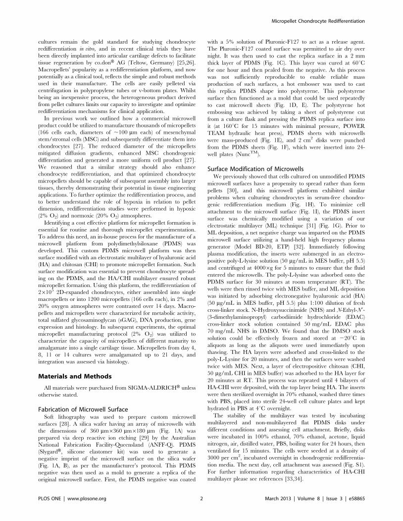
Metabolic Activity, DNA and sGAG ProductionHypoxic micropellets were significantly more metabolically
active than the other pellets over the culture period, as assessed by
alamarBlueH (Fig. 3A). DNA quantification demonstrated that the
proliferation of the cells did not differ significantly in different
conditions (Fig. 3B). To assess the recovery of the chondrogenic
phenotype, the amount of sGAG secreted into the medium over
the culture duration and in the final tissues was quantified. The
amount of the sGAG retained inside the pellet was significantly
higher for the hypoxic macropellets at day 7, 11 and 14 (Fig. 3C).
However, the amount of sGAG released into the media was
highest for the hypoxic micropellets at all time points (Fig. 3D).
The sGAG/DNA ratio was calculated by dividing the total
amount of sGAG produced during the culture to the amount of
DNA measured at the end of the culture. Hypoxic micropellets
had the greatest ratio when compared to other pellets; hypoxic
macropellets also had a significantly higher ratio than normoxic
pellets (Fig. 3E). The amount of total sGAG measured in the
media was higher than the amount measured in the pellets for all
conditions (Fig. 3F). The retained sGAG was the highest for the
hypoxic macropellets, whilst the overall produced sGAG was
greater for the hypoxic micropellets (Fig. 3F).
Chondrogenic and Hypertrophic Gene ExpressionChondrocyte-associated expression of Sox9, aggrecan and
collagen II; and hypertrophy associated expression of Runx2,
collagen I, collagen X, osteocalcin and versican [27] were assessed.
Key matrix genes like aggrecan and collagen II had the highest
expression in the hypoxic micropellets (Fig. 4A, B). Collagen I was
significantly downregulated in macropellets, but remained un-
changed in micropellet cultures relative to day 0 controls (Fig. 4C).
Runx2 (Fig. 4F) expression was greater in normoxic micropellets,
as was collagen X expression in hypoxic micropellets (Fig. 4D).
Sox9 expression was lower in hypoxic macropellets (Fig. 4E).
Macropellets maintained in a normoxic atmosphere had the
highest expression of versican (Fig. 4G) and osteocalcin (Fig. 4F).
Chondrogenic and Hypertrophic Matrix Deposition andDistribution
To visualize the distribution of the ECM molecules within the
macro and micropellets, Alcian blue staining and IF analysis for
collagen I, II and X were performed. DAPI staining was used to
visualize the nuclei (Fig. 5A). Collagen I accumulation was
minimal in all conditions, but appeared even lower in hypoxic
cultures (Fig. 5B). Collagen X was more intense in normoxic
cultures relative to cultures maintained in hypoxic atmospheres
(Fig. 5C). By contrast, collagen II staining was stronger in hypoxic
cultures. Collagen II matrix distribution in hypoxic macropellets
appeared non-uniform, whilst individual micropellets were stained
more homogeneously (Fig. 5D). Alcian blue staining revealed that
sGAG was lower in normoxic macropellets and staining was more
homogeneous in micropellets relative to macropellets (Fig. 5E).
Micropellet Assembly into MacrotissuesTo assess the interaction between individual micropellets, they
were collected from the microwell surface and centrifuged into a
single aggregate at different time points during the chondrogenic
redifferentiation process. Micropellets collected from day 4
cultures that were assembled into larger tissue constructs
integrated in a uniform manner, such that discrete microtissues
were virtually indistinguishable via Alcian blue staining. By
contrast, it was still possible to identify individual micropellets
collected from day 14 cultures that had been assembled into larger
tissue constructs, indicating that full integration had not yet
occurred in these constructs (Fig. 6).
Discussion
A number of strategies are available to facilitate cell aggregate
manufacture (reviewed in [39]), including hanging drop and
various rotary bioreactors. Our group favors microwells, as they
offer an unparalleled capacity to facilitate robust and precise high-
throughput cell aggregate manufacture. Previous studies utilized a
commercial microwell product (AggrewellTM, STEMCELL Tech-
nologies) to efficiently manufacture micropellets [27,30,35].
However, if the unmodified PDMS microwell surface is utilized
directly in micropellet manufacture, the chondrocytes will adhere
to the surface rather than form micropellets (Fig. 1H). In our
studies, modifying the surface to prevent cell attachment further
enhanced the performance of AggrewellTM. Minimizing cell
attachment favoured aggregate formation and facilitated aggregate
harvest. As a cost effective strategy, also to enable full control and
to ease the optimization of the surface properties, we designed and
fabricated our own microwell surface. Figure 1 outlines the
fabrication process that was used to generate PDMS microwell
inserts that fit into 24-well plates. Following surface modification
with the HA/CHI ML (Fig. 1G), the microwell surface enabled
Micropellet Chondrocyte Redifferentiation
PLOS ONE | www.plosone.org 5 March 2013 | Volume 8 | Issue 3 | e58865

robust chondrocyte micropellet formation and harvest (Fig. 1I).
Additionally, the stability of the multilayer was tested, and the
surface modification remained functional even after 24 hour
incubation in 100% ethanol, 70% ethanol, liquid nitrogen, air,
distilled water, PBS or boiling water (Fig. S1).
Other available microwell platforms are also suitable for
chondrocyte micropellet manufacture. For example, the develop-
ment of a microwell platform in which the microwells were cast in
an agarose gel rather than PDMS was recently described [40]. In
this clever strategy, the agarose surface promoted cell aggregate
formation by resisting protein adsorption and subsequent cell
attachment to the agarose surface. However, a more mechanically
robust PDMS microwell platform is compatible with centrifuga-
tion, which enables more rapid and efficient micropellet manu-
facture than the gravity settling method required with an agarose
platform. Additionally, there is comparatively less risk of damaging
the PDMS microwell structure during medium exchange or
culture manipulation processes.
Enhanced sGAG synthesis is critical for effective cartilage tissue
regeneration, as the sGAG content endows cartilage with its
compressive strength [27]. The sGAG outputs in both micropellet
and macropellet cultures were significantly enhanced when these
cultures were maintained in hypoxic atmospheres (Fig. 3E). These
results are consistent with previous studies demonstrating that
hypoxia enhances chondrocyte redifferentiation by stabilizing the
hypoxia inducible factor 1 alpha (HIF1a), which is translocated
into nucleus and activates chondrogenic gene expression [41,42].
However, this is the first study comparing the sGAG production
between micropellets and macropellets, under both hypoxic and
normoxic environments. Consistent greater sGAG production is
observed in hypoxic micropellets, whilst greater sGAG retention
occurs in the hypoxic macropellets (Fig. 3F). The loss of the sGAG
to the medium is a commonly reported challenge in cartilage tissue
engineering, and solutions have been suggested in previous papers
[43,44]. The high surface-area-to-volume ratio in the micropellets
contributes to the significant loss of sGAG to the medium.
However the greater sGAG production and superior gene
expression indicate that the chondrocyte redifferentiation was
enhanced in hypoxic micropellets. If it is possible to improve the
quality of the redifferentiation process, then this short term in vitro
loss of sGAG should be insignificant relative to the overall clinical
benefits. The retention of sGAG would likely be enhanced during
a cartilage repair procedure where a large number of micropellets
would be implanted into a sealed cartilage defect (Fig. 7A).
Consistent with the sGAG results, chondrogenic gene expres-
sion also indicated that the redifferentiation was enhanced in
hypoxic micropellets. No significant difference was observed in
hypoxic micropellet expression profile for collagen I, Sox9, Runx2,
versican and osteocalcin when compared to day 0 measurements
(Fig. 4C, E, F, G, H). The expression of some osteogenic genes was
elevated in the day 0 cultures, and this reflects the fact that the
chondrocytes used in this study were all derived from tissue
discards harvested from elderly patients suffering from severe
osteoarthritis. Whilst collagen X expression was significantly
upregulated in hypoxic micropellets, the overall magnitude of
the expression was very low (0.0012 times) relative to the
expression of housekeeping genes (Fig. 4D). By comparison,
collagen II gene expression was ,10-fold greater in the hypoxic
micropellet cultures than the housekeeping gene expression
(Fig. 4B). Despite having some hypertrophic properties, the
Figure 2. Morphology and size of the pellets. At the end of the 14-day culture, both hypoxic macropellets (A) and micropellets (B) were biggerthan the normoxic pellets. The estimated mean diameter for hypoxic micropellets was 193620 mm (n = 20) whilst the estimated mean diameter ofthe normoxic micropellets was 87610 mm (n = 20). Scale bars: 1 mm.doi:10.1371/journal.pone.0058865.g002
Micropellet Chondrocyte Redifferentiation
PLOS ONE | www.plosone.org 6 March 2013 | Volume 8 | Issue 3 | e58865
hypoxic micropellets exhibit superior redifferentiation when data
are considered cumulatively. The hypoxic micropellets had a
larger volume (Fig. 2B), greater metabolic activity, sGAG
production (Fig. 3A, E) and higher collagen II expression
(Fig. 4B). Importantly, the deposition of the collagen II and
sGAG was more uniform in the micropellets relative to
macropellets (Fig. 5D, E). More uniform cell behavior is consistent
with a smaller diameter pellet with reduced diffusion gradients.
These data mirror our previously reported results indicating that
the micropellet strategy enhanced uniformity in MSC chondro-
genic cultures [27].
Co.donH AG (Teltow, Germany) is currently evaluating the
potential of macropellets for cartilage defect repair in on-going
trials [25,26]. We suggest that there may be legitimate benefits in
using micropellets rather than macropellets, as this should provide
for a more uniform and potent clinical product. Additionally,
because of their smaller geometry, micropellets may be able to
accommodate more complex defect geometries and ultimately
produce a smoother articular surface. This is a rational
expectation, as smaller diameter spheres will always more
uniformly fill a void than larger diameter spheres. A prerequisite
to such applications is that micropellets must demonstrate the
Figure 3. Metabolic activity, growth and sGAG production in pellets. AlamarBlueH graph for metabolic activity (A), DNA quantification (B),sGAG in construct (C) and sGAG in media (D) measurements on days 4, 7, 11, 14. The sGAG/DNA ratio (calculated by dividing the total amount ofsGAG produced during the culture to the amount of DNA measured on day 14) (E) and the total sGAG graph demonstrating the total sGAG in mediaand in construct separately (F).doi:10.1371/journal.pone.0058865.g003
Micropellet Chondrocyte Redifferentiation
PLOS ONE | www.plosone.org 7 March 2013 | Volume 8 | Issue 3 | e58865

Figure 4. Gene expression in pellets. Aggrecan (A), collagen II (B), collagen I (C), collagen X (D), Sox9 (E), Runx2 (F), versican (G), and osteocalcin(H) expressions relative to the geometric mean of housekeeping genes cyclophilin A and GAPDH.doi:10.1371/journal.pone.0058865.g004
Micropellet Chondrocyte Redifferentiation
PLOS ONE | www.plosone.org 8 March 2013 | Volume 8 | Issue 3 | e58865

Micropellet Chondrocyte Redifferentiation
PLOS ONE | www.plosone.org 9 March 2013 | Volume 8 | Issue 3 | e58865

capacity to amalgamate into a contiguous repair tissue. Here we
tested the amalgamation efficiency of cartilage micropellets that
had been cultured for 4, 7, 11 or 14 days. The amalgamation
efficiency depended on the time that the micropellets had been
cultured before assembly (Fig. 6), and the most primitive day 4
micropellets proved the most efficient. This outcome is also
rational, as temporal matrix deposition in the micropellets would
be expected to stabilize with time. These results reflect only short-
term observation, and it may be possible to observe excellent
integration of even day 14 micropellets over an extended time
period. Given that micropellets contain significant matrix content,
we reason that they may be superior to single cell suspensions
lacking any legitimate matrix component, as in procedures like
Autologous Chondrocyte Implantation (ACI). Unlike macropel-
lets, micropellets could easily be injected under ACI-type
membranes, thus facilitating delivery.
Using our microwell system, it is possible to generate 36,000
cartilage micropellets in a single 6-well plate. This manufacturing
efficiency enables the evaluation of micropellets either in the direct
repair of cartilage defects (Fig. 7A), or as a building block in the
in vitro assembly of complex zonal osteochondral tissues (Fig. 7B).
Typically, osteochondral tissues have been made from single cell
suspensions seeded into gels or onto solid scaffolds [45,46,47,48],
although a recent study used spheroids of rabbit MSCs
differentiated into osteoblasts and chondrocytes, which were
subsequently assembled into a zonal tissue [49]. In this study,
the spheroids were manufactured by manually dispensing cells in
collagen droplets. Our more efficient manufacturing process
should enable more sophisticated zonal tissues such as those
shown schematically in Figure 7B. Data from our group indicated
that a similar micropellet manufacturing strategy enabled
enhanced MSC osteogenesis and the generation of bone spheroids
[35] ideal for the assembly of an osteochondral tissue. Ultimately,
the small dimensions of micropellets make them ideal for
identifying the culture conditions necessary to recapitulate the
various zonal tissues found in cartilage [50], and therefore,
micropellets will likely become the preferred building blocks for
reconstructing such tissues.
Figure 5. Cell and matrix localization throughout pellets following 14 days of culture. DAPI staining of nuclei in pellets(A).Immunofluorescence images for collagen I (B), collagen X (C), and collagen II (D), Alcian blue staining for sGAG (E). Scale bars: 100 mm.doi:10.1371/journal.pone.0058865.g005
Figure 6. Hypoxic micropellets assembled into macrotissues. Alcian blue staining for hypoxic micropellets assembled at different time points(indicated days). The total culture duration was 21 days. Scale bars: 100 mm.doi:10.1371/journal.pone.0058865.g006
Micropellet Chondrocyte Redifferentiation
PLOS ONE | www.plosone.org 10 March 2013 | Volume 8 | Issue 3 | e58865

ConclusionHerein we describe the fabrication of a custom microwell system
and a surface modification that enables the efficient manufacture
of thousands of cartilage micropellets. Hypoxic micropellet culture
was shown to be a superior chondrocyte redifferentiation platform,
relative to traditional macropellet cultures. We rationalized that
the micropellets might offer a unique strategy for enhanced
cartilage defect repair, and to investigate this potential the
efficiency of the micropellet amalgamation was examined.
Micropellets that had been cultured for 4–7 days most efficiently
amalgamated, and the composite tissue was nearly seamless at the
end of the 21-day culture. Cumulatively, our results indicate that
the redifferentiation of expanded human articular chondrocytes
can be enhanced using micropellet culture, and that these
micropellets can be assembled into larger more clinically relevant
dimensions.
Supporting Information
Figure S1 Surface modification testing. To assess the
stability and functionality of the surface modification after
incubation in ethanol and PBS, a testing platform was set up as
follows: flat 24 well plate PDMS disks were produced and half of
them were multilayered as explained in the Materials and
Methods section. The disks were incubated under conditions
stated (in 100% ethanol, in 70% ethanol, in acetone, in liquid
nitrogen, in air, in distilled water, in PBS, in boiling water) for 24
hours and the functionality of the surface was assessed by imaging
cell attachment. After 15 minutes of ventilation, cells were seeded
at a density of 3000/cm2, incubated in chondrogenic rediffer-
entiation media overnight. Surface modification was not affected
by any of the conditions. However acetone sensibly decreased the
transparency of the PDMS itself therefore the cell attachment
could not be assessed. For all other conditions the cell spreading
was observed for the surface with no multilayer whereas the cells
were not spreading on the surfaces with multilayer.
(TIF)
Table S1 Primers used for gene expression analysis.
(DOCX)
Author Contributions
Conceived and designed the experiments: BKB TK JJCW RC MRD.
Performed the experiments: BKB PG PL MRD. Analyzed the data: BKB
PG PL WBL TK JJCW RC MRD. Contributed reagents/materials/
analysis tools: RC MRD. Wrote the paper: BKB PG PL WBL TK JJCW
RC MRD.
References
1. Harris JD, Siston RA, Brophy RH, Lattermann C, Carey JL, et al. (2011)
Failures, re-operations, and complications after autologous chondrocyte
implantation–a systematic review. Osteoarthritis Cartilage 19: 779–791.
2. Vavken P, Samartzis D (2010) Effectiveness of autologous chondrocyte
implantation in cartilage repair of the knee: a systematic review of controlled
trials. Osteoarthritis Cartilage 18: 857–863.
3. Lutzner J, Kasten P, Gunther KP, Kirschner S (2009) Surgical options for
patients with osteoarthritis of the knee. Nat Rev Rheumatol 5: 309–316.
4. Perrot S, Menkes CJ (1996) Nonpharmacological approaches to pain in
osteoarthritis. Available options. Drugs 52 Suppl 3: 21–26.
5. Cameron HU, Botsford DJ, Park YS (1997) Prognostic factors in the outcome of
supracondylar femoral osteotomy for lateral compartment osteoarthritis of the
knee. Can J Surg 40: 114–118.
6. Brittberg M, Lindahl A, Nilsson A, Ohlsson C, Isaksson O, et al. (1994)
Treatment of deep cartilage defects in the knee with autologous chondrocyte
transplantation. N Engl J Med 331: 889–895.
7. Gillogly SD, Voight M, Blackburn T (1998) Treatment of articular cartilage
defects of the knee with autologous chondrocyte implantation. J Orthop Sports
Phys Ther 28: 241–251.
8. Kon E, Gobbi A, Filardo G, Delcogliano M, Zaffagnini S, et al. (2009)
Arthroscopic second-generation autologous chondrocyte implantation compared
with microfracture for chondral lesions of the knee: prospective nonrandomized
study at 5 years. Am J Sports Med 37: 33–41.
9. Van Assche D, Staes F, Van Caspel D, Vanlauwe J, Bellemans J, et al. (2010)
Autologous chondrocyte implantation versus microfracture for knee cartilage
injury: a prospective randomized trial, with 2-year follow-up. Knee Surg Sports
Traumatol Arthrosc 18: 486–495.
10. Boeuf S, Richter W (2010) Chondrogenesis of mesenchymal stem cells: role of
tissue source and inducing factors. Stem Cell Res Ther 1: 31.
11. Cherubino P, Grassi FA, Bulgheroni P, Ronga M (2003) Autologous
chondrocyte implantation using a bilayer collagen membrane: a preliminary
report. J Orthop Surg (Hong Kong) 11: 10–15.
12. Darling EM, Athanasiou KA (2005) Rapid phenotypic changes in passaged
articular chondrocyte subpopulations. J Orthop Res 23: 425–432.
13. Holtzer H, Abbott J, Lash J, Holtzer S (1960) The Loss of Phenotypic Traits by
Differentiated Cells in Vitro, I. Dedifferentiation of Cartilage Cells. Proc Natl
Acad Sci U S A 46: 1533–1542.
14. Malda J, Kreijveld E, Temenoff JS, van Blitterswijk CA, Riesle J (2003)
Expansion of human nasal chondrocytes on macroporous microcarriers
enhances redifferentiation. Biomaterials 24: 5153–5161.
15. Yen CN, Lin YR, Chang MDT, Tien CW, Wu YC, et al. (2008) Use of porous
alginate sponges for substantial chondrocyte expansion and matrix production:
Effects of seeding density. Biotechnology Progress 24: 452–457.
16. Malda J, van Blitterswijk CA, Grojec M, Martens DE, Tramper J, et al. (2003)
Expansion of bovine chondrocytes on microcarriers enhances redifferentiation.
Tissue engineering 9: 939–948.
Figure 7. Potential applications of the chondrocyte micropellets. The direct use of chondrocyte micropellets in articular cartilage defectrepair (A). The use of cartilage micropellets in the manufacture of osteochondral tissues in vitro (B).doi:10.1371/journal.pone.0058865.g007
Micropellet Chondrocyte Redifferentiation
PLOS ONE | www.plosone.org 11 March 2013 | Volume 8 | Issue 3 | e58865

17. Banu N, Tsuchiya T (2007) Markedly different effects of hyaluronic acid and
chondroitin sulfate-A on the differentiation of human articular chondrocytes inmicromass and 3-D honeycomb rotation cultures. J Biomed Mater Res A 80:
257–267.
18. Goldberg AJ, Lee DA, Bader DL, Bentley G (2005) Autologous chondrocyteimplantation. Culture in a TGF-beta-containing medium enhances the re-
expression of a chondrocytic phenotype in passaged human chondrocytes inpellet culture. J Bone Joint Surg Br 87: 128–134.
19. Hsieh-Bonassera ND, Wu I, Lin JK, Schumacher BL, Chen AC, et al. (2009)
Expansion and redifferentiation of chondrocytes from osteoarthritic cartilage:cells for human cartilage tissue engineering. Tissue Eng Part A 15: 3513–3523.
20. Imabayashi H, Mori T, Gojo S, Kiyono T, Sugiyama T, et al. (2003)Redifferentiation of dedifferentiated chondrocytes and chondrogenesis of human
bone marrow stromal cells via chondrosphere formation with expressionprofiling by large-scale cDNA analysis. Exp Cell Res 288: 35–50.
21. Tallheden T, Karlsson C, Brunner A, Van Der Lee J, Hagg R, et al. (2004) Gene
expression during redifferentiation of human articular chondrocytes. Osteoar-thritis Cartilage 12: 525–535.
22. Tare RS, Howard D, Pound JC, Roach HI, Oreffo RO (2005) Tissueengineering strategies for cartilage generation–micromass and three dimensional
cultures using human chondrocytes and a continuous cell line. Biochem Biophys
Res Commun 333: 609–621.23. Giovannini S, Diaz-Romero J, Aigner T, Heini P, Mainil-Varlet P, et al. (2010)
Micromass co-culture of human articular chondrocytes and human bonemarrow mesenchymal stem cells to investigate stable neocartilage tissue
formation in vitro. Eur Cell Mater 20: 245–259.24. Croucher LJ, Crawford A, Hatton PV, Russell RG, Buttle DJ (2000)
Extracellular ATP and UTP stimulate cartilage proteoglycan and collagen
accumulation in bovine articular chondrocyte pellet cultures. Biochim BiophysActa 1502: 297–306.
25. Anderer U, Libera J (2002) In vitro engineering of human autogenous cartilage.J Bone Miner Res 17: 1420–1429.
26. Siebold R, Sartory N, Yang Y, Feil S, Paessler HH (2011) Prone position for
minimal invasive or all-arthroscopic autologous chondrocyte implantation at thepatella. Knee Surg Sports Traumatol Arthrosc 19: 2036–2039.
27. Markway BD, Tan GK, Brooke G, Hudson JE, Cooper-White JJ, et al. (2010)Enhanced chondrogenic differentiation of human bone marrow-derived
mesenchymal stem cells in low oxygen environment micropellet cultures. CellTransplant 19: 29–42.
28. Whitesides GM, Ostuni E, Takayama S, Jiang X, Ingber DE (2001) Soft
lithography in biology and biochemistry. Annu Rev Biomed Eng 3: 335–373.29. Fu YQ, Colli A, Fasoli A, Luo JK, Flewitt AJ, et al. (2009) Deep reactive ion
etching as a tool for nanostructure fabrication. Journal of Vacuum Science &Technology B 27: 1520–1526.
30. Cook MM, Futrega K, Osiecki M, Kabiri M, Kul B, et al. (2012)
Micromarrows–three-dimensional coculture of hematopoietic stem cells andmesenchymal stromal cells. Tissue Eng Part C Methods 18: 319–328.
31. Doran MR, Frith JE, Prowse AB, Fitzpatrick J, Wolvetang EJ, et al. (2010)Defined high protein content surfaces for stem cell culture. Biomaterials 31:
5137–5142.32. Bhattacharya S, Datta A, Berg JM, Gangopadhyay S (2005) Studies on surface
wettability of poly(dimethyl) siloxane (PDMS) and glass under oxygen-plasma
treatment and correlation with bond strength. Journal of Microelectromecha-nical Systems 14: 590–597.
33. Bongaerts JH, Cooper-White JJ, Stokes JR (2009) Low biofouling chitosan-
hyaluronic acid multilayers with ultra-low friction coefficients. Biomacromole-cules 10: 1287–1294.
34. Tan GK, Dinnes DL, Butler LN, Cooper-White JJ (2010) Interactions between
meniscal cells and a self assembled biomimetic surface composed of hyaluronicacid, chitosan and meniscal extracellular matrix molecules. Biomaterials 31:
6104–6118.35. Kabiri M, Kul B, Lott WB, Futrega K, Ghanavi P, et al. (2012) 3D
mesenchymal stem/stromal cell osteogenesis and autocrine signalling. Biochem
Biophys Res Commun 419: 142–147.36. Ungrin MD, Joshi C, Nica A, Bauwens C, Zandstra PW (2008) Reproducible,
ultra high-throughput formation of multicellular organization from single cellsuspension-derived human embryonic stem cell aggregates. PLoS One 3: e1565.
37. Liebman J, Goldberg RL (2001) Chondrocyte Culture and Assay. CurrentProtocols in Pharmacology: John Wiley & Sons, Inc.
38. Bookout AL, Mangelsdorf DJ (2003) Quantitative real-time PCR protocol for
analysis of nuclear receptor signaling pathways. Nucl Recept Signal 1: e012.39. Lin RZ, Chang HY (2008) Recent advances in three-dimensional multicellular
spheroid culture for biomedical research. Biotechnol J 3: 1172–1184.40. Moreira Teixeira LS, Leijten JC, Sobral J, Jin R, van Apeldoorn AA, et al.
(2012) High throughput generated micro-aggregates of chondrocytes stimulate
cartilage formation in vitro and in vivo. European cells & materials 23: 387–399.41. Hirao M, Tamai N, Tsumaki N, Yoshikawa H, Myoui A (2006) Oxygen tension
regulates chondrocyte differentiation and function during endochondralossification. J Biol Chem 281: 31079–31092.
42. Coyle CH, Izzo NJ, Chu CR (2009) Sustained hypoxia enhances chondrocytematrix synthesis. J Orthop Res 27: 793–799.
43. Doran MR, Markway BD, Clark A, Athanasas-Platsis S, Brooke G, et al. (2010)
Membrane bioreactors enhance microenvironmental conditioning and tissuedevelopment. Tissue Eng Part C Methods 16: 407–415.
44. Shahin K, Doran PM (2011) Strategies for enhancing the accumulation andretention of extracellular matrix in tissue-engineered cartilage cultured in
bioreactors. PLoS One 6: e23119.
45. Cao Z, Hou S, Sun D, Wang X, Tang J (2012) Osteochondral regeneration by abilayered construct in a cell-free or cell-based approach. Biotechnol Lett 34:
1151–1157.46. Haasper C, Colditz M, Budde S, Hesse E, Tschernig T, et al. (2009) Perfusion
and cyclic compression of mesenchymal cell-loaded and clinically applicableosteochondral grafts. Knee Surg Sports Traumatol Arthrosc 17: 1384–1392.
47. Grayson WL, Bhumiratana S, Grace Chao PH, Hung CT, Vunjak-Novakovic G
(2010) Spatial regulation of human mesenchymal stem cell differentiation inengineered osteochondral constructs: effects of pre-differentiation, soluble factors
and medium perfusion. Osteoarthritis Cartilage 18: 714–723.48. Erisken C, Kalyon DM, Wang H, Ornek-Ballanco C, Xu J (2011)
Osteochondral tissue formation through adipose-derived stromal cell differen-
tiation on biomimetic polycaprolactone nanofibrous scaffolds with gradedinsulin and Beta-glycerophosphate concentrations. Tissue Eng Part A 17: 1239–
1252.49. Cheng HW, Luk KD, Cheung KM, Chan BP (2011) In vitro generation of an
osteochondral interface from mesenchymal stem cell-collagen microspheres.Biomaterials 32: 1526–1535.
50. Klein TJ, Malda J, Sah RL, Hutmacher DW (2009) Tissue engineering of
articular cartilage with biomimetic zones. Tissue engineering Part B, Reviews15: 143–157.
Micropellet Chondrocyte Redifferentiation
PLOS ONE | www.plosone.org 12 March 2013 | Volume 8 | Issue 3 | e58865