NFkB disrupts tissue polarity in 3D by preventing integration of microenvironmental signals
ARTICLE
Received 8 Sep 2014 | Accepted 5 Feb 2015 | Published 13 Mar 2015
The interferon-related developmental regulator 1is used by human papillomavirus to suppress NFkBactivationBart Tummers1, Renske Goedemans1, Laetitia P.L. Pelascini2, Ekaterina S. Jordanova3, Edith M.G. van Esch4,
Craig Meyers5, Cornelis J.M. Melief6, Judith M. Boer7,w & Sjoerd H. van der Burg1
High-risk human papillomaviruses (hrHPVs) infect keratinocytes and successfully evade host
immunity despite the fact that keratinocytes are well equipped to respond to innate and
adaptive immune signals. Using non-infected and freshly established or persistent hrHPV-
infected keratinocytes we show that hrHPV impairs the acetylation of NFkB/RelA K310 in
keratinocytes. As a consequence, keratinocytes display a decreased pro-inflammatory
cytokine production and immune cell attraction in response to stimuli of the innate or
adaptive immune pathways. HPV accomplishes this by augmenting the expression of
interferon-related developmental regulator 1 (IFRD1) in an EGFR-dependent manner.
Restoration of NFkB/RelA acetylation by IFRD1 shRNA, cetuximab treatment or the HDAC1/3
inhibitor entinostat increases basal and induced cytokine expression. Similar observations
are made in IFRD1-overexpressing HPV-induced cancer cells. Thus, our study reveals an
EGFR–IFRD1-mediated viral immune evasion mechanism, which can also be exploited by
cancer cells.
DOI: 10.1038/ncomms7537 OPEN
1 Department of Clinical Oncology, Leiden University Medical Center, Albinusdreef 2, 2333ZA Leiden, The Netherlands. 2 Department of Molecular CellBiology, Leiden University Medical Center, Albinusdreef 2, 2333ZA Leiden, The Netherlands. 3 Center for Gynaecological Oncology, Plesmanlaan 121, 1066CXAmsterdam, The Netherlands. 4 Department of Gynaecology, Leiden University Medical Center, Albinusdreef 2, 2333ZA Leiden, The Netherlands.5 Department of Microbiology and Immunology, The Pennsylvania State University College of Medicine, 500 University Drive, Hershey, Pennsylvania 17033,USA. 6 Department of Immunohematology and Blood Transfusion, Leiden University Medical Center, Albinusdreef 2, 2333ZA Leiden, The Netherlands.7 Department of Human Genetics, Leiden University Medical Center, Albinusdreef 2, 2333ZA Leiden, The Netherlands. w Present addresses: Department ofPediatric Oncology, Erasmus MC—Sophia Children’s Hospital, Wytemaweg 80, 3015CN Rotterdam, The Netherlands; Netherlands Bioinformatics Center,Geert Grooteplein-Zuid 28, 6525GA Nijmegen, The Netherlands. Correspondence and requests for materials should be addressed to S.H.v.d.B.(email: [email protected]).
NATURE COMMUNICATIONS | 6:6537 | DOI: 10.1038/ncomms7537 | www.nature.com/naturecommunications 1
& 2015 Macmillan Publishers Limited. All rights reserved.
High-risk human papillomaviruses (hrHPVs) are absolutelyspecies-specific small double-stranded DNA viruses thatprimarily target undifferentiated keratinocytes (KCs) of
squamous epithelia via micro-wounds and abrasions. HrHPVinfections can last up to 2 years despite viral activity in infectedKCs, the expression of viral antigens and the presence ofKC-expressed pattern recognition receptors (PRRs)1–4 thatshould lead to activation of innate and adaptive immuneresponses. This indicates that hrHPV has evolved mechanismsto transiently evade innate and adaptive immune mechanisms.Ultimately, the majority of hrHPV infections are controlled bythe immune system, in particular by type-1 interferon (IFN)-gand tumour necrosis factor (TNF)-a cytokine-producingT cells5. In case of immune failure, hrHPV causes cancer of theanogenital and/or head and neck regions6.
Upon infection, hrHPV alters the immune-related response ofKCs to various innate and adaptive immune stimuli, resulting inimpaired expression of IFN-stimulated genes, interferon regula-tory transcription factor-induced genes and NFkB-inducedgenes3,7–12, suggesting that HPV hampers STAT1 and NFkBactivation. HPV-infected KCs display downregulated basalexpression of STAT1 and lowered STAT1 protein levels, explain-ing the impaired expression of IFN-stimulated genes13–16.Furthermore, soon after infection HPV upregulates the cellulardeubiquitinase ubiquitin carboxy-terminal hydrolase L1(UCHL1) to impair PRR-induced NFkB activation by upstreaminterference with TRAF3, TRAF6 and NEMO8. The upregulationof UCHL1, however, cannot explain how the virus manages tosuppress the KCs response to adaptive immune signals12. Inaddition, repressing UCHL1 does not fully restore NFkBsignalling via PRR8, suggesting that one or more additionalmechanisms are in play to suppress NFkB signalling.
In this study, we analyse NFkB activation and subsequentcytokine/chemokine production following IFN-g and TNF-astimulation in uninfected and HPV-infected primary KCs. Ourstudy reveals that RelA acetylation, needed for NFkB transcrip-tional activity17, is impaired in hrHPV-infected KCs. The HPV-induced overexpression of the cellular protein interferon-relateddevelopmental regulator 1 (IFRD1) is shown to be instrumentalin this process and involves histone deacetylases (HDACs) 1and/or 3. The augmented expression of IFRD1 is the result ofthe HPV-mediated upregulation of the epidermal growthfactor receptor (EGFR). Blocking of IFRD1 protein expressionby small hairpin RNA (shRNA) or via the anti-EGFR antibodycetuximab restores NFkB/RelA-mediated cytokine expression.Additional data suggest that IFRD1 may have a similar role insuppressing cytokine/chemokine production in HPV-positivecervical cancer cells.
ResultsHrHPV impairs the KCs cytokine response to IFN-c and TNF-a.To evaluate whether the KCs immune response following theexposure to IFN-g and/or TNF-a is attenuated by hrHPV,we utilized a system that resembles the natural infection withhrHPV as closely as possible. Primary KCs stably maintaining thehrHPV genome as episomes (hrHPVþKCs) display similargrowth properties as non-transfected KCs, and upon culture inorganotypic raft cultures, mimic HPV infection in vivo as docu-mented by genome amplification, late gene expression and virusproduction during the differentiation-dependent life cycle ofHPV18–20.
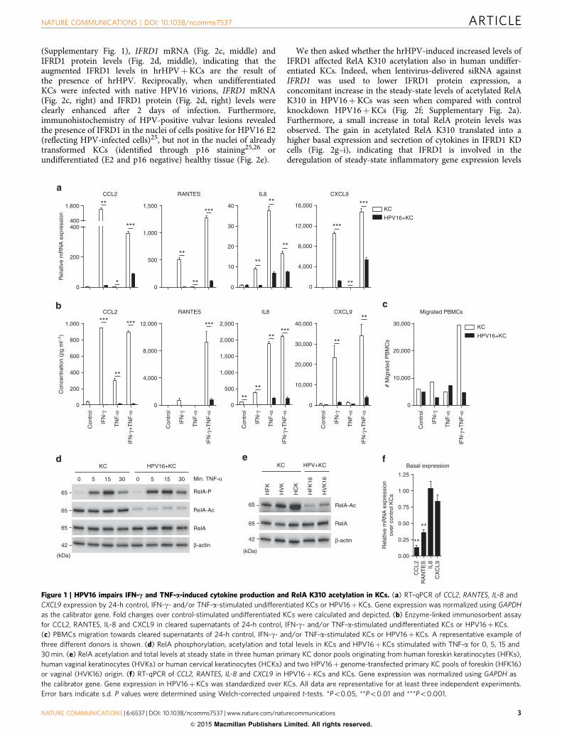
The presence of HPV type 16 (HPV16) was clearly associatedwith an impaired capacity to respond to IFN-g and to TNF-a, asshown by the lower messenger RNA (mRNA) expression andproduction of the IFN-g and/or TNF-a-induced pro-inflammatorycytokines CCL2, RANTES (CCL5), interleukin (IL)-8 and the
chemokines CXCL9, 10 and 11 by KCs (Fig. 1a,b). Not only didthe presence of HPV16 impair the production of cytokines, alsothe migration of peripheral blood mononuclear cells (PBMCs) tosupernatants of IFN-g and TNF-a-stimulated HPV16þKCs wasgreatly impaired (Fig. 1c).
These data suggest that hrHPV, besides impairing theinnate immune response of KCs8, also suppresses the KCsresponse to the adaptive immune signals provided by IFN-g andTNF-a.
The hrHPV-mediated deregulated expression of STAT1(refs 13–16) may explain the impaired cytokine expression byhrHPV-positive KCs upon IFN-g stimulation, but not theimpaired response to TNF-a (IL-8) or to IFN-g and TNF-a(RANTES). Previously, we showed that hrHPV hampersphosphorylation of the NFkB subunit RelA (p65) uponstimulation with the innate PRR ligand poly(I:C)8. As TNF-astimulation rapidly induces the phosphorylation of RelA17, wetested whether hrHPV also hampers rapid TNF-a-induced RelAphosphorylation by stimulating KCs and HPV16þKCs for 0, 5,15 or 30 min with TNF-a. Western blotting showed that RelA wasrapidly phosphorylated similarly in KCs and HPV16þKCs,peaking after 15 min of TNF-a stimulation (Fig. 1d), indicatingthat the impairment of TNF-a-induced responses seen inHPV16þKCs was not due to altered RelA phosphorylationafter short-term TNF-a stimulation. Activated NFkB translocatesto the nucleus where it is modified to regulate its DNA-bindingability and transcriptional activity. Acetylation of the RelAsubunit at lysine 310 (K310) is crucial in this process17.Strikingly, acetylated RelA K310 protein levels were lower inthe HPV16þKCs than in uninfected KCs, both in the absence ofstimulation and after short-term TNF-a stimulation (Fig. 1d).The lowered basal RelA K310 acetylation state was verified inthree independent primary KC and two independentHPV16þKC cultures (Fig. 1e), indicating that HPV hampersthe activity of NFkB already at steady-state levels. This was alsoreflected in a lowered basal cytokine gene expression inunstimulated HPV16þKCs (Fig. 1f).
HrHPV upregulates IFRD1 to impair RelA K310 acetylation.Acetylation of RelA K310 can be regulated by the lysine acetyltransferases (KAT) PCAF (KAT2B), CREBBP (KAT3A), p300(KAT3B) and TIP60 (KAT5), as well as the HDACs 1 and 3(ref. 17). Since our results imply that hrHPV has a mechanismeither to deacetylate or impair the acetylation of RelA, wescreened our validated microarray data12 for genes involved inregulating RelA K310 acetylation. HrHPV did not significantlyinfluence HDAC (HDAC1 to 11) or sirtuin (SIRT1 to 7)expression (Fig. 2a). The only significantly upregulated genewas the lysine acetyl transferase CREBBP (KAT3A), confirmingprevious observations stating that HPV upregulates CREBBP toenhance expression from episomal DNA21,22. However, asCREBBP acetylates RelA its upregulation cannot explain theobserved lower levels of RelA K310 acetylation in hrHPV-infectedKCs under steady-state conditions. Interestingly, the microarraydata also showed the upregulation of IFRD1 (Fig. 2b),which previously was shown to complex HDAC1 (ref. 23) andHDAC3 to RelA causing its deacetylation at lysine 310 in themouse myoblast cell line C2C12 (ref. 24). We hypothesized that itmay fulfil a similar role in human KCs. Therefore, reversetranscriptase (RT)–quantitative PCR (qPCR) and westernblotting was used to confirm that IFRD1 gene expression(Fig. 2c, left) and IFRD1 protein levels (Fig. 2d, left) wereelevated in two independent HPV16þKC cultures. Knockdownof the polycistronic mRNA of HPV16 by a small interfering RNA(siRNA) against HPV16 E2 in HPV16þKCs resulted in thereduction of HPV16 E1, E2, E6 and E7 expression
ARTICLE NATURE COMMUNICATIONS | DOI: 10.1038/ncomms7537
2 NATURE COMMUNICATIONS | 6:6537 | DOI: 10.1038/ncomms7537 | www.nature.com/naturecommunications
& 2015 Macmillan Publishers Limited. All rights reserved.
(Supplementary Fig. 1), IFRD1 mRNA (Fig. 2c, middle) andIFRD1 protein levels (Fig. 2d, middle), indicating that theaugmented IFRD1 levels in hrHPVþKCs are the result ofthe presence of hrHPV. Reciprocally, when undifferentiatedKCs were infected with native HPV16 virions, IFRD1 mRNA(Fig. 2c, right) and IFRD1 protein (Fig. 2d, right) levels wereclearly enhanced after 2 days of infection. Furthermore,immunohistochemistry of HPV-positive vulvar lesions revealedthe presence of IFRD1 in the nuclei of cells positive for HPV16 E2(reflecting HPV-infected cells)25, but not in the nuclei of alreadytransformed KCs (identified through p16 staining25,26 orundifferentiated (E2 and p16 negative) healthy tissue (Fig. 2e).
We then asked whether the hrHPV-induced increased levels ofIFRD1 affected RelA K310 acetylation also in human undiffer-entiated KCs. Indeed, when lentivirus-delivered siRNA againstIFRD1 was used to lower IFRD1 protein expression, aconcomitant increase in the steady-state levels of acetylated RelAK310 in HPV16þKCs was seen when compared with controlknockdown HPV16þKCs (Fig. 2f; Supplementary Fig. 2a).Furthermore, a small increase in total RelA protein levels wasobserved. The gain in acetylated RelA K310 translated into ahigher basal expression and secretion of cytokines in IFRD1 KDcells (Fig. 2g–i), indicating that IFRD1 is involved in thederegulation of steady-state inflammatory gene expression levels
0
200
400400
1,600
CCL2 RANTES
0
500
1,000
1,500
IL8
0
10
20
30
40
CXCL9
0
4,000
8,000
12,000
16,000
Rel
ativ
e m
RN
A e
xpre
ssio
n
KC
HPV16+KC
*
**
***
**
**
***
**
**
**
***
***
**
*** ***
Basal expression
0.00
0.25
0.50
0.75
1.00
1.25
CC
L2
CX
CL9IL8
RA
NT
ES
Rel
ativ
e m
RN
A e
xpre
ssio
nov
er c
ontr
ol K
Cs
**
**
**
0
500
1,000
1,500
2,000
2,500
IL8
0
200
400
600
800
1,000
CCL2
Con
trol
IFN
-γ+
TN
F-α
TN
F-α
IFN
-γ
Con
trol
IFN
-γ+
TN
F-α
TN
F-α
IFN
-γ
Con
trol
IFN
-γ+
TN
F-α
TN
F-α
IFN
-γ
Con
trol
IFN
-γ+
TN
F-α
TN
F-α
IFN
-γ
Con
cent
ratio
n (p
g m
l–1)
KC
HPV16+KC
# M
igra
ted
PB
MC
s
Migrated PBMCs
0
10,000
20,000
30,000
Con
trol
IFN
-γ+
TN
F-α
TN
F-α
IFN
-γ
**
*****
****
0
10,000
20,000
30,000
40,000
CXCL9
**
0
4,000
8,000
12,000
RANTES
***
KC HPV16+KC
0 5 15 30 0 5 15 30
RelA
β-actin
RelA-Ac
RelA-P
Min. TNF-α
65
42
65
65
(kDa)
RelA-Ac
RelA
β-actin
KC HPV+KC
HF
K
HV
K
HC
K
HF
K16
HV
K16
65
65
42
(kDa)
Figure 1 | HPV16 impairs IFN-c and TNF-a-induced cytokine production and RelA K310 acetylation in KCs. (a) RT–qPCR of CCL2, RANTES, IL-8 and
CXCL9 expression by 24-h control, IFN-g- and/or TNF-a-stimulated undifferentiated KCs or HPV16þKCs. Gene expression was normalized using GAPDH
as the calibrator gene. Fold changes over control-stimulated undifferentiated KCs were calculated and depicted. (b) Enzyme-linked immunosorbent assay
for CCL2, RANTES, IL-8 and CXCL9 in cleared supernatants of 24-h control, IFN-g- and/or TNF-a-stimulated undifferentiated KCs or HPV16þKCs.
(c) PBMCs migration towards cleared supernatants of 24-h control, IFN-g- and/or TNF-a-stimulated KCs or HPV16þKCs. A representative example of
three different donors is shown. (d) RelA phosphorylation, acetylation and total levels in KCs and HPV16þKCs stimulated with TNF-a for 0, 5, 15 and
30 min. (e) RelA acetylation and total levels at steady state in three human primary KC donor pools originating from human foreskin keratinocytes (HFKs),
human vaginal keratinocytes (HVKs) or human cervical keratinocytes (HCKs) and two HPV16þ genome-transfected primary KC pools of foreskin (HFK16)
or vaginal (HVK16) origin. (f) RT–qPCR of CCL2, RANTES, IL-8 and CXCL9 in HPV16þKCs and KCs. Gene expression was normalized using GAPDH as
the calibrator gene. Gene expression in HPV16þKCs was standardized over KCs. All data are representative for at least three independent experiments.
Error bars indicate s.d. P values were determined using Welch-corrected unpaired t-tests. *Po0.05, **Po0.01 and ***Po0.001.
NATURE COMMUNICATIONS | DOI: 10.1038/ncomms7537 ARTICLE
NATURE COMMUNICATIONS | 6:6537 | DOI: 10.1038/ncomms7537 | www.nature.com/naturecommunications 3
& 2015 Macmillan Publishers Limited. All rights reserved.
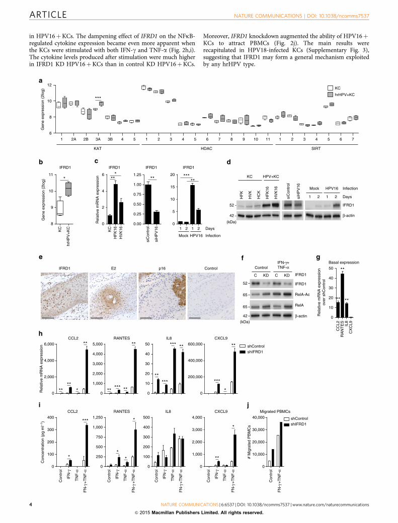
in HPV16þKCs. The dampening effect of IFRD1 on the NFkB-regulated cytokine expression became even more apparent whenthe KCs were stimulated with both IFN-g and TNF-a (Fig. 2h,i).The cytokine levels produced after stimulation were much higherin IFRD1 KD HPV16þKCs than in control KD HPV16þKCs.
Moreover, IFRD1 knockdown augmented the ability of HPV16þKCs to attract PBMCs (Fig. 2j). The main results wererecapitulated in HPV18-infected KCs (Supplementary Fig. 3),suggesting that IFRD1 may form a general mechanism exploitedby any hrHPV type.
IFRD1
8
9
10
11
KC
hrH
PV
+K
C
*
Gen
e ex
pres
sion
(2l
og)
10
12
6
8
1 2A 2B 3A 3B 4 5 1 2 3 4 5 6 7 8 9 10 11 1 2 3 4 5 6 7
KAT SIRTHDAC
KC
hrHPV+KC***
Gen
e ex
pres
sion
(2l
og)
IFRD1 E2 p16 Control
IFRD1
0.00
0.25
0.50
0.75
1.00
1.25
siC
ontr
ol
siH
PV
16
IFRD1
0
2
4
6
Rel
ativ
e m
RN
A e
xpre
ssio
n
HV
K16
HF
K16KC
IFRD1
0
5
10
15
20
Mock Infection
1 2 Days
HPV16
1 2
*** ***
****
CCL2
0
2,000
4,000
6,000
RANTES
0
1,000
2,000
3,000
4,000
5,000
CXCL9
0
200,000
400,000
600,000
IL8
0
10
20
30
40
50 shControlshIFRD1
Rel
ativ
e m
RN
A e
xpre
ssio
n
********
**
** **
**
**
*****
***
**
***
*
Basal expression
0
10
20
30
40
50
CC
L2
CX
CL9IL8
RA
NT
ES
Rel
ativ
e m
RN
A e
xpre
ssio
nov
er s
hCon
trol
**
** **
Con
cent
ratio
n (p
g m
l–1)
Con
trol
IFN
-γ+
TN
F-α
TN
F-α
IFN
-γ
Con
trol
IFN
-γ
TN
F-α
IFN
-γ+
TN
F-α
Con
trol
IFN
-γ
TN
F-α
IFN
-γ+
TN
F-α
Con
trol
IFN
-γ
TN
F-α
FN
-γ+
TN
F-α
Migrated PBMCs
0
10,000
20,000
30,000
40,000 shControlshIFRD1
# M
igra
ted
PB
MC
s
Con
trol
IFN
-γ+
TN
F-α
RANTES
0
250
500
750
1,000
1,250
0
100
200
300
400
500
IL8CCL2
0
100
200
300
400
CXCL9
0
1,000
2,000
3,000
4,000***
**
*
*
**
*
KC HPV+KC
HF
K
HV
K
HC
K
HF
K16
HV
K16
siC
ontr
ol
siH
PV
16
IFRD1
β-actin
Mock HPV16
1 2 1 2
Infection
Days
42
(kDa)
52
IFRD1
ControlIFN-γ+TNF-α
RelA-Ac
RelA
β-actin
C CKD KD IFRD1
42
(kDa)
52
65
65
ARTICLE NATURE COMMUNICATIONS | DOI: 10.1038/ncomms7537
4 NATURE COMMUNICATIONS | 6:6537 | DOI: 10.1038/ncomms7537 | www.nature.com/naturecommunications
& 2015 Macmillan Publishers Limited. All rights reserved.
As the effect of IFRD1 occurred directly at the level of RelA,the influence of IFRD1 on the response of HPV16þKCs topoly(I:C) stimulation, previously shown by us to be impaired inhrHPV-infected KCs8, was also tested. Knockdown of IFRD1resulted in an enhanced expression of CCL2, RANTES, IL-8 andCXCL9 following PRR stimulation with poly(I:C) (SupplementaryFig. 4).
Thus, hrHPV upregulates the expression of IFRD1 soon afterinfection, thereby effectively decreasing the basal levels oftranscriptionally active RelA and as a consequence the levels ofpro-inflammatory cytokines induced via various innate andadaptive immune-mediated NFkB stimulatory pathways.
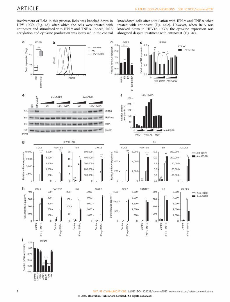
EGFR signalling mediates the increased expression of IFRD1.Growth factors, such as nerve growth factor, fibroblast growthfactor or EGF, have previously been shown to induce theexpression of Tis family genes, which also includes IFRD1, in ratneocortical astrocytes and chromaffin cell line PC12, mouse C243and IEC-18, and mammary epithelial cells27. The hrHPV E5protein is known to affect different aspects of EGFR signallingand expression28. Verification of EGFR expression in our modelshowed that EGFR mRNA expression (Fig. 3a) and membrane-bound protein expression (Fig. 3b) were higher in hrHPVþKCsthan in non-infected KCs. When we transfected complementaryDNA for E2 (as control), E5 or a mix of several other HPVproteins, only E5 enhanced EGFR expression (Fig. 3c). To testwhether EGFR signalling had a similar effect on IFRD1 in humanprimary KCs, the clinically used anti-EGFR antibody cetuximabwas employed to block EGFR signalling. Indeed, IFRD1expression decreased in HPV16þKCs, but not in uninfectedKCs, when treated with cetuximab (Fig. 3d). IFRD1 protein levelsalso decreased dose-dependently in both cetuximab-treated non-infected KCs and HPV16þKCs (Fig. 3e). Notably, the isotypecontrol antibody rituximab (anti-CD20) had no effect (Fig. 3d,e).Thus EGFR signalling does not only induce IFRD1 geneexpression but also stabilizes IFRD1 protein levels. Relativedensity analysis revealed that in cetuximab-treated HPV16þKCsthe protein levels of IFRD1 decreased while concomitantly thelevels of RelA K310 acetylation increased in a dose-dependentfashion. Total RelA levels were unaffected (Fig. 3f). These resultsindicated that the HPV-induced expression of IFRD1 is mediatedvia the EGFR signalling pathway, and implied that cetuximabtreatment may enhance the hrHPVþKCs pro-inflammatorycytokine response to immune stimuli. Indeed, upon IFN-g andTNF-a stimulation cetuximab-treated HPV16þKCs expressedhigher levels of indicated cytokine genes than rituximab-treatedcells (Fig. 3g), as well as higher levels of secreted cytokines
(Fig. 3h). In uninfected KCs, treatment with cetuximab decreasedthe already low levels of IFRD1 protein, and although this led toincreased cytokine gene expression after IFN-g and TNF-a-stimulation, no additional increase in the already high levelsof secreted cytokines was observed (Fig. 3g,h). The absence ofcytokine production in cetuximab-treated HPV16þKC anduninfected KCs that were not stimulated with IFN-g andTNF-a shows that binding of cetuximab to EGFR per se doesnot result in the stimulation of cytokine production (Fig. 3g,h).
As EGFR signalling involves the downstream partners PI3K,mTOR, MEK1, RAF and JNK, we selectively inhibited theseproteins using small-molecule inhibitors in HPV16þKCsand observed that selective inhibition of mTOR (rapamycin),MEK1 (PD98059) and RAF (GW5074), but not PI3K (LY94002)or JNK (SP60025), resulted in decreased expression ofIFRD1 (Fig. 3i). Thus EGFR-mediated upregulation of IFRD1 isfundamental to the impaired NFkB-induced cytokine responseof hrHPV-infected KCs to innate and adaptive immunestimuli.
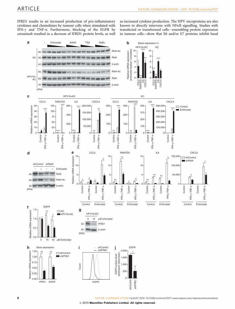
HDAC1/3 inhibition stimulates cytokine production. IFRD1-mediated RelA deacetylation required the recruitment of HDAC1and/or -3 to the RelA–IFRD1 complex in the mouse myoblast cellline C2C12 (ref. 24). To test whether these HDACs played asimilar role in human hrHPVþKCs, the effect of HDACinhibition was tested in HPV16þKCs and non-infected KCs.A dose titration of the HDAC1/3-specific inhibitor entinostat(MS-275), and the prototypic pan-HDAC inhibitors trichostatin A,sodium butyrate (NaBu) and the Food and Drug Administration-approved vorinostat (suberoylanilide hydroxamic acid (SAHA))was performed to study RelA K310 acetylation. All pan-HDACinhibitors increased RelA acetylation in KCs at the lowestconcentration used (Fig. 4a; Supplementary Fig. 2b), but at higherdoses cells suffered from toxic effects as observed by microscopy.However, HPV16þKCs did survive entinostat treatment, andclearly this HDAC1/3 inhibitor increased RelA K310 acetylationin HPV16þKCs (Fig. 4a; Supplementary Fig. 2b). This indicatedthat HDAC1 and/or -3 are indeed specifically involved in thedeacetylation of RelA in hrHPVþKCs. Entinostat treatment ofHPV16þKCs not only restored RelA K310 acetylation but alsoreleased the suppressive effect of IFRD1 on cytokine production.Treated HPV16þKCs displayed a higher basal expression forthree out of four tested cytokines when compared with theiruntreated counterparts (Fig. 4b). Moreover, when stimulated withIFN-g and TNF-a both KCs and HPV16þKCs displayed ahigher expression of CCL2, IL-8 and CXCL9, although theexpression of RANTES was abrogated (Fig. 4c). To confirm the
Figure 2 | HrHPV upregulates IFRD1 to impair RelA K310 acetylation and basal cytokine expression. Microarray intensities for (a) all KATs, HDACs and
SIRTs, and (b) IFRD1 in four independent KCs and four independent hrHPVþKCs represented in a box plot. The box contains the 1st quartile up to the 3rd
quartile; the median is represented as a line; whiskers represent the values of the outer two quartiles. (c) IFRD1 mRNA expression of one representative
control primary KC culture and two HPV16þKC cultures (left panel), in HFK16 cells transfected with siControl or siHPV16 (middle panel) and in primary
KCs that are either mock infected or infected with native HPV16 virions (right panel), as measured by RT–qPCR. (d) IFRD1 protein expression in three
human primary keratinocyte (KC) donor pools originating from human foreskin keratinocytes (HFKs), human vaginal keratinocytes (HVKs) or human
cervical keratinocytes (HCKs) and two HPV16þ genome-transfected primary KC pools of foreskin (HFK16) or vaginal (HVK16) origin (left panel) in HFK16
cells transfected with siControl or siHPV16 (middle panel) and in primary KCs that are either mock infected or infected with native HPV16 virions (right
panel), as measured by western blot. (e) Immunohistochemical staining for IFRD1, HPV16 E2, p16 and negative antibody control of a vulvar intraepithelial
neoplasia (VIN) lesion, one representative donor of two shown. Counterstaining was done using haematoxylin. Scale bar, 500 mm. (f) IFRD1, RelA K310
acetylation and total RelA levels in 24-h non- or IFN-g- and TNF-a-stimulated control or IFRD1 knockdown (KD) HPV16þKCs. (g) RT–qPCR of CCL2,
RANTES, IL-8 and CXCL9 expression in steady-state control or IFRD1 KD HPV16þKCs. (h) RT–qPCR of CCL2, RANTES, IL-8 and CXCL9 expression in 24-h
non- or IFN-g- and/or TNF-a-stimulated control or IFRD1 KD HPV16þKCs. (i) Enzyme-linked immunosorbent assay for CCL2, RANTES, IL-8 and CXCL9 in
cleared supernatants of 24-h non- or IFN-g- and/or TNF-a-stimulated control or IFRD1 KD HPV16þKCs. (j) PBMCs migration towards cleared
supernatants of 24-h non- or IFN-g- and TNF-a-stimulated control or IFRD1 KD HPV16þKCs. A representative example of three different donors is shown.
These data are representative for at least three independent experiments. Error bars indicate s.d. P values were determined using Welch-corrected unpaired
t-tests. *Po0.05, **Po0.01 and ***Po0.001.
NATURE COMMUNICATIONS | DOI: 10.1038/ncomms7537 ARTICLE
NATURE COMMUNICATIONS | 6:6537 | DOI: 10.1038/ncomms7537 | www.nature.com/naturecommunications 5
& 2015 Macmillan Publishers Limited. All rights reserved.
involvement of RelA in this process, RelA was knocked down inHPVþKCs (Fig. 4d), after which the cells were treated withentinostat and stimulated with IFN-g and TNF-a. Indeed, RelAacetylation and cytokine production was increased in the control
knockdown cells after stimulation with IFN-g and TNF-a whentreated with entinostat (Fig. 4d,e). However, when RelA wasknocked down in HPV16þKCs, the cytokine expression wasabrogated despite treatment with entinostat (Fig. 4e).
Con
cent
ratio
n (p
g m
l–1)
Con
cent
ratio
n (p
g m
l–1)
0
50
100
150
200
250HPV16+KC
Rel
ativ
e de
nsity
over
β-a
ctin
(%
)
0 0 0 Anti-EGFR
IFRD1 RelA-Ac RelA
Cou
ntHPV16+KCKC
Unstained
EGFR
EGFR
9
10
11
12
KC
hrH
PV
+K
C
Gen
e ex
pres
sion
(2l
og) ***
IFRD1
0.0
0.5
1.0
1.5
0Anti-EGFR Anti-CD20
Rel
ativ
e m
RN
A e
xpre
ssio
n KCHPV16+KC
**
*** ***
Rel
ativ
e m
RN
A e
xpre
ssio
n
0.00
0.25
0.50
0.75
1.00
1.25IFRD1
Con
trol
DM
SO
PI3
Km
TO
RM
EK
1R
AF
JNK
***
***
***
0
200
400
600
800IL8
0
500
1,000
1,500
2,000
2,500RANTES
0
500
1,000
1,500CCL2
0
1,000
2,000
3,000
4,000
5,000CXCL9
0
200
400
600
CCL2
0
2,000
4,000
6,000
RANTES
0.0
2.5
5.0
7.5
10.0
12.5
IL8
0
50,000
100,000
150,000
200,000
250,000
CXCL9
Rel
ativ
e m
RN
A e
xpre
ssio
n
Con
trol
Con
trol
Con
trol
Con
trol
KC
0
2,500
5,000
7,500
10,000
CCL2
0
500
1,000
1,500
2,000
2,500
RANTES
0
5
10
15
20
IL8
0
100,000
200,000
300,000
400,000
500,000
CXCL9
0
50
100
150
200IL8
0
100
200
300
400
500RANTES
0
100
200
300
400CCL2
0
1,000
2,000
3,000
4,000
5,000CXCL9
Rel
ativ
e m
RN
A e
xpre
ssio
n
Con
trol
Con
trol
Con
trol
Con
trol
HPV16+KC
Anti-CD20Anti-EGFR
Anti-CD20Anti-EGFR
IFN
-γ+
TN
F-α
IFN
-γ+
TN
F-α
IFN
-γ+
TN
F-α
IFN
-γ+
TN
F-α
IF
N-γ
+ T
NF
-α
IFN
-γ+
TN
F-α
IFN
-γ+
TN
F-α
IFN
-γ+
TN
F-α
*** ***
**
***
******
***
** *
***
0.0
0.5
1.0
1.5
2.0
2.5EGFR
Rel
ativ
e m
RN
A e
xpre
ssio
n
Con
trol E2
E5
E1+
E2+
E6+
E7
Anti-CD20Anti-EGFR
HPV16+KC HPV16+KCKC KCHP
V16
+K
C
KC
IFRD1
β-actin
RelA
RelA-Ac
52
42
65
65
(kDa)
ARTICLE NATURE COMMUNICATIONS | DOI: 10.1038/ncomms7537
6 NATURE COMMUNICATIONS | 6:6537 | DOI: 10.1038/ncomms7537 | www.nature.com/naturecommunications
& 2015 Macmillan Publishers Limited. All rights reserved.
Previously, it was shown that HDAC inhibition abrogatesEGFR expression29,30, indicating that EGFR expression isdependent on acetylation events. Indeed, entinostat treatmentdose-dependently abrogated EGFR expression in hrHPVþKCs,but did not influence the expression in KCs (Fig. 4f).Furthermore, entinostat treatment resulted in a reduced level ofIFRD1 protein in hrHPVþKCs (Fig. 4g), which made us wonderwhether IFRD1 could regulate EGFR expression. Therefore,IFRD1 was knocked down in hrHPVþKCs and this resulted inlower EGFR expression (Fig. 4h) and a lower level of membrane-bound EGFR than control-treated hrHPVþKCs (Fig. 4i,j),indicating that IFRD1 can control EGFR expression.
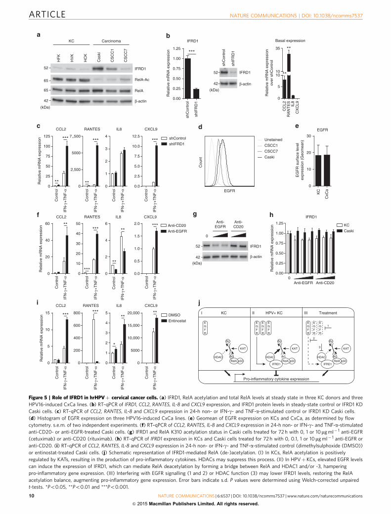
IFRD1 hampers the response of cancer cells to IFN-c and TNF-a.To evaluate whether an increased expression of IFRD1 could alsoplay a role in HPV16-induced squamous cell carcinoma, we ana-lysed the cell line Caski, as well as the two early-passage cervicalcancer cell lines, CSCC1 and CSCC7 (ref. 31). IFRD1 proteinexpression differed between the cell lines (Fig. 5a), but wasincreased in Caski and CSCC1 when compared with normal KCs.RelA K310 acetylation was lower in all three cervical cancer celllines than in uninfected KCs (Fig. 5a; Supplementary Fig. 2c). Forthe Caski and CSCC1 lines this may be explained by the presenceof upregulated IFRD1. However, the lack of RelA K310 acetylationin the CSCC7 line indicates that, besides IFRD1 also othermechanisms can alter the acetylation of RelA K310 in thesesquamous cancer cells.
Because IFRD1 was upregulated in the Caski and CSCC1 cells,we studied the effects of IFRD1 using these cell lines. IFRD1knockdown in the CSCC1 cells did not alter basal cytokineexpression levels (Supplementary Fig. 5a), but IFRD1 knockdownin the Caski cells resulted in a direct increase of the basalexpression levels of CCL2 and RANTES (Fig. 5b). Furthermore,both cell lines showed increased cytokine gene levels upon IFN-gand TNF-a stimulation when IFRD1 was knocked down ascompared with their control knockdown counterparts (Fig. 5c;Supplementary Fig. 5b).
CSCC1 and Caski cells express EGFR (Fig. 5d) at a level that issimilar to that of uninfected KCs (Fig. 5e). However, thedownstream signalling pathway is known to be constitutivelyhigher in HPV-induced cancer cells32. As a consequence, thetreatment of Caski and CSCC1 cancer cells with the anti-EGFRantibody cetuximab resulted in a higher production of IFN-g andTNF-a-induced cytokines than when the cancer cells were treatedwith the control anti-CD20 antibody rituximab (Fig. 5f;Supplementary Fig. 5c). The enhanced response to IFN-g andTNF-a was associated with a concomitant decrease in IFRD1protein levels (Fig. 5g), but not mRNA expression (Fig. 5h), uponEGFR blockade. Similarly, treatment of Caski and CSCC1 cancercells with entinostat resulted in a higher production of CCL2, IL-8and CXCL9 by the cancer cells when stimulated with IFN-g and
TNF-a than dimethylsulphoxide carrier control-treated cells(Fig. 5i; Supplementary Fig. 5d). Congruent with our earlierobservations, RANTES levels diminished after entinostattreatment. These results suggest that IFRD1 may also play arole in suppressing the response of cancer cells to immune stimulisuch as IFN-g and TNF-a.
DiscussionUsing a unique in vitro model, we here show that hrHPVinfection leads to the upregulated expression of endogenousIFRD1 to deregulate the K310 acetylation of NFkB/RelA. As aresult, hrHPV-infected KCs display an impaired production ofpro-inflammatory cytokines and chemokines, and a reducedcapacity to attract immune cells. The increased expression ofIFRD1 in hrHPVþKCs is mediated by EGFR signalling viamTOR, RAF and/or MEK1. Knockdown of IFRD1 with siRNA orindirectly via blockade of EGFR with the clinically used EGFR-specific antibody cetuximab, resulted in decreased IFRD1 mRNAand protein levels, increased NFkB/RelA K310 acetylation andenhanced expression and production of pro-inflammatorycytokines and chemokines by hrHPVþKCs. The use ofentinostat indicated that HDAC1 and/or -3 are involved inlowering K310 acetylation of NFkB/RelA. These conclusions areschematically represented in Fig. 5j.
EGFR activation on epithelial cells has been shown to result ina decreased production of CCL2, RANTES and CXCL10 andincreased production of IL-8. Inhibition of EGFR signalling withblocking antibodies or tyrosine kinase inhibitors can reverse theeffect on these cytokines, as well as result in an increasedepithelial immune infiltrate in vivo33–35. Interestingly, virus-induced EGFR activation has been implicated as a novelmechanism for respiratory viruses to suppress antiviral hostresponses33. The exact underlying mechanism on EGFR-mediated immune suppression remained unclear, albeit thatERK1/2 signalling was shown to be involved in regulatingcytokine production and skin inflammation36. Using the EGFR-blocking antibody cetuximab in the absence of an additionalEGFR stimulus such as the transforming growth factor-a,we found similar effects on the cytokine production ofHPV16þKCs. In KCs, the expression of EGFR and IFRD1 aretightly linked, as EGFR inhibition reduced the expression andprotein levels of IFRD1, via mTOR, RAF and/or MEK1, but notPI3K or JNK. This fits with the involvement of ERK1/2 inregulating cytokine production36, since RAF and MEK1 are justupstream of these kinases. On the basis of our data, the previouslyobserved EGFR activation-induced suppression of cytokineproduction and immune cell infiltration of epithelia can beexplained by upregulation of IFRD1 and subsequent suppressionof NFkB signalling. Our data suggest that EGFR-drivenoverexpression of IFRD1 may also play a role in deregulatingNFkB-signalling in HPV-induced tumour cells. Knockdown of
Figure 3 | Blocking EGFR signalling decreases IFRD1 levels and rescues cytokine production by hrHPVþKCs. (a) Microarray intensities for EGFR in
KCs (n¼4) and hrHPVþKCs (n¼4) represented in a box plot. (b) Histogram of EGFR surface protein expression on KCs and HPV16þKCs, as
determined by flow cytometry. (c) RT–qPCR of EGFR expression in KCs transfected with complementary DNA for E2, E5, E1þ E2þ E6þ E7 or empty
control. (d) RT–qPCR of IFRD1 expression in KCs and HPV16þKCs treated for 72 h with 0, 0.1, 1 or 10mg ml� 1 anti-EGFR or anti-CD20. (e) IFRD1, RelA
K310 acetylation and total RelA levels in KCs and HPV16þKCs treated for 72 h with 0, 0.1, 1 or 10mg ml� 1 anti-EGFR or anti-CD20. (f) Quantified protein
levels of IFRD1, RelA K310 acetylation and RelA over b-actin in HPV16þKCs treated for 72 h with 0, 0.1, 1 or 10mg ml� 1 anti-EGFR (two-dimensional
western blot). The expression levels of the 0mg ml� 1-treated HPVþKCs were set as 100%. (g) RT–qPCR of CCL2, RANTES, IL-8 and CXCL9 expression in
24-h non- or IFN-g- and TNF-a-stimulated, anti-CD20- or anti-EGFR-treated HPV16þKCs (left) and KCs (right). (h) Enzyme-linked immunosorbent assay
for CCL2, RANTES, IL-8 and CXCL9 in cleared supernatants of 24-h non- or IFN-g- and TNF-a-stimulated, anti-CD20- or anti-EGFR-treated HPV16þKCs
(left) and KCs (right). (i) RT–qPCR of IFRD1 expression in HPV16þKCs treated with inhibitors of PI3K (LY94002, 25mM), mTOR (rapamycin, 50 nM),
MEK1 (PD98059, 50mM), RAF (GW5074, 20mM) and JNK (SP60025, 20mM). Gene expression was normalized using GAPDH as the calibrator gene. Fold
changes over control were calculated and depicted. These data are representative for at least three independent experiments, except for h that was
performed once. Error bars indicate s.d. P values were determined using Welch-corrected unpaired t-tests. *Po0.05, **Po0.01 and ***Po0.001.
NATURE COMMUNICATIONS | DOI: 10.1038/ncomms7537 ARTICLE
NATURE COMMUNICATIONS | 6:6537 | DOI: 10.1038/ncomms7537 | www.nature.com/naturecommunications 7
& 2015 Macmillan Publishers Limited. All rights reserved.
IFRD1 results in an increased production of pro-inflammatorycytokines and chemokines by tumour cells when stimulated withIFN-g and TNF-a. Furthermore, blocking of the EGFR bycetuximab resulted in a decrease of IFRD1 protein levels, as well
as increased cytokine production. The HPV oncoproteins are alsoknown to directly intervene with NFkB signalling. Studies withtransfected or transformed cells—resembling protein expressionin tumour cells—show that E6 and/or E7 proteins inhibit basal
Cou
nt
EGFR
shIFRD1shControl
Rel
ativ
e m
RN
A e
xpre
ssio
n
0
10
20
30
40
50
CCL2
0
25
50
75
100
125
RANTES
0
200
400
600
IL8
0
50,000
100,000
150,000
200,000
CXCL9
Con
trol
Con
trol
Con
trol
Con
trol
HPV16+KC
0
100
200
300
CCL2
0
100
200
300
RANTES
0
125
250
375
500
IL8
0
100,000
200,000
300,000
400,000
500,000
CXCL9
Con
trol
Con
trol
Con
trol
Con
trol
ControlEntinostat
KC
IFN
-γ+
TN
F-α
IFN
-γ+
TN
F-α
IFN
-γ+
TN
F-α
IFN
-γ+
TN
F-α
IFN
-γ+
TN
F-α
IFN
-γ+
TN
F-α
IFN
-γ+
TN
F-α
IFN
-γ+
TN
F-α
***
***
***
***
***
**
**
**
*** ***
** ***
******
0
2
4
6
8
10
CC
L2
CX
CL9IL
8R
AN
TE
S
Rel
ativ
e m
RN
A e
xpre
ssio
n(e
ntin
osta
t tre
ated
/ un
trea
ted)
HPV16+KC
0
2
4
6
8
10
CC
L2
CX
CL9IL
8R
AN
TE
S
KC
Basal expression in
**
***
***
***
**
**
0 10 400.0
0.5
1.0
1.5EGFR
KCHPV16+KC
Rel
ativ
e m
RN
A e
xpre
ssio
n
μM Entinostat
***
Rel
ativ
e m
RN
A e
xpre
ssio
n
IFRD1 EGFR0.00
0.25
0.50
0.75
1.00
1.25
Gene expression
shControlshIFRD1
** **
EG
FR
sur
face
leve
lex
pres
sion
(G
eom
ean)
EGFR
0
500
1,000
1,500
2,000
shC
ontr
ol
shIF
RD
1
*
0
5
10
15
CCL2
0
5
10
15
RANTES
0
5
10
15
IL8
0
50,000
100,000
150,000
CXCL9
shRelAshControl
Rel
ativ
e m
RN
A e
xpre
ssio
n
Con
trol
IFN
-γ+
TN
F-α
Con
trol
IFN
-γ+
TN
F-α
Control Entinostat
Con
trol
IFN
-γ+
TN
F-α
Con
trol
IFN
-γ+
TN
F-α
Control Entinostat
Con
trol
IFN
-γ+
TN
F-α
Con
trol
IFN
γ+T
NF
-α
Control Entinostat
Con
trol
IFN
-γ+
TN
F-α
Con
trol
IFN
-γ+
TN
F-α
Control Entinostat
**
* **
**** **
** **
*
*
*
KC
HPV16+KC
RelA-Ac
β-actin
RelA-Ac
β-actin
RelA
RelA
Entinostat SAHA TSA NaBu–
42
(kDa)
65
65
42
65
65
RelA-Ac
RelA
β-actin
– –+ +
shControl shRelA
Entinostat
42
65
65
(kDa)
IFRD1
β-actin
HPV16+KC
0 10 μM entinostat
42
52
(kDa)
ARTICLE NATURE COMMUNICATIONS | DOI: 10.1038/ncomms7537
8 NATURE COMMUNICATIONS | 6:6537 | DOI: 10.1038/ncomms7537 | www.nature.com/naturecommunications
& 2015 Macmillan Publishers Limited. All rights reserved.
and TNF-a-inducible NFkB activity37 by influencing NFkBlocalization38,39 and activation40–43.
Studies in immunosuppressed patients and healthy individualsshow a key role for the adaptive immune response, in particularthat of a strong type-1 (IFN-g and TNF-a)-associated HPV earlyantigen-specific T cells in the protection against progressivedisease5. This notion is sustained by the clinical responses ofpatients treated with HPV-specific therapeutic vaccines5. Amplereasons, therefore, for HPV to also develop strategies preventingKCs to respond to these cytokines. Our data show thatHPV deploys multiple strategies to interfere with inducedRelA-associated NFkB signalling. HPV utilizes the cellulardeubiquitinase UCHL1 to interfere with TRAF3, TRAF6 andNEMO function8, and here we show that HPV also upregulatesthe expression of endogenous IFRD1 to deregulate the K310acetylation of NFkB. Furthermore, the E7 protein of hrHPV hasbeen shown to bind HDAC1 and prevent acetylation of histones,thereby suppressing TLR9 signaling44, but E7 can also displaceHDACs resulting in enhanced hypoxia-inducible factor-1atranscriptional activity45. It is not unusual for viruses to targetNFkB activation46,47, and hampering RelA acetylation is acommon strategy. For instance, the N terminus of the orfvirus protein 002 inhibits acetylation of RelA by blockingphosphorylation of RelA S276 and subsequent recruitment ofacetylases p300 and CBP48, and the A238L protein of the Africanswine fever virus hampers RelA K310 acetylation by inhibitingRelA–p300 interaction49. We here postulate that hrHPV does nothamper KATs in acetylating RelA, but rather recruits a mediatorto enhance HDAC-mediated RelA deacetylation. Together withour observation that HPV lowers basal cytokine expression inresting KCs due to the presence of IFRD1, we suggest thatimpairment of immune-driven RelA-associated NFkB-responsivegene expression is crucial for the virus to persist. This viralstrategy has not been reported before, but as discussed above mayalso be employed by respiratory viruses that activate EGFR33.
All together, our data indicate that HPV upregulates EGFR todrive IFRD1 expression as a tool to decrease basal and adaptive-immune system-driven cytokine expression. This may allowhrHPV to evade the host’s immune response. It is highly likelythat this mechanism plays a role in other viral infections too andeven extends to tumours.
MethodsEthics statement. The use of discarded human foreskin, cervical and vaginal KCtissues to develop cell lines for these studies was approved by the institutionalreview board at the Pennsylvania State University College of Medicine and by theinstitutional review board at Pinnacle Health Hospitals. The Medical EthicalCommittee of the Leiden University Medical Center approved the human tissuesections (healthy foreskin, healthy cervix and HPV16- or 18-positive cervicalneoplasias) used for staining. All sections and cell lines were derived from dis-carded tissues and de-identified, therefore no informed consent was necessary.
Cell culture. Primary cultures of human epithelial KCs were established fromforeskin, vaginal, vulva and cervical tissues, as previously described3, and grown in
KC serum-free medium (Medium 154 supplemented with the HKGS kit,Invitrogen, Breda, The Netherlands). KCs stably maintaining the full episomalHPV genome following electroporation (HPV-positive KCs) were grown inmonolayer culture using E-medium in the presence of mitomycin C (Sigma-Aldrich, Zwijndrecht, The Netherlands)-treated J2 3T3 feeder cells19,20 for twopassages and were then adapted to KC serum-free medium for one passage beforeexperimentation. J2 3T3 mouse fibroblasts, Caski, CSCC1, CSCC7 and SiHacell lines were cultured in Iscove’s modified Dulbecco’s medium (IMDM)supplemented with 8% fetal bovine serum, 2 mM l-glutamine and 1% penicillin–streptomycin (complete IMDM medium) (Gibco-BRL, Invitrogen).
HPV16 infection of non-infected KCs. Primary basal layer human foreskin KCswere seeded at 75,000 cells per well in 24-well plates and allowed to attach for 48 h.Cells received fresh medium (mock infected) or medium containing native HPV16isolated from raft cultures at multiplicity of infection 100 for 24 h. Cells werewashed and harvested for either RT–qPCR or western blotting analysis.
IFRD1 and RelA knockdown in HPV-positive KCs. shRNAs were obtained fromthe MISSION TRC-library of Sigma-Aldrich. The MISSION shRNA clones aresequence-verified shRNA lentiviral plasmids (pLKO.1-puro) provided as frozenbacterial glycerol stocks (Luria broth, carbenicillin at 100 mg ml� 1 and 10%glycerol) in Escherichia coli for propagation and downstream purification of theshRNA clones. pLKO.1 contains the puromycin selection marker for transientor stable transfection. The construct against IFRD1 (NM_001550) wasTRCN0000156194: 50-CCGGCAGTTCTGAAACAGTTTCTTTCTCGAGAAAGAAACTGTTTCAGAACTGTTTTT-30, RelA (NM_021975) was TRCN0000014687:50-CCGGCCTGAGGCTATAACTCGCCTACTCGAGTAGGCGAGTTATAGCCTCAGGTTTTT-30 and the control was: SHC004 (MISSION TRC2-pLKO puroTurboGFP shRNA control vector): 50-CCGGCGTGATCTTCACCGACAAGATCTCGAGATCTTGTCGGTGAAGATCACGTTTTT-30. HPV16-positive KCs atB60% confluence were transduced with lentivirus at multiplicity of infection 5–10overnight, after which the medium was replaced. At least 72 h post transduction,cells were stimulated as indicated and target gene expression was assayed byRT–qPCR or western blotting.
HPV knockdown in HPV-positive KCs. Silencer Select siRNA against HPV16 E2(50-AACACUACACCCAUAGUACAUtt-30) was designed using siRNA TargetFinder software (Ambion, Invitrogen). Blast search revealed that the designed E2siRNA does not match with the known human transcriptome. E2 and negativecontrol #2 siRNA (sequence not provided by the manufacturer) were purchasedfrom Ambion. HPV16þKCs were transfected with 50 nM siRNA E2 or negativecontrol #2 using Lipofectamine 2000 (Invitrogen), according to the manufacturer’sinstructions. Forty-eight hours post transfection, cells were harvested or stimulatedas indicated and target gene expression was assayed by RT–qPCR or westernblotting.
Transfection of HPV genes into non-infected KCs. Non-infected primaryKCs were seeded at 50,000 cells per well in 24-well plates and allowed to attachovernight. Cells were transfected with 500 ng DNA using Lipofectamine(Invitrogen), according to the manufacturer’s instructions. Cells were maintainedin E-medium. Seventy-two hours post transfection, cells were harvested and targetgene expression was assayed by RT–qPCR.
EGFR signalling blocking. Subconfluent cells were cultured in respective completegrowth medium in the presence of cetuximab (0.1, 1 or 10 mg ml� 1; Merck serono),rituximab (0.1, 1 or 10mg ml� 1; Roche), rapamycin (50 nM; Calbiochem),PD98059 (50 mM; Sigma-Aldrich), GW5074 (20 mM; Sigma-Aldrich), LY94002(25 mM; Sigma-Aldrich) or SP60025 (20 mM; Sigma-Aldrich). Medium waschanged every 2–3 days. After at least 72 h, cells were stimulated as indicated andtarget gene expression was assayed by RT–qPCR or western blotting.
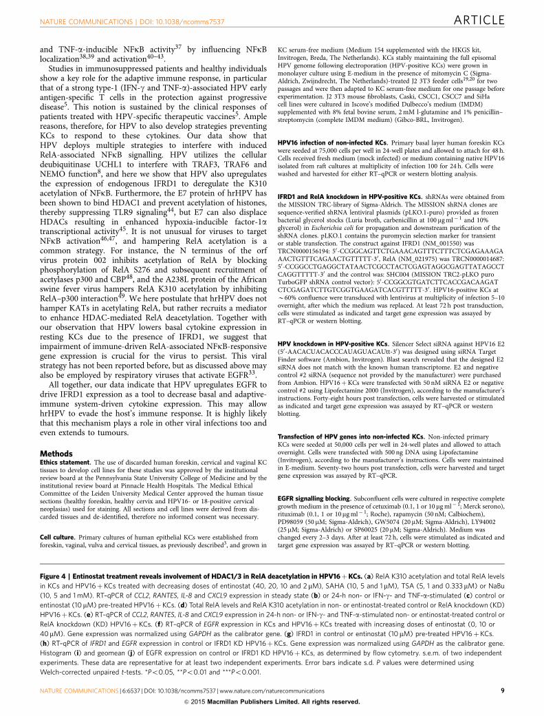
Figure 4 | Entinostat treatment reveals involvement of HDAC1/3 in RelA deacetylation in HPV16þKCs. (a) RelA K310 acetylation and total RelA levels
in KCs and HPV16þKCs treated with decreasing doses of entinostat (40, 20, 10 and 2 mM), SAHA (10, 5 and 1 mM), TSA (5, 1 and 0.333 mM) or NaBu
(10, 5 and 1 mM). RT–qPCR of CCL2, RANTES, IL-8 and CXCL9 expression in steady state (b) or 24-h non- or IFN-g- and TNF-a-stimulated (c) control or
entinostat (10 mM) pre-treated HPV16þKCs. (d) Total RelA levels and RelA K310 acetylation in non- or entinostat-treated control or RelA knockdown (KD)
HPV16þKCs. (e) RT–qPCR of CCL2, RANTES, IL-8 and CXCL9 expression in 24-h non- or IFN-g- and TNF-a-stimulated non- or entinostat-treated control or
RelA knockdown (KD) HPV16þKCs. (f) RT–qPCR of EGFR expression in KCs and HPV16þKCs treated with increasing doses of entinostat (0, 10 or
40mM). Gene expression was normalized using GAPDH as the calibrator gene. (g) IFRD1 in control or entinostat (10 mM) pre-treated HPV16þKCs.
(h) RT–qPCR of IFRD1 and EGFR expression in control or IFRD1 KD HPV16þKCs. Gene expression was normalized using GAPDH as the calibrator gene.
Histogram (i) and geomean (j) of EGFR expression on control or IFRD1 KD HPV16þKCs, as determined by flow cytometry. s.e.m. of two independent
experiments. These data are representative for at least two independent experiments. Error bars indicate s.d. P values were determined using
Welch-corrected unpaired t-tests. *Po0.05, **Po0.01 and ***Po0.001.
NATURE COMMUNICATIONS | DOI: 10.1038/ncomms7537 ARTICLE
NATURE COMMUNICATIONS | 6:6537 | DOI: 10.1038/ncomms7537 | www.nature.com/naturecommunications 9
& 2015 Macmillan Publishers Limited. All rights reserved.
0
10
20
30
EGFR
EG
FR
sur
face
leve
lex
pres
sion
(G
eom
ean)
KC
CxC
a
Cou
nt
CSCC1
Unstained
EGFR
Caski
CSCC7
IFRD1
0.00
0.25
0.50
0.75
1.00
1.25
Rel
ativ
e m
RN
A e
xpre
ssio
n
0Anti-EGFR Anti-CD20
KC
Caski
IFRD1
0.00
0.25
0.50
0.75
1.00
1.25
Rel
ativ
e m
RN
A e
xpre
ssio
n
shC
ontr
ol
shIF
RD
1
***
0
5
1010
35
Basal expression
Rel
ativ
e m
RN
A e
xpre
ssio
nov
er s
hCon
trol
CC
L2
CX
CL9IL
8R
AN
TE
S
**
**
RANTES
0
2,500
5000
7,,500
IL8
0
1
2
3
4
CCL2
0
25
50
75
100
125
Rel
ativ
e m
RN
A e
xpre
ssio
n
CXCL9
0.0
2.5
5.0
7.5
10.0
12.5 shControl
shIFRD1C
ontr
ol
Con
trol
Con
trol
Con
trol
IFN
-γ+
TN
F-α
IFN
-γ+
TN
F-α
IFN
-γ+
TN
F-α
IFN
-γ+
TN
F-α
***
** **
*** ***
CCL2
0
20
40
60
RANTES
0
10
20
30
40
50
IL8
0
2
4
6
Rel
ativ
e m
RN
A e
xpre
ssio
n
CXCL9
0.0
0.5
1.0
1.5
2.0 Anti-CD20
Anti-EGFR
Con
trol
Con
trol
Con
trol
Con
trol
IFN
-γ+
TN
F-α
IFN
-γ+
TN
F-α
IFN
-γ+
TN
F-α
IFN
-γ+
TN
F-α
**
***
***
**
*****
0
5
10
15
CCL2
0
200
400
600
800
RANTES
0
1
2
3
4
5
IL8
0
5000
10,000
15,000
20,000
CXCL9
DMSO
Entinostat
Rel
ativ
e m
RN
A e
xpre
ssio
n
Con
trol
Con
trol
Con
trol
Con
trol
IFN
-γ+
TN
F-α
IFN
-γ+
TN
F-α
IFN
-γ+
TN
F-α
IFN
-γ+
TN
F-α
******
*
**** KC HPV+ KC Treatment
RelA p50
Ac
HDAC
KAT
Ac
EGFR
IFRD1
EGFR
EGFR
RelA p50
Ac
HDAC
KAT
Ac
EGFR
IFRD1
EGFR
EGFR
RelA p50
Ac
HDAC
KAT
Ac
EGFR
I II III
Pro-inflammatory cytokine expression
1
2
3
shIF
RD
1
shC
ontr
ol
IFRD1
β-actin42
52
(kDa)
KC Carcinoma
IFRD1
RelA
β-actin
RelA-Ac
Cas
ki
CS
CC
7
CS
CC
1
HF
K
HC
K
HV
K
42
65
(kDa)
65
52
0
IFRD1
Anti-EGFR
Anti-CD20
β-actin42
52
(kDa)
Figure 5 | Role of IFRD1 in hrHPVþ cervical cancer cells. (a) IFRD1, RelA acetylation and total RelA levels at steady state in three KC donors and three
HPV16-induced CxCa lines. (b) RT–qPCR of IFRD1, CCL2, RANTES, IL-8 and CXCL9 expression, and IFRD1 protein levels in steady-state control or IFRD1 KD
Caski cells. (c) RT–qPCR of CCL2, RANTES, IL-8 and CXCL9 expression in 24-h non- or IFN-g- and TNF-a-stimulated control or IFRD1 KD Caski cells.
(d) Histogram of EGFR expression on three HPV16-induced CxCa lines. (e) Geomean of EGFR expression on KCs and CxCa, as determined by flow
cytometry. s.e.m. of two independent experiments. (f) RT–qPCR of CCL2, RANTES, IL-8 and CXCL9 expression in 24-h non- or IFN-g- and TNF-a-stimulated
anti-CD20- or anti-EGFR-treated Caski cells. (g) IFRD1 and RelA K310 acetylation status in Caski cells treated for 72 h with 0, 1 or 10mg ml� 1 anti-EGFR
(cetuximab) or anti-CD20 (rituximab). (h) RT–qPCR of IFRD1 expression in KCs and Caski cells treated for 72 h with 0, 0.1, 1 or 10mg ml� 1 anti-EGFR or
anti-CD20. (i) RT–qPCR of CCL2, RANTES, IL-8 and CXCL9 expression in 24-h non- or IFN-g- and TNF-a-stimulated control (dimethylsulphoxide (DMSO))
or entinostat-treated Caski cells. (j) Schematic representation of IFRD1-mediated RelA (de-)acetylation. (I) In KCs, RelA acetylation is positively
regulated by KATs, resulting in the production of pro-inflammatory cytokines. HDACs may suppress this process. (II) In HPVþKCs, elevated EGFR levels
can induce the expression of IFRD1, which can mediate RelA deacetylation by forming a bridge between RelA and HDAC1 and/or -3, hampering
pro-inflammatory gene expression. (III) Interfering with EGFR signalling (1 and 2) or HDAC function (3) may lower IFRD1 levels, restoring the RelA
acetylation balance, augmenting pro-inflammatory gene expression. Error bars indicate s.d. P values were determined using Welch-corrected unpaired
t-tests. *Po0.05, **Po0.01 and ***Po0.001.
ARTICLE NATURE COMMUNICATIONS | DOI: 10.1038/ncomms7537
10 NATURE COMMUNICATIONS | 6:6537 | DOI: 10.1038/ncomms7537 | www.nature.com/naturecommunications
& 2015 Macmillan Publishers Limited. All rights reserved.
HDAC inhibition. Subconfluent cells were cultured in presence of a dilution seriesof entinostat (MS-175; 40, 20, 10 and 2 mM; Selleckchem BioConnect), vorinostat(SAHA; 10, 5 and 1 mM; Sigma-Aldrich), trichostatin A (5, 1 and 0.333 mM; Sigma-Aldrich) or sodium butyrate (NaBu; 10, 5 and 1 mM; Sigma-Aldrich) in therespective complete growth medium overnight. Medium was changed for therespective complete growth medium, cells were stimulated as indicated and targetgene expression was assayed by RT–qPCR or western blotting. Since treatmentwith 10 mM entinostat showed a good increase in RelA K310 acetylation withoutsigns of toxicity, subsequent experiments were performed using this dose.
Migration assays. (HPV-positive) KCs were stimulated as indicated for 24 h.Cleared (HPV-positive) KC supernatants were added to the lower compartmentof a transwell plate (Corning). The upper compartment was filled with PBMCsisolated from buffy coats (Sanquin). PBMCs were allowed to migrate for 16 h,after which the cells in the lower compartment were counted by flow cytometry inthe presence of counting beads (Invitrogen), according to the manufacturer’sinstructions. Myeloid cells and lymphocytes were differentiated by their respectivesize in the Forward Scatter (FSC)/Side Scatter (SSC) plot (data not shown).
RNA expression analyses and ELISA. The microarray data12 are accessible in theGene Expression Omnibus database (accession number GSE54181). Plots weregenerated using the webtool R2: microarray analysis and visualization platform(http://r2.amc.nl).
Total RNA was isolated using the NucleoSpin RNA II kit (Machery-Nagel,Leiden, The Netherlands) according to the manufacturer’s instructions. Total RNA(0.5–1.0 mg) was reverse transcribed using the SuperScript III First Strand synthesissystem from Invitrogen. TaqMan PCR was performed using the TaqMan UniversalPCR Master Mix and pre-designed, pre-optimized primers and probe mix forCCL2, RANTES (CCL5), IL-8 (CXCL8), CXCL9 and GAPDH (AppliedBiosystems, Foster City, USA). Threshold cycle numbers (Ct) were determinedusing the CFX PCR System (Bio-Rad, Veenendaal, The Netherlands), and therelative quantities of complementary DNA per sample were calculated using theDDCt method using GAPDH as the calibrator gene.
Enzyme-linked immunosorbent assays (ELISAs) for CCL2, RANTES, IL-8 andCXCL9 were performed according to the manufacturer’s instruction (PeproTech,London, UK).
Flow cytometry. Expression of EGFR on KCs was analysed by flow cytometryusing phycoerythrin (PE)-coupled mouse-anti-human EGFR (1:20, BD Biosciences,Breda, The Netherlands). Per live gate, 50,000 cells were recorded using the BDFACS Calibur with Cellquest software (BD Bioscience) and data were analysedusing Flowjo (Treestar, Olten, Switzerland).
Western blot analysis. For western blotting, polypeptides were resolved bySDS–polyacrylamide gel electrophoresis and transferred to a nitrocellulosemembrane (Bio-Rad). Immunodetection was achieved with anti-p65 (1:1,000,sc-372, Santa Cruz), anti-phospho-p65 (Ser536; 1:1,000, #3033 Cell SignalingTechnology (CST)), anti-acetyl-p65 (Lys310; 1:1,000, #3045 CST), anti-IFRD1(1:400, T2576 Sigma-Aldrich), b-actin (1:10,000, Sigma-Aldrich) primaryantibodies, and horseradish peroxidase-coupled anti-mouse (1:5,000; CST) andhorseradish peroxidase-coupled anti-rabbit (1:5,000, CST) secondary antibodies.Chemoluminescence reagent (Bio-Rad) was used as a substrate and the signal wasscanned using the Chemidoc and accompanying software (Bio-Rad) to quantify theintensity of the bands as a measure of the amount of protein of interest in the blot.The relative amount was determined by calculating the ratio of each proteinover that of the density measured for the housekeeping protein b-actin. Imageshave been cropped for presentation. Full-size images are presented inSupplementary Fig. 6.
Immunohistochemistry. Four mm formalin-fixed, paraffin-embedded tissue sec-tions from two random vulvar intraepithelial neoplasia cases were deparaffinizedand rehydrated using graded concentrations of ethanol to distilled water.Endogenous peroxidise activity was blocked with 0.03% H2O2/MeOH for 20 min.Antigen retrieval was performed in boiling EDTA buffer (pH 9.0) for 12 min. After2 h of cooling down to room temperature, slides were washed twice in distilledwater and twice in PBS. Subsequently, incubation was performed overnight atroom temperature with the primary IFRD1 antibody (T2576 Sigma-Aldrich; 1:500in PBS containing 1% bovine serum albumin); p16 (CINTEC, diluted 1:5) and E2(1:50) (provided by Dr F. Thierry). Second, sections were incubated with Bright-Vision polyhorseradish peroxidase anti-mouse/rabbit/rat immunoglobulin-G(Immunologic BV, Duiven, The Netherlands) for 30 min at room temperature.Washing between incubations was performed three times for 5 min in PBS.Immune complexes were visualized by applying a 0.05-M tris-HCl buffer (pH 7.6)containing 0.05% of 3,30-diamino-benzidine-tetrahydrochloride and 0.0018% ofH2O2. After 10 min, the reaction was stopped by rinsing with demineralized water.Finally, the tissue sections were counterstained with Mayer’s haematoxylin beforeaddition of a coverslip.
Statistical analysis. Statistical analysis was performed using GraphPad InStatversion 3.00. P values were determined using Welch-corrected unpaired t-tests.*Po0.05, **Po0.01 and ***Po0.001.
References1. Doorbar, J. Molecular biology of human papillomavirus infection and cervical
cancer. Clin. Sci. 110, 525–541 (2006).2. Frazer, I. H. Interaction of human papillomaviruses with the host immune
system: a well evolved relationship. Virology 384, 410–414 (2009).3. Karim, R. et al. Human papillomavirus deregulates the response of a cellular
network comprising of chemotactic and proinflammatory genes. PLoS ONE 6,e17848 (2011).
4. Richardson, H. et al. The natural history of type-specific human papillomavirusinfections in female university students. Cancer Epidemiol. Biomarkers Prev. 12,485–490 (2003).
5. van der Burg, S. H. & Melief, C. J. Therapeutic vaccination against humanpapilloma virus induced malignancies. Curr. Opin. Immunol. 23, 252–257 (2011).
6. zur Hausen, H. Papillomaviruses and cancer: from basic studies to clinicalapplication. Nat. Rev. Cancer 2, 342–350 (2002).
7. Hasan, U. A. et al. TLR9 expression and function is abolished by the cervicalcancer-associated human papillomavirus type 16. J. Immunol. 178, 3186–3197(2007).
8. Karim, R. et al. Human papillomavirus (HPV) upregulates the cellulardeubiquitinase UCHL1 to suppress the keratinocyte’s innate immune response.PLoS Pathog. 9, e1003384 (2013).
9. Reiser, J. et al. High-risk human papillomaviruses repress constitutive kappainterferon transcription via E6 to prevent pathogen recognition receptor andantiviral-gene expression. J. Virol. 85, 11372–11380 (2011).
10. Sunthamala, N. et al. E2 proteins of high risk human papillomavirusesdown-modulate STING and IFN-kappa transcription in keratinocytes. PLoSONE 9, e91473 (2014).
11. Termini, L. et al. Characterization of global transcription profile of normal andHPV-immortalized keratinocytes and their response to TNF treatment. BMCMed. Genomics 1, 29 (2008).
12. Tummers, B. et al. CD40-mediated amplification of local immunity byepithelial cells is impaired by HPV. J. Invest. Dermatol. 134, 2918–2927 (2014).
13. Chang, Y. E. & Laimins, L. A. Microarray analysis identifies interferon-inducible genes and Stat-1 as major transcriptional targets of humanpapillomavirus type 31. J. Virol. 74, 4174–4182 (2000).
14. Hong, S., Mehta, K. P. & Laimins, L. A. Suppression of STAT-1 expression byhuman papillomaviruses is necessary for differentiation-dependent genomeamplification and plasmid maintenance. J. Virol. 85, 9486–9494 (2011).
15. Nees, M. et al. Papillomavirus type 16 oncogenes downregulate expressionof interferon-responsive genes and upregulate proliferation-associated andNF-kappaB-responsive genes in cervical keratinocytes. J. Virol. 75, 4283–4296(2001).
16. Zhou, F., Chen, J. & Zhao, K. N. Human papillomavirus 16-encoded E7 proteininhibits IFN-gamma-mediated MHC class I antigen presentation andCTL-induced lysis by blocking IRF-1 expression in mouse keratinocytes. J. Gen.Virol. 94, 2504–2514 (2013).
17. Chen, L. F. & Greene, W. C. Shaping the nuclear action of NF-kappaB. Nat.Rev. Mol. Cell Biol. 5, 392–401 (2004).
18. Conway, M. J. & Meyers, C. Replication and assembly of humanpapillomaviruses. J. Dent. Res. 88, 307–317 (2009).
19. McLaughlin-Drubin, M. E., Christensen, N. D. & Meyers, C. Propagation,infection, and neutralization of authentic HPV16 virus. Virology 322, 213–219(2004).
20. Meyers, C., Mayer, T. J. & Ozbun, M. A. Synthesis of infectious humanpapillomavirus type 18 in differentiating epithelium transfected with viralDNA. J. Virol. 71, 7381–7386 (1997).
21. Lee, D., Lee, B., Kim, J., Kim, D. W. & Choe, J. cAMP response element-bindingprotein-binding protein binds to human papillomavirus E2 protein andactivates E2-dependent transcription. J. Biol. Chem. 275, 7045–7051 (2000).
22. Quinlan, E. J., Culleton, S. P., Wu, S. Y., Chiang, C. M. & Androphy, E. J.Acetylation of conserved lysines in bovine papillomavirus E2 by p300. J. Virol.87, 1497–1507 (2013).
23. Gu, Y. et al. Identification of IFRD1 as a modifier gene for cystic fibrosis lungdisease. Nature 458, 1039–1042 (2009).
24. Micheli, L. et al. PC4/Tis7/IFRD1 stimulates skeletal muscle regeneration and isinvolved in myoblast differentiation as a regulator of MyoD and NF-kappaB.J. Biol. Chem. 286, 5691–5707 (2011).
25. Xue, Y. et al. HPV16 E2 is an immediate early marker of viral infection,preceding E7 expression in precursor structures of cervical carcinoma. CancerRes. 70, 5316–5325 (2010).
26. von Knebel Doeberitz, M., Gissmann, L. & zur Hausen, H. Growth-regulatingfunctions of human papillomavirus early gene products in cervical cancer cellsacting dominant over enhanced epidermal growth factor receptor expression.Cancer Res. 50, 3730–3736 (1990).
NATURE COMMUNICATIONS | DOI: 10.1038/ncomms7537 ARTICLE
NATURE COMMUNICATIONS | 6:6537 | DOI: 10.1038/ncomms7537 | www.nature.com/naturecommunications 11
& 2015 Macmillan Publishers Limited. All rights reserved.
27. Vietor, I. & Huber, L. A. Role of TIS7 family of transcriptional regulators indifferentiation and regeneration. Differentiation 75, 891–897 (2007).
28. Kim, M. K. et al. Human papillomavirus type 16 E5 oncoprotein as a new targetfor cervical cancer treatment. Biochem. Pharmacol. 80, 1930–1935 (2010).
29. Bruzzese, F. et al. HDAC inhibitor vorinostat enhances the antitumor effect ofgefitinib in squamous cell carcinoma of head and neck by modulating ErbBreceptor expression and reverting EMT. J. Cell. Physiol. 226, 2378–2390 (2011).
30. Liu, N. et al. Blocking the class I histone deacetylase ameliorates renal fibrosisand inhibits renal fibroblast activation via modulating TGF-beta and EGFRsignaling. PLoS ONE 8, e54001 (2013).
31. Heusinkveld, M. et al. M2 macrophages induced by prostaglandin E2 and IL-6from cervical carcinoma are switched to activated M1 macrophages byCD4þTh1 cells. J. Immunol. 187, 1157–1165 (2011).
32. Feng, W., Duan, X., Liu, J., Xiao, J. & Brown, R. E. Morphoproteomic evidenceof constitutively activated and overexpressed mTOR pathway in cervicalsquamous carcinoma and high grade squamous intraepithelial lesions. Int. J.Clin. Exp. Pathol. 2, 249–260 (2009).
33. Kalinowski, A. et al. EGFR activation suppresses respiratory virus-inducedIRF1-dependent CXCL10 production. Am. J. Physiol. Lung Cell. Mol. Physiol.307, L186–L196 (2014).
34. Mascia, F., Mariani, V., Girolomoni, G. & Pastore, S. Blockade of the EGFreceptor induces a deranged chemokine expression in keratinocytes leading toenhanced skin inflammation. Am. J. Pathol. 163, 303–312 (2003).
35. Paul, T. et al. Cytokine regulation by epidermal growth factor receptorinhibitors and epidermal growth factor receptor inhibitor associated skintoxicity in cancer patients. Eur. J. Cancer 50, 1855–1863 (2014).
36. Pastore, S. et al. ERK1/2 regulates epidermal chemokine expression and skininflammation. J. Immunol. 174, 5047–5056 (2005).
37. Vandermark, E. R. et al. Human papillomavirus type 16 E6 and E 7 proteinsalter NF-kB in cultured cervical epithelial cells and inhibition of NF-kBpromotes cell growth and immortalization. Virology 425, 53–60 (2012).
38. Caberg, J. H. et al. Increased migration of Langerhans cells in response toHPV16 E6 and E7 oncogene silencing: role of CCL20. Cancer Immunol.Immunother. 58, 39–47 (2009).
39. Havard, L., Rahmouni, S., Boniver, J. & Delvenne, P. High levels of p105(NFKB1) and p100 (NFKB2) proteins in HPV16-transformed keratinocytes:role of E6 and E7 oncoproteins. Virology 331, 357–366 (2005).
40. Avvakumov, N., Torchia, J. & Mymryk, J. S. Interaction of the HPV E7 proteinswith the pCAF acetyltransferase. Oncogene 22, 3833–3841 (2003).
41. Bernat, A., Avvakumov, N., Mymryk, J. S. & Banks, L. Interaction between theHPV E7 oncoprotein and the transcriptional coactivator p300. Oncogene 22,7871–7881 (2003).
42. Huang, S. M. & McCance, D. J. Down regulation of the interleukin-8 promoterby human papillomavirus type 16 E6 and E7 through effects on CREB bindingprotein/p300 and P/CAF. J. Virol. 76, 8710–8721 (2002).
43. Spitkovsky, D., Hehner, S. P., Hofmann, T. G., Moller, A. & Schmitz, M. L. Thehuman papillomavirus oncoprotein E7 attenuates NF-kappa B activation bytargeting the Ikappa B kinase complex. J. Biol. Chem. 277, 25576–25582 (2002).
44. Hasan, U. A. et al. The human papillomavirus type 16 E7 oncoprotein induces atranscriptional repressor complex on the Toll-like receptor 9 promoter. J. Exp.Med. 210, 1369–1387 (2013).
45. Bodily, J. M., Mehta, K. P. & Laimins, L. A. Human papillomavirus E7 enhanceshypoxia-inducible factor 1-mediated transcription by inhibiting binding ofhistone deacetylases. Cancer Res. 71, 1187–1195 (2011).
46. Hiscott, J., Nguyen, T. L., Arguello, M., Nakhaei, P. & Paz, S. Manipulation ofthe nuclear factor-kappaB pathway and the innate immune response by viruses.Oncogene 25, 6844–6867 (2006).
47. Le Negrate, G. Viral interference with innate immunity by preventingNF-kappaB activity. Cell. Microbiol. 14, 168–181 (2012).
48. Ning, Z. et al. The N terminus of orf virus-encoded protein 002 inhibitsacetylation of NF-kappaB p65 by preventing Ser(276) phosphorylation. PloSONE 8, e58854 (2013).
49. Granja, A. G., Sabina, P., Salas, M. L., Fresno, M. & Revilla, Y. Regulation ofinducible nitric oxide synthase expression by viral A238L-mediated inhibitionof p65/RelA acetylation and p300 transactivation. J. Virol. 80, 10487–10496(2006).
AcknowledgementsThis work was supported by the Netherlands Organization for Health Research(NOW/ZonMw) TOP grant 91209012. The funders had no role in study design, datacollection and analysis, decision to publish or preparation of the manuscript.
Author contributionsB.T., R.G., E.S.J., C.J.M.M., J.M.B. and S.H.v.d.B. designed the experiments. B.T., R.G.,L.P.L.P. and E.S.J. performed the experiments. L.P.L.P., E.M.G.M.v.E. and C.M. madeviruses and cells. B.T. and S.H.v.d.B. wrote the paper. C.J.M.M., J.M.B. and S.H.v.d.B.supervised the project. All authors discussed the data.
Additional informationSupplementary Information accompanies this paper at http://www.nature.com/naturecommunications
Competing financial interests: C.M. has received speaker honoraria from Merck,Quest Diagnostics, GSK and Bristol-Myers. C.M. has performed research funded byMerck, The Phillip Morris External Research Program, NexMed, GSK, OriGenixand Interferon Sciences Inc. The remaining authors declare no competing financialinterests.
Reprints and permission information is available online at http://npg.nature.com/reprintsandpermissions/
How to cite this article: Tummers, B. et al. The interferon-related developmentalregulator 1 is used by human papillomavirus to suppress NFkB activation. Nat.Commun. 6:6537 doi: 10.1038/ncomms7537 (2015).
This work is licensed under a Creative Commons Attribution 4.0International License. The images or other third party material in this
article are included in the article’s Creative Commons license, unless indicated otherwisein the credit line; if the material is not included under the Creative Commons license,users will need to obtain permission from the license holder to reproduce the material.To view a copy of this license, visit http://creativecommons.org/licenses/by/4.0/
ARTICLE NATURE COMMUNICATIONS | DOI: 10.1038/ncomms7537
12 NATURE COMMUNICATIONS | 6:6537 | DOI: 10.1038/ncomms7537 | www.nature.com/naturecommunications
& 2015 Macmillan Publishers Limited. All rights reserved.
Copyright © 2022 FDOKUMEN