Attachment-style differences in the ability to suppress negative thoughts: Exploring the neural...
13
Attachment-style differences in the ability to suppress negative thoughts: Exploring the neural correlates i Omri Gillath, a, * Silvia A. Bunge, a Phillip R. Shaver, a Carter Wendelken, a and Mario Mikulincer b a Department of Psychology, University of California, Davis 95616, USA b Department of Psychology, Bar-Ilan University, Ramat Gan 52900, Israel Received 1 December 2004; revised 10 June 2005; accepted 21 June 2005 Available online 8 August 2005 Beginning in infancy, people can be characterized in terms of two dimensions of attachment insecurity: attachment anxiety (i.e., fear of rejection and abandonment) and attachment avoidance (distancing oneself from close others, shunning dependency; Bowlby, J., 1969/1982. Attachment and loss: Vol. 1. Attachment, 2nd ed., Basic Books, New York). The capacity for emotion regulation varies with attachment style, such that attachment-anxious individuals become highly emo- tional when threatened with social rejection or relationship loss, whereas avoidant individuals tend to distance themselves or disengage from emotional situations. In the present study, 20 women participated in an fMRI experiment in which they thought about—and were asked to stop thinking about—various relationship scenarios. When they thought about negative ones (conflict, breakup, death of partner), their level of attachment anxiety was positively correlated with activation in emotion-related areas of the brain (e.g., the anterior temporal pole, implicated in sadness) and inversely correlated with activation in a region associated with emotion regulation (orbitofrontal cortex). This suggests that anxious people react more strongly than non-anxious people to thoughts of loss while under-recruiting brain regions normally used to down-regulate negative emotions. Participants high on avoidance failed to show as much deactivation as less avoidant participants in two brain regions (subcallosal cingulate cortex; lateral prefrontal cortex). This suggests that the avoidant peoples’ suppression was less complete or less efficient, in line with results from previous behavioral experiments. These are among the first findings to identify some of the neural processes underlying adult attachment orientations and emotion regulation. D 2005 Elsevier Inc. All rights reserved. People differ in the ways they form emotional bonds and regulate emotions in close relationships, and the differences have been extensively studied using behavioral methods with people of various ages from birth through early adulthood. We still know relatively little, however, about the neural underpinnings of individual differences in what researchers call ‘‘attachment style’’ (e.g., Shaver and Clark, 1994). The present article reviews some of the behavioral, psychophysiological, and neuroscientific data related to attachment and then describes an initial attempt to use fMRI methods to examine the neural basis of emotion regulation as it relates to attachment style. Attachment theory (Ainsworth et al., 1978; Bowlby, 1969/ 1982, 1973, 1980) concerns the formation of emotional bonds between people and the effects of a person’s attachment history on emotion regulation and other aspects of personality. According to Bowlby (1969/1982), proximity-seeking behavior, beginning early in infancy, is regulated by an innate attachment behavioral system, the function of which is to obtain protection and care from another person, an attachment figure. The system becomes adapted to characteristics of key attachment figures over the life course, and the resulting attachment style (e.g., secure, anxious, or avoidant) becomes relatively stable and can be measured by self-report questionnaires and structured interviews (see Mikulincer and Shaver, 2003 for a review of behavioral studies of attachment processes). These styles of attachment then affect numerous features of adults’ close relationships and emotional experiences. Attachment styles can be assessed in terms of two dimensions of insecurity: attachment-related anxiety and attachment-related avoidance (e.g., Ainsworth et al., 1978, Fig. 10; Brennan et al., 1998; Fraley and Spieker, 2003). People are roughly normally distributed within the space formed by these two orthogonal dimensions. Since 1987, hundreds of studies have shown that a person’s attachment style is a powerful predictor of various psychological and social – relational phenomena including self- and social schemas, self-regulation of stress and emotion, the quality of relations with romantic or marital partners, sexual motivation, and reactions to relationship breakups or losses (see Mikulincer and Shaver, 2003; Shaver and Clark, 1994 for reviews). 1053-8119/$ - see front matter D 2005 Elsevier Inc. All rights reserved. doi:10.1016/j.neuroimage.2005.06.048 i Preparation of this article was facilitated by a grant from the UC Davis Imaging Center. The authors would like to thank Sarah Donohue and Kateri Evans for assistance with data collection and analysis, Shelley Blozis for statistical advice, and Matthew Lieberman for very helpful editorial comments. * Corresponding author. Fax: +1 530 752 2087. E-mail address: [email protected] (O. Gillath). Available online on ScienceDirect (www.sciencedirect.com). www.elsevier.com/locate/ynimg NeuroImage 28 (2005) 835 – 847
-
Upload
independent -
Category
Documents
-
view
0 -
download
0
Transcript of Attachment-style differences in the ability to suppress negative thoughts: Exploring the neural...

www.elsevier.com/locate/ynimg
NeuroImage 28 (2005) 835 – 847
Attachment-style differences in the ability to suppress negative
thoughts: Exploring the neural correlatesi
Omri Gillath,a,* Silvia A. Bunge,a Phillip R. Shaver,a Carter Wendelken,a and Mario Mikulincerb
aDepartment of Psychology, University of California, Davis 95616, USAbDepartment of Psychology, Bar-Ilan University, Ramat Gan 52900, Israel
Received 1 December 2004; revised 10 June 2005; accepted 21 June 2005
Available online 8 August 2005
Beginning in infancy, people can be characterized in terms of two
dimensions of attachment insecurity: attachment anxiety (i.e., fear of
rejection and abandonment) and attachment avoidance (distancing
oneself from close others, shunning dependency; Bowlby, J., 1969/1982.
Attachment and loss: Vol. 1. Attachment, 2nd ed., Basic Books, New
York). The capacity for emotion regulation varies with attachment
style, such that attachment-anxious individuals become highly emo-
tional when threatened with social rejection or relationship loss,
whereas avoidant individuals tend to distance themselves or disengage
from emotional situations. In the present study, 20 women participated
in an fMRI experiment in which they thought about—and were asked
to stop thinking about—various relationship scenarios. When they
thought about negative ones (conflict, breakup, death of partner), their
level of attachment anxiety was positively correlated with activation in
emotion-related areas of the brain (e.g., the anterior temporal pole,
implicated in sadness) and inversely correlated with activation in a
region associated with emotion regulation (orbitofrontal cortex). This
suggests that anxious people react more strongly than non-anxious
people to thoughts of loss while under-recruiting brain regions
normally used to down-regulate negative emotions. Participants high
on avoidance failed to show as much deactivation as less avoidant
participants in two brain regions (subcallosal cingulate cortex; lateral
prefrontal cortex). This suggests that the avoidant peoples’ suppression
was less complete or less efficient, in line with results from previous
behavioral experiments. These are among the first findings to identify
some of the neural processes underlying adult attachment orientations
and emotion regulation.
D 2005 Elsevier Inc. All rights reserved.
1053-8119/$ - see front matter D 2005 Elsevier Inc. All rights reserved.
doi:10.1016/j.neuroimage.2005.06.048
i Preparation of this article was facilitated by a grant from the UC Davis
Imaging Center. The authors would like to thank Sarah Donohue and Kateri
Evans for assistance with data collection and analysis, Shelley Blozis for
statistical advice, and Matthew Lieberman for very helpful editorial
comments.
* Corresponding author. Fax: +1 530 752 2087.
E-mail address: [email protected] (O. Gillath).
Available online on ScienceDirect (www.sciencedirect.com).
People differ in the ways they form emotional bonds and
regulate emotions in close relationships, and the differences have
been extensively studied using behavioral methods with people of
various ages from birth through early adulthood. We still know
relatively little, however, about the neural underpinnings of
individual differences in what researchers call ‘‘attachment style’’
(e.g., Shaver and Clark, 1994). The present article reviews some of
the behavioral, psychophysiological, and neuroscientific data
related to attachment and then describes an initial attempt to use
fMRI methods to examine the neural basis of emotion regulation as
it relates to attachment style.
Attachment theory (Ainsworth et al., 1978; Bowlby, 1969/
1982, 1973, 1980) concerns the formation of emotional bonds
between people and the effects of a person’s attachment history on
emotion regulation and other aspects of personality. According to
Bowlby (1969/1982), proximity-seeking behavior, beginning early
in infancy, is regulated by an innate attachment behavioral system,
the function of which is to obtain protection and care from another
person, an attachment figure. The system becomes adapted to
characteristics of key attachment figures over the life course, and
the resulting attachment style (e.g., secure, anxious, or avoidant)
becomes relatively stable and can be measured by self-report
questionnaires and structured interviews (see Mikulincer and
Shaver, 2003 for a review of behavioral studies of attachment
processes). These styles of attachment then affect numerous
features of adults’ close relationships and emotional experiences.
Attachment styles can be assessed in terms of two dimensions
of insecurity: attachment-related anxiety and attachment-related
avoidance (e.g., Ainsworth et al., 1978, Fig. 10; Brennan et al.,
1998; Fraley and Spieker, 2003). People are roughly normally
distributed within the space formed by these two orthogonal
dimensions. Since 1987, hundreds of studies have shown that a
person’s attachment style is a powerful predictor of various
psychological and social– relational phenomena including self-
and social schemas, self-regulation of stress and emotion, the
quality of relations with romantic or marital partners, sexual
motivation, and reactions to relationship breakups or losses (see
Mikulincer and Shaver, 2003; Shaver and Clark, 1994 for reviews).
1 Many researchers associate the ATP with semantic processing. However
a growing number of studies implicate this region in the experience o
sadness. In the present study we examined ATP activation in relation to sad
thoughts and memories connected with relationship breakups and losses.
O. Gillath et al. / NeuroImage 28 (2005) 835–847836
Secure individuals (those scoring relatively low on both the
anxiety and avoidance dimensions) cope well with stress, either by
seeking support from trusted attachment figures or by calling upon
mental representations of support received in the past (Mikulincer
and Shaver, 2004). Insecure individuals (those scoring relatively
high on either the anxiety or the avoidance dimension, or both), who
do not feel as confident about the availability and responsiveness of
others, are forced to decide between two alternative coping
strategies. Individuals labeled ‘‘anxious’’ by attachment researchers
hyperactivate their ‘‘attachment behavioral system’’ (Bowlby, 1969/
1982), whereas those labeled ‘‘avoidant’’ deactivate the system.
Hyperactivation includes a high degree of emotionality and hyper-
sensitivity to signs of acceptance or rejection. Deactivation includes
suppression or down-regulation of emotion and is accomplished by a
combination of conscious and unconscious processes. (See Cassidy
and Kobak, 1988; Mikulincer and Shaver, 2003, for further
discussion of hyperactivation and deactivation.) These two coping
strategies and their effects on mood and behavior have been
documented in both cross-sectional and longitudinal studies.
Although most studies of adult attachment rely on self-report and
observational methods, some attention has been paid to psychophy-
siological processes as well (see Diamond, 2001 for a review). Most
such studies, however, have relied on surface indicators of
autonomic arousal rather than using brain-imaging techniques
(e.g., Carpenter and Kirkpatrick, 1996; Feeney and Kirkpatrick,
1996; Fraley and Shaver, 1997; Mikulincer, 1998). Recently, there
has been a call for more direct exploration of the neurophysiological
foundations of attachment-related behavior (Diamond, 2001; Hofer,
2003; Schore, 2000). Examination of the neural mechanisms of
attachment would help to answer questions that cannot be fully
answered using cognitive, behavioral, or surface physiological
measures. For example, do attachment-anxious individuals activate
emotions more intensely? Why do they find it difficult to regulate
them? Do avoidant individuals use a particular kind of mental
suppression to keep attachment-related negative emotions out of
awareness? The latter question is particularly interesting because
several recent studies suggest that avoidant suppression strategies are
not as complete or reliable as more secure approaches to emotion
regulation. That is, avoidant suppression is more subject to disruption
by competing mental processes (e.g., Mikulincer et al., 2004).
To date, the few studies that have examined brain correlates of
attachment have focused on laterality differences. Dawson et al.
(2001), using EEG, found that insecurely attached infants exhibited
relatively reduced left frontal brain activity. In preliminary work on
adult emotion regulation (approach and withdrawal tendencies) and
brain laterality, Cohen and Shaver (2004) found that avoidant
individuals made more errors when judging positive attachment-
related words presented to the right hemisphere. A mechanistic
account of these laterality differences, and other attachment-related
cognitive and emotional processing differences, will require in-depth
characterization of attachment-style differences in brain function.
In the study reported here, we used functional magnetic
resonance imaging (fMRI) to explore the neural underpinnings of
emotion-regulation processes identified in previous behavioral
experiments (e.g., Fraley and Shaver, 1997; Mikulincer et al.,
2004). In those experiments, people who were involved in long-
term romantic or marital relationships were asked, first, to vividly
imagine that their partner was leaving them for someone else. After
imagining this stressful experience for a few minutes, participants
were asked to stop thinking about it. Results indicated that, on
average, participants exhibited increased autonomic arousal
(assessed with skin conductance measures) while thinking about
the breakup and loss. When asked to stop thinking about these
negative scenarios, avoidant and secure individuals were able to
down-regulate their negative emotions (as indicated by a reduction
in skin conductance), but anxious individuals were not (Fraley and
Shaver, 1997). Subsequent experiments showed that avoidant
individuals’ ability to down-regulate their emotion in this situation
could be disrupted by adding a cognitive load (remembering a 7-
digit number). In contrast, a cognitive load did not affect secure
individuals’ down-regulation of their emotions. These findings
suggest that suppression is somewhat different for avoidant and
non-avoidant individuals (Mikulincer et al., 2004).
These findings allowed us to make preliminary predictions to be
tested in an fMRI experiment focused on the ability to cope with
attachment-related thoughts and emotions. We expected that, when
avoidant individuals were asked not to think about something, their
pattern of brain activation and deactivation would be different from
the pattern exhibited by non-avoidant (more secure) individuals. The
nature of this difference might help to explain why avoidant, but not
non-avoidant, people experience a rebound of suppressed material
when a cognitive load is added, as if their suppression process was
not reliable or complete. Several brain regions have been associated
in previous studies with regulation and suppression of thoughts and
emotions. These regions include the anterior cingulate cortex (ACC),
orbital frontal cortex (OFC), lateral prefrontal cortex (LPFC), and
more specifically the dorsolateral prefrontal cortex (DLPFC), and the
subcallosal cingulate cortex (SCC) (e.g., Anderson et al., 2004;
Drevets, 2000; Ochsner et al., 2002; Phan et al., 2002; Rolls et al.,
2003;Wyland et al., 2003). These are some of the areas in which we
expected to see ‘‘don’t think vs. think’’ differences as well as
differences between avoidant and non-avoidant people during
thought and emotion suppression.
Turning to the dimension of attachment anxiety, we know that
attachment-anxious individuals become more emotional than their
less anxious counterparts when thinking about breakups and losses.
They also find it difficult to suppress negative thoughts and
emotions even when asked to do so (Fraley and Shaver, 1997;
Mikulincer et al., 2004). Therefore, compared to individuals who
score low on the anxiety dimension, we expected anxious
individuals to exhibit greater activation in brain regions associated
with emotions and less activation in emotion-regulation areas.
Some of the brain regions most consistently associated with
emotional arousal are the amygdala, rostral anterior cingulate
cortex (rACC), dorsal anterior cingulate cortex (dACC), ventro-
medial prefrontal cortex (VMPFC), insula, anterior temporal pole
(ATP), and ventral striatum (for reviews, see Phan et al., 2002,
2004). Not all of these regions are associated with every emotion,
however; and the different regions have different functions related
to experiencing emotion or evaluating emotional states. For
example, the amygdala is strongly associated with calculating
emotional significance (e.g., Hare et al., 2005), whereas the ATP
has been associated with sadness (e.g., Beauregard et al., 2001;
Eugene et al., 2003; Levesque et al., 2003) and the retrieval of
emotional memories (Dolan et al., 2000; Shin et al., 1999).1
We expected attachment anxiety to be positively correlated with
activation in the following regions associated with negative
,
f
O. Gillath et al. / NeuroImage 28 (2005) 835–847 837
emotionality: (1) the amygdala, which is engaged when fear and
anxiety are experienced (e.g., Adolphs, 2004); (2) the ATP, which is
engaged when sadness is experienced (e.g., Blair et al., 1999;
Beauregard et al., 2001; Eugene et al., 2003; Levesque et al., 2003;
Pelletier et al., 2003), especially in a social context (e.g., Calarge et
al., 2003; Ranote et al., 2004); and (3) the ACC (both its rostral and
dorsal subdivisions). The rACC is known to be engaged during
emotion processing (e.g., Bush et al., 2000; Phan et al., 2003), and the
dACC has been associated with subjective distress caused by
physical pain (Rainville et al., 1997), social rejection (Eisenberger
et al., 2003), and neuroticism (Eisenberger et al., in press).
The heightened emotionality of attachment-anxious individuals
might result, not only from heightened emotionality per se, but also
from failure to regulate it. The OFC has been implicated in thought
regulation, particularly in relation to emotional and social stimuli
(e.g., Beer et al., 2004; Miller and Cohen, 2001; Roberts and Wallis,
2000; Rule et al., 2002). Levesque et al. (2003, 2004) reported a
positive correlation between regulation of sadness and OFC
activation in both women and young girls. These authors suggested
that the OFC, through its anatomic projections to the limbic and
paralimbic regions (specifically the ATP), is involved in behavioral
inhibition and voluntary suppression of emotion (see also Cavada et
al., 2000; Mesulam and Mufson, 1982). Thus, we expected attach-
ment anxiety to be negatively correlated with activation in the OFC.
Individual differences related to activation of the ATP and the
OFC have been demonstrated by Eugene et al. (2003) in a study
examining the voluntary expression of sadness. Some study
participants recruited the ATP and the insula (regions associated
with emotional expression) while watching film clips about
bereavement, whereas others recruited OFC and medial PFC. We
therefore expected to find that attachment-anxious people would
have higher activation in the ATP and lower activation in the OFC
when thinking about relationship losses.
Finally, there is considerable evidence indicating that attach-
ment-anxious people tend to ruminate on negative thoughts and
feelings and become overwhelmed by distressing memories. For
example, when asked to recall one negative memory, they tend to
activate many other associated negative memories (Mikulincer and
Orbach, 1995), something not observed in secure or avoidant
individuals. Therefore, we were interested in exploring the
possibility that attachment anxiety would correlate with activation
in the hippocampus, a brain structure known to be important for
associative memory retrieval (e.g., Cohen et al., 1999; Eichen-
baum, 2004). The hippocampus is important for the retrieval of
both emotional and non-emotional memories, including autobio-
graphical memories (e.g., Maguire and Frith, 2003). Our prediction
was that anxious people, who tend to recall previous negative
experiences when thinking about a current negative event, would
retrieve more memories while engaged in our task, which would
involve greater hippocampal activation.
2 The scores on attachment anxiety and avoidance (means on 7-point
scales) for each participant were as follows: 6.00, 2.06; 1.89, 1.03; 4.69,
2.53; 3.92, 1.31; 2.11, 2.19; 5.08, 1.89; 2.22, 5.45; 4.66, 1.25; 3.83, 3.36;
5.92, 2.92; 4.05, 1.39; 2.70, 1; 5.39; 2.80; 4.64, 2.03; 4.58, 2.00; 1.50, 3.81;
2.55, 1.00; 3.64, 1.28; 1.78, 1.22; 1.72, 1.03.
Materials and methods
Participants
Participants were 20 women students from the subject pool at
the University of California, Davis, selected on the basis of their
attachment scores. Each participant had either (1) a relatively high
score on one of the two attachment dimensions (anxiety,
avoidance) and a score close to the median on the other or (2)
low scores on the two dimensions (indicating a relatively secure
attachment style).2 Participants were healthy, right-handed, fluent
English speakers. Their ages ranged from 18 to 25, with a mean
age of 20. None had a prior history of neurological or psychiatric
disorders. All gave written informed consent following instructions
approved by the university’s Institutional Review Board.
Immediately before the fMRI experiment, participants were
given a sample task in which they read a hypothetical scenario
similar to what they would see during the experiment and were
guided through the instructions and the meanings of the different
symbols (traffic lights, as explained below). The structure of a
typical condition in the upcoming experiment (control block,
thinking block, suppressing block, free-thinking block) was
explained and demonstrated using the hypothetical scenario.
Questionnaire measures
In a separate session, several weeks before the experiment took
place, participants completed the Experiences in Close Relation-
ships (ECR) scale (Brennan et al., 1998). Eighteen items on the
scale measured attachment anxiety (e.g., ‘‘I worry about being
abandoned’’) and 18 items measured avoidance (e.g., ‘‘I prefer not
to show a partner how I feel deep down’’). Participants rated the
extent to which each item was descriptive of their experiences in
close relationships on a 7-point scale ranging from ‘‘not at all’’ (1)
to ‘‘very much’’ (7). The reliability and validity of the scales have
been repeatedly demonstrated (e.g., Brennan et al., 1998; Miku-
lincer and Florian, 2000). In the current study, Cronbach alpha
coefficients were high for the anxiety and avoidance scales (0.92
and 0.94, respectively). Neuroticism and general anxiety (i.e., trait
anxiety) were also assessed because these personality traits are
known to be correlated with attachment anxiety and might be
legitimately viewed as offering alternative explanations of our
findings (see Mikulincer and Shaver, 2003 for a review).
Neuroticism was assessed with the 8-item neuroticism subscale
from the Big Five Inventory (BFI; Benet-Martinez and John, 1998).
Participants rated the extent to which an item was self-descriptive
on a 5-point scale ranging from ‘‘disagree strongly’’ (1) to ‘‘agree
strongly’’ (5). Cronbach alpha for the 8-item scale was high (0.87).
Trait anxiety was assessed using Spielberger et al.’s (1983) 20-item
Trait Anxiety Inventory (STAI), on which participants rated
statements on a 4-point Likert scale ranging from ‘‘almost never’’
to ‘‘almost always,’’ with higher scores indicating greater trait
anxiety. Cronbach alpha for the STAI was 0.95.
After the experimental and anatomical scans, participants again
completed the ECR scale, as well as questionnaires relating to
demographics and their relationship history. The correlations
between the pre-study and post-study ECR scores were high (0.83
for anxiety, 0.82 for avoidance). To maximize the reliability of
participants’ attachment scores, the two estimates were averaged.
Behavioral tasks
Participants underwent five MRI scans (9 min, 43 s apiece),
each of which included four task epochs (blocks). The task design

Fig. 1. Order in which the blocks were presented. In the control block,
participants pressed a button every time the yellow light appeared. In the
Think block, they pressed the button whenever they experienced a shift in
thoughts or images. In the Don’t Think block, they pressed the button
whenever they happened to think about the forbidden topic. In the Free-
Thinking block, they pressed the button whenever they happened to think
about the formerly forbidden topic.
O. Gillath et al. / NeuroImage 28 (2005) 835–847838
was based on a prior study of thought suppression (Mitchell et al.,
unpublished manuscript). A green traffic light (see Fig. 1)
instructed participants to think for 2 min about a specific scenario
and press a response button whenever they had a new thought or
image related to the scenario (henceforth referred to as the Think
block). A red traffic light instructed participants to try not to think
about the previous scenario but to press the response button
whenever they did happen to think about it (Don’t Think block). A
green traffic light with the number F2_ superimposed on it
instructed participants to think about whatever came to mind and
press the response button if they happened to think about the
previously excluded topic. Finally, a yellow traffic light meant
simply that participants should press the response button. This
yellow light block served as a visual and motor control for the
other blocks. Participants indicated a change in thoughts (in the
‘‘thinking’’ conditions) or thinking about the forbidden topic (in the
‘‘don’t think’’ conditions) by pressing a button with their right
hand. Each block (120 s) occurred only once per scan. Results for
the Think and Don’t Think blocks are reported here.
The instructions for each block were presented for 20 s
followed by a traffic light for 120 s. For example, just before the
first Think block participants read the following instructions: ‘‘In
the next 2 min (during which you will see a green traffic light with
no number in it), please imagine as vividly as you can that you are
driving alone to Lake Tahoe. Please press the button whenever a
major shift in topic, image, scene, or activity occurs.’’ Each of the
five scenarios was either emotionally neutral, neutral but relation-
ship-related, or emotionally negative and relationship-related. The
order of the scenarios was the same for all participants. The second
scenario involved remembering or imagining a neutral activity
engaged with the participant’s romantic partner, such as shopping
in a mall or supermarket. The third scenario involved remembering
or imagining a conflict or argument with the romantic partner. The
fourth scenario involved remembering or imagining breaking up
with the partner. The fifth scenario involved imagining that their
partner died. At the end of each scan, participants were given a 30-
s rest break. At the end of the fifth scan, they underwent a 10-min
anatomical scan. They then rested and completed a series of
questionnaires and were debriefed and asked about their thoughts
and methods of suppression. Presentation software (Neurobeha-
vioral Systems Inc., San Francisco, CA, USA) was used to present
the traffic lights and instructions synchronously with fMRI data
acquisition. Participants viewed the stimuli on a screen via a mirror
in the head coil of the scanner.
fMRI data acquisition and analysis
Brain images were acquired with a 1.5 T GE Signa scanner at the
UC Davis Imaging Research Center. Head motion was minimized
with comfortable padding around the participant’s head. Functional
images were acquired with gradient-recalled echo EPI sensitive to
the blood oxygen level dependent (BOLD) contrast (TR= 2.5 s, TE =
40 ms, 24 contiguous 4 mm oblique axial slices parallel to the AC–
PC line). After the experimental scans, high-resolution anatomical
images were collected for each participant (TR = 12ms, TE = 4.5ms,
voxel dimensions = 1 � 1 � 1 mm).
Data were preprocessed with SPM2 (Wellcome Department of
Cognitive Neurology, London, UK) on the Matlab 6.5.1 platform
(Mathworks Inc., Natick, MA). The first five brain volumes of each
scan were discarded from the analysis to eliminate nonequilibrium
effects of magnetization. The remaining 224 volumes were used for
the subsequent analyses. Images were corrected for differences in
timing of slice acquisition, and were then submitted to rigid body
motion correction. Structural and functional volumes were spatially
normalized to T1 and EPI templates in SPM, respectively. Templates
are based on theMNI305 stereotaxic space (Cocosco et al., 1997), an
approximation of Talairach space based on the Talairach and
Tournoux (1988) atlas. The normalization algorithm consisted of a
12-parameter affine transformation together with a nonlinear trans-
formation involving cosine basis functions, and resampled the
volumes to 2 � 2 � 2 mm cubic voxels. Functional volumes were
spatially smoothed with an 8-mm FWHM isotropic Gaussian kernel.
Statistical analyses were performed on individual participants’
data with the general linear model implemented in SPM2. The
fMRI time series data were modeled as a series of epochs
convolved with a canonical hemodynamic response function. The
resulting functions were used as covariates in a general linear
model, along with a basis set of cosine functions that high-pass
filtered the data, as well as a covariate for session effects. The data
were high-pass filtered with the maximal frequency of task
alternation (280 s) to cut off baseline drifts and low-pass filtered
with a hemodynamic response function to control for temporal
autocorrelation. The least-squares parameter estimates of height of
the best-fitting synthetic HRF for each condition were used in pair-
wise contrasts, and the resulting contrast images, computed on a
subject-by-subject basis, were submitted to group analyses. At the
group level, contrasts between conditions were computed by
performing one-tailed t tests on these images, treating participants
as a random effect. Task-related responses were generally
considered significant if they consisted of at least 5 contiguous
voxels that exceeded an uncorrected threshold of P < 0.001,
although in a few cases, when exploring details of individual
differences, we used a more liberal threshold, P < 0.005.
Region-of-interest (ROI) analyses were performed with the
MarsBar toolbox in SPM2 (Brett et al., 2002; http://marsbar.
sourceforge.net/). We used functional ROIs for targeted regions
identified in contrasts of interest: namely, the ATP, ACC, hippo-
campus, and SCC. Additionally, we used anatomical ROIs
provided by MarsBar for regions that were of interest but not
identified in the whole-brain contrasts: OFC (BA 11), LPFC (BA
9), and the amygdala. Mean contrast values for each participant
and condition were extracted for each ROI and submitted to a
repeated measures analysis and post-hoc comparisons. The goal of
this exploratory study was to identify candidate regions that might
be differentially engaged by individuals who differed in attachment
style. Thus, a number of ROIs were examined (9 total), and the

O. Gillath et al. / NeuroImage 28 (2005) 835–847 839
resulting ROI analyses were not corrected for multiple compar-
isons. In the whole-brain analyses, we focused on four key
contrasts: Don’t Think > Think, Don’t Think Relationship > Think
Relationship, Don’t Think Negative > Think Negative, and Think
Negative > Think Neutral.
Statistical models
Regression analyses were used to examine associations
between signal intensity in various brain regions and scores on
the two attachment-style dimensions. We conducted another set of
regression analyses predicting activation in various brain regions
from the average number of button presses in each block. When
assessing statistical relations between different brain regions, we
examined only relations that made sense anatomically (Buchel and
Friston, 1997). Based on anatomical studies in non-human
primates, we assumed that the ACC, OFC, and the ATP are
interconnected in humans (e.g., Cavada et al., 2000; Mesulam and
Mufson, 1982; Moran et al., 1987; Morecraft et al., 1992).
Results
Questionnaire measures
Attachment anxiety and avoidance were uncorrelated (r =
0.034, P = 0.88), as intended. As expected, there was a positive
correlation between attachment anxiety and trait anxiety (r = 0.46,
P < 0.05) and a trend toward a positive correlation between
attachment anxiety and neuroticism (r = 0.42, P = 0.065).
Neuroticism and trait anxiety were highly correlated (r = 0.81,
P < 0.001).
Behavioral data
Participants made fewer button presses in the not-thinking con-
ditions than in the thinking conditions overall (t(1,19) = 5.52, P <
0.001), in line with our instructions. Otherwise, the amount of button
pressing did not differ significantly across the different scenarios and
was not correlated with either attachment-style dimension. Never-
theless, there were small correlations within conditions compatible
with previous studies (e.g., Fraley and Shaver, 1997; Mikulincer et
al., 2004): i.e., more button presses for more anxious participants in
the rebound, or Free Thought (green light with a ‘‘2’’ superimposed
Fig. 2. Neural correlates of thought suppression revealed at the group level. Th
MPFC (b).
on it), conditions, and fewer button presses for the more avoidant
participants. The failure of these trends to reach statistical signifi-
cance may be due to the small sample size (in contrast, for example,
Fraley and Shaver, 1997 studied the reactions of 200 people, and
Mikulincer et al., 2004 studied the reactions of 120 people).
Neuroimaging data
Whole-brain comparisons
Our study combines two lines of research, the first dealing with
individual differences in attachment style and their effects on
emotion regulation and cognitive control and the other dealing with
neural correlates of thought suppression. Although our main
interest was attachment style, we began our analyses at the level
of the entire group of 20 participants to see whether our
suppression-related findings were in line with previous findings.
In these group-level analyses, thought suppression was associated
with higher activation in a region known to be related to control,
conflict, and self-regulation: the dorsal ACC (see Fig. 2a; for
review of relevant studies, see Botvinick et al., 2004). Specifically,
participants exhibited greater activation in this region when trying
not to think about a given scenario (collapsing across the five
conditions that involved ‘‘not thinking’’: neutral alone, neutral with
partner, conflict with partner, breakup, and partner’s death) than
when ‘‘thinking’’ about that same scenario (t(1,19) = 4.92, P <
0.001). Activation while suppressing was also found in the MPFC
(Fig. 2b) (t(1,19) = 4.32, P < 0.001), a region known to be
associated with self-related processing and monitoring (e.g.,
Gusnard et al., 2001; Johnson et al., 2002; Kelley et al., 2002).
Similar results were achieved when only attachment-related
conditions were included in the comparison (ACC: t(1,19) = 4.37,
P < 0.001; MPFC: t(1,19) = 4.13, P < 0.001) or when only
negative conditions were included in it (ACC: t(1,19) = 4.19, P <
0.001; MPFC: t(1,19) = 3.36, P = 0.002). In summary, the group-
level analyses revealed increased activation during thought
suppression in several midline PFC structures, compatible with
previous studies (Gusnard et al., 2001; Johnson et al., 2002;
Kelley et al., 2002; Wyland et al., 2003).
Correlations with avoidance
We hypothesized that avoidant participants, as compared with
non-avoidant ones, would have a different pattern of activation
related to not thinking about a particular topic because, in behavioral
studies, such people seem to retain some activation or availability of
e Don’t Think > Think contrast revealed activation in the ACC (a) and
O. Gillath et al. / NeuroImage 28 (2005) 835–847840
loss-related thoughts even when some indexes (e.g., skin conduc-
tance) indicate that they have successfully suppressed them.
To test this prediction, we first examined whether avoidance is
correlated with the pattern of activation noted in the whole-brain
comparisons described above. We started by regressing the contrast
‘‘Don’t Think–Think’’ (collapsed across the five scenarios) on
avoidance. This analysis revealed a positive association between
avoidance and activation in the subcallosal cingulate cortex (SCC
(0, 22, �2); t(1,19) = 3.21, P = 0.002; see Fig. 3a). After looking at
the whole-brain results, we examined the way in which attachment
avoidance correlated with activation in the SCC while controlling
for attachment anxiety (an unlikely confound but one we wished to
rule out completely). In the first step of the regression analysis, we
entered attachment anxiety, which turned out not to be significantly
associated with relative activation in the SCC. In the second step,
we entered attachment avoidance, confirming that relative activa-
tion in the SCC during ‘‘not thinking’’ was positively associated
with avoidance (b = 0.54, t(2,17) = 2.70, P = 0.015).
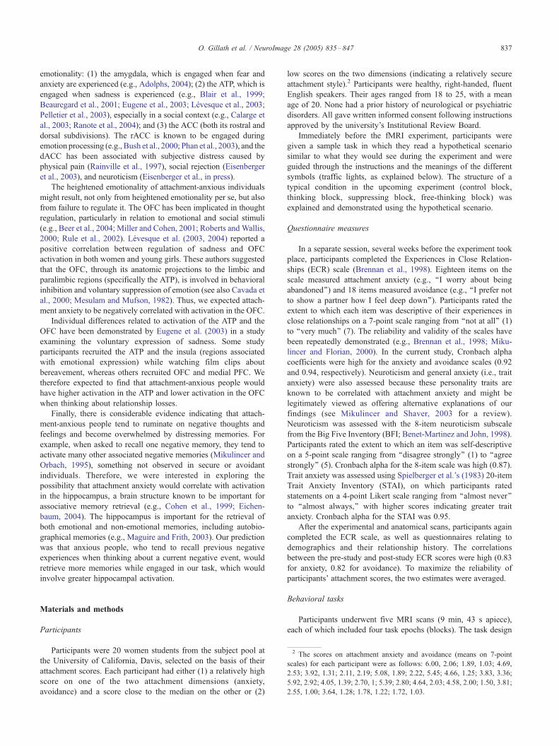
Fig. 3. Avoidant attachment was positively correlated with thought suppression (D
attachment and Don’t Think vs. Think; the Y-axis in Panel (b) represents the co
condition (thinking about driving alone). A similar pattern was revealed for the s
between avoidant attachment and ‘‘Don’t Think Relationships vs. Think Relations
(Think relationship or Don’t think relationships) and the control condition (think
To better understand the role of the SCC in the suppression
process and to examine the differences in its activation between
avoidant and non-avoidant individuals, we conducted a repeated
measures analysis of variance, in which the ‘‘Don’t Think’’ and
‘‘Think’’ conditions were treated as a repeated measure and
avoidance was treated as a predictor. The analysis revealed a
significant interaction between avoidance and the ‘‘Don’t Think–
Think’’ contrast (F(1,17) = 5.64, P < 0.05; see Fig. 3b). Tests for
simple main effects revealed that, among participants scoring low
on avoidance, not thinking was associated with lower SCC
activation than thinking (Don’t Think: M = �0.68, SD = 0.27;
Think: M = �0.22, SD = 0.18; F(1,9) = 6.03, P < 0.05). No such
relative deactivation in the SCCwas evident among individuals who
scored high on avoidance (M =�0.09, SD = 0.20 for thinking;M =
�0.07, SD = 0.25 for not thinking; F < 1). Thus, non-avoidant
participants tended to increase deactivation in the SCC when not
thinking, whereas avoidant people did not. This suggests that, while
everyone activated the same regions (ACC and MPFC) to suppress
on’t Think > Think) in the SCC (BA 25) (a). Interaction between avoidant
ntrast between each task condition (Think or Don’t think) and the control
uppression of relationship-related thoughts. Panel (c) shows the interaction
hips’’; again, the Y-axis represents the contrast between each task condition
ing about driving alone).
O. Gillath et al. / NeuroImage 28 (2005) 835–847 841
thoughts or ‘‘stop thinking,’’ non-avoidant participants focused their
efforts in those regions while deactivating other regions. More
avoidant participants activated the ACC and MPFC but did not
show corresponding deactivation in the SCC.
The above analyses focused only on individual differences in
attachment style, without regard to the nature of the material
participants were thinking about or trying not to think about;
however, previous studies of attachment-related cognitive and
emotional processes focused mainly on relationship-related
thoughts and emotions. Therefore, we conducted a series of
analyses involving only relationship-related content. The relevant
contrast was ‘‘Don’t Think Relationship–Think Relationship’’
(which excludes the condition in which participants thought about
or tried not to think about driving alone to Lake Tahoe). As already
noted, the whole-brain comparisons revealed similar patterns of
activation in the ACC and the MPFC regardless of thought content.
We therefore examined possible effects of avoidance on
suppression of relationship-related thoughts. We did this by
regressing the contrast ‘‘Don’t Think Relationship–Think Rela-
tionship’’ on the avoidance score. The results were similar to those
for the regression of the ‘‘Don’t Think–Think’’ contrast on
avoidance; that is, although everyone activated certain brain
regions during suppression (ACC and MPFC), the more avoidant
participants showed more activation (or less deactivation) in
another region. However, this time, unlike the previous regression
involving avoidance, greater deactivation for the less avoidant
participants occurred in the LPFC (BA9 (�38, 12, 26); t(1,19) =
3.36, P = 0.0017; see Fig. 3c) rather than in the SCC. (As in the
previous analysis, as indicated in Table 1, we used a more liberal
threshold of P < 0.005).
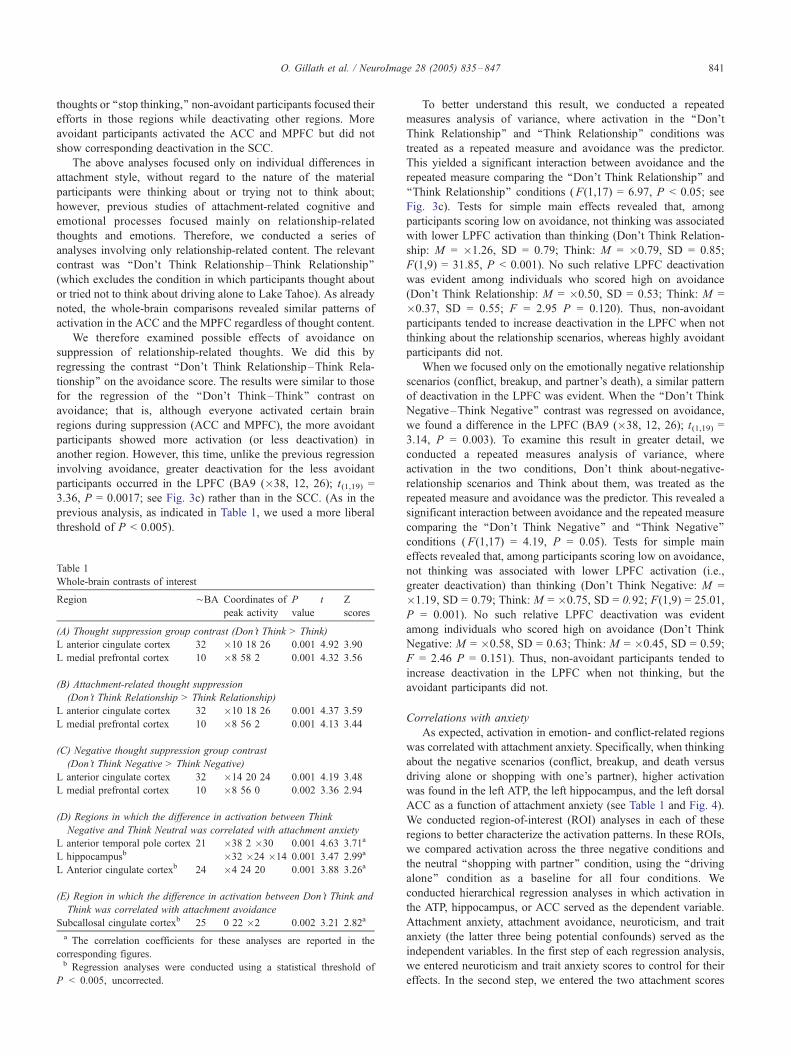
Table 1
Whole-brain contrasts of interest
Region ¨BA Coordinates of
peak activity
P
value
t Z
scores
(A) Thought suppression group contrast (Don’t Think > Think)
L anterior cingulate cortex 32 �10 18 26 0.001 4.92 3.90
L medial prefrontal cortex 10 �8 58 2 0.001 4.32 3.56
(B) Attachment-related thought suppression
(Don’t Think Relationship > Think Relationship)
L anterior cingulate cortex 32 �10 18 26 0.001 4.37 3.59
L medial prefrontal cortex 10 �8 56 2 0.001 4.13 3.44
(C) Negative thought suppression group contrast
(Don’t Think Negative > Think Negative)
L anterior cingulate cortex 32 �14 20 24 0.001 4.19 3.48
L medial prefrontal cortex 10 �8 56 0 0.002 3.36 2.94
(D) Regions in which the difference in activation between Think
Negative and Think Neutral was correlated with attachment anxiety
L anterior temporal pole cortex 21 �38 2 �30 0.001 4.63 3.71a
L hippocampusb �32 �24 �14 0.001 3.47 2.99a
L Anterior cingulate cortexb 24 �4 24 20 0.001 3.88 3.26a
(E) Region in which the difference in activation between Don’t Think and
Think was correlated with attachment avoidance
Subcallosal cingulate cortexb 25 0 22 �2 0.002 3.21 2.82a
a The correlation coefficients for these analyses are reported in the
corresponding figures.b Regression analyses were conducted using a statistical threshold of
P < 0.005, uncorrected.
To better understand this result, we conducted a repeated
measures analysis of variance, where activation in the ‘‘Don’t
Think Relationship’’ and ‘‘Think Relationship’’ conditions was
treated as a repeated measure and avoidance was the predictor.
This yielded a significant interaction between avoidance and the
repeated measure comparing the ‘‘Don’t Think Relationship’’ and
‘‘Think Relationship’’ conditions (F(1,17) = 6.97, P < 0.05; see
Fig. 3c). Tests for simple main effects revealed that, among
participants scoring low on avoidance, not thinking was associated
with lower LPFC activation than thinking (Don’t Think Relation-
ship: M = �1.26, SD = 0.79; Think: M = �0.79, SD = 0.85;
F(1,9) = 31.85, P < 0.001). No such relative LPFC deactivation
was evident among individuals who scored high on avoidance
(Don’t Think Relationship: M = �0.50, SD = 0.53; Think: M =
�0.37, SD = 0.55; F = 2.95 P = 0.120). Thus, non-avoidant
participants tended to increase deactivation in the LPFC when not
thinking about the relationship scenarios, whereas highly avoidant
participants did not.
When we focused only on the emotionally negative relationship
scenarios (conflict, breakup, and partner’s death), a similar pattern
of deactivation in the LPFC was evident. When the ‘‘Don’t Think
Negative–Think Negative’’ contrast was regressed on avoidance,
we found a difference in the LPFC (BA9 (�38, 12, 26); t(1,19) =
3.14, P = 0.003). To examine this result in greater detail, we
conducted a repeated measures analysis of variance, where
activation in the two conditions, Don’t think about-negative-
relationship scenarios and Think about them, was treated as the
repeated measure and avoidance was the predictor. This revealed a
significant interaction between avoidance and the repeated measure
comparing the ‘‘Don’t Think Negative’’ and ‘‘Think Negative’’
conditions (F(1,17) = 4.19, P = 0.05). Tests for simple main
effects revealed that, among participants scoring low on avoidance,
not thinking was associated with lower LPFC activation (i.e.,
greater deactivation) than thinking (Don’t Think Negative: M =
�1.19, SD = 0.79; Think: M = �0.75, SD = 0.92; F(1,9) = 25.01,
P = 0.001). No such relative LPFC deactivation was evident
among individuals who scored high on avoidance (Don’t Think
Negative: M = �0.58, SD = 0.63; Think: M = �0.45, SD = 0.59;
F = 2.46 P = 0.151). Thus, non-avoidant participants tended to
increase deactivation in the LPFC when not thinking, but the
avoidant participants did not.
Correlations with anxiety
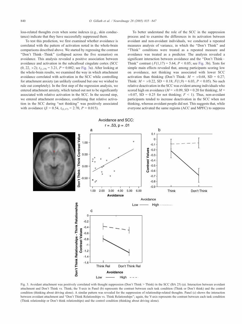
As expected, activation in emotion- and conflict-related regions
was correlated with attachment anxiety. Specifically, when thinking
about the negative scenarios (conflict, breakup, and death versus
driving alone or shopping with one’s partner), higher activation
was found in the left ATP, the left hippocampus, and the left dorsal
ACC as a function of attachment anxiety (see Table 1 and Fig. 4).
We conducted region-of-interest (ROI) analyses in each of these
regions to better characterize the activation patterns. In these ROIs,
we compared activation across the three negative conditions and
the neutral ‘‘shopping with partner’’ condition, using the ‘‘driving
alone’’ condition as a baseline for all four conditions. We
conducted hierarchical regression analyses in which activation in
the ATP, hippocampus, or ACC served as the dependent variable.
Attachment anxiety, attachment avoidance, neuroticism, and trait
anxiety (the latter three being potential confounds) served as the
independent variables. In the first step of each regression analysis,
we entered neuroticism and trait anxiety scores to control for their
effects. In the second step, we entered the two attachment scores
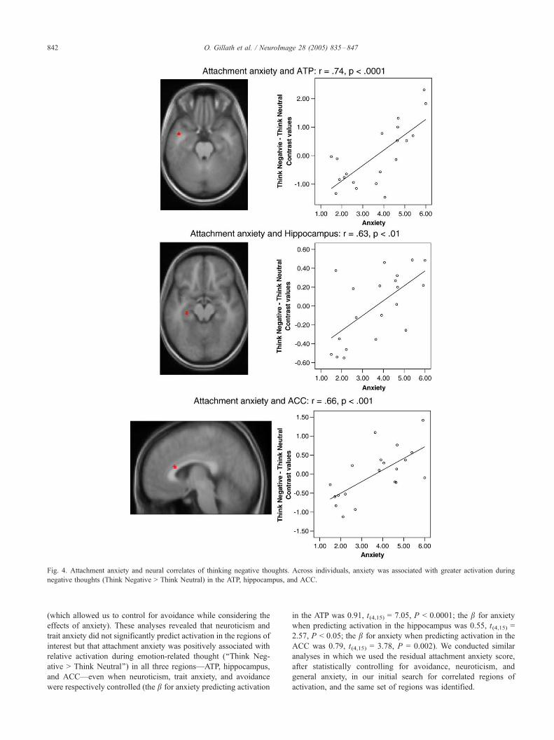
Fig. 4. Attachment anxiety and neural correlates of thinking negative thoughts. Across individuals, anxiety was associated with greater activation during
negative thoughts (Think Negative > Think Neutral) in the ATP, hippocampus, and ACC.
O. Gillath et al. / NeuroImage 28 (2005) 835–847842
(which allowed us to control for avoidance while considering the
effects of anxiety). These analyses revealed that neuroticism and
trait anxiety did not significantly predict activation in the regions of
interest but that attachment anxiety was positively associated with
relative activation during emotion-related thought (‘‘Think Neg-
ative > Think Neutral’’) in all three regions—ATP, hippocampus,
and ACC—even when neuroticism, trait anxiety, and avoidance
were respectively controlled (the b for anxiety predicting activation
in the ATP was 0.91, t(4,15) = 7.05, P < 0.0001; the b for anxiety
when predicting activation in the hippocampus was 0.55, t(4,15) =
2.57, P < 0.05; the b for anxiety when predicting activation in the
ACC was 0.79, t(4,15) = 3.78, P = 0.002). We conducted similar
analyses in which we used the residual attachment anxiety score,
after statistically controlling for avoidance, neuroticism, and
general anxiety, in our initial search for correlated regions of
activation, and the same set of regions was identified.
O. Gillath et al. / NeuroImage 28 (2005) 835–847 843
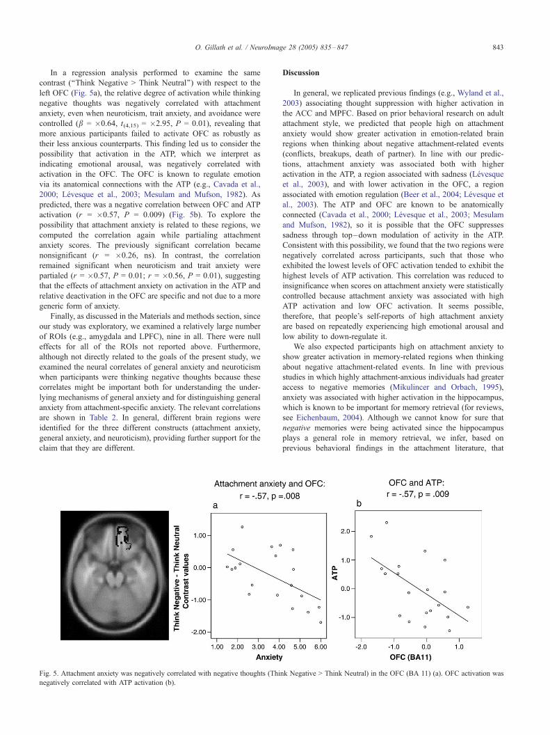
In a regression analysis performed to examine the same
contrast (‘‘Think Negative > Think Neutral’’) with respect to the
left OFC (Fig. 5a), the relative degree of activation while thinking
negative thoughts was negatively correlated with attachment
anxiety, even when neuroticism, trait anxiety, and avoidance were
controlled (b = �0.64, t(4,15) = �2.95, P = 0.01), revealing that
more anxious participants failed to activate OFC as robustly as
their less anxious counterparts. This finding led us to consider the
possibility that activation in the ATP, which we interpret as
indicating emotional arousal, was negatively correlated with
activation in the OFC. The OFC is known to regulate emotion
via its anatomical connections with the ATP (e.g., Cavada et al.,
2000; Levesque et al., 2003; Mesulam and Mufson, 1982). As
predicted, there was a negative correlation between OFC and ATP
activation (r = �0.57, P = 0.009) (Fig. 5b). To explore the
possibility that attachment anxiety is related to these regions, we
computed the correlation again while partialing attachment
anxiety scores. The previously significant correlation became
nonsignificant (r = �0.26, ns). In contrast, the correlation
remained significant when neuroticism and trait anxiety were
partialed (r = �0.57, P = 0.01; r = �0.56, P = 0.01), suggesting
that the effects of attachment anxiety on activation in the ATP and
relative deactivation in the OFC are specific and not due to a more
generic form of anxiety.
Finally, as discussed in the Materials and methods section, since
our study was exploratory, we examined a relatively large number
of ROIs (e.g., amygdala and LPFC), nine in all. There were null
effects for all of the ROIs not reported above. Furthermore,
although not directly related to the goals of the present study, we
examined the neural correlates of general anxiety and neuroticism
when participants were thinking negative thoughts because these
correlates might be important both for understanding the under-
lying mechanisms of general anxiety and for distinguishing general
anxiety from attachment-specific anxiety. The relevant correlations
are shown in Table 2. In general, different brain regions were
identified for the three different constructs (attachment anxiety,
general anxiety, and neuroticism), providing further support for the
claim that they are different.
Fig. 5. Attachment anxiety was negatively correlated with negative thoughts (Thin
negatively correlated with ATP activation (b).
Discussion
In general, we replicated previous findings (e.g., Wyland et al.,
2003) associating thought suppression with higher activation in
the ACC and MPFC. Based on prior behavioral research on adult
attachment style, we predicted that people high on attachment
anxiety would show greater activation in emotion-related brain
regions when thinking about negative attachment-related events
(conflicts, breakups, death of partner). In line with our predic-
tions, attachment anxiety was associated both with higher
activation in the ATP, a region associated with sadness (Levesque
et al., 2003), and with lower activation in the OFC, a region
associated with emotion regulation (Beer et al., 2004; Levesque et
al., 2003). The ATP and OFC are known to be anatomically
connected (Cavada et al., 2000; Levesque et al., 2003; Mesulam
and Mufson, 1982), so it is possible that the OFC suppresses
sadness through top–down modulation of activity in the ATP.
Consistent with this possibility, we found that the two regions were
negatively correlated across participants, such that those who
exhibited the lowest levels of OFC activation tended to exhibit the
highest levels of ATP activation. This correlation was reduced to
insignificance when scores on attachment anxiety were statistically
controlled because attachment anxiety was associated with high
ATP activation and low OFC activation. It seems possible,
therefore, that people’s self-reports of high attachment anxiety
are based on repeatedly experiencing high emotional arousal and
low ability to down-regulate it.
We also expected participants high on attachment anxiety to
show greater activation in memory-related regions when thinking
about negative attachment-related events. In line with previous
studies in which highly attachment-anxious individuals had greater
access to negative memories (Mikulincer and Orbach, 1995),
anxiety was associated with higher activation in the hippocampus,
which is known to be important for memory retrieval (for reviews,
see Eichenbaum, 2004). Although we cannot know for sure that
negative memories were being activated since the hippocampus
plays a general role in memory retrieval, we infer, based on
previous behavioral findings in the attachment literature, that
k Negative > Think Neutral) in the OFC (BA 11) (a). OFC activation was
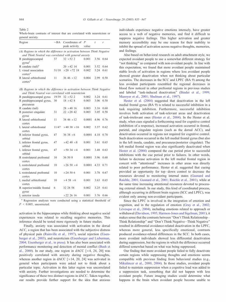
Table 2
Whole-brain contrasts of interest that are correlated with neuroticism or
general anxiety
Region ¨BA Coordinates of
peak activity
P
value
t r
(A) Regions in which the difference in activation between Think Negative
and Think Neutral was correlated with general anxiety
R parahippocampal
gyrusa37 32 �52 2 0.001 3.56 0.64
R caudate (tail)a 20 �42 14 0.001 3.52 0.64
L visual association
cortexa31/18 �20 �72 18 0.002 3.24 0.61
R lateral orbitofrontal
cortexa11 36 46 �12 0.004 2.99 0.58
(B) Regions in which the difference in activation between Think Negative
and Think Neutral was correlated with neuroticism
R parahippocampal gyrus 19/39 32 �52 2 0.002 3.24 0.61
R parahippocampal gyrus,
precuneus
30 18 �42 8 0.003 3.06 0.58
R caudate (tail) 28 �40 16 0.003 3.16 0.60
R posterior cingulate
gyrus
31 22 �28 42 0.003 3.05 0.58
R lateral orbitofrontal
cortex
11 38 46 �12 0.0001 4.96 0.76
L lateral orbitofrontal
cortex
11/47 �40 38 �16 0.002 3.37 0.62
R inferior frontal gyrus,
orbital
47 36 38 �6 0.0001 4.18 0.70
L inferior frontal gyrus,
orbital
47 �42 40 �8 0.001 3.61 0.65
L inferior frontal gyrus,
orbital
47 �50 34 �4 0.001 3.48 0.63
R rostrolateral prefrontal
cortex
10 36 50 8 0.0001 3.96 0.68
L rostrolateral prefrontal
cortex
10 �26 50 �4 0.0001 4.33 0.71
L rostrolateral prefrontal
cortex
10 �24 50 6 0.001 3.78 0.67
L medial orbitofrontal
cortex
10 �4 58 �6 0.001 3.63 0.65
R superior/middle frontal
gyrus
6 32 28 58 0.002 3.25 0.61
L anterior insula �22 26 16 0.001 3.76 0.66
a Regression analyses were conducted using a statistical threshold of
P < 0.005, uncorrected.
O. Gillath et al. / NeuroImage 28 (2005) 835–847844
activation in the hippocampus while thinking about negative social
experiences was related to recalling negative memories. This
inference should be tested more rigorously in future studies.
Finally, anxiety was associated with activation in the dorsal
ACC, a region that has been associated with the subjective distress
of physical pain (Rainville et al., 1997), social rejection (Eisen-
berger et al., 2003), and neuroticism (Eisenberger and Lieberman,
2004; Eisenberger et al., in press). It has also been associated with
performance monitoring and detection of mental conflict (Bush et
al., 2000). In our study, one region in dACC [�4, 24, 20] was
positively correlated with anxiety during negative thoughts,
whereas another region in dACC [�14, 20, 24] was activated in
general when participants were asked not to think about a
particular topic. Activation in this latter region was uncorrelated
with anxiety. Further investigations are needed to determine the
significance of these two distinct regions in dACC. Taken together,
our results provide further support for the idea that anxious
individuals experience negative emotions intensely, have greater
access to a web of negative memories, and find it difficult to
suppress negative feelings. This higher activation and greater
memory accessibility may be one reason for their inability to
inhibit the spread of activation across negative thoughts, memories,
and feelings.
Also based on behavioral research on adult attachment style, we
expected avoidant people to use a somewhat different strategy for
‘‘not thinking’’ as compared with non-avoidant people. In line with
this expectation, we found that more avoidant people maintained
stable levels of activation in regions where less avoidant people
showed greater deactivation when not thinking about particular
scenarios. The decreases in the SCC and LPFC (BA 9) among the
less avoidant participants resembled the regional decreases in
blood flow noticed in other prefrontal regions in previous studies
and labeled ‘‘task-induced deactivation’’ (Binder et al., 1999;
Mazoyer et al., 2001; Shulman et al., 1997).
Hester et al. (2004) suggested that deactivation in the left
medial frontal gyrus (BA 9) is related to successful inhibition in a
task requiring inhibition. Furthermore, successful inhibition
involves both activation of task-relevant areas and deactivation
of task-irrelevant ones (Hester et al., 2004). In the Hester et al.
study, when cues signaled a forthcoming need for cognitive control
(inhibition of a response), increased activation occurred in frontal,
parietal, and cingulate regions (such as the dorsal ACC), and
deactivation occurred in regions not required for cognitive control.
Such deactivation occurred in the left medial frontal gyrus (but also
in the left insula, caudate, and precuneus/posterior cingulate). The
left medial frontal region was also significantly deactivated when
Hester et al. (2004) compared the cue period prior to successful
inhibitions with the cue period prior to failures. In other words,
failure to decrease activation in the left medial frontal region in
concert with ‘‘attentional’’ increases in other areas was directly
related to poor performance. Hester et al. suggested that cueing
provided an opportunity for top–down control to decrease the
resources devoted to monitoring internal states (Gusnard and
Raichle, 2001; Gusnard et al., 2001; Raichle et al., 2001), while at
the same time increasing attentional resources devoted to process-
ing external stimuli. In our study, this kind of coordinated process,
although occurring in different brain regions (SCC and LPFC), was
evident only among non-avoidant participants.
Since the LPFC is involved in the integration of emotion and
cognition, and in the regulation of emotion (Gray et al., 2002;
Levesque et al., 2004), including emotions related to approach or
withdrawal (Davidson, 1995; Harmon-Jones and Sigelman, 2001), it
makes sense that the contrasts between ‘‘Don’t Think Relationship–
Think Relationship’’ and ‘‘Don’t Think Negative–Think Negative’’
resulted in differential avoidance-related deactivation in the LPFC,
whereas more general, less specifically emotional, contrasts
produced avoidance-related differences in the SCC. In both cases,
more avoidant individuals showed less differential deactivation
during suppression, but the regions in which the difference occurred
differed somewhat based on what was being suppressed.
Our finding that more avoidant people failed to fully deactivate
certain regions while suppressing thoughts and emotions seems
compatible with previous finding from behavioral studies (e.g.,
Mikulincer et al., 2000, 2004). In those studies, avoidant people
failed to maintain suppression when a cognitive load was added to
a suppression task, something that did not happen with less
avoidant people. Future imaging studies could determine what
happens in the brain when avoidant people become unable to

O. Gillath et al. / NeuroImage 28 (2005) 835–847 845
suppress certain mental contents while operating under a cognitive
load and whether the failure to deactivate regions that are directly
related to the suppression task is related to the disruptive effects of
the load.
Although preliminary, our study yielded promising results. The
questions under investigation are rooted in a well-established
theory, and the experimental procedures employed have been used
in previous cognitive and behavioral experiments. Moreover, the
inclusion of individual differences enabled us to look beyond
seemingly general processes that are likely to be clouded by such
differences. For example, another interpretation of Eugene et al.’s
(2003) study, described in the introduction, is that their first group
of participants included a relatively high proportion of anxious
individuals, whereas their second group included a relatively high
proportion of avoidant individuals. Many supposedly general
mental processes documented by social cognition researchers have
turned out to be moderated by attachment style (for a review, see
Mikulincer and Shaver, 2005). Using brain imaging techniques to
study such social cognitive processes as they are moderated by
attachment style should reveal where and how the differences arise.
This new methodology may clarify individual differences that
could previously be studied only behaviorally or via self-reports
and interviews.
Although we view this study as encouraging, it is important to
consider some of its limitations. First, we studied only women to
eliminate gender-related variance in the issues under investigation.
Second, our sample size was small for an individual-differences
study, and we therefore included relatively extreme scorers on the
attachment scales. While doing this, we discovered that it was
easier to recruit secure and anxious participants for a study of
‘‘close relationships and the brain’’ than to recruit avoidant
participants. We ended up excluding many secure and anxious
volunteers while working hard to attract avoidant ones. This might
have been related to our requirement that participants be involved
in a serious long-term relationship, which many avoidant college
students are not. Whatever the reasons, we were able to represent
the anxiety dimension of attachment style more completely than
the avoidance dimension, making it possible that more or clearer
findings related to avoidance will be obtained in future studies with
larger, more representative samples. This is especially important in
light of the liberal threshold (P < 0.005) we used for identifying
the differences between avoidant and non-avoidant participants.
We were not able to include all relevant control conditions in
our experiment. Inclusion of a non-attachment-related negative
emotion condition will be important in future studies because we
cannot yet be sure how many of our findings can be attributed to
attachment-related negative emotions alone and how many might
be replicated when we include non-attachment-related stimuli and
situations. Finally, it is difficult to infer, based on our findings,
what psychological process is associated with ATP activation.
Although some investigators have linked ATP activation to
affective processing, as we are inclined to do here, others have
linked it to semantic processing (e.g., Noppeney and Price, 2002;
Vandenberghe et al., 2002). Thus, further investigation of the ATP
activations in relation to attachment anxiety is necessary.
Conclusion
Despite these limitations, the study revealed associations
between self-reported attachment style and brain activation in
regions associated with emotion, memory, and emotion regulation.
The results fit well with other neuroscientific studies of emotion
regulation (e.g., Ochsner and Gross, 2004), which suggests that
emotional processes are modulated by top–down control from the
OFC and PFC and bottom–up processes in the anterior temporal
pole and hippocampus. Our findings indicate that individual
differences play an important role in emotion regulation, suggest-
ing that future studies should include relevant individual-difference
measures. Many psychological processes related to people’s
immersion in a social environment are likely to be moderated by
such individual differences.
References
Adolphs, R., 2004. Processing of emotional and social information by the
human amygdala. In: Gazzaniga, M.S. (Ed.), Cogn. Neurosci., vol. III,
3rd edR MIT Press, Cambridge, MA, pp. 1017–1031.
Ainsworth, M.D.S., Blehar, M.C., Waters, E., Wall, S., 1978. Patterns of
Attachment: Assessed in the Strange Situation and at Home. Erlbaum,
Hillsdale, NJ.
Anderson, M.C., Ochsner, K.N., Kuhl, B., Cooper, J., Robertson, E.,
Gabrieli, S.W., Glover, G.H., Gabrieli, J.D., 2004. Neural systems
underlying the suppression of unwanted memories. Science 303 (5655),
232–235.
Beauregard, M., Levesque, J., Bourgouin, P., 2001. Neural correlates of
conscious self-regulation of emotion. J. Neurosci. 21, 1–6.
Beer, J.S., Shimamura, A.P., Knight, R.T., 2004. Frontal lobe contribution
to executive control of cognitive and social behavior. In: Gazzaniga,
M.S. (Ed.), Cogn. Neurosci., vol. III, 3rd edR MIT Press, Cambridge,
MA, pp. 1091–1104.
Benet-Martinez, V., John, O.P., 1998. Los cinco grandes across cultures and
ethnic groups: multitrait multimethod analysis of the Big Five in
Spanish and English. J. Pers. Soc. Psychol. 75, 729–750.
Binder, J.R., Frost, J.A., Hammeke, T.A., Bellgowan, P.S.F., Rao, S.M.,
Cox, R.W., 1999. Conceptual processing during the conscious resting
state: a functional MRI study. J. Cogn. Neurosci. 11, 80–93.
Blair, R.J., Morris, J.S., Frith, C.D., Perrett, D.I., Dolan, R.J., 1999.
Dissociable neural responses to facial expressions of sadness and anger.
Brain 122, 883–893.
Botvinick, M.M., Cohen, J.D., Carter, C.S., 2004. Conflict monitoring and
anterior cingulate cortex: an update. Trends Cogn. Sci. 8, 539–546.
Bowlby, J., 1969/1982. Attachment, 2nd edR Attachment and Loss, vol. 1.
Basic Books, New York.
Bowlby, J., 1973. Separation: anxiety and anger. Attachment and Loss,
vol. 2. Basic Books, New York.
Bowlby, J., 1980. Sadness and depression. Attachment and Loss, vol. 3.
Basic Books, New York.
Brennan, K.A., Clark, C.L., Shaver, P.R., 1998. Self-report measurement of
adult romantic attachment: an integrative overview. In: Simpson, J.A.,
Rholes, W.S. (Eds.), Attachment Theory and Close Relationships.
Guilford Press, New York, pp. 46–76.
Brett, M., Anton, J.L., Valabregue, R., Poline, J.B. 2002. Region of
interest analysis using an SPM toolbox. Abstract presented at the 8th
International Conference on Functional Mapping of the Human Brain,
June 2–6, 2002, Sendai, Japan. Available on CD-ROM in Neuro-
Image, Vol. 16, No. 2.
Buchel, C., Friston, K.J., 1997. Modulation of connectivity in visual
pathways by attention: cortical interactions evaluated with structural
equation modeling and fMRI. Cereb. Cortex 7 (8), 768–778.
Bush, G., Luu, P., Posner, M.I., 2000. Cognitive and emotional influences
in anterior cingulate cortex. Trends Cogn. Sci. 4, 215–222.
Carpenter, E.M., Kirkpatrick, L.A., 1996. Attachment style and presence of
a romantic partner as moderators of psychophysiological responses to a
stressful laboratory situation. Pers. Relat. 3, 351–367.

O. Gillath et al. / NeuroImage 28 (2005) 835–847846
Cassidy, J., Kobak, R., 1988. Avoidance and its relation to other defensive
processes. In: Belsky, J., Nezworski, T. (Eds.), Clinical Implications of
Attachment. Erlbaum, Hillsdale, NJ, pp. 300–326.
Calarge, C., Andreasen, N.C., O’Leary, D.S., 2003. Visualizing how one
brain understands another: a PET study of theory of mind. Am. J.
Psychiatry 160, 1954–1964.
Cavada, C., Company, T., Tejedor, J., Cruz-Rizzolo1, R.J., Reinoso-Suarez,
F., 2000. The anatomical connections of the macaque monkey
orbitofrontal cortex: a review. Cereb. Cortex 10, 220–242.
Cohen, M.X., Shaver, P.R., 2004. Avoidant attachment and hemispheric
lateralisation of the processing of attachment- and emotion-related
words. Cogn. Emot. 18, 799–813.
Cohen, N.J., Ryan, J., Hunt, C., Romine, L., Wszalek, T., Nash, C., 1999.
Hippocampal system and declarative (relational) memory: summariz-
ing the data from functional neuroimaging studies. Hippocampus 9,
83–98.
Cocosco, C.A., Kollokian, V., Kwan, R.K.S., Evans, A.C., 1997.
Brainweb: online interface to a 3D MRI simulated brain database.
NeuroImage 5, 425.
Davidson, R.J., 1995. Cerebral asymmetry, emotion, and affective style.
In: Davidson, R.J., Hugdahl, K. (eds.), MIT Press, Cambridge, MA,
pp. 361–387.
Dawson, G., Ashman, S.B., Hessl, D., Spieker, S., Frey, K., Panagiotides,
H., Embry, L., 2001. Autonomic and brain electrical activity in
securely- and insecurely-attached infants of depressed mothers. Infant
Behav. Dev. 24, 135–149.
Diamond, L.M., 2001. Contributions of psychophysiology to research on
adult attachment: review and recommendations. Pers. Soc. Psychol.
Rev. 5, 276–295.
Dolan, R.J., Lane, R., Chua, P., Fletcher, P., 2000. Dissociable temporal
lobe activations during emotional episodic memory retrieval. Neuro-
Image 11, 203–209.
Drevets, W.C., 2000. Neuroimaging studies of mood disorders. Biol.
Psychiatry 48, 813–829.
Eichenbaum, H., 2004. An information processing framework for memory
representation by the hippocampus. In: Gazzaniga, M.S. (Ed.), Cogn.
Neurosci., vol. III, 3rd edR MIT Press, Cambridge, MA, pp. 679–690.
Eisenberger, N.I., Lieberman, M.D., 2004. Why rejection hurts: a common
neural alarm system for physical and social pain. Trends Cogn. Sci. 8,
294–300.
Eisenberger, N.I., Lieberman, M.D., Williams, K.D., 2003. Does rejection
hurt? An FMRI study of social exclusion. Science 302, 290–292.
Eisenberger, N.I., Lieberman, M.D., Satpute, A.B., in press. Personality
from a controlled processing perspective: an fMRI study of neuroticism,
extraversion, and self-consciousness. Cogn. Affect. Behav. Neurosci.
Eugene, F., Levesque, J., Mensour, B., Leroux, J.M., Beaudoin, G.,
Bourgouin, P., Beauregard, M., 2003. The impact of individual
differences on the neural circuitry underlying sadness. NeuroImage 19
(2 Pt. 1), 354–364.
Feeney, B.C., Kirkpatrick, L.A., 1996. Effects of adult attachment and
presence of romantic partners on physiological responses to stress.
J. Pers. Soc. Psychol. 70, 255–270.
Fraley, R.C., Shaver, P.R., 1997. Adult attachment and the suppression of
unwanted thoughts. J. Pers. Soc. Psychol. 73, 1080–1091.
Fraley, R.C., Spieker, S.J., 2003. Are infant attachment patterns continu-
ously or categorically distributed? A taxometric analysis of strange
situation behavior. Dev. Psychol. 39, 387–404.
Gray, J.R., Braver, T.S., Raichle, M.E., 2002. Integration of emotion and
cognition in the lateral prefrontal cortex. Proc. Natl. Acad. Sci. 99,
4115–4120.
Gusnard, D.A., Raichle, M.E., 2001. Searching for a baseline: func-
tional imaging and the resting human brain. Nat. Rev., Neurosci. 2,
685–694.
Gusnard, D.A., Akbudak, E., Shulman, G.L., Raichle, M.E., 2001. Medial
prefrontal cortex and self-referential mental activity: relation to a
default mode of brain function. Proc. Natl. Acad. Sci. U. S. A. 98,
4259–4264.
Hare, T.A., Tottenham, N., Davidson, M.C., Glover, G.H., Casey, B.J.,
2005. Contributions of amygdala and striatal activity in emotion
regulation. Biol. Psychiatry 57, 624–632.
Harmon-Jones, E., Sigelman, J., 2001. State anger and prefrontal brain
activity: evidence that insult-related relative left prefrontal activation is
associated with experienced anger and aggression. J. Pers. Soc. Psychol.
80, 797–803.
Hester, R.L., Murphy, K., Foxe, J.J., Foxe, D.M., Javitt, D.C., Garavan, H.,
2004. Predicting success: patterns of cortical activation and deactivation
prior to response inhibition. J. Cogn. Neurosci. 16, 776–785.
Hofer, M.A., 2003. The emerging neurobiology of attachment and
separation: how parents shape their infant’s brain and behavior. In:
Coates, S., Rosenthal, W.J.L., Schechter, D.S. (Eds.), September 11:
Trauma and Human Bonds. Analytic Press, Hillsdale, NJ, pp. 191–209.
Johnson, S.C., Baxter, L.C., Wilder, L.S., Pipe, J.G., Heiserman, J.E.,
Prigatano, G.P., 2002. Neural correlates of self-reflection. Brain 125,
1808–1814.
Kelley, W.M., Macrae, C.N., Wyland, C.L., Caglar, S., Inati, S., Heatherton,
T.F., 2002. Finding the self? An event-related fMRI study. J. Cogn.
Neurosci. 14, 785–794.
Levesque, J., Eugene, F., Joanette, Y., Paquette, V., Mensour, B., Beaudoin,
G., Leroux, J.M., Bourgouin, P., Beauregard, M., 2003. Neural
circuitry underlying voluntary suppression of sadness. Biol. Psychiatry
53, 502–510.
Levesque, J., Joanette, Y., Mensour, B., Beaudoin, G., Leroux, J.M.,
Bourgouin, P., Beauregard, M., 2004. Neural basis of emotional self-
regulation in childhood. Neuroscience 129, 361–369.
Maguire, E.A., Frith, C.D., 2003. Aging affects the engagement of the
hippocampus during autobiographical memory retrieval. Brain 126,
1511–1523.
Mazoyer, B., Zago, L., Mellet, E., Bricogne, S., Etard, O., Houde, O.,
Crivello, F., Joliot, M., Petit, L., Tzourio-Mazoyer, N., 2001.
Cortical networks for working memory and executive functions
sustain the conscious resting state in man. Brain Res. Bull. 54,
287–298.
Mesulam, M.M., Mufson, E.J., 1982. Insula of the old world monkey:
architectonics in the insulo-orbital temporal component of the para-
limbic brain. J. Comp. Neurol. 212, 1–22.
Mikulincer, M., 1998. Adult attachment style and affect regulation: strategic
variations in self-appraisals. J. Pers. Soc. Psychol. 75, 420–435.
Mikulincer, M., Florian, V., 2000. Exploring individual differences in
reactions to mortality salience: does attachment style regulate terror
management mechanisms? J. Pers. Soc. Psychol. 79, 260–273.
Mikulincer, M., Orbach, I., 1995. Attachment styles and repressive
defensiveness: the accessibility and architecture of affective memories.
J. Pers. Soc. Psychol. 68, 917–925.
Mikulincer, M., Shaver, P.R., 2003. The attachment behavioral system in
adulthood: activation, psychodynamics, and interpersonal processes. In:
Zanna, M.P. (Ed.), Adv. Exp. Soc. Psychol., vol. 35. Academic Press,
San Diego, pp. 53–152.
Mikulincer, M., Shaver, P.R., 2004. Security-based self-representations in
adulthood: contents and processes. In: Rholes, W.S., Simpson, J.A.
(Eds.), Adult Attachment: Theory, Research, and Clinical Implications.
Guilford Press, New York, pp. 159–195.
Mikulincer, M., Shaver, P.R., 2005. Mental representations of attachment
security: theoretical foundation for a positive social psychology. In:
Baldwin, M.W. (Ed.), Interpersonal Cognition. Guilford Press, New
York, pp. 233–266.
Mikulincer, M., Birnbaum, G., Woddis, D., Nachmias, O., 2000. Stress and
accessibility of proximity-related thoughts: exploring the normative and
intraindividual components of attachment theory. J. Pers. Soc. Psychol.
78, 509–523.
Mikulincer, M., Dolev, T., Shaver, P.R., 2004. Attachment-related strategies
during thought-suppression: ironic rebounds and vulnerable self-
representations. J. Pers. Soc. Psychol. 87, 940–956.
Miller, E.K., Cohen, J.D., 2001. An integrative theory of prefrontal cortex
function. Annu. Rev. Neurosci. 24, 167–202.
O. Gillath et al. / NeuroImage 28 (2005) 835–847 847
Mitchell, J.P., Heatherton, T.F., Kelley, W.M., Wyland, C.L., Macrae, C.N.,
unpublished manuscript. Controlling the contents of consciousness: the
neural substrates of thought suppression.
Moran, M.A., Mufson, E.J., Mesulam, M.M., 1987. Neural inputs into
the temporopolar cortex of the rhesus monkey. J. Comp. Neurol. 256,
88–103.
Morecraft, R.J., Geula, C., Mesulam, M.M., 1992. Cytoarchitecture and
neural afferents of orbitofrontal cortex in the brain of the monkey.
J. Comp. Neurol. 323, 341–358.
Noppeney, U., Price, C.J., 2002. A PET study of stimulus- and task-induced
semantic processing. NeuroImage 15 (4), 927–935.
Ochsner, K.N., Gross, J.J., 2004. Thinking makes it so: a social cognitive
neuroscience approach to emotion regulation. In: Baumeister, R.F.,
Vohs, K.D. (Eds.), Handbook of Self-regulation: Research, Theory, and
Applications. Guilford Press, New York, pp. 229–255.
Ochsner, K.N., Bunge, S.A., Gross, J.J., Gabrieli, J.D., 2002. Rethinking
feelings: an fMRI study of the cognitive regulation of emotion. J. Cogn.
Neurosci. 14 (8), 1215–1229.
Phan, K.L., Wager, T., Taylor, S.F., Liberzon, I., 2002. Functional
neuroanatomy of emotion: a meta-analysis of emotion activation studies
in PET and fMRI. NeuroImage 162, 331–348.
Phan, K.L., Liberzon, I., Welsh, R.C., Britton, J.C., Taylor, S.F., 2003.
Habituation of rostral anterior cingulate cortex to repeated emotionally
salient pictures. Neuropsychopharmacology 28, 1344–1350.
Phan, K.L., Wager, T., Taylor, S.F., Liberzon, I., 2004. Functional
neuroimaging studies of human emotions. CNS Spectr. 9 (4), 258–266.
Pelletier, M., Bouthillier, A., Levesque, J., Carrier, S., Breault, C., Paquette,
V., Mensour, B., Leroux, J.M., Beaudoin, G., Bourgouin, P., Beau-
regard, M., 2003. Separate neural circuits for primary emotions? Brain
activity during self-induced sadness and happiness in professional
actors. NeuroReport 14, 1111–1116.
Ranote, S., Elliott, R., Abel, K.M., Mitchell, R., Deakin, J.F., Appleby, L.,
2004. The neural basis of maternal responsiveness to infants: an fMRI
study. NeuroReport 15, 1825–1829.
Rainville, P., Duncan, G.H., Proce, D.D., Carrier, B., Bushnell, M.C., 1997.
Pain affect encoded in human anterior cingulate but not somatosensory
cortex. Science 277, 968–971.
Raichle, M.E., MacLeod, A.M., Snyder, A.Z., Powers, W.J., Gusnard, D.A.,
Shulman, G.L., 2001. A default mode of brain function. Proc. Natl.
Acad. Sci. U. S. A. 98, 676–682.
Roberts, A.C., Wallis, J.D., 2000. Inhibitory control and affective
processing in the prefrontal cortex: neuropsychological studies in the
common marmoset. Cereb. Cortex 10, 252–262.
Rolls, E.T., Inoue, K., Browning, A., 2003. Activity of primate subgenual
cingulate cortex neurons is related to sleep. J. Neurophysiol. 90 (1),
134–142.
Rule, R.R., Shimamura, A.P., Knight, R.T., 2002. Orbitofrontal cortex and
dynamic filtering of emotional stimuli. Cogn. Affect. Behav. Neurosci.
2 (3), 264–270.
Schore, A.N., 2000. Attachment and the regulation of the right brain.
Attach. Hum. Dev. 2, 23–47.
Shaver, P.R., Clark, C.L., 1994. The psychodynamics of adult romantic
attachment. In: Masling, J.M., Bornstein, R.F. (Eds.), Empirical
Perspectives on Object Relations Theories. American Psychological
Association, Washington, DC, pp. 105–156.
Shin, L.M., McNally, R.J., Kosslyn, S.M., Thompson, W.L., Rauch, S.L.,
Alpert, N.M., Metzger, L.J., Lasko, N.B., Orr, S.P., Pitman, R.K., 1999.
Regional cerebral blood flow during script-driven imagery in childhood
sexual abuse-related PTSD: a PET investigation. Am. J. Psychiatry 156,
575–584.
Spielberger, C.D., Gorsuch, R.L., Lushene, R., Vagg, P.R., Jacobs, G.A.,
1983. Manual for the State-Trait Anxiety Inventory, Form Y. Consulting
Psychologists Press, Palo Alto, CA.
Shulman, G.L., Fiez, J.A., Corbetta, M., Buckner, R.L., Meizin, F.M.,
Raichle, M.E., Petersen, S.E., 1997. Common blood flow changes
across visual tasks: II. Decreases in cerebral cortex. J. Cogn. Neurosci.
9, 648–663.
Talairach, J., Tournoux, P., 1988. Co-planar Stereotaxic Atlas of the Human
Brain. Thieme, Stuttgart.
Vandenberghe, R., Nobre, A.C., Price, C.J., 2002. The response of left
temporal cortex to sentences. J. Cogn. Neurosci. 14 (4), 550–560.
Wyland, C.L., Kelley, W.M., Macrae, C.N., Gordon, H.L., Heatherton, T.F.,
2003. Neural correlates of thought suppression. Neuropsychologia 41
(14), 1863–1867.