The Indian drawings of the poet Cesare Pascarella: non-destructive analyses and conservation...
12
ORIGINAL PAPER The Indian drawings of the poet Cesare Pascarella: non-destructive analyses and conservation treatments Marina Bicchieri & Michela Monti & Giovanna Piantanida & Flavia Pinzari & Simonetta Iannuccelli & Silvia Sotgiu & Lorena Tireni Received: 13 May 2011 /Revised: 17 June 2011 /Accepted: 30 June 2011 /Published online: 13 July 2011 # Springer-Verlag 2011 Abstract The Italian dialect poet Cesare Pascarella trav- elled all around the world, noting down in notebooks his keen and caustic observations, and drawing sketches that are a visual reportage of his journeys. The sketches were mounted as a random collage over acidic cardboards that were exposed to direct sunlight in his studio. Their poor state of conservation is related to the use of modern paper: chemical instability of raw materials caused acidification and strong oxidation of the support, with intense yellowing of the surfaces and brittleness of the paper. To ensure future preservation of the drawings, chemical stabilisation with simultaneous alcoholic treatment by deacidification (calcium propionate) and reduction (borane tert-butylamine complex) appeared necessary. To verify its applicability, it was indispensible to characterise the support and identify the nature of all the graphic media. The use of Raman, Infrared, X-ray fluorescence spectroscopies and scanning electron microscopy coupled with X-ray microanalysis allowed us to clear the problems related to the different penetration depth of each analytical technique and the different responses of pigments/dyes to each spectroscopy. The palette, how it varied along the journeys, the different supports used and preparations were completely identified showing a choice of colours compatible with the reduction treatment. Keywords Paper . Micro-Raman . ATR-FTIR . SEM-EDS . XRF . Reduction Introduction The drawings, executed by the roman poet and painter Cesare Pascarella (1858–1940) from the second half of nineteenth and the beginning of twentieth century, are occasional sketches on machine-made papers drawn in his various journeys in North Africa, India, Japan and America. To compose his drawings, the author employed a wide range of graphic media. When he returned to Italy, Pascarella realised a peculiar reportage, mounting the sketches over ten wood pulp boards in a sort of random collage. Every cardboard (665×995 mm 2 ) displays 20–30 items, fixed directly on the acidic support by means of glue spots; the sketches were often displaced with a partial overlap. The cardboards have been hanging on the wall of the artist’ s studio for a long time being directly exposed to the sunlight. Pascarella regrouped them in a way that is supposed to be a sort of collection of ‘visual notes’ that he would have used as a reference for future painting works. At present, 78 drawings mounted on three cardboards are undergoing conservation treatments at the ICPAL Published in the special issue Analytical Techniques in Art, Archaeology and Conservation Science with guest editor Oliver Hahn. M. Bicchieri (*) : M. Monti : G. Piantanida Laboratory of Chemistry, Istituto centrale restauro e conservazione patrimonio archivistico e librario, Via Milano 76, 00184 Rome, Italy e-mail: [email protected] F. Pinzari Laboratory of Biology, Istituto centrale restauro e conservazione patrimonio archivistico e librario, Via Milano 76, 00184 Rome, Italy S. Iannuccelli : S. Sotgiu : L. Tireni Laboratory of Conservation, Istituto centrale restauro e conservazione patrimonio archivistico e librario, Via Milano 76, 00184 Rome, Italy Anal Bioanal Chem (2012) 402:1517–1528 DOI 10.1007/s00216-011-5229-3
-
Upload
independent -
Category
Documents
-
view
0 -
download
0
Transcript of The Indian drawings of the poet Cesare Pascarella: non-destructive analyses and conservation...
ORIGINAL PAPER
The Indian drawings of the poet Cesare Pascarella:non-destructive analyses and conservation treatments
Marina Bicchieri & Michela Monti &Giovanna Piantanida & Flavia Pinzari &Simonetta Iannuccelli & Silvia Sotgiu & Lorena Tireni
Received: 13 May 2011 /Revised: 17 June 2011 /Accepted: 30 June 2011 /Published online: 13 July 2011# Springer-Verlag 2011
Abstract The Italian dialect poet Cesare Pascarella trav-elled all around the world, noting down in notebooks hiskeen and caustic observations, and drawing sketches thatare a visual reportage of his journeys. The sketches weremounted as a random collage over acidic cardboards thatwere exposed to direct sunlight in his studio. Their poorstate of conservation is related to the use of modern paper:chemical instability of raw materials caused acidificationand strong oxidation of the support, with intense yellowingof the surfaces and brittleness of the paper. To ensurefuture preservation of the drawings, chemical stabilisationwith simultaneous alcoholic treatment by deacidification(calcium propionate) and reduction (borane tert-butylaminecomplex) appeared necessary. To verify its applicability, itwas indispensible to characterise the support and identifythe nature of all the graphic media. The use of Raman,Infrared, X-ray fluorescence spectroscopies and scanning
electron microscopy coupled with X-ray microanalysisallowed us to clear the problems related to the differentpenetration depth of each analytical technique and thedifferent responses of pigments/dyes to each spectroscopy.The palette, how it varied along the journeys, the differentsupports used and preparations were completely identifiedshowing a choice of colours compatible with the reductiontreatment.
Keywords Paper .Micro-Raman . ATR-FTIR . SEM-EDS .
XRF. Reduction
Introduction
The drawings, executed by the roman poet and painterCesare Pascarella (1858–1940) from the second half ofnineteenth and the beginning of twentieth century, areoccasional sketches on machine-made papers drawn in hisvarious journeys in North Africa, India, Japan and America.To compose his drawings, the author employed a widerange of graphic media.
When he returned to Italy, Pascarella realised a peculiarreportage, mounting the sketches over ten wood pulpboards in a sort of random collage. Every cardboard(665×995 mm2) displays 20–30 items, fixed directly onthe acidic support by means of glue spots; the sketcheswere often displaced with a partial overlap. The cardboardshave been hanging on the wall of the artist’s studio for along time being directly exposed to the sunlight. Pascarellaregrouped them in a way that is supposed to be a sort ofcollection of ‘visual notes’ that he would have used as areference for future painting works.
At present, 78 drawings mounted on three cardboardsare undergoing conservation treatments at the ICPAL
Published in the special issue Analytical Techniques in Art,Archaeology and Conservation Science with guest editor Oliver Hahn.
M. Bicchieri (*) :M. Monti :G. PiantanidaLaboratory of Chemistry, Istituto centrale restauroe conservazione patrimonio archivistico e librario,Via Milano 76,00184 Rome, Italye-mail: [email protected]
F. PinzariLaboratory of Biology, Istituto centrale restauroe conservazione patrimonio archivistico e librario,Via Milano 76,00184 Rome, Italy
S. Iannuccelli : S. Sotgiu : L. TireniLaboratory of Conservation, Istituto centrale restauroe conservazione patrimonio archivistico e librario,Via Milano 76,00184 Rome, Italy
Anal Bioanal Chem (2012) 402:1517–1528DOI 10.1007/s00216-011-5229-3
Conservation Department. They belong to the AccademiaNazionale dei Lincei-Biblioteca Corsiniana, Rome.
The poor state of conservation of the drawings isrepresentative of the problems connected with papers thathave been produced by modern papermaking industries,in particular, between 1850 and end of the Second WorldWar. Chemical instability of raw materials causes acidi-fication and strong oxidation of the support, with intenseyellowing of the surfaces and brittleness of the paper.
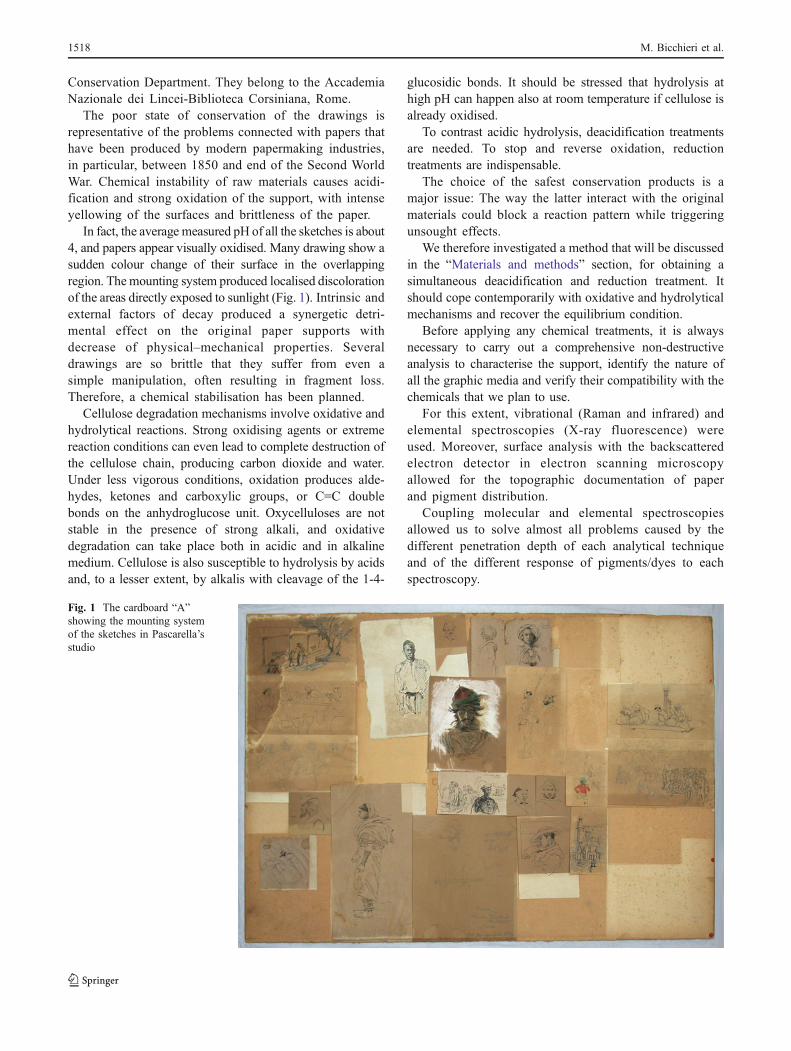
In fact, the average measured pH of all the sketches is about4, and papers appear visually oxidised. Many drawing show asudden colour change of their surface in the overlappingregion. Themounting system produced localised discolorationof the areas directly exposed to sunlight (Fig. 1). Intrinsic andexternal factors of decay produced a synergetic detri-mental effect on the original paper supports withdecrease of physical–mechanical properties. Severaldrawings are so brittle that they suffer from even asimple manipulation, often resulting in fragment loss.Therefore, a chemical stabilisation has been planned.
Cellulose degradation mechanisms involve oxidative andhydrolytical reactions. Strong oxidising agents or extremereaction conditions can even lead to complete destruction ofthe cellulose chain, producing carbon dioxide and water.Under less vigorous conditions, oxidation produces alde-hydes, ketones and carboxylic groups, or C=C doublebonds on the anhydroglucose unit. Oxycelluloses are notstable in the presence of strong alkali, and oxidativedegradation can take place both in acidic and in alkalinemedium. Cellulose is also susceptible to hydrolysis by acidsand, to a lesser extent, by alkalis with cleavage of the 1-4-
glucosidic bonds. It should be stressed that hydrolysis athigh pH can happen also at room temperature if cellulose isalready oxidised.
To contrast acidic hydrolysis, deacidification treatmentsare needed. To stop and reverse oxidation, reductiontreatments are indispensable.
The choice of the safest conservation products is amajor issue: The way the latter interact with the originalmaterials could block a reaction pattern while triggeringunsought effects.
We therefore investigated a method that will be discussedin the “Materials and methods” section, for obtaining asimultaneous deacidification and reduction treatment. Itshould cope contemporarily with oxidative and hydrolyticalmechanisms and recover the equilibrium condition.
Before applying any chemical treatments, it is alwaysnecessary to carry out a comprehensive non-destructiveanalysis to characterise the support, identify the nature ofall the graphic media and verify their compatibility with thechemicals that we plan to use.
For this extent, vibrational (Raman and infrared) andelemental spectroscopies (X-ray fluorescence) wereused. Moreover, surface analysis with the backscatteredelectron detector in electron scanning microscopyallowed for the topographic documentation of paperand pigment distribution.
Coupling molecular and elemental spectroscopiesallowed us to solve almost all problems caused by thedifferent penetration depth of each analytical techniqueand of the different response of pigments/dyes to eachspectroscopy.
Fig. 1 The cardboard “A”showing the mounting systemof the sketches in Pascarella’sstudio
1518 M. Bicchieri et al.
Raman spectroscopy is widely used for pigment and inkcharacterization [1, 2], but it is also a powerful techniquethat allows for paper degradation description [3, 4].Complementary results, in particular, on binding mediaand on possible vibrational modes of carbonyl groups in thecellulose, not very well resolved in Raman, can be obtainedapplying infrared spectroscopy [5], while X-ray fluores-cence takes into account the presence of the mainimpurities, fillers, coating materials in the substrate or inthe main components of inks and pigments [6, 7].
Variable pressure scanning electron microscopy (SEM)technique can be successfully applied to the study ofancient manuscripts, inks, illuminated parchments andwriting supports. In this field, several applications arepossible: the study of manufacturing materials andprocesses, the analysis of degradation phenomena, theevaluation of conservation practices or the description ofmicroscopic effects of chemical treatments. Variablepressure (VP) SEM allows for the description of thesurfaces of the samples without prior preparation andtherefore represents a micro-invasive methodology or anon-destructive technique. Samples can, in fact, bemounted reversibly on a metal stub using double-sidedcarbon adhesive tape, or can be arranged on non-invasivesupports that can be as large as the sample-chamber of theinstrument, thus allowing the observation of whole objectswithout or with very limited damage.
The impact of X-ray microanalysis, usually associated toSEM, on organic materials is more invasive than simpleSEM observation. Paper and parchment deteriorate ifelectronic irradiation is too long. For example, papercontains 5% or more water, and electron irradiation mayvaporise the water, thus modifying the material. To obtaingood results in X-ray microanalysis, measurements have tobe long enough to obtain satisfactory precision andcounting statistics but short enough to limit sampledeterioration. Moreover, the variability in density of thematerials makes the measurements scarcely repeatable. Thenumber of points to analyse on each sample/area dependson the chosen spot size (around 200 nm to obtain anacceptable X-ray microanalysis detector dead time ofaround 20–25%) [8].
Materials and methods
Instrumentation
A Renishaw inVia Reflex Raman microscope equippedwith a Renishaw diode LASER at 785 nm was used toperform the measurements. For each sample, testingmeasurements were attempted, starting from 0.1 mW atthe surface and gradually increasing the LASER intensity
until an acceptable signal-to-noise ratio was obtainedwithout sample degradation. The final experimental con-ditions varied between 1.3 and 2 mW. The backscatteredlight was dispersed by a 1,200-line/mm grating, and theRaman signal was detected by a Peltier cooled (−70 °C)deep depletion charge-coupled device (CCD RD-VIU,578×384 pixel, with spectral response in the range 200–1,025 nm) optimised for near-infrared and ultraviolet.Nominal spectral resolution was about 3 cm-1. The system,equipped with a Leica DMLM microscope to focus thelaser on the sample and a colour video camera, allows forpositioning of the sample and the selection of a specificregion for investigation. Spectral acquisitions (5–50 accu-mulations, 50 s each) were performed with a ×50 objective(NA=0.75). Under these conditions, the laser spot measuresabout 2×10 μm2.
Attenuated total reflectance measurements were per-formed using a Nexus Nicolet interferometer, equippedwith a KBr beam splitter and extended with a ZnSe cell anda liquid nitrogen cooled MCT/A detector. Measurementswere performed in the 4,000–650 cm-1 range at a resolutionof 8 cm-1, averaging 400 acquisitions per sample.
X-ray fluorescence (XRF) spectra were recorded bymeans of an Assing Lithos 3000 portable spectrometer,equipped with a Mo X-ray tube. The radiation can becollimated at different beam diameters (from 0.5 to 4 mm),depending on the area of interest. In this experiment, the2 mm collimator was used together with a Zr filter. A redlaser (695 nm) and a camera, integrated into the system andcontrolled by the instrument software, allow for thepositioning on the area to be sampled. Acquisitions(3,600 s each) were performed with the tube operating at24 kV, 0.300 mA, in the 0–25 keV range, resolution of160 eV at 5.9 keV.
The analysis of the drawings was conducted using aSEM instrument (EVO 50, Carl-Zeiss Electron MicroscopyGroup) equipped with both a detector for electron-backscattered diffraction and for secondary electron scan-ning in variable pressure mode. SEM observations weremade at 20 kV accelerating voltage with a tungstenfilament. Qualitative and quantitative chemical character-isations of the inorganic constituents of the supports andpigments were obtained by means of energy dispersive X-ray spectroscopy (EDS, INCA Energy 250), which allowsfor X-ray scanning of the area focused in SEM images witha very low penetration (1–2 μm), thereby creating acompositional map of the drawing’s surface. The analysiswas not invasive since it was possible to insert each sketchintegrally in the SEM chamber, thanks to their smalldimension. Reference elemental intensities acquired frompure compounds (standards) were utilised for calibratingscanning electron microscopy–energy dispersive X-rayspectroscopy (SEM/EDS) systems. Conventional ZAF
The Indian drawings of the poet Cesare Pascarella 1519
correction [9] integrated into Oxford INCA 250 microanal-ysis package was applied to the spectrum dataset (OxfordInstruments).
Chemicals
Ethanolic solution containing 3.0 g/l of calcium propionate[Ca(CH3CH2COO)2] and 6.0 g/l of borane tert-butylaminecomplex [(CH3)3CNH2·BH3] was used to carry outsimultaneous deacidification and reduction treatments.
Combined deacidification and reduction
The main degradation mechanisms of cellulosic sub-strates are acidic hydrolysis and oxidation. The alkalinehydrolysis, on the contrary, is not a common degradationproblem, but it can play a very important role only ifcellulose chains contain oxidised groups.
In this case, a ß-alkoxyelimination can occur, leading tocellulose depolymerisation, even with diluted alkalis [10]and the degradation increases with increasing of pH, inparticular, for values higher than 12.5 [11].
A big effort has been performed to contrast acidification:At the state of the art, aqueous and non-aqueous solutions,spray compounds and mass-deacidification are available.Besides, only a few products can be applied on the paper tocontrast oxidation without damaging the fibre structure andthe inks. Boron compounds and complexes are veryeffective and chemoselective in reducing carbonylic func-tions that are the most important oxidised groups in thecellulose polymer chain [12–15].
The first reducer that has been used in conservation fieldwas sodium borohydride [16, 17].
During the reduction, hydrogen evolves and, if thereaction is too fast (that means that big amounts ofhydrogen are produced in a very little time), the fibrewall could be broken. This effect has been observed andreported by restorers when they used sodium borohy-dride as reducing product in the treatment of stronglyoxidised papers.
Therefore, to minimise this negative effect, mild and“slow” reducers should be chosen.
Furthermore, to avoid ß-alkoxyelimination mechanisms,the employed reducers should have a pH lower than 10when applied in water solution.
In 1997, we published the first results of our research onthe application on original documents and laboratorysamples of borane tert-butylamine complex (TBAB) usedas a mild reagent in reducing carbonyl groups of oxidisedpaper [18], and we then measured its effectiveness withchemical and instrumental methods such as nuclear mag-netic resonance, differential scanning calorimetry andthermogravimetry [19–21].
Almost in the same period, TBAB was proposed for thebleaching of pulp in the paper industry, and the researchersdemonstrated that amine boranes were better than sodiumborohydride and even more than sodium hydrosulphite forobtaining a more stable, white and not-degraded cellulosepulp [22].
In order to offer to the conservators a larger choice ofproducts that would apply in different solvents or fordifferent restoring needs, we decided, within the ItalianNational Research Council Special Project “Cultural Heri-tage”, to set up an investigation on different reducingproducts for paper conservation [23] to test the possibilityof simultaneous non-aqueous deacidification and reductionand to study the chemistry of the reducing reaction betweenTBAB and cellulosic materials [24].
The use of aqueous solutions in restoration of papermaterials is not always possible, since often, the originalinks/pigments appear to be water-soluble.
Moreover, the use of aqueous solution is limited, in casewe need to operate keeping the bookbinding intact.
Since historical glues used in paper manufacturing areinsoluble in ethyl alcohol, we focused our attention onthe borane complexes that, in our previous research [23],had shown the best effectiveness in ethanol, i.e. boranetert-butylamine complex [(CH3)3CNH2
.BH3] and boraneammonia complex (NH3BH3).
As deacidification agent, we chose calcium propionate[Ca(CH3CH2COO)2] that is soluble both in water and inethyl alcohol [25].
We want to stress that the use of a non-aqueous liquidmedium prevents the major damages due to water–paperinteractions (drop of fold endurance, fibre swelling andstructural change due to bonded water, removal of stabilis-ing complexes) and allows for an optimal delivery of thestabilising products on the material. The deacidifier/reduc-er–ethanol solution can be sprayed on the supports allowingfor direct intervention over books without dismantling thebinding. The results obtained from simultaneous alcoholictreatment of deacidification and reduction [26] showed thatthe examined products were perfectly compatible with thepaper support and effective. They also underlined that thecombined treatment produces, over time, a better stabilisa-tion of the paper support than the deacidification alone.
Ten years after the treatment and the accelerated ageing,the same samples that were left in uncontrolled ambient, inorder to age in the worst natural ageing conditions, were re-measured, as well as some samples treated at the end of the1990s, and the results were presented at the XI. IADACongress in Wien “Non-aqueous restoring treatments.Evaluation of stability after artificial ageing and verificationafter 10 years natural ageing”). The complete set ofmeasurements showed that all samples remained alkalineand around of 40% of the original calcium added with the
1520 M. Bicchieri et al.

deacidification was still available to contrast furtherpossible interaction with external acidity. No sensibledecrease of degree of polymerization was measured and,at the same time, an increase of carbonyl groups less than2% was recorded. No variation in colour coordinates wasdetected. The same samples are now under measurementfor a further control of the effectiveness of the deacidifica-tion and reduction treatment. Atomic force microscopymeasurements [27] confirmed the goodness of the treat-ments also at the topographic level.
In 2008, the three tested alcoholic treatments, i.e.deacidification, reduction and combined method, wereinserted in the official conservation protocol of the ItalianCultural Heritage Ministry with Circ. 89/agosto 2008,Segretariato Generale MiBAC [28].
From 1995 till now, we tested the effect of the reductionon printed books, manuscripts containing iron-gall, Chinaor logwood inks, on many different kind of mineralpigments, on printed works of art (aquatint, mezzotint,etching, wood engraving, calcography, lito and chromoli-thography) on watercolours, pastels and tempera, treatingaround 1,000 original documents of different authors,among them R. Lorrain, G. De Chirico, G. De Nittis, PirroLigorio, Domenichino, G. Vasi, G.B. Nolli, D. Cambellotti,P. Savorelli, G. Ottaviani, B. Pinelli, T. Greuter, A.Tempesta, S. Rosa, C. Mellan, G. B. Bonacina, B. Biscaino,G. B. Castiglione, G. Carpioni, J. Ribeira, G. Lauro, and C.de la Have [29–34]. We never recorded negative interac-tions with either the support or with any colour.
All these reasons and the extensive research executedconvinced us to apply the combined treatment to Pascar-ella’s drawing, after characterising all used graphic media.
The use of a reducing treatment able to act on carbonylicgroups, in fact, cannot be applied to dyes containingcarbonylic chromophores.
Results and discussion
The ICPAL conservation department chose 11 drawings,listed in Table 1, as representative of the different kinds ofgraphical techniques used by Pascarella. On these sketches,all the described measurements were performed. In Table 2,we report the attribution of the whole palette via 123Raman spectra and 68 attenuated total reflectance–Fouriertransform infrared (ATR/FTIR spectra).
XRF (68 spectra) and SEM (61 spectra) elemental data wereboth used to confirm or modify the correct attribution of thepigments composition and to obtain the necessary informationon the impurities present in the paper and on its surface.
Raman spectroscopy proved to be extremely effective inrecognising almost all the pigments in the drawings, bothwhen used pure or in mixtures (Fig. 2).
In the sketches, the usage of white colour was duplex;whites were used both as painting colours and aspreparation layers for the drawings.
The Raman characterization of the whites presentedseveral difficulties, since, on the one hand, the artistinexplicably tended to mix several white pigments together(Fig. 3), and on the other hand, the whites were applied inlayers that were so thin that the signal coming from thepigments was often masked by the signal coming from theunderlying cellulose.
To characterise thin layers, ATR/FTIR analysis proved tobe extremely effective. With this technique, a signal, even ifproduced by the extremely thin preparation layer ofgypsum, appeared so dominant that it even masked thesignal coming from the cellulose. ATR/FTIR could more-
Table 1 Description and numbering of the analysed drawings
Name Description Colour orblack pencil
A9 Barrel with oxen and figure Colour
A12 r Man with turban Black pencil
A12 v Soldiers (battle scene?), horse Colour
B6 Black woman whit traditionalcostume
Black pencil
B9 Side view of a man with turban Black pencil
B12 Man with turban Colour
B17 Soldier with turban in frontof a mosque
Colour
C3 Flute and drum players withIndian dancing women
Colour/blackpencil
C7L Man's profile Red pencil
C9 Man whipping a horse, peoplein the street
Colour
C13 Lady on a deck chair Colour
Nr.1 English lady Colour
Table 2 composition of the Pascarella’s palette
Colour Composition
Black Carbon (amorphous or graphite);iron-gall ink (1 evidence)
White Zinc oxide, gypsum, calcite, barite,kaolin, weddellite (degradation ofcalcium carbonate)
Red Cinnabar, Mars red, martite
Blue Prussian blue, cobalt blue, indigo(one evidence)
Yellow Chrome yellow
Green Chrome yellow mixed with Prussian blue
Other colours Mixture of two or more pigmentsin very variable proportions
The Indian drawings of the poet Cesare Pascarella 1521

over easily detect the presence of kaolin mixed with calcite,e.g. in C3 and C13 drawings (Fig. 4).
The two vibrational techniques detected calcium oxalatein the form of weddellite (Fig. 5), in several white regionsof the samples. Weddellite is not a white pigment but is areaction product of a microbiological attack on paper. It isin fact known [35, 36] that fungi are able to metabolise thecalcium carbonate present in paper, producing oxalates.
As for the other colours, the blues are made by a mixtureof distinct products. In the majority of the cases, thepigment is mainly made by a mixture of Prussian blue andcobalt blue in extremely variable proportions; only in theC13 sketch did we also find indigo. No trace of pure greenpigments was found in the drawings. Green was alwaysprepared by Pascarella as a mixture of chrome yellow andPrussian blue (the so-called green cinnabar), and thevarious hues were obtained by adding red, black or whiteto the basic green mixture.
Black was used as a darkener, to obtain shades in thepictures, and to mark the contour lines; all the spectrarelated to the latter pigment in the analysed drawings showthe typical features of carbon black, in its amorphous stateor in the form of graphite.
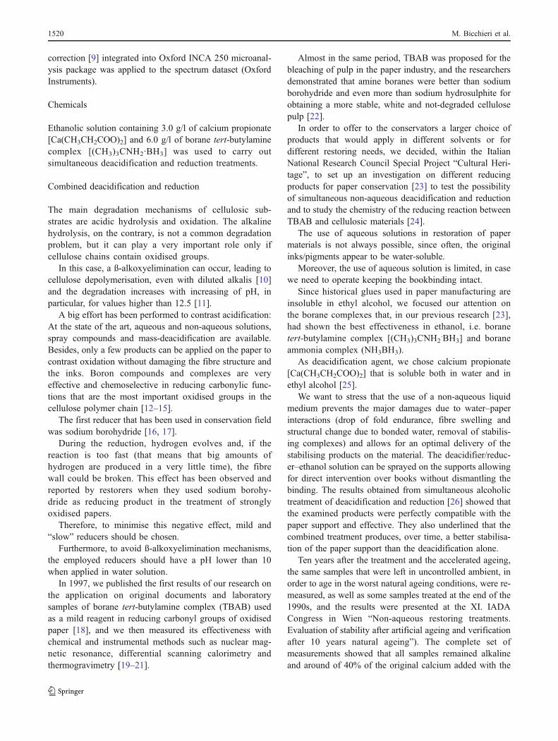
We could collect the spectrum of an iron-gall ink only inthe black region of C13. In Fig. 6, we report someexamples of the different typologies of black.
Ordered, disordered, crystalline and amorphous carbonsgive rise to different and distinguishable spectra [37].
The Raman shift of carbon-based materials is dividedinto first- and second-order regions. The first-order regionlies in the 1,110–1,800 cm-1 range, while the second-ordercovers the range 2,200–3,400 cm-1. The first-order spec-trum of graphite shows a single line at 1,583 cm-1 and avery intense second-order band at 2,750 cm-1.
In the presence of disorder, an additional line at about1,370 cm-1 appears. The position and the intensity of the Dband (in literature, D is referred to the main defect band) and
Fig. 3 Raman spectrum of a white pigment in drawing A9, showingthe presence of barite and gypsum that were used in mixture andwhose contribution to the spectrum is easily distinguished
Fig. 2 Raman spectrum collected from a brownish-black colour insketch A12. The colour is obtained by mixing carbon black, cinnabarand Prussian blue. For comparison, spectra of standard cinnabar,Prussian blue and carbon black are reported. All components are well-detected by Raman
Fig. 5 Raman spectrum of a white colour in drawing C13. In thiswhite, presence of weddellite (reaction product of microbiologicalattack) was recorded
Fig. 4 ATR/FTIR spectrum of a white colour in drawing C13,showing the use of kaolin that was not detected by Raman due anintense fluorescence background
1522 M. Bicchieri et al.
the G band (referred to graphite) depends on the disorder/order ratio, on the presence of sp2 rings or chains, on thesp2/sp3 ratio and on the wavelength of the excitation source.
The third band (usually D*) is an overtone of the D bandin partially disordered graphite.
In B and C spectra (Fig. 6), the two typical bands ofcarbon are not well resolved. It indicates that the analysedsamples have low crystallinity and that defects andamorphous regions are prevailing.
The D and E spectra, on the contrary, show sharp D andG bands. Moreover, the presence of the D* band thatindicates a higher amount of crystallinity is related to thegraphite content of the analysed samples.
All the other colours used in the drawings were obtainedwith variable mixtures of yellow, blue, red, white and black,as already shown in Fig. 2.
Very interesting results were obtained when analysingthe red colours; we found cinnabar and Mars red, twopigments that have historically been widely used, and anextensive usage of red pencils, made of martite (Fig. 7), e.g.in drawing C7L.
Martite is a hematite pseudomorph after magnetite,widely diffused in India and in the other countries wherePascarella travelled, but not in Italy.
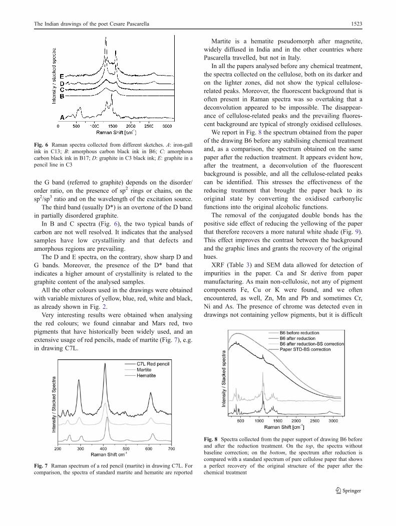
In all the papers analysed before any chemical treatment,the spectra collected on the cellulose, both on its darker andon the lighter zones, did not show the typical cellulose-related peaks. Moreover, the fluorescent background that isoften present in Raman spectra was so overtaking that adeconvolution appeared to be impossible. The disappear-ance of cellulose-related peaks and the prevailing fluores-cent background are typical of strongly oxidised celluloses.
We report in Fig. 8 the spectrum obtained from the paperof the drawing B6 before any stabilising chemical treatmentand, as a comparison, the spectrum obtained on the samepaper after the reduction treatment. It appears evident how,after the treatment, a deconvolution of the fluorescentbackground is possible, and all the cellulose-related peakscan be identified. This stresses the effectiveness of thereducing treatment that brought the paper back to itsoriginal state by converting the oxidised carbonylicfunctions into the original alcoholic functions.
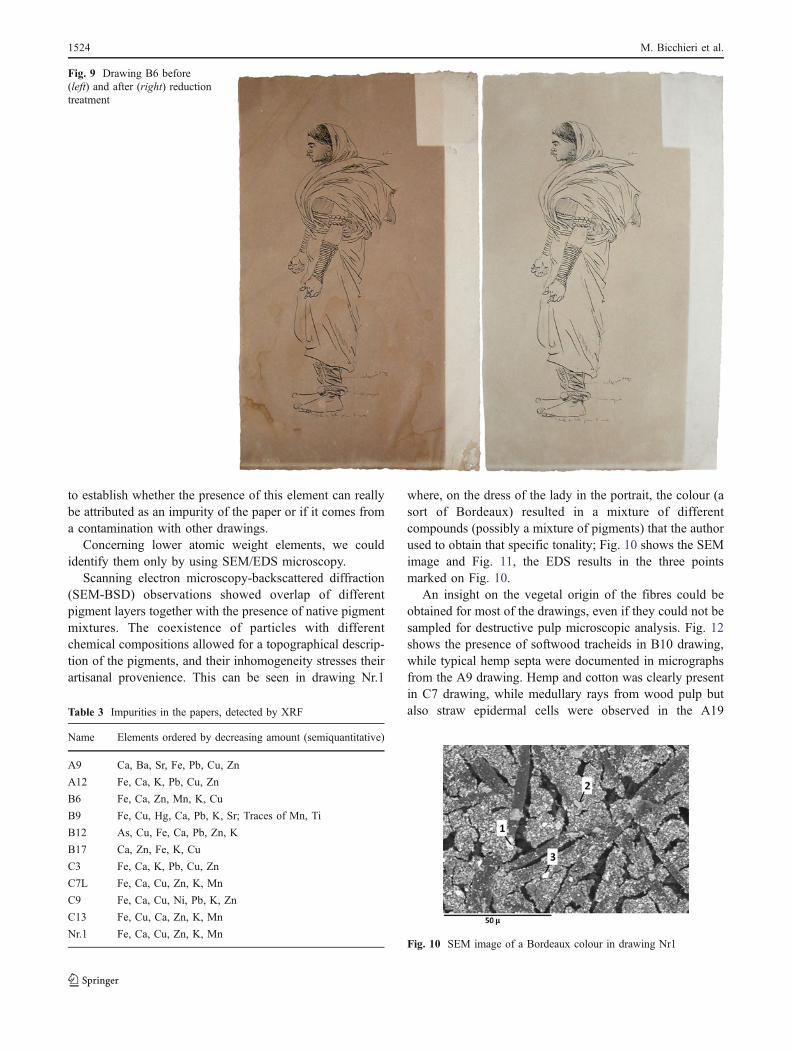
The removal of the conjugated double bonds has thepositive side effect of reducing the yellowing of the paperthat therefore recovers a more natural white shade (Fig. 9).This effect improves the contrast between the backgroundand the graphic lines and grants the recovery of the originalhues.
XRF (Table 3) and SEM data allowed for detection ofimpurities in the paper. Ca and Sr derive from papermanufacturing. As main non-cellulosic, not any of pigmentcomponents Fe, Cu or K were found, and we oftenencountered, as well, Zn, Mn and Pb and sometimes Cr,Ni and As. The presence of chrome was detected even indrawings not containing yellow pigments, but it is difficult
Fig. 8 Spectra collected from the paper support of drawing B6 beforeand after the reduction treatment. On the top, the spectra withoutbaseline correction; on the bottom, the spectrum after reduction iscompared with a standard spectrum of pure cellulose paper that showsa perfect recovery of the original structure of the paper after thechemical treatment
Fig. 7 Raman spectrum of a red pencil (martite) in drawing C7L. Forcomparison, the spectra of standard martite and hematite are reported
Fig. 6 Raman spectra collected from different sketches. A: iron-gallink in C13; B: amorphous carbon black ink in B6; C: amorphouscarbon black ink in B17; D: graphite in C3 black ink; E: graphite in apencil line in C3
The Indian drawings of the poet Cesare Pascarella 1523
to establish whether the presence of this element can reallybe attributed as an impurity of the paper or if it comes froma contamination with other drawings.
Concerning lower atomic weight elements, we couldidentify them only by using SEM/EDS microscopy.
Scanning electron microscopy-backscattered diffraction(SEM-BSD) observations showed overlap of differentpigment layers together with the presence of native pigmentmixtures. The coexistence of particles with differentchemical compositions allowed for a topographical descrip-tion of the pigments, and their inhomogeneity stresses theirartisanal provenience. This can be seen in drawing Nr.1
where, on the dress of the lady in the portrait, the colour (asort of Bordeaux) resulted in a mixture of differentcompounds (possibly a mixture of pigments) that the authorused to obtain that specific tonality; Fig. 10 shows the SEMimage and Fig. 11, the EDS results in the three pointsmarked on Fig. 10.
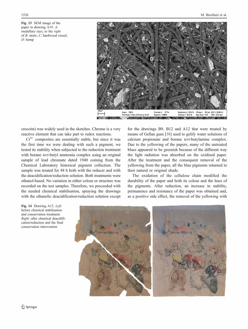
An insight on the vegetal origin of the fibres could beobtained for most of the drawings, even if they could not besampled for destructive pulp microscopic analysis. Fig. 12shows the presence of softwood tracheids in B10 drawing,while typical hemp septa were documented in micrographsfrom the A9 drawing. Hemp and cotton was clearly presentin C7 drawing, while medullary rays from wood pulp butalso straw epidermal cells were observed in the A19
Fig. 9 Drawing B6 before(left) and after (right) reductiontreatment
Table 3 Impurities in the papers, detected by XRF
Name Elements ordered by decreasing amount (semiquantitative)
A9 Ca, Ba, Sr, Fe, Pb, Cu, Zn
A12 Fe, Ca, K, Pb, Cu, Zn
B6 Fe, Ca, Zn, Mn, K, Cu
B9 Fe, Cu, Hg, Ca, Pb, K, Sr; Traces of Mn, Ti
B12 As, Cu, Fe, Ca, Pb, Zn, K
B17 Ca, Zn, Fe, K, Cu
C3 Fe, Ca, K, Pb, Cu, Zn
C7L Fe, Ca, Cu, Zn, K, Mn
C9 Fe, Ca, Cu, Ni, Pb, K, Zn
C13 Fe, Cu, Ca, Zn, K, Mn
Nr.1 Fe, Ca, Cu, Zn, K, MnFig. 10 SEM image of a Bordeaux colour in drawing Nr1
1524 M. Bicchieri et al.
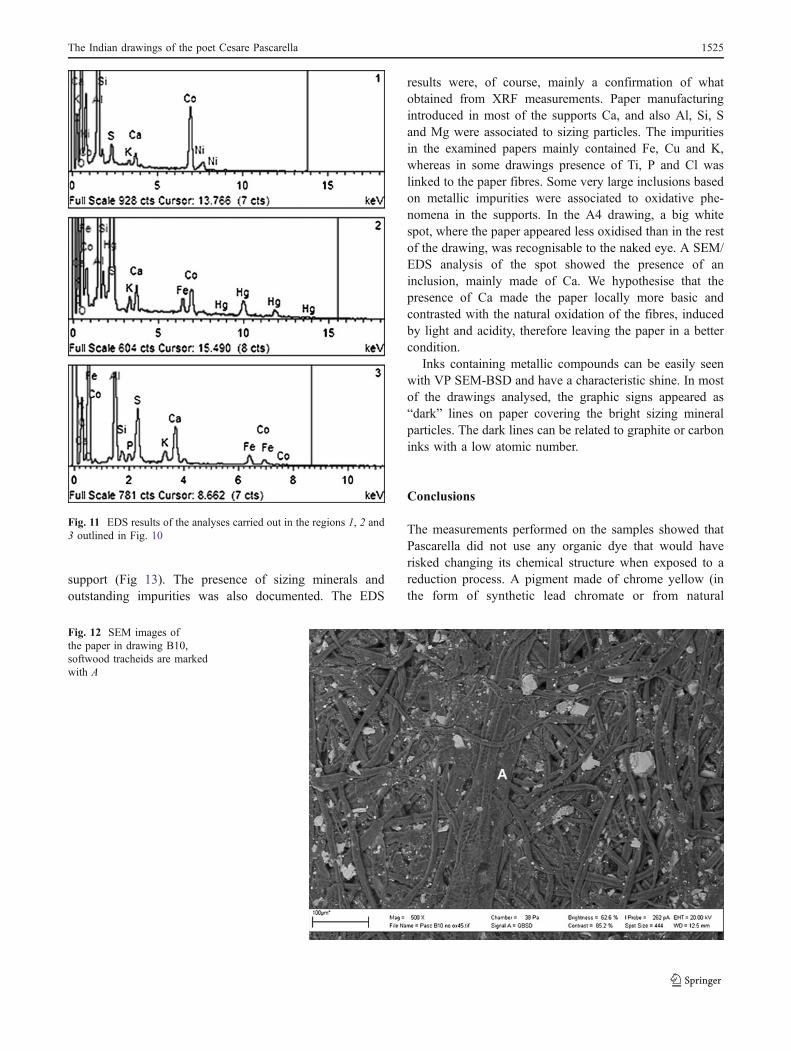
support (Fig 13). The presence of sizing minerals andoutstanding impurities was also documented. The EDS
results were, of course, mainly a confirmation of whatobtained from XRF measurements. Paper manufacturingintroduced in most of the supports Ca, and also Al, Si, Sand Mg were associated to sizing particles. The impuritiesin the examined papers mainly contained Fe, Cu and K,whereas in some drawings presence of Ti, P and Cl waslinked to the paper fibres. Some very large inclusions basedon metallic impurities were associated to oxidative phe-nomena in the supports. In the A4 drawing, a big whitespot, where the paper appeared less oxidised than in the restof the drawing, was recognisable to the naked eye. A SEM/EDS analysis of the spot showed the presence of aninclusion, mainly made of Ca. We hypothesise that thepresence of Ca made the paper locally more basic andcontrasted with the natural oxidation of the fibres, inducedby light and acidity, therefore leaving the paper in a bettercondition.
Inks containing metallic compounds can be easily seenwith VP SEM-BSD and have a characteristic shine. In mostof the drawings analysed, the graphic signs appeared as“dark” lines on paper covering the bright sizing mineralparticles. The dark lines can be related to graphite or carboninks with a low atomic number.
Conclusions
The measurements performed on the samples showed thatPascarella did not use any organic dye that would haverisked changing its chemical structure when exposed to areduction process. A pigment made of chrome yellow (inthe form of synthetic lead chromate or from natural
Fig. 11 EDS results of the analyses carried out in the regions 1, 2 and3 outlined in Fig. 10
Fig. 12 SEM images ofthe paper in drawing B10,softwood tracheids are markedwith A
The Indian drawings of the poet Cesare Pascarella 1525
crocoite) was widely used in the sketches. Chrome is a veryreactive element that can take part to redox reactions.
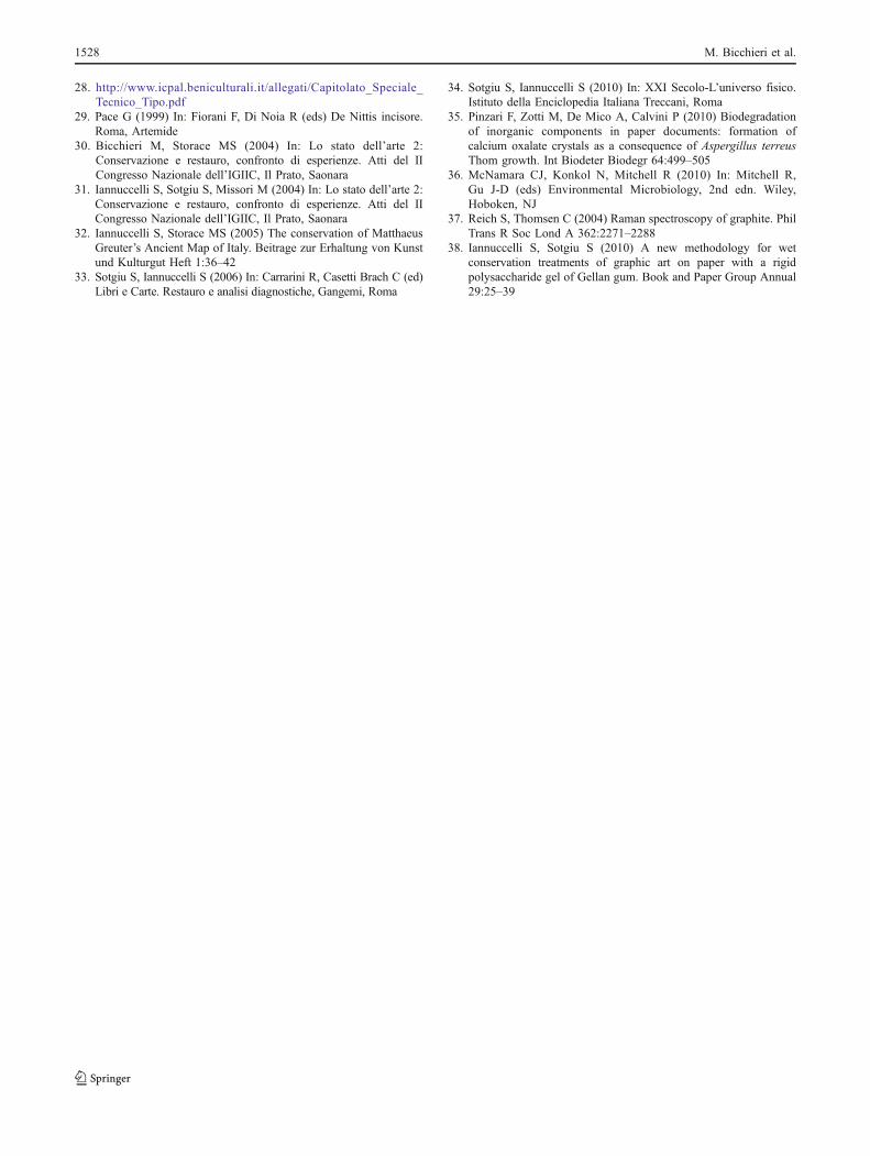
Cr6+ composites are essentially stable, but since it wasthe first time we were dealing with such a pigment, wetested its stability when subjected to the reduction treatmentwith borane tert-butyl ammonia complex using an originalsample of lead chromate dated 1940 coming from theChemical Laboratory historical pigment collection. Thesample was treated for 48 h both with the reducer and withthe deacidification/reduction solution. Both treatments wereethanol-based. No variation in either colour or structure wasrecorded on the test samples. Therefore, we proceeded withthe needed chemical stabilisation, spraying the drawingswith the ethanolic deacidification/reduction solution except
for the drawings B9, B12 and A12 that were treated bymeans of Gellan gum [38] used to gelify water solutions ofcalcium propionate and borane tert-butylamine complex.Due to the yellowing of the papers, many of the untreatedblues appeared to be greenish because of the different waythe light radiation was absorbed on the oxidised paper.After the treatment and the consequent removal of theyellowing from the paper, all the blue pigments returned totheir natural or original shade.
The oxidation of the cellulose chain modified thedurability of the paper and both its colour and the hues ofthe pigments. After reduction, an increase in stability,permanence and resistance of the paper was obtained and,as a positive side effect, the removal of the yellowing with
Fig. 13 SEM image of thepaper in drawing A19. A:medullary rays; to the rightof B: straw; C: hardwood vessel;D: hemp
Fig. 14 Drawing A12. Left:before chemical stabilisationand conservation treatment.Right: after chemical deacidifi-cation/reduction and the finalconservation intervention
1526 M. Bicchieri et al.

recovering of the original hues. Consequent to the chemicaltreatment, all the drawings underwent to the second step ofconservation–preservation treatment: Fragments were re-assembled; tears and lacunae were mended using machine-made Japanese paper (100% Kozo fibres, 3 g/m2) andhandmade woven western paper “Anna Amalia” made byGangolf Ulbricht (pure cotton and flax fibres, 50 g/m2). Thefinal inpainting of restored areas was carried out with“Winsor & Newton” watercolours (Fig. 14).
The images obtained with SEM allowed for a non-destructive recognition of the vegetal fibres employed inpaper manufacturing. Moreover, some fragments thatcould not be assigned to any of the re-assembledshredded drawings were used for the preparation ofmicroscopic slides that were used for pulp analysis. Wecould characterise the vegetable species in the paper andrecognise that some of them were typical of specificgeographic regions (e.g. the Indian drawings are madeon papers that show the presence of vegetables that canonly be found in India). In the same way, most of thewriting materials can be collocated geographically; wealso found watermarks belonging to Indian manufac-turers. We can therefore hypothesise that Pascarella used“local” materials.
The cross-information obtained by using different spec-troscopies made possible the establishment of the correctand successful conservation method. With this work, wewant to stress that the successful preservation of works ofart requires a cautious and comprehensive scientificapproach. Problems can arise from different scales andmaterials, therefore no single non-destructive technique canbe claimed to be the “resolving” one, and it becomesessential to fill the gap of the various techniques by usingcomplementary ones.
Acknowledgements We would like to acknowledge Dr. MarcoGuardo, Director of the Biblioteca Corsiniana, Accademia Nazionaledei Lincei, Rome, for cooperation and for allowing us to present theimages of Pascarella’s sketches.
References
1. Bioletti S, Leahy R, Fields J, Meehan B, Blau W (2009) Theexamination of the Book of Kells using micro-Raman spectros-copy. J Raman Spectrosc 40:1043–1049
2. Bicchieri M, Monti M, Piantanida G, Sodo A (2008) All that isiron ink is not always iron-gall. J Raman Spectrosc 39:1074–1078
3. Bicchieri M, Sodo A, Piantanida G, Coluzza C (2006) Analysis ofdegraded papers by non-destructive spectroscopic techniques. JRaman Spectrosc 37:1186–1192
4. Mansoa M, Carvalho ML (2009) Application of spectroscopictechniques for the study of paper documents: A survey. Spec-trochim Acta B 64:482–490
5. Workman JJ Jr (2001) Infrared and Raman spectroscopy in paperand pulp analysis. Appl Spectrosc Rev 36:139–168
6. Hochleitner B, Desnica V, Mantler M, Schreiner M (2003)Historical pigments: a collection analyzed with X-ray diffractionanalysis and X-ray fluorescence analysis in order to create adatabase*1. Spectrochim Acta B 58:641–649
7. Ferrero JL, Roldán C, Navarro E, Ardid M, Marzal M, AlmiranteJ, Ineba P, Vergara J, Mata C (1999) Applications of the X-rayfluorescence analysis to the cultural patrimony of the ComunidadValenciana (Spain): Painting, metal and paper. J Radioanal NuclCh 240:523–528
8. Roux S, Feugeas F, Bach M, Fagon M, Bettinger C, Mourad S,Cornet A (2008) Determination of paper filler Z-distribution bylow-vacuum SEM and EDX. J Microsc 229:44–59
9. Goldstein J, Newbury D, Joy D, Lyman C, Echlin P, Lifshin E,Sawyer L, Michael E (2003) Scanning Electron Microscopy andX-ray Microanalysis 3 rd edn. Kluver, New York
10. Richards GN (1971) In: Bikales NM, Segal L (eds) Cellulose andcellulose derivatives Part V, 2nd edn. New York, Wiley
11. Van Loon LR, Glaus MA (1997) Review of the kinetics of alkalinedegradation of cellulose in view of its relevance for safetyassessment of radioactive waste repositories. J Polym Environ 5(2):97–109
12. Walker ERH (1976) The functional group selectivity of complexhydride reducing agents. Chem Soc Rev 5(1):23–50
13. Block I, Kim HK (1986) In: Needles H, Zeronian SH (eds)Historic Textile and Paper Materials. Washington DC, ACS
14. Andrews GC (1980) Chemoselectivity in the reduction ofaldehydes and ketones with amine boranes. Tetrahedron Lett21:679–700
15. Raber DJ, Guida WC, Shoenberger DC (1981) Reduction ofaldehydes and ketones with tetraalkylammonium borohydrides.Tetrahedron Lett 22:5107–5110
16. Tang LC (1986) In: Needles H, Zeronian SH (eds) Historic Textileand Paper Materials. Washington DC, ACS
17. Burgess HD (1990) The stabilization of cellulosic fibres byborohydride derivatives. ICOM Committe For Conservation2:447–452
18. Bicchieri M, Brusa P (1997) The bleaching of paper by reductionwith borane tert-butylamine complex. Restaurator 18(1):1–11
19. Bicchieri M, Bella M, Sementilli F (1999) A quantitative measureof borane tert–butylamine complex effectiveness in carbonylreduction of aged papers. Restaurator 20(1):22–29
20. Bicchieri M, Curini R, D’Ascenzo G, Orrù MA (2000) Charac-terization of oxidized and reduced papers by thermogravimetricanalysis—effectiveness of reducing treatments. Qvinio 2:93–101
21. Capitani D, Segre AL, Pentimalli M, Bicchieri M,Munafò PF (2000)Ancient deteriorated paper: washing and restoring processes asstudied by 13 C CP-MAS NMR spectroscopy. Qvinio 2:37–43
22. Pedneault C, Robert S, Pellerin C (1997) Bleaching with newreductive chemicals: replacement of hydrosulfite. Pulp Pap Can98(3):51–55
23. Bicchieri M, Sementilli FM, Sodo A (2000) Application of sevenborane complexes in paper conservation. Restaurator 21(4):213–228
24. Sanna C, Sodo A, Laguzzi G, Mancini G, Bicchieri M (2009) Tert-butyl amine borane complex: an unusual application of a reducingagent on model molecules of cellulose based materials. J CultHerit 10(3):356–361
25. Plossi Zappalà M (1994) Il propionato di calcio nella deacidifi-cazione e stabilizzazione della carta. Cellulosa e Carta 45:53–60
26. Bicchieri M, Monti M, Antonelli ML (2001) In: J. Apuente (ed)Proceedings of the 3 rd International Conference on Science andTechnology for the safeguard of cultural heritage in the mediter-ranean basin Vol. I. Universitad de Alcalà, Alcalà
27. Coluzza C, Bicchieri M, Monti M, Piantanida G, Sodo A (2008)Atomic force microscopy application for degradation diagnosticsin library heritage. Surf Interface Anal 40(9):1248–1253
The Indian drawings of the poet Cesare Pascarella 1527
28. http://www.icpal.beniculturali.it/allegati/Capitolato_Speciale_Tecnico_Tipo.pdf
29. Pace G (1999) In: Fiorani F, Di Noia R (eds) De Nittis incisore.Roma, Artemide
30. Bicchieri M, Storace MS (2004) In: Lo stato dell’arte 2:Conservazione e restauro, confronto di esperienze. Atti del IICongresso Nazionale dell’IGIIC, Il Prato, Saonara
31. Iannuccelli S, Sotgiu S, Missori M (2004) In: Lo stato dell’arte 2:Conservazione e restauro, confronto di esperienze. Atti del IICongresso Nazionale dell’IGIIC, Il Prato, Saonara
32. Iannuccelli S, Storace MS (2005) The conservation of MatthaeusGreuter’s Ancient Map of Italy. Beitrage zur Erhaltung von Kunstund Kulturgut Heft 1:36–42
33. Sotgiu S, Iannuccelli S (2006) In: Carrarini R, Casetti Brach C (ed)Libri e Carte. Restauro e analisi diagnostiche, Gangemi, Roma
34. Sotgiu S, Iannuccelli S (2010) In: XXI Secolo-L’universo fisico.Istituto della Enciclopedia Italiana Treccani, Roma
35. Pinzari F, Zotti M, De Mico A, Calvini P (2010) Biodegradationof inorganic components in paper documents: formation ofcalcium oxalate crystals as a consequence of Aspergillus terreusThom growth. Int Biodeter Biodegr 64:499–505
36. McNamara CJ, Konkol N, Mitchell R (2010) In: Mitchell R,Gu J-D (eds) Environmental Microbiology, 2nd edn. Wiley,Hoboken, NJ
37. Reich S, Thomsen C (2004) Raman spectroscopy of graphite. PhilTrans R Soc Lond A 362:2271–2288
38. Iannuccelli S, Sotgiu S (2010) A new methodology for wetconservation treatments of graphic art on paper with a rigidpolysaccharide gel of Gellan gum. Book and Paper Group Annual29:25–39
1528 M. Bicchieri et al.