Ion-pair chromatographic monitoring of photographic effluents
Upload
khangminh22Category
view
4download
0
separations
Article
Non-Destructive X-ray Spectrometric andChromatographic Analysis of Metal Containers andTheir Contents, from Ancient Macedonia
Christos S. Katsifas 1,2,* ID , Despina Ignatiadou 3, Anastasia Zacharopoulou 1,Nikolaos Kantiranis 4 ID , Ioannis Karapanagiotis 5 ID and George A. Zachariadis 2
1 Laboratory of Physico-Chemical Studies & Archaeometry, Archaeological Museum of Thessaloniki,54013 Thessaloniki, Greece; [email protected]
2 Laboratory of Analytical Chemistry, Department of Chemistry, Aristotle University, 54124 Thessaloniki,Greece; [email protected]
3 Department of Sculpture, National Archaeological Museum, 10682 Athens, Greece; [email protected] Department of Mineralogy, Petrology and Economic Geology, School of Geology, Aristotle University,
54124 Thessaloniki, Greece; [email protected] Department of Management and Conservation of Ecclesiastical Cultural Heritage Objects, University
Ecclesiastical Academy of Thessaloniki, N. Plastira 65, 54250 Thessaloniki, Greece; [email protected]* Correspondence: [email protected], Tel.: +30-6944-262-282
Received: 1 April 2018; Accepted: 31 May 2018; Published: 11 June 2018�����������������
Abstract: This work describes a holistic archaeometric approach to ancient Macedonian specimens.In the region of the ancient city Lete, the deceased members of a rich and important family wereinterred in a cluster of seven tombs (4th century BC). Among the numerous grave goods, there wasalso a set of metal containers preserving their original content. The physico-chemical analysis of thecontainers and their contents was performed in order to understand the purpose of their use. For thecontainers, Energy Dispersive micro-X-Ray Fluorescence (EDµXRF) spectroscopy was implementedtaking advantage of its non-invasive character. The case (B35) and the small pyxis (B37) were made ofa binary Cu-Sn alloy accompanied by a slight amount of impurities (Fe, Pb, As) and the two miniaturebowls were made of almost pure Cu. For the study of the contents, a combination of EDµXRF,X-Ray Diffraction (XRD), and Gas Chromatography—Mass Spectrometry (GC-MS) was carried out.Especially for the extraction of the volatile compounds, the Solid Phase Micro-Extraction (SPME)technique was used in the headspace mode. Because of the detection of Br, High Pressure LiquidChromatography coupled to a Diode-Array-Detector (HPLC-DAD) was implemented, confirmingthe existence of the ancient dye shellfish purple (porphyra in Greek). The analytical results of thecombined implementation of spectrometric and chromatographic analytical techniques of the metalcontainers and their contents expand our knowledge about the pharmaceutical practices in Macedoniaduring the 4th century BC.
Keywords: Derveni; Ancient Macedonia; micro-XRF; XRD; HPLC-DAD; HS-SPME/GC-MS; ancientmedicines; ancient pharmaceuticals; shellfish purple; porphyra; high-tin bronzes; bronzes
1. Introduction
The Derveni tombs were accidentally revealed in 1962, 9.5 km NW of Thessaloniki, Macedonia,Greece. The six cist graves and one Macedonian tomb that were excavated then had not been lootedand contained rich offerings, mainly dated to the 4th century BC. The deceased were members of arich and important Thessalian family that probably lived in the nearby ancient city Lete. In grave A,the so-called Derveni papyrus was found; fragments of a papyrus roll with the most important Orphic
Separations 2018, 5, 32; doi:10.3390/separations5020032 www.mdpi.com/journal/separations

Separations 2018, 5, 32 2 of 17
religious text of the 4th century BC, preserving excerpts of an earlier poem. The biggest and richestgrave was grave B. It contained the cremated remains of a man and his female consort, which hadbeen placed in an elaborate bronze vessel, today known as the famous Derveni krater. That maleindividual was an important member of the elite, probably a royal companion who died when he wasapproximately 35–50 years old. In addition to the bronze krater, the burial contained a gold wreathand other gold jewelry, twenty silver vessels, many bronze vessels, stone alabastra, glass vessels andpottery vases, the iron weapons of the dead, a folding board gaming set with glass gaming counters,pieces of a leather cuirass, bronze greaves, and a gold coin of King Philip II [1,2].
Among the numerous grave goods, from grave B, was a lidded box (B35), preserving its originalcontent (Figure 1). It is a semi-cylindrical case divided into three compartments. Each compartment isfilled with a mass of “clay”. The case has a hinged lid that protects the contents. According to the firstestimations [1], B35 is a case for storing cosmetics and its contents are materials for makeup. Ongoingresearch on the history of medicine in Macedonia and the comparison with other metal cases whichhave been unearthed in Macedonian burials [3,4], after B35, had led to suspicions that the Derveni casewas a medical case. Other metal finds from the same grave that were more or less associated with thecase B35 are: two bowls (B43a, B43b) and a pyxis (B37), all preserving their original content. In the twobowls, there are preserved pieces of a thin dark-coloured cake and in the pyxis there is a red powder.
Separations 2018, 5, x FOR PEER REVIEW 2 of 17
Orphic religious text of the 4th century BC, preserving excerpts of an earlier poem. The biggest and richest grave was grave B. It contained the cremated remains of a man and his female consort, which had been placed in an elaborate bronze vessel, today known as the famous Derveni krater. That male individual was an important member of the elite, probably a royal companion who died when he was approximately 35–50 years old. In addition to the bronze krater, the burial contained a gold wreath and other gold jewelry, twenty silver vessels, many bronze vessels, stone alabastra, glass vessels and pottery vases, the iron weapons of the dead, a folding board gaming set with glass gaming counters, pieces of a leather cuirass, bronze greaves, and a gold coin of King Philip II [1,2].
Among the numerous grave goods, from grave B, was a lidded box (B35), preserving its original content (Figure 1). It is a semi-cylindrical case divided into three compartments. Each compartment is filled with a mass of “clay”. The case has a hinged lid that protects the contents. According to the first estimations [1], B35 is a case for storing cosmetics and its contents are materials for makeup. Ongoing research on the history of medicine in Macedonia and the comparison with other metal cases which have been unearthed in Macedonian burials [3,4], after B35, had led to suspicions that the Derveni case was a medical case. Other metal finds from the same grave that were more or less associated with the case B35 are: two bowls (B43a, B43b) and a pyxis (B37), all preserving their original content. In the two bowls, there are preserved pieces of a thin dark-coloured cake and in the pyxis there is a red powder.
The aims of the present physico-chemical study are: (a) to determine the composition of the alloy used for the construction of the metal containers, as well as the differentiation according to each part of the artefact (e.g., body, lid, handle, nails); and (b) to identify the inorganic and organic components of the contents using chemical and mineralogical analysis. The analytical results will contribute to the effort to determine the nature of these artefacts and consequently their purpose of use, as well as to illuminate the identity of their owner.
(a) (b)
(c) (d)
Figure 1. Metal containers and their contents. (a) Lidded case B35 and its content (cakes B35-I, B35-II, B35-III). (b) Case B35 without the lid. (c) Bowls B43a and B43b. (d) Lidded pyxis B37 (with and without lid).
2. Materials and Methods
The metal containers under study are: one lidded case (B35), two miniature bowls (B43a, B43b), and a small pyxis (B37), all found in Derveni grave B. The metal case B35 consists of different parts:
Figure 1. Metal containers and their contents. (a) Lidded case B35 and its content (cakes B35-I, B35-II,B35-III). (b) Case B35 without the lid. (c) Bowls B43a and B43b. (d) Lidded pyxis B37 (with and withoutlid).
The aims of the present physico-chemical study are: (a) to determine the composition of the alloyused for the construction of the metal containers, as well as the differentiation according to each partof the artefact (e.g., body, lid, handle, nails); and (b) to identify the inorganic and organic componentsof the contents using chemical and mineralogical analysis. The analytical results will contribute to theeffort to determine the nature of these artefacts and consequently their purpose of use, as well as toilluminate the identity of their owner.

Separations 2018, 5, 32 3 of 17
2. Materials and Methods
The metal containers under study are: one lidded case (B35), two miniature bowls (B43a, B43b),and a small pyxis (B37), all found in Derveni grave B. The metal case B35 consists of different parts:body, lid, handle, the lid’s hinges, the rim of the case proper, and the nails the latter is fastened with.The bowls B43a and B43b have been made from a single metal sheet and pyxis B37 consists of its bodyand a lid. Case B35 is preserved in very good condition. It has been chemically cleaned in the pastand does not carry any corrosion products. Its color, after the cleaning, is golden. On the other handthe two bowls (B43a, B43b) are reddish, denoting a differentiation of the alloy compared to that ofB35. They are also preserved in a very good state and apparently do not present corrosion layers.Finally, pyxis B37 presents, at the body, a thick reddish corrosion layer in combination with extensiverestoration works. Its lid maintains a better condition with areas where the metal does not presentextensive corrosion layers.
For the analysis of the metal containers, the choice of a non-invasive and non-destructive analyticaltechnique is obligatory. Sampling is largely prohibited, according to the Greek Archaeological Law,due to the uniqueness, integrity, and small size of the artefacts. So the choice of a non-invasivetechnique like Energy Dispersive micro X-ray Fluorescence (EDµXRF) spectroscopy—which is awidespread technique for the analysis of ancient metals [5,6]—was a requisite instead of a techniquethat provides bulk analysis, such as Inductively Coupled Plasma Spectroscopy (ICP) or AtomicAbsorption Spectroscopy (AAS). On the other hand, EDµXRF provides a chemical profile of the surface,which may differ from the bulk composition and perhaps is not representative of the whole [7,8].When bronze artefacts are exposed to the atmosphere or are buried in the ground, their surface acquiresa more or less thick patina under which the metal core may remain substantially unchanged [9].Before the implementation of the EDµXRF analysis, optical macro- and microscopic examination ofeach artefact was applied in order to select the spots for analysis. Areas free of corrosion products ormaterials from conservation treatments were selected. Especially at the pyxis B37, mechanical removalof the corrosion products took place in order to measure from the bulk of the metal.
The contents of case B35 bear similar hues. Starting from left to right, they were numbered: B35-I,B35-II, and B35-III. Especially at the middle compartment of B35 and on the top of cake B35-II, a fourthcake can be seen which has a different hue than the others. Instead of an earthy hue, it is reddish andwas hence numbered B35-IIa. Macroscopically, cakes B35-I, B35-II, and B35-III resemble dried out clays.Respectively, the contents of bowls B43a and B43b were numbered B43a-I and B43b-I. These cakesare harder than those in case B35 and their hue is brownish red with a dark top. To characterize thecomponents of the four cakes that are preserved in the lidded case B35, as well as the two from thebronze bowls (B43a, B43b) and the red powder from the pyxis (B37), a physico-chemical analysis wasundertaken in combination with a mineralogical examination. After the preliminary morphologicalexamination by stereomicroscopy, EDµXRF spectroscopy was carried out for the analysis of theinorganic constituents. In order to determine their mineralogical composition, XRD diffractometrywas implemented. For the study of the organic constituents, the samples were extracted with theHead Space—Solid Phase Micro-Extraction (HS-SPME) technique and the absorbed volatiles wereanalyzed by Gas Chromatography—Mass Spectrometry (GC-MS). Because of the significance of thematerial under study, an effort has been made to implement the above analytical techniques, as muchas possible, in a non-destructive way. Analysis using High Pressure Liquid Chromatography coupledto a Diode-Array-Detector (HPLC-DAD) was carried out in only one sample from cake B35-IIa in orderto ascertain the constituent which is responsible for its color. The cause of this implementation was theinteresting results of EDµXRF in combination with its reddish hue.
2.1. Energy Dispersive Micro-X-ray Fluorescence spectrometry (EDµXRF)
The instrument ARTAX 400 (Bruker AXS Microanalysis GmbH, Berlin, Germany) was used forthe implementation of the micro-X-ray Fluorescence spectroscopy technique (µXRF). This EnergyDispersive spectrometer has been specially designed for the demands of archaeometry [10].

Separations 2018, 5, 32 4 of 17
The measuring head, which is placed on a x,y,z—motor driven positioning stage, is comprised of: (a) anair cooled Mo X-ray fine focus tube, (b) a peltier cooled silicon drift detector (SDD), and (c) a ChargeCoupled Device (CCD) camera for the visual inspection of the sample. The X-ray beam is restricted bya collimator and on the surface of the sample has a diameter between 200 µm and 1500 µm, dependingon the type of collimator used. All excitation and detection paths can be rinsed with Helium (He) gaswhich is directed, by use of two gas jets, towards the sample surface in order to reduce the absorptionof the beam through the air and thus improve the excitation conditions for elements with a loweratomic number. Acquired spectra were processed with software SPECTRA 7.4. This program enablesthe acquisition of measurement data including the control of all Artax 400 components. Parallel tothe measurement, it is possible to carry out qualitative elemental analysis, data reduction (integralcalculation of the different spectral lines through deconvolution), and calculation of the artefactelemental concentration.
For the analysis of the metal containers, EDµXRF measurements (ARTAX 400, Bruker AXSMicroanalysis GmbH, Berlin, Germany) were taken in order to determine their elemental composition.For the metal case B35, measurements were taken from every different part (body, lid, hinge, rim,nails). Each final reported result is the mean value of three measurements. The measurement timefor each spot analysis was 100s. The voltage of the exciting X-ray beam was 50 kV and the currentwas 700 µA. The collimator with a 1500 µm diameter was used in order to get a representative resultand to avoid alterations due to micro structural inhomogeneity [11,12]. Especially for the analysis ofthe pyxis B37, which presents corrosion layers, and in order to avoid the removal of corrosion in anextended area (since it is considered as a destructive action), the collimator with a diameter of 650 µmwas used. The certified data from Certified Reference Material (CRM) 32XSN1 (MBH—Analytical Ltd.,Barnet, UK) was used as a calibration file. The quantified results were balanced to 100%. In order tocheck the accuracy of the implemented method, measurements were taken with the same parametersas those employed for the artefacts, from Certified Reference Materials (CRMs). BCR-691 (EuropeanCommission—Joint Research Centre, Institute for Reference Materials and Measurements, Brussels,Belgium), a set of five (5) copper alloys, was used (Table 1).
Table 1. Results of the analysis of Certified Reference Materials (CRM’s) and detection limits (wt %).
BCR-691 Sn Zn Pb As
Quaternary bronze (A) Certified value ± unsertainty 7.16 ± 0.21 6.02 ± 0.22 7.90 ± 0.7 0.19 ± 0.01
Measured value ± std 7.00 ± 0.3 6.20 ± 0.45 7.50 ± 0.50 0.20 ± 0.00
Brass (B)Certified value ± unsertainty 2.06 ± 0.07 14.80 ± 0.50 0.39 ± 0.04 0.10 ± 0.01
Measured value ± std 2.00 ± 0.10 14.50 ± 0.21 0.30 ± 0.06 0.10 ± 0.00
Arsenic copper (C) Certified value ± unsertainty 0.202 ± 0.029 0.05 ± 0.005 0.175 ± 0.014 4.6 ± 0.27
Measured value ± std 0.17 ± 0.06 0.03 ± 0.01 0.18 ± 0.04 4.5 ± 0.50
Lead bronze (D)Certified value ± unsertainty 10.1 ± 0.80 0.148 ± 0.024 9.2 ± 1.7 0.285 ± 0.022
Measured value ± std 9.60 ± 0.50 0.17 ± 0.02 9.05 ± 0.416 0.26 ± 0.05
Tin bronze (E)Certified value ± unsertainty 7 ± 0.60 0.157 ± 0.025 0.204 ± 0.018 0.194 ± 0.02
Measured value ± std 7.33 ± 0.50 0.17 ± 0.01 0.2 ± 0.00 0.2 ± 0.00
Estimated detectionlimits (%) 0.02 0.01 0.03 0.01
For the elemental analysis of the contents of the metal containers, with the EDµXRF technique,the voltage of the exciting X-ray beam was 35 kV and the current was 900 µA. It was also implementedin an He gas atmosphere. The 1500 µm diameter collimator was used to counterbalance theinhomogeneity of the materials under study. Each final reported result is the mean value of sevenmeasurements since the contents present greater inhomogeneity than the metal containers. For thequantification of the analytical results, the CRM soil sample SO-3 (Canada Centre for Mineral & EnergyTechnology, Ottawa, ON, Canada) was used in combination with software SPECTRA 7.4 (Bruker AXSMicroanalysis GmbH, Berlin, Germany).

Separations 2018, 5, 32 5 of 17
2.2. X-ray Diffraction (XRD)
The content of the metal containers were archaeological material and therefore were studied intheir bulk form without any grinding, homogenization, or pretreatment. In order to identify theirmineralogical composition, X-ray diffraction analysis (XRD) was applied directly on the surface ofthe samples.
A Phillips PW1820/00 diffractometer equipped with a PW1710/00 microprocessor (PHILIPS,Almelo, The Netherlands) was used and the samples were scanned over the 3◦–63◦ 2θ interval at ascanning speed of 1.2◦/min. Semi-quantitative analysis estimates of the abundance of the mineralphases were derived from the XRD data, using the intensity of a certain reflection [13], the density,and the mass absorption coefficient for Cu-Kα radiation for the minerals present. The identificationof the minerals present was made using the DDView+/SiIeve+ ICDD’s viewing/search indexingsoftware provided with the PDF-4+ (PDF-4+, 2009, Powder diffraction fileTM, International Centre forDiffraction Data, Newtown Square, PA, USA) relational database. Corrections were made using theexternal standard mixtures of minerals (i.e., standard mixtures of the identified minerals in the studiedsamples such as quartz and cristobalite, feldspars, micas and chlorite, amphibole, calcite and sulfateminerals, as well as iron oxides). The detection limit of the method was ±1% w/w [14]. The degreeof crystallinity and the calculation of the amorphous phase amount were calculated according to themethod described by Kantiranis et al. (2004) [14].
2.3. Head Space—Solid Phase Micro-Extraction/Gas Chromatography—Mass Spectrometry(HS—SPME/GC—MS)
The pre-treatment and extraction of the samples was done by the HS-SPME technique usinga polydimethylsiloxane (PDMS) coated fiber (100 µm film) in the head space above the heatedsamples. GC-MS (Agilent 6873 K gas chromatograph—Agilent 5973 quadrupole mass detector,Agilent Technologies, Santa Clara, CA, USA) analysis of the absorbed volatiles from the sampleswas carried out and finally the identification of the compounds was succeeded by using the NISTlibrary. The GC-MS operating conditions are listed in Table 2.
Table 2. GC-MS operating conditions.
Operating Conditions Value
GC model Agilent 6873 K gas chromatograph (Electron Ionization mode)Column DB-5MS (capillary) 30 m × 0.25 mm × 0.10 µm
Injector port temperature 200 ◦C (SPME fiber was remained there for 6 min)Carrier gas, Flow-rate Helium, 2.0 mL/min (constant pressure at 29.8 psi)
Oven temperature program 60 ◦C (3 min) to 270 ◦C, 10 ◦C/min ramp timeTransfer line temperature 250 ◦C
MS model Agilent 5973 quadrupole mass detector (Scan mode)Total time of chromatographic analysis 40 min
2.4. High Pressure Liquid Chromatography—Diode Array Detector (HPLC-DAD)
High Pressure Liquid Chromatography was coupled to a Diode-Array-Detector (Ultimate 3000,Dionex, Sunnyvale, CA, USA,) and consisted of a LPG-3000 quaternary HPLC pump with a vacuumdegasser, a WPS-3000SL auto sampler, a column compartment TCC-3000SD, and a UV-Vis Diode ArrayDetector (DAD-3000) (Dionex, Sunnyvale, CA, USA) Analyses were carried out by injecting 20 µL intoan Alltima (Grace-Alltech, Deerfield, IL, USA) HP C18 (250 mm × 3 mm, i.d. 5 µm) column at a stabletemperature of 35 ◦C.
Two solvent reservoirs, containing (A) water + 0.1% (v/v) Tri-Fluoro-Acetic Acid (TFA) and (B)acetonitrile + 0.1% (v/v) TFA, were used under a gradient elution program which was developed andevaluated for the analysis of shellfish purple components offering extremely low limits of detection [15].
The sample was immersed in a hot (80 ◦C) dimethyl sulfoxide (DMSO) bath and kept therefor 15 min. After centrifugation, the upper liquid phase was immediately submitted to HPLC.

Separations 2018, 5, 32 6 of 17
By employing this method, polar and apolar compounds are detected. The method was previouslydevised and optimised for the extraction and solubilisation of shellfish purple, as described in detailelsewhere [16].
3. Results
3.1. Metal Containers
According to the results of Table 3, the main parts (body, lid, and rim) of the metal case B35were made of copper-tin (Cu-Sn) alloy. Lead (Pb), iron (Fe), and calcium (Ca) were detected, at allparts, at quantities much lower than 1 wt %. Arsenic (As) is also detected, except at the four nailswhich present significant differentiation, at their chemical composition, compared to the main partsof B35. They consist of almost pure Cu which is detected at levels higher than 99.0 wt %. Nickel (Ni)and titanium (Ti) are only detected at the nails of B35, at very low quantities (rounded average forthe four nails are: 0.17 ± 0.03 and 0.01 ± 0.00 wt % for Ni and Ti, respectively), while Sn is presentat levels below 1 wt %. Another part of B35 that presents a significant differentiation is the hingeof the lid where the quantity of Sn (6.56 ± 0.30 wt %) is very low compared to the body of the case(12.11 ± 0.20 wt %).
Bowls B43a and B43b present the same qualitative and quantitative analysis. Cu is detected atlevels around 99 wt %. The two bowls present also the same chemical elements Sn, Pb, Fe, As, Ni,and Ca at similar quantities (lower than 0.5 wt %). At pyxis B37, measurements were only taken fromits lid after the removal of superficial corrosion products. The body presents thick corrosion layerswhich will not provide us with a safe quantitative result. The lid of pyxis B37 mainly consists of Cu(86.96 ± 0.40 wt %) and Sn (12.16 ± 0.20 wt %) at quantities similar to the main parts of the metal caseB35, e.g., the body (Cu: 87.61 ± 0.60 wt % and Sn: 12.16 ± 0.20 wt %). Similar to B35, Pb and Fe are theminor elements, at quantities lower than 1 wt %. However, Ni is only detected at a very low quantity(0.15 ± 0.50 wt %).
3.2. Contents of the Metal Containers
According to the EDµXRF analysis (Table 4), cakes B35-I, B35-II, and B35-III, which present asimilar appearance, texture, and colour, also present similar analytical results. Major constituents(concentration higher than 1 wt %) are: SiO2, Al2O3, Fe2O3(total), MgO, CaO, CuO, and K2O; and minorconstituents (concentration between 0.1 wt % and 1.0 wt %) are: V2O3, and MnO. Finally, at the levelof traces (concentration lower than 0.1 wt %), the following were detected: Cr2O3, NiO, ZnO, Rb2O,SrO, and PbO.
Cake B35-IIa, which presents a reddish hue, presents some differentiations compared to the othercakes. Al2O3, which is a main constituent in the other cakes, is a minor one at B35-IIa (0.28 ± 0.30 wt %).Furthermore, the total percentage of the inorganic constituents is lower in than the other cakes andranges from 16.56 wt % to 42.27 wt %. Finally, B35-IIa is the only cake where the chemical elementbromine (Br) was detected (Figure 2). This result, in combination with the differentiation of the colourof the cake (reddish hue), created suspicions for the possible existence of a dye. In the cakes (B43a-I,B43b-I) of the two bowls, there is a similar distribution between major and minor constituents and thosethat detected as traces. Compared to cakes of the metal case B35, the total percentage of the inorganicoxides (30.98 wt % for cake B43a-Iand 15.03 wt % for cakeB43b-I) is significantly lower. The powder inpyxis B37 presents great differentiations, in terms of the analytical results, compared to the contents ofthe others containers. There are only two major constituents: Fe2O3(t) (11.44 ± 1.10 wt %) and CuO(9.71 ± 1.10 wt %). The minor constituent detected is CaO, and ZnO and K2O are only detectedin traces.

Separations 2018, 5, 32 7 of 17
Table 3. Chemical composition (wt % ± std) of metal containers—results of EDµXRF analysis (* nd: not detected).
No Description Cu Sn Pb Fe As Ni Ca Ti
1 B35-body 87.61 ± 0.60 12.11 ± 0.20 0.19 ± 0.04 0.06 ± 0.02 0.02 ± 0.01 nd* 0.01 ± 0.00 nd2 B35-lid 87.68 ± 0.50 12.07 ± 0.30 0.12 ± 0.02 0.08 ± 0.03 0.04 ± 0.01 nd 0.02 ± 0.01 nd3 B35-lid-handle 85.33 ± 0.30 14.42 ± 0.20 0.15 ± 0.03 0.06 ± 0.02 0.01 ± 0.00 nd 0.03 ± 0.01 nd4 B35-lid-hinge 93.00 ± 0.60 6.59 ± 0.30 0.23 ± 0.04 0.13 ± 0.05 0.03 ± 0.01 nd 0.02 ± 0.01 nd5 B35 body-rim 83.44 ± 0.30 15.62 ± 0.50 0.72 ± 0.04 0.04 ± 0.02 0.01 ± 0.00 nd 0.02 ± 0.01 nd6 B35-nail-01 99.20 ± 0.60 0.06 ± 0.01 0.35 ± 0.03 0.20 ± 0.03 nd 0.17 ± 0.03 0.01 ± 0.00 0.01 ± 0.007 B35-nail-02 98.95 ± 0.50 0.76 ± 0.20 0.40 ± 0.02 0.18 ± 0.04 nd 0.17 ± 0.03 0.02 ± 0.00 0.01 ± 0.008 B35-nail-03 99.22 ± 0.50 0.10 ± 0.02 0.34 ± 0.04 0.15 ± 0.02 nd 0.16 ± 0.03 0.02 ± 0.01 nd9 B35-nail-04 99.09 ± 0.60 0.12 ± 0.40 0.43 ± 0.06 0.17 ± 0.08 nd 0.16 ± 0.03 0.01 ± 0.00 0.01 ± 0.0010 B43a-body 99.20 ± 0.40 0.04 ± 0.01 0.45 ± 0.05 0.14 ± 0.05 0.01 ± 0.00 0.16 ± 0.03 0.01 ± 0.00 nd11 B43b-body 99.12 ± 0.50 0.04 ± 0.01 0.48 ± 0.02 0.18 ± 0.03 0.01 ± 0.00 0.17 ± 0.03 0.01 ± 0.00 nd12 B37-lid 86.96 ± 0.40 12.16 ± 0.20 0.66 ± 0.04 0.07 ± 0.02 nd 0.15 ± 0.05 nd nd
Table 4. Contents of metal containers—results of the EDµXRF analysis (wt %).
Description SiO2 TiO2 Al2O3 Fe2O3(t) V2O3 Cr2O3 MnO MgO CaO CuO NiO ZnO Rb2O SrO PbO K2O Br TOTAL
B35-I 42.66 ± 2.10 1.42 ± 0.30 15.53 ± 1.20 10.38 ± 1.10 0.18 ± 0.06 0.09 ± 0.04 0.31 ± 0.10 2.19 ± 0.20 4.90 ± 0.42 2.30 ± 0.32 0.01 ± 0.00 0.01 ± 0.00 0.02 ± 0.01 0.04 ± 0.02 0.01 ± 0.00 3.36 ± 0.45 nd 83.41B35-II 29.84 ± 1.40 0.82 ± 0.20 10.75 ± 0.40 5.95 ± 0.40 0.10 ± 0.04 0.04 ± 0.02 0.18 ± 0.02 0.86 ± 0.40 3.12 ± 0.25 4.12 ± 0.70 nd 0.01 ± 0.00 0.01 ± 0.00 0.02 ± 0.01 0.02 ± 0.01 1.84 ± 0.28 nd 57.70B35-IIa 16.58 ± 1.20 0.43 ± 0.12 0.28 ± 0.30 4.65 ± 1.00 0.04 ± 0.03 0.15 ± 0.05 0.06 ± 0.03 0.90 ± 0.20 4.67 ± 0.32 11.85 ± 1.56 0.03 ± 0.01 0.02 ± 0.01 0.02 ± 0.01 0.01 ± 0.00 0.01 ± 0.00 1.02 ± 0.04 0.40 ± 0.05 41.14B35-III 43.94 ± 2.15 1.22 ± 0.30 18.06 ± 1.80 9.92 ± 1.10 0.22 ± 0.04 0.07 ± 0.03 0.10 ± 0.02 1.84 ± 0.35 2.00 ± 0.24 2.02 ± 0.35 0.01 ± 0.00 0.01 ± 0.00 0.02 ± 0.01 0.02 ± 0.01 0.14 ± 0.03 2.92 ± 0.67 nd 82.53B43a-I 6.48 ± 1.20 0.35 ± 0.15 2.19 ± 0.25 3.57 ± 0.70 0.75 ± 0.25 0.12 ± 0.02 0.03 ± 0.01 0.86 ± 0.25 3.65 ± 0.18 12.02 ± 1.32 0.04 ± 0.02 0.01 ± 0.00 nd 0.01 ± 0.00 0.16 ± 0.00 0.73 ± 0.25 nd 30.98B43b-I 4.43 ± 0.80 0.22 ± 0.30 1.10 ± 0.20 2.42 ± 0.40 0.44 ± 0.10 0.07 ± 0.03 0.01 ± 0.00 0.63 ± 0.18 1.78 ± 0.16 3.39 ± 0.88 0.03 ± 0.01 nd nd 0.01 ± 0.00 0.10 ± 0.00 0.41 ± 0.25 nd 15.03B37-I nd nd nd 11.44 ± 1.10 nd nd nd nd 0.27 ± 0.08 9.71 ± 1.10 nd 0.02 ± 0.00 nd nd nd 0.02 ± 0.00 nd 21.47
nd: not detected.
Separations 2018, 5, 32 8 of 17Separations 2018, 5, x FOR PEER REVIEW 8 of 17
Figure 2. EDμXRF spectrum of cake B-35-IIa—detection of Br (peak Ka1 at 11.88 keV and Kb1 at 13.29 keV).
The detection of Br in combination with the colour differentiation of the sample B35-IIa led to the investigation of the possible existence of a dye that contains Br. The implementation of the HPLC-DAD technique resulted in the detection of the compound 6,6′-dibromoindigotin (DBI) (Figure 3), which was detected at 288 nm. The limit of daltons is 2.4 × 10¹⁴, as described in detail elsewhere [13].
Figure 3. 6,6′-dibromoindigotin (DBI).
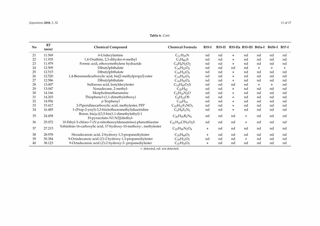
According to the results of the mineralogical analysis (Table 5) of the cakes in case B35, similar amounts of quartz (SiO2) (39–44 wt %) were detected at high quantities. Other constituents in abundance are: plagioclase (8–29 wt %), mica (6–14 wt %) that belongs to the group of silicate minerals, and chlorite (4–8 wt %). Minerals like K-feldspar and calcite are only detected in cakes B35-II and B35-III. Amorphous matter estimated at high level ranges from 16% to 31%, except in cake B35-I, where it is only 5 wt %. In cakes B43a-I and B43b-I, the amorphous matter estimated at even higher levels ranges from 65 wt % to 78 wt %. Except for the siliceous minerals (quartz, plagioclase, and mica), cristobalite (6–12 wt %), gypsum (5 wt %), and graphite (1 wt %) were also detected. Graphite was possible to identify (Figure 4) and measure using the 89-8487 ICDD card (main peak 3.3540Å, in comparison with the 3.3434 Å main peak of quartz, 46-1045 ICDD card). Especially 2 wt % bassanite was detected in cake B43b-I, a calcium sulfate mineral. Finally, powder B37-I presented a very different mineralogical composition than the others. The dominant constituents are the ferrous oxides (hematite and magnetite: 71 wt %), a copper sulphate mineral antlerite (15 wt %), gypsum (8 wt %), bassanite (5 wt %), and a low quantity of a rare borate mineral: inderite (1 wt %).
O
NBrH
C CNH
Br
O
Figure 2. EDµXRF spectrum of cake B-35-IIa—detection of Br (peak Ka1 at 11.88 keV and Kb1 at 13.29 keV).
The detection of Br in combination with the colour differentiation of the sample B35-IIa led to theinvestigation of the possible existence of a dye that contains Br. The implementation of the HPLC-DADtechnique resulted in the detection of the compound 6,6′-dibromoindigotin (DBI) (Figure 3), which wasdetected at 288 nm. The limit of daltons is 2.4 × 10¹⁴, as described in detail elsewhere [13].
Separations 2018, 5, x FOR PEER REVIEW 8 of 17
Figure 2. EDμXRF spectrum of cake B-35-IIa—detection of Br (peak Ka1 at 11.88 keV and Kb1 at 13.29 keV).
The detection of Br in combination with the colour differentiation of the sample B35-IIa led to the investigation of the possible existence of a dye that contains Br. The implementation of the HPLC-DAD technique resulted in the detection of the compound 6,6′-dibromoindigotin (DBI) (Figure 3), which was detected at 288 nm. The limit of daltons is 2.4 × 10¹⁴, as described in detail elsewhere [13].
Figure 3. 6,6′-dibromoindigotin (DBI).
According to the results of the mineralogical analysis (Table 5) of the cakes in case B35, similar amounts of quartz (SiO2) (39–44 wt %) were detected at high quantities. Other constituents in abundance are: plagioclase (8–29 wt %), mica (6–14 wt %) that belongs to the group of silicate minerals, and chlorite (4–8 wt %). Minerals like K-feldspar and calcite are only detected in cakes B35-II and B35-III. Amorphous matter estimated at high level ranges from 16% to 31%, except in cake B35-I, where it is only 5 wt %. In cakes B43a-I and B43b-I, the amorphous matter estimated at even higher levels ranges from 65 wt % to 78 wt %. Except for the siliceous minerals (quartz, plagioclase, and mica), cristobalite (6–12 wt %), gypsum (5 wt %), and graphite (1 wt %) were also detected. Graphite was possible to identify (Figure 4) and measure using the 89-8487 ICDD card (main peak 3.3540Å, in comparison with the 3.3434 Å main peak of quartz, 46-1045 ICDD card). Especially 2 wt % bassanite was detected in cake B43b-I, a calcium sulfate mineral. Finally, powder B37-I presented a very different mineralogical composition than the others. The dominant constituents are the ferrous oxides (hematite and magnetite: 71 wt %), a copper sulphate mineral antlerite (15 wt %), gypsum (8 wt %), bassanite (5 wt %), and a low quantity of a rare borate mineral: inderite (1 wt %).
O
NBrH
C CNH
Br
O
Figure 3. 6,6′-dibromoindigotin (DBI).
According to the results of the mineralogical analysis (Table 5) of the cakes in case B35, similaramounts of quartz (SiO2) (39–44 wt %) were detected at high quantities. Other constituents inabundance are: plagioclase (8–29 wt %), mica (6–14 wt %) that belongs to the group of silicateminerals, and chlorite (4–8 wt %). Minerals like K-feldspar and calcite are only detected in cakesB35-II and B35-III. Amorphous matter estimated at high level ranges from 16% to 31%, except in cakeB35-I, where it is only 5 wt %. In cakes B43a-I and B43b-I, the amorphous matter estimated at evenhigher levels ranges from 65 wt % to 78 wt %. Except for the siliceous minerals (quartz, plagioclase,and mica), cristobalite (6–12 wt %), gypsum (5 wt %), and graphite (1 wt %) were also detected.Graphite was possible to identify (Figure 4) and measure using the 89-8487 ICDD card (main peak3.3540Å, in comparison with the 3.3434 Å main peak of quartz, 46-1045 ICDD card). Especially 2 wt %bassanite was detected in cake B43b-I, a calcium sulfate mineral. Finally, powder B37-I presented avery different mineralogical composition than the others. The dominant constituents are the ferrousoxides (hematite and magnetite: 71 wt %), a copper sulphate mineral antlerite (15 wt %), gypsum(8 wt %), bassanite (5 wt %), and a low quantity of a rare borate mineral: inderite (1 wt %).

Separations 2018, 5, 32 9 of 17
Separations 2018, 5, x FOR PEER REVIEW 9 of 17
Figure 4. XRD pattern of cake B43b-I.
Table 5 presents the organic constituents of the contents of the metal containers according to the results of the HS-SPME/GC-MS analysis. In cake B35-I, only three (3) organic compounds were detected, none were detected at B35-II, and only three (3) (most of them fatty acids) were detected at B35-III. Contrary to the above, in cake B35-IIa, which presents a reddish hue, twenty-two (22) organic constituents (fatty acids, fatty acid esters, hydrocarbons, quinolines, phthalate esters, and amines) were detected (Figure 5). At contents B43a-I and B43b-I, the same three organic constituents were detected and at the red powder B37-I, seven (7) (thioamides, lactam, hydrocarbons, halide, and phthalate ester) were detected. Except for B43a-I and B43b-I, the other contents do not share any of the organic compounds. Fatty acids are the class of organic compounds which is common in the B35 and B43 contents. On the other hand, classes like thioamides and lactam are only detected at powder B37.
Figure 5. GC-MS chromatogram of volatile constituents of the sample B35-IIa (retention times correspond to analytes as described in Table 6).
Figure 4. XRD pattern of cake B43b-I.
Table 5 presents the organic constituents of the contents of the metal containers according tothe results of the HS-SPME/GC-MS analysis. In cake B35-I, only three (3) organic compounds weredetected, none were detected at B35-II, and only three (3) (most of them fatty acids) were detected atB35-III. Contrary to the above, in cake B35-IIa, which presents a reddish hue, twenty-two (22) organicconstituents (fatty acids, fatty acid esters, hydrocarbons, quinolines, phthalate esters, and amines) weredetected (Figure 5). At contents B43a-I and B43b-I, the same three organic constituents were detectedand at the red powder B37-I, seven (7) (thioamides, lactam, hydrocarbons, halide, and phthalateester) were detected. Except for B43a-I and B43b-I, the other contents do not share any of the organiccompounds. Fatty acids are the class of organic compounds which is common in the B35 and B43contents. On the other hand, classes like thioamides and lactam are only detected at powder B37.
Separations 2018, 5, x FOR PEER REVIEW 9 of 17
Figure 4. XRD pattern of cake B43b-I.
Table 5 presents the organic constituents of the contents of the metal containers according to the results of the HS-SPME/GC-MS analysis. In cake B35-I, only three (3) organic compounds were detected, none were detected at B35-II, and only three (3) (most of them fatty acids) were detected at B35-III. Contrary to the above, in cake B35-IIa, which presents a reddish hue, twenty-two (22) organic constituents (fatty acids, fatty acid esters, hydrocarbons, quinolines, phthalate esters, and amines) were detected (Figure 5). At contents B43a-I and B43b-I, the same three organic constituents were detected and at the red powder B37-I, seven (7) (thioamides, lactam, hydrocarbons, halide, and phthalate ester) were detected. Except for B43a-I and B43b-I, the other contents do not share any of the organic compounds. Fatty acids are the class of organic compounds which is common in the B35 and B43 contents. On the other hand, classes like thioamides and lactam are only detected at powder B37.
Figure 5. GC-MS chromatogram of volatile constituents of the sample B35-IIa (retention times correspond to analytes as described in Table 6).
Figure 5. GC-MS chromatogram of volatile constituents of the sample B35-IIa (retention timescorrespond to analytes as described in Table 6).

Separations 2018, 5, 32 10 of 17
Table 5. Contents of metal containers—results of the XRD analysis (wt %).
Sample Quartz Plagioclase Mica Chlorite Dolomite Amorphous K-Feldspar Calcite Amphibole Cristobalite Gypsum Graphite Hydrozincite Hematite Magnetite Bassanite Inderite Antlerite
B35-I 39 28 14 8 nd 5 nd nd 5 nd nd nd 1 nd nd nd nd ndB35-II 43 8 9 5 nd 18 7 7 3 nd nd nd nd nd nd nd nd ndB35-IIa 42 9 8 7 3 31 nd nd nd nd nd nd nd nd nd nd nd ndB35-III 44 13 6 4 nd 16 8 6 3 nd nd nd nd nd nd nd nd ndB43a-I 7 1 1 1 nd 78 nd nd nd 6 5 1 nd nd nd nd nd ndB43b-I 11 1 1 nd nd 65 2 nd nd 12 5 1 nd nd nd 2 nd ndB37-I nd nd nd nd nd nd nd nd nd nd 8 nd nd 62 9 5 1 15
nd: not detected
Table 6. Contents of the metal containers: results of HS-SPME/GC-MS analysis.
No RT(min) Chemical Compound Chemical Formula B35-I B35-II B35-IIa B35-III B43a-I B43b-I B37-I
1 1.205 Ethanethioamide C2H5NS nd nd nd nd nd nd +2 1.422 2,4-Hexadiyne C6H6 nd nd + nd nd nd nd3 1.827 2,4,6,8-tetramethyl-1-undecene C15H30 nd nd nd nd nd nd +4 1.841 Caprolactam C6H11NO nd nd nd nd nd nd +5 2.587 Pentane, 2,3-dimethyl C7H16 nd nd nd nd nd nd +6 3.136 Napthalene-1-isocyano C11H7N nd nd nd nd nd nd +7 3.151 1-Octanol, 2-butyl- C12H26O nd nd + nd nd nd nd8 3.939 Propanoic acid, 2-methyl-,butylester C8H16O2 nd nd + nd nd nd nd9 5.848 Pentanoic acid C19H30O3 nd nd + nd nd nd nd
10 7.267 Didodecyl phthalate C32H54O4 nd nd + nd nd nd nd11 7.926 1,3-Dioxolane-2-propanoic acid, 2-methyl-,ethylester C9H16O4 nd nd + nd nd nd nd12 8.855 Hexane, 3,3-dimethyl C8H18 nd nd + nd nd nd nd13 10.333 Nonadecane C19H40 nd nd + nd nd nd nd14 10.925 2,5,8,-Triphenylbenzotristriazole C24H15N9 nd nd + nd nd nd nd15 10.932 8-Benzylquinoline C16H13N nd nd + nd nd nd nd16 10.962 Benzimidazo[2,1-a]isoquinoline C15H10N2 nd nd + nd nd nd nd17 11.105 Butane, 1-chloro-3,3-dimethyl C6H13Cl nd nd nd nd + + +18 11.249 1,2-Benzenedicarboxylic acid, bis[2-methylpropyl] ester C16H22O4 nd nd + nd nd nd nd19 11.284 Phthalic acid, 6-ethyl-3-octylisobutylester C22H34O4 nd nd + nd nd nd nd20 11.313 1,2-Benzenedicarboxylic acid, bis[2-methylpropyl] ester C16H22O4 nd nd + nd nd nd nd
Separations 2018, 5, 32 11 of 17
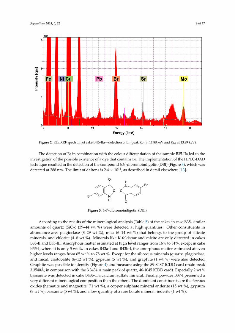
Table 6. Cont.
No RT(min) Chemical Compound Chemical Formula B35-I B35-II B35-IIa B35-III B43a-I B43b-I B37-I
21 11.569 6-Undecylamine C11H25N nd nd + nd nd nd nd22 11.935 1,4-Oxathiin, 2,3-dihydro-6-methyl C5H8OS nd nd + nd nd nd nd23 11.979 Formic acid, ethoxymethylene hydrazide C4H8N2O2 nd nd + nd nd nd nd24 12.509 Dibutylphthalate C16H22O4 nd nd nd nd + + +25 12.515 Dibutylphthalate C16H22O4 nd nd + nd nd nd nd26 12.520 1,4-Benzenedicarboxylic acid, bis[2-methylpropyl] ester C16H22O4 nd nd + nd nd nd nd27 12.586 Dibutylphthalate C16H22O4 nd nd + nd nd nd nd28 13.007 Sulfurous acid, butyldecylester C14H30O3S nd nd nd nd + + nd29 13.047 Nonadecane, 2-methyl- C20H42 nd nd + nd nd nd nd30 14.166 Morpholineethanamine C6H14N2O nd nd + nd nd nd nd31 14.203 Thiophene3-(1,1-dimethylethoxy) C8H12OS nd nd + nd nd nd nd32 14.956 p-Terphenyl C18H14 nd nd + nd nd nd nd33 15.417 2-Piperidinecarboxylic acid, methylester, PFP C10H12F5NO3 nd nd + nd nd nd nd34 16.485 1-(Prop-2-ynyl)-3,3-bis(trifluoromethyl)diaziridine C6H4F6N2 nd nd + nd nd nd nd
35 24.458 Boron, bis{µ-[(3,5-bis(1,1-dimethylethyl)-1H-pyrazolato-N1:N2]}diethyl- C26H50B2N4 nd nd nd + nd nd nd
36 25.072 10-Ethyl-3-chloro-7-(N-p-nitrobenzyldeneamino)-phenothiazine C21H16ClN3O2S nd nd nd + nd nd nd
37 27.215 Yohimban-16-carboxylic acid, 17-hydroxy-10-methoxy-, methylester. . . C22H28N2O4 + nd nd nd nd nd nd
38 28.978 Hexadecanoic acid, 2-hydroxy-1,3-propanediylester C35H68O5 + nd nd nd nd nd nd39 30.384 9-Octadecanoic acid (Z)-2 hydroxy-1,3-propanediylester C39H72O5 nd nd nd + nd nd nd40 38.123 9-Octadecanoic acid (Z)-2 hydroxy-3- propanediylester C37H70O5 + nd nd nd nd nd nd
+: detected, nd: not detected.

Separations 2018, 5, 32 12 of 17
4. Discussion
4.1. Metal Containers
Metal case B35, which was found in Derveni grave B, presents differentiation in terms of thechemical composition of its various parts (Table 2). These parts have been made from hammered metalsheets (except the lid’s handle which was cast). In general when Sn exceeds the limit of 1% in a Cualloy, it is characterized as a deliberate addition [17]. The main parts of B35 (body, lid, the lid’s handle,and the rim) were made of tin bronze (Cu-Sn) alloy accompanied by a low amount of impurities(Pb, Fe, As, Ca). This type of alloy was employed in Macedonia during the Classical period for themanufacture of vessels and utensils [7,18,19]. There is a classification of bronzes from low to highSn depending on the amount of Sn that was added to the alloy. According to the related literature,there is no single definition of the concentration of Sn that would define an ancient bronze artefact as a“high-tin” [8,12,20]. The most accepted compromise, to characterize an ancient bronze as “high-tin”,is an Sn concentration between 14 wt % and 16 wt %. [21,22]. Given that the lid’s handle and the rimcontain enough Sn (14.42–15.62 wt %) to characterize them as “high-tin” bronzes, the body and lid areclose to this definition since they contain around 12 wt % Sn. On the contrary, the hinge contains only6.59 ± 0.30 wt % Sn. The addition of Sn to Cu increases the strength and hardness of the alloy [23–25].It also increases the resistance of the bronze artefact to corrosive parameters [21]. On the other hand,a Sn content higher than 15 wt % strongly increases the embrittlement of the alloy, raising problems(e.g., cracking) during hammering [18,25]. The role of the quantity of Sn is also significant to theappearance of bronze artefacts. A high Sn content gives them a “golden” hue which probably imitatesvessels of precious metals [8,21,26] and denotes the high social status of the owner of B35; alreadysuggested for grave goods in Derveni grave B [1]. Only the four nails are different and those containSn as an impurity and not as an additive [17]. Its very low level (0.06–0.76 wt %) makes the alloy easyto work with [27], but the authors also express their reservation as to whether the nails and hingeare ancient, or are modern additions. The chemical composition of bowls B43a and B43b, which arealso hammered, is also different. Their main constituent is Cu (at level of 99 wt %) accompanied by alow amount of impurities (Sn, Pb, Fe, Ni, Ca). Sn is detected at very low quantities (0.04 ± 0.01 wt %)and is characterized as an impurity and not as an additive [17]. Finally, the lid of pyxisB37 presentsa relatively high amount of Sn (12.16 ± 0.20 wt %), similar to the lid of case B35 (12.07 ± 0.01 wt %).The criteria of the ancient smith and their customers were more the social status and prestige impartedby the appearance of the objects [28] (ensuring the golden appearance of the artefacts with the additionof a relatively high amount of Sn) and not so much the workability. The smith of the 4th centuryBC probably developed skills to work with such brittle alloys without causing any cracks. The factthat pyxis B37 presents extensive corrosion compared to the good preservation of case B35 must beattributed to the taphonomic environment (e.g, the coexistence with other metal artefacts [27]) and notso with the chemical composition.
Fe is detected in all metal artefacts of the present study. In all cases, it is characterized as an impuritydue to its very low concentration (0.04–0.20 wt %) [23,29]. Fe readily enters Cu during smelting anddifferences in Fe content arise through differences in the smelting process. The implementation of asimple and short smelting process results in an average Fe content of around 0.05 wt % instead of aprocess that involves slagging where the average Fe content rises at 0.5 wt % [30,31]. In this study,it is estimated that the first procedure was implemented. Moreover, for cold hammering procedures,Fe must not exceed 0.5 wt % because the formability of the artefact decreases due to precipitationphenomena [32]. Finally, the detection of very low Fe concentrations is an indicator that native andunrefined Cu was used [23,30].
Pb is considered as an impurity for all results of Table 2 since its concentration is lower than2 wt % [33]. This is an expected result for hammered objects but not for cast ones (e.g., B35 lid’s handlewhich contains only 0.15 ± 0.03 wt % Pb). This result is unexpected since Pb was a common additive

Separations 2018, 5, 32 13 of 17
in ancient bronzes. It improved fluidity and castability as it causes a reduction of the melting point.Most Hellenistic bronze cast artefacts contain more than 2 wt % Pb [34].
Arsenic was detected at all parts of B35 case (except the four nails) and bowls B43a and B43b atvery low levels (0.01–0.04 wt %). This very small quantity may indicate that the initial ore deposit wasnot rich in As [33]. According to a previous study [7], Greek bronzes of the 4th century BC contain,in general, less than 1 wt % As, which is confirmed by the present investigation.
4.2. Contents of the Metal Containers
Elements like Si, Al, Fe Mg, Ca, and K which were detected as the main constituents, accordingto the EDµXRF analysis (Table 4), are in abundance in the earth’s crust [21,27] and are consequentlycommon soil elements. The only chemical element that is not common and is detected in all contentsis Cu. A possible interpretation for this is contamination from the metal containers due to thelong-term coexistence at the burial environment [35]. The other result, not common in soil, is thedetection of Br in cake B35-IIa. Br was only detected in this cake, which also presents a reddish hue.The consequent implementation of the HPLC-DAD technique led to the detection of the compound6,6′-dibromoindigotin (DBI), which is used as an index for the identification of shellfish purple(or porphyra in Greek or mollusc purple). Purple was the colour of royalty and a designator ofsocial status.
According to the EDµXRF results of (Table 4), the powder of pyxis B37 presents a very differentchemical composition. Elements which are the main constituents in the other contents (Si, Al, Mg, Ca,K) have not been detected in B37-I. Fe and Cu oxides comprises almost 20 wt % of the powder. The restof it could be organic material, carbon oxides, or chemical elements of a very low atomic number (lowerthan 12) for which the excitation conditions of the EDµXRF technique are poor [36]. The high contentof powder B37-I in ferrous constituents was also confirmed by the XRD results (Table 5), which havebeen normalized at 100 wt % contrary to the results of EDµXRF (Table 4). According to them, B37-I ismostly comprised of hematite (Fe2O3), magnetite (Fe3O4), and antlerite (Cu3(SO4)(OH)4). Hematite isa red mineral pigment well known in antiquity [37]. Antlerite is a possible corrosion product of thereaction of the bronze antiquities, which are located in urban areas, with sulfur dioxide (a commonpollutant). Especially in sheltered areas, the low weathering permits the accumulation of copper ionsand enhancement in the acidity of water films [38].
A possible grouping according to the XRD results is: group A of the four B35 cakes wheresilicate minerals dominate, group B of the two B43 cakes which are mostly comprised of amorphousmaterial, and finally group C of the red ferrous mineral powder which comprises entirely crystallinephases (Table 5). Gypsum (CaSO4·2H2O) is a common constituent in groups B and C. It is known,in antiquity, for adhesive and quick binding properties, especially regarding constructions [27].Especially and only in the B37-I bassanite [CaSO4·0.5(H2O)], a semi-hydrous form of sulfate saltis detected. Bassanite probably resulted from the dehydration of gypsum under dried conditions.The authors, however, express their reservation that the presence of gypsum and bassanite in B37-Imay be explained by contamination with reservation materials applied. In cake B35-I, the mineralhydrozincite Zn5(CO3)2(OH)6, which was used in antiquity for ophtalmic purposes, is detected [39].The mineral cristobalite is only detected in the contents of B43a and B43b bowls. It is a member ofthe quartz group minerals and has the same chemical formula as quartz, SiO2, but a distinct crystalstructure. The above grouping of the contents under study implies a targeted selection of raw materialsin order to have the desired effects during use.
In the sample B35-IIa, pentanoic acid has been detected (Table 6). Pentanoic acid or valericacid is a compound with an unpleasant rancid odour and naturally occurs in the roots of the plant“valeriana officinalis”. It is used in pharmaceuticals, as well as in cosmetics and perfumes, in itspure form or as an ester [40]. “Valeriana officinalis” was traditionally used in ancient Greece as atreatment against stress and insomnia [41]. At the same sample, another organic substance detected isnonadecane. Nonadecane is used as a fragrance agent and it is one of the components of the essential

Separations 2018, 5, 32 14 of 17
oils of some of the species that belong to the genus “Artemisia” [42]. More specifically, “Artemisiaabsinthium” is a species of Artemisia native to Eurasia and analysis has shown that, among severalother chemical substances, its essential oil also contains nonadecane [43]. In ancient Greece, “Artemisiaabsinthium” was used for its medicinal properties against gastrointestinal disorders, as an astringent,helminthicide, diuretic, and emmenagogue [41]. In this case, nonadecane could have been used forits sole properties or as a masking agent to cover the unpleasant odour deriving from pentanoic acid.Nevertheless, access to other species of Artemisia or even completely different genuses, not native tothe Mediterranean or Eurasia, cannot be excluded. As traces of fatty acid esters can be observed in allthe compartments of the lidded case (B35), a possible explanation could be a preparation in the formof an ointment or even a formulation to be consumed orally, which would target the gastrointestinalsystem, simultaneously combining the sedative properties of “Valeriana officinalis” and the therapeuticactivity of Artemisia absinthium, as gastrointestinal disorders are commonly associated with stress.
Hexadecanoic acid was detected in the B-35-I cake. This chemical substance is also known bythe name palmitic acid and is used widely as a cleansing agent and in the production of soaps [44].Palmitic acid can be found naturally in meat, dairy products, palm oil, and palm kernel oil, as well asin other plant oils, in smaller amounts [45]. More specifically, palmitic acid is one of the constituents ofthe essential oil of the plant “carum carvi”, widely known as “caraway”, which was used in Europe asa traditional medicine for stomach disorders and flatulence [41]. The content of sample B-35-I couldpotentially constitute a soap or a cleansing agent, some kind of medicinal ointment, or a decoction fororal consumption to treat stomach disorders.
It is well known that fatty acids and their derivatives were used in antiquity as constituents ofpharmaceutical products [41,46]. According to the results of the HS-SPME/GC-MS analysis (Table 6),many of the twenty-two organic compounds that were detected in cake B35-IIa belong to the aboveclasses. In the same cake, 6,6′-dibromoindigotin (DBI) was detected, which in combination with thereddish colour, denotes the existence of the royal dye shellfish purple. In the other cakes, very feworganic compounds were detected. A possible interpretation is that cake B35-IIa was the basis thatcontained a large number of drastic substances and the reddish—purple colour a useful indicator fordistinguishing it easily from other preparations. Alternatively, cake B35-IIa was perhaps contaminatedby purple-dyed items that had been deposited in the burial. Dioscuridies Pedanius, in his work“De Materia Medica” [46], refers to the storage of fats in tin containers for pharmaceutical purposes.The metal case B35 and pyxis B37 were made of a high tin bronze alloy, but on the other hand, bowlsB43a and B43b were made of almost pure Cu.
5. Conclusions
The combination of spectrometric (EDµXRF, XRD) and chromatographic (HPLC-DAD,HS SPME/GC-MS) analytical techniques was used as a holistic archaeometric approach in orderto determine the construction technology of a group of metal containers and the chemical synthesis oftheir contents. The analytical methodology gave priority to a non-destructive implementation, as far aspossible, due to the uniqueness and significance of the analyzed material. For the elemental analysis ofthe metal containers, EDµXRF spectrometry was implemented, in a non-invasive way, which resultedin the determination of their raw materials. Moreover, differentiations in their constituent parts werefound. Generally, two kinds of alloy were used: a high tin bronze alloy for the construction of the mainparts of metal case B35 and pyxis B37 and pure Cu accompanied by a low amount of impurities for themanufacture of bowls B43a and B43b. Furthermore, their chemical composition reveals the ancientarchaeometallurgical procedures of the 4th century BC.
The implementation of a non-invasive technique like EDµXRF could be used as a guide for theselective implementation of a destructive one, e.g., the detection of Br in cake B35-IIa, which led tothe implementation of HPLC-DAD and the detection of the royal dye shellfish purple. Althoughthe archaeological approach led to the conclusion that this set was used for medical purposes [4],
Separations 2018, 5, 32 15 of 17
the archaeometric approach provides us with further information on medical knowledge in Macedoniaduring the 4th century BC
Author Contributions: Conceptualization, C.S.K., D.I.; Methodology, C.S.K., D.I., G.A.Z.; Investigation, C.S.K.,A.Z., N.K., I.K.; Writing-Original Draft Preparation, C.S.K., D.I., N.K., I.K.; Writing-Review & Editing, C.S.K.;G.A.Z.; Supervision, G.A.Z.
Funding: This research received no external funding.
Acknowledgments: The authors wish to thank: P. Adam—Veleni, then Director of the Archaeological Museum ofThessaloniki (A.M.Th.) for the permission to analyze the artefacts of the article; V. Michalopoulou, Conservator ofAntiquities, A.M.Th., for providing support with issues concerning ancient technology and metal conservation;and the two anonymous reviewers for their recommendations which resulted in a greatly improved paper.
Conflicts of Interest: The authors declare no conflict of interest.
References
1. Themelis, P.; Touratsoglou, G. The tombs of Derveni; Ministry of Culture–Archaeological Receipts Fund:Athens, Greece, 1977. (In Greek) (Θέµελησ, Π., Toυράτσoγλoυ, Γ., 1997. O ι τάϕoι τoυ ∆ερβενίoυ,Υπoυργείo Πoλιτισµoύ—Tαµείo Aρχαιoλoγικών Πóρων και Aπαλλoτριώσεων, Aθήνα)
2. Tsigarida, B.; Ignatiadou, D. The Gold of Macedon: Archaeological Museum of Thessaloniki, Archaeological ReceiptsFund; Archaeological Museum of Thessaloniki: Thessaloniki, Greece, 2000.
3. Chrysostomou, P. Contributions to the history of medicine in ancient Macedonia. Eulimene 2002, 3, 99–116. (InGreek) (Xρυσoστóµoυ, Π. 2002, «Συµβoλέσ στην Iστoρία της Iατρικής στην Aρχαία Mακεδoνία», Eυλιµένη3, 99–116»)
4. Ignatiadou, D. The warrior priest in Derveni grave B was a healer too, Histoire. Méd. Santé 2016, 8, 89–113.[CrossRef]
5. Guerra, M.F. Analysis of archaeological metals, The place of XRF & PIXE in the determination of technologyand provenance. X-ray Spectrom. 1998, 27, 73–80.
6. Cechak, T.; Hlozek, M.; Musilek, L.; Trojek, T. X-ray fluorescence in investigations of archaeological finds.Nucl. Instrum. Methods Phys. Res. Sect. B 2007, B263, 54–57. [CrossRef]
7. Karydas, A.G. Application of a portable XRF spectrometer for the non invasive analysis of museum metalartefacts. Ann. Chim. 2007, 97, 419–432. [CrossRef] [PubMed]
8. Charalambous, A.; Kassianidou, V.; Papasavvas, G. A compositional study of Cypriot bronzes dating to theEarly Iron Age using portable X-ray fluorescence spectrometry (pXRF). J. Archaeol. Sci. 2014, 46, 205–216.[CrossRef]
9. Stuart, B. Analytical Techniques in Materials Conservation; John Wiley & Sons Ltd.: Chichester, UK, 2007; p. 194.10. Bronk, H.; Rohrs, S.; Bjeoumikhov, A.; Langhoff, N.; Schmalz, J.; Wedell, R.; Gorny, H.-E.; Herold, A.;
Waldschlager, U. Artax—A new mobile spectrometer for EDXRF spectrometry on art & archaeologicalobjects, Freseniu’s. J. Anal. Chem. 2001, 371, 307–316.
11. Dylan, S. Handheld XRF analysis of Renaissance bronzes: Practical approaches to quantification andacquisition. In Handheld XRF for Art and Archaeology; Shugar, A.N., Mass, J.L., Eds.; Leuven University Press:Leunen, The Netherlands, 2012; pp. 37–74.
12. Scott, D.A. Ancient Metals: Microstructure and Metallurgy; Lulu.com: Morrisville, NC, USA, 2010; Volume I.13. Klug, H.P.; Alexander, L.E. X-ray Diffraction Procedures for Polycrystalline and Amorphous Materials; John Wiley
& Sons: New York, NY, USA; Sydney, Australia; Toronto, ON, Canada, 1974; 966p.14. Kantiranis, N.; Stergiou, A.; Filippidis, A.; Drakoulis, A. Calculation of the percentage of amorphous material
using PXRD patterns. Bull. Geol. Soc. Greece 2004, 36, 446–453.15. Vasileiadou, A.; Karapanagiotis, I.; Zotou, A. Determination of Tyrian purple by high performance liquid
chromatography with diode array detection. J. Chromatogr. A 2016, 1448, 67–72. [CrossRef] [PubMed]16. Karapanagiotis, I.; Mantzouris, D.; Cooksey, C.; Mubarak, M.S.; Tsiamyrtzis, P. An improved HPLC method
coupled to PCA for the identification of Tyrian Purple in archaeological and historical samples. Microchem. J.2013, 110, 70–80. [CrossRef]
17. Healy, J.F. Mining and Metallurgy in the Greek and Roman World; Thames and Hudson: London, UK, 1978.

Separations 2018, 5, 32 16 of 17
18. Varoufakis, G. Technological specifications of the 4th century BC. Contribution to the historic metallurgy.Archaeol. Ephemer. 1974, 113, 57–62. (In Greek). (Bαρoυϕάκησ, Γ., 1974. Tεχνικαί πρoδιαγραϕαί τoυ 4oυ αι.π.X. Συµβoλή εισ την ιστoρικήν µεταλλoυργίαν, Aρχαιoλoγική Eϕηµερίσ, σελ. 57–62)
19. Asimenos, K. Macedonian metalworking and alloy composition of the classical era. In Proceedings ofthe XII International Congress of Classical Archaeology, Athens, Greece, 4–10 September 1983; Volume C,pp. 283–288. (In Greek) (Aσηµενóσ, K., 1988. Mακεδoνική µεταλλoτεχνία και σύσταση κραµάτων τησκλασικήσ επoχήσ, στo XII ∆ιεθνέσ Συνέδριo Kλασσικήσ Aρχαιoλoγίασ, τόµoσ Γ’, Aθήνα, σελ. 283–288)
20. Scott, D.A. Metallography and Microstructure of Ancient and Historic Metals; The Getty Conservation Institute:Singapore, 1991.
21. Ashkenazi, D.; Iddan, N.; Tal, O. Archaeometallurgical characterization of Hellenistic metal objects:The contribution of the objects for Rishon Le-Zion (Israel). Archaeometry 2012, 54, 528–548. [CrossRef]
22. Konecná, R.; Fintová, S. Copper and copper alloys: Casting, classification and characteristic microstructures.In Copper Alloys—Early Applications and Current Performance—Enhancing Processes; Collini, L., Ed.; InTechWeb.Org: Rijeka, Croatia, 2012; pp. 3–30.
23. Tylecote, R.F. The Early History of Metallurgy in Europe; Longman Inc.: New York, NY, USA, 1987.24. Craddock, P.T. The composition of the copper alloys used by the Greek, Etruscan and Roman civilisations:
2. the Archaic, Classical and Hellenistic Greeks. J. Archaeol. Sci. 1977, 4, 103–123. [CrossRef]25. Craddock, P.T. The composition of the copper alloys used by the Greek, Etruscan and Roman Civilizations.
J. Archaeol. Sci. 1976, 3, 93–113. [CrossRef]26. Figueiredo, E.; Silva, R.J.C.; Senna-Martinez, J.C.; Araújo, M.F.; Braz Fernandes, F.M.; Inês Vaz, J. Smelting
and recycling evidences from the Late Bronze Age habitat site of Baiões (Viseu, Portugal). J. Archaeol. Sci.2010, 37, 1623–1634. [CrossRef]
27. Goffer, Z. Archaeological Chemistry, 2nd ed.; John Wiley & Sons, Inc.: Hoboken, NJ, USA, 2007.28. Descamps- Lequime, S. The color of bronze—Polychromy and the aesthetics of bronze surfaces. In Power
and Pathos—Bronze Sculpture of the Hellenistic World; Daehner, J.M., Lapatin, K., Eds.; J. Paul Getty Museum:Los Angeles, CA, USA, 2015; pp. 150–165.
29. Craddock, P.T.; Meeks, N.D. Iron in ancient copper. Archaeometry 1987, 29, 187–204. [CrossRef]30. Henderson, J. The Science and Archaeology of Materials—An Investigation of Inorganic Materials; Routledge:
New York, NY, USA, 2000.31. Cooke, S.R.B.; Aschenbenner, S. The occurrence of metallic iron in ancient copper. J. Field Arcahaeol. 1975, 2,
251–266.32. Papadimitriou, G. Copper and bronze metallurgy in ancient Greece. In Archaeometry 90, Proceedings of the
International Symposium in Archaeometry, 2–6 April 1990, Heidelberg, Germany; Wagner, G.A., Pernicka, E., Eds.;Birkhauser Verlag: Basel, Switzerland, 1991; pp. 117–126.
33. Oudbashi, O.; Davami, P. Metallography and microstructure interpretation of some archaeological tin bronzevessels from Iran. Mater. Charact. 2014, 97, 74–82. [CrossRef]
34. Craddock, P.T.; Giumlia-Mair, A. Problems and possibilities for provenancing bronzes by chemicalcomposition. In Bronzeworking Centers of Western Asia, c.100-539 B.C., Kegan Paul International in Associationwith the British Museum; Curtis, J., Ed.; Kegan Paul International in association with the British Museum;Distributed by Routledge; Chapman & Hall: London, UK, 1988.
35. Rovira, S.; Montero, I. Natural tin–bronze alloy in Iberian Peninsula metallurgy: Potentiality and reality.In The Problem of Early Tin; Giumlia-Mair, A., Schiavo, F.L., Eds.; Archaeopress: Oxford, UK, 2003; pp. 15–22.
36. Jenkins, R. X-ray Fluorescence Spectrometry; John Whiley & Sons Inc.: Hoboken, NJ, USA, 1999.37. Gettens, R.J.; Stout, G.L. Paintings Materials: A Short Encyclopedia; Dover Publications: New York, NY,
USA, 1966.38. Leygraf, C.; Graedel, T.E. Atmospheric Corrosion; Wiley-Interscience: New York, NY, USA, 2000.39. Giachi, G.; Pallecchi, P.; Romualdi, A.; Ribechini, E.; Lucejko, J.J.; Colombini, M.P.; Lippi, M.M. Ingredients
of a 2000-y-old medicine revealed by chemical, mineralogical and botanical investigations. PNAS 2013, 110,1193–1196. [CrossRef] [PubMed]
40. Goldberg, I.; Rokem, J.S. Organic and Fatty Acid Production, Microbial. In Encyclopedia of Microbiology,3rd ed.; Elsevier: New York, NY, USA, 2009; pp. 421–442.
Separations 2018, 5, 32 17 of 17
41. Skaltsa, E. History of Pharmacy; Kallipos-Hellenic Academic Books: Athens, Greece, 2015; p. 104. (In Greek)(Σκαλτσά, E., 2015. «Iστoρία τησ Φαρµακευτικήσ», Kάλλιπoσ-Eλληνικά Aκαδηµαϊκά Συγγράµµατα καιBoηθήµατα, Aθήνα, 104)
42. Mojarrab, M.; Delazar, A.; Esnaashari, S.; Afshar, F.H. Chemical composition and general toxicity of essentialoils extracted from the aerial parts of Artemisia armeniaca Lam. and A. incana (L.) Druce growing in Iran.Res. Pharm. Sci. 2013, 1, 65–69.
43. Kordali, S.; Cakir, A.; Mavi, A.; Kilic, H.; Yildirim, A. Screening of Chemical Composition and Antifungaland Antioxidant Activities of the Essential Oils from Three Turkish Artemisia Species. Agric. Food Chem.2005, 53, 1408–1416. [CrossRef] [PubMed]
44. Anneken, D.J.; Both, S.; Christoph, R.; Fieg, G.; Steinberner, U.; Westfechtel, A. Fatty Acids. In Ullmann’sEncyclopedia of Industrial Chemistry; Wiley-VCH: Weinheim, Germany, 2006.
45. Kim, S.; Thiessen, P.A.; Bolton, E.E.; Chen, J.; Fu, G.; Gindulyte, A.; Han, L.; He, J.; He, S.; Shoemaker, B.A.;et al. PubChem Substance and Compound databases. Nucleic Acids Res. 2016, 44, D1202–D1213. [CrossRef][PubMed]
46. Dioscorides, P. De Materia Medica; Beck, L.Y., Translator; Altertumswissenschaftliche Texte und Studien,Band 38; Olms-Weidmann: Hildesheim, Germany, 2005.
© 2018 by the authors. Licensee MDPI, Basel, Switzerland. This article is an open accessarticle distributed under the terms and conditions of the Creative Commons Attribution(CC BY) license (http://creativecommons.org/licenses/by/4.0/).
Copyright © 2022 FDOKUMEN